Low Oil-Water Ratio Invert Emulsion Mud for Unconventional ...
Two-Dimensional DPPC Based Emulsion-like Structures Stabilized bySilica NanoparticlesEduardo Guzman,† Davide Orsi,‡ Luigi Cristofolini,†,‡ Libero Liggieri,† and Francesca Ravera*,†
†Consiglio Nazionale delle Ricerche-Istituto per l’Energetica e le Interfasi, U.O.S. Genova (CNR-IENI), Via De Marini 6, 16149Genova, Italy‡Dipartimento di Fisica e Scienze della Terra, Universita degli Studi di Parma, Viale Usberti 7/A, 43100 Parma, Italy
ABSTRACT: We studied the mechanical and structural properties of mixedsurface layers composed by 1,2-dipalmitoyl-sn-glycero-3-phosphocholine(DPPC) and silica nanoparticles (NPs). These layers are obtained byspreading a DPPC Langmuir monolayer on a colloidal silica dispersion. Thetransfer/incorporation of NPs into the DPPC monolayer, driven byelectrostatic interactions, alters the molecular orientation, the mechanismsof domain formation, and consequently the phase behavior of the surfacelayer during compression. The investigation of these systems by means ofcomplementary techniques (Langmuir trough, fluorescence microscopy,ellipsometry, and scanning electron microscopy (SEM)) shows that theincorporated NPs preferentially distribute along the liquid expanded phase ofDPPC. The layer assumes the stable and homogeneous bidimensionalstructure of a two-dimensional (2D) analogue of a Pickering emulsion. Infact, the presence of particles provides a circular shape to the DPPC domainsand stabilizes them against growth and coalescence during the monolayer compression.
1. INTRODUCTION
The interaction between nanoparticles (NPs) and adsorbed orspread surfactant layers at liquid interfaces is relevant in severalscientific and technological fields such as material science,biomaterials, emulsion-foam technology, pharmaceuticals, andnanomedicine.1−4 The potential effects of NPs on themechanical and physicochemical properties of surfactantadsorbed layers are well-known from previous studies basedon surface tension and rheological measurements.5 On theother side, insoluble surfactant layers have been widelyinvestigated in the presence of NPs as Langmuir monolayersspread on nanoparticle dispersions. This experimental approachis especially effective to assess the properties of biologicallyrelevant systems such as mixtures of lipids.In this context, NPs of different chemical composition, size,
and hydrophobicity/philicity have been investigated asinteracting with lipid monolayers, containing 1,2-dipalmitoyl-sn-glycero-3-phosphocholine (DPPC) as the main compo-nent.6−11 These studies have evidenced that NPs maysignificantly alter the phase behavior of the surface layer andits structure12−15 because of their interaction with the lipidmolecules, which induces changes in the surface molecularpacking. Such interactions may have hydrophobic or electro-static origin depending on the chemical nature of theparticles.10
The same type of interactions are also responsible for theability of DPPC to bind to several biological relevant moleculesif they are present in the water phase; e.g., interaction withinterface-related proteins such as myelin basic protein (MBP, a
protein deemed to be involved in degenerative neuronaldiseases) was demonstrated for DPPC as well as for otherphospholipid monolayers.16 At the same time, incorporation ofDNA in DPPC monolayers was indeed demonstrated bydifferent techniques,17 including X-ray reflectivity,17 and couldbe exploited for therapeutic as well as for biosensingpurposes.18
The work presented here focuses on the properties of amixed DPPC−silica nanoparticles layer obtained by spreading aDPPC monolayer at the surface of dispersions of colloidal silica.Under these conditions, in fact, the silica NPs tend to beincorporated into the DPPC monolayers driven by electrostaticinteractions.19,20
This system has been investigated in refs 12 and 13 byLangmuir trough measurements and Brewster angle micros-copy (BAM), in the framework of studies on the nanoparticleimpact on the pulmonary surfactant functionality. In thosestudies, as well as in many other toxicological investigations, thechoice of DPPC is justified by the fact that it is the maincomponent of biosystems such as lung surfactant or cellmembranes.21 Nevertheless, the motivation of the present workis related to the development of innovative soft materials, suchas DPPC and the silica representative of organic−inorganicmixed systems, undergoing, under certain conditions, interest-ing processes of surface reorganization.
Received: June 5, 2014Revised: September 11, 2014Published: September 11, 2014
Article
pubs.acs.org/Langmuir
© 2014 American Chemical Society 11504 dx.doi.org/10.1021/la502183t | Langmuir 2014, 30, 11504−11512

The analysis reported in the following is based on diversesurface diagnostics implemented in the Langmuir troughtechnique, such as fluorescence microscopy and ellipsometry,combined with scanning electron microscopy (SEM) ofLangmuir−Schaefer (LS) films obtained from the mixedmonolayers. Those techniques proved successful in identifyingthe morphology and, therefore, also in the interpretation of themechanical properties of similar interfacial systems realized by acompletely different route, i.e., 2D interfacial gels formed byLangmuir monolayers of gold nanoparticles.22−24 In that case,the steps leading to the formation of the gel could be identifiedby epi-microscopy,22 and those could be used to provide anexplanation of the observed dynamical properties23,24 Theresults obtained here by this deeper characterization, discussedin view of the previous thermodynamic and rheologicalfindings, provide new insights into the stable and homogeneousbidimensional structures obtained as well as on their rheologicalproperties.
2. MATERIALS1,2-Dipalmitoyl-sn-glycerol-3-phosphocholine (DPPC) was purchasedfrom Sigma (Germany) at 99% purity and used without furtherpurification. DPPC spreading solutions (1 mg/mL) were preparedusing high-performance liquid chromatography (HPLC)-grade chloro-form (Sigma, Germany).Colloidal silica dispersions were obtained by diluting commercial
samples (Levasil-Stark, Germany). Three different samples were used,Levasil 300/30, 200/30, and 50/50, presenting three differentBrunauer−Emmett−Teller (BET) values, 300, 200, and 50 cm2/g,respectively, corresponding to different average diameters. As declaredby the producer and confirmed in previous studies on the samesystems,5,25 these commercial dispersions are characterized by a veryhigh stability, without stabilizing additive, due to an important negativesurface charge obtained during the specific production process. Thesurface tension measurements of these silica dispersions provided, infact, the same value of the pure water, i.e., 72.5 mN/m, constant overseveral hours.The bare silica dispersions have been also characterized by dynamic
light scattering (DLS) and ζ-potential measurements, which confirmedtheir good stability and provided the particle size distribution and theparticle surface charge. It was found that d = 9.0, 15.0, and 60.0 ± 0.2nm, as average particle diameters, in agreement with the nominalvalues by the BET data, i.e., 300, 200, and 50 cm2/g, respectively, and aζ-potential of −42 ± 4 mV, in agreement with what was found in ref25.Water for all the experiments was purified by a multicartridge Elix
plus Milli-Q (Millipore) system, providing a resistivity greater than 18MΩ·cm.
3. METHODSA conventional Langmuir trough of a total area At = 243 cm2 (KSVMinitrough, Finland) was used, equipped with two Delrin barriers,allowing for symmetric compression of the surface layer, and with aWhilhelmy plate tensiometer for the surface pressure measurement.The mixed layers were obtained by spreading at the surface of theaqueous silica dispersions a volume of 30 μL of DPPC solution inchloroform at a concentration of 1.36 mM.In this way, the spread DPPC monolayer, after evaporation of the
solvent, presented a density Γ0 = 10 μmol/m2. All the colloidal silicadispersions used as subphase were at a concentration of 1 wt %, andtheir volume was ∼200 cm3.As discussed in ref 12, after a time of ∼1 h, appropriate for the
complete evaporation of the solvent and the transfer of silica NPs fromthe subphase to the surface, the layer can be assumed to be atequilibrium. In all the experiments presented here, the compression ofthe surface layer was performed at a rate of 2 cm2/min, corresponding
to 0.25 nm2 molecule−1 min−1, which is sufficiently low to ensure theequilibrium conditions during the layer compression.
The 2D morphology of the monolayers was investigated byfluorescence microscopy using a Nikon Eclipse-Ti optical microscope(Nikon Corp., Japan) with a 20× objective coupled to a fluorescencemodule. A small quantity (1 mol %) of fluorescence-marked lipidNBD-PC (1-palmitoyl-2-{12-[(7-nitro-2-1,3-benzoxadizole-4-yl)-amino]dodecanoyl}phosphatidylcholine), purchased from AvantiPolar Lipids, U.S.A., was added to the DPPC solution used for thespreading. The image analysis was done using our own softwaredeveloped in the Matlab computing environment22 as well as thefreeware software Image.26
For ellipsometry measurements a multipurpose apparatus (Multi-skop Optrel, Germany)27 was used with a single wavelength of 632.8nm. This technique was employed to evaluate the thickness of themixed layer at water/air interface under various compression, always inthe Drude approximation. The optical model adopted was thatdescribed in detail in ref 17. This specific apparatus provides anaverage thickness value calculated on an area of a few squaremillimeters.
Langmuir−Schaeffer films of the monolayers were deposited ontosilicon wafers and have been analyzed by scanning electron microscopy(SEM-FEG Zeiss SUPRA 40), a field-emission SEM with improvedspatial resolution (5−10 nm) and minimized sample charging/damage.All the measurements presented in this study were performed at acontrolled temperature of 22 °C.
4. RESULTS AND DISCUSSIONDPPC is a zwitterionic natural surfactant that can be spread onan aqueous subphase as a Langmuir monolayer with thetrimethylammonium headgroup protruding into the water.Thus, the attachment of silica NPs to the DPPC monolayer isdriven by electrostatic interactions. Silica particles that are welldispersible in water due to their negative surface charge may becaptured and incorporated in the layer, affecting its phasebehavior and forming mixed layers with particular thermody-namic and structural properties.The compression isotherm and the quasi-equilibrium
dilational elasticity of the mixed DPPC−silica NP withintermediate average diameter (Levasil 200/30) were pre-viously obtained in ref 12. In Figure 1 the analoguecompression isotherms obtained for silica dispersions atdifferent NP sizes are reported, compared with that of pureDPPC.As already discussed in ref 12, the presence of NPs affects the
principal characteristics of the well-known surface pressure−
Figure 1. Π−A isotherm of mixed DPPC−silica nanoparticlemonolayers, obtained by spreading DPPC on dispersions of silicaparticles at different sizes d, and Π−A isotherm of pure DPPC asfound in ref 12.
Langmuir Article
dx.doi.org/10.1021/la502183t | Langmuir 2014, 30, 11504−1151211505
area (Π−A) isotherm of the pure DPPC,28,29 which is thetypical plateau in the surface pressure corresponding to thecoexistence of liquid expanded (LE) and liquid condensed(LC) phases, and reduces the collapse surface pressure. Theseeffects, also observed here independently of the size of the silicaparticles, can be explained by the NP incorporation in themonolayer. Such incorporation also explains the surfacepressure increase at smaller compression degrees, or highersurface areas, as due to the increased effective DPPC surfaceconcentration or excluded area effect.30
For pure DPPC, the coexistence region is characterized bythe nucleation and growth of bean-shaped domains thatcoalesce during compression to form a total condensedphase. As pointed out in ref 12 by BAM, in the presence ofparticles these domains assume a different, less elongated, shapeand their area is almost 1 order of magnitude smaller comparedto those found for DPPC spread at the pure water surface.In this work the fluorescence microscopy technique is used
to obtain a more accurate characterization of this mixedbiphasic layers. As shown below, using this technique providesnew insights on how the 2D structure of the layer evolvesduring the compression. The fluorescence probe added in smallquantity (∼1% of the total molar content of the lipid solution)to the spreading solution of DPPC, preferentially distributesinside the liquid expanded phase.31 This provides a highly
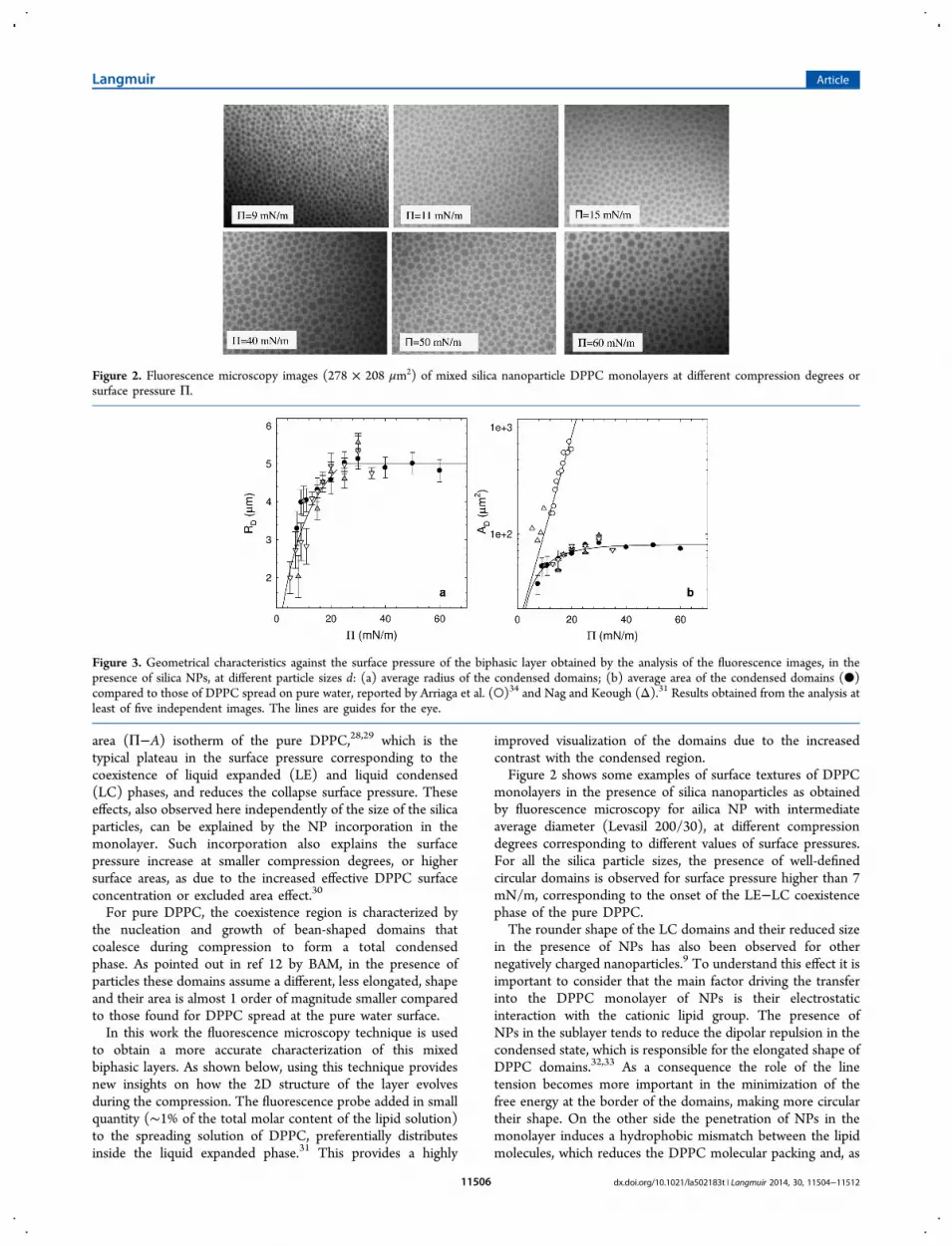
improved visualization of the domains due to the increasedcontrast with the condensed region.Figure 2 shows some examples of surface textures of DPPC
monolayers in the presence of silica nanoparticles as obtainedby fluorescence microscopy for ailica NP with intermediateaverage diameter (Levasil 200/30), at different compressiondegrees corresponding to different values of surface pressures.For all the silica particle sizes, the presence of well-definedcircular domains is observed for surface pressure higher than 7mN/m, corresponding to the onset of the LE−LC coexistencephase of the pure DPPC.The rounder shape of the LC domains and their reduced size
in the presence of NPs has also been observed for othernegatively charged nanoparticles.9 To understand this effect it isimportant to consider that the main factor driving the transferinto the DPPC monolayer of NPs is their electrostaticinteraction with the cationic lipid group. The presence ofNPs in the sublayer tends to reduce the dipolar repulsion in thecondensed state, which is responsible for the elongated shape ofDPPC domains.32,33 As a consequence the role of the linetension becomes more important in the minimization of thefree energy at the border of the domains, making more circulartheir shape. On the other side the penetration of NPs in themonolayer induces a hydrophobic mismatch between the lipidmolecules, which reduces the DPPC molecular packing and, as
Figure 2. Fluorescence microscopy images (278 × 208 μm2) of mixed silica nanoparticle DPPC monolayers at different compression degrees orsurface pressure Π.
Figure 3. Geometrical characteristics against the surface pressure of the biphasic layer obtained by the analysis of the fluorescence images, in thepresence of silica NPs, at different particle sizes d: (a) average radius of the condensed domains; (b) average area of the condensed domains (●)compared to those of DPPC spread on pure water, reported by Arriaga et al. (○)34 and Nag and Keough (Δ).31 Results obtained from the analysis atleast of five independent images. The lines are guides for the eye.
Langmuir Article
dx.doi.org/10.1021/la502183t | Langmuir 2014, 30, 11504−1151211506
a matter of fact, hinders the nucleation and the growth of LCdomains. The combination of these two effects favors thepersistence of a big number of circular-shaped domains duringthe compression.The average radius and area of the domains, RD and AD, as
obtained by the image analysis are reported in Figure 3. It isevident that the size of the domains increases for low surfacepressure until reaching a plateau value for Π > 25 mN/m. Thecomparison of these AD data with those of pure DPPC obtainedby Nag and Keough31 and Arriaga et al.,34 reported in Figure3b, clearly points out the reduction of the average domain sizeinduced by the NPs. These results confirm that the presence ofNPs avoids the growth of the domains above a threshold valuethat corresponds to the size of the initial nucleus of condensedphase observed for pure DPPC. In addition, the transition to atotal condensed phase, occurring in pure DPPC monolayersaround Π ≈ 25 mN/m, is not observed here.The dependence on the surface pressure of the fraction of
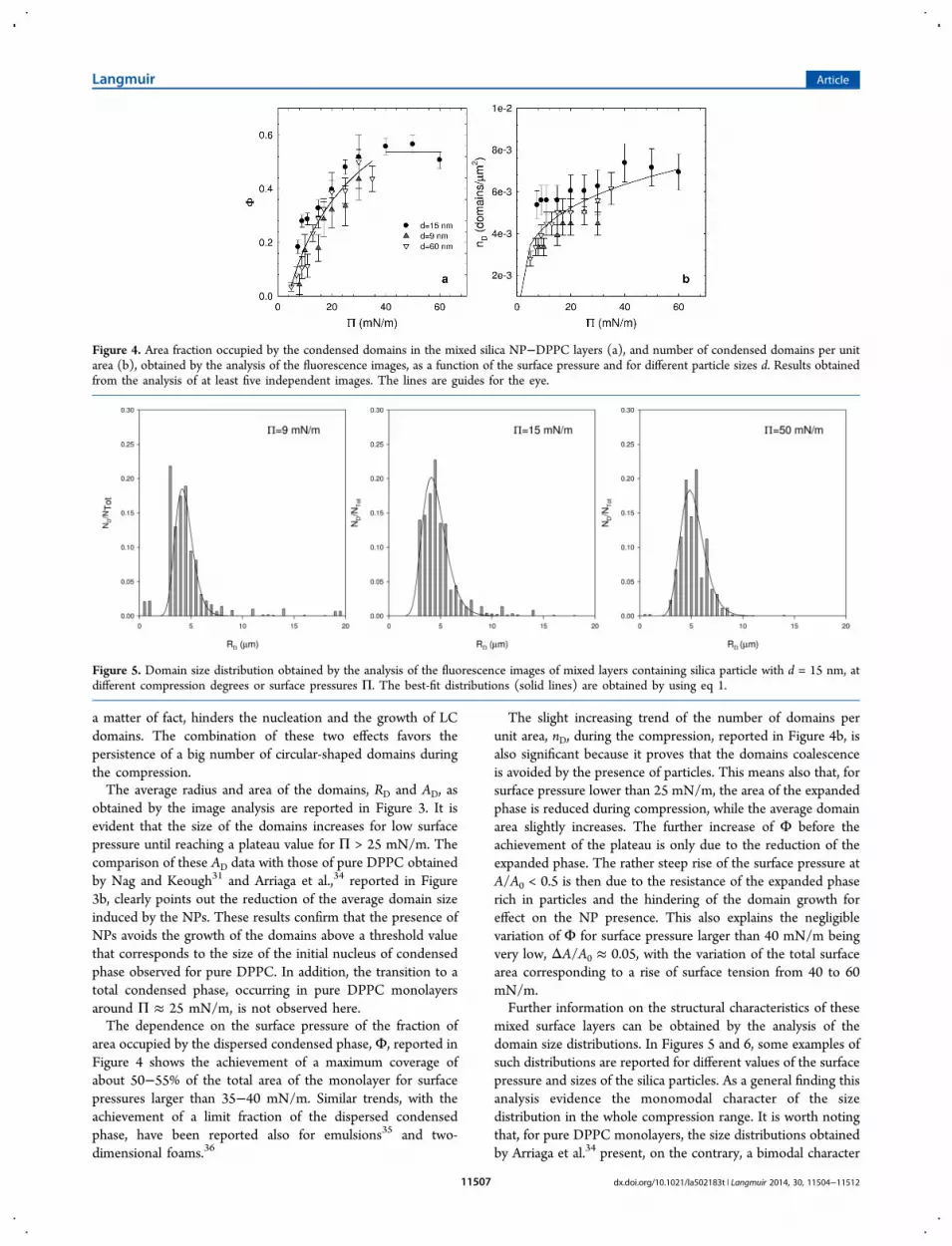
area occupied by the dispersed condensed phase, Φ, reported inFigure 4 shows the achievement of a maximum coverage ofabout 50−55% of the total area of the monolayer for surfacepressures larger than 35−40 mN/m. Similar trends, with theachievement of a limit fraction of the dispersed condensedphase, have been reported also for emulsions35 and two-dimensional foams.36
The slight increasing trend of the number of domains perunit area, nD, during the compression, reported in Figure 4b, isalso significant because it proves that the domains coalescenceis avoided by the presence of particles. This means also that, forsurface pressure lower than 25 mN/m, the area of the expandedphase is reduced during compression, while the average domainarea slightly increases. The further increase of Φ before theachievement of the plateau is only due to the reduction of theexpanded phase. The rather steep rise of the surface pressure atA/A0 < 0.5 is then due to the resistance of the expanded phaserich in particles and the hindering of the domain growth foreffect on the NP presence. This also explains the negligiblevariation of Φ for surface pressure larger than 40 mN/m beingvery low, ΔA/A0 ≈ 0.05, with the variation of the total surfacearea corresponding to a rise of surface tension from 40 to 60mN/m.Further information on the structural characteristics of these
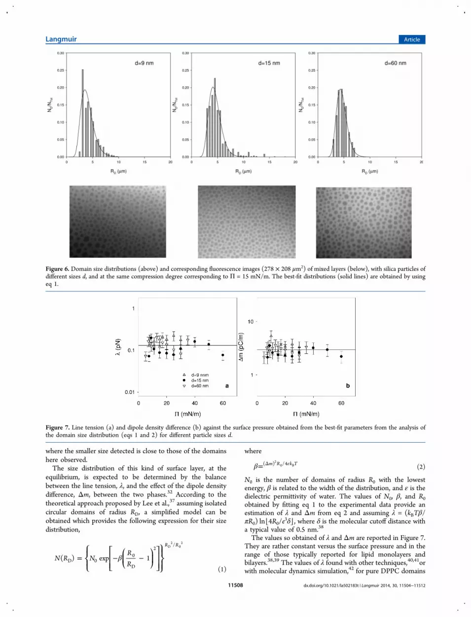
mixed surface layers can be obtained by the analysis of thedomain size distributions. In Figures 5 and 6, some examples ofsuch distributions are reported for different values of the surfacepressure and sizes of the silica particles. As a general finding thisanalysis evidence the monomodal character of the sizedistribution in the whole compression range. It is worth notingthat, for pure DPPC monolayers, the size distributions obtainedby Arriaga et al.34 present, on the contrary, a bimodal character
Figure 4. Area fraction occupied by the condensed domains in the mixed silica NP−DPPC layers (a), and number of condensed domains per unitarea (b), obtained by the analysis of the fluorescence images, as a function of the surface pressure and for different particle sizes d. Results obtainedfrom the analysis of at least five independent images. The lines are guides for the eye.
Figure 5. Domain size distribution obtained by the analysis of the fluorescence images of mixed layers containing silica particle with d = 15 nm, atdifferent compression degrees or surface pressures Π. The best-fit distributions (solid lines) are obtained by using eq 1.
Langmuir Article
dx.doi.org/10.1021/la502183t | Langmuir 2014, 30, 11504−1151211507
where the smaller size detected is close to those of the domainshere observed.The size distribution of this kind of surface layer, at the
equilibrium, is expected to be determined by the balancebetween the line tension, λ, and the effect of the dipole densitydifference, Δm, between the two phases.32 According to thetheoretical approach proposed by Lee et al.,37 assuming isolatedcircular domains of radius RD, a simplified model can beobtained which provides the following expression for their sizedistribution,
β= − −⎪⎪⎧⎨⎩
⎡⎣⎢⎢
⎛⎝⎜
⎞⎠⎟
⎤⎦⎥⎥⎫⎬⎪⎭⎪
N R NRR
( ) exp 1
R R
D 00
D
2/D
20
2
(1)
where
β= εΔm R k T( ) /420 B (2)
N0 is the number of domains of radius R0 with the lowestenergy, β is related to the width of the distribution, and ε is thedielectric permittivity of water. The values of N0, β, and R0obtained by fitting eq 1 to the experimental data provide anestimation of λ and Δm from eq 2 and assuming λ = (kBTβ/πR0) ln⌊4R0/e
3δ⌋, where δ is the molecular cutoff distance witha typical value of 0.5 nm.38
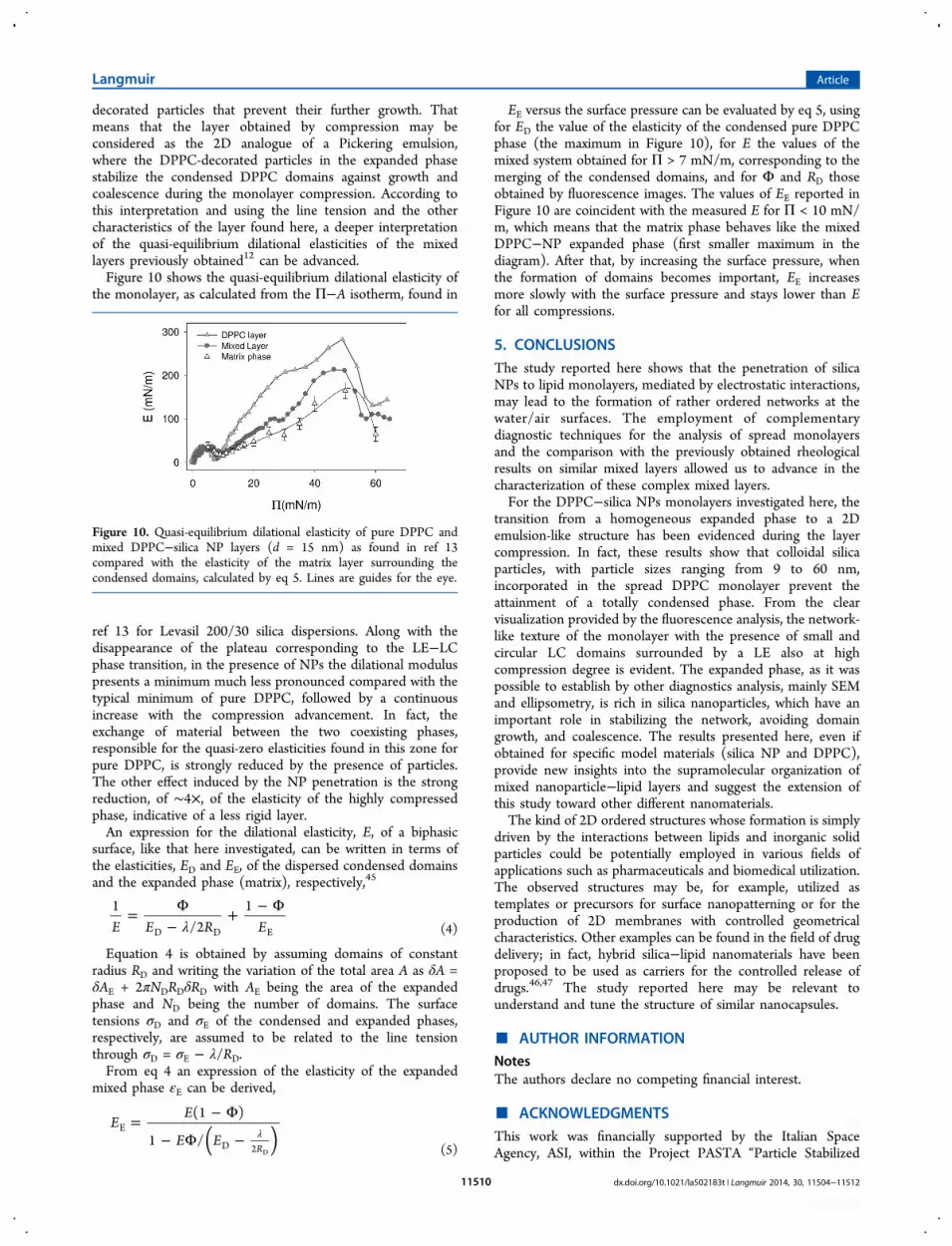
The values so obtained of λ and Δm are reported in Figure 7.They are rather constant versus the surface pressure and in therange of those typically reported for lipid monolayers andbilayers.38,39 The values of λ found with other techniques,40,41orwith molecular dynamics simulation,42 for pure DPPC domains
Figure 6. Domain size distributions (above) and corresponding fluorescence images (278 × 208 μm2) of mixed layers (below), with silica particles ofdifferent sizes d, and at the same compression degree corresponding to Π = 15 mN/m. The best-fit distributions (solid lines) are obtained by usingeq 1.
Figure 7. Line tension (a) and dipole density difference (b) against the surface pressure obtained from the best-fit parameters from the analysis ofthe domain size distribution (eqs 1 and 2) for different particle sizes d.
Langmuir Article
dx.doi.org/10.1021/la502183t | Langmuir 2014, 30, 11504−1151211508
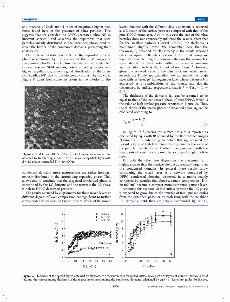
and mixtures of lipids are ∼1 order of magnitude higher thanthose found here in the presence of silica particles. Thissuggests that we consider the DPPC-decorated silica NP aslinactant species43 and advances the hypothesis that suchparticles, mainly distributed in the expanded phase, tend tocover the border of the condensed domains, preventing theircoalescence.The preferred distribution of NP in the expanded external
phase is confirmed by the analysis of the SEM images ofLangmuir−Schaeffer (LS) films transferred at controlledsurface pressure. SEM analysis, beside providing images withhigher magnification, allows a good visualization of the phaserich in silica NP, due to the electronic contrast. As shown inFigure 8, apart from some inclusions in the interior of the
condensed domains, most nanoparticles are rather homoge-neously distributed in the surrounding expanded phase. Thatallows one to conclude that the dispersed condensed phase isconstituted by the LC domains and the matrix is the LE phaseis rich in DPPC-decorated particles.The results obtained by ellipsometry for these mixed layers at
different degrees of layer compression are significant to furthercorroborate this scenario. In Figure 9 the thickness of the mixed
layers obtained with the different silica dispersions is reportedas a function of the surface pressure compared with that of thepure DPPC monolayer. Also in this case the size of the silicaparticles does not appreciably influence the results, apart thatfor the smallest particles (Levasil 300/30) the thickness ismaintained slightly lower. We remember here that thethickness, h, obtained by ellipsometry is the result averagedon a few square millimeters portion of the mixed two-phaselayer. In principle, height inhomogeneities on the nanometricscale should be dealt with within an effective mediumapproximation, such as the Lorentz−Lorenz one.44 However,given the reduced value of the film thickness, which neverexceeds the Drude approximation, we can model the roughlayer with an “average” homogeneous layer whose thickness h isexpressed as a combination of the matrix and domainthicknesses, hE and hD, respectively, that is h = ΦhD + (1 −Φ)hE.The thickness of the domains, hD, can be assumed to be
equal to that of the condensed phase of pure DPPC, which isthe value at high surface pressure reported in Figure 9a. Thus,the thickness of the matrix phase, or expanded phase, hE, can becalculated according to
=− Φ− Φ
hh h
1ED
(3)
In Figure 9b hE versus the surface pressure is reported ascalculated by eq 3 with Φ obtained by the fluorescence images(Figure 6). It is interesting to notice that hE, obtained forLevasil 300/30 at high layer compression, assumes the value ofthe particle diameter (9 nm), which is in agreement with thehypothesis of a matrix composed by a compact single particlelayer.For both the other two dispersions the maximum hE is
slightly smaller than the particle size but appreciably larger thanthe condensed domains. In general these results allowconsidering the mixed layer as a network composed byDPPC condensed domains dispersed in a matrix mainlycomposed by particles that above a certain compression (Π >30 mN/m) become a compact monodistributed particle layer.Assuming this scenario, at low surface pressure the LC phase
is expected to grow due to the transfer of free lipid moleculesfrom the expanded phase or by coalescing with the neighborLC domains, until they are totally surrounded by DPPC-
Figure 8. SEM image (180 × 150 μm2) of a Langmuir−Schaeffer filmobtained by transferring a mixed DPPC−silica nanoparticle layer withd = 15 nm, at controlled Π = 20 mN/m.
Figure 9. Thickness of the spread layers obtained by ellipsometric measurements for mixed DPPC silica particles layers, at different particle sizes d(a), and the corresponding thickness of the matrix layers surrounding the condensed domains, calculated by eq 3 (b). Lines are guides for the eye.
Langmuir Article
dx.doi.org/10.1021/la502183t | Langmuir 2014, 30, 11504−1151211509
decorated particles that prevent their further growth. Thatmeans that the layer obtained by compression may beconsidered as the 2D analogue of a Pickering emulsion,where the DPPC-decorated particles in the expanded phasestabilize the condensed DPPC domains against growth andcoalescence during the monolayer compression. According tothis interpretation and using the line tension and the othercharacteristics of the layer found here, a deeper interpretationof the quasi-equilibrium dilational elasticities of the mixedlayers previously obtained12 can be advanced.Figure 10 shows the quasi-equilibrium dilational elasticity of
the monolayer, as calculated from the Π−A isotherm, found in
ref 13 for Levasil 200/30 silica dispersions. Along with thedisappearance of the plateau corresponding to the LE−LCphase transition, in the presence of NPs the dilational moduluspresents a minimum much less pronounced compared with thetypical minimum of pure DPPC, followed by a continuousincrease with the compression advancement. In fact, theexchange of material between the two coexisting phases,responsible for the quasi-zero elasticities found in this zone forpure DPPC, is strongly reduced by the presence of particles.The other effect induced by the NP penetration is the strongreduction, of ∼4×, of the elasticity of the highly compressedphase, indicative of a less rigid layer.An expression for the dilational elasticity, E, of a biphasic
surface, like that here investigated, can be written in terms ofthe elasticities, ED and EE, of the dispersed condensed domainsand the expanded phase (matrix), respectively,45
λ= Φ
−+ − Φ
E E R E1
/21
D D E (4)
Equation 4 is obtained by assuming domains of constantradius RD and writing the variation of the total area A as δA =δAE + 2πNDRDδRD with AE being the area of the expandedphase and ND being the number of domains. The surfacetensions σD and σE of the condensed and expanded phases,respectively, are assumed to be related to the line tensionthrough σD = σE − λ/RD.From eq 4 an expression of the elasticity of the expanded
mixed phase εE can be derived,
= − Φ
− Φ − λ( )E
E
E E
(1 )
1 /R
E
D 2 D (5)
EE versus the surface pressure can be evaluated by eq 5, usingfor ED the value of the elasticity of the condensed pure DPPCphase (the maximum in Figure 10), for E the values of themixed system obtained for Π > 7 mN/m, corresponding to themerging of the condensed domains, and for Φ and RD thoseobtained by fluorescence images. The values of EE reported inFigure 10 are coincident with the measured E for Π < 10 mN/m, which means that the matrix phase behaves like the mixedDPPC−NP expanded phase (first smaller maximum in thediagram). After that, by increasing the surface pressure, whenthe formation of domains becomes important, EE increasesmore slowly with the surface pressure and stays lower than Efor all compressions.
5. CONCLUSIONSThe study reported here shows that the penetration of silicaNPs to lipid monolayers, mediated by electrostatic interactions,may lead to the formation of rather ordered networks at thewater/air surfaces. The employment of complementarydiagnostic techniques for the analysis of spread monolayersand the comparison with the previously obtained rheologicalresults on similar mixed layers allowed us to advance in thecharacterization of these complex mixed layers.For the DPPC−silica NPs monolayers investigated here, the
transition from a homogeneous expanded phase to a 2Demulsion-like structure has been evidenced during the layercompression. In fact, these results show that colloidal silicaparticles, with particle sizes ranging from 9 to 60 nm,incorporated in the spread DPPC monolayer prevent theattainment of a totally condensed phase. From the clearvisualization provided by the fluorescence analysis, the network-like texture of the monolayer with the presence of small andcircular LC domains surrounded by a LE also at highcompression degree is evident. The expanded phase, as it waspossible to establish by other diagnostics analysis, mainly SEMand ellipsometry, is rich in silica nanoparticles, which have animportant role in stabilizing the network, avoiding domaingrowth, and coalescence. The results presented here, even ifobtained for specific model materials (silica NP and DPPC),provide new insights into the supramolecular organization ofmixed nanoparticle−lipid layers and suggest the extension ofthis study toward other different nanomaterials.The kind of 2D ordered structures whose formation is simply
driven by the interactions between lipids and inorganic solidparticles could be potentially employed in various fields ofapplications such as pharmaceuticals and biomedical utilization.The observed structures may be, for example, utilized astemplates or precursors for surface nanopatterning or for theproduction of 2D membranes with controlled geometricalcharacteristics. Other examples can be found in the field of drugdelivery; in fact, hybrid silica−lipid nanomaterials have beenproposed to be used as carriers for the controlled release ofdrugs.46,47 The study reported here may be relevant tounderstand and tune the structure of similar nanocapsules.
■ AUTHOR INFORMATIONNotesThe authors declare no competing financial interest.
■ ACKNOWLEDGMENTSThis work was financially supported by the Italian SpaceAgency, ASI, within the Project PASTA “Particle Stabilized
Figure 10. Quasi-equilibrium dilational elasticity of pure DPPC andmixed DPPC−silica NP layers (d = 15 nm) as found in ref 13compared with the elasticity of the matrix layer surrounding thecondensed domains, calculated by eq 5. Lines are guides for the eye.
Langmuir Article
dx.doi.org/10.1021/la502183t | Langmuir 2014, 30, 11504−1151211510
Emulsion and Foams” (n. 2013-028-R.O)”, by the “FondazioneCassa di Risparmio di Parma” within the Project “Microscopiain fluorescenza in sistemi molecolari complessi di interessebiomedico” and carried out in the framework of the ERFCOST Action CM1101 “Colloidal Aspects of Nanoscience forInnovative Processes and Materials” and ERF COST MP1106“Smart and Green Interfaces”.
■ REFERENCES(1) Velev, O.D.; Gupta, S. Materials fabricated by micro- andnanoparticle assemblythe challenging path from science toengineering. Adv. Mater. 2009, 21, 1897−1905.(2) Mata, D.; Oliveira, F.J.; Ferro, M.; Gomes, P.S.; Fernandes, M.H.;Lopes, M.A.; Silva, R.F. Multifunctional Carbon Nanotube/Biocer-amics Modulate the Directional Growth and Activity of OsteoblasticCells. J. Biomed. Nanotechnol. 2014, 10, 725−743.(3) Fadeel, B.; Garcia-Bennett, A.E. Better safe than sorry:Understanding the toxicological properties of inorganic nanoparticlesmanufactured for biomedical applications. Adv. Drug Delivery Rev.2010, 62, 362−374.(4) Moghimi, S.M.; Hunter, A.C.; Andresen, T.L. Factors ControllingNanoparticle Pharmacokinetics: An Integrated Analysis and Perspec-tive. Annu. Rev. Pharmacol. Toxicol. 2012, 52, 481−503.(5) Liggieri, L.; Santini, E.; Guzman, E.; Maestro, A.; Ravera, F.Wide-frequency dilational rheology investigation of mixed silicananoparticle−CTAB interfacial layers. Soft Matter 2011, 7, 7699−7709.(6) Guzman, E.; Liggieri, L.; Santini, E.; Ferrari, M.; Ravera, F. MixedDPPC−Cholesterol Langmuir Monolayers in Presence of HydrophilicSilica Nanoparticles. Colloids Surf., B 2013, 105, 284−293.(7) Wang, Z.; Li, X.; Yang, S. Studies of Dipalmitoylphosphatidylcho-line (DPPC) Monolayers Embedded with Endohedral Metallofuller-ene (Dy@C82). Langmuir 2009, 25, 12968−12973.(8) Chiu, C.-C.; Shinoda, W.; DeVane, R.H.; Nielsen, S.O. Effects ofspherical fullerene nanoparticles on a dipalmitoyl phosphatidylcholinelipid monolayer: a coarse grain molecular dynamics approach. SoftMatter 2012, 8, 9610−9616.(9) Farnoud, A. M.; Fiegel, J. Low Concentrations of NegativelyCharged Sub-Micron Particles Alter the Microstructure of DPPC atthe Air−Water Interface. Colloids Surf., A 2012, 415, 320−327.(10) Harishchandra, R. K.; Saleem, M.; Galla, H.-J. Nanoparticleinteraction with model lung surfactant monolayers. J. Royal Soc.Interface 2010, 7, S15−S26.(11) Sachan, A.K.; Harishchandra, R.K.; Bantz, C.; Maskos, M.;Reichelt, R.; Galla, H.-J. High-Resolution Investigation of NanoparticleInteraction with a Model Pulmonary Surfactant Monolayer. ACS Nano2012, 6, 1677−1687.(12) Guzman, E.; Liggieri, L.; Santini, E.; Ferrari, M.; Ravera, F.Influence of silica nanoparticles on phase behaviour and structuralproperties of DPPC−Palmitic acid Langmuir monolayers. ColloidsSurf., A 2012, 413, 280−287.(13) Guzman, E.; Liggieri, L.; Santini, E.; Ferrari, M.; Ravera, F.Influence of silica nanoparticles on dilational rheology of DPPC−palmitic acid Langmuir monolayers. Soft Matter 2012, 8, 3938−3948.(14) Tatur, S.; Badia, A. Influence of Hydrophobic Alkylated GoldNanoparticles on the Phase Behavior of Monolayers of DPPC andClinical Lung Surfactant. Langmuir 2012, 28, 628−639.(15) Matshay, T.J.; Lanterna, A.E.; Granados, A.M.; Krause, R. W.M.; Maggio, B.; Vico, R.W. Distinctive Interactions of Oleic AcidCovered Magnetic Nanoparticles with Saturated and UnsaturatedPhospholipids in Langmuir Monolayers. Langmuir 2014, 30, 5888−5896.(16) Polverini, E.; Arisi, S.; Cavatorta, P.; Berzina, T.; Cristofolini, L.;Fasano, A.; Fontana, M. P. Interaction of Myelin Basic Protein withPhospholipid Monolayers: Mechanism of Protein Penetration.Langmuir 2003, 19, 872−877.(17) Cristofolini, L.; Berzina, T.; Erokhina, S.; Konovalov, O.;Erokhin, V. Structural Study of the DNA−Dipalmitoylphosphatidyl-
choline Complex at the Air−Water Interface. Biomacromolecules 2007,8, 2270−2275.(18) Matharu, Z.; Bandodkar, AJ; Gupta, V.; Malhotra, BD.Fundamentals and application of ordered molecular assemblies toaffinity biosensing. Chem. Soc. Rev. 2012, 41, 1363−1402.(19) McNamme, C.E.; Kappl, M.; Butt, H.-J.; Higashitani, K.; Graf,K. Interfacial Forces between a Silica Particle and PhosphatidylcholineMonolayers at the Air−Water Interface. Langmuir 2010, 26, 14574−14581.(20) Honciuc, A.; Howard, A.L; Schwartz, D.K. Single MoleculeObservations of Fatty Acid Adsorption at the Silica/Water Interface:Activation Energy of Attachment. J. Phys. Chem. C 2009, 113, 2078−2081.(21) Guidotti, G. The Composition of Biological Membranes. Arch.Int. Med. 1972, 129, 194−201.(22) Orsi, D.; Baldi, G.; Cicuta, P.; Cristofolini, L. On the relationbetween hierarchical morphology and mechanical properties of acolloidal 2D gel system. Colloids Surf., A 2012, 413, 71−77.(23) Orsi, D.; Cristofolini, L.; Baldi, G.; Madsen, A. Heterogeneousand anisotropic dynamics of a 2D Gel. Phys. Rev. Lett. 2012, 108,105701.(24) Orsi, D.; Ruta, B.; Chushkin, Y.; Pucci, A.; Ruggeri, G.; Baldi,G.; Cristofolini, L. Controlling the dynamics of a bidimensional gelabove and below its percolation transition. Phys. Rev. E 2014, 89,042308.(25) Maestro, A.; Guzman, E.; Santini, E.; Ravera, F.; Liggieri, L.;Ortega, F.; Rubio, R.G. Wettability of silica nanoparticle−surfactantnanocomposite interfacial layers. Soft Matter 2012, 8, 837−843.(26) Schneider, C.A.; Rasband, W.S.; Eliceiri, K.W. NIH Image toImageJ: 25 years of image analysis. Nat. Methods 2012, 9, 671−675.(27) Harke, M.; Teppner, R.; Schulz, O.; Motschmann, H.; Orendi,H. Description of a single modular optical setup for ellipsometry,surface plasmons, waveguide modes, and their corresponding imagingtechniques including Brewster angle microscopy. Rev. Sci. Instrum.1997, 68, 3130−3134.(28) Phillips, M. C.; Chapman, D. Monolayer characteristics ofsaturated 1,2-diacyl phosphatidylcholines (lecithins) and phosphatidy-lethanolamines at the air−water interface. Biochim. Biophys. Acta 1968,163, 301−313.(29) Klopfer, K. J.; Vanderlick, T. K. Isotherms of Dipalmitoylphos-phatidylcholine (DPPC) Monolayers: Features Revealed and FeaturesObscured. J. Colloid Sci. 1996, 182, 220−229.(30) Grigoriev, D. O.; Kragel, J.; Dutschk, V.; Miller, R.; Mohwald,H. Contact angle determination of micro- and nanoparticles at fluid/fluid interfaces: The excluded area concept. Phys. Chem. Chem. Phys.2007, 9, 6447−6454.(31) Nag, K.; Keough, K.M. Epifluorescence microscopic studies ofmonolayers containing mixtures of dioleoyl- and dipalmitoylphospha-tidylcholines. Biophys. J. 1993, 65, 1019−1026.(32) Mercado, F. V.; Maggio, B.; Wilke, N. Modulation of thedomain topography of biphasic monolayers of stearic acid anddimyristoyl phosphatidylcholine. Chem. Phys. Lipids 2012, 165, 232−237.(33) Sriram, I.; Schwartz, D. K. Line tension between coexistingphases in monolayers and bilayers of amphiphilic molecules. SurfaceSci. Rep. 2012, 67, 143−159.(34) Arriaga, L. R.; Lopez-Montero, I.; Ignes-Mullol, J.; Monroy, F.Domain-Growth Kinetic Origin of Nonhorizontal Phase CoexistencePlateaux in Langmuir Monolayers: Compression Rigidity of a Raft-Like Lipid Distribution. J. Phys. Chem. B 2010, 114, 4509−4520.(35) Maestro, A.; Drenckhan, W.; Rio, E.; Hohler, R. Liquiddispersions under gravity: Volume fraction profile and osmoticpressure. Soft Matter 2012, 9, 2531−2540.(36) Fortuna, I.; Thomas, G. L.; de Almeida, R. M. C.; Graner, F.Growth Laws and Self-Similar Growth Regimes of Coarsening Two-Dimensional Foams: Transition from Dry to Wet Limits. Phys. Rev.Lett. 2012, 108, 248301.(37) Lee, D. W.; Min, Y.; Dhar, P.; Ramachandran, A.; Israelachvili, J.N.; Zasadzinski, J. A. Relating domain size distribution to line tension
Langmuir Article
dx.doi.org/10.1021/la502183t | Langmuir 2014, 30, 11504−1151211511
and molecular dipole density in model cytoplasmic myelin lipidmonolayers. Proc. Nat. Acad. Sci. U. S. A. 2011, 108, 9425−9430.(38) Honerkamp-Smith, A. R.; Cicuta, P.; Collins, M. D.; Veatch, S.L.; Nijs, M. d.; Schick, M.; Keller, S. L. Line Tensions, CorrelationLengths, and Critical Exponents in Lipid Membranes Near CriticalPoints. Biophys. J. 2008, 95, 236−246.(39) Kuzmin, P.I.; Akhimov, S.A.; Chizmadzhev, Y.A.; Zimmerberg,J.; Cohen, F.S. Line tension and interaction energies of membrane raftscalculated from lipid splay and tilt. Biophys. J. 2005, 88, 1120−1133.(40) Bischof, A. A.; Wilke, N. Molecular determinants for the linetension of coexisting liquid phases in monolayers. Chem. Phys. Lipids2012, 165, 737−744.(41) Zhelev, D.V.; Needham, D. Tension-stabilized pores in giantvesicles: determination of pore size and pore line tension. Biochim.Biophys. Acta 1993, 1147, 89−104.(42) Marrink, S.J.; Mark, A.E. Molecular Dynamics Simulation of theFormation, Structure, and Dynamics of Small Phospholipid Vesicles. J.Am. Chem. Soc. 2003, 125, 15233−15242.(43) Trabelsi, S.; Zhang, S.; Lee, T.R; Schwartz, D.S. Linactants:Surfactant Analogues in Two Dimensions. Phys. Rev. Lett. 2008, 100,037802.(44) Tompkins, H.G. A user’s guide to ellipsometry; Academic Press:Boston, 1993.(45) Lucassen, J. Dynamic dilational properties of composite surfaces.Colloids Surf. 1991, 65, 139−149.(46) Mohanraj, V.J.; Barnes, T.J.; Prestidge, C.A. Silica nanoparticlecoated liposomes: A new type of hybrid nanocapsule for proteins. Int.J. Pharm. 2010, 392, 285−293.(47) Begu, S.; Pouessel, A.A.; Lerner, D.A.; Tourne-Peteilh, C.;Devoisselle, J.M. Liposil, a promising composite material for drugstorage and release. J. Controlled Release 2007, 118, 1−6.
Langmuir Article
dx.doi.org/10.1021/la502183t | Langmuir 2014, 30, 11504−1151211512
Copyright © 2022 FDOKUMEN