Transport of gabapentin by LAT1 (SLC7A5)
12
Transport of gabapentin by LAT1 (SLC7A5) David Dickens a , Steven D. Webb b , Svetlana Antonyuk c , Athina Giannoudis d , Andrew Owen a , Steffen Ra ¨ disch a , S. Samar Hasnain c , Munir Pirmohamed a, * a Department of Molecular and Clinical Pharmacology, University of Liverpool, Liverpool, UK b Centre for Drug Safety Science, Department of Molecular and Clinical Pharmacology, University of Liverpool, Liverpool, UK c Molecular Biophysics Group, Institute of Integrative Biology, Faculty of Health and Life Sciences, University of Liverpool, Liverpool, UK d Department of Haematology, University of Liverpool, Liverpool, UK 1. Introduction Gabapentin (l-(aminomethyl)cyclohexaneacetic acid) has a branched chain amino acid like structure and is used in the treatment of epilepsy and neuropathic pain. Gabapentin is a chemical analogue of GABA but has no activity in GABAergic neuronal systems [1]. Instead gabapentin has been found to be a a2d ligand (subunit of the voltage-gated calcium channel) that disrupts calcium channel trafficking [2] and is proposed to function therapeutically by blocking new synapse formation [3]. In humans, gabapentin plasma concentrations do not increase proportionally with increasing dose resulting in large inter-patient variability [4]. A saturable transport mechanism in the intestinal absorption of gabapentin is the underlying cause of the lack of proportionality between increasing dose and drug concentrations in plasma [5]. Additionally, an in-situ rat intestinal perfusion model found an interaction of gabapentin with dipeptides that enhanced the uptake of gabapentin [6]. Gabapentin is not bound to plasma proteins, metabolites account for <1% of the dose and the drug is excreted unchanged in the urine [7]. The excretion rate of gabapentin into the urine in healthy individuals has been correlated with a single nucleotide polymorphism in the OCTN1 transporter gene (SLC22A4) and gabapentin has been shown to be a substrate of this transporter in vitro [8]. OCTN1 plays an important role in gabapentin urinary excretion but does not influence gabapentin plasma concentrations [8] As gabapentin acts in the brain, the blood–brain barrier (BBB) will be important in regulating drug penetration. Gabapentin can readily cross the BBB, but the blood and brain concentrations are not proportional to dose [9]. The passage of gabapentin across the Biochemical Pharmacology xxx (2013) xxx–xxx A R T I C L E I N F O Article history: Received 4 February 2013 Accepted 27 March 2013 Available online xxx Keywords: LAT1 Blood–brain barrier Gabapentin Alternative access mechanism Brain endothelial cells Mathematical modelling A B S T R A C T Gabapentin is used in the treatment of epilepsy and neuropathic pain. Gabapentin has high and saturable permeability across the BBB, but no mechanistic studies underpinning this process have been reported. The aim of the current study was to investigate the transport of gabapentin in a model of the BBB, identify the important drug transporter(s) and to use mathematical modelling to quantify the processes involved. A human brain endothelial cell line (hCMEC/D3) was utilised as an in-vitro model of the BBB. Uptake of radiolabeled gabapentin into cells in the presence of chemical inhibitors, siRNA or overexpressed drug transporters of interest was investigated. Gabapentin was demonstrated to be a LAT1 substrate in brain endothelial cells (LAT1-process; K m = 530 mM and V max = 7039 pmoles/million cells/min versus other- processes; K m = 923 mM and V max = 3656 pmoles/million cells/min) and in transfected HEK 293 LAT1 cells (LAT1-process; K m = 217 mM and V max = 5192 pmoles/million cells/min versus otherprocesses; K m = 1546 mM and V max = 3375 pmoles/million cells/min). At physiological concentrations of gabapen- tin, LAT1 mediated transport was 3 or 10-fold higher than the other transport processes in the two systems, respectively, demonstrating clear selectivity for gabapentin. In-silico structural homology modelling confirmed that LAT1 could have the LeuT conserved fold and functions by the alternative access mechanism. Mathematical modelling of this mechanism revealed revised significance of V max and K m so that a low K m may not necessarily imply a high affinity transport process. Gabapentin was negative for OCT like transport and LAT2 activity in the hCMEC/D3 and OCT1 transfected cells. Our data shows that gabapentin is a substrate for the influx transporter LAT1 at therapeutic concentrations. ß 2013 Elsevier Inc. All rights reserved. Abbreviations: LAT1, L-type amino acid transporter 1; BBB, blood–brain barrier; FBS, fetal bovine serum; HBSS, hanks balanced salt solution; OCT, organic cation transporters; TEA, tetraethylammonium chloride; OCT1, organic cation transporter 1.. * Corresponding author at: Department of Molecular and Clinical Pharmacology, Wolfson Centre for Personalised Medicine, University of Liverpool, Block A: Waterhouse Building, 1-5 Brownlow Street, Liverpool, L69 3GL, UK. Tel.: +44 151 794 5549; fax: +44 151 794 5549. E-mail address: [email protected] (M. Pirmohamed). G Model BCP-11602; No. of Pages 12 Please cite this article in press as: Dickens D, et al. Transport of gabapentin by LAT1 (SLC7A5). Biochem Pharmacol (2013), http:// dx.doi.org/10.1016/j.bcp.2013.03.022 Contents lists available at SciVerse ScienceDirect Biochemical Pharmacology jo u rn al h om epag e: ww w.els evier.c o m/lo cat e/bio c hem p har m 0006-2952/$ – see front matter ß 2013 Elsevier Inc. All rights reserved. http://dx.doi.org/10.1016/j.bcp.2013.03.022
Transcript of Transport of gabapentin by LAT1 (SLC7A5)

Biochemical Pharmacology xxx (2013) xxx–xxx
G Model
BCP-11602; No. of Pages 12
Transport of gabapentin by LAT1 (SLC7A5)
David Dickens a, Steven D. Webb b, Svetlana Antonyuk c, Athina Giannoudis d,Andrew Owen a, Steffen Radisch a, S. Samar Hasnain c, Munir Pirmohamed a,*a Department of Molecular and Clinical Pharmacology, University of Liverpool, Liverpool, UKb Centre for Drug Safety Science, Department of Molecular and Clinical Pharmacology, University of Liverpool, Liverpool, UKc Molecular Biophysics Group, Institute of Integrative Biology, Faculty of Health and Life Sciences, University of Liverpool, Liverpool, UKd Department of Haematology, University of Liverpool, Liverpool, UK
A R T I C L E I N F O
Article history:
Received 4 February 2013
Accepted 27 March 2013
Available online xxx
Keywords:
LAT1
Blood–brain barrier
Gabapentin
Alternative access mechanism
Brain endothelial cells
Mathematical modelling
A B S T R A C T
Gabapentin is used in the treatment of epilepsy and neuropathic pain. Gabapentin has high and saturable
permeability across the BBB, but no mechanistic studies underpinning this process have been reported.
The aim of the current study was to investigate the transport of gabapentin in a model of the BBB, identify
the important drug transporter(s) and to use mathematical modelling to quantify the processes involved.
A human brain endothelial cell line (hCMEC/D3) was utilised as an in-vitro model of the BBB. Uptake of
radiolabeled gabapentin into cells in the presence of chemical inhibitors, siRNA or overexpressed drug
transporters of interest was investigated. Gabapentin was demonstrated to be a LAT1 substrate in brain
endothelial cells (LAT1-process; Km = 530 mM and Vmax = 7039 pmoles/million cells/min versus other-
processes; Km = 923 mM and Vmax = 3656 pmoles/million cells/min) and in transfected HEK 293 LAT1
cells (LAT1-process; Km = 217 mM and Vmax = 5192 pmoles/million cells/min versus otherprocesses;
Km = 1546 mM and Vmax = 3375 pmoles/million cells/min). At physiological concentrations of gabapen-
tin, LAT1 mediated transport was 3 or �10-fold higher than the other transport processes in the two
systems, respectively, demonstrating clear selectivity for gabapentin. In-silico structural homology
modelling confirmed that LAT1 could have the LeuT conserved fold and functions by the alternative
access mechanism. Mathematical modelling of this mechanism revealed revised significance of Vmax and
Km so that a low Km may not necessarily imply a high affinity transport process. Gabapentin was negative
for OCT like transport and LAT2 activity in the hCMEC/D3 and OCT1 transfected cells. Our data shows that
gabapentin is a substrate for the influx transporter LAT1 at therapeutic concentrations.
� 2013 Elsevier Inc. All rights reserved.
Contents lists available at SciVerse ScienceDirect
Biochemical Pharmacology
jo u rn al h om epag e: ww w.els evier .c o m/lo cat e/b io c hem p har m
1. Introduction
Gabapentin (l-(aminomethyl)cyclohexaneacetic acid) has abranched chain amino acid like structure and is used in thetreatment of epilepsy and neuropathic pain. Gabapentin is achemical analogue of GABA but has no activity in GABAergicneuronal systems [1]. Instead gabapentin has been found to be aa2d ligand (subunit of the voltage-gated calcium channel) thatdisrupts calcium channel trafficking [2] and is proposed to functiontherapeutically by blocking new synapse formation [3].
Abbreviations: LAT1, L-type amino acid transporter 1; BBB, blood–brain barrier; FBS,
fetal bovine serum; HBSS, hanks balanced salt solution; OCT, organic cation
transporters; TEA, tetraethylammonium chloride; OCT1, organic cation
transporter 1..
* Corresponding author at: Department of Molecular and Clinical Pharmacology,
Wolfson Centre for Personalised Medicine, University of Liverpool, Block A:
Waterhouse Building, 1-5 Brownlow Street, Liverpool, L69 3GL, UK.
Tel.: +44 151 794 5549; fax: +44 151 794 5549.
E-mail address: [email protected] (M. Pirmohamed).
Please cite this article in press as: Dickens D, et al. Transport of gadx.doi.org/10.1016/j.bcp.2013.03.022
0006-2952/$ – see front matter � 2013 Elsevier Inc. All rights reserved.
http://dx.doi.org/10.1016/j.bcp.2013.03.022
In humans, gabapentin plasma concentrations do not increaseproportionally with increasing dose resulting in large inter-patientvariability [4]. A saturable transport mechanism in the intestinalabsorption of gabapentin is the underlying cause of the lack ofproportionality between increasing dose and drug concentrationsin plasma [5]. Additionally, an in-situ rat intestinal perfusion modelfound an interaction of gabapentin with dipeptides that enhancedthe uptake of gabapentin [6]. Gabapentin is not bound to plasmaproteins, metabolites account for <1% of the dose and the drug isexcreted unchanged in the urine [7]. The excretion rate ofgabapentin into the urine in healthy individuals has beencorrelated with a single nucleotide polymorphism in the OCTN1transporter gene (SLC22A4) and gabapentin has been shown to bea substrate of this transporter in vitro [8]. OCTN1 plays animportant role in gabapentin urinary excretion but does notinfluence gabapentin plasma concentrations [8]
As gabapentin acts in the brain, the blood–brain barrier (BBB)will be important in regulating drug penetration. Gabapentin canreadily cross the BBB, but the blood and brain concentrations arenot proportional to dose [9]. The passage of gabapentin across the
bapentin by LAT1 (SLC7A5). Biochem Pharmacol (2013), http://
D. Dickens et al. / Biochemical Pharmacology xxx (2013) xxx–xxx2
G Model
BCP-11602; No. of Pages 12
BBB has been shown to be saturable [10]. Interestingly gabapentinpermeability across the BBB in rodents is vastly higher than wouldhave been predicted taking into account the drugs lipophilicity(predictive Log P of 1.2) [11]. The mechanism or specifictransporter that confers this high permeability across the BBB isunknown with no mechanistic studies having been published [12].
Gabapentin transport has been linked to amino acid transpor-ters in a variety of studies. In three different cell lines (astrocytes,synaptosomes and CHO cells) gabapentin transport into cells wasinhibited by L-phenylalanine, suggesting the involvement of L-alpha amino acid transporters (LAT) [13]. LATs are a generic familyof transporters that transport neutral amino acids such as L-phenylalanine and include LAT1 (SLC7A5), LAT2 (SLC7A8), LAT3(SLC43A1) and LAT4 (SLC43A2). A study utilising oocytes expres-sing rodent LAT1 and an adapter protein (4f2hc) showed thatgabapentin was an inhibitor of LAT1-mediated transport ofphenylalanine uptake [14]. The LAT1 transporter is an antiporterthat has been found to be highly expressed at the BBB [12,15]. IfLAT1 transports gabapentin this could have an effect on brainuptake of the drug due to inter-patient differences in activity orexpression of the transporter. This could lead to variations inresponse to treatment with gabapentin. Other studies investigat-ing an interaction with gabapentin and drug transporters,including P-glycoprotein (ABCB1) and OCTN2 (SLC22A5), havebeen negative [16,17].
As the BBB expresses a variety of influx as well as effluxfunctions [18] and many CNS active drugs have been shown to betransported at the BBB [19], we hypothesised that gabapentin isalso actively transported into the brain. To investigate thetransport of gabapentin in an in-vitro model of the BBB, chemicalinhibitors, siRNA and specific overexpression of transporters ofinterest were utilised.
2. Materials and methods
2.1. Materials
[3H]-gabapentin was acquired from American RadiolabeledChemicals (ARC, St. Louis, USA) with specific activity of 110 Ci/mmol. [14C]-tetraethylammonium bromide was purchased fromPerkinElmer (MA, USA) with specific activity of 3.2 mCi/mmol.[3H]-phenylalanine was acquired from PerkinElmer with specificactivity of 110 Ci/mmol. Tariquidar was synthesised by Dr. OliverLanger, Medical University of Vienna, Austria. Gabapentin wasobtained from Abcam Biochemicals (Cambridge, UK). MK571 wasacquired from Tocris Biosciences (Bristol, UK). All other drugs andchemicals, unless otherwise stated, were purchased from Sigma(Poole, Dorset, UK).
2.2. Distribution coefficient (Log D, pH 7.4)
The lipophilicity of a drug in a biphasic system is termed thedistribution coefficient (Log D). The Log D (pH 7.4) of gabapentinwas experimentally determined as previously described [20]. Inbrief [3H]-gabapentin was suspended in Hanks balanced saltsolution (HBSS) at pH 7.4 with 25 mM HEPES and mixed vigorouslyfor 15 min with 1-octanol. The layers were separated bycentrifugation for 5 min at 250 � g. Radioactivity was determinedby scintillation counting of aliquots from both phases. The log D
pH7.4 was calculated as; log D = log (DPMoctanol/DPM buffer).
2.3. Cell culture
HEK 293 cells were cultured in DMEM supplemented withpenicillin–streptomycin (1%) and 10% FCS. hCMEC/D3, an immor-talised human brain endothelial cell line (kind gift from
Please cite this article in press as: Dickens D, et al. Transport of gadx.doi.org/10.1016/j.bcp.2013.03.022
Pierre-Olivier Couraud, INSERM, Paris, France), was maintainedin EGM-2 medium (Lonza, Slough, UK) supplemented withpenicillin-streptomycin (1%), hydrocortisone (1.4 mM), ascorbicacid (5 mg/ml), chemically defined lipid concentrate (1/100)(Invitrogen, Paisley, UK), HEPES (10 mM), 5% foetal calf serum(FCS) and basic fibroblast growth factor (1 ng/ml). hCMEC/D3 werepassaged every 3–4 days and seeded onto collagen type I(Invitrogen) coated cell culture plasticware (Nunc, Fisher Scientific,Loughborough, UK). KCL22 wild type cells are a chronic myelocyticleukaemia (CML) cell line and was maintained in RPMI supple-mented with penicillin-streptomycin (1%) and 10% FCS. TheCountess Automated Cell Counter (Invitrogen, Paisley, UK) wasused to determine cell density for plating and transport assays.
2.4. Transfection of KCL22 with hOCT1, OCTN1 and OCTN2
The CML cell line KCL22 was selected for transfectionexperiments concerning OCTs since it expresses a low basalamount of those transporters in comparison to other CML cell lines[21,22]. The previously established hOCT1 overexpressing cell linewas used for the transport studies and the stable cell line carryingthe empty vector pcDNA3.1 was used as a control in all theexperiments [22,23]. Similarly, KCL22 cells were transfected byAMAXA nucleofection technology (Lonza) with the pcDNA3.1plasmids carrying human OCTN1 and human OCTN2 (kind gift ofProfs A. Tsuji and Y. Kato, Kanazawa, Japan) and positive cloneswere selected using 1 mg/ml neomycin (G418). The mRNA levels ofthe generated clones were screened by real-time PCR in order toselect those over-expressing the transporter gene of interest.
2.5. Sub-cloning and transfection of LAT1 into 293 cells
LAT1 full length cDNA (SLC7A5, IMAGE clone 5551612) wassub-cloned into pcDNA3.1 using pcDNA3.1/V5-His TOPO TAExpression Kit (Invitrogen) following the manufacturer’s instruc-tions. Successful sub-cloning of LAT1 was confirmed by sequencingand HEK 293 cells were transfected with pcDNA3.1 and pcDNA3.1LAT1 plasmids utilising lipofectamine 2000 (Invitrogen) accordingto the manufacturer’s instructions. The clones were selected using800 mg/ml G418. The mRNA levels of the generated single cellclones were screened by real-time PCR to select clones over-expressing LAT1.
2.6. siRNA transfection of hCMEC/D3 cells
hCMEC/D3 cells were plated onto 6-well plates and lipofecta-mine RNAiMAX (Invitrogen) was used to transfect cells with a finalconcentration of 10 nM siRNA as per manufacture’s protocol. Thecells were used, after 48 h transfection, for cellular accumulationassays or mRNA extraction. The silencer select pre-designedsiRNAs (Invitrogen) utilised in this study were: negative controlsiRNA #1 (4390843), LAT1 siRNA #1 (15653), LAT1 siRNA #2(15655) & LAT2 siRNA (23808).
2.7. RNA extraction and quantitative RT-PCR
hCMEC/D3, KCL22 and 293 cells were washed with HBSS andlysed in Tri reagent for subsequent RNA extraction as described inthe manufacturer’s instructions. Following RNA extraction, reversetranscription utilising TaqMan reverse transcription reagents(Applied Biosystems, Paisley, UK) was performed. Assays ondemand primer and probe mixes for OCTN1 (SLC22A4,Hs00268200), OCTN2 (SLC22A5, Hs00161895), LAT1 (SLC7A5,Hs00185826), LAT2 (SLC7A8, Hs00794796) and GAPDH (4310884)were purchased from Applied Biosystems. GAPDH was included asa housekeeping gene. Thermal cycling conditions for all assays
bapentin by LAT1 (SLC7A5). Biochem Pharmacol (2013), http://
D. Dickens et al. / Biochemical Pharmacology xxx (2013) xxx–xxx 3
G Model
BCP-11602; No. of Pages 12
consisted of 15 min at 95 8C followed by 50 cycles of 15 s at 95 8Cand 60 s at 60 8C with quantification of PCR products in real timeusing a real-time qPCR machine (AB 7900HT). Expression datawere normalised to GAPDH expression using the comparative Ct
method to determine relative expression of OCTN1, OCTN2, LAT1and LAT2 mRNA [24].
2.8. Western blotting
To generate whole cell lysates, cells were lysed in RIPA buffer(Thermo Scientific, Loughborough, UK) containing protease inhibi-tor cocktail and incubated for 30 min on ice. The lysate wassubsequently centrifuged at 13,000 � g and protein quantificationperformed on the supernatant. The whole cell lysates were used forsubsequent immunoblotting applications. Primary antibodies weredirected against poly-his tag (1:1000, Mouse monoclonal, Abcam,Cambridge, UK), and b actin (1:8000, Mouse monoclonal).
2.9. Cellular drug accumulation assay
Studies of drug uptake utilised a tracer concentration ofradiolabelled compound (0.1 mCi/ml) with sufficient non-radiola-belled compound added to give a final concentration of drug from0.01 mM to 1000 mM in transport media. Transport media consistedof HBSS with 25 mM HEPES at pH 7.4 in addition to 0.1% BSA.
The KCL22 cells transfected with hOCT1, OCTN1 and OCTN2were used for drug transporter assays by adding 2 million cells totransport medium containing radiolabelled drug at 37 8C. Afterincubation, three wash steps with ice-cold HBSS were performedand the cells were solubilised by incubation with 10% sodiumdodecyl sulfate (SDS) for 30 min. The resultant solution was mixedwith scintillation fluid and radioactivity was counted using ascintillation-counter (1500 Tri Carb LS Counter; Packard, Meriden,CT 06450, USA).
On the day of the assay, hCMEC/D3 cells or HEK 293 cellscultured on 6 well plates were equilibrated in transport media.Transport media containing the radiolabelled drug with vehicle orinhibitor were added to the cells and incubated at 37 8C for 30 minor for the indicated time. For the inhibition studies, the followinginhibitors were used: verapamil (blocks OCTs, OCTN1, OCTN2 andP-gp), amantadine (blocks OCT1 and OCT2), tariquidar (blocks P-gpand BCRP), Ko-143 (blocks BCRP) and MK-571 (blocks MRPs).Amino acids at 1 mM were utilised as competitive inhibitors and1 mM BCH (2-aminobicyclo-(2,2,1)-heptane-2-carboxylic acid)was used to inhibit system L amino acid transporters.
2.10. Mechanistic model of gabapentin transport
Kinetics of gabapentin uptake were determined by selecting atime point within a linear range (1 min) and then determininginflux at different concentrations of drug. The rate of drugtransport by LAT1 and other transport processes (pmoles/min/million cells) was plotted against gabapentin concentration (mM).A Nelder–Mead simplex direct search algorithm was used tocalculate Michaelis–Menten values for gabapentin uptake into inthe cells. Corresponding mechanistic equations describing the rateof change over time of the intra- and extracellular gabapentinconcentrations, denoted here by si(t) and se(t), are given by
siðtÞ ¼ ve
viðseð0Þ � seðtÞÞ; ve
dseðtÞdt¼ �vi jother � vi jLAT1
where vi and ve are the total volumes of the intra- and extracellularcompartments (which we assume are fixed over the timescale ofthe experiment: 1 ml transfer buffer gives ve = 1012 mm3;vi = 2.107 � 109 mm3 for hCMEC/D3 cells – calculated from a
Please cite this article in press as: Dickens D, et al. Transport of gadx.doi.org/10.1016/j.bcp.2013.03.022
single cell diameter of 13.6 mm as measured on a Countess cellcounter and assuming a spherical cell shape multiplied by anaverage of 1.6 million cells; similarly, vi = 4.451 � 109 mm3 for HEK293 cells – single cell diameter 12.9 mm, average number of cells3.96 million), se(0) is the initial concentration of drug (5 mM in Figs3G and 4H) and JLAT1 and Jother describe the (per volume)Michaelis–Menten uptake rates for LAT1 and the other processes,respectively
jLAT1 ¼ aV1
maxseðtÞK1
m þ seðtÞ; Jother ¼
V0maxseðtÞ
K0m þ seðtÞ
:
The value of a is taken to be 1 and 0 for brain endothelialcells transfected with NC siRNA and LAT1 siRNA#1, respectively.For HEK 293 cells, a is taken to be 1 for the LAT1 transfectedcells and a < 1 for the control. However, as it is difficult toestimate how much of the 160-fold increase in LAT1 mRNA istransferred to activity, it is then difficult to estimate exactly thevalue of a. Instead we vary a to represent a 20- to 160-foldincrease in activity (i.e. a 2 [1/160,1/20]). Our estimates for theMichaelis–Menten values are then taken to be the averages ofthose obtained for this range of a. Note that fold increases below20 fail to give statistically significant comparisons between theMichaelis–Menten uptake rates and the corresponding data,indicating that at least a 20-fold increase in activity occurs. Themechanistic equations were solved using a Runge–Kutta fourthorder method.
2.11. In silico 3D modelling of LAT1
The human LAT1 amino acid sequence (NP_003477.4) wasinputted into the iterative threading assembly refinement (I-TASSER) server to generate a structural prediction [25]. The I-TASSER server generates three-dimensional atomic models frommultiple threading alignments and iterative structural assemblysimulations. PROMALS3D is an alignment algorithm that takes intoaccount the amino acid sequences and structure to align proteinsequences [26] and was used to identify putative LAT1 binding site.The 3D model of LAT1 was viewed and annotated using PyMOL(http://www.pymol.org).
2.12. Mathematical modelling of the alternative access mechanism
The symmetrical states of the alternative access mechanism forthe LAT1 transporter (illustrated in Fig. 5C) can be re-written in thefollowing reaction equations:
ce þ se�!k1
sce�!k
sci�!k�1
ci þ si;
ai þ si�!k2
aci�!k
ace�!k�2
ce þ ae;
where ai and ae denote the concentrations of intra- andextrcellular amino acid and the remaining notation describesthe concentration of transporters in the following states: ce
unbound outward facing; sce bound to drug, outward facing; sci
bound to drug, inward facing; ci unbound inward facing; aci
bound to amino acid, inward facing; ace bound to amino acid,outward facing. We use k1 and k�1 to denote the binding anddissociation rates of the transporter to the drug; k2 and k�2 todenote the binding and dissociation rates of the transporter toamino acid; and k to denote the rate at which the conformationalchange takes place (i.e. 1/k is the time taken for the bound carrierto change configuration from outward to inward facing and viceversa). Ordinary differential equations describing the rate ofchange over time of the number of transporters in the variousstates can be derived using law of mass action kinetics on theabove reaction scheme. We assume that the total number of
bapentin by LAT1 (SLC7A5). Biochem Pharmacol (2013), http://
D. Dickens et al. / Biochemical Pharmacology xxx (2013) xxx–xxx4
G Model
BCP-11602; No. of Pages 12
transporters is conserved, namely ce + sce + sci + ci + aci + ace = C
where C is a constant (total transporter concentration). Hence,there are only five independent equations, not six. The flux, JLAT1,is
JLAT1 ¼ k�1sci ¼ �k1cesc;
where we have described the flux from outside to inside as beingpositive. Assuming steady-state flux conditions and solving theresulting algebraic systems, we can then obtain
JLAT1 ¼Vmaxse
Km þ se; Vmax ¼ kc
Kai
KKd þ 2aiðKd þ KÞ
� �; Km
¼ KKdai
KKd þ 2aiðKd þ KÞ
� �;
where for simplicity we have set k1 = k2 = k+ and k�1 = k�2 = k�,K = k�/k+, Kd = k/k+, and we have assumed that the leading order ai
is constant. Note that kC and K are the typical definitions for Vmax
and Km and are usually taken to denote measures of transportercapacity and 1/affinity, respectively.
2.13. Statistical tests
All data are presented as mean � standard deviation from threeindependent experiments performed in triplicate (n = 3). To assessstatistical significance GraphPad Prism 5 (GraphPad Software, Inc., LaJolla, USA) was used for drug uptake in the presence of inhibitors ortransfected cells with the samples compared by one way ANOVAfollowed by Tukey–Kramer test for multiple comparisons. A P value of<0.05 was taken to indicate significance.
3. Results
3.1. Assessment of gabapentin uptake by organic cation transporters
in transfected cells (OCT1, OCTN1 & OCTN2) and in human brain
endothelial cells
The distribution coefficient (Log D, pH 7.4) of gabapentin wasexperimentally determined and was found to be �1.21, indicatingthat gabapentin is a hydrophilic compound and therefore morelikely to be taken up into cells by a transporter mediated process.
To assess the importance of carrier mediated transport ofgabapentin, organic cation transporters were investigated (Fig. 1).KCL22 cells transfected with OCT1, OCTN1 and OCTN2 wereutilised (Fig. 1A–C). Increased expression of the respective drugtransporter in the transfected cells was confirmed by QRTPCR forOCTN1 (98-fold) and OCTN2 (133-fold) with OCT1 expressionpreviously determined [19]. TEA was used as a positive control andwas significantly higher in the OCT1, OCTN1 and OCTN2transfected cells compared to the control cells. Gabapentin uptakewas increased in the OCTN1 transfected cells (Fig. 1C) but wasnegative in the OCT1 (Fig. 1A) and OCTN2 cells (Fig. 1B). Todetermine if an organic cation transporter was responsible for thetransport of gabapentin into hCMEC/D3 cells, a panel of chemicalinhibitors was utilised (Fig. 1D): no inhibition of transport wasdemonstrated with these inhibitors.
3.2. Transport of gabapentin into human brain endothelial cells is
inhibited by neutral amino acids
As no interaction was observed with the chemical inhibitorsagainst organic cation transporters, a panel of compounds wasused to determine an inhibitory profile for gabapentin transport.None of these compounds, which are known to act as inhibitors ofvarious transporters, had an effect on gabapentin uptake apart
Please cite this article in press as: Dickens D, et al. Transport of gadx.doi.org/10.1016/j.bcp.2013.03.022
from L-phenylalanine which significantly decreased transport(Fig. 2A). Temperature also affected drug transport. As classicaldrug transporters did not seem to be important, while temperatureand L-phenylalanine had an effect, we went onto define whichamino acids inhibited uptake. Neutral amino acids and BCHinhibited the uptake of gabapentin (Fig. 2B) but not glycine,arginine or glutamic acid suggesting that a neutral amino acidtransporter was involved. A time course analysis of gabapentinuptake with L-phenylalanine inhibition showed differences inuptake over time (Fig. 2C).
3.3. Gabapentin influx is mediated by LAT1 in brain endothelial cells
To determine the specific neutral amino transporter responsi-ble for the uptake of gabapentin, knockdown of two L-type aminoacid transporters (LAT1 and LAT2) was performed by RNAi.Transient transfection with siRNA achieved >75% suppression oftransporter mRNA (Fig. 3A). A LAT1 targeting siRNA #1significantly reduced uptake when compared to a non-targetingcontrol siRNA (NC) while a LAT2 targeting siRNA had no effect(Fig. 3B). An additional independent targeting siRNA of LAT1 (#2)was utilised to confirm the phenotype observed with the firsttargeting siRNA. The second LAT1 (#2) siRNA caused a significantdecrease in gabapentin uptake compared to the negative controlsiRNA transfected cells. To determine a linear time point fortransport kinetic studies, a time course for gabapentin uptake intohCMEC/D3 cells transfected with NC siRNA and LAT1 siRNA wasperformed (Fig. 3C). This identified the 1 min time point for linearuptake that was then used to determine the velocity of gabapentinuptake in the siRNA transfected hCMEC/D3 cells at a range ofconcentrations (Fig. 3D). Significantly more gabapentin uptakewas observed in the NC siRNA transfected cells compared to thesiRNA LAT1 #1 transfected cells over the concentrations tested.
Fig. 3E and F shows a comparison between the Michaelis–Menten gabapentin uptake rate functions for LAT1 and the othertransport processes, highlighting the relative Km values to thetherapeutic concentrations of gabapentin (Fig. 3E) and uptakerates at therapeutic concentrations (Fig. 3F). If Vmax and Km areinterpreted in the conventional way, LAT1 (Km of 530 mM and Vmax
of 7039 pmoles/million cells/min) has almost twice the capacitycompared to the sum of the other transport processes (which hasVmax 3656 pmoles/million cells/min) and almost twice the affinity(Km for the other processes is 923 mM). However, at therapeuticconcentrations, LAT1 transport actually turns out to be 3-foldhigher than the other transport processes (Fig. 3G). LAT1 transportat 37 mM of gabapentin (mean plasma concentration) is459.3 pmoles/million cells/min versus 140.8 pmoles/millioncells/min for the other transport processes.
Solving the corresponding mechanistic equations with theappropriate Michaelis–Menten uptake terms (see Section 2.8 fulldetails) and comparing these analytical solutions to the timeprofile data for gabapentin uptake, we observed that both the NCsiRNA and LAT 1 siRNA #1 show good agreement untilapproximately 4 min after which time gabapentin uptake intothe cells decreased in LAT1 siRNA transfected cells (Fig. 3G). Thissuggests an additional process occurs that is not included in themathematical model and appears to be a time-dependent ratherthan a concentration dependent process.
3.4. Gabapentin is transported in LAT1 stably transfected cells
To investigate in a second model system whether gabapentin wasa substrate for LAT1, HEK 293 cells stably transfected with pcDNA3.1(control) and pcDNA3.1 LAT1 were generated. Increased expressionof LAT1 in the pcDNA3.1 LAT1 cell line was confirmed by QRTPCR(Fig. 4A) and by Western blotting (Fig. 4B). L-phenylalanine was
bapentin by LAT1 (SLC7A5). Biochem Pharmacol (2013), http://
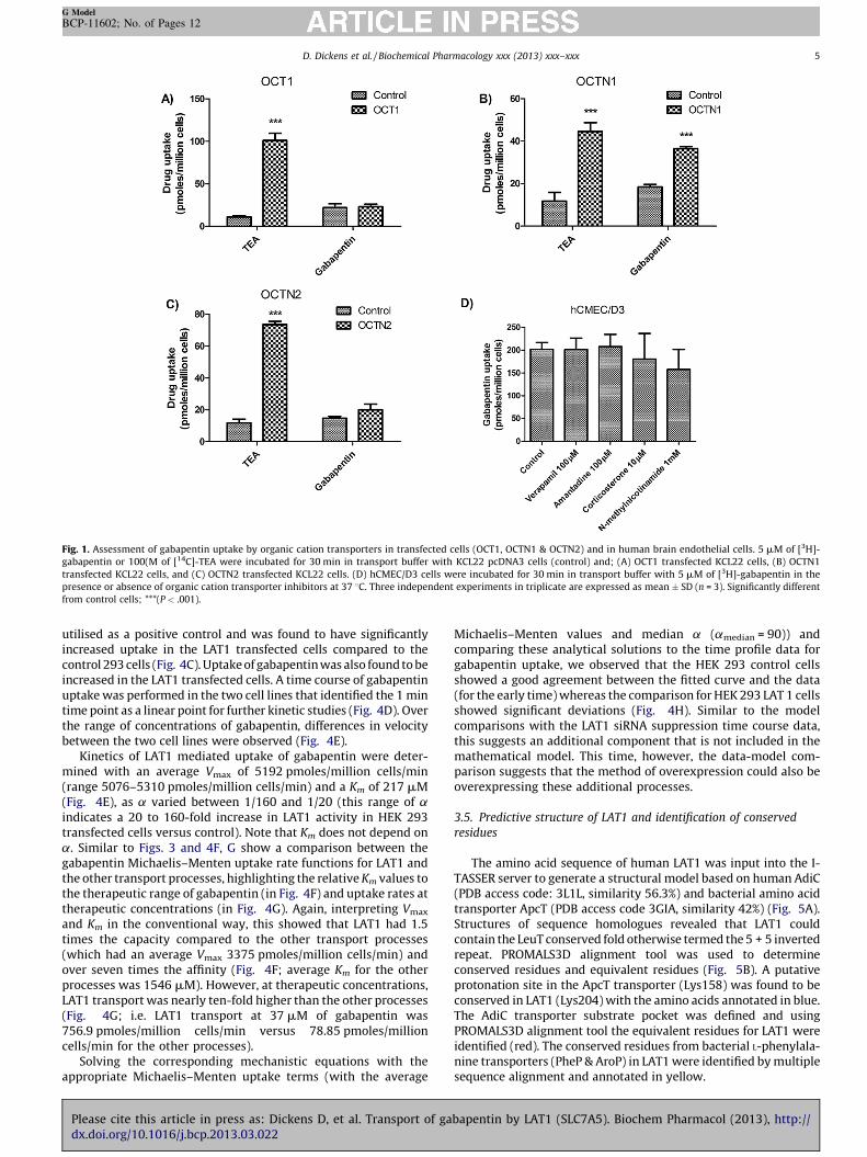
Fig. 1. Assessment of gabapentin uptake by organic cation transporters in transfected cells (OCT1, OCTN1 & OCTN2) and in human brain endothelial cells. 5 mM of [3H]-
gabapentin or 100(M of [14C]-TEA were incubated for 30 min in transport buffer with KCL22 pcDNA3 cells (control) and; (A) OCT1 transfected KCL22 cells, (B) OCTN1
transfected KCL22 cells, and (C) OCTN2 transfected KCL22 cells. (D) hCMEC/D3 cells were incubated for 30 min in transport buffer with 5 mM of [3H]-gabapentin in the
presence or absence of organic cation transporter inhibitors at 37 8C. Three independent experiments in triplicate are expressed as mean � SD (n = 3). Significantly different
from control cells; ***(P < .001).
D. Dickens et al. / Biochemical Pharmacology xxx (2013) xxx–xxx 5
G Model
BCP-11602; No. of Pages 12
utilised as a positive control and was found to have significantlyincreased uptake in the LAT1 transfected cells compared to thecontrol 293 cells (Fig. 4C). Uptake of gabapentin was also found to beincreased in the LAT1 transfected cells. A time course of gabapentinuptake was performed in the two cell lines that identified the 1 mintime point as a linear point for further kinetic studies (Fig. 4D). Overthe range of concentrations of gabapentin, differences in velocitybetween the two cell lines were observed (Fig. 4E).
Kinetics of LAT1 mediated uptake of gabapentin were deter-mined with an average Vmax of 5192 pmoles/million cells/min(range 5076–5310 pmoles/million cells/min) and a Km of 217 mM(Fig. 4E), as a varied between 1/160 and 1/20 (this range of aindicates a 20 to 160-fold increase in LAT1 activity in HEK 293transfected cells versus control). Note that Km does not depend ona. Similar to Figs. 3 and 4F, G show a comparison between thegabapentin Michaelis–Menten uptake rate functions for LAT1 andthe other transport processes, highlighting the relative Km values tothe therapeutic range of gabapentin (in Fig. 4F) and uptake rates attherapeutic concentrations (in Fig. 4G). Again, interpreting Vmax
and Km in the conventional way, this showed that LAT1 had 1.5times the capacity compared to the other transport processes(which had an average Vmax 3375 pmoles/million cells/min) andover seven times the affinity (Fig. 4F; average Km for the otherprocesses was 1546 mM). However, at therapeutic concentrations,LAT1 transport was nearly ten-fold higher than the other processes(Fig. 4G; i.e. LAT1 transport at 37 mM of gabapentin was756.9 pmoles/million cells/min versus 78.85 pmoles/millioncells/min for the other processes).
Solving the corresponding mechanistic equations with theappropriate Michaelis–Menten uptake terms (with the average
Please cite this article in press as: Dickens D, et al. Transport of gadx.doi.org/10.1016/j.bcp.2013.03.022
Michaelis–Menten values and median a (amedian = 90)) andcomparing these analytical solutions to the time profile data forgabapentin uptake, we observed that the HEK 293 control cellsshowed a good agreement between the fitted curve and the data(for the early time) whereas the comparison for HEK 293 LAT 1 cellsshowed significant deviations (Fig. 4H). Similar to the modelcomparisons with the LAT1 siRNA suppression time course data,this suggests an additional component that is not included in themathematical model. This time, however, the data-model com-parison suggests that the method of overexpression could also beoverexpressing these additional processes.
3.5. Predictive structure of LAT1 and identification of conserved
residues
The amino acid sequence of human LAT1 was input into the I-TASSER server to generate a structural model based on human AdiC(PDB access code: 3L1L, similarity 56.3%) and bacterial amino acidtransporter ApcT (PDB access code 3GIA, similarity 42%) (Fig. 5A).Structures of sequence homologues revealed that LAT1 couldcontain the LeuT conserved fold otherwise termed the 5 + 5 invertedrepeat. PROMALS3D alignment tool was used to determineconserved residues and equivalent residues (Fig. 5B). A putativeprotonation site in the ApcT transporter (Lys158) was found to beconserved in LAT1 (Lys204) with the amino acids annotated in blue.The AdiC transporter substrate pocket was defined and usingPROMALS3D alignment tool the equivalent residues for LAT1 wereidentified (red). The conserved residues from bacterial L-phenylala-nine transporters (PheP & AroP) in LAT1 were identified by multiplesequence alignment and annotated in yellow.
bapentin by LAT1 (SLC7A5). Biochem Pharmacol (2013), http://
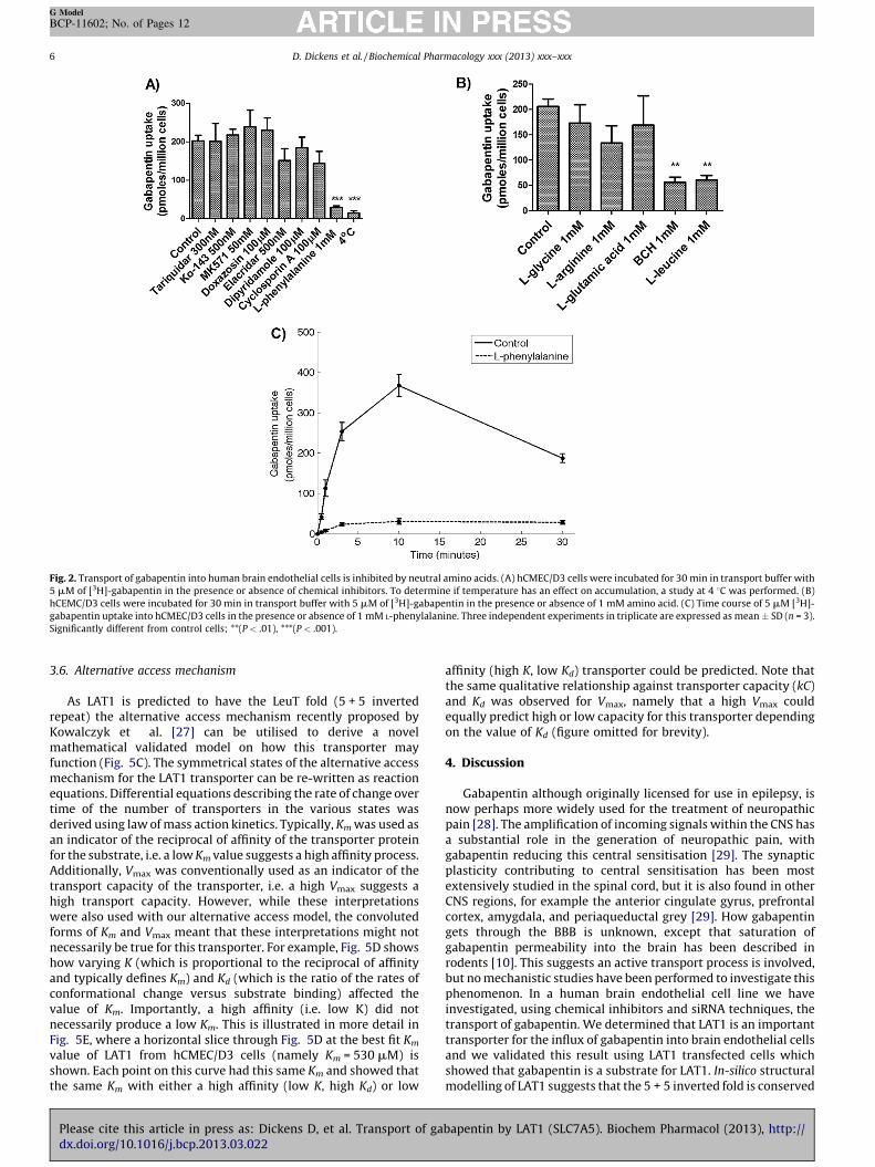
Fig. 2. Transport of gabapentin into human brain endothelial cells is inhibited by neutral amino acids. (A) hCMEC/D3 cells were incubated for 30 min in transport buffer with
5 mM of [3H]-gabapentin in the presence or absence of chemical inhibitors. To determine if temperature has an effect on accumulation, a study at 4 8C was performed. (B)
hCEMC/D3 cells were incubated for 30 min in transport buffer with 5 mM of [3H]-gabapentin in the presence or absence of 1 mM amino acid. (C) Time course of 5 mM [3H]-
gabapentin uptake into hCMEC/D3 cells in the presence or absence of 1 mM L-phenylalanine. Three independent experiments in triplicate are expressed as mean � SD (n = 3).
Significantly different from control cells; **(P < .01), ***(P < .001).
D. Dickens et al. / Biochemical Pharmacology xxx (2013) xxx–xxx6
G Model
BCP-11602; No. of Pages 12
3.6. Alternative access mechanism
As LAT1 is predicted to have the LeuT fold (5 + 5 invertedrepeat) the alternative access mechanism recently proposed byKowalczyk et al. [27] can be utilised to derive a novelmathematical validated model on how this transporter mayfunction (Fig. 5C). The symmetrical states of the alternative accessmechanism for the LAT1 transporter can be re-written as reactionequations. Differential equations describing the rate of change overtime of the number of transporters in the various states wasderived using law of mass action kinetics. Typically, Km was used asan indicator of the reciprocal of affinity of the transporter proteinfor the substrate, i.e. a low Km value suggests a high affinity process.Additionally, Vmax was conventionally used as an indicator of thetransport capacity of the transporter, i.e. a high Vmax suggests ahigh transport capacity. However, while these interpretationswere also used with our alternative access model, the convolutedforms of Km and Vmax meant that these interpretations might notnecessarily be true for this transporter. For example, Fig. 5D showshow varying K (which is proportional to the reciprocal of affinityand typically defines Km) and Kd (which is the ratio of the rates ofconformational change versus substrate binding) affected thevalue of Km. Importantly, a high affinity (i.e. low K) did notnecessarily produce a low Km. This is illustrated in more detail inFig. 5E, where a horizontal slice through Fig. 5D at the best fit Km
value of LAT1 from hCMEC/D3 cells (namely Km = 530 mM) isshown. Each point on this curve had this same Km and showed thatthe same Km with either a high affinity (low K, high Kd) or low
Please cite this article in press as: Dickens D, et al. Transport of gadx.doi.org/10.1016/j.bcp.2013.03.022
affinity (high K, low Kd) transporter could be predicted. Note thatthe same qualitative relationship against transporter capacity (kC)and Kd was observed for Vmax, namely that a high Vmax couldequally predict high or low capacity for this transporter dependingon the value of Kd (figure omitted for brevity).
4. Discussion
Gabapentin although originally licensed for use in epilepsy, isnow perhaps more widely used for the treatment of neuropathicpain [28]. The amplification of incoming signals within the CNS hasa substantial role in the generation of neuropathic pain, withgabapentin reducing this central sensitisation [29]. The synapticplasticity contributing to central sensitisation has been mostextensively studied in the spinal cord, but it is also found in otherCNS regions, for example the anterior cingulate gyrus, prefrontalcortex, amygdala, and periaqueductal grey [29]. How gabapentingets through the BBB is unknown, except that saturation ofgabapentin permeability into the brain has been described inrodents [10]. This suggests an active transport process is involved,but no mechanistic studies have been performed to investigate thisphenomenon. In a human brain endothelial cell line we haveinvestigated, using chemical inhibitors and siRNA techniques, thetransport of gabapentin. We determined that LAT1 is an importanttransporter for the influx of gabapentin into brain endothelial cellsand we validated this result using LAT1 transfected cells whichshowed that gabapentin is a substrate for LAT1. In-silico structuralmodelling of LAT1 suggests that the 5 + 5 inverted fold is conserved
bapentin by LAT1 (SLC7A5). Biochem Pharmacol (2013), http://

Fig. 3. Gabapentin influx is mediated by in brain endothelial cells. (A) Expression of target gene mRNA following transient transfection with siRNA oligos in hCMEC/D3 cells.
Relative mRNA expression compared to the negative control siRNA (NC siRNA). (B) hCMEC/D3 cells transfected with NC siRNA, LAT1 siRNA#1, LAT1 siRNA#2 and LAT2 siRNA
were incubated with 5 mM of [3H]-gabapentin for 30 min. Significantly different from control cells; **(P < .01). (C) Time course of 5 mM [3H]-gabapentin uptake into hCMEC/
D3 cells transfected with NC siRNA or LAT1 siRNA. (D) Kinetics of gabapentin influx into hCMEC/D3 cells. hCMEC/D3 cells transfected with NC siRNA or LAT1 siRNA#1 were
D. Dickens et al. / Biochemical Pharmacology xxx (2013) xxx–xxx 7
G Model
BCP-11602; No. of Pages 12
Please cite this article in press as: Dickens D, et al. Transport of gabapentin by LAT1 (SLC7A5). Biochem Pharmacol (2013), http://dx.doi.org/10.1016/j.bcp.2013.03.022
D. Dickens et al. / Biochemical Pharmacology xxx (2013) xxx–xxx8
G Model
BCP-11602; No. of Pages 12
from bacterial amino acid transporter homologues and that analternative access mechanism can therefore be put forward for itsmode of action. The kinetic data for LAT1 mediated transport wasinput into a two compartment model which yielded a newinterpretation for the Michaelis–Menten Vmax and Km parametersbased on the alternative access mechanism.
Studies by Summerfield et al. [11] and Uchino et al. [30] bothquote a predictive Log P of 1.2 for gabapentin in their investiga-tions. To clarify this situation, we experimentally determined theLog D at pH7.4 and found it to be �1.2. This would suggest minimalpassive diffusion and the potential importance of carrier mediatedtransporters to get gabapentin into cells. OCTN1 has been shown totransport gabapentin and affects the renal excretion of the drug [8].Since gabapentin is a substrate of another organic cationtransporter member (OCTN1) we used a panel of organic cationtransporter transfected cell lines and chemical inhibitors of OCTson human brain endothelial cells. However gabapentin wasnegative for transport by OCT1 transfected cells and OCT chemicalinhibitors had no effect.
L-phenylalanine affected the uptake of gabapentin in humanbrain endothelial cells, which can therefore be added to the list ofcells (astrocytes, synaptosomes, and CHO cells) where similarobservations have been reported [13]. Assuming that L-phenylala-nine is working as a competitive inhibitor in these cell types,gabapentin uptake must be mediated by a L-phenylalaninesensitive transporter. Previously, gabapentin has been shown toinhibit the LAT1-mediated transport of phenylalanine in oocytesexpressing rodent LAT1 and an adapter protein (4f2hc) [14].However at least 7 high affinity L-phenylalanine transporters areencoded by the human genome and we narrowed our investigationby known expression patterns. At the BBB, LAT1 has been shown tobe highly expressed [15] with possible expression of LAT2 [31].Additionally, gabapentin has been shown to be an inhibitor ofrodent LAT1 function in a Xenopus Laevis oocyte expression systemwith indirect measures suggesting a competitive mode of action[14]. We show for the first time that gabapentin is a substrate ofhuman LAT1 in two mammalian cell systems. Also, LAT2involvement at least in brain endothelial cell type can be ruledout because it was functionally negative for transport even thoughmRNA was detectable in the cells. The hCMEC/D3 cell line is anestablished in-vitro model of the BBB and siRNA mediatedsuppression of LAT1 with two independent oligonucleotidesprovided evidence that gabapentin is a substrate. We then utiliseda stable overexpression cell line of LAT1. This two-step process thatwe previously used for lamotrigine [19] and imatinib [21] providesadditional evidence that gabapentin is a LAT1 substrate andprovides kinetic information that can be derived and interpreted. Afuture topic of experimentation would be to investigate theexpression and potential involvement of additional phenylalaninetransporters such as LAT3 and LAT4 in the transport of gabapentin.
Patients on gabapentin for the treatment of neuropathic painhave an average peak serum concentration of 37 mM with a rangeof 23 mM to 80 mM [32]. Therefore the Km values derived for LAT1in the two cell lines are higher than the physiologically achievableconcentrations. However, the rate of LAT1 transport in our twocompartment model compared to the other process at atherapeutic concentration range, indicates that in both cell
incubated for 1 min in transport buffer containing 0.01–1000 mM of [3H]-gabapentin
V0maxse=ðK0
m þ seÞ þ V1maxse=ðK1
m þ seÞ (solid curve) and V0maxse=ðK0
m þ seÞ (dashed curve).
expressed as mean � SD (n = 3). (E) A comparison of the gabapentin uptake rate functions f
LAT1 (K1m , solid line) and the other transport processes (K0
m , dashed line). The vertical shaded
the thick vertical line indicating the mean (37 mM). (F) Uptake rates at physiological concentr
the mechanistic model predicted uptake. The experimentally observed uptake of gabapenti
lines) and LAT1 siRNA #1 (circles and thin dashed lines). The thick curves show the model pre
(NC siRNA = thick solid curve; LAT 1 siRNA #1 = thick dashed curve).
Please cite this article in press as: Dickens D, et al. Transport of gadx.doi.org/10.1016/j.bcp.2013.03.022
systems, LAT1 transport predominates for gabapentin by either3 or 10-fold. Therefore, LAT1 appears to be the predominanttransport process in brain endothelial cells at therapeuticconcentrations. Other processes that might include other influxtransporters or a passive diffusion component are therefore lessimportant. The suppression of LAT1 expression is achievable in-
vitro for this transporter but no LAT1 knock out mouse has beendescribed which could in part be due to an essential role of aminoacid transport into cells leading to embryonic lethality.
Due to LAT1 expression at the BBB [15] strategies have beendeveloped for drug delivery by using prodrugs of L-type amino acidtransporter substrates that deliver compounds to the brain [33]. Amedicinal chemistry approach with pregabalin has determinedthat both the system L-type amino acid transporter activity anda2d binding affinity are important for the in vivo activity of thiscompound [34]. The structural similarities to pregabalin mean thatthis might also be the case for gabapentin. Therefore, understand-ing the specific L-type amino acid transporters responsible at aparticular biological barrier could be relevant to treatmentoutcome. In this study we show that LAT1 is important fortransport at the BBB and thus is likely to be important forgabapentin penetrating to its site of action. The importance of L-type amino acid transporters at tissues such as the gut needsfurther investigation as they may also be involved in the saturableabsorption of gabapentin in patients [5] where expression of atleast LAT1 and LAT2 has been observed [35]. An in-silico study byBolger et al. found a predicted association between LAT2expression in the human intestine and gabapentin absorptionbut no mechanistic studies were performed [36]. LAT2 has beensuggested to be the functionally active L-type amino acidtransporter at the kidney [37] and could be involved in the renalextraction of gabapentin. Additional studies are required toinvestigate if it is a substrate. Changes in expression, or regulationof LAT1 expression have not been investigated in detail, but LAT1expression is altered in cancer [38,39] and hypoxia [40], and thiswould be a potentially important area for further investigation.
Depending on the cell type, LAT1 expressed on its own caninduce L-type transporter activity, without the need of an adapterprotein [30] and in the current study HEK 293 cells only requiredoverexpression of LAT1 for transport activity. We have identifiedsites of conservation of LAT1 that might be important for functionand these include a putative site of protonation, putative bindingpocket and residues that are conserved with bacterial L-phenylal-anine transporters. This is a starting point for further studies thatcould investigate which of these residues are important forfunction (e.g. by site directed mutagenesis). How LAT1 works as ananti-porter is unclear but it has been suggested that it might pumpextracellular substrate in at the same time as intracellularsubstrate goes out [12]. However this is based on limited evidence.With a structural modelling approach we propose that LAT1 mightfunction by the alternative access mechanism but future work interms of X-ray crystallography is required to confirm this in-silico
finding. Due to the conservation of the LeuT fold, the alternativeaccess mechanism [27] is a highly attractive assumption at thisstage. As far as we are aware, our mathematical model for thealternate access mechanism is the first theoretical application tothis LeuT fold mechanism. It is interesting that despite the
with uptake velocity plotted against concentration with curves best fitted to;
Data shown are three independent experiments performed in triplicate and are
or LAT1 and the other transport processes. The vertical lines indicate the Km values for
region denotes the physiological concentration range of gabapentin (23–80 mM) with
ations. (G) A comparison of the experimentally determined gabapentin time course and
n is shown versus time in hCMEC/D3 transfected with NC siRNA (circles and thin solid
dicted uptake profiles with the uptake rates as with the appropriate best fit parameters
bapentin by LAT1 (SLC7A5). Biochem Pharmacol (2013), http://

Fig. 4. Gabapentin is transported in LAT1 stably transfected cells. (A) The relative mRNA expression of LAT1 in HEK 293 cells stably transfected with pcDNA3.1 (control) or
pcDNA3.1 LAT1. (B) Immunoblotting for poly-his tag and b-actin of whole protein lysate from HEK 293 cells stably transfected with pcDNA3.1 or pcDNA3.1 LAT1.
Representative immunoblot is shown. (C) Uptake of 5 mM [3H]-L-phenylalanine & 5 mM [3H]-gabapentin at 1 min in HEK 293 cells transfected with pcDNA3.1 (control) or
pcDNA3.1 LAT1. Data shown are three independent experiments performed in triplicate and are expressed as mean � SD (n = 3). Significantly different from control cells;
D. Dickens et al. / Biochemical Pharmacology xxx (2013) xxx–xxx 9
G Model
BCP-11602; No. of Pages 12
Please cite this article in press as: Dickens D, et al. Transport of gabapentin by LAT1 (SLC7A5). Biochem Pharmacol (2013), http://dx.doi.org/10.1016/j.bcp.2013.03.022
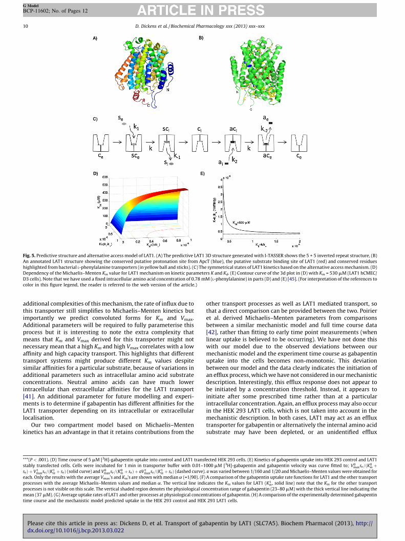
Fig. 5. Predictive structure and alternative access model of LAT1. (A) The predictive LAT1 3D structure generated with I-TASSER shows the 5 + 5 inverted repeat structure, (B)
An annotated LAT1 structure showing the conserved putative protonation site from ApcT (blue), the putative substrate binding site of LAT1 (red) and conserved residues
highlighted from bacterial L-phenylalanine transporters (in yellow ball and sticks). (C) The symmetrical states of LAT1 kinetics based on the alternative access mechanism. (D)
Dependency of the Michaelis–Menten Km value for LAT1 mechanism on kinetic parameters K and Kd. (E) Contour curve of the 3d plot in (D) with Km = 530 mM (LAT1 hCMEC/
D3 cells). Note that we have used a fixed intracellular amino acid concentration of 0.78 mM (L-phenylalanine) in parts (D) and (E) [45]. (For interpretation of the references to
color in this figure legend, the reader is referred to the web version of the article.)
D. Dickens et al. / Biochemical Pharmacology xxx (2013) xxx–xxx10
G Model
BCP-11602; No. of Pages 12
additional complexities of this mechanism, the rate of influx due tothis transporter still simplifies to Michaelis–Menten kinetics butimportantly we predict convoluted forms for Km and Vmax.Additional parameters will be required to fully parameterise thisprocess but it is interesting to note the extra complexity thatmeans that Km and Vmax derived for this transporter might notnecessary mean that a high Km and high Vmax correlates with a lowaffinity and high capacity transport. This highlights that differenttransport systems might produce different Km values despitesimilar affinities for a particular substrate, because of variations inadditional parameters such as intracellular amino acid substrateconcentrations. Neutral amino acids can have much lowerintracellular than extracellular affinities for the LAT1 transport[41]. An additional parameter for future modelling and experi-ments is to determine if gabapentin has different affinities for theLAT1 transporter depending on its intracellular or extracellularlocalisation.
Our two compartment model based on Michaelis–Mentenkinetics has an advantage in that it retains contributions from the
***(P < .001). (D) Time course of 5 mM [3H]-gabapentin uptake into control and LAT1 trans
stably transfected cells. Cells were incubated for 1 min in transporter buffer with 0.01–1
seÞ þ V1maxse=ðK1
m þ seÞ (solid curve) and V0maxse=ðK0
m þ seÞ þ aV1maxse=ðK1
m þ seÞ (dashed curve)
each. Only the results with the average Vmax’s and Km’s are shown with median a (=1/90). (F)
processes with the average Michaelis–Menten values and median a. The vertical line indi
processes is not visible on this scale. The vertical shaded region denotes the physiological co
mean (37 mM). (G) Average uptake rates of LAT1 and other processes at physiological concen
time course and the mechanistic model predicted uptake in the HEK 293 control and HEK
Please cite this article in press as: Dickens D, et al. Transport of gadx.doi.org/10.1016/j.bcp.2013.03.022
other transport processes as well as LAT1 mediated transport, sothat a direct comparison can be provided between the two. Poirieret al. derived Michaelis–Menten parameters from comparisonsbetween a similar mechanistic model and full time course data[42], rather than fitting to early time point measurements (whenlinear uptake is believed to be occurring). We have not done thiswith our model due to the observed deviations between ourmechanistic model and the experiment time course as gabapentinuptake into the cells becomes non-monotonic. This deviationbetween our model and the data clearly indicates the initiation ofan efflux process, which we have not considered in our mechanisticdescription. Interestingly, this efflux response does not appear tobe initiated by a concentration threshold. Instead, it appears toinitiate after some prescribed time rather than at a particularintracellular concentration. Again, an efflux process may also occurin the HEK 293 LAT1 cells, which is not taken into account in themechanistic description. In both cases, LAT1 may act as an effluxtransporter for gabapentin or alternatively the internal amino acidsubstrate may have been depleted, or an unidentified efflux
fected HEK 293 cells. (E) Kinetics of gabapentin uptake into HEK 293 control and LAT1
000 mM [3H]-gabapentin and gabapentin velocity was curve fitted to; V0maxse=ðK0
m þ. a was varied between 1/160 and 1/20 and Michaelis–Menten values were obtained for
A comparison of the gabapentin uptake rate functions for LAT1 and the other transport
cates the Km values for LAT1 (K1m , solid line) note that the Km for the other transport
ncentration range of gabapentin (23–80 mM) with the thick vertical line indicating the
trations of gabapentin. (H) A comparison of the experimentally determined gabapentin
293 LAT1 cells.
bapentin by LAT1 (SLC7A5). Biochem Pharmacol (2013), http://
D. Dickens et al. / Biochemical Pharmacology xxx (2013) xxx–xxx 11
G Model
BCP-11602; No. of Pages 12
transporter may begin to pump gabapentin out of the cell. A moredetailed theoretical and experimental investigation of in this isrequired and this will be the feature of future work.
As LAT1 is the important influx transporter for gabapentin in anin-vitro model of the BBB, we would speculate that variation in theactivity or expression could affect drug uptake and thus clinicaloutcomes. Interaction with foods containing phenylalanine at thelevel of the BBB is also possible, although a high protein meal didnot affect the disposition of gabapentin in epileptics indicating thattransporters other than LAT1 may be important for GI absorption[43]. This has been shown for another LAT1 substrate, L-DOPA: inmonkeys, a high protein meal prior to the administration of L-DOPAreduced transport into the brain [44]. Similar studies looking atgabapentin brain uptake have not been performed, and studiesthat focus on GI absorption [43] may give false reassurance of thelack of an interaction at the site of action.
In conclusion, we show for the first time that LAT1 can transportgabapentin and that LAT1 is an important influx transporter for thecarriage of this drug in an in-vitro model of the BBB. We havedemonstrated that the alternative access mechanism may beimportant for LAT1 transport. LAT1 transport activity may varybetween individuals and could therefore determine clinicaloutcome in patients treated with gabapentin. However, thisrequires further work.
Conflict of interest
The authors declare no conflict of interest.
Acknowledgements
MP is a NIHR Senior Investigator and is also supported by theDept. of Health (NHS Chair of Pharmacogenetics), MRC andWellcome Trust. AO would like to thank the MRC and EPSRC forresearch support. SVA is supported by the Wellcome Trust strategicfunds.
References
[1] Timmerman W, Bouma M, De Vries JB, Davis M, Westerink BH. A microdialysisstudy on the mechanism of action of gabapentin. Eur J Pharmacol 2000;398:53–7.
[2] Hendrich J, Van Minh AT, Heblich F, Nieto-Rostro M, Watschinger K,Striessnig J, et al. Pharmacological disruption of calcium channel traffickingby the alpha2delta ligand gabapentin. Proc Natl Acad Sci U S A 2008;105:3628–33.
[3] Eroglu C, Allen NJ, Susman MW, O‘Rourke NA, Park CY, Ozkan E, et al.Gabapentin receptor alpha2delta-1 is a neuronal thrombospondin receptorresponsible for excitatory CNS synaptogenesis. Cell 2009;139:380–92.
[4] Gidal BE, Radulovic LL, Kruger S, Rutecki P, Pitterle M, Bockbrader HN. Inter-and intra-subject variability in gabapentin absorption and absolute bioavail-ability. Epilepsy Res 2000;40:123–7.
[5] Stewart BH, Kugler AR, Thompson PR, Bockbrader HN. A saturable transportmechanism in the intestinal absorption of gabapentin is the underlying causeof the lack of proportionality between increasing dose and drug levels inplasma. Pharm Res 1993;10:276–81.
[6] Nguyen TV, Smith DE, Fleisher D. PEPT1 enhances the uptake of gabapentin viatrans-stimulation of b0,+ exchange. Pharm Res 2007;24:353–60.
[7] Bockbrader HN, Wesche D, Miller R, Chapel S, Janiczek N, Burger P. A compari-son of the pharmacokinetics and pharmacodynamics of pregabalin and gaba-pentin. Clin Pharmacokinet 2010;49:661–9.
[8] Urban TJ, Brown C, Castro RA, Shah N, Mercer R, Huang Y, et al. Effects ofgenetic variation in the novel organic cation transporter, OCTN1, on the renalclearance of gabapentin. Clin Pharmacol Ther 2008;83:416–21.
[9] Welty DF, Schielke GP, Vartanian MG, Taylor CP. Gabapentin anticonvulsantaction in rats: disequilibrium with peak drug concentrations in plasma andbrain microdialysate. Epilepsy Res 1993;16:175–81.
[10] Luer MS, Hamani C, Dujovny M, Gidal B, Cwik M, Deyo K, et al. Saturabletransport of gabapentin at the blood–brain barrier. Neurol Res 1999;21:559–62.
[11] Summerfield SG, Read K, Begley DJ, Obradovic T, Hidalgo IJ, Coggon S, et al.Central nervous system drug disposition: the relationship between in situbrain permeability and brain free fraction. J Pharmacol Exp Ther 2007;322:205–13.
Please cite this article in press as: Dickens D, et al. Transport of gadx.doi.org/10.1016/j.bcp.2013.03.022
[12] del Amo EM, Urtti A, Yliperttula M. Pharmacokinetic role of L-type amino acidtransporters LAT1 and LAT2. Eur J Pharm Sci 2008;35:161–74.
[13] Su TZ, Lunney E, Campbell G, Oxender DL. Transport of gabapentin, a gamma-amino acid drug, by system l alpha-amino acid transporters: a comparativestudy in astrocytes, synaptosomes, and CHO cells. J Neurochem 1995;64:2125–31.
[14] Uchino H, Kanai Y, Kim DK, Wempe MF, Chairoungdua A, Morimoto E, et al.Transport of amino acid-related compounds mediated by L-type amino acidtransporter 1 (LAT1): insights into the mechanisms of substrate recognition.Mol Pharmacol 2002;61:729–37.
[15] Boado RJ, Li JY, Nagaya M, Zhang C, Pardridge WM. Selective expression of thelarge neutral amino acid transporter at the blood–brain barrier. Proc Natl AcadSci U S A 1999;96:12079–84.
[16] Crowe A, Teoh YK. Limited P-glycoprotein mediated efflux for anti-epilepticdrugs. J Drug Target 2006;14:291–300.
[17] Grigat S, Fork C, Bach M, Golz S, Geerts A, Schomig E, et al. The carnitinetransporter SLC22A5 is not a general drug transporter, but it efficientlytranslocates mildronate. Drug Metab Dispos 2009;37:330–7.
[18] Abbott NJ, Patabendige AA, Dolman DE, Yusof SR, Begley DJ. Structure andfunction of the blood–brain barrier. Neurobiol Dis 2010;37:13–25.
[19] Dickens D, Owen A, Alfirevic A, Giannoudis A, Davies A, Weksler B, et al.Lamotrigine is a substrate for OCT1 in brain endothelial cells. BiochemPharmacol 2012;83:805–14.
[20] Davies A, Jordanides NE, Giannoudis A, Lucas CM, Hatziieremia S, Harris RJ,et al. Nilotinib concentration in cell lines and primary CD34(+) chronicmyeloid leukemia cells is not mediated by active uptake or efflux by majordrug transporters. Leukemia 2009.
[21] Thomas J, Wang L, Clark RE, Pirmohamed M. Active transport of imatinibinto and out of cells: implications for drug resistance. Blood 2004;104:3739–45.
[22] Wang L, Giannoudis A, Lane S, Williamson P, Pirmohamed M, Clark RE.Expression of the uptake drug transporter hOCT1 is an important clinicaldeterminant of the response to imatinib in chronic myeloid leukemia. ClinPharmacol Ther 2008;83:258–64.
[23] Giannoudis A, Davies A, Lucas CM, Harris RJ, Pirmohamed M, Clark RE. Effectivedasatinib uptake may occur without human organic cation transporter 1(hOCT1): implications for the treatment of imatinib-resistant chronic myeloidleukemia. Blood 2008;112:3348–54.
[24] Pfaffl MW. A new mathematical model for relative quantification in real-timeRT-PCR. Nucleic Acids Res 2001;29:e45.
[25] Roy A, Kucukural A, Zhang Y. I-TASSER: a unified platform for automatedprotein structure and function prediction. Nat Protoc 2010;5:725–38.
[26] Pei J, Kim BH, Grishin NV. PROMALS3D: a tool for multiple protein sequenceand structure alignments. Nucleic Acids Res 2008;36:2295–300.
[27] Kowalczyk L, Ratera M, Paladino A, Bartoccioni P, Errasti-Murugarren E,Valencia E, et al. Molecular basis of substrate-induced permeation by anamino acid antiporter. Proc Natl Acad Sci U S A 2011;108:3935–40.
[28] Finnerup NB, Sindrup SH, Jensen TS. The evidence for pharmacological treat-ment of neuropathic pain. Pain 2010;150:573–81.
[29] von Hehn CA, Baron R, Woolf CJ. Deconstructing the neuropathic pain pheno-type to reveal neural mechanisms. Neuron 2012;73:638–52.
[30] Campbell WA, Thompson NL. Overexpression of LAT1/CD98 light chain issufficient to increase system L-amino acid transport activity in mouse hepa-tocytes but not fibroblasts. J Biol Chem 2001;276:16877–84.
[31] Kido Y, Tamai I, Uchino H, Suzuki F, Sai Y, Tsuji A. Molecular and functionalidentification of large neutral amino acid transporters LAT1 and LAT2 and theirpharmacological relevance at the blood–brain barrier. J Pharm Pharmacol2001;53:497–503.
[32] Carlsson KC, van de Schootbrugge M, Eriksen HO, Moberg ER, Karlsson MO,Hoem NO. A population pharmacokinetic model of gabapentin developed innonparametric adaptive grid and nonlinear mixed effects modeling. Ther DrugMonit 2009;31:86–94.
[33] Gynther M, Laine K, Ropponen J, Leppanen J, Mannila A, Nevalainen T, et al.Large neutral amino acid transporter enables brain drug delivery via prodrugs.J Med Chem 2008;51:932–6.
[34] Belliotti TR, Capiris T, Ekhato IV, Kinsora JJ, Field MJ, Heffner TG, et al.Structure–activity relationships of pregabalin and analogues that target thealpha(2)-delta protein. J Med Chem 2005;48:2294–307.
[35] Fraga S, Pinho MJ, Soares-da-Silva P. Expression of LAT1 and LAT2 amino acidtransporters in human and rat intestinal epithelial cells. Amino Acids2005;29:229–33.
[36] Bolger MB, Lukacova V, Woltosz WS. Simulations of the nonlinear dosedependence for substrates of influx and efflux transporters in the humanintestine. AAPS J 2009;11:353–63.
[37] Broer S. Amino acid transport across mammalian intestinal and renal epithelia.Physiol Rev 2008;88:249–86.
[38] Fuchs BC, Bode BP. Amino acid transporters ASCT2 and LAT1 in cancer:partners in crime. Semin Cancer Biol 2005;15:254–66.
[39] Ganapathy V, Thangaraju M, Prasad PD. Nutrient transporters in cancer:relevance to Warburg hypothesis and beyond. Pharmacol Ther 2009;121:29–40.
[40] Boado RJ, Li JY, Tsukamoto H, Pardridge WM. Hypoxia induces de-stabilizationof the LAT1 large neutral amino acid transporter mRNA in brain capillaryendothelial cells. J Neurochem 2003;85:1037–42.
[41] Meier C, Ristic Z, Klauser S, Verrey F. Activation of system L heterodimericamino acid exchangers by intracellular substrates. EMBO J 2002;21:580–9.
bapentin by LAT1 (SLC7A5). Biochem Pharmacol (2013), http://

D. Dickens et al. / Biochemical Pharmacology xxx (2013) xxx–xxx12
G Model
BCP-11602; No. of Pages 12
[42] Poirier A, Lave T, Portmann R, Brun ME, Senner F, Kansy M, et al.Design, data analysis, and simulation of in vitro drug transport kineticexperiments using a mechanistic in vitro model. Drug Metab Dispos 2008;36:2434–44.
[43] Benetello P, Furlanut M, Fortunato M, Baraldo M, Pea F, Tognon A, et al. Oralgabapentin disposition in patients with epilepsy after a high-protein meal.Epilepsia 1997;38:1140–2.
Please cite this article in press as: Dickens D, et al. Transport of gadx.doi.org/10.1016/j.bcp.2013.03.022
[44] Alexander GM, Schwartzman RJ, Grothusen JR, Gordon SW. Effect of plasmalevels of large neutral amino acids and degree of parkinsonism on the blood-to-brain transport of levodopa in naive and MPTP parkinsonian monkeys.Neurology 1994;44:1491–9.
[45] Hansen HA, Extra- Emborg C. intracellular amino acid concentrations incontinuous Chinese hamster ovary cell culture. Appl Microbiol Biotechnol1994;41:560–4.
bapentin by LAT1 (SLC7A5). Biochem Pharmacol (2013), http://