Transforming growth factor-?1 expression in cultured corneal fibroblasts in response to injury
13
ORIGINAL ARTICLE TGF-β1 regulates TGF-β1 and FGF-2 mRNA expression during fibroblast wound healing Q H Song, V E Klepeis, M A Nugent, V Trinkaus-Randall ............................................................................................................................. J Clin Pathol: Mol Pathol 2002;55:164–176 Aims: To evaluate the expression of transforming growth factor β1 (TGF-β1) and fibroblast growth fac- tor 2 (FGF-2) mRNA in stromal cells in response to injury in the presence of either TGF-β1 or FGF-2. It has been shown previously that heparan sulfate proteoglycans and FGF-2 are present transiently dur- ing wound repair in vivo and that an increase in TGF-β1 mRNA is detected rapidly after injury. Methods: Primary corneal fibroblasts were cultured to confluency, serum starved, and linear wound(s) were made in medium containing TGF-β1 or FGF-2. TGF-β1 and FGF-2 mRNA expression were evalu- ated using both northern blot analysis and in situ hybridisation. Both dose dependent and time course experiments were performed. Whole eye organ culture experiments were also carried out and growth factor expression was assessed. Results: Injury and exogenous TGF-β1 increased TGF-β1 mRNA values. The increase in expression of FGF-2 mRNA was not detected until wound closure. In contrast, FGF-2 inhibited the expression of TGF- β1. TGF-β1 increased TGF-β1 mRNA stability but did not alter that of FGF-2. Migration assay data demonstrated that unstimulated stromal cells could be activated to migrate to specific growth factors. Conclusions: TGF-β1 specifically enhances cellular responsiveness, as shown by increased stability after injury and the acquisition of a migratory phenotype. These data suggest that there is an integral relation during wound repair between TGF-β1 and FGF-2. W ound repair is a complex process beginning with the rapid disruption of cell–cell and cell–matrix interac- tions and the activation of signalling mechanisms. This is followed by a repair phase that encompasses inflamma- tion, cell proliferation, matrix degradation and deposition, and tissue remodelling. The corneal stroma is a logical tissue to use for the evaluation of signalling mechanisms in wound repair because it is an avascular tissue with a highly organised archi- tecture, where cells make contact via gap junctions between collagen lamellae. The stroma is composed mainly of collagen types I, V, and VI and proteoglycans possessing either keratan sulfate side chains or chondroitin/dermatan sulfate side chains. It is believed that the highly ordered lamellae and the inter-relation of collagens and proteoglycans maintain corneal transparency. 12 Unlike other tissues, heparan sulfate is not detected in the unwounded cornea, but is present in the injured stroma and cultured cells. 3–5 Our goal was to characterise the cellular changes that occur in response to injury and the addition of exogenous growth factors (transforming growth factor β1 (TGF-β1) and fibro- blast growth factor 2 (FGF-2)). Previously, it was shown that injury to the corneal stroma causes glycosaminoglycan side chains to become more highly sulfated, with increased amounts of iduronic acid. 6 In addition, the ratio of chondroi- tin sulfate to keratan sulfate increases and heparan sulfate proteoglycans are detected at the edge of the injury, where cells are migrating. 3 6–8 We showed previously that TGF-β1 and FGF-2 were present after injury in vivo. 9 Growth factors were also localised to the region in the stroma where migrating cells moved into an implanted porous polymer. 39 TGF-β1 has also been localised to epidermal wounds in rabbit, porcine, and human models. 10–13 The transient appearance of these growth factors in vivo suggests that they play a role in regulating the synthesis of matrix molecules and are crucial to the early and delayed phases in wound repair. “The corneal stroma is a logical tissue to use for the evaluation of signalling mechanisms in wound repair because it is an avascular tissue with a highly organised architecture, where cells make contact via gap junctions between collagen lamellae” The TGF-β family includes several structurally homologous proteins and their effects depend on cell type and the character- istics of the extracellular matrix. 14–16 Although TGF-β inhibits epithelial and leucocyte proliferation, it can stimulate the proliferation of smooth muscle cells, skin fibroblasts, and stromal fibroblasts. 4 11 15 17 18 TGF-β molecules are typically secreted in a biologically latent form and activation is induced in vivo through a complex process of proteolytic activation and the dissociation of latency protein subunits. 19 Active TGF-β is a 25 kDa disulfide linked homodimer. TGF-β receptors I and II are transmembrane glycoproteins of 55 and 70 kDa, whereas TGF-β receptor III (betaglycan) is a large cell surface proteoglycan that can have both heparan and chondroitin sulfate chains on its extracellular domain. Intracellular signalling occurs as a result of TGF-β binding to cell surface receptors, primarily through the type I and type II receptors. 20 Betaglycan may have several roles, one being to bind TGF-β and present it to the type II receptor. 21 FGF-2 is a member of a large family of growth factors impli- cated in numerous biological processes, including cell prolifera- tion, differentiation, migration, angiogenesis, and wound healing. 5 22 FGF-2 is an 18 kDa protein, and three high molecu- lar mass forms have been described (22, 22.5, and 24 kDa). 23–25 FGF-2 belongs to a family containing more than 20 heparin binding proteins and its activity is mediated by binding to heparan sulfate proteoglycans and to high affinity cell surface ............................................................. Abbreviations: BSA, bovine serum albumin; DRB, 5,6-dichlorobenzmidazole riboside; FBS, fetal bovine serum; FGF-2, fibroblast growth factor 2; FISH, fluorescent in situ hybridisation; FITC, fluorescein isothiocyanate; PBS, phosphate buffered saline; RT-PCR, reverse transcriptase polymerase chain reaction; SDS, sodium dodecyl sulfate; SSC, saline sodium citrate; TGF-β1, transforming growth factor β1 See end of article for authors’ affiliations ....................... Correspondence to: Dr V Trinkaus-Randall, Boston University School of Medicine, L903, 80 East Concord Street, Boston, MA 02118, USA; [email protected]. bu.edu Accepted for publication 22 November 2001 ....................... 164 www.molpath.com
-
Upload
independent -
Category
Documents
-
view
2 -
download
0
Transcript of Transforming growth factor-?1 expression in cultured corneal fibroblasts in response to injury

ORIGINAL ARTICLE
TGF-β1 regulates TGF-β1 and FGF-2 mRNA expressionduring fibroblast wound healingQ H Song, V E Klepeis, M A Nugent, V Trinkaus-Randall. . . . . . . . . . . . . . . . . . . . . . . . . . . . . . . . . . . . . . . . . . . . . . . . . . . . . . . . . . . . . . . . . . . . . . . . . . . . . . . . . . . . . . . . . . . . . . . . . . . . . . . . . . . . . . . . . . . . . . . . . . . . .
J Clin Pathol: Mol Pathol 2002;55:164–176
Aims: To evaluate the expression of transforming growth factor β1 (TGF-β1) and fibroblast growth fac-tor 2 (FGF-2) mRNA in stromal cells in response to injury in the presence of either TGF-β1 or FGF-2. Ithas been shown previously that heparan sulfate proteoglycans and FGF-2 are present transiently dur-ing wound repair in vivo and that an increase in TGF-β1 mRNA is detected rapidly after injury.Methods: Primary corneal fibroblasts were cultured to confluency, serum starved, and linear wound(s)were made in medium containing TGF-β1 or FGF-2. TGF-β1 and FGF-2 mRNA expression were evalu-ated using both northern blot analysis and in situ hybridisation. Both dose dependent and time courseexperiments were performed. Whole eye organ culture experiments were also carried out and growthfactor expression was assessed.Results: Injury and exogenous TGF-β1 increased TGF-β1 mRNA values. The increase in expression ofFGF-2 mRNA was not detected until wound closure. In contrast, FGF-2 inhibited the expression of TGF-β1. TGF-β1 increased TGF-β1 mRNA stability but did not alter that of FGF-2. Migration assay datademonstrated that unstimulated stromal cells could be activated to migrate to specific growth factors.Conclusions: TGF-β1 specifically enhances cellular responsiveness, as shown by increased stabilityafter injury and the acquisition of a migratory phenotype. These data suggest that there is an integralrelation during wound repair between TGF-β1 and FGF-2.
W ound repair is a complex process beginning with therapid disruption of cell–cell and cell–matrix interac-tions and the activation of signalling mechanisms.
This is followed by a repair phase that encompasses inflamma-tion, cell proliferation, matrix degradation and deposition, andtissue remodelling. The corneal stroma is a logical tissue to usefor the evaluation of signalling mechanisms in wound repairbecause it is an avascular tissue with a highly organised archi-tecture, where cells make contact via gap junctions betweencollagen lamellae. The stroma is composed mainly of collagentypes I, V, and VI and proteoglycans possessing either keratansulfate side chains or chondroitin/dermatan sulfate side chains.It is believed that the highly ordered lamellae and theinter-relation of collagens and proteoglycans maintain cornealtransparency.1 2 Unlike other tissues, heparan sulfate is notdetected in the unwounded cornea, but is present in the injuredstroma and cultured cells.3–5
Our goal was to characterise the cellular changes that occurin response to injury and the addition of exogenous growthfactors (transforming growth factor β1 (TGF-β1) and fibro-blast growth factor 2 (FGF-2)). Previously, it was shown thatinjury to the corneal stroma causes glycosaminoglycan sidechains to become more highly sulfated, with increasedamounts of iduronic acid.6 In addition, the ratio of chondroi-tin sulfate to keratan sulfate increases and heparan sulfateproteoglycans are detected at the edge of the injury, wherecells are migrating.3 6–8 We showed previously that TGF-β1 andFGF-2 were present after injury in vivo.9 Growth factors werealso localised to the region in the stroma where migrating cellsmoved into an implanted porous polymer.3 9 TGF-β1 has alsobeen localised to epidermal wounds in rabbit, porcine, andhuman models.10–13 The transient appearance of these growthfactors in vivo suggests that they play a role in regulating thesynthesis of matrix molecules and are crucial to the early anddelayed phases in wound repair.
“The corneal stroma is a logical tissue to use for theevaluation of signalling mechanisms in wound repair
because it is an avascular tissue with a highly organisedarchitecture, where cells make contact via gap junctionsbetween collagen lamellae”
The TGF-β family includes several structurally homologous
proteins and their effects depend on cell type and the character-
istics of the extracellular matrix.14–16 Although TGF-β inhibits
epithelial and leucocyte proliferation, it can stimulate the
proliferation of smooth muscle cells, skin fibroblasts, and
stromal fibroblasts.4 11 15 17 18 TGF-β molecules are typically
secreted in a biologically latent form and activation is induced
in vivo through a complex process of proteolytic activation and
the dissociation of latency protein subunits.19 Active TGF-β is a
25 kDa disulfide linked homodimer. TGF-β receptors I and II are
transmembrane glycoproteins of 55 and 70 kDa, whereas TGF-βreceptor III (betaglycan) is a large cell surface proteoglycan that
can have both heparan and chondroitin sulfate chains on its
extracellular domain. Intracellular signalling occurs as a result
of TGF-β binding to cell surface receptors, primarily through the
type I and type II receptors.20 Betaglycan may have several roles,
one being to bind TGF-β and present it to the type II receptor.21
FGF-2 is a member of a large family of growth factors impli-
cated in numerous biological processes, including cell prolifera-
tion, differentiation, migration, angiogenesis, and wound
healing.5 22 FGF-2 is an 18 kDa protein, and three high molecu-
lar mass forms have been described (22, 22.5, and 24 kDa).23–25
FGF-2 belongs to a family containing more than 20 heparin
binding proteins and its activity is mediated by binding to
heparan sulfate proteoglycans and to high affinity cell surface
. . . . . . . . . . . . . . . . . . . . . . . . . . . . . . . . . . . . . . . . . . . . . . . . . . . . . . . . . . . . .
Abbreviations: BSA, bovine serum albumin; DRB,5,6-dichlorobenzmidazole riboside; FBS, fetal bovine serum; FGF-2,fibroblast growth factor 2; FISH, fluorescent in situ hybridisation; FITC,fluorescein isothiocyanate; PBS, phosphate buffered saline; RT-PCR,reverse transcriptase polymerase chain reaction; SDS, sodium dodecylsulfate; SSC, saline sodium citrate; TGF-β1, transforming growth factor β1
See end of article forauthors’ affiliations. . . . . . . . . . . . . . . . . . . . . . .
Correspondence to:Dr V Trinkaus-Randall,Boston University School ofMedicine, L903, 80 EastConcord Street, Boston,MA 02118, USA;[email protected]
Accepted for publication22 November 2001. . . . . . . . . . . . . . . . . . . . . . .
164
www.molpath.com

receptor tyrosine kinases.22 The transient localisation of FGF-2six weeks after injury in vivo and the coincident expression ofheparan sulfate suggest that changes in growth factors in anavascular tissue may be used to mediate the crucial regulation ofproteoglycans.3 9
Previously, we showed that TGF-β modulates the interac-tion of stromal cells with their extracellular matrix by induc-
ing the synthesis of specific proteoglycan core proteins andtheir respective glycosaminoglycans.4 More recently, Song andcolleagues26 demonstrated that injury rapidly increasesTGF-β1 mRNA expression along the wound margin. In ourpresent study, we aimed to evaluate the relation betweeninjury and growth factors in mediating the expression ofTGF-β1 and FGF-2. The addition of exogenous TGF-β1 rapidly
Figure 1 Transforming growth factor β1 (TGF-β1) and injury mediate expression of TGF-β 1 mRNA. (A) TGF-β1 mRNA and fibroblast growthfactor 2 (FGF-2) mRNA expression in response to TGF-β1 and injury. Cultures were serum starved, multiple wounds were made throughout theculture (+), and cells were incubated in serum free DMEM containing a series of concentrations of TGF-β1 for six hours. Parallel unwoundedcultures (−) were also harvested. RNA extractions represent the entire culture and ethidium bromide staining of 18S and 28S rRNA indicatesthe integrity of RNA. Northern blots were probed with TGF-β1, FGF-2, and cyclophilin cDNA probes; n = 3. (B) Densitometric analysis ofnorthern blots. TGF-β1 and FGF-2 mRNA were normalised to cyclophilin mRNA.
2.5
2
1.5
1
0.5
0
TGF-β1 (ng/ml)
TGF-β1 mRNA, unwounded
TGF-β1 (2.3 kb)
TGF-β1
FGF-2 (7.0 kb)
Cyclophilin (0.7 kb)
28S
18S
– +
0
A
B
Inte
nsity
(TG
F-β1
or F
GF-
2 m
RNA
/cyc
loph
ilin
mRN
A)
0 0.1 0.25 0.5 2.5 5 10
TGF-β1 mRNA, wounded
FGF-2 mRNA, unwounded
FGF-2 mRNA, wounded
– +
0.1
– +
0.25
– +
0.5
– +
2.5
– +
5
– +
10 ng
TGF-β1 and injury mediate TGF-β1 and FGF-2 expression 165
www.molpath.com

increased the level of its own mRNA. TGF-β1 also played a rolein the increase in expression of FGF-2 following woundclosure. In contrast, exogenous FGF-2 did not alter theexpression of itself and inhibited that of TGF-β1. These resultssuggest a process whereby injury activates TGF-β and thenprimes cells to respond to other growth factors. These events
may have implications in the regulation of wound repair.
MATERIALS AND METHODSCell cultureRabbit eyes were obtained from Pel-Freeze (Rogers, Arkansas,
USA), shipped on ice, and used within 24 hours of
enucleation. Briefly, epithelium and endothelium were re-
moved and stromas were cut into small pieces and digested in
2 mg/ml collagenase A (Sigma, St Louis, Missouri).4 Cells were
Figure 2 Fibroblast growth factor 2 (FGF-2) mediates the expression of transforming growth factor β1 (TGF-β1) mRNA in injured cultures. (A)TGF-β1 mRNA and FGF-2 mRNA expression in response to FGF-2 and injury. Multiple wounds were made throughout the culture (+) and cellswere incubated in serum free DMEM containing a series of concentrations of FGF-2 for six hours. Parallel unwounded cultures (−) were alsoharvested. RNA extractions represent the entire culture and ethidium bromide staining of 18S and 28S rRNA indicates the integrity of RNA.Northern blots were probed with TGF-β1, FGF-2, and cyclophilin cDNA probes; n = 3. (B) Densitometric analysis of northern blots. TGF-β1 andFGF-2 mRNA were normalised to cyclophilin mRNA.
0.8
0.6
0.4
0.2
0
FGF-2 (ng/ml)
TGF-β1 mRNA, unwounded
TGF-β1 (2.3 kb)
FGF-2
FGF-2 (7.0 kb)
Cyclophilin (0.7 kb)
28S
18S
– +
0
A
B
Inte
nsity
(TG
F-β1
or F
GF-
2 m
RNA
/cyc
loph
ilin
mRN
A)
0 0.1 0.25 0.5 2.5 5 10
TGF-β1 mRNA, wounded
FGF-2 mRNA, unwounded
FGF-2 mRNA, wounded
– +
0.1
– +
0.25
– +
0.5
– +
2.5
– +
5
– +
10 ng
166 Song, Klepeis, Nugent, et al
www.molpath.com

cultured in DMEM containing 4% fetal bovine serum (FBS),
100 U/ml penicillin, and 100 µg/ml streptomycin, 1% non-
essential amino acids, and antifungal agents (Life Technolo-
gies, Grand Island, New York, USA) and passed once. At con-
fluency, cells were serum starved for 24 hours and linear
wounds were made every 2 mm with a 25G7/8 needle, as
described previously.26 27 To evaluate the role of exogenous
growth factors on the injury response, wounds were made,
growth factors (TGF-β1 or FGF-2) were added (at concentra-
tions of 0, 0.1, 0.25, 0.5, 2.5, 5.0, and 10 ng/ml) to serum free
medium, and the response was assessed over a period of 48
hours. Morphological and biochemical assays were performed
to evaluate the expression and localisation of mRNA.
Organ cultureEyes were stabilised on customised paraffin wax posts in tissue
culture wells. Full thickness wounds were made using a 25
gauge needle and injured whole eyes were cultured in DMEM
containing TGF-β1 (5 ng/ml), 100 U/ml penicillin, 100 µg/ml
streptomycin, 1% non-essential amino acids, and antifungal
agents for 15 minutes, one, three, six, and 24 hours. During the
incubation period, the wells were rotated on a nutator and
viability was assessed at the end of each time period. Eyes were
washed using phosphate buffered saline (PBS) containing
5.0 mM MgCl2, pH 7.4, and fixed in 4% paraformaldehyde at 4°C
overnight. Corneas were removed and maintained in PBS
containing 0.1% azide until the completion of the experiment.
They were then evaluated using immunohistochemical and/or
fluorescent in situ hybridisation (FISH).
AntibodiesThe following antibodies were used: polyclonal anti-TGF-βreceptor I (human TGF-β RI rabbit IgG) from Research Diag-
nostics (Flanders, New Jersey, USA), anti-TGF-β1 receptor II
(human TGF-β RII goat IgG), and anti-TGF-β receptor III
(human TGF-β RIII goat IgG) from R & D Systems
(Minneapolis, Minnesota, USA).
ProbesThe plasmid pGEM3zf (−) human TGF-β1 (host strain, DH5α)
was a gift from Dr R Derynck, NIH NCI, Bethesda, USA.
TGF-β1 cDNA restriction fragments 322 bp (Sac I/BstE II)
were generated for the non-isotopic in situ hybridisation
experiments, whereas a 523 bp restriction fragment (SacI/
BamHI) was used for northern blot analysis. A 1400 bp FGF-2
cDNA was cut by EcoR-I from the plasmid pGEM3zf (−)
bovine FGF-2 (host strain, DH5α) for northern blot analysis.
Reverse transcriptase polymerase chain reaction (RT-PCR) was
used to amplify the 407 bp cDNA for in situ hybridisation
experiments. The sequence of the upstream FGF-2 primer was
5′-CCG CCC TGC CGG AGG ATG GAG GCA-3′ and the down-
stream FGF-2 primer was 5′-GCC TTC TGC CCA GGT CCT
GT-3′. The primers were complementary to the sequence of the
rabbit FGF-2 cDNA (L12034).28 The 216 bp cDNA of cyclophi-
lin was also amplified by RT-PCR. The forward PCR primer for
cyclophilin was 5′-CCA TCG TGT CAT CAA GGA CTT CAT-3′and the reverse PCR primer was 5′-TTG CCA TCC AGC CAG
GAG GTC T-3′ (Ambion, Austin, Texas, USA). Cyclophilin was
used for normalisation in northern blot analysis.
RNA isolation and northern blot analysisTotal cellular RNA was isolated from stromal fibroblasts using
TRIzol reagent (Gibco/BRL, Gaithersburg, Massachusetts,
USA). A 15 µg aliquot of total RNA was denatured and
separated by electrophoresis using a 1% agarose gel containing
1.9% formaldehyde. Equal gel loading and the integrity of the
18S and 28S RNAs were verified using ethidium bromide.
RNA was transferred to a Duralose membrane (Stratagene, La
Jolla, California, USA) and UV crosslinked. Blots were hybrid-
ised with TGF-β1, FGF-2, and cyclophilin cDNA probes that
were labelled by random priming with [α-32P] dCTP (New Life
Science Products, Boston, Massachusetts, USA) using the
random prime DNA labelling kit (Pharmacia, Piscataway, New
Jersey, USA). Prehybridisation and hybridisation were per-
formed in Rapid-Hybridisation buffer (Stratagene) at 68°C.
Blots were washed twice at room temperature in 2× saline
sodium citrate (SSC) buffer (1× SSC: 0.15 M NaCl, 15 mM
sodium citrate) containing 0.1% sodium dodecyl sulfate
(SDS), once in 1× SSC buffer containing 0.1% SDS at 68°C, and
once in 0.1× SSC containing 0.1% SDS at 60°C. Membranes
were exposed to Amersham Hyperfilm for 24 to 48 hours and
mRNA was quantified using NIH Imaging Software (NIH).
Stability of TGF-β1 and FGF-2 mRNATo investigate whether the increases in TGF-β1 and FGF-2
mRNA were related to a change in mRNA turnover, quiescent
unwounded cultures were preincubated with TGF-β1 (5 ng/
ml) for three hours. 5,6-Dichlorobenzmidazole riboside (DRB;
Sigma) (30 µg/ml) was added to the cultures to inhibit new
transcription. Parallel cultures were incubated in DRB alone.
Cells were evaluated at 30, 60, 90, 120, and 180 minutes after
the addition of DRB. Total RNA was isolated at each time point
and northern blots were probed with TGF-β1 and FGF-2
cDNA probes.
Non-isotopic in situ hybridisationExperiments were performed to evaluate the localisation of
cells expressing TGF-β1 and FGF-2 mRNA in response to
TGF-β1 or FGF-2 during the wound healing process. Non-
isotopic in situ hybridisation for TGF-β1 and FGF-2 mRNA was
performed.26 29 30 Cells or organs were fixed in 4% paraformalde-
hyde containing 5.0 mM MgCl2, pH 7.4, for 15 minutes,
washed, and stored in PBS at 4°C overnight. Before hybridisa-
tion, cells were incubated in a Tris/HCl buffer, pH 7.4, contain-
ing 0.1 M glycine. The cDNA restriction fragments of TGF-β1
(322 bp SacI/BstEII) and FGF-2 (407 bp; see probes section)
were prepared and labelled using the nick translational kit
(Roche Molecular Biochemicals, Indianapolis, Indiana, USA)
and digoxigenin-11-UTP. Southern blot analysis of the labelled
probe was carried out each time to ensure that the probe was
labelled specifically. The cDNA probe, melted in 100%
formamide at 90°C, was combined with an equivalent volume
of hybridisation buffer containing 20× SSC, 2% bovine serum
albumin (BSA), 50% dextran sulfate, and vanadyl ribonucle-
oside complex (1/1/2/1), added to cells, and incubated in a
humidified chamber at 37°C for 15 minutes. After hybridisation
and extensive washing with 2× SSC, cells were incubated in
antidigoxigenin–fluorescein isothiocyanate (FITC) for one
hour at 37°C. Cells were washed and coverslipped, images were
recorded using a Zeiss LSM 510 confocal laser scanning micro-
scope, and image analysis was performed using Zeiss LSM 510
software 2.5. Control cells (those lacking cDNA probe) were
analysed first and the gain and per cent laser transmission
were set at a value where the fluorescence of the control was
negligible. The background staining for the secondary antibody
was set at zero mean pixel intensity for each unit area; this
parameter was measured for each field. All experimental
images were scanned using these parameters. Simultaneous
transmitted light images were recorded in the second channel
and the total number of cells in the field determined. The pro-
portion of cells expressing mRNA was determined (cells
expressing mRNA/total number of cells). To calculate the aver-
age fluorescence intensity of an image, the wound region was
not included and only cells exceeding a minimum threshold
pixel unit of 25 were counted. A minimum of 50 cells was
counted for each parameter.
Localisation of TGF-β receptorsTo localise TGF-β1 binding sites at various times after injury
(zero, three, six, and 24 hours), wounded and unwounded cul-
tures were incubated in binding buffer (serum free DMEM with
TGF-β1 and injury mediate TGF-β1 and FGF-2 expression 167
www.molpath.com

0.05% gelatin and 25 mM HEPES) containing TGF-β (1.5 nM)
at 4°C for 2.5 hours. Cells were rinsed twice with binding buffer,
once with PBS, and fixed for 10 minutes with formaldehyde
(3.7% in PBS, pH 7.2). Cells were rinsed again in PBS and
blocked with PBS/ΒSA (3%) for one hour at room temperature.
Anti-TGF-β antibody (150 µg/ml) in PBS/BSA (2%) was hybrid-
ised for one hour at 37°C. After three washes with PBS/BSA
(2%) and two washes with PBS, the secondary anti-IgG–FITC
(1/100 dilution) in PBS/BSA (2%) was hybridised for one hour
at 37°C. Cells were washed and antifade (Molecular Probes,
Eugene, Oregon, USA) was added to the cover slip. To localise
TGF-β receptors (I, II, and III) at various times in response to
wounding and/or TGF-β1, wounded and unwounded cultures
were incubated in DMEM and TGF-β1 (5 ng/ml) for 15
minutes, 30 minutes, one hour, and three hours. Cells were
washed with PBS and fixed in 3.7% formaldehyde for 15
minutes at room temperature. Cells were washed with PBS and
blocked in a solution containing 3% BSA. Cells were incubated
with one of the three antibodies to receptors I, II, and III
(150 µg/ml) in a solution containing 1% BSA on a rocker at 4°C.
After extensive washes with PBS, cells were incubated in
PBS/BSA (1%) containing the appropriate secondary antibody
(anti-IgG–FITC; 1/100 dilution) for one hour at 37°C. Cells were
washed and antifade was added to the coverslip.
Confocal microscopyCells were imaged using a Zeiss inverted LSM 510 confocal
laser scanning microscope (CLSM) equipped with one argon
Figure 3 Exogenous transforming growth factor β1 (TGF-β1) enhances the stability of TGF-β1 mRNA. (A) Quiescent unwounded cultures wereincubated in the presence or absence of TGF-β1 (5 ng/ml) for three hours, and 5,6-dichlorobenzimidazole riboside (DRB), an inhibitor oftranscription, was added. Total RNA was extracted every 30 minutes for three hours. Ethidium bromide staining of 18 and 28S rRNA. (B) Thehalf life of TGF-β1 treated cultures was 90 minutes compared with a shorter half life in untreated cultures. Fibroblast growth factor 2 (FGF-2)mRNA stability was not altered by TGF-β1. Data are representative of at least three experiments.
1.1
1
0.9
0.8
0.7
0.6
0.5
0.4
0.3210
Time (minutes)
TGF-β1 mRNA (–TGF-β1)
TGF-β1
TGF-β1 mRNA (+TGF-β1)
FGF-2 mRNA (–TGF-β1)
FGF-2 mRNA (+TGF-β1)
Inte
nsity
(TG
F-β1
/18S
rRN
A)t/
(TG
F-β1
/18S
rRN
A)to
0 180150120906030
FGF-2
28S
No DRB
A
B
30' 60
Unwounded –TGF-β1
' 90' 120' 180'
18S
No DRB 30' 60
Unwounded +TGF-β1 (5 ng/ml)
' 90' 120' 180'
168 Song, Klepeis, Nugent, et al
www.molpath.com
and two helium lasers, as described previously.26 29 31–33 Control
cultures (cells lacking either primary antibody or cDNA probe)
were imaged at settings where the fluorescence was negligible.
All experimental images were then captured at these control
settings. All images were acquired using an optical slice of
1.5 µm. Image analysis was performed using Zeiss LSM 510
software 2.5.
Migration assayTo evaluate the functional role of TGF-β, the migration of stro-
mal cells was measured using Transwell migration chambers
(24 mm diameter polycarbonate membrane; 5 µm pore size).
Binding buffer (0.05% gelatin and 25 mM HEPES in DMEM)
was used for both diluting growth factors and resuspending
the cells. Stromal cells were isolated under serum free condi-
tions and cultured in P-100s for 48 hours.34 Cells were
incubated in the presence or absence of 5 ng/ml TGF-β1 for 72
hours. After inducing quiescence by incubating the cells for 24
hours in serum and factor free medium, cultures were
suspended with trypsin and trypsin was inhibited with an
equal volume of soybean trypsin inhibitor (1 mg/ml in
DMEM). The viability of the cells was monitored using dye
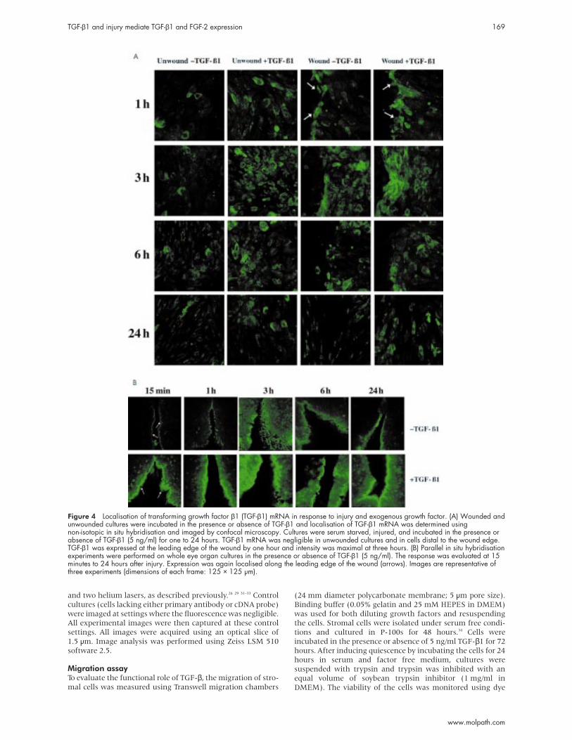
Figure 4 Localisation of transforming growth factor β1 (TGF-β1) mRNA in response to injury and exogenous growth factor. (A) Wounded andunwounded cultures were incubated in the presence or absence of TGF-β1 and localisation of TGF-β1 mRNA was determined usingnon-isotopic in situ hybridisation and imaged by confocal microscopy. Cultures were serum starved, injured, and incubated in the presence orabsence of TGF-β1 (5 ng/ml) for one to 24 hours. TGF-β1 mRNA was negligible in unwounded cultures and in cells distal to the wound edge.TGF-β1 was expressed at the leading edge of the wound by one hour and intensity was maximal at three hours. (B) Parallel in situ hybridisationexperiments were performed on whole eye organ cultures in the presence or absence of TGF-β1 (5 ng/ml). The response was evaluated at 15minutes to 24 hours after injury. Expression was again localised along the leading edge of the wound (arrows). Images are representative ofthree experiments (dimensions of each frame: 125 × 125 µm).
TGF-β1 and injury mediate TGF-β1 and FGF-2 expression 169
www.molpath.com

exclusion assays and only cultures with a minimum of 90%
viability were used. Cells were centrifuged and resuspended in
binding buffer to a final cell density of 30 000 cells/100 µl. A
100 µl sample of the cell suspension was added to the top of
each Transwell chamber and binding buffer (0.600 µl) with
varying concentrations of TGF-β1 (0.1, 1, 10, and 100 ng/ml)
was added to the bottom chambers (binding buffer alone was
used as a negative control for migration, whereas 10% FBS in
binding buffer was used as a positive control). The migration
assay was conducted at 37°C for 12 hours, after which time the
cells were rinsed and fixed with methanol for 10 minutes at
room temperature. The non-migrated cells were removed and
the migrated cells were permeabilised with 0.1% Triton X-100
in PBS for one minute and stained with propidium iodide for
10 minutes at room temperature (1/200 dilution). This was
done to assess migratory cells, and only those cells with
detectable nuclei were counted as positive. The polycarbonate
membranes were removed, mounted on to glass slides,
antifade was added to each membrane, and coverslips were
applied. For each membrane, the total number of cells was
counted in each of six random fields (1.5 mm2/field). The
migration experiments were repeated four times and the
average number of cells migrating in each field for each con-
dition was calculated (± SE of the mean).
RESULTSPrevious work has shown that linear wounds made in primary
cells cultured in serum free medium close by 24 hours.27 We
have also shown that injury elicits a highly defined response
that includes a rapid upregulation in the expression of TGF-β1
mRNA at the leading edge of the wound, along with a minimal
increase in signal a distance from the wound.26 The aim of our
present study was to determine the response of cells to exog-
enous TGF-β1 and/or FGF-2 and to evaluate the regulatory
role of both growth factors upon injury. In all experiments,
cells were maintained in serum free medium to evaluate the
response to specific growth factors. Confirmatory experiments
were performed using whole eye organ culture and expression
was evaluated with FISH.
Expression of TGF-β1 and FGF-2 mRNA in response toinjury and growth factorsTo characterise the response of wounded cultures to growth fac-
tors, both dose dependent and time course experiments were
performed. In the dose dependent experiments, cells were
injured mechanically in serum free medium, incubated in
several concentrations of either FGF-2 or TGF-β1 (0, 0.1, 0.25,
0.5, 2.5, 5.0, and 10 ng/ml), and evaluated at a single time point
using northern blot analysis. Northern blots were subjected to
densitometric analysis and the expression of growth factor was
normalised to cyclophilin. The response represents an average of
the entire culture, including wounded and unwounded cells.The expression of TGF-β1 and FGF-2 mRNA was evaluated
six hours after cells were injured and cultured in serum freemedium containing several concentrations of TGF-β1. Theintegrity of the RNA was monitored using ethidium bromidestaining of 18S and 28S RNA and expression was normalisedto cyclophilin. There was an increase in the expression ofTGF-β1 mRNA in response to injury and this was used to
Figure 5 Percentage of cells expressing fibroblast growth factor 2 (FGF-2) mRNA. The response of wounded and control cultures in thepresence or absence of growth factor was evaluated. Cultures were serum starved, injured, and incubated in the presence or absence oftransforming growth factor β1 (TGF-β1; 5 ng/ml) for one to 24 hours. Confocal microscopy was used to quantitate the in situ hybridisationexperiments by calculating the background pixel intensity for three images collected at each time point. The total number of cells in each fieldwas counted, as was the number of cells with an intensity above background. The data are expressed as the percentage of cells expressingFGF-2 mRNA. Time of wound closure is noted (arrow). Injured cultures treated with exogenous TGF-β1 display an increase in the expression ofFGF-2 coincident with wound closure. Data are representative of three experiments.
25
20
15
10
5
0
Time (hours)
–TGF-β1, unwounded
% C
ells
expr
essi
ng F
GF-
2 m
RNA
0.25 0.5 3 6 24 48
+TGF-β1, unwounded
–TGF-β1, wounded
+TGF-β1, wounded
170 Song, Klepeis, Nugent, et al
www.molpath.com
establish a baseline to monitor changes in response to growth
factors and injury. Exogenous TGF-β1 further increased
TGF-β1 mRNA values in both wounded and unwounded cul-
tures (on average fourfold increase over the wounded control
(no addition of growth factor)) (fig 1A,B). The increase was
detected at concentrations as low as 0.1 ng/ml, and the
response was maximal between 0.5 and 5.0 ng/ml (fig 1A,B).
In contrast, FGF-2 was expressed constitutively and neither
injury nor TGF-β1 significantly altered its expression at six
hours (fig 1A,B). These data indicate that TGF-β1 mediates
the expression of its own mRNA, but does not alter that of
FGF-2 mRNA when cells are actively migrating.
Reciprocal experiments were performed and the expression
of TGF-β1 and FGF-2 was evaluated in response to exogenous
FGF-2 after six hours of incubation. In the control lacking
growth factor, there was a similar increase in the TGF-β1
mRNA ratio of wounded to unwounded cultures when
expression was normalised to cyclophilin (figs 1B, 2A,B). The
addition of FGF-2 to the wounded cultures inhibited the
expression of TGF-β1 mRNA in a dose dependent manner (fig
2A,B). Interestingly, low concentrations of exogenous FGF-2
(0–0.5 ng/ml) enhanced the expression of TGF-β1 over control
in unwounded cultures. In contrast, exogenous FGF-2 did not
significantly alter its own regulation of FGF-2 mRNA.
The increase in expression of TGF-β1 may reflect an
increase in transcriptional activity, an increase in message sta-
bility, or potentially a combination of both. Previously, we
demonstrated that injury alone induced an increase in
stability.26 Here, we evaluated whether the addition of
exogenous TGF-β1 altered the stability of TGF-β1 and/or
FGF-2 mRNA. Cells were incubated in the presence or absence
of DRB (an inhibitor of new gene transcription) and TGF-β1
(5 ng/ml). RNA was extracted every 30 minutes for three
hours. Replicate control experiments were conducted in the
absence of exogenous TGF-β1. The half life of TGF-β1 mRNA
in TGF-β1 treated cultures increased from 45 to 90 minutes
compared with untreated cultures (fig 3A,B). The addition of
TGF-β1 did not alter the stability of FGF-2 mRNA.
Expression and localisation of TGF-β1 and FGF-2mRNA in response to TGF-β1 and injuryTo assess changes in localisation of FGF-2 and TGF-β1 mRNA
over time, cultures were incubated as described and evaluated
using FISH. Figure 4A shows representative confocal images of
unwounded and wounded cells (arrows indicate wound)
cultured in the presence or absence of TGF-β1 (5 ng/ml) at one,
three, six, and 24 hours. As described previously, wound closure
occurs by 24 hours.27 In the control unwounded cultures, there
was minimal expression of TGF-β1 mRNA. Between 3% and 5%
of the cells in each field exceeded the threshold pixel intensity
unit of 25. When the unwounded cultures were incubated in the
presence of TGF-β1, the proportion of cells expressing over the
threshold increased to 7% (fig 4A).
In the wounded cultures TGF-β1 was localised along the
wound margins at one hour and expression was negligible
distal to the wound edge. By three hours there was an increase
in cells that expressed TGF-β1 mRNA at a distance from the
wound (8–9%) (fig 4A). When exogenous TGF-β was present
in the medium of injured cultures, maximal expression was
detected at three hours, with 10–12% of cells a distance from
the wound expressing TGF-β1 mRNA.
Parallel experiments were performed on whole eye organ
cultures in serum free medium in the presence or absence of
growth factor and the intact cornea was evaluated using con-
focal microscopy. A response was detected and localisation
was similar to that detected in the in vitro experiments—
wounded corneas showed an increase in the number of cells
expressing TGF-β1 mRNA along the wound edge. Expression
was enhanced further when the corneas were wounded and
incubated in serum free medium containing 5 ng/ml TGF-β1
at 15 minutes and one hour (fig 4B; arrows). By three hours,
there was a significant increase in the number of cells
expressing TGF-β1. At each time point, phase images were
taken simultaneously to evaluate the integrity of the tissue
(data not shown). In the organ culture experiments, the
wound was not sutured and because each side of the wound is
at a distance from the other, we predict that the prolonged
increased response correlated with the open wound.
Parallel experiments were conducted to evaluate whether
injury and TGF-β1 affected FGF-2 mRNA values in our cell
culture system. The cells were found to express FGF-2 mRNA
constitutively, with minimal change in expression of FGF-2
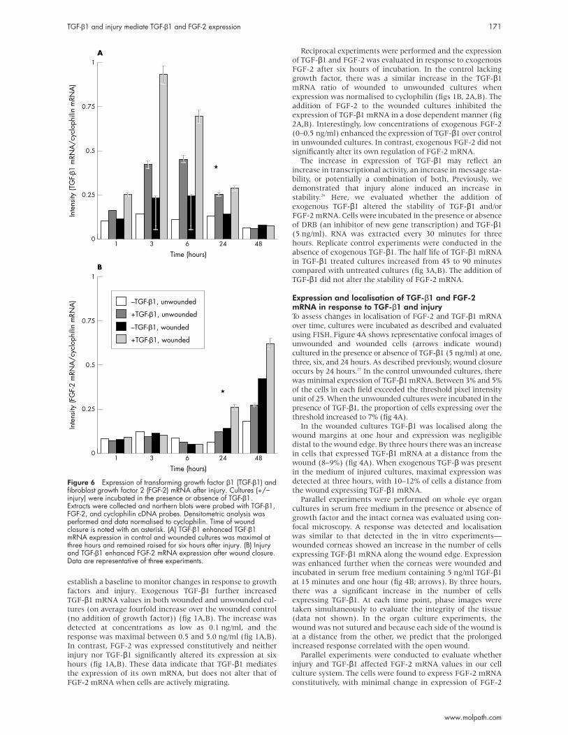
Figure 6 Expression of transforming growth factor β1 (TGF-β1) andfibroblast growth factor 2 (FGF-2) mRNA after injury. Cultures (+/−injury) were incubated in the presence or absence of TGF-β1.Extracts were collected and northern blots were probed with TGF-β1,FGF-2, and cyclophilin cDNA probes. Densitometric analysis wasperformed and data normalised to cyclophilin. Time of woundclosure is noted with an asterisk. (A) TGF-β1 enhanced TGF-β1mRNA expression in control and wounded cultures was maximal atthree hours and remained raised for six hours after injury. (B) Injuryand TGF-β1 enhanced FGF-2 mRNA expression after wound closure.Data are representative of three experiments.
1
0.75
0.5
0.25
0
Time (hours)
B
Inte
nsity
(FG
F-2
mRN
A/c
yclo
phili
n m
RNA
)
1 3 6 24 48
1
0.75
0.5
0.25
0
Time (hours)
A
–TGF-β1, unwounded
Inte
nsity
(TG
F-β1
mRN
A/c
yclo
phili
n m
RNA
)
1 3 6 24 48
+TGF-β1, unwounded
–TGF-β1, wounded
+TGF-β1, wounded
TGF-β1 and injury mediate TGF-β1 and FGF-2 expression 171
www.molpath.com

mRNA for the first six hours after injury (fig 5). To evaluatewhether there was a delayed response, cultures wereincubated over a period of 48 hours. In the presence of TGF-β,there was a slight increase in the proportion of cells express-ing FGF-2 mRNA (fig 5). In the wounded cultures, there wasno significant increase in expression until 24 hours (woundclosure), and this was only detected in the cultures containingexogenous TGF-β1. By 48 hours, there was a similar increasein both wounded cultures (fig 5). In addition, experimentswere conducted to determine whether TGF-β1 altered the rateof wound closure, and micrographs were taken at specifictimes after injury and morphometry was carried out.18 Wefound that the rate of repair was accelerated at the six hourtime point in cultures containing growth factor, but at later
time points there was no detectable difference, and in both
wounds closure occurred by 24 hours.
Northern blot analysis was carried out on the entire cell
population to evaluate the effects of injury and growth factors
on the overall degree of expression over a 48 hour period.
Densitometric analysis was performed at each time point and
changes in either TGF-β1 or FGF-2 were normalised to cyclo-
philin. When cells were incubated under control conditions
there was negligible change over time. When TGF-β1 was
added there was an increase in TGF-β1 mRNA that was
detected at one hour, became maximal at three hours (three-
fold increase in the unwounded and a 3.9-fold increase in
wounded cultures), and remained raised for six hours after
injury (fig 6A). There was no detectable change in the expres-
sion of FGF-2 mRNA for the first 24 hours in the control
wounded cultures. In cultures containing TGF-β1 there was an
increase in FGF-2 mRNA at 24 hours. At 48 hours, there was a
1.4-fold increase in wounded controls and a sixfold increase in
the wounded cultures containing TGF-β1. The increase
occurred at a time when the expression of TGF-β1 was negli-
gible (fig 6A,B). These results indicate that a crucial balance in
growth factor regulation is required, and that TGF-β1 may
play a role in mediating cellular events that elicit an increase
in FGF-2 mRNA. To maintain the balance, an increase in
FGF-2 (at a concentration greater than 0.5 ng/ml) decreases
the expression of TGF-β1, suggesting a regulatory feedback
mechanism.
Changes in the localisation of TGF-β receptors afterinjuryIn immunocytochemical studies, we found that both injury
and TGF-β1 alter the localisation of TGF-β receptors I and III.
In the absence of exogenous growth factors, TGF-β receptors I
and III were detected along the margins of the wound (figs
7,8; arrows), and by 30 minutes there was an increase in the
number of cells that expressed TGF-β receptors I and III at a
distance from the wound (figs 7,8). The change in localisation
of receptors along the wound edge (in the absence of TGF-β1)
agreed with the increase in binding localised to the wound
edge and detected within minutes after injury.26 The addition
of exogenous TGF-β1 caused an enhanced response in TGF-βreceptors I and III between 30 minutes and one hour (figs 7,8).
After three hours, the enhanced expression returned to
unwounded control values. Interestingly, the addition of
TGF-β1 to the unwounded cultures did not result in the
enhancement that was detected with injury. Under these con-
ditions, binding was detected rapidly after injury, was present
for three hours, and then decreased to background values
(data not shown). In contrast, the only noticeable change in
TGF-β receptor II was present at the earliest timepoint with
Figure 7 Expression of transforming growth factor β1 (TGF-β1) receptors in response to injury. Cultures were wounded and incubated in thepresence or absence of TGF-β1 (5 ng/ml). Wounded cultures were compared with control cultures. Arrows indicate the leading edge of thewound and the orientation is kept the same throughout the time course. TGF-β receptor I is detected in control cultures and was foundpredominantly along the margins of the wound. By three hours, the localisation of receptors was diffuse and resembled unwounded controls.Images are representative of three experiments (dimensions of each frame: 125 × 125 µm).
172 Song, Klepeis, Nugent, et al
www.molpath.com

both injury and exogenous growth factor (fig 9). Therefore,
the combination of exogenous TGF-β1 and injury elicit a
maximal response, indicating that these factors play a role in
mediating the availability of receptors.
TGF-β induces a migratory cellular phenotypeTo isolate a specific biological process of wound repair, namely
motility, a migration assay was used. Because TGF-β1 was
detected after injury, and injury increased the stability of
TGF-β1 mRNA, unstimulated stromal cells were primed with
this growth factor to migrate in response to TGF-β1 or FGF-2.
These cells were isolated and cultured under serum free condi-
tions and possessed a dendritic-like phenotype (modification
of Beales et al).34 Their motility was compared with that of
unstimulated cells that were cultured in serum free medium
containing TGF-β1 (5 ng/ml) for three days. These cells under-
went a transformation from the small dendritic-like cells to
large myofibroblast-like cells (“TGF-β cells”). Both phenotypes
were cultured in serum free medium for 24 hours before the
migration assay and allowed to migrate for 12 hours in the
presence of TGF-β1, FGF-2, binding buffer (negative control),
or binding buffer containing 10% FBS (positive control).
Both cell phenotypes migrated in the presence of 10% FBS,
indicating that the cells could be stimulated with a rich com-
posite of factors (fig 10). However, the migrational ability was
altered when cells were exposed to a single growth factor.
When the unstimulated cells were migrated for 12 hours in
the presence of TGF-β1 there was a twofold increase in the
number of cells migrating when compared with the negative
control. These cells also showed similar migration in the
presence of FGF-2, indicating that over time quiescent cells
can be stimulated by exposure to specific growth factors (fig
10). The migration of TGF-β cells in the presence of TGF-β1 or
FGF-2 was enhanced in comparison with the unstimulated
cells. However, it should be noted that the TGF-β cells achieved
the same high degree of motility even when migrated in the
absence of any growth factor. These results indicate that the
initial stimulation required to induce the myofibroblast-like
phenotype caused a highly migratory state that was not
enhanced by additional exposure to either growth factor
alone. Cells were also evaluated for proliferative ability in
response to either growth factor alone and none was detected
(data not shown).
DISCUSSIONMany investigators have studied growth factors and their
receptors to evaluate the mechanisms of scarring. Injury
induces the recruitment of cells to the wound site, including
activated macrophages, neutrophils, and platelets, which play
a role in secreting such factors (see Clark Richard for a
review).35 In the cornea, an additional source of mediators is
the lacrimal gland.36–38 Our study evaluated the expression of
TGF-β1 and FGF-2 in response to injury and exogenous
growth factors using both a primary cell line derived from
stromal cells and an organ culture system. We found that
TGF-β1 mediates the early phases of wound repair, whereas
FGF-2 plays a role in modulating later responses. In addition,
the combination of injury and exogenous TGF-β1 induces a
synergistic response with respect to TGF-β mRNA expression
Figure 8 Expression of transforming growth factor β1 (TGF-β1) receptors in response to injury. Cultures were wounded and incubated in thepresence or absence of TGF-β1 (5 ng/ml). Wounded cultures were compared with control cultures. Arrows indicate the leading edge of thewound and the orientation is kept the same throughout the time course. Low amounts of TGF-β receptor III (betaglycan) are present in controlcultures and are increased in the presence of growth factor. There is a change in localisation in response to injury and/or exogenous TGF-β1that is detected between 30 minutes and one hour. TGF-β receptor III returned to background values by three hours. Images are representativeof three experiments (dimensions of each frame: 125 × 125 µm).
TGF-β1 and injury mediate TGF-β1 and FGF-2 expression 173
www.molpath.com

during the phase of wound repair associated with cell migra-
tion. These data support the observation of others that growth
factors act in a coordinated manner.39 40
Our current in vitro studies were developed from a large
body of work where polymers or synthetic devices were placed
into corneas and wound repair was monitored over a period of
months and years (reviews by Trinkaus-Randall).1 2 In those
studies the synthesis, deposition, and localisation of colla-
gen(s), glycosaminoglycans, and growth factors (TGF-β1 and
FGF-2) were monitored and the localisation of the growth
factors was found to correlate with the appearance of specific
glycosaminoglycans. These results suggested that there was
an interaction between changes that occur in injury and the
bioavailability of growth factors. Initial studies performed in
vitro showed that cells respond to injury in serum free
medium with an increase in tyrosine phosphorylation of FAK
and paxillin,27 and an increase in TGF-β1 mRNA that is local-
ised initially along the wound edge.26
Our results indicate that injury itself may regulate the
availability and expression of receptors and that this response
is enhanced by TGF-β1. Furthermore, preliminary results have
shown that connective tissue growth factor is also affected by
injury in our wound model, and mediates the expression of
TGF-β1 and is stimulated by TGF-β1 in a dose dependent
manner, indicating the presence of a potential feedback
loop.41 We also found that if exogenous FGF-2 is added during
wound repair, there is an inhibition in the expression of
TGF-β1 during the active phase of repair. This suggests that
TGF-β1 expression is regulated by the status of the wound
repair. The increase in the expression of FGF-2 mRNA at alater time in culture correlates well with the delayed and tran-sient appearance of FGF-2 seen in the tissue in vivo.9 The mul-tiplicity of roles that FGF-2 plays has been shown by otherinvestigators who have demonstrated that FGF-2 plays a cru-cial role in mediating cell proliferation, differentiation, migra-tion, angiogenesis, and wound healing.22
“Our results indicate that injury itself may regulate theavailability and expression of receptors and that thisresponse is enhanced by transforming growth factor β1”
The maximal response in FGF-2 mRNA occurred when the
system was altered by both injury and exogenous TGF-β1,
suggesting that injury activates TGF-β, which primes cells to
respond to other growth factors. Previously, Nugent and
Edelman42 showed that TGF-β1 synergistically regulated FGF-2
activity by altering FGF-2–proteoglycan interactions. In those
experiments, TGF-β1 increased [125I] FGF-2 binding to the extra-
cellular matrix of BALB/c3T3 cells by increasing the number of
FGF-2 binding sites. However, it is likely that there are several
potential pathways, because the addition of TGF-β1 to primary
fibroblasts did not result in increased FGF-2 binding.5
In other similar cell culture systems it has been shown thatTGF-β1 can enhance the expression of perlecan and otherheparan sulfate proteoglycans in a dose dependent manner.4
Perlecan has been shown to be present at wound margins invivo.43 Because TGF-β interacts with extracellular matrixmolecules, it is also probable that changes in the sulfated
Figure 9 Expression of transforming growth factor β1 (TGF-β1) receptors in response to injury. Cultures were wounded and incubated in thepresence or absence of TGF-β1 (5 ng/ml). Wounded cultures were compared with control cultures. Arrows indicate the leading edge of thewound and the orientation is kept the same throughout the time course.(C) The localisation and expression of TGF-β receptor II did not changein the presence of exogenous growth factor or injury over three hours. Images are representative of three experiments (dimensions of eachframe: 125 × 125 µm).
174 Song, Klepeis, Nugent, et al
www.molpath.com

proteoglycans that occur with injury may modulate TGF-βreceptor activation and binding.3 4 Thus, alterations in the
environment surrounding the injury, such as cell–cell and
cell–substrate disruption, can trigger a cellular response that is
further mediated by changes in the bioavailability of growth
factors. In our system, there was an increase in the localisation
of TGF-β receptors I and III detected at the wound margin,
which decreased with time and correlated with a transient
increase in growth factor binding.26 The specific increase in the
type I receptor could suggest a role in regulating or limiting
activity by mediating a single receptor in the entire complex.
These variables ultimately add a level of complexity to the
wound repair cascade.
TGF-β molecules are known to have different functions in
specific cell types. Although TGF-β1 is mitogenic for fibro-
blasts, it inhibits the proliferation of epithelial and endothelial
cells. TGF-β has been shown to cause varied responses in
migration and these again correlate with cell type. Andresen
and colleagues44 showed that concentrations between 0.1 and
1 ng/ml were necessary to induce cellular migration of
fibroblasts. However, concentrations greater than 1 ng/ml
were inhibitory. Our migration studies using a Costar
Transwell system demonstrated that quiescent cells that have
never been exposed to serum or growth factors can be stimu-
lated to acquire a migratory phenotype when treated with
higher concentrations of TGF-β1. In our assay, quiescent cells
were activated after 12 hours of exposure to either FGF-2 or
TGF-β1. In addition, when the quiescent cells were cultured in
the presence of TGF-β1 for three days, they acquired a myofi-
broblast phenotype (TGF-β cells), which was conserved even
after removing the growth factor for 24 hours before
migration. Other laboratories have also shown that TGF-β or
cell density can alter cell phenotype and expression of stromal
cells.45 46 These TGF-β cells were highly motile and did not lose
the ability to migrate when growth factor was removed. We
predict that migration was not enhanced further with eithergrowth factor because the receptors were saturated. Whensimilar migration experiments were performed using cellscultured with serum rather than TGF-β1, the cells failed tomigrate in the presence of TGF-β1 (data not shown). Theseresults indicate that a factor(s) present in the serum inhibitsthe stimulatory action of TGF-β and may potentially functionto mediate expression of cells after wound closure whenmigration is no longer necessary.
Several potential therapeutic roles for TGF-β1 have beenexplored. The administration of TGF-β1 enhances the repair ofinjured tissue in several models. When TGF-β1 is applied topi-cally, healing is improved in several wound models, includingincisional and excisional wounds, punch wounds, andulcers.44 Others have hypothesised that TGF-β increases therate of healing and the breaking strength of repairing tissue bystimulating the local secretion of other growth factors.47 48
These findings may underlie why the stability of TGF-β1mRNA is naturally enhanced with injury or exogenousTGF-β1—because it promotes healing. In addition, investiga-tors have shown that in mice overexpressing TGF-β there wasno scarring in linear wounds, but there was a compensatorychange in the regulation of other isoforms of TGF-β.49
Our data suggest that there is an integral relation duringwound repair between TGF-β1 and FGF-2. TGF-β1 primes cellsand may cause a delayed increase in FGF-2 by first altering thesynthesis and degradation of specific proteins. In addition,FGF-2 inhibits the expression of TGF-β1. Thus, the increase inFGF-2 values may prevent the continuous upregulation ofTGF-β1 during the later phases of wound repair.
ACKNOWLEDGEMENTSThis work was supported by NEI Grant EY11000–4 (to MN) and bydepartmental grants from the Massachusetts Lions Eye ResearchFund, Research to Prevent Blindness, and from the New EnglandCorneal Transplant Fund.
Figure 10 Comparison of migratory ability of stimulated and unstimulated cells. Cells were isolated under serum free conditions andincubated for three days in either serum free medium (unstimulated cells) or 5 ng/ml transforming growth factor β1 (TGF-β1) (myofibroblast-likecells). Before the migration assay, cells were quiescent for 24 hours and then allowed to migrate for 12 hours in the presence of 10 ng/mlTGF-β1, 10 ng/ml fibroblast growth factor 2 (FGF-2), binding buffer (negative control), or 10% fetal bovine serum (FBS; positive control) usingthe Costar Transwell system. The average number of cells migrating in a 1.5 mm2 area was calculated for each condition (average of sixareas). Both cell phenotypes migrated in the presence of 10% FBS. Unstimulated cells showed a twofold increase in migration in the presenceof TGF-β1 over control. Myofibroblast-like cells were highly migratory in the presence or absence of either growth factor. The experiment wasrepeated three times and the mean and SEM are shown.
600
500
400
300
200
100
0
Unstimulated cells
No growth factor
Num
ber o
f cel
ls m
igra
ting
in e
ach
field
TGF-β cells
10 ng/ml FGF-2
10 ng/ml TGF-β1
10% FBS
TGF-β1 and injury mediate TGF-β1 and FGF-2 expression 175
www.molpath.com
. . . . . . . . . . . . . . . . . . . . .Authors’ affiliationsM A Nugent, V Trinkaus-Randall, Department of Ophthalmology,Boston University School of Medicine, Boston, MA 02118, USAV E Klepeis, Department of Pathology, Boston University School ofMedicineQ H Song, Department of Biochemistry, Boston University School ofMedicine
REFERENCES1 Trinkaus-Randall V. Cornea. In: Lanza R, Langer R, Chick W, eds.
Principles of tissue engineering. Austin, Texas: RG Landes Company,1997:383–402.
2 Trinkaus-Randall V. Cornea. In: Lanza R, Langer R, Chick W, eds.Principles of tissue engineering, 2nd ed. Boston: MA Academic Press,2000:471–91.
3 Brown CT, Vural M, Johnson M, et al. Age-related changes of scleralhydration and sulfated glycosaminoglycans. Mech Ageing Dev1994;77:97–107.
4 Brown CT, Nugent MA, Lau FW, et al. Characterization of proteoglycanssynthesized by cultured corneal fibroblasts in response to transforminggrowth factor beta and fetal calf serum. J Biol Chem 1999;274:7111–19.
5 Richardson TP, Trinkaus-Randall V, Nugent MA. Regulation of basicfibroblast growth factor binding and activity by cell density and heparansulfate. J Biol Chem 1999;274:13534–40.
6 Hassell JR, Cintron C, Kublin C, et al. Proteoglycan changes duringrestoration of transparency in corneal scars. Arch Biochem Biophys1983;222:362–9.
7 Cintron E, Gilula LA, Murphy WA, et al. The widened disk space: a signof cervical hyperextension injury. Radiology 1981;141:639–44.
8 Funderburgh JL, Chandler JW. Proteoglycans of rabbit corneas withnonperforating wounds. Invest Ophthalmol Vis Sci 1989;30:435–42.
9 Trinkaus-Randall V, Nugent MA. Biological response to a syntheticcornea. J Control Release 1998;53:205–14.
10 Assoian RK, Komoriya A, Meyers CA, et al. Transforming growthfactor-beta in human platelets: identification of a major storage site,purification, and characterization. J Biol Chem 1983;258:7155–60.
11 Assoian RK, Fleurdelys BE, Stevenson HC, et al. Expression andsecretion of type beta transforming growth factor by activated humanmacrophages. Proc Natl Acad Sci U S A 1987;84:6020–4.
12 Gailit J, Welch MP, Clark RA. TGF-beta 1 stimulates expression ofkeratinocyte integrins during re-epithelialization of cutaneous wounds.Invest Dermatol 1994;103:221–7.
13 Shipley GD, Pittelkow MR, Wille JJ, Jr, et al. Reversible inhibition ofnormal human prokeratinocyte proliferation by type beta transforminggrowth factor-growth inhibitor in serum-free medium. Cancer Res1986;46:2068–71.
14 Attisano L, Wrana JL, Lopez-Casillas F, et al. TGF-beta receptors andactions. Biochim Biophys Acta 1994;1222:71–80.
15 Cross M, Dexter TM. Growth factors in development, transformation,and tumorigenesis. Cell 1991;64:271–80.
16 Massague J. The transforming growth factor-beta family. Annu Rev CellBiol 1990;6:597–641.
17 Grotendorst GR, Smale G, Pencev D. Production of transforming growthfactor beta by human peripheral blood monocytes and neutrophils. J CellPhysiol 1989;40:396–402.
18 Kay EP, Lee MS, Seong GJ, et al. TGF-betas stimulate cell proliferationvia an autocrine production of FGF-2 in corneal stromal fibroblasts. CurrEye Res 1998;17:286–93.
19 Massague J. TGF-beta signal transduction. Annu Rev Biochem1998;67:753–91.
20 Massague J. TGF-beta signaling: receptors, transducers, and Madproteins. Cell 1996;85:947–50.
21 Derynck R, Feng XH. TGF-beta receptor signaling. Biochim Biophys Acta1997;1333:105–50.
22 Nugent MA, Iozzo RV. Fibroblast growth factor-2. Int J Biochem Cell Biol2000;32:115–20.
23 Arese M, Chen Y, Florkiewicz RZ, et al. Nuclear activities of basicfibroblast growth factor: potentiation of low-serum growth mediated bynatural or chimeric nuclear localization signals. Mol Biol Cell1999;10:1429–44.
24 Patry V, Bugler B, Maret A, et al. Endogenous basic fibroblast growthfactor isoforms involved in different intracellular protein complexes.Biochem J 1997;326:259–64.
25 Powell PP, Klagsbrun M. Three forms of rat basic fibroblast growthfactor are made from a single mRNA and localize to the nucleus. CellPhysiology 1991;148:202–10.
26 Song QH, Singh RP, Richardson TP, et al. Transforming growthfactor-beta1 expression in cultured corneal fibroblasts in response toinjury. J Cell Biochem 2000;77:186–99.
27 Haq F, Trinkaus-Randall V. Injury of stromal fibroblasts inducesphosphorylation of focal adhesion proteins. Curr Eye Res1998;17:512–23.
28 Winkles JA, Friesel R, Alberts GF, et al. Elevated expression of basicfibroblast growth factor in an immortalized rabbit smooth muscle cell line.Am J Pathol 1993;143:518–27.
29 Grushkin-Lerner LS, Kewalramani R, Trinkaus-Randall V. Expression ofintegrin receptors on plasma membranes of primary corneal epithelialcells is matrix specific. Exp Eye Res 1997;64:323–34.
30 Singer RH, Lawrence JB, Villnave C. Optimization of in situhybridization using isotopic and nonisotopic detection methods.Biotechniques 1986;4:230–50.
31 Song QH, Singh RP, Trinkaus-Randall V, et al. Injury and EGF mediatethe expression of alpha6beta4 integrin subunits in corneal epithelium. JCell Biochem 2001;80:397–414.
32 Trinkaus-Randall V, Tong M, Thomas P, et al. Confocal imaging of thealpha 6 and beta 4 integrin subunits in the human cornea with aging.Invest Ophthalmol Vis Sci 1993;34:3103–9.
33 Wu XY, Svoboda KK, Trinkaus-Randall V. Distribution of F-actin, vinculinand integrin subunits (alpha 6 and beta 4) in response to cornealsubstrata. Exp Eye Res 1995;60:445–58.
34 Beales MP, Funderburgh JL, Jester JV, et al. Proteoglycan synthesis bybovine keratocytes and corneal fibroblasts: maintenance of the keratocytephenotype in culture. Invest Ophthalmol Vis Sci 1995;40:1658–63.
35 Clark Richard AF. Wound repair: overview and general considerations.In: Clark Richard AF, ed. The molecular and cellular biology of woundrepair, 2nd ed. New York: Plenum Press, 1996:293–5.
36 Sobrin L, Liu Z, Monroy DC, et al. Regulation of MMP-9 activity inhuman tear fluid and corneal epithelial culture supernatant. InvestOphthalmol Vis Sci 2000;411:1703–9.
37 Tervo T, van Setten GB, Paallysaho T, et al. Wound healing of theocular surface. Ann Med 1992;24:19–27.
38 Vesaluoma MH, Tervo TT. Tenascin and cytokines in tear fluid afterphotorefractive keratectomy. J Refract Surg 1998;14:447–54.
39 Bennett NT, Schultz GS. Growth factors and wound healing: part II.Role in normal and chronic wound healing. Am J Surg1993;166:74–81.
40 Schultz GS, Strelow S, Stern GA, et al. Treatment of alkali-injured rabbitcorneas with a synthetic inhibitor of matrix metalloproteinases. InvestOphthalmol Vis Sci 1992;33:3325–31.
41 Duncan MR, Frazier KS, Abramson S, et al. Connective tissue growthfactor mediates transforming growth factor beta-induced collagensynthesis: down-regulation by cAMP. FASEB J 1999;13:1774–86.
42 Nugent MA, Edelman ER. Transforming growth factor beta 1 stimulatesthe production of basic fibroblast growth factor binding proteoglycans inBalb/c3T3 cells. J Biol Chem 1992;267:21256–64.
43 Nathan A, Nugent MA, Edelman ER. Tissue engineered perivascularendothelial cell implants regulate vascular injury. Proc Natl Acad Sci U SA 1995;92:8130–4.
44 Andresen JL, Ledet T, Ehlers N. Keratocyte migration and peptidegrowth factors: the effect of PDGF, bFGF, EGF, IGF-I, aFGF and TGF-betaon human keratocyte migration in a collagen gel. Curr Eye Res1997;16:605–13.
45 Masur SK, Dewal HS, Dinh TT, et al. Myofibroblasts differentiate fromfibroblasts when plated at low density. Proc Natl Acad Sci U S A1996;93:4219–23.
46 Petroll WM, Jester JV, Bean JJ, et al. Myofibroblast transformation of catcorneal endothelium by transforming growth factor-beta1, -beta2, and-beta3. Invest Ophthalmol Vis Sci 1998;39:2018–32
47 Amento EP, Beck LS. TGF-beta and wound healing. Ciba Found Symp1991;157:115–23.
48 Roberts AB, Sporn MB. Physiological actions and clinical applications oftransforming growth factor-beta (TGF-beta). Growth Factors 1993;8:1–9.
49 Shah M, Revis D, Herrick S, et al. Role of elevated plasma transforminggrowth factor-beta1 levels in wound healing. Am J Pathol 1999;154:1115–24.
Take home messages
• Transforming growth factor β1 (TGF-β1) appears tomediate the early phases of wound repair, whereasfibroblast growth factor 2 (FGF-2) seems to play a role inmodulating later responses
• TGF-β1 and FGF-2 appear to act together during wound repair:TGF-β1 primes cells and may cause a delayed increase inFGF-2 and FGF-2 inhibits the expression of TGF-β1
• Thus, the increase in FGF-2 values may prevent the continu-ous upregulation of TGF-β1 during the later phases ofwound repair
• The combination of injury and exogenous TGF-β1 inducesa synergistic response with respect to TGF-β mRNA expres-sion during the phase of wound repair associated with cellmigration
• Injury and exogenous increased TGF-β1 and fibroblastgrowth factor 2 (FGF-2) mRNA values, although theincrease in FGF-2 was not seen until wound closure
• FGF-2 inhibited the expression of TGF-β1• TGF-β1 increased TGF-β1 mRNA stability but had no effect
upon FGF-2• Migration assays indicated that unstimulated stromal cells
could be activated to migrate to specific growth factors. TGF-β1specifically enhances cellular responsiveness, as shown byincreased stability after injury and the acquisition of amigratory phenotype. These data suggest that there is an inte-gral relation during wound repair between TGF-β1 and FGF-2.
176 Song, Klepeis, Nugent, et al
www.molpath.com