Extracellular matrix proteins and displacement of cultured fibroblasts from duodenal biopsies in...
8
RESEARCH Open Access Extracellular matrix proteins and displacement of cultured fibroblasts from duodenal biopsies in celiac patients and controls Leda Roncoroni 1,3* , Luca Elli 1 , Maria Teresa Bardella 1 , Gianluca Perrucci 2 , Michele Ciulla 2 , Vincenza Lombardo 1 , Carolina Tomba 1 , Dario Conte 1 and Luisa Doneda 3 Abstract Background: Celiac disease (CD) is mainly characterised by villous atrophy and mucosal architectural rearrangement. The fibroblasts (FBs) are the most abundant mesenchymal cell type in the intestinal mucosa and are responsible for both the architectural arrangement of the villi and the formation of the extracellular matrix (ECM). This study aimed at the evaluation of both the intracellular distribution of different proteins involved in ECM and FBs characterisation, and the cellular displacement of primary FBs obtained from duodenal endoscopic biopsies of healthy subjects and celiac patients. Methods: Primary healthy and celiac duodenal FBs were evaluated by means of immuno-fluorescence assay for collagen type I and IV, fibronectin, actin, alpha-Smooth Muscle Actin (alpha-SMA), Fibroblast Surface Protein (FSP) and transglutaminase type 2 (TG2). The geometric indexes of the fluorescence signals were investigated by image analysis software (Image J, NIH). Both morphology and kinetic were evaluated during a 72 hours time course movie. TG2 medium activity was evaluated by means of ELISA. Results: All the cells examined were immunopositive for FSP, alpha-SMA, actin, collagen I, collagen IV and TG2. CD cells showed a signet collagen-I and collagen-IV pattern, as compared to the controls being characterised by a spindle geometry. Moreover, the collagen signals in CD FBs showed a significantly higher circularity index (major orthogonal diameter ratio) than the controls (p < 0.0001), whereas the perimeter and area ratio were significantly lower (p < 0.0001). The TG2 signal had a decreased area (p < 0.05), but a two-fold increased medium activity. The time course highlighted a reduction of the displacement of CD FBs. Conclusions: The isolated primary CD FBs showed a different collagen and TG2 pattern of distribution associated with a different cellular displacement. The reasons for such CD cell peculiar characteristics are yet unknown but they might represent a factor in the progression of the intestinal damage. Keywords: Celiac disease, Fibroblasts, Extracellular matrix, Collagen, Tissue transglutaminase, Transglutaminase type 2 Background The small bowel mucosal architecture is maintained through a delicate balance among cell production in the crypt compartment, enterocytes migration along the villi, and extrusion of mature epithelial cells from the tip of the villi into the intestinal lumen [1]. This continuous process of cellular differentiation and natural apoptosis arises and is controlled by interactions among epithelium, mesenchymal cells, gut associated lymphoid tissue and the structure of the extracellular matrix (ECM), through mech- anisms still largely unknown [2]. Among the different cell types resident in the intestinal mucosa, the fibroblasts (FBs) are the most represented mesenchymal ones and are re- sponsible for both the architectural arrangement of the villi and the formation of the ECM, mainly composed by colla- gen fibrils [3,4]. As concerns the intestinal wall, ECM (i.e. the structural basis of the human tissues) includes the base- ment membrane that sustains the epithelium and the * Correspondence: [email protected] 1 Center for the Prevention and Diagnosis of Celiac Disease, Gastroenterology Unit II, Fondazione IRCCS Ca’ Granda - Ospedale Maggiore Policlinico and Università degli Studi di Milano, Via F. Sforza 35, Milan 20122, Italy 3 Department of Biomedical, Surgical and Odontoiatric Sciences, Università degli Studi di Milano, Via Festa del Perdono 7, Milan 20122, Italy Full list of author information is available at the end of the article © 2013 Roncoroni et al.; licensee BioMed Central Ltd. This is an Open Access article distributed under the terms of the Creative Commons Attribution License (http://creativecommons.org/licenses/by/2.0), which permits unrestricted use, distribution, and reproduction in any medium, provided the original work is properly cited. Roncoroni et al. Journal of Translational Medicine 2013, 11:91 http://www.translational-medicine.com/content/11/1/91
Transcript of Extracellular matrix proteins and displacement of cultured fibroblasts from duodenal biopsies in...

Roncoroni et al. Journal of Translational Medicine 2013, 11:91http://www.translational-medicine.com/content/11/1/91
RESEARCH Open Access
Extracellular matrix proteins and displacement ofcultured fibroblasts from duodenal biopsies inceliac patients and controlsLeda Roncoroni1,3*, Luca Elli1, Maria Teresa Bardella1, Gianluca Perrucci2, Michele Ciulla2, Vincenza Lombardo1,Carolina Tomba1, Dario Conte1 and Luisa Doneda3
Abstract
Background: Celiac disease (CD) is mainly characterised by villous atrophy and mucosal architectural rearrangement. Thefibroblasts (FBs) are the most abundant mesenchymal cell type in the intestinal mucosa and are responsible for both thearchitectural arrangement of the villi and the formation of the extracellular matrix (ECM). This study aimed at theevaluation of both the intracellular distribution of different proteins involved in ECM and FBs characterisation, and thecellular displacement of primary FBs obtained from duodenal endoscopic biopsies of healthy subjects and celiac patients.
Methods: Primary healthy and celiac duodenal FBs were evaluated by means of immuno-fluorescence assay for collagentype I and IV, fibronectin, actin, alpha-Smooth Muscle Actin (alpha-SMA), Fibroblast Surface Protein (FSP) andtransglutaminase type 2 (TG2). The geometric indexes of the fluorescence signals were investigated by image analysissoftware (Image J, NIH). Both morphology and kinetic were evaluated during a 72 hours time course movie. TG2 mediumactivity was evaluated by means of ELISA.
Results: All the cells examined were immunopositive for FSP, alpha-SMA, actin, collagen I, collagen IV and TG2. CD cellsshowed a signet collagen-I and collagen-IV pattern, as compared to the controls being characterised by a spindlegeometry. Moreover, the collagen signals in CD FBs showed a significantly higher circularity index (major orthogonaldiameter ratio) than the controls (p < 0.0001), whereas the perimeter and area ratio were significantly lower (p < 0.0001).The TG2 signal had a decreased area (p < 0.05), but a two-fold increased medium activity. The time course highlighted areduction of the displacement of CD FBs.
Conclusions: The isolated primary CD FBs showed a different collagen and TG2 pattern of distribution associated with adifferent cellular displacement. The reasons for such CD cell peculiar characteristics are yet unknown but they mightrepresent a factor in the progression of the intestinal damage.
Keywords: Celiac disease, Fibroblasts, Extracellular matrix, Collagen, Tissue transglutaminase, Transglutaminase type 2
BackgroundThe small bowel mucosal architecture is maintainedthrough a delicate balance among cell production in thecrypt compartment, enterocytes migration along the villi,and extrusion of mature epithelial cells from the tip ofthe villi into the intestinal lumen [1]. This continuous
* Correspondence: [email protected] for the Prevention and Diagnosis of Celiac Disease, GastroenterologyUnit II, Fondazione IRCCS Ca’ Granda - Ospedale Maggiore Policlinico andUniversità degli Studi di Milano, Via F. Sforza 35, Milan 20122, Italy3Department of Biomedical, Surgical and Odontoiatric Sciences, Universitàdegli Studi di Milano, Via Festa del Perdono 7, Milan 20122, ItalyFull list of author information is available at the end of the article
© 2013 Roncoroni et al.; licensee BioMed CenCreative Commons Attribution License (http:/distribution, and reproduction in any medium
process of cellular differentiation and natural apoptosisarises and is controlled by interactions among epithelium,mesenchymal cells, gut associated lymphoid tissue and thestructure of the extracellular matrix (ECM), through mech-anisms still largely unknown [2]. Among the different celltypes resident in the intestinal mucosa, the fibroblasts (FBs)are the most represented mesenchymal ones and are re-sponsible for both the architectural arrangement of the villiand the formation of the ECM, mainly composed by colla-gen fibrils [3,4]. As concerns the intestinal wall, ECM (i.e.the structural basis of the human tissues) includes the base-ment membrane that sustains the epithelium and the
tral Ltd. This is an Open Access article distributed under the terms of the/creativecommons.org/licenses/by/2.0), which permits unrestricted use,, provided the original work is properly cited.

Roncoroni et al. Journal of Translational Medicine 2013, 11:91 Page 2 of 8http://www.translational-medicine.com/content/11/1/91
villous axis [5]. ECM is mainly formed by fibrillar collagenand provides the support for the different cell types residentin the mucosa (type I collagen) and for the epithelial line ofenterocytes forming the intestinal barrier (type IV collagen).ECM also includes the glycoproteins (fibronectin),connecting collagen and cells. All the proteins participatingin ECM are interconnected to form a three-dimensionalstructure by enzymes. The most important of such enzymesis the transglutaminases type 2 (TG2), capable to catalyzestable isopeptide bonds in physiological conditions charac-terized by neutral pH and low calcium concentrations [6].Collagen and enzymes are secreted by FBs, which also par-ticipate in the tissue re-modelling and repair through theirmovements along the protein fibrils of the ECM, secretingmolecules when and where needed and in association withthe other resident cells. Besides this tissutal morphogeneticfunction FBs actively participate in the inflammatoryresponse by producing chemokines and cytokines, and inthe antigen presentation by acting as non-professional anti-gen presenting cells [7-12]. Moreover, in the autoimmuneprocesses, FBs are frequently targeted by autoantibodies,leading to a biological dysfunction [9,13,14]. Based on theabove characteristics, FBs are involved in diseases charac-terized by tissue architectural alterations, inflammation andautoimmunity.In the celiac disease (CD), which is the most common
autoimmune enteropathy in Western countries, gluten in-gestion progressively leads to villous atrophy, crypt hyper-plasia with a two to three-fold increased depth of thelamina propria, an increased number of immunologicalcells (B and T lymphocytes, macrophages and eosinophils)and intraepithelial lymphocytosis [6,15]. These abnormal-ities, responsible for any functional derangement, are as-cribed to the primary cytotoxic effects of gliadin and IgAautoantibodies against TG2 on the surface of FBs and inthe ECM [16-18]. In particular, Verbeke et al. reported a de-rangement of collagen distribution in the duodenal mucosaof CD subjects [16]. As of earlier studies by the Authors[19], duodenal CD FBs can be successfully cultured; theyare morphologically different from healthy FBs and poten-tially carry intrinsic alterations, suggesting their primaryinvolvement in both the CD pathogenesis and the develop-ment of the atrophic duodenal lesions.The present study was aimed at evaluating, in
consecutive naïf CD patients and matched controls,the cellular distribution of type I and IV collagens,fibronectin, actin, TG2 and the cellular displacementof primary FBs obtained from duodenal endoscopicbiopsies.
MethodsPatientsFrom February 2009 to December 2011, 50 consecutivesubjects selected from a cohort of patients undergoing
upper GI tract endoscopy for suspected CD, who gavetheir written informed consent to the study, were en-rolled: there were 22 CD patients (10 males and 12 fe-males, mean age 45, range 35–55) and 28 non-CDcontrols (CTRs) (16 males and 12 females, mean age 42,range 30–60). The CD diagnosis was based on theserological presence of anti-TG2 IgA (ELISA or radio-im-munoassay tests) and/or anti-endomysium IgA (immuno-fluorescence technique) auto-antibodies and, at least, aMarsh-Oberhuber grade III duodenal histology. The non-CD group was composed of serologically negative subjectswithout endoscopic or histological lesions, not reportingother autoimmune or intestinal diseases.The study plan was approved by the Ethics Committee of
the “Fondazione IRCCS Cà Granda-Ospedale MaggiorePoliclinico”.
Duodenal specimens and cell culturesThree duodenal biopsies obtained from all the partici-pants by standard endoscopic forceps (Boston Scientific,USA) were rapidly dipped into sterile tubes (Becton andDickinson, Italy) containing an appropriate medium.The samples were finely chopped with disposable sur-
gery knives and incubated in Ham’s F12 medium(Euroclone, Italy), containing liberase blendzyme 2(1.4 W/mL) (Roche, Italy) and the cells so derived cul-tured. Viability was routinely checked by a trypan bluedye exclusion assay (Sigma, Italy) and only thosecultures with viability > 95% were used. Mycoplasmacontamination was routinely checked and ruled outusing the Hoechst method.
ImmunocytochemistryThe different types of proteins were analysed by im-munocytochemistry: characterising proteins i.e., the “FBsurface protein” (FSP, monoclonal anti-human FSP,Clone 1B10; Sigma, Italy), the α-smooth muscle actin(αSMA) (ABCAM, Italy) and actin (Sigma, Italy), andthe ECM components and enzymes i.e., the type I andIV collagens (ABCAM, Italy), fibronectin (Sigma, Italy)and TG2 (Zedira, Germany). Primary antibodies wereused following the manufacturer’s recommended dilu-tions. Cells, seeded onto 24-well plates at a concentra-tion of 20,000 cells/plate, were washed twice 48 hourslater in PBS and fixed with 3.7% formaldehyde in PBSfor 15 minutes at room temperature (RT). Fixed cellswere permeabilised with 0.1% triton X-100 (Sigma, Italy)in PBS for 15 minutes at RT. The non specific bindingof any secondary antibody was blocked by incubationwith normal foetal serum for 30 minutes at RT. Theimmunostaining cells were then rinsed with PBS and in-cubated with a fluorochrome conjugated secondary anti-body for 45 minutes at RT, according to the donorspecies of the primary antibodies. PBS was used as the

Roncoroni et al. Journal of Translational Medicine 2013, 11:91 Page 3 of 8http://www.translational-medicine.com/content/11/1/91
negative control in place of the primary antibody.Counter-staining was performed using DAPI; the glasscoverslip was mounted on glass slides with ProLongGold Antifade Reagent (Invitrogen, Italy). Images wereobtained by fluorescence microscope (Leica, Italy).The epithelial cellular type was carefully excluded in
performing cytokeratine analysis by using anti-humanCytokeratine 20 (Sigma, Italy).
Medium transglutaminase activityTG activity was evaluated by a colorimetric technique(Covalab, France) according to the manufacturer’s in-structions. Briefly, 50 μL of the cell culture medium wasdispensed in each well of the 96-well microtiter platewith covalently coupled CBZ-Gln-Gly and incubatedwith calcium, dithiothereitol and biotinylated cadaverine;strepavidin-labelled peroxidase was added to the wellsand the peroxidase activity revealed using H2O2 andtetramethyl benzidine. Optical density was measured at450 nm with a microplate reader (Bouty Diagnostici,Italy). The results were normalized referring to the cellnumber.
Image analysisThe digital images were analysed as previously described[19,20]. Briefly, after the localization of fluorescence sig-nals, multiple adjacent high-power fields in each sectionwere selected, acquired, and stored in a personal com-puter (Power Mac G4, 867 MHz, 640 MB RAM, Apple,USA) in JPEG format (5:1) at 32 bits/pixel on a 764×560pixel matrix. The stored images were then read by anexpert blinded to both image sequence and assignment,and analyzed to determine the differences between CDand CTRs FBs. A 3D interactive surface plot was digit-ally reconstructed for every image and based on a 256-level scale (0–255).Fluorescent signal analysis was performed on the digit-
ally elaborated images. Analysis algorithms were devel-oped as a set of macros executed with NIH Image(Interactive 3D Surface Plot Plugin), which is an inte-grated image-processing software distributed as freewareby the National Institutes of Health (Bethesda, USA).Prior to analysis an automated threshold process wasperformed on the images to minimize the influence oflight variation in the microscope field and in the opera-tor’s subjective settings. This process cuts off any objectbelow the minimum signal intensity. FBs were recog-nized on the basis of their size and intensity signal usinga cell count algorithm which draws a region of interest(ROI) around each discrete object within the image. Theminimum size in pixels of the objects to be includedin the count was previously defined by accurately meas-uring twelve representative FBs. Objects below theminimum size were not included in the analysis. The
evaluation of the fluorescent signal included, on onehand, the major orthogonal diameters and their ratio asindex of circularity and, on the other hand, the perim-eter, the area and their ratio, as index of complexity.
Shape and motility evaluationMorphology and growth were evaluated during a 72-hour time course movie. Images were acquired with aLeica DMI AF6000LX microscope at 10× magnification.After three days from seeding, the cell culture plates
(7 CD and 7 CTRs) were placed under the microscopeconnected to a camera. The camera was set for captur-ing frames every 10 minutes over 72 hours. The com-pressed frames were composed into the experimentalmovies (one for each plate); the movies were acceleratedby software (LAS-AF imaging software), obtaining a 40 -seconds long one. Thus, one second of the final digitalmovie corresponded to a 1.8 hours long period ofculture. Fifteen randomly selected cells from 3 differentmovies from each group were selected at time 0 (i.e. atthe beginning of the movie). The nucleus was fixed totrack the displacement after 12, 24, 36, 48, 60 and 72 -hours (Photoshop Adobe, USA). Data were expressed astotal displacement (μm) and velocity (μm/h). Also per-imeter, circularity index and feret diameters wereanalysed as previously shown at time 0 and after 36 and72 hours.
Statistical analysisData were expressed as mean ± standard deviation(SD) or median and range. Comparisons of the param-eters were carried out by one-way ANOVA. Statisticalanalysis was performed using statistical software (SPSSversion 13, SPSS, USA), considering a p value < 0.05 assignificant.
ResultsBoth CD and CTR cells expressed FSP, α-SMA, actin,type I and IV collagens, fibronectin and TG2 (Figure 1)but not cytokeratine 20. CD cells showed a signet type Iand IV collagen patterns, whereas CTRs had a spindlegeometry (Figure 1). Type I and IV collagen morphomet-ric analysis, performed on CD and CTR FBs obtained onthe same day of culture, showed some differences. Asdetailed in Table 1 and Figure 2, which shows the digit-ally elaborated images, the collagen fluorescent signalinvolved a larger part of the FB cytoplasm with anincreased circularity index in CD FBs compared to CTRones. Statistically significant differences between theTG2 fluorescent signal of CD and CTR FBs were found.In particular, in the CD FBs the area covered by the TG2fluorescent signal decreased by 50% (Table 2), whileTG activity increased in the culture medium of CDFBs (Figure 3). No statistically different findings were

Figure 1 Fibroblast surface protein, α-smooth muscle actin,actin, type I and IV collagens, fibronectin and transglutaminasetype 2 immuno-fluorescence analysis of fibroblasts from celiacsand healthy controls.
Table 1 Morphometrical characteristics of intracellular fluoresceliacs and healthy controls
Type I collagen
Parameter Controls (#12) Celiacs (#12)
Feret Diameter (μm) * 53.00 ± 11.00 35.20 ± 5.00
Perimeter (μm) 121.30 ± 24.00 107.60 ± 16.80
Area (μm2) 377.20 ± 86.90 563.40 ± 122.80
Circularity index° 0.30 ± 0.10 0.62 ± 0.11
Complexity index (1/μm)& 0.30 ± 0.03 0.19 ± 0.02
* Feret diameter: longest axis length.° Circularity index: Feret diameter/short axis length.& Complexity index: perimeter/area (1/μm).
Roncoroni et al. Journal of Translational Medicine 2013, 11:91 Page 4 of 8http://www.translational-medicine.com/content/11/1/91
observed for αSMA, actin and fibronectin signals (datanot shown). The cellular shapes of CD and CTR FBs aredetailed in Table 3.The CD FBs showed a decreased motility and velocity.
In detail, during the 72 hours period of the observedculture the CD FBs moved 113 ± 39 μm while the CTRFBs 222 ± 88 μm with a velocity of 1.5 ± 0.5 μm/h vs3.0 ± 1.2 μm/h, the difference being statistically different(see Figure 4 for movement tracking). Two additionalmovie files show the FBs displacement in more detail(Additional file 1: Video S1 and Additional file 2: VideoS2 for CD and CTR FBs respectively).
DiscussionThe results from the present study are consistent withan alteration of the collagen and TG2 immunofluores-cence signal in the CD FBs compared to the CTR ones.These abnormalities could be involved in the decreasedmotility observed during the time-course experimentsand in turn in the villous damage observed in CD.The maintenance of a normal duodenal mucosal morph-
ology results from a continuous interaction between theepithelium, the ECM represented by the basement mem-brane and the underlying FBs network, responsible for thesecretion of collagen molecules and matrix stabilizingenzymes (TG2) [6]. In CD the mucosal structure is deeplyaltered as proven by the typical “stigmata” of CD repre-sented by mucosal atrophy, crypt hyperplasia and lympho-cytic infiltration. Also the lamina propria, composed bytype IV collagen and controlling the interface between theepithelium and the subepithelial compartment, is altered inCD [21,22]. These aforementioned ECM alterations can beattributed to the secreted autoantibodies (anti-TG2 andanti-gliadin) interacting with the matrix proteins or alterna-tively to a primary defect of CD FBs which could abnor-mally express these proteins or enzymes [23]. Moreover,FBs control the degradation of ECM through the secretionof lytic enzymes, as the metalloproteases (MMP), and theirinhibitors [24-26]. Previous findings suggest that both thesemechanisms can play a role in the CD progress. Schuppanet al. for example have demonstrated that ECM could act
cent signal of type I and IV collagen fibroblasts from
Type IV collagen
P value Controls (#12) Celiacs (#12) P value
< 0.005 62.90 ± 12.05 32.90 ± 4.90 < 0.0001
< 0.005 145.90 ± 27.20 99.60 ± 12.06 < 0.001
< 0.05 375.60 ± 118.60 502.70 ± 98.10 < 0.05
< 0.005 0.20 ± 0.06 0.60 ± 0.11 < 0.0001
< 0.0001 0.40 ± 0.08 0.20 ± 0.03 < 0.0001

Figure 2 Digitally reconstructed images from immunofluorescence analysis of type I and type IV collagen of fibroblasts from healthycontrols (panels A and C) and celiacs (panels B and D), respectively.
Roncoroni et al. Journal of Translational Medicine 2013, 11:91 Page 5 of 8http://www.translational-medicine.com/content/11/1/91
as a reservoir of autoantibodies, fuelling the mucosal in-flammation and eventually modifying the pH and calciumconcentrations, making the environment suitable for pro-tein lysis and TG2 activation in terms of switching fromisopeptide bonds to the deamidation using H2O as acyl ac-ceptor [6,23]. On the other hand, Verbeke et al. havereported a decreased immunofluorescence signal of type IVcollagen and laminin with a “leaky” basement membrane[16]. Accordingly, the increase of type I and IV collagen sig-nals in FBs and the increase of TG2 activity in the mediumcould represent an attempt of CD FBs to restore the de-creased collagen levels in the ECM of CD patients and toincrease the formation of isopeptide bonds stabilizing thematrix fibrils. Although demonstrated only in vitro this
Table 2 Morphometrical characteristics of theintracellular fluorescent signal of the transglutaminasetype 2 in fibroblasts from celiacs and healthy controls
Parameter Controls (#12) Celiacs (#12) P value
Feret Diameter (μm)* 26.60 ± 5.40 19.10 ± 4.40 <0.05
Perimeter (μm) 73.00 ± 23.10 53.10 ± 13.80 NS
Area (μm2) 143.90 ± 50.70 90.00 ± 34.80 <0.05
Circularity index° 0.38 ± 0.20 0.40 ± 0.10 NS
Complexity index (1/μm)& 0.50 ± 0.10 0.60 ± 0.20 NS
* Feret diameter: longest axis length.° Circularity index: Feret diameter/short axis length.& Complexity index: perimeter/area (1/μm); NS, Not significant.
scenario could also be present in the flattened CD mucosa;in other words, as primary cell cultures maintain the mem-ory of their site of origin even after different passages, a res-toration of the original in vivo scenario could be possible[27,28]. To underline these mechanisms, the fibronectinlevels did not differ between CD and healthy FBs, in linewith the results obtained by Korhonen et al. who reporteda comparable pattern both in CD and non-CD duodenalmucosa [29].
Figure 3 Transglutaminase activity in the culture medium offibroblasts from celiacs and healthy controls.
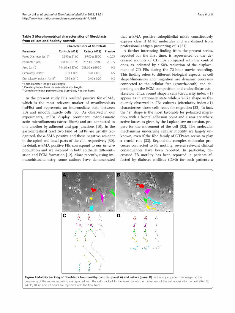
Table 3 Morphometrical characteristics of fibroblastsfrom celiacs and healthy controls
Characteristics of fibroblasts
Parameter Controls (#12) Celiacs (#12) P value
Feret Diameter (μm)* 62.90 ± 21.60 89.00 ± 28.00 < 0.05
Perimeter (μm) 188.30 ± 61.90 252.30 ± 99.80 < 0.05
Area (μm2) 744.60 ± 357.60 933.60 ± 649.30 NS
Circularity index° 0.30 ± 0.20 0.20 ± 0.10 NS
Complexity index (1/μm)& 0.30 ± 0.10 0.40 ± 0.20 NS
* Feret diameter: longest axis length.° Circularity index: Feret diameter/short axis length.& Complexity index: perimeter/area (1/μm); NS, Not significant.
Roncoroni et al. Journal of Translational Medicine 2013, 11:91 Page 6 of 8http://www.translational-medicine.com/content/11/1/91
In the present study FBs resulted positive for αSMA,which is the most relevant marker of myofibroblasts(mFBs) and represents an intermediate state betweenFBs and smooth muscle cells [30]. As observed in ourexperiments, mFBs display prominent cytoplasmaticactin microfilaments (stress fibers) and are connected toone another by adherent and gap junctions [10]. In thegastrointestinal tract two kind of mFBs are usually rec-ognized, the α-SMA positive and those negative, residentin the apical and basal parts of the villi, respectively [30].In detail, α-SMA positive FBs correspond to our in vitropopulation and are involved in both epithelial differenti-ation and ECM formation [12]. More recently, using im-munohistochemistry, some authors have demonstrated
Figure 4 Motility tracking of fibroblasts from healthy controls (panelbeginning of the movie recording are reported with the cells tracked. In th24, 36, 48, 60 and 72 hours are reported with the final trace.
that α-SMA positive subepithelial mFBs constitutivelyexpress class II MHC molecules and are distinct fromprofessional antigen presenting cells [31].A further interesting finding from the present series,
reported for the first time, is represented by the de-creased motility of CD FBs compared with the controlones, as indicated by a 50% reduction of the displace-ment of CD FBs during the 72-hour movie recording.This finding refers to different biological aspects, as cellshape/dimension and migration are dynamic processesconnected to the cellular fate (growth/death) and de-pending on the ECM composition and endocellular cyto-skeleton. Thus, round shapen cells (circularity index = 1)appear as in stationary state while a Y-like shape as fre-quently observed in FBs cultures (circularity index < 1)characterizes those cells ready for migration [32]. In fact,the “Y” shape is the most favorable for polarized migra-tion, with a frontal adhesion point and a rear arc whereactive forces as given by the Laplace law on tension, pre-pare for the movement of the cell [32]. The molecularmechanisms underlying cellular motility are largely un-known, even if the Rho family of GTPases seems to playa crucial role [33]. Beyond the complex molecular pro-cesses connected to FB motility, several relevant clinicalconsequences have been reported. In particular, de-creased FB motility has been reported in patients af-fected by diabetes mellitus (DM): for such patients a
A) and celiacs (panel B). In the upper panels the images at thee lower panels the movement of the cell nuclei into the field after 12,

Roncoroni et al. Journal of Translational Medicine 2013, 11:91 Page 7 of 8http://www.translational-medicine.com/content/11/1/91
migratory defect represents a relevant impairment forwound healing, responsible for chronic skin ulcerationseven requiring amputation [34]. The discussed findingcould represent a connection between CD and DM,which are supposed to present some common factorssuch as genetic background. Moreover, DM animalmodels are currently used to investigate particularaspects of CD as they are capable to develop duodenalatrophy when exposed to gluten feeding [35]. It remainsan interesting point for future investigation whether theobserved alterations are common to other gastrointes-tinal autoimmune disorders (inflammatory bowel dis-ease, autoimmune enteropathy) or they could bereverted in case of FBs derived from CD non atrophicmucosa, where a different FB population has been se-lected from duodenal micro-environment.
ConclusionOverall, the results from the present study highlight theimportance of FBs in CD pathogenesis, stronglysupporting the need for further investigations on thistopic in order to improve our understanding of the CDrelated injury processes. Transitionally, if the defect oftissue repair process in CD FBs should be confirmed aspivotal in the normalization of duodenal mucosa aftergluten withdrawal from a patient’s diet, the possibility ofECM targeted therapies could play a role in the CDresearch.
Additional files
Additional file 1: Video S1. CD FBs.
Additional file 2: Video S2. CTR FBs.
Competing interestsThe authors declare that they have no competing interests.
Authors’ contributionsLR and VL perfomed the cell cultures, immunofluorescence eperiments andELISA. GP and MC dealt with the FB movies and image analysis. LE and CTplanned the study, dealt with the patients and performed upperendoscopies with duodenal biopsies. MTB, DC and LD analyzed data andprepared the manuscript. All the authors read and approved the finalmanuscript.
AcknowledgementsWe are deeply indebted to Professor Elena Cattaneo of the Department ofBioSciences at Università degli Studi di Milano – Milan, Italy for grantingaccess to use the DMI6000LX microscope.
Author details1Center for the Prevention and Diagnosis of Celiac Disease, GastroenterologyUnit II, Fondazione IRCCS Ca’ Granda - Ospedale Maggiore Policlinico andUniversità degli Studi di Milano, Via F. Sforza 35, Milan 20122, Italy.2Department of Clinical and Community Sciences, Università degli Studi diMilano, Via F. Sforza 35, Milan 20122, Italy. 3Department of Biomedical,Surgical and Odontoiatric Sciences, Università degli Studi di Milano, Via Festadel Perdono 7, Milan 20122, Italy.
Received: 13 January 2013 Accepted: 23 March 2013Published: 8 April 2013
References1. MacDonald TT, Monteleone I, Fantini MC, Monteleone G: Regulation of
homeostasis and inflammation in the intestine. Gastroenterology 2011,140:1768–1775.
2. Burke JP, Mulsow JJ, O’Keane C, Docherty NG, Watson RW, O’Connell PR:Fibrogenesis in Crohn’s disease. Am J Gastroenterol 2007, 102:439–448.
3. Powell DW, Pinchuk IV, Saada JI, Chen X, Mifflin RC: Mesenchymal cells ofthe intestinal lamina propria. Annu Rev Physiol 2011, 73:213–237.
4. Simon-Assmann P, Kedinger M, De Arcangelis A, Rousseau V, Simo P:Extracellular matrix components in intestinal development. Experientia1995, 51:883–900.
5. Shi YB, Li Q, Damjanovski S, Amano T, Ishizuya-Oka A: Regulation ofapoptosis during development: input from the extracellular matrix(review). Int J Mol Med 1998, 2:273–282.
6. Elli L, Bergamini CM, Bardella MT, Schuppan D: Transglutaminases ininflammation and fibrosis of the gastrointestinal tract and the liver.Dig Liver Dis 2009, 41:541–550.
7. Darby I, Skalli O, Gabbiani G: Alpha-smooth muscle actin is transientlyexpressed by myofibroblasts during experimental wound healing.Lab Invest 1990, 63:21–29.
8. Sappino AP, Masouye I, Saurat JH, Gabbiani G: Smooth muscledifferentiation in scleroderma fibroblastic cells. Am J Pathol 1990,137:585–591.
9. Smith TJ: Insights into the role of fibroblasts in human autoimmunediseases. Clin Exp Immunol 2005, 141:388–397.
10. Villaschi S, Nicosia RF: Paracrine interactions between fibroblasts andendothelial cells in a serum-free coculture model. Modulation ofangiogenesis and collagen gel contraction. Lab Invest 1994, 71:291–299.
11. Pang G, Couch L, Batey R, Clancy R, Cripps A: GM-CSF, IL-1 alpha, IL-1 beta,IL-6, IL-8, IL-10, ICAM-1 and VCAM-1 gene expression and cytokineproduction in human duodenal fibroblasts stimulated withlipopolysaccharide, IL-1 alpha and TNF-alpha. Clin Exp Immunol 1994,96:437–443.
12. Saada JI, Pinchuk IV, Barrera CA, Adegboyega PA, Suarez G, Mifflin RC, DiMari JF, Reyes VE, Powell DW: Subepithelial myofibroblasts are novel non-professional APCs in the human colonic mucosa. J Immunol 2006,177:5968–5979.
13. Hogaboam CM, Smith RE, Kunkel SL: Dynamic interactions between lungfibroblasts and leukocytes: implications for fibrotic lung disease. ProcAssoc Am Physicians 1998, 110:313–320.
14. Khoo TK, Bahn RS: Pathogenesis of Graves’ ophthalmopathy: the role ofautoantibodies. Thyroid 2007, 17:1013–1018.
15. Elli L, Bardella MT: Motility disorders in patients with celiac disease.Scand J Gastroenterol 2005, 40:743–749.
16. Verbeke S, Gotteland M, Fernandez M, Bremer J, Rios G, Brunser O:Basement membrane and connective tissue proteins in intestinalmucosa of patients with coeliac disease. J Clin Pathol 2002, 55:440–445.
17. Maiuri L, Ciacci C, Ricciardelli I, Vacca L, Raia V, Rispo A, Griffin M, Issekutz T,Quaratino S, Londei M: Unexpected role of surface transglutaminase typeII in celiac disease. Gastroenterology 2005, 129:1400–1413.
18. Elli L, Dolfini E, Bardella MT: Gliadin cytotoxicity and in vitro cell cultures.Toxicol Lett 2003, 146:1–8.
19. Roncoroni L, Elli L, Doneda L, Piodi L, Ciulla MM, Paliotti R, Bardella MT:Isolation and culture of fibroblasts from endoscopic duodenal biopsiesof celiac patients. J Transl Med 2009, 7:40.
20. Elli L, Roncoroni L, Doneda L, Ciulla MM, Colombo R, Braidotti P, Bonura A,Bardella MT: Imaging analysis of the gliadin direct effect on tightjunctions in an in vitro three-dimensional Lovo cell line culture system.Toxicol In Vitro 2011, 25:45–50.
21. Koning F, Schuppan D, Cerf-Bensussan N, Sollid LM: Pathomechanisms inceliac disease. Best Pract Res Clin Gastroenterol 2005, 19:373–387.
22. Schuppan D: Current concepts of celiac disease pathogenesis.Gastroenterology 2000, 119:234–242.
23. Dieterich W, Esslinger B, Trapp D, Hahn E, Huff T, Seilmeier W, Wieser H,Schuppan D: Cross linking to tissue transglutaminase and collagenfavours gliadin toxicity in coeliac disease. Gut 2006, 55:478–484.

Roncoroni et al. Journal of Translational Medicine 2013, 11:91 Page 8 of 8http://www.translational-medicine.com/content/11/1/91
24. Ciccocioppo R, Di Sabatino A, Bauer M, Della Riccia DN, Bizzini F, Biagi F,Cifone MG, Corazza GR, Schuppan D: Matrix metalloproteinase pattern inceliac duodenal mucosa. Lab Invest 2005, 85:397–407.
25. Daum S, Bauer U, Foss HD, Schuppan D, Stein H, Riecken EO, Ullrich R:Increased expression of mRNA for matrix metalloproteinases-1 and −3and tissue inhibitor of metalloproteinases-1 in intestinal biopsyspecimens from patients with coeliac disease. Gut 1999, 44:17–25.
26. Daum S, Bauer U, Foss HD, Wahnschaffe U, Schuppan D, Stein H, RieckenEO, Ullrich R: Expression of matrix metalloprotease-1 and collagen ImRNA in biopsies from patients with celiac disease. Ann N Y Acad Sci1998, 859:254–257.
27. Motoyama T, Hojo H, Watanabe H: Comparison of seven cell lines derivedfrom human gastric carcinomas. Acta Pathol Jpn 1986, 36:65–83.
28. Sciaky D, Brazer W, Center DM, Cruikshank WW, Smith TJ: Cultured humanfibroblasts express constitutive IL-16 mRNA: cytokine induction of activeIL-16 protein synthesis through a caspase-3-dependent mechanism.J Immunol 2000, 164:3806–3814.
29. Korhonen M, Ormio M, Burgeson RE, Virtanen I, Savilahti E: Unaltereddistribution of laminins, fibronectin, and tenascin in celiac intestinalmucosa. J Histochem Cytochem 2000, 48:1011–1020.
30. Mifflin RC, Pinchuk IV, Saada JI, Powell DW: Intestinal myofibroblasts:targets for stem cell therapy. Am J Physiol Gastrointest Liver Physiol 2011,300:G684–G696.
31. Barrera CA, Pinchuk IV, Saada JI, Suarez G, Bland DA, Beswick E, AdegboyegaPA, Mifflin RC, Powell DW, Reyes VE: Class II MHC-expressingmyofibroblasts play a role in the immunopathogenesis associated withstaphylococcal enterotoxins. Ann N Y Acad Sci 2004, 1029:313–318.
32. Mogilner A, Keren K: The shape of motile cells. Curr Biol 2009, 19:R762–R771.33. Buonomo R, Giacco F, Vasaturo A, Caserta S, Guido S, Pagliara V, Garbi C,
Mansueto G, Cassese A, Perruolo G, et al: PED/PEA-15 controls fibroblastmotility and wound closure by ERK1/2-dependent mechanisms. J CellPhysiol 2012, 227:2106–2116.
34. Lerman OZ, Galiano RD, Armour M, Levine JP, Gurtner GC: Cellulardysfunction in the diabetic fibroblast: impairment in migration, vascularendothelial growth factor production, and response to hypoxia. Am JPathol 2003, 162:303–312.
35. Busch R, De Riva A, Hadjinicolaou AV, Jiang W, Hou T, Mellins ED: On theperils of poor editing: regulation of peptide loading by HLA-DQ and H2-A molecules associated with celiac disease and type 1 diabetes. ExpertRev Mol Med 2012, 14:e15.
doi:10.1186/1479-5876-11-91Cite this article as: Roncoroni et al.: Extracellular matrix proteins anddisplacement of cultured fibroblasts from duodenal biopsies in celiacpatients and controls. Journal of Translational Medicine 2013 11:91.
Submit your next manuscript to BioMed Centraland take full advantage of:
• Convenient online submission
• Thorough peer review
• No space constraints or color figure charges
• Immediate publication on acceptance
• Inclusion in PubMed, CAS, Scopus and Google Scholar
• Research which is freely available for redistribution
Submit your manuscript at www.biomedcentral.com/submit