Increased Mercury Levels in Patients with Celiac Disease following a Gluten-Free Regimen
7
Research Article Increased Mercury Levels in Patients with Celiac Disease following a Gluten-Free Regimen Luca Elli, 1 Valentina Rossi, 1,2 Dario Conte, 1 Anna Ronchi, 3 Carolina Tomba, 1 Manuela Passoni, 2 Maria Teresa Bardella, 1 Leda Roncoroni, 1,4 and Gianpaolo Guzzi 2 1 Center for the Prevention and Diagnosis of Celiac Disease, Gastroenterology and Endoscopy Unit, Fondazione IRCCS Ca’ Granda Ospedale Maggiore Policlinico, Department of Pathophysiology and Transplantation, Universit` a degli Studi di Milano, Via Francesco Sforza 35, 20122 Milan, Italy 2 Italian Association for Metals and Biocompatibility Research (AIRMEB), Via Banfi 4, 20122 Milan, Italy 3 Pavia Poison Control Center and National Toxicology Information Centre, Toxicology Unit, IRCCS Maugeri Foundation and University of Pavia, Via Salvatore Maugeri 10, 27100 Pavia, Italy 4 Department of Biomedical, Surgical and Dental Sciences, Universit` a degli Studi di Milano, Via Festa del Perdono 7, 20122 Milan, Italy Correspondence should be addressed to Luca Elli; [email protected] Received 9 November 2014; Accepted 2 February 2015 Academic Editor: Paul Enck Copyright © 2015 Luca Elli et al. is is an open access article distributed under the Creative Commons Attribution License, which permits unrestricted use, distribution, and reproduction in any medium, provided the original work is properly cited. Background and Aim. Although mercury is involved in several immunological diseases, nothing is known about its implication in celiac disease. Our aim was to evaluate blood and urinary levels of mercury in celiac patients. Methods. We prospectively enrolled 30 celiac patients (20 treated with normal duodenal mucosa and 10 untreated with duodenal atrophy) and 20 healthy controls from the same geographic area. Blood and urinary mercury concentrations were measured by means of flow injection inductively coupled plasma mass spectrometry. Enrolled patients underwent dental chart for amalgam fillings and completed a food-frequency questionnaire to evaluate diet and fish intake. Results. Mercury blood/urinary levels were 2.4 ± 2.3/1.0 ± 1.4, 10.2 ± 6.7/2.2 ± 3.0 and 3.7±2.7/1.3±1.2 in untreated CD, treated CD, and healthy controls, respectively. Resulting mercury levels were significantly higher in celiac patients following a gluten-free diet. No differences were found regarding fish intake and number of amalgam fillings. No demographic or clinical data were significantly associated with mercury levels in biologic samples. Conclusion. Data demonstrate a fourfold increase of mercury blood levels in celiac patients following a gluten-free diet. Further studies are needed to clarify its role in celiac mechanism. 1. Introduction Mercury (Hg) is ubiquitous environmental heavy metal, naturally originating from erosion of the volcanic rocks and accumulating in the food chain. Besides its natural presence, Hg environmental concentration is progressively increasing due to its employ in human industry and manufactures (medications, thermometers, blood-pressure cuffs, batteries, switches, and fluorescent light bulbs) [1–4]. us, Hg is actually considered a pollutant and, due to its deleterious effects on humans, it is generally considered toxic especially for the nervous system [5]. Mercury main sources for human being are represented by fish consumption, dental amalgams, and vaccines [4, 6]. Once in the body, Hg atoms bind the proteic thiol groups and deposit in all tissues, where they can remain for a long time, triggering its chronic consequences on health [7, 8]; in fact, behind the Hg acute intoxication syndrome (exemplified by Minamata Bay disaster) [9], recent scientific advances have demonstrated that Hg is a cofactor in several multifactorial diseases (cardiovascular, neurodegenerative, and autoimmune), as a consequence of its biological effect on inflammation and immune system [1, 10–14]. Hg is mainly an HLA class II- restricted immunostimulator, leading to the proliferation of B and T lymphocytes and formation of autoantibodies and immunocomplexes [15]. In the last decades, the concomitant increase of autoimmune disorders and Hg environmental pollution represents an intriguing point [16]. In particular, Hindawi Publishing Corporation Gastroenterology Research and Practice Volume 2015, Article ID 953042, 6 pages http://dx.doi.org/10.1155/2015/953042
-
Upload
independent -
Category
Documents
-
view
2 -
download
0
Transcript of Increased Mercury Levels in Patients with Celiac Disease following a Gluten-Free Regimen
Research ArticleIncreased Mercury Levels in Patients with Celiac Diseasefollowing a Gluten-Free Regimen
Luca Elli,1 Valentina Rossi,1,2 Dario Conte,1 Anna Ronchi,3 Carolina Tomba,1
Manuela Passoni,2 Maria Teresa Bardella,1 Leda Roncoroni,1,4 and Gianpaolo Guzzi2
1Center for the Prevention and Diagnosis of Celiac Disease, Gastroenterology and Endoscopy Unit,Fondazione IRCCS Ca’ Granda Ospedale Maggiore Policlinico, Department of Pathophysiology and Transplantation,Universita degli Studi di Milano, Via Francesco Sforza 35, 20122 Milan, Italy2Italian Association for Metals and Biocompatibility Research (AIRMEB), Via Banfi 4, 20122 Milan, Italy3Pavia Poison Control Center and National Toxicology Information Centre, Toxicology Unit,IRCCS Maugeri Foundation and University of Pavia, Via Salvatore Maugeri 10, 27100 Pavia, Italy4Department of Biomedical, Surgical and Dental Sciences, Universita degli Studi di Milano, Via Festa del Perdono 7, 20122Milan, Italy
Correspondence should be addressed to Luca Elli; [email protected]
Received 9 November 2014; Accepted 2 February 2015
Academic Editor: Paul Enck
Copyright © 2015 Luca Elli et al.This is an open access article distributed under the Creative Commons Attribution License, whichpermits unrestricted use, distribution, and reproduction in any medium, provided the original work is properly cited.
Background and Aim. Although mercury is involved in several immunological diseases, nothing is known about its implication inceliac disease. Our aim was to evaluate blood and urinary levels of mercury in celiac patients.Methods. We prospectively enrolled30 celiac patients (20 treated with normal duodenal mucosa and 10 untreated with duodenal atrophy) and 20 healthy controlsfrom the same geographic area. Blood and urinary mercury concentrations were measured by means of flow injection inductivelycoupled plasmamass spectrometry. Enrolled patients underwent dental chart for amalgam fillings and completed a food-frequencyquestionnaire to evaluate diet and fish intake. Results. Mercury blood/urinary levels were 2.4±2.3/1.0±1.4, 10.2±6.7/2.2±3.0 and3.7±2.7/1.3±1.2 in untreated CD, treated CD, and healthy controls, respectively. Resultingmercury levels were significantly higherin celiac patients following a gluten-free diet. No differences were found regarding fish intake and number of amalgam fillings. Nodemographic or clinical data were significantly associated with mercury levels in biologic samples. Conclusion. Data demonstratea fourfold increase of mercury blood levels in celiac patients following a gluten-free diet. Further studies are needed to clarify itsrole in celiac mechanism.
1. Introduction
Mercury (Hg) is ubiquitous environmental heavy metal,naturally originating from erosion of the volcanic rocks andaccumulating in the food chain. Besides its natural presence,Hg environmental concentration is progressively increasingdue to its employ in human industry and manufactures(medications, thermometers, blood-pressure cuffs, batteries,switches, and fluorescent light bulbs) [1–4]. Thus, Hg isactually considered a pollutant and, due to its deleteriouseffects on humans, it is generally considered toxic especiallyfor the nervous system [5]. Mercury main sources for humanbeing are represented by fish consumption, dental amalgams,and vaccines [4, 6]. Once in the body, Hg atoms bind
the proteic thiol groups and deposit in all tissues, wherethey can remain for a long time, triggering its chronicconsequences on health [7, 8]; in fact, behind the Hgacute intoxication syndrome (exemplified by Minamata Baydisaster) [9], recent scientific advances have demonstratedthat Hg is a cofactor in several multifactorial diseases(cardiovascular, neurodegenerative, and autoimmune), as aconsequence of its biological effect on inflammation andimmune system [1, 10–14]. Hg is mainly an HLA class II-restricted immunostimulator, leading to the proliferation ofB and T lymphocytes and formation of autoantibodies andimmunocomplexes [15]. In the last decades, the concomitantincrease of autoimmune disorders and Hg environmentalpollution represents an intriguing point [16]. In particular,
Hindawi Publishing CorporationGastroenterology Research and PracticeVolume 2015, Article ID 953042, 6 pageshttp://dx.doi.org/10.1155/2015/953042
2 Gastroenterology Research and Practice
the hypothesis of Hg-autoimmunity connection appearsplausible for disorders characterized by a HLA restrictedgenetic background [17, 18] as celiac disease (CD), an HLAclass II-dependent autoimmune disease of the small bowel[19–21]. CD is a common (prevalence rate 1 : 100) chronicenteropathy triggered, in genetically predisposed subjects(carrying the HLA DQ2 and/or DQ8 haplotypes), by theingestion of gluten [19, 22, 23]. In CD, gluten induces andfuels an immunological response inducing a small bowelmucosa damage characterized by intraepithelial lymphocy-tosis, crypt hyperplasia, and villous atrophy [24]. However,other-than-gluten environmental factors are supposed to bepresent in the development of CD. Among them, infectiousagents (virus) [25] and microbiota [26] have been evaluatedwithout conclusive data.
In this context, considering the absence of pertinentfindings, Hg role in the CD pathomechanism could behypothesized.
The present study aimed to evaluate the Hg levels in CDpatients.
2. Materials and Methods
2.1. Patients. From January 1, 2007, to June 6, 2010, sub-jects signing an informed consent were consecutively andprospectively enrolled at the “Center for Prevention andDiagnosis of Celiac Disease” of the “Fondazione IRCCS Ca’Granda Ospedale Maggiore Policlinico,” Milan, Italy. CDdiagnosis was based on the presence of serological anti-tissuetransglutaminase (tTGA, ELISA, or radioimmunoassay tests)and/or anti-endomisium (EmA, immunofluorescence tech-nique) IgA antibodies and a duodenal histology presentingvillous atrophy (grade 3 according to the Marsh-Oberhuberclassification) [27]. The enrolment included both newlydiagnosed and treated (following a gluten-free diet, GFD)CD patients. Treated CD patients were compliant to theGFD with normalization of serological tests and restorationof the duodenal villous architecture (grade 0 accordingto the Marsh-Oberhuber classification). A group of non-CD subjects was enrolled as controls. Patients reporting anoccupational exposure to Hg were excluded such as patientsreporting renal or liver pathologies. To avoid environmentalpollution differences, all participants were resident in thesame urban area (Milan, Northern Italy).
Enrolled patients underwent an odontostomatologic visitto evaluate dental amalgam fillings and completed an opera-tor assisted questionnaire investigating the intake of potentialHg containing food (fish, days of intake/month), the presenceof possible factors influencing the Hg levels (nocturnalbruxism, chewing gum use in presence of Hg amalgams),and the presence of symptoms possibly correlated with Hgexposure (metallic taste, foggy mind, chronic fatigue, andtremor) [28]. Moreover, a detailed seven-day alimentarydiary was completed by participants.
The study was approved by the ethical committee ofthe “Fondazione IRCCS Ca’ Granda Ospedale MaggiorePoliclinico.”
2.2.MercuryAnalysisofBiological Samples. TotalHg levelswereassessed by using flow injection inductively coupled plasma
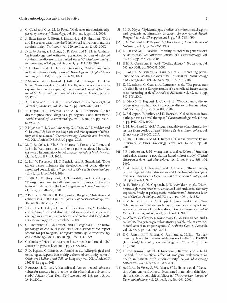
Treated CD Untreated CD Controls0
10
20
30
40P < 0.05
Mer
cury
blo
od co
nten
t (𝜇
g/L)
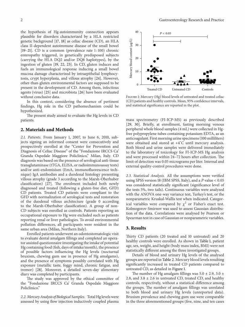
Figure 1: Mercury (Hg) blood levels of untreated and treated celiac(CD) patients and healthy controls. Mean, 95% confidence intervals,and statistical significance are reported in the plot.
mass spectrometry (FI-ICP-MS) as previously described[29, 30]. Briefly, at enrollment, fasting morning venousperipheral whole blood samples (4mL) were collected in Hg-free polypropylene tubes containing potassium EDTA, as ananticoagulant. Firstmorning urine specimens (100milliliters)were obtained and stored at +4∘C until mercury analysis.Both blood and urine samples were delivered immediatelyto the laboratory of toxicology for FI-ICP-MS Hg analysisand were processed within 24–72 hours after collection. Thelimit of detection was 0.05 micrograms per liter. Internal andexternal quality-control procedures were done.
2.3. Statistical Analysis. All the assumptions were verifiedusing SPSS version 18 (IBM SPSS, Italy), and a 𝑃 value < 0.05was considered statistically significant (significance level ofthe tests 5%, two tails). Continuous variables were analyzedwith the ANOVA one-way variance test, Turkey’s test, or thenonparametric Kruskal-Wallis test when indicated. Categor-ical variables were compared by 𝜒2 or Fisher’s exact test.Kolmogorov-Smirnov was used to assess Gaussian distribu-tion of the data. Correlations were analysed by Pearson orSpearman test in case ofGaussian or nonparametric variables.
3. Results
Thirty CD patients (20 treated and 10 untreated) and 20healthy controls were enrolled. As shown in Table 1, patientage, sex, weight, and height (bodymass index, BMI) were notstatistically different among the three investigated groups.
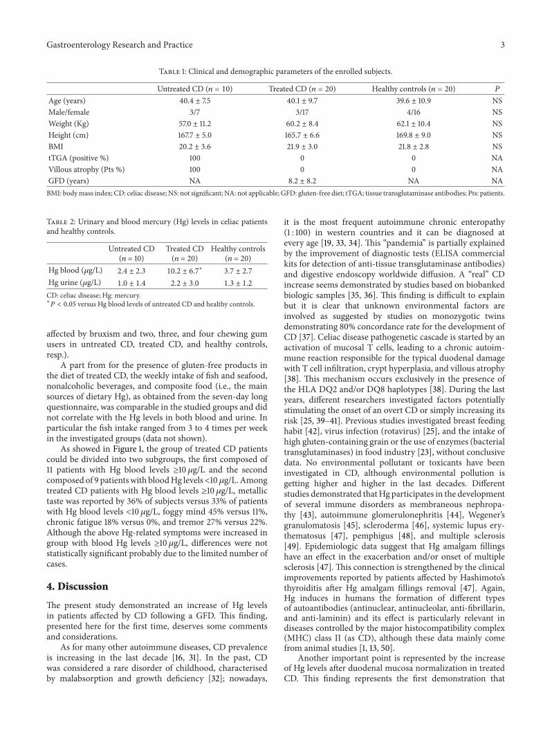
Details of blood and urinary Hg levels of the analysedgroups are reported in Table 2.Mercury blood levels resultingsignificantly increased in treated CD patients compared tountreated CD, as detailed in Figure 1.
The number of Hg amalgam fillings was 5.0 ± 2.9, 3.0 ±2.8, and 3.8 ± 2.6 in untreated CD, treated CD, and healthycontrols, respectively, without a statistical difference amongthe groups. The number of amalgam fillings was unrelatedto both blood and urinary Hg levels (unreported data).Bruxism prevalence and chewing gum use were comparablein the three aforementioned groups (five, nine, and ten cases
Gastroenterology Research and Practice 3
Table 1: Clinical and demographic parameters of the enrolled subjects.
Untreated CD (𝑛 = 10) Treated CD (𝑛 = 20) Healthy controls (𝑛 = 20) 𝑃
Age (years) 40.4 ± 7.5 40.1 ± 9.7 39.6 ± 10.9 NSMale/female 3/7 3/17 4/16 NSWeight (Kg) 57.0 ± 11.2 60.2 ± 8.4 62.1 ± 10.4 NSHeight (cm) 167.7 ± 5.0 165.7 ± 6.6 169.8 ± 9.0 NSBMI 20.2 ± 3.6 21.9 ± 3.0 21.8 ± 2.8 NStTGA (positive %) 100 0 0 NAVillous atrophy (Pts %) 100 0 0 NAGFD (years) NA 8.2 ± 8.2 NA NABMI: bodymass index; CD: celiac disease; NS: not significant; NA: not applicable; GFD: gluten-free diet; tTGA; tissue transglutaminase antibodies; Pts: patients.
Table 2: Urinary and blood mercury (Hg) levels in celiac patientsand healthy controls.
Untreated CD(𝑛 = 10)
Treated CD(𝑛 = 20)
Healthy controls(𝑛 = 20)
Hg blood (𝜇g/L) 2.4 ± 2.3 10.2 ± 6.7∗
3.7 ± 2.7
Hg urine (𝜇g/L) 1.0 ± 1.4 2.2 ± 3.0 1.3 ± 1.2
CD: celiac disease; Hg: mercury.∗
𝑃 < 0.05 versus Hg blood levels of untreated CD and healthy controls.
affected by bruxism and two, three, and four chewing gumusers in untreated CD, treated CD, and healthy controls,resp.).
A part from for the presence of gluten-free products inthe diet of treated CD, the weekly intake of fish and seafood,nonalcoholic beverages, and composite food (i.e., the mainsources of dietary Hg), as obtained from the seven-day longquestionnaire, was comparable in the studied groups and didnot correlate with the Hg levels in both blood and urine. Inparticular the fish intake ranged from 3 to 4 times per weekin the investigated groups (data not shown).
As showed in Figure 1, the group of treated CD patientscould be divided into two subgroups, the first composed of11 patients with Hg blood levels ≥10 𝜇g/L and the secondcomposed of 9 patientswith bloodHg levels<10𝜇g/L.Amongtreated CD patients with Hg blood levels ≥10 𝜇g/L, metallictaste was reported by 36% of subjects versus 33% of patientswith Hg blood levels <10 𝜇g/L, foggy mind 45% versus 11%,chronic fatigue 18% versus 0%, and tremor 27% versus 22%.Although the above Hg-related symptoms were increased ingroup with blood Hg levels ≥10 𝜇g/L, differences were notstatistically significant probably due to the limited number ofcases.
4. Discussion
The present study demonstrated an increase of Hg levelsin patients affected by CD following a GFD. This finding,presented here for the first time, deserves some commentsand considerations.
As for many other autoimmune diseases, CD prevalenceis increasing in the last decade [16, 31]. In the past, CDwas considered a rare disorder of childhood, characterisedby malabsorption and growth deficiency [32]; nowadays,
it is the most frequent autoimmune chronic enteropathy(1 : 100) in western countries and it can be diagnosed atevery age [19, 33, 34]. This “pandemia” is partially explainedby the improvement of diagnostic tests (ELISA commercialkits for detection of anti-tissue transglutaminase antibodies)and digestive endoscopy worldwide diffusion. A “real” CDincrease seems demonstrated by studies based on biobankedbiologic samples [35, 36]. This finding is difficult to explainbut it is clear that unknown environmental factors areinvolved as suggested by studies on monozygotic twinsdemonstrating 80% concordance rate for the development ofCD [37]. Celiac disease pathogenetic cascade is started by anactivation of mucosal T cells, leading to a chronic autoim-mune reaction responsible for the typical duodenal damagewith T cell infiltration, crypt hyperplasia, and villous atrophy[38]. This mechanism occurs exclusively in the presence ofthe HLA DQ2 and/or DQ8 haplotypes [38]. During the lastyears, different researchers investigated factors potentiallystimulating the onset of an overt CD or simply increasing itsrisk [25, 39–41]. Previous studies investigated breast feedinghabit [42], virus infection (rotavirus) [25], and the intake ofhigh gluten-containing grain or the use of enzymes (bacterialtransglutaminases) in food industry [23], without conclusivedata. No environmental pollutant or toxicants have beeninvestigated in CD, although environmental pollution isgetting higher and higher in the last decades. Differentstudies demonstrated thatHg participates in the developmentof several immune disorders as membraneous nephropa-thy [43], autoimmune glomerulonephritis [44], Wegener’sgranulomatosis [45], scleroderma [46], systemic lupus ery-thematosus [47], pemphigus [48], and multiple sclerosis[49]. Epidemiologic data suggest that Hg amalgam fillingshave an effect in the exacerbation and/or onset of multiplesclerosis [47]. This connection is strengthened by the clinicalimprovements reported by patients affected by Hashimoto’sthyroiditis after Hg amalgam fillings removal [47]. Again,Hg induces in humans the formation of different typesof autoantibodies (antinuclear, antinucleolar, anti-fibrillarin,and anti-laminin) and its effect is particularly relevant indiseases controlled by the major histocompatibility complex(MHC) class II (as CD), although these data mainly comefrom animal studies [1, 13, 50].
Another important point is represented by the increaseof Hg levels after duodenal mucosa normalization in treatedCD. This finding represents the first demonstration that

4 Gastroenterology Research and Practice
duodenal atrophy could lead to a reversible Hg “malabsorp-tion.” Actually, very few are known about the Hg trans-port/absorption in human small bowel. Recent in vitro dataon Caco-2 cell line suggest the existence of an active Hgtransport into enterocytes although the transporter remainsunknown [51]. This “scenario” could be comparable to thatobserved for iron intestinal transport when a geneticallydetermined hemochromatosis is present in association withCD: the concomitant duodenal atrophy preserves patientsby iron overload and the development of clinically rele-vant hemochromatosis, usually developing during the GFD(i.e., duodenal normalization) [52, 53]. Interpretation of thepresent finding leads to important observations: (i) intestinalHg absorption is mainly localized in the duodenum; (ii) Hgseems to be absorbed by transporters mainly localised on theapical part of the villi, typically damaged in CD. However,another factor increasing the Hg levels could be the GFDitself; in fact, GFD could have a positive effect favoring theHg release from the intracellular stores to extracellular fluids(i.e., blood). If this process could be related to the presenceof symptoms (metallic taste, foggymind, chronic fatigue, andtremor) resemblingHg poisoning as detected in our cohort, itremains an interesting point to be investigated in large seriesof patients.
The relevant increase of Hg blood levels in treated CD isnot justified by differences in seafood consumption and den-tal amalgam fillings, which are considered important sourcesof Hg for humans by the World Health Organization, WHO[11]. Fish generally accumulates Hg through the alimentarychain and for these reasons fish-eating fishes (swordfish, tuna,etc.) aremore likelyHg-contaminated, while amalgamfillingscontinuously release Hg in the mouth, especially in patientsaffected by bruxism or using chewing gum [11].
Looking at these data, Hg accumulation could also be dueto a genetic predisposition of CD subjects to retain it (see alsolowHg levels in urine). In our study we analysed whole bloodand urine Hg levels, believed to be reliable marker for Hgexposure; however this choice could have some implications.When exposure continues, tissue levels of Hg in humansare increased, mainly in brain (pituitary gland and cerebralcortex), central nervous system, thyroid, and kidneys. Hence,concentrations of Hg in blood and urine may underestimateretention of Hg in the organism. In other words, there is thepossibility that measurements of Hg in blood and urine donot fully reflect the real body content.
Another limitation of the study, besides the limitednumber of patients, is that blood and urineHg concentrationsnot always correlate with the noxious effects in humans. Infact, response toHg is influenced by different factors inducingthe onset of the immune or neurologic alteration. Thesefactors are largely unknown but genetic ones are the stronglysuspected: subjects with a predisposition to accumulate Hgin determined cells or tissues could be more susceptible tospecific toxic effects [4].
In conclusion, our study demonstrates an alteration of Hgcontent in CD when a gluten-free regimen is followed. Thisresult could be due to an altered response to Hg exposure,with the tendency to accumulate it. Further studies areneeded to clarify if CD genetic background could generate
“sensitivity” to Hg proinflammatory effect and inspire newrules for the surveillance of Hg content in food.
Conflict of Interests
The authors declare that there is no conflict of interestsregarding the publication of this paper.
Acknowledgment
The study was supported by “Fondazione IRCCS Ca’ GrandaOspedale Maggiore Policlinico, Milano.”
References
[1] T. W. Clarkson, “The three modern faces of mercury,” Envi-ronmental Health Perspectives, vol. 110, supplement 1, pp. 11–23,2002.
[2] K. Suresh Kumar Reddy, A. Al Shoaibi, and C. Srinivasakannan,“Elemental mercury adsorption on sulfur-impregnated porouscarbon-a review,” Environmental Technology, vol. 35, no. 1, pp.18–26, 2014.
[3] R. D. Day, P. R. Becker, O. F. X. Donard, R. S. Pugh, and S.A. Wise, “Environmental specimen banks as a resource formercury and mercury isotope research in marine ecosystems,”Environmental Sciences: Processes and Impacts, vol. 16, no. 1, pp.10–27, 2014.
[4] S. M. Koral, “The toxicology of mercury,” The New EnglandJournal of Medicine, vol. 350, no. 9, pp. 945–947, 2004.
[5] C. Sanfeliu, J. Sebastia, and S. U. Kim, “Methylmercury neuro-toxicity in cultures of human neurons, astrocytes, neuroblas-toma cells,” NeuroToxicology, vol. 22, no. 3, pp. 317–327, 2001.
[6] S. M. Koral, “Mercury from dental amalgam: exposure and riskassessment,”Compendiumof Continuing Education inDentistry,vol. 34, no. 2, pp. 138–144, 2013.
[7] J. D. Park and W. Zheng, “Human exposure and health effectsof inorganic and elemental mercury,” Journal of PreventiveMedicine and Public Health, vol. 45, no. 6, pp. 344–352, 2012.
[8] X. Xu, C. Mathieu, S. E. Boitard et al., “Skeletal muscle glycogenphosphorylase is irreversibly inhibited by mercury: molecular,cellular and kinetic aspects,” FEBS Letters, vol. 588, no. 1, pp.138–142, 2014.
[9] S. Ekino, M. Susa, T. Ninomiya, K. Imamura, and T. Kitamura,“Minamata disease revisited: an update on the acute and chronicmanifestations of methyl mercury poisoning,” Journal of theNeurological Sciences, vol. 262, no. 1-2, pp. 131–144, 2007.
[10] K. K. Aminzadeh and M. Etminan, “Dental amalgam and mul-tiple sclerosis: a systematic review and meta-analysis,” Journalof Public Health Dentistry, vol. 67, no. 1, pp. 64–66, 2007.
[11] G. Guzzi, G. B. Fogazzi, M. Cantu et al., “Dental amalgam,mercury toxicity, and renal autoimmunity,” Journal of Environ-mental Pathology, Toxicology and Oncology, vol. 27, no. 2, pp.147–155, 2008.
[12] C.-C. Kuo, K. Moon, K. A. Thayer, and A. Navas-Acien, “Envi-ronmental chemicals and type 2 diabetes: an updated system-atic review of the epidemiologic evidence,” Current DiabetesReports, vol. 13, no. 6, pp. 831–849, 2013.
[13] J. F. Nyland, M. Fillion, F. Barbosa Jr. et al., “Biomarkersof methylmercury exposure immunotoxicity among fish con-sumers in amazonian Brazil,” Environmental Health Perspec-tives, vol. 119, no. 12, pp. 1733–1738, 2011.
Gastroenterology Research and Practice 5
[14] G. Guzzi and C. A. M. La Porta, “Molecular mechanisms trig-gered by mercury,” Toxicology, vol. 244, no. 1, pp. 1–12, 2008.
[15] S. Havarinasab, E. Bjorn, J. Ekstrand, and P. Hultman, “DoseandHg species determine the T-helper cell activation inmurineautoimmunity,” Toxicology, vol. 229, no. 1-2, pp. 23–32, 2007.
[16] D. L. Jacobson, S. J. Gange, N. R. Rose, and N. M. H. Graham,“Epidemiology and estimated population burden of selectedautoimmune diseases in theUnited States,”Clinical Immunologyand Immunopathology, vol. 84, no. 3, pp. 223–243, 1997.
[17] P. Hultman and H. Hansson-Georgiadis, “Methyl mercury-induced autoimmunity in mice,” Toxicology and Applied Phar-macology, vol. 154, no. 3, pp. 203–211, 1999.
[18] P.Moszczynski, S. Slowinski, J. Rutkowski, S. Bem, andD. Jakus-Stoga, “Lymphocytes, T and NK cells, in men occupationallyexposed to mercury vapours,” International Journal of Occupa-tional Medicine and Environmental Health, vol. 8, no. 1, pp. 49–56, 1995.
[19] A. Fasano and C. Catassi, “Celiac disease,” The New EnglandJournal of Medicine, vol. 367, no. 25, pp. 2419–2426, 2012.
[20] N. Gujral, H. J. Freeman, and A. B. R. Thomson, “Celiacdisease: prevalence, diagnosis, pathogenesis and treatment,”World Journal of Gastroenterology, vol. 18, no. 42, pp. 6036–6059, 2012.
[21] P. Nijeboer, R. L. J. vanWanrooij, G. J. Tack, C. J. J. Mulder, andG. Bouma, “Update on the diagnosis andmanagement of refrac-tory coeliac disease,” Gastroenterology Research and Practice,vol. 2013, Article ID 518483, 9 pages, 2013.
[22] M. T. Bardella, L. Elli, S. D. Matteis, I. Floriani, V. Torri, andL. Piodi, “Autoimmune disorders in patients affected by celiacsprue and inflammatory bowel disease,”Annals ofMedicine, vol.41, no. 2, pp. 139–143, 2009.
[23] L. Elli, V. Discepolo, M. T. Bardella, and S. Guandalini, “Doesgluten intake influence the development of celiac disease-associated complications?” Journal of Clinical Gastroenterology,vol. 48, no. 1, pp. 13–20, 2014.
[24] L. Elli, C. M. Bergamini, M. T. Bardella, and D. Schuppan,“Transglutaminases in inflammation and fibrosis of the gas-trointestinal tract and the liver,”Digestive and Liver Disease, vol.41, no. 8, pp. 541–550, 2009.
[25] P. Pavone, E. Nicolini, R. Taibi, andM. Ruggieri, “Rotavirus andceliac disease,” The American Journal of Gastroenterology, vol.102, no. 8, article 1831, 2007.
[26] E. Sanchez, I. Nadal, E. Donat, C. Ribes-Koninckx,M. Calabuig,and Y. Sanz, “Reduced diversity and increased virulence-genecarriage in intestinal enterobacteria of coeliac children,” BMCGastroenterology, vol. 8, article 50, 2008.
[27] G. Oberhuber, G. Granditsch, and H. Vogelsang, “The histo-pathology of coeliac disease: time for a standardized reportscheme for pathologists,” European Journal of Gastroenterologyand Hepatology, vol. 11, no. 10, pp. 1185–1194, 1999.
[28] C. Cooksey, “Health concerns of heavy metals and metalloids,”Science Progress, vol. 95, no. 1, pp. 73–88, 2012.
[29] P. D. Pigatto, C. Minoia, A. Ronchi et al., “Allergological andtoxicological aspects in a multiple chemical sensitivity cohort,”Oxidative Medicine and Cellular Longevity, vol. 2013, Article ID356235, 12 pages, 2013.
[30] P. Apostoli, I. Cortesi, A.Mangili et al., “Assessment of referencevalues for mercury in urine: the results of an Italian polycentricstudy,” Science of the Total Environment, vol. 289, no. 1–3, pp.13–24, 2002.
[31] M. D. Mayes, “Epidemiologic studies of environmental agentsand systemic autoimmune diseases,” Environmental HealthPerspectives, vol. 107, supplement 5, pp. 743–748, 1999.
[32] S. G. Cole andM. F. Kagnoff, “Celiac disease,” Annual Review ofNutrition, vol. 5, pp. 241–266, 1985.
[33] L. Elli and M. T. Bardella, “Motility disorders in patients withceliac disease,” Scandinavian Journal of Gastroenterology, vol.40, no. 7, pp. 743–749, 2005.
[34] P. H. R. Green and B. Jabri, “Coeliac disease,” The Lancet, vol.362, no. 9381, pp. 383–391, 2003.
[35] S. Lohi, K. Mustalahti, K. Kaukinen et al., “Increasing preva-lence of coeliac disease over time,” Alimentary PharmacologyandTherapeutics, vol. 26, no. 9, pp. 1217–1225, 2007.
[36] K. Mustalahti, C. Catassi, A. Reunanen et al., “The prevalenceof celiac disease in Europe: results of a centralized, internationalmass screening project,” Annals of Medicine, vol. 42, no. 8, pp.587–595, 2010.
[37] L. Nistico, C. Fagnani, I. Coto et al., “Concordance, diseaseprogression, and heritability of coeliac disease in Italian twins,”Gut, vol. 55, no. 6, pp. 803–804, 2006.
[38] D. Schuppan, Y. Junker, and D. Barisani, “Celiac disease: frompathogenesis to novel therapies,” Gastroenterology, vol. 137, no.6, pp. 1912–1933, 2009.
[39] L.M. Sollid and B. Jabri, “Triggers and drivers of autoimmunity:lessons from coeliac disease,” Nature Reviews Immunology, vol.13, no. 4, pp. 294–302, 2013.
[40] L. Elli, E. Dolfini, and M. T. Bardella, “Gliadin cytotoxicity andin vitro cell cultures,” Toxicology Letters, vol. 146, no. 1, pp. 1–8,2003.
[41] J. F. Ludvigsson, S. M. Montgomery, and A. Ekbom, “Smokingand celiac disease: a population-based cohort study,” ClinicalGastroenterology and Hepatology, vol. 3, no. 9, pp. 869–874,2005.
[42] L. A. Persson, A. Ivarsson, and O. Hernell, “Breast-feedingprotects against celiac disease in childhood—epidemiologicalevidence,” Advances in Experimental Medicine and Biology, vol.503, pp. 115–123, 2002.
[43] R. R. Tubbs, G. N. Gephardt, J. T. McMahon et al., “Mem-branous glomerulonephritis associated with industrial mercuryexposure. Study of pathogenetic mechanisms,” American Jour-nal of Clinical Pathology, vol. 77, no. 4, pp. 409–413, 1982.
[44] S. Miller, S. Pallan, A. S. Gangji, D. Lukic, and C. M. Clase,“Mercury-associated nephrotic syndrome: a case report andsystematic review of the literature,” The American Journal ofKidney Diseases, vol. 62, no. 1, pp. 135–138, 2013.
[45] D. Albert, C. Clarkin, J. Komoroski, C. M. Brensinger, and J.A. Berlin, “Wegener’s granulomatosis: possible role of environ-mental agents in its pathogenesis,” Arthritis Care & Research,vol. 51, no. 4, pp. 656–664, 2004.
[46] F. C. Arnett, M. J. Fritzler, C. Ahn, and A. Holian, “Urinarymercury levels in patients with autoantibodies to U3-RNP(fibrillarin),” Journal of Rheumatology, vol. 27, no. 2, pp. 405–410, 2000.
[47] J. Prochazkova, I. Sterzl, H. Kucerova, J. Bartova, and V. D. M.Stejskal, “The beneficial effect of amalgam replacement onhealth in patients with autoimmunity,” NeuroendocrinologyLetters, vol. 25, no. 3, pp. 211–218, 2004.
[48] A. M. Abreu Velez, G. Warfvinge, W. L. Herrera et al., “Detec-tion ofmercury and other undeterminedmaterials in skin biop-sies of endemic pemphigus foliaceus,” The American Journal ofDermatopathology, vol. 25, no. 5, pp. 384–391, 2003.

6 Gastroenterology Research and Practice
[49] A. M. Attar, A. Kharkhaneh, M. Etemadifar, K. Keyhanian, V.Davoudi, andM. Saadatnia, “Serummercury level andmultiplesclerosis,” Biological Trace Element Research, vol. 146, no. 2, pp.150–153, 2012.
[50] P. D. Pigatto, C. Minoia, L. Brambilla, S. Ferrucci, and G. Guzzi,“Auto-antibodies to nuclear and nucleolar antigen and long-term exposure to inorganic mercury,” Environmental Research,vol. 110, no. 8, article 821, 2010.
[51] M. Vazquez, M. Calatayud, D. Velez, and V. Devesa, “Intestinaltransport of methylmercury and inorganic mercury in variousmodels of Caco-2 andHT29-MTX cells,”Toxicology, vol. 311, no.3, pp. 147–153, 2013.
[52] E. Zubizarreta, E. Zapata, and A. Castiella, “Celiac diseaseand hemochromatosis,” European Journal of Gastroenterology &Hepatology, vol. 20, no. 6, p. 589, 2008.
[53] D. Barisani, S. Ceroni, S. del Bianco, R. Meneveri, and M. T.Bardella, “Hemochromatosis gene mutations and iron metab-olism in celiac disease,”Haematologica, vol. 89, no. 11, pp. 1299–1305, 2004.

Submit your manuscripts athttp://www.hindawi.com
Stem CellsInternational
Hindawi Publishing Corporationhttp://www.hindawi.com Volume 2014
Hindawi Publishing Corporationhttp://www.hindawi.com Volume 2014
MEDIATORSINFLAMMATION
of
Hindawi Publishing Corporationhttp://www.hindawi.com Volume 2014
Behavioural Neurology
EndocrinologyInternational Journal of
Hindawi Publishing Corporationhttp://www.hindawi.com Volume 2014
Hindawi Publishing Corporationhttp://www.hindawi.com Volume 2014
Disease Markers
Hindawi Publishing Corporationhttp://www.hindawi.com Volume 2014
BioMed Research International
OncologyJournal of
Hindawi Publishing Corporationhttp://www.hindawi.com Volume 2014
Hindawi Publishing Corporationhttp://www.hindawi.com Volume 2014
Oxidative Medicine and Cellular Longevity
Hindawi Publishing Corporationhttp://www.hindawi.com Volume 2014
PPAR Research
The Scientific World JournalHindawi Publishing Corporation http://www.hindawi.com Volume 2014
Immunology ResearchHindawi Publishing Corporationhttp://www.hindawi.com Volume 2014
Journal of
ObesityJournal of
Hindawi Publishing Corporationhttp://www.hindawi.com Volume 2014
Hindawi Publishing Corporationhttp://www.hindawi.com Volume 2014
Computational and Mathematical Methods in Medicine
OphthalmologyJournal of
Hindawi Publishing Corporationhttp://www.hindawi.com Volume 2014
Diabetes ResearchJournal of
Hindawi Publishing Corporationhttp://www.hindawi.com Volume 2014
Hindawi Publishing Corporationhttp://www.hindawi.com Volume 2014
Research and TreatmentAIDS
Hindawi Publishing Corporationhttp://www.hindawi.com Volume 2014
Gastroenterology Research and Practice
Hindawi Publishing Corporationhttp://www.hindawi.com Volume 2014
Parkinson’s Disease
Evidence-Based Complementary and Alternative Medicine
Volume 2014Hindawi Publishing Corporationhttp://www.hindawi.com