The Assessment of Autoimmunological Status and Prevalence of Different Forms of Celiac Disease among...
7
Hindawi Publishing Corporation Mediators of Inflammation Volume 2008, Article ID 285989, 6 pages doi:10.1155/2008/285989 Research Article The Assessment of Autoimmunological Status and Prevalence of Different Forms of Celiac Disease among Children with Type 1 Diabetes Mellitus and Celiac Disease Grazyna Deja, 1 Anna Myrda, 2 Przemyslawa Jarosz-Chobot, 1 and Urszula Siekiera 3 1 Department of Pediatrics, Endocrinology and Diabetes, Medical University of Silesia, 40-752 Katowice, Poland 2 Schwarzwald-Baar Klinikum, 78011 Villingen-Schwenningen, Germany 3 Blood Center, 40-074 Katowice, Poland Correspondence should be addressed to Grazyna Deja, [email protected] Received 5 August 2007; Accepted 3 January 2008 Recommended by Freek J. Zijlstra This study aims to assess the autoimmunological status and forms of celiac disease (CD) among children with type 1 diabetes mellitus (T1DM) . The study group comprises 27 patients at the mean age of 12.30 years (±SD 3.12). The measurement of the level of diabetes-specific antibodies and organ-specific antibodies was gained at the T1DM-onset and repeated annually. The following risk factors influencing time of CD diagnosis were analyzed: age, sex, T1DM duration, autoantibodies, and HLA-haplotype. The prevalence of antibodies was GADA-74%, IAA-63%, IA2A-67%, ATA-11%, and ATG-4%. The intestinal biopsy revealed in 19% no changes and in 77% stage 3 (Marsh scale). In most cases, no clinical manifestation of CD was observed. The diagnosis of Hashimoto’s disease was made twice. The negative correlation between the age at T1DM-onset and the interval between onset of T1DM and CD (r =−0.35, p<.05) was noted. The high-comorbidity ratio of CD and thyroiditis with T1DM demands regular screening tests especially in the first years after T1DM-onset. Copyright © 2008 Grazyna Deja et al. This is an open access article distributed under the Creative Commons Attribution License, which permits unrestricted use, distribution, and reproduction in any medium, provided the original work is properly cited. 1. INTRODUCTION Due to a common genetic background and interaction be- tween environmental and immunological factors, patients with type 1 diabetes mellitus (T1DM) are at a high risk of having other autoimmunological diseases. The autoimmune polyendocrine syndromes type 1 (APS-1) and type 2 (APS- 2) are rare but most dramatic. APS-1 is caused by a sin- gle mutation in the AIRE gene (transcriptional factor) as opposite to APS-2, which is a “complex” genetic disorder with strong HLA-genes association (haplotype DQA1∗0501, DQB1∗0201, DRB1∗0301) [1]. Single autoimmune diseases like celiac disease (CD) or thyroid gland disease are more common among patients with T1DM. They are also con- nected with above-mentioned predisposing HLA-genes. In the course of T1DM, no clinical manifestation of both coex- isting disorders is noted in most cases [2, 3]. That is why reg- ular investigations of the autoimmunological status should be provided. Severe complications of untreated celiac disease necessitate an early diagnosis and the prompt introduction of a gluten-free diet. Undiagnosed thyroid gland disease that remains untreated can lead to subclinical or clinical obvious hypothyroidism. At the time of the diagnosis of diabetes, various antibod- ies are evaluated. To make a statement of autoimmune back- ground of the disease, the diabetes-associated antibodies are detected. Blood samples are screened for islet cell antibodies, insulin autoantibodies, GAD antibodies, and anti-IA-2 anti- bodies [4, 5]. Additionally, screening for celiac disease and autoimmune thyroiditis is usually performed. The presence of humoral immune markers allows an immediate medical intervention, but their absence should not mislead a physi- cian, since there is a possibility of developing these disorders some years after diabetes onset [5]. The purpose of our study was to assess retrospectively the autoimmunological status from the time of the first diabetes presentation until celiac disease was revealed and assess the risk factors of influencing the time of the CD diagnosis. The next objective of the study was to evaluate the stages of celiac disease most commonly appearing among children with type 1 diabetes mellitus.
-
Upload
independent -
Category
Documents
-
view
0 -
download
0
Transcript of The Assessment of Autoimmunological Status and Prevalence of Different Forms of Celiac Disease among...

Hindawi Publishing CorporationMediators of InflammationVolume 2008, Article ID 285989, 6 pagesdoi:10.1155/2008/285989
Research ArticleThe Assessment of Autoimmunological Status and Prevalenceof Different Forms of Celiac Disease among Children withType 1 Diabetes Mellitus and Celiac Disease
Grazyna Deja,1 Anna Myrda,2 Przemyslawa Jarosz-Chobot,1 and Urszula Siekiera3
1 Department of Pediatrics, Endocrinology and Diabetes, Medical University of Silesia, 40-752 Katowice, Poland2 Schwarzwald-Baar Klinikum, 78011 Villingen-Schwenningen, Germany3 Blood Center, 40-074 Katowice, Poland
Correspondence should be addressed to Grazyna Deja, [email protected]
Received 5 August 2007; Accepted 3 January 2008
Recommended by Freek J. Zijlstra
This study aims to assess the autoimmunological status and forms of celiac disease (CD) among children with type 1 diabetesmellitus (T1DM) . The study group comprises 27 patients at the mean age of 12.30 years (±SD 3.12). The measurement of the levelof diabetes-specific antibodies and organ-specific antibodies was gained at the T1DM-onset and repeated annually. The followingrisk factors influencing time of CD diagnosis were analyzed: age, sex, T1DM duration, autoantibodies, and HLA-haplotype. Theprevalence of antibodies was GADA-74%, IAA-63%, IA2A-67%, ATA-11%, and ATG-4%. The intestinal biopsy revealed in 19%no changes and in 77% stage 3 (Marsh scale). In most cases, no clinical manifestation of CD was observed. The diagnosis ofHashimoto’s disease was made twice. The negative correlation between the age at T1DM-onset and the interval between onset ofT1DM and CD (r = −0.35, p < .05) was noted. The high-comorbidity ratio of CD and thyroiditis with T1DM demands regularscreening tests especially in the first years after T1DM-onset.
Copyright © 2008 Grazyna Deja et al. This is an open access article distributed under the Creative Commons Attribution License,which permits unrestricted use, distribution, and reproduction in any medium, provided the original work is properly cited.
1. INTRODUCTION
Due to a common genetic background and interaction be-tween environmental and immunological factors, patientswith type 1 diabetes mellitus (T1DM) are at a high risk ofhaving other autoimmunological diseases. The autoimmunepolyendocrine syndromes type 1 (APS-1) and type 2 (APS-2) are rare but most dramatic. APS-1 is caused by a sin-gle mutation in the AIRE gene (transcriptional factor) asopposite to APS-2, which is a “complex” genetic disorderwith strong HLA-genes association (haplotype DQA1∗0501,DQB1∗0201, DRB1∗0301) [1]. Single autoimmune diseaseslike celiac disease (CD) or thyroid gland disease are morecommon among patients with T1DM. They are also con-nected with above-mentioned predisposing HLA-genes. Inthe course of T1DM, no clinical manifestation of both coex-isting disorders is noted in most cases [2, 3]. That is why reg-ular investigations of the autoimmunological status shouldbe provided. Severe complications of untreated celiac diseasenecessitate an early diagnosis and the prompt introductionof a gluten-free diet. Undiagnosed thyroid gland disease that
remains untreated can lead to subclinical or clinical obvioushypothyroidism.
At the time of the diagnosis of diabetes, various antibod-ies are evaluated. To make a statement of autoimmune back-ground of the disease, the diabetes-associated antibodies aredetected. Blood samples are screened for islet cell antibodies,insulin autoantibodies, GAD antibodies, and anti-IA-2 anti-bodies [4, 5]. Additionally, screening for celiac disease andautoimmune thyroiditis is usually performed. The presenceof humoral immune markers allows an immediate medicalintervention, but their absence should not mislead a physi-cian, since there is a possibility of developing these disorderssome years after diabetes onset [5].
The purpose of our study was to assess retrospectively theautoimmunological status from the time of the first diabetespresentation until celiac disease was revealed and assess therisk factors of influencing the time of the CD diagnosis. Thenext objective of the study was to evaluate the stages of celiacdisease most commonly appearing among children with type1 diabetes mellitus.

2 Mediators of Inflammation
2. MATERIALS AND METHODS
The study group comprises 27 diabetic children (14 girls, 13boys) at the mean age of 12.3 years (±3.12) with diagnosedceliac disease. They were selected from about 450 new pa-tients with T1DM. The retrospective analysis concerns pa-tients’ data collected in the years 2001–2006. All patientstaken into consideration were hospitalized in the Depart-ment of Pediatrics, Endocrinology and Diabetes in Katowiceat the time of the first diabetes presentation. Patients wereregularly observed in outpatient diabetes care at least till CDwas revealed. The diagnosis of T1DM was made according toWHO and ISPAD criteria [4, 5].
The measurement of the level of disease-associatedautoantibodies to glutamic acid decarboxylase (GADA),protein-tyrosine phosphatase (IA2A), insulin (IAA) wasgained at the time of T1DM onset. The serum samples weredrawn before the initiation of insulin treatment. Samplesfor organ-specific antibodies: tissue transglutaminase (tTG),thyroperoxidase (ATA), and thyroglobuline (ATG) as wellas thyroid hormones (TSH, fT4) were also obtained fromdiabetes-onset patients. In all children, serum immunoglob-ulin A (IgA) concentration was determined to exclude IgAdeficiency and a subsequent false interpretation of receivedresults of IgA tTG. The control measurements of tTG, ATG,ATA, and thyroid hormones were repeated annually. The titerof GADA, IA2A, and IAA was gauged by using the radioim-mune assay technique (RIA-CIS Bio International) in con-formity with the protocols included in laboratory originalkits. The measurement ranges were as follows: for GADA 0–300 U/mL, for IA2A 0–50 U/mL, and for IAA 0–100%. Theresults for GADA and IA2A above 0.75 U/mL and for IAAabove 7% were considered positive. The CD-associated IgAtTG were detected by the ELISA method. The level of thy-roid hormones and thyroid antibodies was estimated by thechemiluminescence method (DPC, USA). Normal ranges forfT4 for children under the age of 12 are 0.65–2.3 ng/dl andfor older ones are 0.8–1.9 ng/dl. TSH was assessed accord-ing to age-adequate normal levels. The concentration of ATA> 35 IU/mL and of ATG > 40 IU/mL was considered to bepositive. IgA concentration was assessed by immunoturbidi-metric measurements (Dade Behring, Germany). IgA defi-ciency was defined as an IgA below-normal level adequate forage.
If autoimmunity markers were present, the ultrasoundexamination of thyroid gland or the small bowel biopsy wasordered. Intestinal mucosa specimens were obtained duringperoral endoscopy and afterwards examined by a pathol-ogist. Changes in mucosa were assessed by employing theMarsh stage. Type 1 (infiltrative lesion) is characterized bynormal structure of mucosa with presence of intraepitheliallymphocytes infiltration. Type 2 (hyperplastic lesion) com-prises type 1-mentioned changes and additionally enlargedjejunum crypts. Type 3 (destructive lesion) is divided into 3groups. The characteristics of all of them include inflamma-tory infiltration, hyperplastic crypts, and the villous atrophy,which in each group is of a different degree. In type 3a par-tial, in type 3b subtotal, and in type 3c, total destruction ofvilli is observed. Type 4 (hypoplastic lesion) comprises to-
2
49
5
22 2
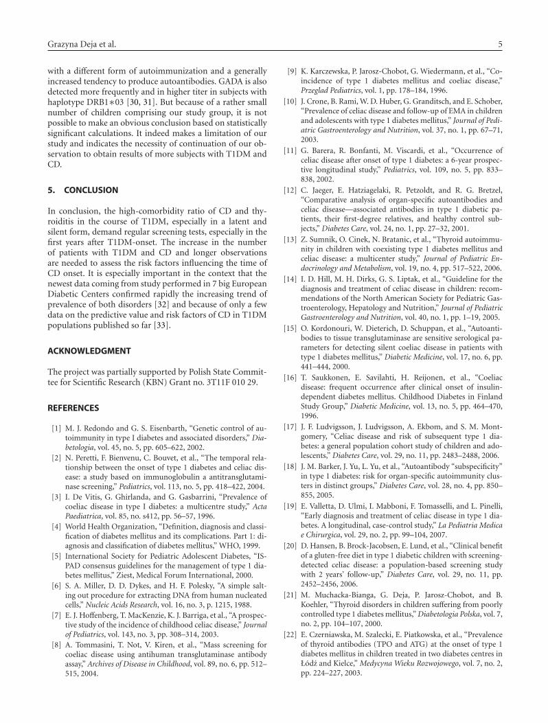
GADA 74%
IA2A 67%IAA 63%
Figure 1: Diabetes-associated autoantibodies in children at thetime of T1DM onset.
tal villous atrophy without inflammatory infiltration and thenormal architecture of the crypts.
HLA tests were performed in the Regional Blood Cen-tre in Katowice. HLA class II alleles were typed by using thepolymerase chain reaction—the single strand polymorphismmethod (PCR-SSP). Allele-specific tests One Lambda (Cy-togen, USA) were employed for HLA-DRB1∗ and DQB1∗typing. For genetic assays, human leukocytes were used. ThePCR program was performed according to the protocol de-scribed earlier [6].
The statistical analysis was carried out with the applica-tion SigmaStat. To estimate the risk factors influencing thetime of CD diagnosis among children with T1DM, the fol-lowing parameters were analyzed: age at T1DM onset, gen-der, T1DM duration, presence of autoantibodies, and HLAhaplotype. The normality of the distribution of the contin-uous variables was evaluated by using the Shapiro-Wilk test.The correlation between analyzed parameters was calculatedby using Pearson correlation.The results with the p < .05were considered to be statistically significant.
3. RESULTS
The mean age at T1DM diagnosis was 7.39 years (±SD 3.12),and the mean age of CD diagnosis was 8.43 years (±3.69).The prevalence of antibodies was as follows: GADA 74%,IAA 63%, and IA2A 67%. One third (33%) of the probandswas tested positive for all three autoantibodies. In 11 (41%)cases, the presence of two autoantibodies was identified. In5 cases GADA+IA2A, in 4 cases GADA+IAA, and in 2 casesIAA+IA2A were detected. Only in one case no autoantibod-ies were detected (Figure 1).
Thyroid autoimmunity markers were present in 3 (11%)children: 3 (11%) ATA and 1 (4%) ATG. All of them wereeuthyroid. In 2 (7%) children, abnormal ultrasound thyroidgland image was observed, suggesting Hashimoto’s disease.In these children, therapy with L-thyroxine was introduced.A total of 27 intestinal biopsies were performed. The biop-sies revealed: 5 (19%) x no changes (latent form), 1 (4%) xtype 1, 6 (22%) x type 3a, 6 (22%) x type 3b, and 9 (33%) x
Grazyna Deja et al. 3
−2
0
2
4
6
8
10
12
14
16
18
Tim
ebe
twee
nD
Man
dC
Dre
cogn
itio
n
0 2 4 6 8 10 12 14
DM onset
95% Cl
r = −0.3452
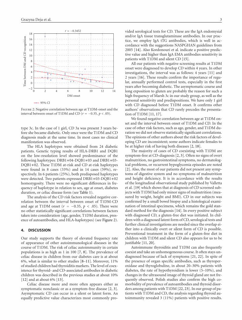
Figure 2: Negative correlation between age at T1DM-onset and theinterval between onset of T1DM and CD (r = −0.35, p < .05).
type 3c. In the case of 1 girl, CD 3a was present 3 years be-fore she became diabetic. Only once were the T1DM and CDdiagnosis made at the same time. In most cases no clinicalmanifestation was observed.
The HLA haplotypes were obtained from 24 diabeticpatients. Genetic typing results of HLA-DRB1 and DQB1on the low-resolution level showed predominance of thefollowing haplotypes: DRB1∗04-DQB1∗03 and DRB1∗03-DQB1∗02. These T1DM at-risk and CD at-risk haplotypeswere found in 8 cases (33%) and in 14 cases (59%), re-spectively. In 6 patients (25%), both predisposed haplotypeswere detected. Two patients expressed DRB1∗03-DQB1∗02homozygosity. There were no significant differences in fre-quency of haplotype in relation to sex, age at onset, diabetesduration, or celiac disease form (see Table 1).
The analysis of the CD risk factors revealed negative cor-relation between the interval between onset of T1DM-CDand age at T1DM onset (r = −0.35, p < .05). There wereno other statistically significant correlations between factorstaken into consideration (age, gender, T1DM duration, pres-ence of autoantibodies, and HLA-haplotypes) (see Figure 2).
4. DISCUSSION
Our study supports the theory of elevated frequency rateof appearance of other autoimmunological diseases in thecourse of T1DM. The risk of celiac autoimmunity in certainpopulations is as high as 1 in 100 [7, 8]. The prevalence ofceliac disease in children from our diabetes care is at about6%, what is similar to other studies [8–11]. Moreover, 11%of studied children had thyroiditis markers. The level of coex-istence for thyroid- and CD-associated antibodies in diabeticchildren was described in the previous studies at about 10%[12] and at about 6% [13].
Celiac disease more and more often appears either assymptomatic nonclassic or as a symptom-free disease [2, 3].Asymptomatic CD can occur in a silent or latent form. Anequally predictive value characterizes most commonly pro-
vided serological tests for CD. These are the IgA endomysialand/or IgA tissue transglutaminase antibodies. In our prac-tice, we employ IgA tTG antibodies, which is well in ac-cordance with the suggestions NASPGHAN guidelines from2005 [14]. Also Kordonouri et al. indicate a positive predic-tive value and higher than IgA EMA antibodies sensitivity inpatients with T1DM and silent CD [15].
All our patients with negative screening results at T1DMonset were diagnosed to develop CD within 4 years. In otherinvestigations, the interval was as follows: 4 years [11] and2 years [16]. These results confirm the importance of regu-lar, annually performed control tests, especially in the firstyears after becoming diabetic. The asymptomatic course andlong exposition to gluten are probably the reason for such ahigh frequency of Marsh 3c in our study group, as well as thepersonal sensitivity and predispositions. We have only 1 girlwith CD diagnosed before T1DM onset. It confirms otherauthors’ observations that CD rarely precedes the presenta-tion of T1DM [11, 17].
We found negative correlation between age at T1DM on-set and the interval between onset of T1DM and CD. In thecase of other risk factors, such as age, gender, and T1DM du-ration we did not observe statistically significant correlations.The opinions of other authors about the risk factors of devel-oping CD are inconsistent; some authors indicate females tobe at higher risk of having both diseases [2, 18].
The majority of cases of CD coexisting with T1DM aresymptom-free at CD-diagnosis [2, 3]. Often no signs of overtmalnutrition, no gastrointestinal symptoms, no dermatolog-ical problems, or recurrent hypoglycemia episodes are noted[2]. Also, the most of our patients did not present any symp-toms of digestive system and no symptoms of malnutritionand height deficiency. It is in accordance with the resultsof the longitudinal observational study published by Vallettaet al. [19] which shows that at diagnosis of CD screened sub-jects with T1DM had only minor signs of malnutrition (mea-sured by weight, height and BMI). CD in our subjects wasconfirmed by a small bowel biopsy and a histological exami-nation of intestinal specimens, which remains the gold stan-dard method for the diagnosis [14]. In every positive subjectwith diagnosed CD, a gluten-free diet was initiated. In chil-dren with a diagnosed latent form of CD, serological tests andfurther clinical investigations are needed since the overlap ei-ther into a clinically overt or silent form of CD is possible.Preventional treatment in the form of a gluten-free diet inchildren with T1DM and silent CD also appears for us to bejustifiable [11, 20].
Autoimmune thyroiditis and T1DM can also frequentlycoexist and take an unhomogeneous course. It often stays un-diagnosed because of lack of symptoms [21, 22]. In spite ofthe presence of organ specific antibodies, such as thyroper-oxidase and thyroglobuline, in about 20–30% patients withdiabetes, the rate of hypothyroidism is lower (5–10%), andchanges in the ultrasound image of thyroid gland are not fre-quently observed. Polish studies also confirm the high co-morbidity of prevalence of autoantibodies and thyroid disor-ders among patients with T1DM [22, 23]. In our group of pa-tients with T1DM and CD, the analysis regarding thyroid au-toimmunity revealed 3 (11%) patients with positive results.

4 Mediators of Inflammation
Table 1: Study group characteristics.
Gender (female = 1) Age at T1DM onset GADA IAA IA2A ATA ATG Biopsy HLA
1 0 11.5 − + + + + 3b DRB1∗03 DQB1∗02 DRB1∗04 DQB1∗03
2 1 2.50 + + + − − 3b DRB1 07 DQB1 02 DRB1∗04 DQB1∗03
3 1 5.50 + − + + − 3a DRB1∗03 DQB1∗02 DRB1∗16 DQB1∗05
4 0 5.00 − + − − − 0 DRB1∗03 DQB1∗02 DRB1∗04 DQB1∗03
5 0 10.00 + + − − − 1 —
6 1 5.50 + + − − − 0 —
7 0 2.50 + + + − − 3c DRB1∗03 DQB1∗03 DRB1∗04 DQB1∗02
8 1 5.50 − + + − − 3a DRB1 07 DQB1 03 DRB1 16 DQB1 06
9 1 7.00 + − − − − 3c DRB1∗03 DQB1∗02 DRB1∗04 DQB1∗03
10 0 3.00 + + + − − 0 DRB1∗03 DQB1∗02 DRB1∗04 DQB1∗03
11 1 2.00 + + + − − 3a DRB1 01 DQB1 02 DRB1 16 DQB1 03
12 1 3.00 − − + − − 0 —
13 1 8.00 + + − − − 3c DRB1∗03 DQB1∗03 DRB1∗04 DQB1∗02
14 0 1.50 + + + − − 3b DRB1∗04-DQB1∗03 DRB1 07 DQB1 02
15 0 2.00 − + − − − 3c DRB1∗03-DQB1∗02 DRB1 06 DQB1 05
16 1 2.50 + − + − − 0 DRB1∗03 DQB1∗02 DRB1∗09 DQB1∗02
17 1 9.00 − − − − − 3b DRB1∗03 DQB1∗02 DRB1∗04 DQB1∗03
18 0 5.00 + − + − − 3c DRB1 01 DQB1 04 DRB1∗03 DQB1∗02
19 1 8.50 + − + + − 3b DRB1∗011DQB1∗03 DRB1∗011DQB1∗02
20 0 10.00 − − + − − 3a DRB1∗03 DQB1∗02 DRB1∗07 DQB1∗03
21 1 6.50 + + + − − 3c DRB1∗03 DQB1∗02 DRB1∗13 DQB1∗03
22 1 2.50 + + + − − 3c DRB1∗03 DQB1∗03 DRB1∗04 DQB1∗02
23 0 8.50 + − + − − 3a DRB1∗03 DQB1∗03 DRB1∗04 DQB1∗02
24 0 11.00 + + − − − 3a DRB1∗03 DQB1∗02 DRB1∗03 DQB1∗02
25 0 9.00 + + + − − 3c DRB1∗03 DQB1∗02 DRB1∗03 DQB1∗02
26 0 8.50 + + + − − 3b DRB1∗07 DQB1∗03 DRB1∗15 DQB1∗06
27 1 12.00 + − − − − 3c DRB1∗03 DQB1∗03 DRB1∗04 DQB1∗02
Maybe such a small number of positive results is caused byour study inclusion criteria (having both disorders: T1DMand CD). The second possible explanation of this findingis the fact that children included to the study were ratheryoung (only 3 of them had the T1DM-onset above 10 years)and thyroid disorders usually developed in older age. In two(8%) of 27 children, the diagnosis of Hashimoto’s diseasewas made on the basis of the serum findings and ultrasoundimages. All these patients had negative screening results atT1DM onset and also were euthyroid and asymptomatic atthe time of diagnosis. Data coming from literature pointedthat the asymptomatic course of the disease is rather typicaland indicated the benefits of annual TSH screening to hy-pothyroidism development [24].
Similar to previously investigated Caucasian popula-tions, also in our study haplotypes DRB1∗04-DQB1∗03and DRB1∗03-DQB1∗02 revealed the predominance, sincethese are most strongly connected with T1DM haplotypes[25]. Nevertheless, in the studied group of children with co-existing CD and T1DM, we observed more frequent pres-ence of allele DQB1∗02 compared with DQB1∗03 (59% ver-sus 33%, resp.). Such predominance of allele DQB1∗02 hasnot been noted in our previous study concerning a geneticsusceptibility to T1DM in the same population (65% ver-
sus 62%, resp.) [26]. It confirms earlier observations that themost strongly connected with CD allele is DQB1∗02, whichis present in about 90% of CD patients [27, 28]. The highimportance of the allele DQB1∗02 in developing CD espe-cially confirms the Bao study [25]. In this study it was shownthat approximately 1 out of 3 patients with T1DM, who areDQB1∗02 homozygous, expresses tTG-antibodies, and halfof them have celiac disease on biopsy. In our study group,we had only 2 children expressing DQB1∗02 homozygosity.In both cases, the biopsy was positive: stage 3a and 3c wererecognized.
Diabetes-associated autoantibodies were found in mostsubjects at the time of T1DM presentation. Our group ofpatients is positive to all antibodies: GADA, IA2A, and IAA;(33%) seems to be rather large in comparison to a group pre-sented for example by Sabbah et al. (2, 1%) [29]. The expla-nation for such a large group of children positive to multi-ple autoantibodies could be the fact of having additionally2 or 3 autoimmune disorders and an individually increasedtendency to autoimmunization. Furthermore, we noted theGADA dominance in comparison to other markers. Prob-ably these both facts can result from more frequent pres-ence of the allele DRB1∗03 in the studied group. The alleleDRB1∗03 is known as the most classic haplotype connected
Grazyna Deja et al. 5
with a different form of autoimmunization and a generallyincreased tendency to produce autoantibodies. GADA is alsodetected more frequently and in higher titer in subjects withhaplotype DRB1∗03 [30, 31]. But because of a rather smallnumber of children comprising our study group, it is notpossible to make an obvious conclusion based on statisticallysignificant calculations. It indeed makes a limitation of ourstudy and indicates the necessity of continuation of our ob-servation to obtain results of more subjects with T1DM andCD.
5. CONCLUSION
In conclusion, the high-comorbidity ratio of CD and thy-roiditis in the course of T1DM, especially in a latent andsilent form, demand regular screening tests, especially in thefirst years after T1DM-onset. The increase in the numberof patients with T1DM and CD and longer observationsare needed to assess the risk factors influencing the time ofCD onset. It is especially important in the context that thenewest data coming from study performed in 7 big EuropeanDiabetic Centers confirmed rapidly the increasing trend ofprevalence of both disorders [32] and because of only a fewdata on the predictive value and risk factors of CD in T1DMpopulations published so far [33].
ACKNOWLEDGMENT
The project was partially supported by Polish State Commit-tee for Scientific Research (KBN) Grant no. 3T11F 010 29.
REFERENCES
[1] M. J. Redondo and G. S. Eisenbarth, “Genetic control of au-toimmunity in type I diabetes and associated disorders,” Dia-betologia, vol. 45, no. 5, pp. 605–622, 2002.
[2] N. Peretti, F. Bienvenu, C. Bouvet, et al., “The temporal rela-tionship between the onset of type 1 diabetes and celiac dis-ease: a study based on immunoglobulin a antitransglutami-nase screening,” Pediatrics, vol. 113, no. 5, pp. 418–422, 2004.
[3] I. De Vitis, G. Ghirlanda, and G. Gasbarrini, “Prevalence ofcoeliac disease in type I diabetes: a multicentre study,” ActaPaediatrica, vol. 85, no. s412, pp. 56–57, 1996.
[4] World Health Organization, “Definition, diagnosis and classi-fication of diabetes mellitus and its complications. Part 1: di-agnosis and classification of diabetes mellitus,” WHO, 1999.
[5] International Society for Pediatric Adolescent Diabetes, “IS-PAD consensus guidelines for the management of type 1 dia-betes mellitus,” Ziest, Medical Forum International, 2000.
[6] S. A. Miller, D. D. Dykes, and H. F. Polesky, “A simple salt-ing out procedure for extracting DNA from human nucleatedcells,” Nucleic Acids Research, vol. 16, no. 3, p. 1215, 1988.
[7] E. J. Hoffenberg, T. MacKenzie, K. J. Barriga, et al., “A prospec-tive study of the incidence of childhood celiac disease,” Journalof Pediatrics, vol. 143, no. 3, pp. 308–314, 2003.
[8] A. Tommasini, T. Not, V. Kiren, et al., “Mass screening forcoeliac disease using antihuman transglutaminase antibodyassay,” Archives of Disease in Childhood, vol. 89, no. 6, pp. 512–515, 2004.
[9] K. Karczewska, P. Jarosz-Chobot, G. Wiedermann, et al., “Co-incidence of type 1 diabetes mellitus and coeliac disease,”Przeglad Pediatrics, vol. 1, pp. 178–184, 1996.
[10] J. Crone, B. Rami, W. D. Huber, G. Granditsch, and E. Schober,“Prevalence of celiac disease and follow-up of EMA in childrenand adolescents with type 1 diabetes mellitus,” Journal of Pedi-atric Gastroenterology and Nutrition, vol. 37, no. 1, pp. 67–71,2003.
[11] G. Barera, R. Bonfanti, M. Viscardi, et al., “Occurrence ofceliac disease after onset of type 1 diabetes: a 6-year prospec-tive longitudinal study,” Pediatrics, vol. 109, no. 5, pp. 833–838, 2002.
[12] C. Jaeger, E. Hatziagelaki, R. Petzoldt, and R. G. Bretzel,“Comparative analysis of organ-specific autoantibodies andceliac disease—associated antibodies in type 1 diabetic pa-tients, their first-degree relatives, and healthy control sub-jects,” Diabetes Care, vol. 24, no. 1, pp. 27–32, 2001.
[13] Z. Sumnik, O. Cinek, N. Bratanic, et al., “Thyroid autoimmu-nity in children with coexisting type 1 diabetes mellitus andceliac disease: a multicenter study,” Journal of Pediatric En-docrinology and Metabolism, vol. 19, no. 4, pp. 517–522, 2006.
[14] I. D. Hill, M. H. Dirks, G. S. Liptak, et al., “Guideline for thediagnosis and treatment of celiac disease in children: recom-mendations of the North American Society for Pediatric Gas-troenterology, Hepatology and Nutrition,” Journal of PediatricGastroenterology and Nutrition, vol. 40, no. 1, pp. 1–19, 2005.
[15] O. Kordonouri, W. Dieterich, D. Schuppan, et al., “Autoanti-bodies to tissue transglutaminase are sensitive serological pa-rameters for detecting silent coeliac disease in patients withtype 1 diabetes mellitus,” Diabetic Medicine, vol. 17, no. 6, pp.441–444, 2000.
[16] T. Saukkonen, E. Savilahti, H. Reijonen, et al., “Coeliacdisease: frequent occurrence after clinical onset of insulin-dependent diabetes mellitus. Childhood Diabetes in FinlandStudy Group,” Diabetic Medicine, vol. 13, no. 5, pp. 464–470,1996.
[17] J. F. Ludvigsson, J. Ludvigsson, A. Ekbom, and S. M. Mont-gomery, “Celiac disease and risk of subsequent type 1 dia-betes: a general population cohort study of children and ado-lescents,” Diabetes Care, vol. 29, no. 11, pp. 2483–2488, 2006.
[18] J. M. Barker, J. Yu, L. Yu, et al., “Autoantibody “subspecificity”in type 1 diabetes: risk for organ-specific autoimmunity clus-ters in distinct groups,” Diabetes Care, vol. 28, no. 4, pp. 850–855, 2005.
[19] E. Valletta, D. Ulmi, I. Mabboni, F. Tomasselli, and L. Pinelli,“Early diagnosis and treatment of celiac disease in type 1 dia-betes. A longitudinal, case-control study,” La Pediatria Medicae Chirurgica, vol. 29, no. 2, pp. 99–104, 2007.
[20] D. Hansen, B. Brock-Jacobsen, E. Lund, et al., “Clinical benefitof a gluten-free diet in type 1 diabetic children with screening-detected celiac disease: a population-based screening studywith 2 years’ follow-up,” Diabetes Care, vol. 29, no. 11, pp.2452–2456, 2006.
[21] M. Muchacka-Bianga, G. Deja, P. Jarosz-Chobot, and B.Koehler, “Thyroid disorders in children suffering from poorlycontrolled type 1 diabetes mellitus,” Diabetologia Polska, vol. 7,no. 2, pp. 104–107, 2000.
[22] E. Czerniawska, M. Szalecki, E. Piatkowska, et al., “Prevalenceof thyroid antibodies (TPO and ATG) at the onset of type 1diabetes mellitus in children treated in two diabetes centres inŁodz and Kielce,” Medycyna Wieku Rozwojowego, vol. 7, no. 2,pp. 224–227, 2003.

6 Mediators of Inflammation
[23] L. Machnica, A. Osior, P. Jarosz-Chobot, G. Deja, J. Polanska,and E. Otto-Buczkowska, “An analysis of the prevalence of thy-roid autoantibodies: thyroid peroxidase antibodies (ATA) andthyroglobulin antibodies (ATG) in children with newly diag-nosed diabetes mellitus type 1 during 2000–2004 in the UpperSilesia region, Poland,” Acta Diabetologica, vol. 45, no. 1, pp.37–40, 2008.
[24] S. J. Glastras, M. E. Craig, C. F. Verge, A. K. Chan, J. M.Cusumano, and K. C. Donaghue, “The role of autoimmunityat diagnosis of type 1 diabetes in the development of thyroidand celiac disease and microvascular complications,” DiabetesCare, vol. 28, no. 9, pp. 2170–2175, 2005.
[25] F. Bao, L. Yu, S. Babu, et al., “One third of HLA DQ2 ho-mozygous patients with type 1 diabetes express celiac disease-associated transglutaminase autoantibodies,” Journal of Au-toimmunity, vol. 13, no. 1, pp. 143–148, 1999.
[26] G. Deja, P. Jarosz-Chobot, J. Polanska, U. Siekiera, and E.Malecka-Tendera, “Is the association between TNF-α-308 Aallele and T1DM independent of HLA-DRB1, DQB1 alle-les?” Mediators of Inflammation, vol. 2006, Article ID 19724,7 pages, 2006.
[27] G. Contreas, E. Valletta, D. Ulmi, S. Cantoni, and L. Pinelli,“Screening of coeliac disease in North Italian children withtype 1 diabetes: limited usefulness of HLA-DQ typing,” ActaPaediatrica, vol. 93, no. 5, pp. 628–632, 2004.
[28] A. Doolan, K. Donaghue, J. Fairchild, M. Wong, and A. J.Williams, “Use of HLA typing in diagnosing celiac disease inpatients with type 1 diabetes,” Diabetes Care, vol. 28, no. 4, pp.806–809, 2005.
[29] E. Sabbah, K. Savola, P. Kulmala, et al., “Diabetes-associatedautoantibodies in relation to clinical characteristics and natu-ral course in children with newly diagnosed type 1 diabetes.The Childhood Diabetes in Finland Study Group,” Journalof Clinical Endocrinology and Metabolism, vol. 84, no. 5, pp.1534–1539, 1999.
[30] M. Knip, M. Kukko, P. Kulmala, et al., “Humoral beta-cell au-toimmunity in relation to HLA-defined disease susceptibilityin preclinical and clinical type 1 diabetes,” American Journal ofMedical Genetics, vol. 115, no. 1, pp. 48–54, 2002.
[31] G. Deja, P. Jarosz-Chobot, M. Muchacka-Bianga, et al., “Hu-moral autoimmunity in relation to HLA genotype in childrenwith newly diagnosed diabetes mellitus type 1,” DiabetologiaPolska, vol. 11, no. 1, pp. 12–16, 2004.
[32] O. Cinek, M. Kulich, G. Deja, et al., “The risk of celiac diseaseamong children with diabetes has increased three-fold over thelast ten years,” Pediatric Diabetes, p. 15, 2007.
[33] L. C. de Graaff, J. W. A. Smit, and J. K. Radder, “Prevalence andclinical significance of organ-specific autoantibodies in type1 diabetes mellitus,” Netherlands Journal of Medicine, vol. 65,no. 7, pp. 235–247, 2007.

Submit your manuscripts athttp://www.hindawi.com
Stem CellsInternational
Hindawi Publishing Corporationhttp://www.hindawi.com Volume 2014
Hindawi Publishing Corporationhttp://www.hindawi.com Volume 2014
MEDIATORSINFLAMMATION
of
Hindawi Publishing Corporationhttp://www.hindawi.com Volume 2014
Behavioural Neurology
EndocrinologyInternational Journal of
Hindawi Publishing Corporationhttp://www.hindawi.com Volume 2014
Hindawi Publishing Corporationhttp://www.hindawi.com Volume 2014
Disease Markers
Hindawi Publishing Corporationhttp://www.hindawi.com Volume 2014
BioMed Research International
OncologyJournal of
Hindawi Publishing Corporationhttp://www.hindawi.com Volume 2014
Hindawi Publishing Corporationhttp://www.hindawi.com Volume 2014
Oxidative Medicine and Cellular Longevity
Hindawi Publishing Corporationhttp://www.hindawi.com Volume 2014
PPAR Research
The Scientific World JournalHindawi Publishing Corporation http://www.hindawi.com Volume 2014
Immunology ResearchHindawi Publishing Corporationhttp://www.hindawi.com Volume 2014
Journal of
ObesityJournal of
Hindawi Publishing Corporationhttp://www.hindawi.com Volume 2014
Hindawi Publishing Corporationhttp://www.hindawi.com Volume 2014
Computational and Mathematical Methods in Medicine
OphthalmologyJournal of
Hindawi Publishing Corporationhttp://www.hindawi.com Volume 2014
Diabetes ResearchJournal of
Hindawi Publishing Corporationhttp://www.hindawi.com Volume 2014
Hindawi Publishing Corporationhttp://www.hindawi.com Volume 2014
Research and TreatmentAIDS
Hindawi Publishing Corporationhttp://www.hindawi.com Volume 2014
Gastroenterology Research and Practice
Hindawi Publishing Corporationhttp://www.hindawi.com Volume 2014
Parkinson’s Disease
Evidence-Based Complementary and Alternative Medicine
Volume 2014Hindawi Publishing Corporationhttp://www.hindawi.com