Etiologies and Predictors of Diagnosis in Nonresponsive Celiac Disease
6
Etiologies and Predictors of Diagnosis in Nonresponsive Celiac Disease DANIEL A. LEFFLER, MELINDA DENNIS, BRIAN HYETT, EOIN KELLY, DETLEF SCHUPPAN, and CIARAN P. KELLY Department of Gastroenterology, Beth Israel Deaconess Medical Center, Boston, Massachusetts See Barone MV et al on page 1245 for companion article in the April 2007 issue of Gastroenterology. Background & Aims: Nonresponsive celiac disease (NRCD) is a common problem affecting from 7% to 30% of celiac patients. Because NRCD comprises varied and poten- tially morbid entities, efficient and cost-effective patient care requires knowledge of the specific causes of this dis- order. The aim of this study was to determine the common etiologies of NRCD in a tertiary referral center. Methods: All cases of biopsy examination–proven celiac disease (CD) seen at our institution over the preceding 5 years were included in this study. NRCD was defined as a failure to respond to at least 6 months of treatment with a gluten-free diet or the re-emergence of symptoms or laboratory abnor- malities typical of CD while still on treatment with a gluten- free diet. Results: A total of 113 patients with NRCD meeting the earlier-described criteria were seen from a total of 603 patients with CD (19%), however, among patients for whom we provided primary specialist care the incidence of NRCD was 10% (P < .001). Gluten expo- sure was the most common cause of NRCD (36%), fol- lowed by irritable bowel syndrome (22%), refractory CD (10%), lactose intolerance (8%), and microscopic colitis (6%). The mean immunoglobulin A tissue transglutami- nase level in the gluten-exposed group was 67 vs 17 U/mL (normal, <20) for other diagnoses (P < .05). Weight loss and male sex were highly predictive of refractory CD (P < .05 and < .001, respectively). Conclusions: NRCD is a common phenomenon affecting 10%–19% of celiac patients. A limited number of etiologies account for the majority of cases. Clinical factors may be used to guide evaluation. C eliac disease (CD) is a small-intestinal inflammatory dis- ease defined by characteristic histologic changes including villous atrophy and increases in intraepithelial lymphocytes. CD is triggered by gluten proteins from wheat, rye, and barley, in genetically predisposed individuals who carry the human lymphocyte antigen (HLA)-DQ2 or -DQ8. 1 Mostly because of the availability of accurate serology-based tests, 2–5 increasing numbers of individuals are being diagnosed with CD, therefore clinicians must be aware of the frequency and common etiolo- gies of incomplete response to gluten withdrawal. Although the majority of individuals with CD have substan- tial improvement within the first few weeks of gluten with- drawal, between 7% and 30% continue to have symptoms or clinical manifestations suggestive of CD despite being on a gluten-free diet. 6,7 This clinical problem, which encompasses many distinct diagnoses, is known as nonresponsive celiac dis- ease (NRCD). NRCD may be defined further as primary if there is initial failure to respond to a gluten-free diet or secondary if signs, symptoms, or laboratory abnormalities consistent with CD re-emerge after initial normalization while maintaining a gluten-free diet (Table 1). In smaller studies the most common cause of NRCD was unintentional gluten intake, accounting for approximately 50% of cases of NRCD, 6,8 but etiologies can vary greatly to include lym- phoma, 9 small-intestinal bacterial overgrowth (SIBO), 10,11 micro- scopic colitis, 12 pancreatic insufficiency, 8,12 disaccharidase defi- ciency, 12 and irritable bowel syndrome (IBS), 8 all of which may present similarly but require very different therapies. Because NRCD comprises a number of potentially severe conditions with disparate treatment and prognosis, efficient and cost-effective care of patients with this syndrome may be challenging. In contrast to prior investigations, our data allow differen- tiation of patients referred from outside practices and those receiving primary gastroenterology care though our referral center. In addition, the large number of consecutive patients evaluated allows for a more definitive determination of clinical characteristics, especially as correlated to refractory CD. We sought, therefore, to better define the prevalence of NRCD in current clinical practice in the United States, to identify the range of specific etiologies for this disorder, and to determine which, if any, clinical factors are predictive of the final etiology. Methods A database of all patients seen at our institution from January 1, 2000, to April 1, 2006, coded for CD under the International Classification of Diseases 9th edition code 579.0 was compiled and predetermined clinical data were recorded. From this list, 603 patients were found to have biopsy exami- nation–proven CD. Individuals without definitive evidence of CD in the form of duodenal biopsy examination, or skin biopsy examination in cases of dermatitis herpetiformis, were not included in this study. HLA typing was performed for patients in whom there was doubt regarding the validity of the diagno- sis. Analysis of tissue transglutaminase (tTG) titers was per- formed by enzyme-linked immunosorbent assay with recombi- nant human antigen (INOVA Quanta Lite human-tTG immunoglobulin [Ig]A; San Diego, CA: sensitivity, 94%; speci- ficity, 99%). For patients with CD, electronic medical records that included all clinician notes, laboratory data, and results of Abbreviations used in this paper: CD, celiac disease; EATL, enterop- athy associated T-cell lymphoma; ELISA, enzyme-linked immunosor- bent assay; HLA, human lymphocyte antigen; IBS, irritable bowel syndrome; Ig, immunoglobulin; NRCD, nonresponsive celiac disease; SIBO, small intestinal bacterial overgrowth; tTG, tissue transglutami- nase. © 2007 by the AGA Institute 1542-3565/07/$32.00 doi:10.1016/j.cgh.2006.12.006 CLINICAL GASTROENTEROLOGY AND HEPATOLOGY 2007;5:445– 450
-
Upload
hms-harvard -
Category
Documents
-
view
1 -
download
0
Transcript of Etiologies and Predictors of Diagnosis in Nonresponsive Celiac Disease

E
D
D
B(ctcoeAsirdmfmtptsl((n(a.cAc
CvCiltncg
tdcgm
CLINICAL GASTROENTEROLOGY AND HEPATOLOGY 2007;5:445– 450
tiologies and Predictors of Diagnosis in Nonresponsive Celiac Disease
ANIEL A. LEFFLER, MELINDA DENNIS, BRIAN HYETT, EOIN KELLY, DETLEF SCHUPPAN, and CIARAN P. KELLY
epartment of Gastroenterology, Beth Israel Deaconess Medical Center, Boston, Massachusetts
eisCg
ucpscpNdo
trcecscrw
JIwFnCeiisfnifit
absSn
See Barone MV et al on page 1245 forcompanion article in the April 2007 issue ofGastroenterology.
ackground & Aims: Nonresponsive celiac diseaseNRCD) is a common problem affecting from 7% to 30% ofeliac patients. Because NRCD comprises varied and poten-ially morbid entities, efficient and cost-effective patientare requires knowledge of the specific causes of this dis-rder. The aim of this study was to determine the commontiologies of NRCD in a tertiary referral center. Methods:ll cases of biopsy examination–proven celiac disease (CD)
een at our institution over the preceding 5 years werencluded in this study. NRCD was defined as a failure toespond to at least 6 months of treatment with a gluten-freeiet or the re-emergence of symptoms or laboratory abnor-alities typical of CD while still on treatment with a gluten-
ree diet. Results: A total of 113 patients with NRCDeeting the earlier-described criteria were seen from a
otal of 603 patients with CD (19%), however, amongatients for whom we provided primary specialist care
he incidence of NRCD was 10% (P < .001). Gluten expo-ure was the most common cause of NRCD (36%), fol-owed by irritable bowel syndrome (22%), refractory CD10%), lactose intolerance (8%), and microscopic colitis6%). The mean immunoglobulin A tissue transglutami-ase level in the gluten-exposed group was 67 vs 17 U/mL
normal, <20) for other diagnoses (P < .05). Weight lossnd male sex were highly predictive of refractory CD (P <
05 and < .001, respectively). Conclusions: NRCD is aommon phenomenon affecting 10%–19% of celiac patients.
limited number of etiologies account for the majority ofases. Clinical factors may be used to guide evaluation.
eliac disease (CD) is a small-intestinal inflammatory dis-ease defined by characteristic histologic changes including
illous atrophy and increases in intraepithelial lymphocytes.D is triggered by gluten proteins from wheat, rye, and barley,
n genetically predisposed individuals who carry the humanymphocyte antigen (HLA)-DQ2 or -DQ8.1 Mostly because ofhe availability of accurate serology-based tests,2–5 increasingumbers of individuals are being diagnosed with CD, thereforelinicians must be aware of the frequency and common etiolo-ies of incomplete response to gluten withdrawal.
Although the majority of individuals with CD have substan-ial improvement within the first few weeks of gluten with-rawal, between 7% and 30% continue to have symptoms orlinical manifestations suggestive of CD despite being on aluten-free diet.6,7 This clinical problem, which encompasses
any distinct diagnoses, is known as nonresponsive celiac dis-ase (NRCD). NRCD may be defined further as primary if theres initial failure to respond to a gluten-free diet or secondary ifigns, symptoms, or laboratory abnormalities consistent withD re-emerge after initial normalization while maintaining aluten-free diet (Table 1).
In smaller studies the most common cause of NRCD wasnintentional gluten intake, accounting for approximately 50% ofases of NRCD,6,8 but etiologies can vary greatly to include lym-homa,9 small-intestinal bacterial overgrowth (SIBO),10,11 micro-copic colitis,12 pancreatic insufficiency,8,12 disaccharidase defi-iency,12 and irritable bowel syndrome (IBS),8 all of which mayresent similarly but require very different therapies. BecauseRCD comprises a number of potentially severe conditions withisparate treatment and prognosis, efficient and cost-effective caref patients with this syndrome may be challenging.
In contrast to prior investigations, our data allow differen-iation of patients referred from outside practices and thoseeceiving primary gastroenterology care though our referralenter. In addition, the large number of consecutive patientsvaluated allows for a more definitive determination of clinicalharacteristics, especially as correlated to refractory CD. Weought, therefore, to better define the prevalence of NRCD inurrent clinical practice in the United States, to identify theange of specific etiologies for this disorder, and to determinehich, if any, clinical factors are predictive of the final etiology.
MethodsA database of all patients seen at our institution from
anuary 1, 2000, to April 1, 2006, coded for CD under thenternational Classification of Diseases 9th edition code 579.0as compiled and predetermined clinical data were recorded.rom this list, 603 patients were found to have biopsy exami-ation–proven CD. Individuals without definitive evidence ofD in the form of duodenal biopsy examination, or skin biopsy
xamination in cases of dermatitis herpetiformis, were notncluded in this study. HLA typing was performed for patientsn whom there was doubt regarding the validity of the diagno-is. Analysis of tissue transglutaminase (tTG) titers was per-ormed by enzyme-linked immunosorbent assay with recombi-ant human antigen (INOVA Quanta Lite human-tTG
mmunoglobulin [Ig]A; San Diego, CA: sensitivity, 94%; speci-city, 99%). For patients with CD, electronic medical recordshat included all clinician notes, laboratory data, and results of
Abbreviations used in this paper: CD, celiac disease; EATL, enterop-thy associated T-cell lymphoma; ELISA, enzyme-linked immunosor-ent assay; HLA, human lymphocyte antigen; IBS, irritable bowelyndrome; Ig, immunoglobulin; NRCD, nonresponsive celiac disease;IBO, small intestinal bacterial overgrowth; tTG, tissue transglutami-ase.
© 2007 by the AGA Institute1542-3565/07/$32.00
doi:10.1016/j.cgh.2006.12.006

dtCNscbr
idtwn
pppcetwa
etciodoogttcRtismo
edl
ssf
ttat
(Pvnc
C
frrf3onTtpr2tFt
ncpbsdmopcf
plmoator8iw4n
1tc
T
446 LEFFLER ET AL CLINICAL GASTROENTEROLOGY AND HEPATOLOGY Vol. 5, No. 4
iagnostic tests preformed at our hospital and affiliated insti-utions (Beth Israel Deaconess Medical Center, Joslin Diabetesenter, New England Baptist Hospital, Beth Israel Deaconesseedham, and Beth Israel Deaconess Nashoba) then were
earched individually for evidence of NRCD and prespecifiedlinical characteristics, including age at diagnosis, sex, comor-id diseases, and initial IgA anti-tTG level. All entries wereeviewed twice for accuracy.
NRCD was defined as: (1) referral to a clinician specializingn CD for the evaluation of a lack of response to a gluten-freeiet, (2) failure of clinical symptoms or laboratory abnormali-ies typical of CD to improve within 6 months of glutenithdrawal, (3) recurrence of symptoms and/or laboratory ab-ormalities typical of CD while on a gluten-free diet (Table 1).
Refractory CD was defined as the persistence of villous atro-hy despite strict gluten withdrawal and no evidence of anotherathology including overt lymphoma.13 In addition, selectedatients believed to be at high risk were evaluated for T-celllonality and aberrant T-cell markers, which suggest the pres-nce of more severe type II refractory CD vs the more indolentype I refractory CD.14,15 For analysis, cases of refractory CDere grouped with ulcerative jejunitis and enteropathy-associ-ted T-cell lymphoma (EATL).
Gluten exposure was determined to be the cause if, duringvaluation, likely sources of gluten were elicited and the removal ofhese sources led to sustained clinical improvement. Lymphocyticolitis was diagnosed in individuals with a grossly normal–appear-ng colon and more than 20 lymphocytes per 100 epithelial cellsn colonic biopsy examination.16 Disaccharidase deficiency wasiagnosed by clinical history with sustained symptom resolutionn removal of the specific sugar. SIBO was diagnosed in the settingf significantly increased hydrogen or methane excretion duringlucose or lactulose breath testing with sustained clinical responseo antibiotic therapy.17 Eating disorders were defined according tohe Diagnostic and Statistical Manual of Mental Disorders-IV18
riteria. IBS was diagnosed when patients had symptoms meetingome criteria19 in the absence of red flag signs and symptoms and
he presence of normal duodenal biopsy specimens. Pancreaticnsufficiency was diagnosed when individuals had sustained remis-ion of symptoms in response to the pancreatic enzyme replace-
ent regimen chosen by the treating physician. Other diagnoses inur population were made according to established criteria.20–24
With the exception of terminal diseases, diagnosis was consid-red final if the patient experienced a durable clinical response toirected therapy. Patients seen only a single time were considered
ost to follow-up evaluation and no diagnosis was recorded.A total of 113 consecutive patients meeting the earlier-de-
cribed criteria were identified. All patients were evaluated by akilled nutritionist trained in CD. Diagnostic testing was per-
able 1. Diagnostic Criteria for NRCD
Symptoms/signsFatigueAbdominal painDiarrheaWeight loss
Laboratory abnormalitiesAnemiatTG�50% above normal limit
ormed according to clinical necessity and the evaluation con- (
inued until a secure diagnosis was reached. In most instanceshis was followed by subsequent improvement in symptomsnd normalization of laboratory abnormalities in cases of non-erminal disease.
Statistical analysis was completed using SPSS for Windowsrelease 13.0; SPSS Inc., Chicago, IL). A 2-sample t test, theearson �2 test, and the Fisher exact test were used to compareariables between groups of patients with specific final diag-oses and between NRCD and responsive CD. Results wereonsidered significant with a P value of less than .05.
This study was reviewed and approved by the Committee forlinical Investigations at Beth Israel Deaconess Medical Center.
ResultsA total of 113 of 603 (18.7%) individuals with CD were
ound to have NRCD. Of the 113 patients, 74 (65%) wereeferred by clinicians from outside our institution and theemaining 39 (35%) patients were cared for at our institutionrom the initial diagnosis of CD onward. NRCD accounted for5% of the 211 new referrals to our celiac center. The majorityf the remaining referrals were for either confirmation of diag-osis or general management of previously diagnosed CD.hirty-nine of the 392 patients (9.9%) with CD who received
heir primary care at our institution developed NRCD com-ared with 35% of referrals (P � .001). Of the individualseceiving primary care at our institution who developed NRCD,5 (64%) were primary in onset. The overall duration of symp-oms at initial evaluation was 15.3 months (range, 1–138 mo).or primary and secondary nonresponse the duration of symp-oms before evaluation was 31.7 and 7.7 months, respectively.
The mean age at onset of NRCD was 42.2 years, which wasot significantly different from the mean age at diagnosis of allases of CD in our population. Gender distribution and therevalence of comorbid autoimmune disorders were similaretween groups. In individuals with NRCD, there was a non-ignificant increase in the prevalence of psychiatric disorders,efined by past diagnosis with one or more mental illnesses,ost commonly anxiety and depression, or current prescription
f psychoactive medication (Table 2). Of the 34 individuals withsychiatric diagnoses, 22 (64.7%) had B12 and folate levelshecked. Two (9%) were found to have low B12 levels, whereasolate levels were normal in all individuals.
The reliability of diagnosis is based on repeated visits withersistent clinical improvement. The mean duration of fol-
ow-up evaluation was 19.8 months (range, 2–126 mo), theean number of visits was 5.8 (range, 2–30) Individuals seen
nly for a single visit were considered lost to follow-up evalu-tion with no confirmed diagnosis. In this cohort of 113 pa-ients, 14 did not meet the earlier-described criteria at the timef review and were not included in further analysis. Of theemaining 99 individuals, IgA anti-tTG titers were available for9 patients at the initial evaluation. Of these, 38 patients had
ncreased titers, with levels greater than 20 (42.7%). The groupith increased IgA anti-tTG had been on the diet for a mean of9.2 months compared with 60.5 months for the patients withormal anti-tTG titers (P � .50).
Among the 99 with confirmed diagnoses, we found a total of2 etiologies of NRCD. The most common cause was (inadver-ent) gluten exposure, accounting for 36% of patients. Otherommon etiologies of NRCD included IBS (22%), refractory CD
10%), lactose deficiency (8%), SIBO (6%), and microscopic co-
lpgntlaie
trritgvtaii
gU1adabrs
cacoasmctgsdgrr
rrnhhaiptf
T
M%I
%
%
%
a
b
R
T
GIMRD
ESM
Da
b
c
i
April 2007 DIAGNOSIS IN NRCD 447
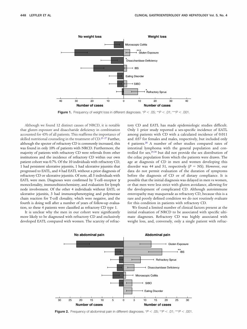
itis (6%). The remaining 13% consisted of eating disorders,eptic ulcer disease, gastroparesis, Crohn’s disease, food aller-ies, common variable immune deficiency, and duodenal ade-ocarcinoma. Predominant symptoms in nonresponsive pa-ients included diarrhea (54%), abdominal pain (55%), weightoss (20%), and, to a lesser extent, fatigue (5%). Laboratorybnormalities prompting evaluation were found in 10% andncluded persistent increases in IgA anti-tTG autoantibody lev-ls and iron-deficiency anemia.
A limited number of factors were found to be predictive ofhe final diagnosis in NRCD. The most significant of these wasecent weight loss per patient report, which was predictive ofefractory CD with an odds ratio of 31.1 (95% confidencenterval, 5.9 –163.1). An increased IgA anti-tTG titer greaterhan 20 U/mL (the upper limit of normal) was predictive ofluten exposure at an odds ratio of 11.3 (95% confidence inter-al, 3.7–34.4) and a mean of 67 U/mL (range, 2–135 U/mL). Ofhe 35 patients with persistent gluten exposure, anti-tTG titerst evaluation were available for 28. Of these, 22 (78%) werencreased with tTG levels greater than 20. The absence of abdom-nal pain also was associated with gluten exposure (P � .01).
able 2. Characteristics of Nonresponsive vs ResponsiveCD
Nonresponsive(n � 113)
Responsive(n � 490) P
ean age at diagnosis, y 42.2 44.0 .29Female 79.6 70.3 .11
nitial IgA tTG (ELISAunits)
114.7 103.6 .50
Comorbid psychiatricconditiona
31.9 23.7 .09
Other autoimmunedisorderb
31.0 23.9 .15
Outside referral 64.6 27.6 �.0001
Predominantly depression and anxiety.Predominantly thyroid disease, type 1 diabetes mellitus, andaynaud’s phenomenon.
able 3. Summary of Final Diagnoses and Associated Charac
DiagnosisNumber ofpatients Mean tTG
Male (%),n � 21
luten exposure 35 67a 9 (43)BS 22 9 1 (5)icroscopic colitis 6 7 0 (0)efractory sprueb 10 45 6 (29)isaccharidasedeficiency
8 16 1 (5)
ating disorder 4 34 0 (0)IBO 6 25 2 (9)iscellaneousc 8 14 2 (10)
, diarrhea; A, abdominal discomfort; W, weight loss.P � .05 vs combined other diagnoses.Includes refractory sprue, ulcerative duodenitis/jejunitis, and small-Duodenal adenocarcinoma (1), diabetic gastroparesis (1), Crohn’s
mmune deficiency (1).Beyond the gluten-exposure group, a mean IgA anti-tTG titerreater than 20 U/mL also was found in refractory CD (mean, 45/mL; range, 3–127 U/mL) and SIBO (mean, 25 U/mL; range,–82 U/mL). IBS was found in 22 of 99 NRCD patients and wasssociated with the presence of abdominal pain and the absence ofiarrhea (P � .01). Microscopic colitis and SIBO also were associ-ted with diarrhea (P � .05). Female patients had a greater risk ofeing diagnosed with IBS (P � .04), but a decreased risk ofefractory CD (P � .006) (Table 3). Comparisons of presentingymptoms across diagnoses can be found in Figures 1–3.
DiscussionIn this study we report the incidence, etiology, and
linical characteristics of 113 consecutive NRCD patients seent our center over the preceding 5 years. Our findings areonsistent with those of other reports; however, the larger sizef our patient population allows for increased statistical powernd for us to make diagnostic predictions based on presentingymptoms. As in past studies,8 gluten exposure remains the
ost common cause of NRCD and is the only diagnosticategory independently associated with an increased IgA anti-TG titer. It is difficult to classify purposeful vs inadvertentluten exposure. During the study period, patients were in-tructed to avoid all oat products and the majority of patientsiagnosed with gluten exposure did not have gross blatantluten intake, but rather had not been adequately diligent inemoving sources of cross-contamination or hidden gluten inestaurant foods, medications, or cosmetics.
Although gluten exposure is prominent, the 35% prevalenceate seen in this study was lower than the 50% previouslyeported.8 Although this difference is not yet statistically sig-ificant (P � .06) and may be a chance finding, the Boston areaas a very large and active CD advocacy group and a number ofighly skilled celiac nutritionists. These factors may account forrelatively good adherence to a gluten-free diet, which may vary
n different regions. This view is supported by the much lowerrevalence of NRCD in those patients who were treated in ourertiary center initially compared with patients who were re-erred to us (9.9% vs 39%, P � .05).
tics in NRCD
le (%),78
Predominant symptoms(%)
Primary nonresponse
Male(%)
Female(%)
33) D (55) A (35)a W (15) 4/9 (44) 18/26 (69)27)a D (34)a A (91)a W (5) 1/1 (100) 13/21 (62)8) D (100)a A (17) W (0) N/A 2/6 (33)5)a D (70) A (80) W (90)a 3/6 (50) 2/4 (50)9) D (67) A (67) W (0) 0/1 (0) 7/7 (100)
5) D (50) A (100) W (0) N/A 4/4 (100)5) D (100)a A (67) W (33) 1/2 (50) 2/4 (50)8) N/A N/A N/A
inal lymphoma.se (1), peptic ulcer disease (2), food allergy (2), common variable
teris
Feman �
26 (21 (6 (4 (7 (
4 (4 (6 (
intestdisea
tasawmip1prEmnucft
md
tOaa4ittaddbpoterf
imw
ent d
448 LEFFLER ET AL CLINICAL GASTROENTEROLOGY AND HEPATOLOGY Vol. 5, No. 4
Although we found 12 distinct causes of NRCD, it is notablehat gluten exposure and disaccharide deficiency in combinationccounted for 43% of all patients. This reaffirms the importance ofkilled nutritional counseling in the treatment of CD.25–27 Further,lthough the specter of refractory CD is commonly increased, thisas found in only 10% of patients with NRCD. Furthermore, theajority of patients with refractory CD were referrals from other
nstitutions and the incidence of refractory CD within our ownatient cohort was 0.7%. Of the 10 individuals with refractory CD,had persistent ulcerative jejunitis, 1 had ulcerative jejunitis that
rogressed to EATL, and 4 had EATL without a prior diagnosis ofefractory CD or ulcerative jejunitis. Of note, all 5 individuals withATL were men. Diagnoses were confirmed by T-cell–receptor �onoclonality, immunohistochemistry, and evaluation for lymph
ode involvement. Of the other 4 individuals without EATL orlcerative jejunitis, 3 had immunophenotyping and polymerasehain reaction for T-cell clonality, which were negative, and theourth is doing well after a number of years of follow-up evalua-ion, so these 4 patients were classified as refractory CD type 1.
It is unclear why the men in our cohort were significantlyore likely to be diagnosed with refractory CD and exclusively
eveloped EATL compared with women. The scarcity of refrac-
Figure 1. Frequency of weight loss in differ
Figure 2. Frequency of abdominal pain in different
ory CD and EATL has made epidemiologic studies difficult.nly 1 prior study reported a sex-specific incidence of EATL
mong patients with CD with a calculated incidence of 0.011nd .037 for females and males, respectively, but included only
patients.28 A number of other studies compared rates ofntestinal lymphoma with the general population and con-rolled for sex,29,30 but did not provide the sex distribution ofhe celiac population from which the patients were drawn. Thege at diagnosis of CD in men and women developing thisisorder was 44 and 51, respectively (P � NS). However, ourata do not permit evaluation of the duration of symptomsefore the diagnosis of CD or of dietary compliance. It isossible that the initial diagnosis was delayed in men vs women,r that men were less strict with gluten avoidance, allowing forhe development of complicated CD. Although autoimmunenteropathy may masquerade as refractory CD, because this is aare and poorly defined condition we do not routinely evaluateor this condition in patients with refractory CD.
We found a limited number of clinical factors present at thenitial evaluation of NRCD to be associated with specific ulti-
ate diagnoses. Refractory CD was highly associated witheight loss, and, conversely, only a single patient with refrac-
iagnoses. �P � .05; ��P � .01; ���P � .001.
diagnoses. �P � .05; ��P � .01; ���P � .001.

twcarpCtss
SImtbiIapeabuatbcspipo
ssbmcia
dscpuddcen
vdgbNdAtise
wnbtsbcweacpbpv
p
nt dia
April 2007 DIAGNOSIS IN NRCD 449
ory CD did not present with this sign. The IgA anti-tTG titeras increased significantly only in the gluten-exposure group
ompared with all other groups and this finding should triggersearch for inadvertent gluten exposure. Notably, although not
eaching statistical significance, the only other diagnoses thatresented with an increased tTG level were SIBO and refractoryD. The reasons for this are unclear and warrant further inves-
igation, although we hypothesize that nonspecific immunetimulation of small intestinal mucosal B and/or T cells mayustain antibody production.
Diarrhea was present in all cases of microscopic colitis andIBO but was significantly less common in individuals withBS. The lack of diarrhea in NRCD patients diagnosed with IBS
ay stem from the well-documented fiber deficiency in theypical gluten-free diet,31 however, it also is notable that the lineetween SIBO and IBS has blurred recently32 and it may be that
ndividuals who in the past would have been diagnosed withBS now appear more consistent with SIBO. IBS also wasssociated commonly with abdominal pain whereas gluten ex-osure was not. The lack of abdominal pain in the gluten-xposure group, which differs from the common complaint ofbdominal pain among newly diagnosed CD patients,33,34 maye explained by the fact that most patients had low-level,nintentional exposure, which may be enough to cause diarrheand/or laboratory abnormalities without causing pain. All pa-ients underwent esophagogastroduodenoscopy/colonoscopyefore diagnosis with IBS. Other imaging modalities includingapsule endoscopy, computerized tomography scans, and ultra-ounds were performed according to clinical acumen. Of the 22atients with IBS, 15 (68%) underwent imaging studies, includ-
ng 9 small-bowel follow-throughs, 8 computerized tomogra-hy scans, 6 ultrasounds, and 1 magnetic resonance imaging, allf which were, by definition, normal.
No diagnostic group was significantly different in primary vsecondary nonresponse, however, disaccharidase deficiency pre-ented only in primary NRCD. This is plausible because brush-order enzymes are reconstituted with healing of the intestinalucosa in treated celiac patients. This finding supports the
ommon practice of limiting lactose intake in CD during thenitial weeks to months of dietary therapy.12,35 In addition,
Figure 3. Frequency of diarrhea in differe
lthough approximately 20 individuals are known to have un- T
ergone a therapeutic trial of pancreatic exocrine hormoneupplementation, none responded to this treatment, whichontrasts with other studies of NRCD.8,12 Whether clinicalancreatic exocrine sufficiency is associated with treated orntreated CD currently is unclear and needs to be addressedefinitively in future studies. Finally, eating disorders wereetermined to be the cause of NRCD in 4 patients. While theo-existence of eating disorders with CD has been reported,36,37
ating disorders as a cause of NRCD is not commonly recog-ized and is an area deserving of further study.
In general, measured characteristics were similar between indi-iduals with responsive CD and NRCD, except for psychiatricisorders, which trended toward an increase in the nonresponsiveroup. The reason for this finding is unclear at this time. Possi-ilities include that pre-existing psychiatric disorders predispose toRCD because a number of etiologies (gluten exposure, eatingisorders, and IBS) may be influenced by psychiatric disorders.lternatively, psychiatric disorders could be reactive as a result of
he stresses associated with NRCD. A third possibility is that thenflammatory and/or nutritional abnormalities of CD are moreevere or persistent in NRCD and these may contribute to thevolution of psychiatric disease.38–40
There were a number of limitations to this study. First, dataere collected retrospectively and patients were evaluated by aumber of clinicians. Differences in the diagnostic strategies usedy various clinicians may have biased the final diagnoses. However,he end point of clinical improvement with directed treatmentuggests that, although different intermediate paths may haveeen taken, the final diagnoses are reliable. Another limitationould be that the study was undertaken at a tertiary referral centerith decreased relevance for a general community practice. How-
ver, the fact that there was no significant difference in the prev-lence of diagnoses between patients referred to us from outsidelinicians and hospitals and those cared for primarily by ourhysicians suggests that our findings are generalizable. Finally,ecause we are the only CD referral center in New England, ouratient population was limited geographically and there may beariations in etiologies in different regions.
In conclusion, NRCD is a common condition, affecting 19% ofatients with CD in our sample, 10% of whom have refractory CD.
gnoses. �P � .05; �P � .01; ���P � .001.
he calculated incidence of NRCD and refractory CD in our

psaopwwNnmwf
1
1
1
1
1
1
1
1
1
1
2
2
2
2
2
2
2
2
2
2
3
3
3
3
3
3
3
3
3
3
4
G3d
450 LEFFLER ET AL CLINICAL GASTROENTEROLOGY AND HEPATOLOGY Vol. 5, No. 4
opulation is 10% and 0.7%, respectively. Similar to smaller priortudies,6,8 gluten exposure is the most common etiology of NRCD,nd a limited number of other diagnoses make up more than 90%f cases. There is evidence that certain readily available clinicalarameters including IgA anti-tTG titer, sex, and the presence ofeight loss, diarrhea, or abdominal pain can guide the diagnosticork-up, and these should be considered when evaluating a case ofRCD. With the rapidly growing number of individuals diag-osed with CD, NRCD is likely to become an increasingly com-on clinical problem in gastroenterologic practice. Further studyill have to prospectively evaluate the utility of the predictive
actors described in this study.
References
1. Farrell RJ, Kelly CP. Celiac sprue. N Engl J Med 2002;346:180–188.2. Dieterich W, Ehnis T, Bauer M, et al. Identification of tissue
transglutaminase as the auto-antigen of celiac disease. Nat Med1997;3:797–801.
3. Dieterich W, Esslinger B, Schuppan D. Pathomechanisms in ce-liac disease. Int Arch Allergy Immunol 2003;132:98–108.
4. Dieterich W, Laag E, Schopper H, et al. Autoantibodies to tissuetransglutaminase as predictors of celiac disease. Gastroenterol-ogy 1998;115:1317–1321.
5. Sulkanen S, Halttunen T, Laurila K, et al. Tissue transglutaminaseautoantibody enzyme-linked immunosorbent assay in detecting ce-liac disease. Gastroenterology 1998;115:1322–1328.
6. O’Mahony S, Howdle PD, Losowsky MS. Review article: manage-ment of patients with non-responsive coeliac disease. AlimentPharmacol Ther 1996;10:671–680.
7. Wong RC, Steele RH, Reeves GE, et al. Antibody and genetictesting in coeliac disease. Pathology 2003;35:285–304.
8. Abdulkarim AS, Burgart LJ, See J, et al. Etiology of nonresponsiveceliac disease: results of a systematic approach. Am J Gastro-enterol 2002;97:2016–2021.
9. Honemann D, Prince HM, Hicks RJ, et al. Enteropathy-associatedT-cell lymphoma without a prior diagnosis of coeliac disease:diagnostic dilemmas and management options. Ann Hematol2005;84:118–121.
0. Ghoshal UC, Ghoshal U, Misra A, et al. Partially responsive celiacdisease resulting from small intestinal bacterial overgrowth andlactose intolerance. BMC Gastroenterol 2004;4:10.
1. Tursi A, Brandimarte G, Giorgetti G. High prevalence of smallintestinal bacterial overgrowth in celiac patients with persistenceof gastrointestinal symptoms after gluten withdrawal. Am J Gas-troenterol 2003;98:839–843.
2. Fine KD, Meyer RL, Lee EL. The prevalence and causes of chronicdiarrhea in patients with celiac sprue treated with a gluten-freediet. Gastroenterology 1997;112:1830–1838.
3. Trier JS, Falchuk ZM, Carey MC, et al. Celiac sprue and refractorysprue. Gastroenterology 1978;75:307–316.
4. Daum S, Weiss D, Hummel M, et al. Frequency of clonal intra-epithelial T lymphocyte proliferations in enteropathy-type intesti-nal T cell lymphoma, coeliac disease, and refractory sprue. Gut2001;49:804–812.
5. Cellier C, Patey N, Mauvieux L, et al. Abnormal intestinal intra-epithelial lymphocytes in refractory sprue. Gastroenterology1998;114:471–481.
6. Lazenby AJ, Yardley JH, Giardiello FM, et al. Lymphocytic (“micro-scopic”) colitis: a comparative histopathologic study with particularreference to collagenous colitis. Hum Pathol 1989;20:18–28.
7. Kerlin P, Wong L. Breath hydrogen testing in bacterial overgrowthof the small intestine. Gastroenterology 1988;95:982–988.
8. Wilson GT, Walsh BT. Eating disorders in the DSM-IV. J Abnorm
Psychol 1991;100:362–365.9. Longstreth GF, Thompson WG, Chey WD, et al. Functional boweldisorders. Gastroenterology 2006;130:1480–1491.
0. Burks W, Ballmer-Weber BK. Food allergy. Mol Nutr Food Res2006;50:595–603.
1. Kalha I, Sellin JH. Common variable immunodeficiency and thegastrointestinal tract. Curr Gastroenterol Rep 2004;6:377–383.
2. Luzi G, Zullo A, Iebba F, et al. Duodenal pathology and clinical-immunological implications in common variable immunodefi-ciency patients. Am J Gastroenterol 2003;98:118–121.
3. Friedenberg FK, Parkman HP. Delayed gastric emptying: whom totest, how to test, and what to do. Curr Treat Options Gastroen-terol 2006;9:295–304.
4. Hanauer SB, Present DH. The state of the art in the managementof inflammatory bowel disease. Rev Gastroenterol Disord 2003;3:81–92.
5. Lovik A, Fausa O, Motzfeldt K, et al. [Diet among patients withceliac disease. Do patients comply with a gluten-free diet?]Tidsskr Nor Laegeforen 1989;109:1153–1155.
6. Lovik A, Lundin KE. [Dietary treatment of coeliac disease anddermatitis herpetiformis.] Tidsskr Nor Laegeforen 2003;123:3237–3240.
7. Case S. The gluten-free diet: how to provide effective education andresources. Gastroenterology 2005;128(Suppl 1):S128–S134.
8. Cottone M, Termini A, Oliva L, et al. Mortality and causes of deathin celiac disease in a Mediterranean area. Dig Dis Sci 1999;44:2538–2541.
9. Askling J, Linet M, Gridley G, et al. Cancer incidence in a population-based cohort of individuals hospitalized with celiac disease or der-matitis herpetiformis. Gastroenterology 2002;123:1428–1435.
0. Catassi C, Fabiani E, Corrao G, et al. Risk of non-Hodgkin lym-phoma in celiac disease. JAMA 2002;287:1413–1419.
1. Thompson T, Dennis M, Higgins LA, et al. Gluten-free diet survey:are Americans with coeliac disease consuming recommendedamounts of fibre, iron, calcium and grain foods? J Hum Nutr Diet2005;18:163–169.
2. Sharara AI, Aoun E, Abdul-Baki H, et al. A randomized double-blind placebo-controlled trial of rifaximin in patients with abdom-inal bloating and flatulence. Am J Gastroenterol 2006;101:326–333.
3. Rashid M, Cranney A, Zarkadas M, et al. Celiac disease: evalu-ation of the diagnosis and dietary compliance in Canadian chil-dren. Pediatrics 2005;116:e754–e759.
4. Murray JA, Watson T, Clearman B, et al. Effect of a gluten-freediet on gastrointestinal symptoms in celiac disease. Am J ClinNutr 2004;79:669–673.
5. Murphy MS, Sood M, Johnson T. Use of the lactose H2 breathtest to monitor mucosal healing in coeliac disease. Acta Paediatr2002;91:141–144.
6. Yucel B, Ozbey N, Demir K, et al. Eating disorders and celiacdisease: a case report. Int J Eat Disord 2006;39:530–532.
7. Ricca V, Mannucci E, Calabro A, et al. Anorexia nervosa and celiacdisease: two case reports. Int J Eat Disord 2000;27:119–122.
8. Sverker A, Hensing G, Hallert C. ’Controlled by food’—lived experi-ences of coeliac disease. J Hum Nutr Diet 2005;18:171–180.
9. Wei J, Hemmings GP. Gene, gut and schizophrenia: the meetingpoint for the gene-environment interaction in developing schizo-phrenia. Med Hypotheses 2005;64:547–552.
0. Pynnonen PA, Isometsa ET, Aronen ET, et al. Mental disorders inadolescents with celiac disease. Psychosomatics 2004;45:325–335.
Address requests for reprints to: Daniel Leffler, MD, Department ofastroenterology, Beth Israel Deaconess Medical Center, Dana 501,30 Brookline Avenue, Boston, Massachusetts 02215. e-mail:[email protected]; fax: (617) 667-2767.
Supported by a National Institutes of Health T32 research grant.