Non-celiac Gluten Sensitivity and Rheumatic Diseases
7
Reumatol Clin. 2016;12(1):4–10 ww w . r eumatologiaclinica.org Special Article Non-celiac Gluten Sensitivity and Rheumatic Diseases Carlos Isasi, a,* Eva Tejerina, b Luz M. Morán c a Servicio de Reumatología, Hospital Puerta de Hierro Majadahonda, Majadahonda, Madrid, Spain b Servicio de Anatomía Patológica, Hospital Puerta de Hierro Majadahonda, Majadahonda, Madrid, Spain c Servicio de Radiodiagnóstico, Hospital Puerta de Hierro Majadahonda, Majadahonda, Madrid, Spain a r t i c l e i n f o Article history: Received 9 January 2015 Accepted 2 March 2015 Available online 11 December 2015 Keywords: Celiac disease Non-celiac gluten sensitivity Fibromyalgia Spondyloarthritis Autoimmunity a b s t r a c t Celiac disease is an autoimmune systemic disease having among its clinical manifestations frequent symptoms common to rheumatologic diseases such as musculoskeletal pain, asthenia, and cognitive fatigue. It is associated with other autoimmune diseases like Sjögren disease. It is a well-characterized disease with specific diagnostic tests. Non-celiac gluten sensitivity is an emerging entity with symptoms similar to celiac disease, but without specific diagnostic tests. The concept of non-celiac gluten sensitivity and its diagnostic problems are reviewed, and the hypothesis of its association with fibromyalgia, spondyloarthritis, and autoimmune conditions is proposed. Clinical observations supporting the hypothesis are described, highlighting the benefit of treating non-celiac gluten sensitivity. © 2015 Elsevier Espa ˜ na, S.L.U. and Sociedad Espa ˜ nola de Reumatología y Colegio Mexicano de Reumatología. All rights reserved. Sensibilidad al gluten no celíaca y enfermedades reumatológicas Palabras clave: Enfermedad celíaca Sensibilidad al gluten no celíaca Fibromialgia Espondiloartritis Autoinmunidad r e s u m e n La enfermedad celíaca es una enfermedad autoinmune sistémica que tiene entre sus manifestaciones clínicas síntomas frecuentes en las enfermedades reumatológicas, como dolor musculoesquelético crónico, astenia y fatiga mental. Se asocia a otras enfermedades autoinmunes, como la enfermedad de Sjögren. Es una enfermedad bien caracterizada con pruebas diagnósticas específicas. La sensibilidad al gluten no celíaca es una entidad emergente, con sintomatología similar a la de la enfermedad celíaca, pero sin pruebas diagnósticas específicas. Se revisan el concepto y los problemas diagnósticos de la sensibilidad al gluten no celíaca y se propone como hipótesis la asociación de la sensibilidad al gluten no celíaca a la fibromialgia, las espondiloartropatías y las enfermedades autoin- munes. Se describen observaciones clínicas que apoyan esta hipótesis, destacando el beneficio clínico del tratamiento de la sensibilidad al gluten. © 2015 Elsevier Espa ˜ na, S.L.U. and Sociedad Espa ˜ nola de Reumatología y Colegio Mexicano de Reumatología. Todos los derechos reservados. Celiac Disease and Non-celiac Gluten Sensitivity Celiac disease (CD) has traditionally been considered to be a pediatric gastrointestinal disease, characterized by malabsorp- tion and failure to thrive; however, this perspective has changed substantially in recent years. It is now considered a common Please cite this article as: Isasi C, Tejerina E, Morán LM. Sensibilidad al gluten no celíaca y enfermedades reumatológicas. Reumatol Clin. 2016;12:4–10. * Corresponding author. E-mail address: [email protected] (C. Isasi). autoimmune disease that can present at any age, with both intesti- nal and extraintestinal manifestations. 1–6 Although the objective of this article is not to review CD, we think it necessary to point out certain aspects that rheumatologists should take into account: (a) CD can be present in the absence of gastrointestinal symptoms; in fact, nearly half of the CD patients diagnosed in adulthood do not have relevant gastrointestinal symptoms; (b) in addition to the classic iron-deficiency anemia, diarrhea and osteoporosis, CD is the cause of symptoms such as asthenia, mental fatigue and chronic musculoskeletal pain, which accompany many systemic diseases 7 ; in fact, it has been referred to as the great imposter 8 ; and (c) CD is known to be associated with other autoimmune diseases, most 2173-5743/© 2015 Elsevier Espa ˜ na, S.L.U. and Sociedad Espa ˜ nola de Reumatología y Colegio Mexicano de Reumatología. All rights reserved.
-
Upload
khangminh22 -
Category
Documents
-
view
3 -
download
0
Transcript of Non-celiac Gluten Sensitivity and Rheumatic Diseases

Reumatol Clin. 2016;12(1):4–10
ww w . r eumato logiac l in ica .org
Special Article
Non-celiac Gluten Sensitivity and Rheumatic Diseases�
Carlos Isasi,a,∗ Eva Tejerina,b Luz M. Moránc
a Servicio de Reumatología, Hospital Puerta de Hierro Majadahonda, Majadahonda, Madrid, Spainb Servicio de Anatomía Patológica, Hospital Puerta de Hierro Majadahonda, Majadahonda, Madrid, Spainc Servicio de Radiodiagnóstico, Hospital Puerta de Hierro Majadahonda, Majadahonda, Madrid, Spain
a r t i c l e i n f o
Article history:
Received 9 January 2015
Accepted 2 March 2015
Available online 11 December 2015
Keywords:
Celiac disease
Non-celiac gluten sensitivity
Fibromyalgia
Spondyloarthritis
Autoimmunity
a b s t r a c t
Celiac disease is an autoimmune systemic disease having among its clinical manifestations frequent
symptoms common to rheumatologic diseases such as musculoskeletal pain, asthenia, and cognitive
fatigue. It is associated with other autoimmune diseases like Sjögren disease. It is a well-characterized
disease with specific diagnostic tests.
Non-celiac gluten sensitivity is an emerging entity with symptoms similar to celiac disease, but without
specific diagnostic tests. The concept of non-celiac gluten sensitivity and its diagnostic problems are
reviewed, and the hypothesis of its association with fibromyalgia, spondyloarthritis, and autoimmune
conditions is proposed. Clinical observations supporting the hypothesis are described, highlighting the
benefit of treating non-celiac gluten sensitivity.
© 2015 Elsevier Espana, S.L.U. and Sociedad Espanola de Reumatología y Colegio Mexicano de
Reumatología. All rights reserved.
Sensibilidad al gluten no celíaca y enfermedades reumatológicas
Palabras clave:
Enfermedad celíaca
Sensibilidad al gluten no celíaca
Fibromialgia
Espondiloartritis
Autoinmunidad
r e s u m e n
La enfermedad celíaca es una enfermedad autoinmune sistémica que tiene entre sus manifestaciones
clínicas síntomas frecuentes en las enfermedades reumatológicas, como dolor musculoesquelético
crónico, astenia y fatiga mental. Se asocia a otras enfermedades autoinmunes, como la enfermedad de
Sjögren. Es una enfermedad bien caracterizada con pruebas diagnósticas específicas.
La sensibilidad al gluten no celíaca es una entidad emergente, con sintomatología similar a la de la
enfermedad celíaca, pero sin pruebas diagnósticas específicas. Se revisan el concepto y los problemas
diagnósticos de la sensibilidad al gluten no celíaca y se propone como hipótesis la asociación de la
sensibilidad al gluten no celíaca a la fibromialgia, las espondiloartropatías y las enfermedades autoin-
munes. Se describen observaciones clínicas que apoyan esta hipótesis, destacando el beneficio clínico del
tratamiento de la sensibilidad al gluten.
© 2015 Elsevier Espana, S.L.U. and Sociedad Espanola de Reumatología y Colegio
Mexicano de Reumatología. Todos los derechos reservados.
Celiac Disease and Non-celiac Gluten Sensitivity
Celiac disease (CD) has traditionally been considered to be
a pediatric gastrointestinal disease, characterized by malabsorp-
tion and failure to thrive; however, this perspective has changed
substantially in recent years. It is now considered a common
� Please cite this article as: Isasi C, Tejerina E, Morán LM. Sensibilidad al gluten no
celíaca y enfermedades reumatológicas. Reumatol Clin. 2016;12:4–10.∗ Corresponding author.
E-mail address: [email protected] (C. Isasi).
autoimmune disease that can present at any age, with both intesti-
nal and extraintestinal manifestations.1–6 Although the objective
of this article is not to review CD, we think it necessary to point
out certain aspects that rheumatologists should take into account:
(a) CD can be present in the absence of gastrointestinal symptoms;
in fact, nearly half of the CD patients diagnosed in adulthood do
not have relevant gastrointestinal symptoms; (b) in addition to the
classic iron-deficiency anemia, diarrhea and osteoporosis, CD is
the cause of symptoms such as asthenia, mental fatigue and chronic
musculoskeletal pain, which accompany many systemic diseases7;
in fact, it has been referred to as the great imposter8; and (c) CD
is known to be associated with other autoimmune diseases, most
2173-5743/© 2015 Elsevier Espana, S.L.U. and Sociedad Espanola de Reumatología y Colegio Mexicano de Reumatología. All rights reserved.

C. Isasi et al. / Reumatol Clin. 2016;12(1):4–10 5
Table 1
Groups at Risk for Celiac Disease.
First-degree relatives
Patients with associated diseases
Autoimmune disease
Type 1 diabetes mellitus
Autoimmune thyroiditis
Selective IgA deficiency
Inflammatory bowel disease
Sjögren’s syndrome
Systemic lupus erythematous
Addison’s disease
IgA nephropathy
Chronic autoimmune hepatitis
Primary biliary cirrhosis
Rheumatoid arthritis
Psoriasis, vitiligo and alopecia areata
Neurological and psychiatric disorders
Progressive encephalopathy
Cerebellar syndromes
Dementia with brain atrophy
Leukoencephalopathy
Epilepsy and calcifications
Schizophrenia
Other associations
Down syndrome
Williams syndrome
Turner syndrome
Cystic fibrosis
Hartnup disease
Cystinuria
Microscopic colitis
Cardiomyopathy
Fibromyalgia
Chronic fatigues syndrome
Taken from Diagnóstico precoz de la enfermedad celíaca. Spanish Ministry of Health
and Consumer Affairs, 2008.9
frequently, autoimmune thyroid disease and Sjögren’s syndrome.
Table 1 shows the diseases associated with CD.9 The presence of
the rheumatic diseases associated with CD is reason enough to con-
sider performing serologic testing for CD in rheumatic patients with
asthenia, anemia, chronic musculoskeletal pain or systemic dis-
eases. In a study carried out in 211 patients with unexplained signs
of joint disease, the rate of positive CD serology was much higher
than that of the control population. Positivity for immunoglobulin
A (IgA) endomysial antibodies (the most specific serologic test for
CD) was detected in 2.3% of the patients and in only 0.28% of blood
donors used as the control group.10
Celiac disease is considered to be an autoimmune enteropathy
caused by exposure to gluten in genetically predisposed individ-
uals. Those who are positive for human leukocyte antigen (HLA)
DQ2.5 (DQA1*05-DQB1*02) or DQ8 (DQA1*0301-DQB1*0302) may
have an adaptive immune response to gluten, with production
of antibodies (to tissue transglutaminase [tTG] and endomysium)
and infiltration of the intestinal epithelium by CD3+ lymphocytes
(intraepithelial lymphocytosis), which, when severe, leads to the
atrophy of the intestinal villi observed in the duodenal biopsy
(Marsh type 3 lesion). The genetic susceptibility triad consisting of
HLA DQ2 or DQ8, specific antibodies (to tTG and endomysium) and
intestinal villous atrophy on duodenal biopsy is what characterizes
and defines CD.
Non-celiac gluten sensitivity (NCGS) is an emerging entity
characterized by gluten-related intestinal and extraintestinal
symptoms in patients with negative CD tests who, thus, are not con-
sidered to be celiac patients.11–14 Clinical observations of patients
who responded to a gluten-free diet (GFD) in whom CD could not be
confirmed date back at least to 1978. In recent years, this entity is
gaining increasing prominence and is no longer regarded as a rare
condition. Non-celiac gluten sensitivity is considered to be more
prevalent than CD, which affects 1% of the population. Although
Table 2
Symptoms of Non-celiac Gluten Sensitivity.
Intestinal
Abdominal pain 68%
Diarrhea 33%
Nausea
Weight loss
Bloating, flatulence
Cutaneous 40%
Erythema
Eczema
General
Headache 35%
Bone and joint pain 11%
Muscle contractures 34%
Numbness of hands and feet 20%
Chronic tiredness 33%
Behavioral
Attention deficit
Depression 22%
Hyperactivity
Oral
Chronic ulcerative stomatitis
Taken from Czaja-Bulsa.11
there are no systematic epidemiological studies, due to the fact
that there is no diagnostic marker that enables them to be
performed, NCGS is estimated to affect around 5% of the
population.
The most prevalent concept, according to a consensus reached
at an expert meeting held in Oslo, is that CD and NCGS are 2 dif-
ferent conditions within the spectrum of gluten-related disorders,
which also includes wheat allergy.15 As there is no diagnostic test
for NCGS, the diagnosis is based on the exclusion of CD (absence
of anti-tTG antibodies and of intestinal villous atrophy on duode-
nal biopsy) and of wheat allergy in patients with gluten-related
symptoms. With regard to the symptoms, CD and NCGS are indis-
tinguishable, although NCGS is considered to have no relationship
to systemic diseases. Table 2 shows the symptoms observed in 347
cases. With respect to HLA, only about half the patients with NCGS
carry DQ2.5 or DQ8. In CD, the adaptive immune system predomi-
nates, responding with production of anti-endomysial and anti-tTG
antibodies, which leads to an increase in intestinal permeability
and to intestinal villous atrophy. Antibodies to deamidated gliadin
peptide, which are nearly as specific for CD as anti-tTG antibodies,
are also present. In NCGS, the innate immune system is considered
to predominate. IgG antibodies against native gliadin, which have
a low specificity for intestinal villous atrophy and are not useful
in the diagnosis of CD, do prove to be of use in detecting NCGS,
although the sensitivity is limited.
However, the dichotomous working approach of considering CD
and NCGS as different entities does not depict the complexity of
a disease that is probably the expression of a biological contin-
uum. There are many examples of patients who, following strict
criteria, cannot be considered celiacs, but whose profile overlaps
substantially with CD. The clearest examples are the patients with
gluten-sensitive enteropathy without villous atrophy. In healthy
individuals, there are few intraepithelial lymphocytes and they
are distributed predominantly at the base of the villus. In gluten-
sensitive enteropathy, there may be no villous atrophy, only an
increase in the number of intraepithelial lymphocytes all along
the intestinal villus, over 25 CD3+ lymphocytes per 100 entero-
cytes, characteristically present at the tip of the villus.16 There
are patients with CD-like symptoms, negative tests for specific
antibodies and no villous atrophy, but they have HLA suscepti-
bility and intraepithelial lymphocytosis on duodenal biopsy and
respond to the GFD. It is considered that these patients have

6 C. Isasi et al. / Reumatol Clin. 2016;12(1):4–10
gluten-sensitive enteropathy despite the absence of villous atro-
phy, and that the serological markers of CD are sensitive in
the detection of villous atrophy, but have a low sensitivity for
non-atrophic gluten-sensitive enteropathy. This gluten-sensitive
enteropathy, which corresponds to Marsh type 1 lesions and, if
strict criteria are applied, is “non-celiac”, has been demonstrated
in several clinical conditions, as well as in relatives of celiacs and
patients with irritable bowel syndrome.17–20 It is important to point
out that intraepithelial lymphocytosis on duodenal biopsy can be
underestimated. A good evaluation requires immunohistochemi-
cal staining and the biopsies should be analyzed by a pathologist
with experience in this disease. On the other hand, the increase
in intraepithelial lymphocytes is a nonspecific findings that can be
observed in other diseases, such as Helicobacter infection or lactose
intolerance. Moreover, some of the patients with NCGS that clearly
overlaps with CD have a sensitivity to gluten that evidently falls
outside the CD spectrum, as they do not carry either DQ2.5 or DQ8,
but have a clinical picture similar to that of CD and respond to the
GFD, with or without changes in the duodenal biopsy.
Diagnosing NCGS is complicated because there is no diagnostic
test. HLA typing and duodenal biopsy are merely suggestive of the
direction to take. It is the whole series of clinical data, HLA typing
and intraepithelial lymphocytosis on duodenal biopsy that point to
NCGS, which is confirmed by the response to the GFD and, ideally,
by a worsening following double-blind gluten challenge. To com-
plicate things more, in our experience, often the response to the
GFD is not immediate and can take months, as occurs in CD.
The clinical approach to suspected CD and NCGS should take into
account this complexity. If the question is: “Does the patient have
celiac disease?”, arriving at the response is relatively simple when
based on the tests that confirm or rule out CD: anti-tTG antibodies
and duodenal biopsy if that first test is positive. If there is a strong
clinical suspicion of CD and HLA susceptibility, a duodenal biopsy is
justified even if the test for anti-tTG antibody is negative. However,
if the question is: “Is the patient sensitive to gluten?”, the problem
is much more complex, as there are no tests to confirm or rule out
NCGS. It must be taken into account that the serology and biopsy
should be performed prior to starting a test GFD.
The idea that has guided the clinical development dealt with in
this article is that NCGS occurs frequently and is the cause of a num-
ber of rheumatic complaints. The following sections provide case
reports that support this hypothesis and describe the development
of the model that relates gluten sensitivity to rheumatic conditions.
Non-celiac Gluten Sensitivity and Fibromyalgia
The first clinical situation in which we looked for gluten sensi-
tivity was in patients with refractory fibromyalgia (FM). Tiredness,
chronic musculoskeletal pain, irritable bowel syndrome and mental
fatigue are all characteristic of FM. This approach seemed reason-
able, given that these symptoms are also associated with CD and
that the diagnosis of FM is merely a descriptive, syndromic diagno-
sis. The concept of NCGS had not yet been coined, but even then, it
was known that there were celiac patients with negative anti-tTG
antibody tests, and the March type 1 gluten-sensitive enteropathy,
which did not meet the criteria for CD, had been characterized. At
the beginning, it was frustrating; CD serology was positive in very
few cases, an experience that coincides with that of another group
that found no increase in the incidence of CD in patients with FM
who underwent CD serology.21 With the authorization of the Clini-
cal Research Ethics Committee and the informed consent from each
of the patients, gastroscopy and duodenal biopsy were performed
despite the negative CD serology. Once again, the findings were
frustrating, as villous atrophy was hardly observed in any of the
patients in our study population. Despite this fact, a strict GFD was
recommended with the aid of the Asociación de Celíacos de Madrid
(Association of Celiac Patients of Madrid), which, since then, has
come to be called the Asociación de Celíacos y Sensibles al Gluten
de Madrid (Association of Celiac and Gluten-Sensitive Patients of
Madrid). Other recommendations were the elimination from the
diet of dairy products and lactose if there was any clinical suspi-
cion of intolerance to either and the incorporation of vitamin and
mineral supplements, both of which are frequently suggested to
celiac patients. The use of anti-inflammatory agents, proton pump
inhibitors and psychotropic drugs was also minimized because of
their secondary effects on the small intestine and on the central
nervous system.
The first clinical observations were surprisingly favorable, sur-
passing our expectations. A report on a selection of 20 of the first
patients with a clear clinical response and Marsh type 1 lesions has
been published.22 Relevant clinical improvement was defined as
achieving at least one of the following objectives: remission of FM,
return to work or to normal life, or the discontinuation of treatment
with opioids. The mean follow-up time was 16 months (range: 5–
31 months). Only 11 patients carried DQ2.5 or DQ8. Follow-up data
are available for 246 FM patients in whom this GFD-based strategy
was applied, and relevant clinical improvement has been observed
in 90 (36%). We consider it quite probable that patients who carry
HLA DQ2 or DQ8 and have intraepithelial lymphocytosis on duode-
nal biopsy will respond to a GFD, although a lack of response does
not exclude them. On the basis of the clinical data, the findings that
we consider most suggestive of NCGS are: having a relative with
CD, recurrent aphthous stomatitis, diarrhea-predominant irritable
bowel syndrome and iron-deficiency anemia.
Non-celiac Gluten Sensitivity and Spondyloarthritis
Next, we considered the presence of NCGS in patients with
spondyloarthritis. Symptom overlap between spondyloarthritis
and FM has been widely reported as the chronic low back and
polyenthesitic pain associated with spondyloarthritis can be simi-
lar to the symptoms of FM. There is no analytical test to confirm or
rule out either of the two diseases, and, in our experience, it is not
uncommon to examine FM patients for preradiographic spondy-
loarthritis and even administer a test treatment. In a study of
30 patients with ankylosing spondylitis, 11 had anti-gliadin anti-
bodies, whereas none of the subjects in the control group did.
However, CD was confirmed in only one patient.23 As the intesti-
nal etiology and pathogenesis are important in spondyloarthritides,
and sacroiliitis has been associated with CD in some reports,24,25 it
seems reasonable to postulate that NCGS may be a cause of entero-
pathic spondyloarthritis.
We describe a number of clinical observations that support this
hypothesis. The cases involve 4 patients with axial spondyloarthri-
tis, 2 of them with ankylosing spondylitis and 1 with psoriatic
spondyloarthritis. Celiac disease was ruled out in all 4 patients, and
a clear response to the GFD was observed, with resolution of their
chronic inflammatory low back pain, which recurred after gluten
intake. Three patients underwent duodenal biopsy, which revealed
intraepithelial lymphocytosis.
Case no. 1: a 28-year-old woman with a 10-year history of
chronic inflammatory low back pain and asthenia. Plain radio-
graphy revealed evident bilateral sacroiliitis. Magnetic resonance
images (MRI) of the sacroiliac joints showed the following
signs of sacroiliitis: sclerosis, erosions and marked bilateral
periarticular bone marrow edema (Fig. 1). As she was HLA-B27-
positive, a diagnosis of ankylosing spondylitis was established. She
had a sister with CD. She had no associated gastrointestinal symp-
toms. Serological tests for CD, consisting of screening for anti-tTG
and anti-deamidated gliadin peptide antibodies, both IgG and IgA,

C. Isasi et al. / Reumatol Clin. 2016;12(1):4–10 7
Fig. 1. Case no. 1. (A) Radiography of sacroiliac joints showing widening, erosions
and bilateral sclerosis, which corresponds to bilateral grade 3 sacroiliitis. (B) Mag-
netic resonance image of sacroiliac joints showing sclerosis, erosions and marked
bone marrow edema, all signs of active sacroiliitis.
was negative. HLA typing demonstrated that she was homozygous
for DQ7 and the absence of DQ2 and DQ8. The duodenal biopsy
revealed intraepithelial lymphocytosis with 37 CD3+ lymphocytes
per 100 enterocytes, without villous atrophy. There was a clear
improvement in low back pain and asthenia 3 months after initiat-
ing the GFD. At 10 months, her chronic low back pain had resolved,
but recurred after inadvertent gluten ingestion.
Case no. 2: a 28-year-old woman with a 1-year history of
chronic low back pain with inflammatory characteristics and gen-
eralized pain that met the criteria for FM. She tested negative
for HLA-B27 and positive for antinuclear antibodies (ANA), with
a low titer of 1/80, which showed no specificity. Plain radiography
revealed sclerosis of both sacroiliac joints, with a difficult differ-
ential diagnosis between condensing osteitis and sacroiliitis. On
MRI, sclerosis, erosions and bone marrow edema were observed,
demonstrating sacroiliitis (Fig. 2). Thus, the patient met the Assess-
ment of SpondyloArthritis International Society (ASAS) criteria for
axial spondyloarthritis. Moreover, she had gastrointestinal symp-
toms associated with dyspepsia, nausea and vomiting. Serological
screening for CD was negative. HLA typing revealed the presence of
DQ7 and DQ6 and absence of DQ2 and DQ8. The duodenal biopsy
revealed intraepithelial lymphocytosis with 44 CD3+ lymphocytes
per 100 enterocytes, without villous atrophy. After 7 months of a
GFD, there was remission of both low back pain and generalized
pain, as well as the gastrointestinal symptoms. After 20 months of
follow-up, clinical remission was maintained, and she reported the
Fig. 2. Case no. 2. (A) Radiography showing marked bilateral sclerosis in both bone
margins of the sacroiliac joints. (B) Magnetic resonance image of sacroiliac joints
showing sclerosis, erosions and bone marrow edema, signs of active sacroiliitis.
recurrence of the pain and gastrointestinal symptoms after having
ingested gluten.
Case no. 3: a 50-year-old woman who had been diagnosed with
ankylosing spondylitis 3 years earlier, with bilateral sacroiliitis on
plain radiography and HLA-B27 positivity. She had severe refrac-
tory, disabling, inflammatory low back pain, with an insufficient
response to nonsteroidal anti-inflammatory drugs, and difficult
management due to the coexistence of obesity, spondyloarthrosis,
hypertension, chronic elevations of transaminase levels, diabetes
mellitus, autoimmune hypothyroidism and diarrhea. The pain con-
fined her to a life of crutches and wheelchairs, with dependence
on others for her personal care. Ileocolonoscopy with biopsies
of ileum and colon was normal. Magnetic resonance imaging of
sacroiliac joints revealed marked bone marrow edema, demon-
strating sacroiliitis. Serological screening for CD was negative; HLA
typing revealed homozygosity and absence of DQ2 and DQ8. Duo-
denal biopsy was proposed to her, but she opted for trying the GFD
without duodenal biopsy. Her response to the GFD and vitamin D
supplements was very good, with remission of the disabling low
back pain and diarrhea after 7 months. Mechanical low back pain
and asthenia persisted. Milk and dairy products were eliminated
from her diet and the asthenia improved. Magnetic resonance imag-
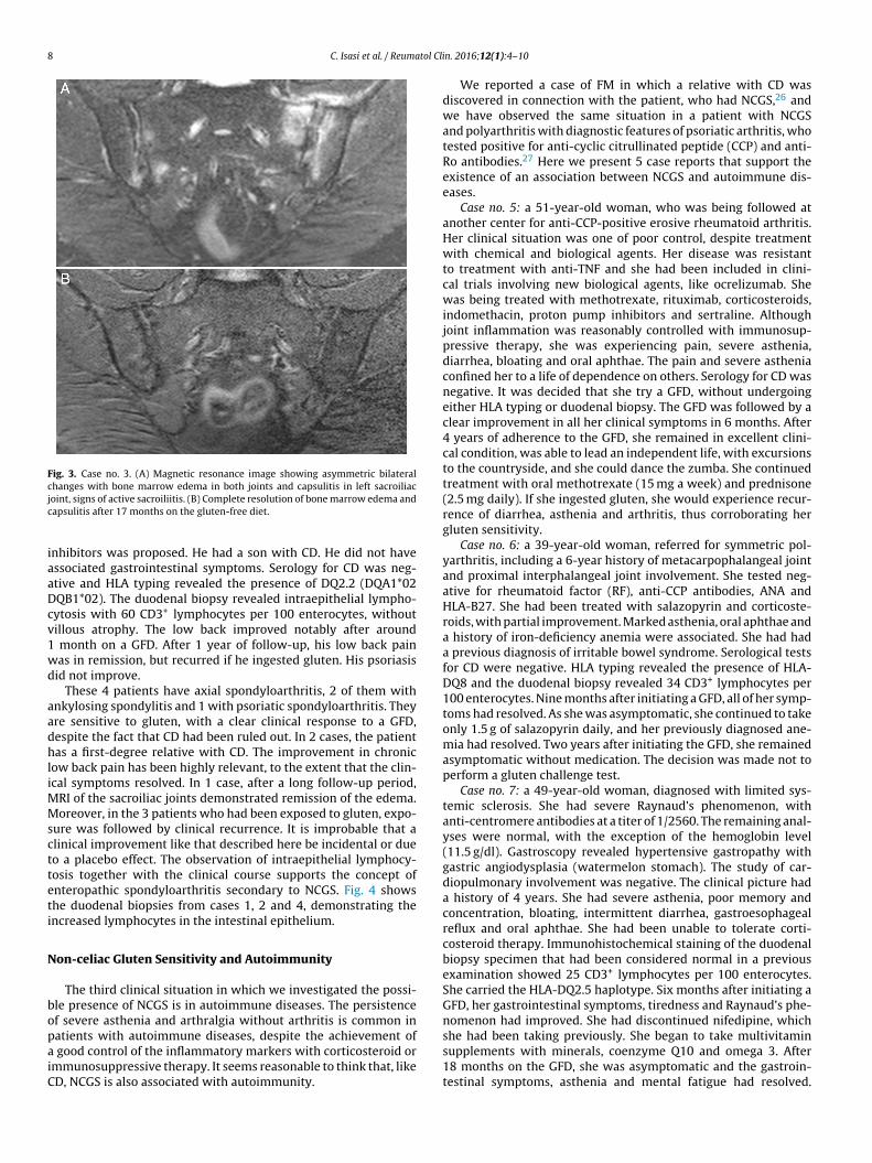
ing of the sacroiliac joints performed after 17 months of follow-up
showed remission of bone marrow edema (Fig. 3). She had returned
to a normal active life and to work. She did not ingest gluten again.
Case no. 4: a 39-year-old man, with a history of over 15 years
of chronic low back pain. He had been diagnosed with psoriatic
spondyloarthritis on the basis of sacroiliitis, with bone marrow
edema on MRI of the sacroiliac joints, and psoriasis. His condi-
tions were refractory and treatment with tumor necrosis factor
8 C. Isasi et al. / Reumatol Clin. 2016;12(1):4–10
Fig. 3. Case no. 3. (A) Magnetic resonance image showing asymmetric bilateral
changes with bone marrow edema in both joints and capsulitis in left sacroiliac
joint, signs of active sacroiliitis. (B) Complete resolution of bone marrow edema and
capsulitis after 17 months on the gluten-free diet.
inhibitors was proposed. He had a son with CD. He did not have
associated gastrointestinal symptoms. Serology for CD was neg-
ative and HLA typing revealed the presence of DQ2.2 (DQA1*02
DQB1*02). The duodenal biopsy revealed intraepithelial lympho-
cytosis with 60 CD3+ lymphocytes per 100 enterocytes, without
villous atrophy. The low back improved notably after around
1 month on a GFD. After 1 year of follow-up, his low back pain
was in remission, but recurred if he ingested gluten. His psoriasis
did not improve.
These 4 patients have axial spondyloarthritis, 2 of them with
ankylosing spondylitis and 1 with psoriatic spondyloarthritis. They
are sensitive to gluten, with a clear clinical response to a GFD,
despite the fact that CD had been ruled out. In 2 cases, the patient
has a first-degree relative with CD. The improvement in chronic
low back pain has been highly relevant, to the extent that the clin-
ical symptoms resolved. In 1 case, after a long follow-up period,
MRI of the sacroiliac joints demonstrated remission of the edema.
Moreover, in the 3 patients who had been exposed to gluten, expo-
sure was followed by clinical recurrence. It is improbable that a
clinical improvement like that described here be incidental or due
to a placebo effect. The observation of intraepithelial lymphocy-
tosis together with the clinical course supports the concept of
enteropathic spondyloarthritis secondary to NCGS. Fig. 4 shows
the duodenal biopsies from cases 1, 2 and 4, demonstrating the
increased lymphocytes in the intestinal epithelium.
Non-celiac Gluten Sensitivity and Autoimmunity
The third clinical situation in which we investigated the possi-
ble presence of NCGS is in autoimmune diseases. The persistence
of severe asthenia and arthralgia without arthritis is common in
patients with autoimmune diseases, despite the achievement of
a good control of the inflammatory markers with corticosteroid or
immunosuppressive therapy. It seems reasonable to think that, like
CD, NCGS is also associated with autoimmunity.
We reported a case of FM in which a relative with CD was
discovered in connection with the patient, who had NCGS,26 and
we have observed the same situation in a patient with NCGS
and polyarthritis with diagnostic features of psoriatic arthritis, who
tested positive for anti-cyclic citrullinated peptide (CCP) and anti-
Ro antibodies.27 Here we present 5 case reports that support the
existence of an association between NCGS and autoimmune dis-
eases.
Case no. 5: a 51-year-old woman, who was being followed at
another center for anti-CCP-positive erosive rheumatoid arthritis.
Her clinical situation was one of poor control, despite treatment
with chemical and biological agents. Her disease was resistant
to treatment with anti-TNF and she had been included in clini-
cal trials involving new biological agents, like ocrelizumab. She
was being treated with methotrexate, rituximab, corticosteroids,
indomethacin, proton pump inhibitors and sertraline. Although
joint inflammation was reasonably controlled with immunosup-
pressive therapy, she was experiencing pain, severe asthenia,
diarrhea, bloating and oral aphthae. The pain and severe asthenia
confined her to a life of dependence on others. Serology for CD was
negative. It was decided that she try a GFD, without undergoing
either HLA typing or duodenal biopsy. The GFD was followed by a
clear improvement in all her clinical symptoms in 6 months. After
4 years of adherence to the GFD, she remained in excellent clini-
cal condition, was able to lead an independent life, with excursions
to the countryside, and she could dance the zumba. She continued
treatment with oral methotrexate (15 mg a week) and prednisone
(2.5 mg daily). If she ingested gluten, she would experience recur-
rence of diarrhea, asthenia and arthritis, thus corroborating her
gluten sensitivity.
Case no. 6: a 39-year-old woman, referred for symmetric pol-
yarthritis, including a 6-year history of metacarpophalangeal joint
and proximal interphalangeal joint involvement. She tested neg-
ative for rheumatoid factor (RF), anti-CCP antibodies, ANA and
HLA-B27. She had been treated with salazopyrin and corticoste-
roids, with partial improvement. Marked asthenia, oral aphthae and
a history of iron-deficiency anemia were associated. She had had
a previous diagnosis of irritable bowel syndrome. Serological tests
for CD were negative. HLA typing revealed the presence of HLA-
DQ8 and the duodenal biopsy revealed 34 CD3+ lymphocytes per
100 enterocytes. Nine months after initiating a GFD, all of her symp-
toms had resolved. As she was asymptomatic, she continued to take
only 1.5 g of salazopyrin daily, and her previously diagnosed ane-
mia had resolved. Two years after initiating the GFD, she remained
asymptomatic without medication. The decision was made not to
perform a gluten challenge test.
Case no. 7: a 49-year-old woman, diagnosed with limited sys-
temic sclerosis. She had severe Raynaud’s phenomenon, with
anti-centromere antibodies at a titer of 1/2560. The remaining anal-
yses were normal, with the exception of the hemoglobin level
(11.5 g/dl). Gastroscopy revealed hypertensive gastropathy with
gastric angiodysplasia (watermelon stomach). The study of car-
diopulmonary involvement was negative. The clinical picture had
a history of 4 years. She had severe asthenia, poor memory and
concentration, bloating, intermittent diarrhea, gastroesophageal
reflux and oral aphthae. She had been unable to tolerate corti-
costeroid therapy. Immunohistochemical staining of the duodenal
biopsy specimen that had been considered normal in a previous
examination showed 25 CD3+ lymphocytes per 100 enterocytes.
She carried the HLA-DQ2.5 haplotype. Six months after initiating a
GFD, her gastrointestinal symptoms, tiredness and Raynaud’s phe-
nomenon had improved. She had discontinued nifedipine, which
she had been taking previously. She began to take multivitamin
supplements with minerals, coenzyme Q10 and omega 3. After
18 months on the GFD, she was asymptomatic and the gastroin-
testinal symptoms, asthenia and mental fatigue had resolved.

C. Isasi et al. / Reumatol Clin. 2016;12(1):4–10 9
Fig. 4. Duodenal biopsies after immunohistochemical staining for CD3+ lymphocytes showing an increase in the number of intraepithelial lymphocytes, without villous
shortening or crypt hyperplasia. (A) Increase in the number of intraepithelial lymphocytes all along the intestinal villus (20×), case no. 1. (B) A close view of the increase in
intraepithelial lymphocytes (40×), case no. 2. (C) A close view of the cluster of intraepithelial lymphocytes at the tip of the intestinal villus (40×), case no. 4, courtesy of Dr.
Fernando Casco, HistoCitoMed.
Raynaud’s phenomenon reappeared occasionally. She tested posi-
tive for anti-centromere antibodies, with a titer of 1/160. Occasional
gluten ingestion had been followed immediately by asthenia, oral
aphthae and recurrence of the gastrointestinal symptoms.
Case no 8: a 46-year-old woman with a 10-year history of
polyarthritis and sicca syndrome, with later development of Ray-
naud’s phenomenon, telangiectasia and digital ulcers. Her analyses
showed RF 556 IU, antibodies to extractable nuclear antigens
(ENA) SSA/Ro > 600, (U)1 ribonucleoprotein (RNP) 140 and RNP-
70 114. Capillaroscopy revealed changes compatible with systemic
sclerosis. Salivary gland biopsy was compatible with Sjögren’s syn-
drome. Treatment with corticosteroids, methotrexate, antimalarial
drugs, azathioprine, leflunomide, statins, antiplatelet therapy, non-
steroidal anti-inflammatory drugs, proton pump inhibitors and
antidepressants had been prescribed. She had experienced sec-
ondary effects of the medication and poor adherence to treatment.
Severe asthenia, diarrhea and oral aphthae were associated. Sero-
logical test for CD was negative and the duodenal biopsy was
normal. HLA typing revealed DQ2.2 (DQA1*02 DQB1*02) and DQ8.
Four months after initiating a GFD, she had a clear improvement
in the digestive symptoms, asthenia and oral aphthae, as well as
improvement in the hand swelling and digital ulcers. If she ingested
gluten, diarrhea recurred. Two years later, the patient feels well and
continues the GFD, and in addition, is being treated with 10 mg a day
of leflunomide, 2.5 mg a day of prednisone and vitamin and min-
eral supplements. She continues to test positive for autoimmune
markers at similar titers.
Case no. 9: a 44-year-old woman, referred from another cen-
ter for recurrent arthritis. She had shown variable positivity for
ANA and had tested positive for IgG and IgM anticardiolipin anti-
bodies at low titers. She also had diarrhea and marked asthenia.
She had been diagnosed with irritable bowel syndrome, and had
a daughter with CD. Celiac disease had been ruled out by a nega-
tive serological test and duodenal biopsy, despite which, she had
been following a GFD diet for 3 years, although not strictly, and her
symptoms had clearly improved, and worsened following expo-
sure to gluten. She did not want to undergo gluten challenge. She
tested positive for ANA (1/160) and negative for anticardiolipin
antibodies. She carried the HLA-DQ2 haplotype, and a review of
the previous duodenal biopsy revealed Marsh type 1 lesions, with
33 CD3+ lymphocytes per 100 enterocytes. It was recommended
that she follow a strict GFD diet and eliminate milk and dairy prod-
ucts. Vitamin and mineral supplements and omega 3 were added.
After 2 years of follow-up, her gastrointestinal symptoms, asthe-
nia and joint symptoms had remitted. She tested positive for ANA
at a titer of 1/80 and continued to test negative for anticardiolipin
antibodies.
It has recently been reported that 14% of the patients with
NCGS have an associated autoimmune disease, mainly autoimmune
thyroiditis and psoriasis.14 These case reports include polyarthritis
of unknown cause, polyarthritis associated with ANA and with anti-
cardiolipin antibodies, refractory rheumatoid arthritis, systemic
sclerosis and mixed connective tissue disease. They show that
NCGS can be associated with a variety of autoimmune diseases.
These patients, whose clinical condition was very poor, despite the
intensification of immunosuppressive therapy, showed a striking
improvement after following a GFD. As in FM and spondyloarthritis,
it is improbable that this be due to a coincidence or a placebo effect
and, thus, the question is to what extent can the GFD play an impor-
tant role in the treatment of these patients. It is important to point
out that the clinical improvement permitted the reduction or dis-
continuation of immunosuppressive therapy. The favorable clinical
course was produced despite the persistence of positivity for mark-
ers of autoimmune disease, although there were cases in which
they also improved. As in FM and spondyloarthritis, the findings
that point to the presence of NCGS are severe asthenia, oral aph-
thae, associated gastrointestinal symptoms and having a relative
with CD. These clinical observations contradict the study of Vives
et al., who found very few cases of gluten-sensitive enteropathy
among their patients with systemic diseases.28 This may be due to
the fact that we identify NCGS on the basis of the clinical response,
including both the forms nearest to CD, with disease-susceptible
HLA and Marsh type 1 enteropathy, and the forms farthest from
CD, even with no need to demonstrate the presence of enteropa-
thy. In their study, Vives et al. attempt to detect gluten-sensitive
enteropathy by performing duodenal biopsy in patients with pos-
itive CD serology or disease-susceptible HLA, or with symptoms
indicative of CD, and the diagnosis of gluten-sensitive enteropa-
thy requires the demonstration of enteropathy and, aside from the
clinical response, an analytical or histological response.
These clinical observations of NCGS associated with systemic
diseases, in which it may take months for the benefits of the diet to
be noted, differ from the accepted ideas about NCGS, in which it is
considered to be a nonautoimmune condition, in which the clinical
response to the GFD can easily be observed in a matter of a few days
or weeks.
Conclusions
Celiac disease is an autoimmune disease with a wide spectrum
of rheumatic manifestations, which should be taken into account
by rheumatologists. The incidence of NCGS is probably higher than
that of CD, with similar symptoms, but it is difficult to diagnose due
to the lack of specific diagnostic tests. In this article, we express
the hypothesis, based on reasonable arguments and clinical obser-
vations, that NCGS is associated with a wide range of rheumatic
manifestations that include FM, the spondyloarthritides and sys-
temic autoimmune diseases. In our experience, the most important
10 C. Isasi et al. / Reumatol Clin. 2016;12(1):4–10
clinical data that indicate the presence of NCGS are severe asthenia
of unknown etiology, oral aphthae, the associated gastrointesti-
nal symptoms, iron-deficiency anemia and having a relative with
CD. It seems that these clinical data should particularly be taken
into account when a patient with a systemic disease has associated
symptoms of FM. The favorable course following the GFD, observed
in both FM-like manifestations and in arthritis and sacroiliitis, leads
us to think that gluten sensitivity may play an etiological and
pathogenic role that acts as a trigger in some patients with systemic
autoimmune diseases. Of course, prospective studies and clinical
trials with double-blind challenge will be necessary to establish the
extent to which the occurrence of NCGS can be considered frequent,
and its treatment relevant, in rheumatic diseases.
Ethical Disclosures
Protection of human and animal subjects. The authors declare
that no experiments were performed on humans or animals for
this study.
Confidentiality of data. The authors declare that they have fol-
lowed the protocols of their work center on the publication of
patient data.
Right to privacy and informed consent. The authors have
obtained the written informed consent of the patients or subjects
mentioned in the article. The corresponding author is in possession
of this document.
Funding
The contribution of MSD Inmunología was decisive during the
initial phases of this study, providing financial resources for
the performance of immunohistochemistry in the duodenal biop-
sies. Genyca Innova contributed to the performance of HLA typing
of the patients.
Conflicts of Interest
The authors declare that they have no conflicts of interest.
Acknowledgments
We acknowledge the authors of this report who are not among
the signatories due to an editorial norm that limits the number
of authors: Natalia Fernández Puga, digestive system specialist at
Hospital Puerta de Hierro, Madrid; Isabel Colmenero and Fernando
Casco, pathologists at Birmingham Children’s Hospital, Birming-
ham, U.K. and at Labco, Madrid, respectively; María José Castro
Panete, immunologist at Hospital Doce de Octubre, Madrid, and
the Association of Celiac and Gluten Sensitive Patients of Madrid
(la Asociación de Celíacos y Sensibles al Gluten de Madrid).
References
1. Fasano A, Catassi C. Clinical practice. Celiac disease. N Engl J Med.2012;367:2419–26.
2. Green PH, Cellier C. Celiac disease. N Engl J Med. 2007;357:1731–43.
3. Castillo N, Theethira TG, Leffler DA. The present and the future in the diagnosisand management of celiac disease. Gastroenterol Rep (Oxf). 2015;3:3–11.
4. Troncone R, Discepolo V. Celiac disease and autoimmunity. J Pediatr Gastroen-terol Nutr. 2014;59 Suppl. 1:S9–11.
5. Mooney PD, Hadjivassiliou M, Sanders DS. Coeliac disease. BMJ. 2014:348.6. National Institute for Health and Clinical Excellence. Coeliac disease: recognition
and assessment of coeliac disease. 2009. Coeliac disease. BMJ. 2013;348. Avail-able from: www.nice.org.uk/nicemedia/pdf/CG86FullGuideline.pdf [accessed30.09.14].
7. Zipser RD, Patel S, Yahya KZ, Baisch DW, Monarch E. Presentations of adultceliac disease in a nationwide patient support group. Dig Dis Sci. 2003;48:761–4.
8. Lee SK, Green PH. Celiac sprue (the great modern-day imposter). Curr OpinRheumatol. 2006;18:101–7.
9. Diagnóstico precoz de la enfermedad celíaca. Ministerio de Sanidad yConsumo; 2008. Available from: http://www.msssi.gob.es/profesionales/prestacionesSanitarias/publicaciones/Celiaquia/enfermedadCeliaca.pdf[accessed 30.09.14].
10. Ghozzi M, Sakly W, Mankaï A, Bouajina E, Bahri F, Nouira R, et al. Screening forceliac disease, by endomysial antibodies, in patients with unexplained articularmanifestations. Rheumatol Int. 2014;34:637–42.
11. Czaja-Bulsa G. Non coeliac gluten sensitivity – a new disease with gluten intol-erance. Clin Nutr. 2015;34:189–94.
12. Troncone R, Jabri B. Coeliac disease and gluten sensitivity. J Intern Med.2011;269:582–90.
13. Catassi C, Bai JC, Bonaz B, Bouma G, Calabro A, Carroccio A, et al. Non-celiacgluten sensitivity: the new frontier of gluten related disorders. Nutrients.2013;5:3839–53.
14. Volta U, Bardella MT, Calabrò A, Troncone R, Corazza GR, Study Groupfor Non-Celiac Gluten Sensitivity. An Italian prospective multicenter sur-vey on patients suspected of having non-celiacgluten sensitivity. BMC Med.2014;12:85, http://dx.doi.org/10.1186/1741-7015-12-85.
15. Ludvigsson JF, Leffler DA, Bai JC, Biagi F, Fasano A, Green PH, et al. The Oslodefinitions for coeliac disease and related terms. Gut. 2013;62:43–52.
16. Ensari A. Gluten-sensitive enteropathy (celiac disease): controversies in diag-nosis and classification. Arch Pathol Lab Med. 2010;134:826–36.
17. Molina-Infante J, Santolaria S, Montoro M, Esteve M, Fernández-BanaresF. Sensibilidad al gluten no celiaca: una revisión crítica de la eviden-cia actual. Gastroenterol Hepatol. 2014;37:362–71, http://dx.doi.org/10.1016/j.gastrohep.2014.01.005.
18. Fernández-Banares F, Esteve M, Salas A, Alsina M, Farré C, González C, et al.Systematic evaluation of the causes of chronic watery diarrhea with functionalcharacteristics. Am J Gastroenterol. 2007;102:2520–8.
19. Molina-Infante J, Santolaria S, Fernandez-Banares F, Montoro M, Esteve M.Lymphocytic enteropathy. HLA-DQ2/DQ8 genotype and wheat-dependentsymptoms: non-celiac wheat sensitivity or Marsh I celiac disease? Am J Gas-troenterol. 2013;108:451.
20. Esteve M, Rosinach M, Fernández-Banares F, Farré C, Salas A, Alsina M,et al. Spectrum of gluten-sensitive enteropathy in first-degree relatives ofpatients with coeliac disease: clinical relevance of lymphocytic enteritis. Gut.2006;55:1739–45.
21. Tovoli F, Giampaolo L, Caio G, Monti M, Piscaglia M, Frisoni M, et al. Fibromyalgiaand coeliac disease: a media hype or an emerging clinical problem? Clin ExpRheumatol. 2013;31 Suppl. 79:S50–2.
22. Isasi C, Colmenero I, Casco F, Tejerina E, Fernandez N, Serrano-Vela JI, et al.Fibromyalgia and non-celiac gluten sensitivity: a description with remission offibromyalgia. Rheumatol Int. 2014;34:1607–12.
23. Togrol RE, Nalbant S, Solmazgül E, Ozyurt M, Kaplan M, Kiralp MZ, et al. The sig-nificance of coeliac disease antibodies in patients with ankylosing spondylitis:a case-controlled study. J Int Med Res. 2009;37:220–6.
24. Usai P, Boi MF, Piga M, Cacace E, Lai MA, Beccaris A, et al. Adult celiac disease isfrequently associated with sacroiliitis. Dig Dis Sci. 1995;40:1906–8.
25. Vereckei E, Mester A, Hodinka L, Temesvári P, Kiss E, Poór G. Back pain andsacroiliitis in long-standing adult celiac disease: a cross-sectional and follow-upstudy. Rheumatol Int. 2010;30:455–60.
26. Isasi C, Tejerina E, Fernandez-Puga N, Serrano-Vela JI. Fibromialgia yfatiga crónica causada por sensibilidad al gluten no celíaca. Reumatol Clin.2015;11:567, http://dx.doi.org/10.1016/j.reuma.2014.06.005.
27. Isasi C, Colmenero I, Casco F, Tejerina E, Fernandez-Puga N. Non-coeliacgluten sensitivity and autoimmunity: a case report. EJCRIM. 2014;1,http://dx.doi.org/10.12890/2014 000156.
28. Vives MJ, Esteve M, Mariné M, Fernández-Banares F, Alsina M, Salas A, et al.Prevalence and clinical relevance of enteropathy associated with systemicautoimmune diseases. Dig Liver Dis. 2012;44:636–42.