Toxicity profiling of several common RNAi-based nanomedicines: a comparative study
10
1 23 Drug Delivery and Translational Research An Official Journal of the Controlled Release Society ISSN 2190-393X Drug Deliv. and Transl. Res. DOI 10.1007/s13346-013-0158-7 Toxicity profiling of several common RNAi- based nanomedicines: a comparative study Dalit Landesman-Milo & Dan Peer
-
Upload
independent -
Category
Documents
-
view
4 -
download
0
Transcript of Toxicity profiling of several common RNAi-based nanomedicines: a comparative study

1 23
Drug Delivery and TranslationalResearchAn Official Journal of the ControlledRelease Society ISSN 2190-393X Drug Deliv. and Transl. Res.DOI 10.1007/s13346-013-0158-7
Toxicity profiling of several common RNAi-based nanomedicines: a comparative study
Dalit Landesman-Milo & Dan Peer

1 23
Your article is protected by copyright and
all rights are held exclusively by Controlled
Release Society. This e-offprint is for personal
use only and shall not be self-archived
in electronic repositories. If you wish to
self-archive your article, please use the
accepted manuscript version for posting on
your own website. You may further deposit
the accepted manuscript version in any
repository, provided it is only made publicly
available 12 months after official publication
or later and provided acknowledgement is
given to the original source of publication
and a link is inserted to the published article
on Springer's website. The link must be
accompanied by the following text: "The final
publication is available at link.springer.com”.

RESEARCH ARTICLE
Toxicity profiling of several common RNAi-basednanomedicines: a comparative study
Dalit Landesman-Milo & Dan Peer
# Controlled Release Society 2013
Abstract RNAi-based nanomedicine platforms (RNPs) haveprogressed from tools to study gene expression in vitro intoclinical trials. Numerous RNPs strategies have been document-ed with an efficient ability to condense RNAi payloads andinduce potent gene silencing. Moreover, some of these RNPshave been explored in various animal models, and some haveeven made it to the clinic. Still, there is lack of a clinicallyapproved RNAi-based delivery strategy most probably due tounpredicted clinical toxicity. In this study,we prepared commonRNPs such as cationic liposomes, polyamines, and hyaluronan-coated lipid-based nanoparticles and tested these strategies forglobal toxicity parameters such as changes in bodyweight, liverenzyme release, and hematological profiling. We found thatpolyamines such as polyethyleneimine and Poly-L-lysinereleased high levels of liver enzymes into the serum andreduced C57BL/6 mice bodyweight upon three intravenousinjections. In addition, these polyamines dramatically reducedthe total number of leukocytes, suggesting an immunesuppression mechanism, while cationic liposomes, which alsoincreased liver enzymes levels in the serum, elevated the totalnumber of leukocytes probably by activation of Toll-likereceptors 2 and 4. Coating the liposomes with hyaluronan, ahydrophilic glycosaminoglycan, provided a protective layer anddid not induce adverse effects upon multiple intravenousinjections. These findings suggest that there is an urgent need todevelop gold standards for nanotoxicity in the field of RNAithat will be embraced by the RNAi community.
Keywords RNAi .Nanomedicines .Nanotoxicity .Cationicliposomes . Polyamines
Introduction
RNAi is a specific regulatory mechanism of most eukaryoticcells, in which small double-stranded ribonucleic acidsefficiently control gene expression in a complementarity-dependent manner. This natural mechanism, which involvesdiverse families of small non-coding RNA regulators, ispresumed to protect against pathogenic infections as well asregulate various biological pathways [1, 2]. Utilizing thisnative mechanism for therapeutic and diagnostic purposes canbe achieved by exogenous delivery of synthetic RNAimolecules. However, due to their net negative charge andrelatively large size compared with vasculature effective poresize, RNAimolecules are less likely to readily cross biologicalbarriers, thus, efficient systemic delivery necessitates the useof physical protection (i.e., carriers/reagents) and chemicalmodifications that together protect RNAi molecules untilreaching their site of action and facilitate cell penetration, yetmaintaining their functionality [2, 3]. Numerous strategies todeliver RNAi have been developed with some promising invivo results [4, 5].
Despite the promise, developing RNAi as therapeuticshas proven challenging. Like most drug development, thereis quick fix, and many pharmaceutical companies thatinvested considerable sums in developing RNA-based drugsannounced they are closing their RNAi subsidiaries.
Although many of the hurdles in developing RNAi-basedtherapeutics have been already addressed, the mainchallenge is figuring out the way of delivering smallRNAs into cells in a therapeutically relevant manner withminimal adverse effects. Several delivery strategies forRNAi payloads have been well documented over the pastyears, among them polyamines such as poly-L-lysine andpolyethyleneimine (PEI) [6–8], cationic liposomes [9–11],and neutral lipid-based nanoparticles [12–15]. In thesedelivery strategies, the efficiency of gene silencing andphenotypic genes have been documented, yet comprehen-sive analyses of adverse effects were not reported for all
D. Landesman-Milo :D. PeerLaboratory of NanoMedicine, Department of Cell Researchand Immunology, Tel Aviv University, Tel Aviv 69978, Israel
D. Landesman-Milo :D. Peer (*)Center for Nanosciences and Nanotechnology,Tel Aviv University, Tel Aviv 69978, Israele-mail: [email protected]
Drug Deliv. and Transl. Res.DOI 10.1007/s13346-013-0158-7
Author's personal copy

these strategies. Herein, we profiled global toxicity offour representative delivery strategies, poly-L-lysine,PEI, cationic liposomes, and neutral lipid-basednanoparticles all entrapping or condensing siRNApayloads with an effective knockdown capabilities. Weexamined global changes in bodyweight upon threeintravenous administrations of each of the deliverysystems with its RNAi payloads, tested liver enzymerelease in the serum of mice, and profiled totalleukocytes and specific subsets count upon multipleintravenous (IV) injections into mice.
Materials and methods
Materials Linear PEI (750 kDa), poly-L-lysine, werepurchased from Sigma-Aldrich (St. Louis, MO, USA).1,2-Dioleoyl-3-trimethylammonium-propane (DOTAP)was obtained by Avanti Polar Lipid (Albaster, AL,USA). Other lipids were from Avanti Polar Lipids Inc.(Alabaster, AL, USA) or Phospholipid GMBH(Germany). Sodium hyaluronate (HA) with an averagemolecular weight of 700 KDa was obtained fromLifecore Biomedical Co. (Chaska, MN). All otherreagents were from Sigma-Aldrich (St. Louis, MO,USA).
Preparation of lipid-based nanoparticles (LNPs)
Multilamellar vesicles (MLV) composed of pure soybeanphosphatidylcholine (Phospholipon 90G), which was a kindgift from Phospholipid GMBH (Germany). 1,2-Dipalmitoyl-sn-glycero-3-phosphoethanolamine (DPPE)and cholesterol (Chol) were purchased from Avanti PolarLipids Inc. (Alabaster, AL, USA). PC/Chol/DPPE were atmole ratios of 60:20:19.9, with the addition of 0.1 % DPPElabeled with rhodamine fluorophore (Invitrogen) wereprepared by the traditional lipid-film method [16–19].Briefly, the lipids were dissolved in ethanol and evaporatedto dryness under reduced pressure in a rotary evaporator(Buchi Rotary Evaporator Vacuum System Flawil,Switzerland). Following evaporation, the dry lipid filmwas hydrated in 10 ml of HEPES (pH 7.4). This wasfollowed by extensive agitation using a vortex device and2 h incubation in a shaker bath at 65 °C. The MLV wereextruded through a Lipex extrusion device (Northern lipids,Vancouver, Canada), operated at 65 °C and under nitrogenpressures of 200–500 psi. The extrusion was carried out instages using progressively smaller pore-size polycarbonatemembranes (Whatman Inc, UK), with several cycles perpore-size, for achieving unilamellar vesicles in a final sizerange of ∼100 nm in diameter. Lipid mass was quantified aspreviously reported [19, 20].
Hyaluronan-coated–LNPs
We have previously shown that high-molecular weighthyaluronan (HA), of an average of 700 kDa from LifecoreBiomedical LLC (Chaska, MN, U.S.A) when coated ontothe surface of LNPs, endow these particles with highaffinity to CD44, its natural receptor [19]. The HA wasdissolved in 0.2 M MES buffer (pH 5.5) to a finalconcentration of 5 mg/ml. HA was activated with EDC andsulfo-NHS at a molar ratio of 1:1:6. After 30 min ofactivation, the LNPs were added, and the pH was adjustedto 7.4. The solution was incubated at room temp (2 h). Thefree HA was removed by 3 cycles of repeated washing bycentrifugation (1.3×10⁵×g, 4 °C, 60 min). HA wasquantified as previously demonstrated [19, 20]. The finalHA/lipid ratio was typically 75 μg HA/μmole lipid asassayed by 3H-HA (ARC, Saint Louis, MI) with an averageof 2 % of the particles that had the HA decoration.
Particle size distribution and zeta potential measurements
Particle size distribution and mean diameter of the HA-coatedlipid-based nanoparticles (HA-LNPs), PEI-siRNA, and PLL–siRNA complexes as well as cationic liposomes and control,non-surface-modified NPs (LNPs) were measured on aMalvern Zetasizer Nano ZS zeta potential and dynamic lightscattering instrument (Malvern Instruments, Southborough,MA) using the automatic algorithm mode and analyzed withthe PCS 1.32a. All measurements were done in 0.01 mol/lNaCl, pH 6.7, at 20 °C.
siRNA encapsulation or complex formation.
siRNA sequences against the CCND1 gene NM_053056(siD1, sense strand: GUAGGACUCUCAUUCGGGATT)and the luciferase gene as a control sequence (siLuc, sensestrand: CUUACGCUGAGUACUUCGA) were designedand screened by Alnylam Pharmaceuticals (CambridgeMA, USA) and previously published [21]. HA-LNPssiRNAs were entrapped in HA-LNPs or LNPs as previouslyreported [15]. Briefly, HA-LNPs and LNPs were lyophi-lized until complete water removal was ensured (48 h). Thelyophilized particles were hydrated with DEPC-treatedwater with or without siRNAs. The particles were shakengently for 30 min at room temperature. UnencapsulatedsiRNA was removed via ultra-fast centrifugation at 6.4×10⁵×g, 4 °C, 20 min prior to adding the suspension to thecells or to intravenous injection into C57BL/6 mice.
PEI–siRNA complex formation To formulate the siRNA–PEI complex, 200 μ l of siRNA (0.1 mg/ml) was condensedwith 100 μ l of PEI (0.6 mM) in 10 mM HEPES buffer(pH 7.4), at a nitrogen/phosphate ratio of 1.0.
Drug Deliv. and Transl. Res.
Author's personal copy

Poly-L-lysin (PLL)–siRNA complex formation To formulatethe siRNA–PLL complex, 200 μl of siRNA (0.1 mg/ml)was condensed with 200 μl of PLL (10KDa , 0.3 mM) in10 mM HEPES buffer (pH 7.4).
Preparation of Cationic (+) LNPs and siRNA entrap-ment (+)LNPs were composed of HSPC/Chol/DOTAP at3:1:1 mole ratio and prepared as detailed above for LNPs.
siRNAs entrapment was done similar to the one reportedin Kedmi et al. [22]. Briefly, the lipids were dissolved inchloroform/methanol (4:1, v/v). The organic solvent wasevaporated under pressure at 60°C for 30 min, and the lipidfilm was flushed with N2 gas to remove residual solvent.The lipid film was hydrated using a solution of siRNAs(Luciferase , as control or CyD1) with previously describedsequence [21] in 5 % dextrose (w/v) prepared using RNase-free dH2O. Size reduction was performed as withconventional particles (see above).
siRNA quantification of the efficiency of entrapment
siRNA encapsulation efficiency was determined by theQuant-iT RiboGreen RNA assay (Invitrogen) as previouslydescribed by us and others [13, 15, 20, 23]. Briefly, theentrapment efficiency was determined by comparingfluorescence of the RNA binding dye RiboGreen in the (+)LNP, HA-LNP, PLL-siRNA, and PEI-siRNA complexessamples, in the presence and absence of 0.5 % Triton X-100[13, 15]. In the absence of detergent, fluorescence can bemeasured from accessible (un-entrapped or condensed)siRNA only. Whereas, in the presence of the detergent,fluorescence is measured from total siRNA [23]; thus, the %encapsulation is described by the equation:
% siRNA encapsulation=complexation
¼ 1− free siRNA conc:=total siRNA conc:ð Þ½ � � 100:
Based on the entrapment efficiency, we have calculatedthe specific amount needed for each formulation for in vitroand in vivo experiments so no bias will occur in the resultsor while interpretation of the data.
Delivery of siRNAs into cancer cells by RNPs
Human lung adenocarcinoma cell line (A549) were seededinto six-well cell culture plates at 0.1×10⁶ cells/well inRPMI medium, supplemented with antibiotics, L-glutamine,and 10 % fetal calf serum (Biological industries, BeitHaemek, Israel). Twenty-four hours post-seeding themedium was removed and replaced with RPMI only. Thecells were then treated with empty vehicles (HA-LNPs) orwith tested RNAi-based nanomedicine platform (RNP)(HA-LNPs, PLL, PEI, or (+)LNPs) encapsulating Cyclin
D1 (CyD1) or luciferase (Luci) siRNAs. CyD1 chosen as asurrogated marker to evaluate the delivery strategy. Fourhours post-incubation, the medium was removed, and thecells were washed and supplemented with completemedium. Three days after transfection, the cells were split0.2×106 cell/well into fresh 6 wells cell culture plate. FinalsiRNA concentration on the cells was 100 nM.
Quantitative real-time PCR
QPCR was done as previously published [21]. Briefly, totalRNA was isolated using EZ-RNA kit (biological industries,Israel), and cDNAwas generated with high-capacity cDNA kit(Life Technologies, Carlsbad, CA, USA) according to themanufacturers' protocols. qRT-PCR was performed with FastSYBR® Green Master Mix and the ABI StepOnePlus™instrument (Life Technologies). CCND1 (F:GAGGAGCCCCAACAACTTCC, R:GTCCGGGTCACACTTGATCAC)expression was normalized to the house keeping genes eIF3a(F:TCCAGAGAGCCAGTCCATGC, R:CCTGCCACAATTCA TGCT) and eIF3c (F:ACCAAGAGAGTTGTCCGCAGTG, R:TCATGGCATTACG GATGGTCC). Analysiswas done with the StepOne™ software V 2.1 (LifeTechnologies) using the multiple endogenous controls option.When using multiple endogenous controls, the software treatsall endogenous controls as a single population and calculates theexperiment-appropriate mean to establish a single value againstwhich the target of interest is normalized.
Animal care and treatment
Animals (healthy C57BL/6 mice) were obtained from theanimal-breeding center, Tel-Aviv University (Tel Aviv,Israel). Animals were maintained and treated according toNational Institutes of Health guidelines. All animalprotocols were approved by the Tel-Aviv InstitutionalAnimal Care and Use Committee.
Bodyweight measurements
Healthy C57BL/6 mice (n=7/group) were given either asingle or three bolus IV injections of saline or treated with(+)LNPs-siRNA, PLL-siRNA, PEI-siRNA, and HA-LNP(with siRNAs) at days 4, 8, and 12 from experimentinitiation at a dose of 1 mg/kg body siRNA/dose/mouse),and individual mouse bodyweight was monitored every4 days using an Avery Berkel scale (Fairmont, MN, USA).Cyclin D1 –siRNA was used in all tested groups.
Blood biochemistry and hematology assays
Healthy C57BL/6 mice (n=7/group) were given three bolusIV injections of saline (+)LNPs-siRNA, PLL-siRNA, PEI-
Drug Deliv. and Transl. Res.
Author's personal copy

siRNA, and HA-LNP (with siRNAs) at days 0, 2, and 4from experiment initiation at a dose of 1 mg/kg bodysiRNA/dose/mouse); 24 h after the last injection, bloodwas drawn, and the serum was obtained by centrifuga-tion of the whole blood at 850×g for 15 min in aprotocol we previously reported [22]. Liver enzymelevels of alanine aminotransferase (ALT), aspartateaminotransferase (AST), and cholesterol levels weredetermined by COBAS MIRA auto analyzer (Roche). Inaddition, the number of major groups of leukocytes wasalso measured.
Statistical analysis In vitro data were analyzed usingStudent's t test. Differences between treatment groups wereevaluated by one-way ANOVA with significance deter-mined by Bonferroni-adjusted t tests.
Results and discussion
Structural and physicochemical characterizationof RNAi-based nanomedicine platforms
In order to compare different toxicity parameters ofRNPs in vivo, we prepared four different RNPs andcomplexed or entrapped siRNAs (all with same siRNAconcentration, 100 nM for in vitro use and 1 mg/kgbody siRNAs for in vivo application). Size distribution,zeta potential, and efficiency of RNAi payload entrap-ment or complexation of PEI-, PLL-based siRNAcomplexes, (+)LNP-siRNA, and HA-LNPs entrappingsiRNA are detailed in Table 1.
All RNPs-siRNA had mean diameter of <200 nmwith narrow distribution pattern. PEI, PLL, and (+)LNPs had a positive zeta potential that wasdecreased with the addition of siRNAs (data notshown), while HA-coated LNPs had negatively zetapotential.
Robust knockdown of cyclin D1 in cancer cells by RNPs
We compared all four strategies to assess the knockdownefficiency in a model cancer cell, a human lung adenocar-cinoma cell line (A549). These cells also highly expressingthe HA receptor, CD44, as we recently showed [15]. Theexperimental setting is detailed in the “Materials andmethods” section, and we chose CyD1 as a surrogatedmarker since A549 highly express CyD1 [24]. Around 65 %knockdown of CyD1 mRNA levels was observed 20 h post-transfection with all four delivery systems (Fig. 1a) with nosignificant differences between the four strategies. Thisknockdown was specific for all four types of deliverystrategies as observed from a control study with Luci-siRNA (Fig. 1b).
Decrease in bodyweight is observed upon intravenousinjections of cationic formulations
Next, we examined global changes in bodyweight inhealthy mice injected with all four delivery systems uponsingle and multiple [3] intravenous injections (Fig. 2).
We chose a therapeutically relevant dose of 1 mg/kgbody siRNA that was shown effective in vivo in allformulations [22, 25–27]. A single IV injection 4 days post-initiation of the experiment shows that, in all cationicformulations (PLL-siRNAs, PEI-siRNAs and (+)LNPs-siRNAs), a slight reduction in bodyweight was observedto a certain degree; however, due to large standarddeviations, it is not significantly different than the mock-treated group (Fig. 2a). Screening the results for multiple IVinjections (once every 4 days) reduced the bodyweight of allformulations except that of the HA-coated LNPs and themock-treated groups (Fig. 2b). No reduction in bodyweightwas observed when siRNAs were applied in saline with 5 %glucose (data not shown), implying that the siRNAs itself isnot the cause for this general toxicity in the cationicformulations.
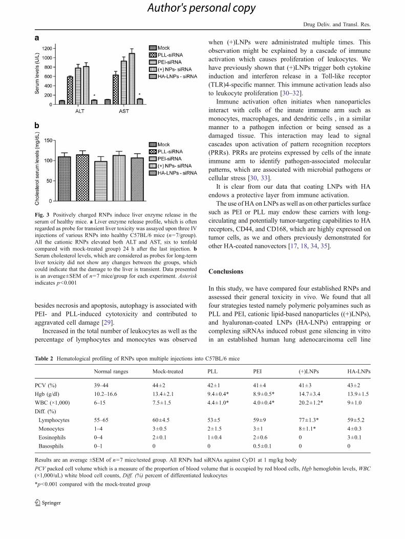
Positively charged RNPs induce liver enzyme releasein the serum of healthy mice
Potential systemic toxicity can be assayed by global toxicitymarkers [20, 28] such as serum liver enzymes and bodyweight loss upon multiple injections. Since we have alreadywitnessed bodyweight loss upon multiple IV injections (inthe case of the cationic RNPs), we wanted to examine thelevels of liver enzyme release (as probes for short-termhepatotoxicity and cholesterol levels as long-term hepato-toxicity) [22].
Serum levels of the liver enzymes, ALT and AST(Fig. 3a), and cholesterol levels (Fig. 3b) were determinedin healthy C57BL/6 mice (n=7/group) administrated
Table 1 Physicochemical and structural characterization of RNAi-based nanomedicine platforms
RNAi-basednanomedicineplatform
Sizedistribution(nm)
Zetapotential(mV)
siRNA entrapment/complexationefficacy (%)
PEI-siRNA 155.5±7.4 15.3±2.1 88.3±4.6
PLL-siRNA 162.2±9.4 9.7±1.8 62.8±7.2
HA-LNPs–siRNA 130.9±11.2 −25.4±2.9 60.4±2.2
(+)LNPs–siRNA 189.5±8.8 42.4±2.7 90.3 ±2.7
Each result is an average (±SD) of six independent measurements.Particles were measured at pH 6.7, in ddH2O with 10 mM NaCl, at20° C using a Malvern ZS Zetasizer
Drug Deliv. and Transl. Res.
Author's personal copy

intravenously with saline or with 1 mg/kg body of PLL-siRNA, PEI-siRNA, (+)LNPs, and HA-coated LNPs, and24 h after the last IV injection, blood was drawn to assesspotential hepatotoxicity.
Examining liver enzyme release into the serum after 24 hpost-injection is considered to be a gold standard forexamining acute hepatotoxicity [13, 22]; therefore, wechose to draw blood at this time point.
Administration of (+)NPs–, PLL–, and PEI–siRNAsignificantly enhanced liver enzymes release 24 h after thelast injection (six- to tenfold higher than the mock-treatedmice), indicating possible short-term liver toxicity.However, serum cholesterol levels remain the same in alltested RNPs, indicating that the potential toxicity istransient, and long-term cholesterol synthesis was not
affected by these short-term hepatotoxicity. Administrationof naked siRNA did not trigger any release of liver enzymesor changed cholesterol levels (data not shown), demon-strating that these modified molecules are safe for use.
Cationic NPs may cause immune suppression or immuneactivation
We next examined the total number of leukocytes uponthree IV administrations into C57BL/6 mice. HA-coatedLNPs had comparable numbers of leukocytes to the mock-treated group (Table 2), suggesting a protective mechanismof the HA layer. PLL- and PEI-based platforms showeddramatic decrease in the total number of leukocytes as wellas in hemoglobin levels (Table 2) with no change in thedis t r ibut ion pat tern between lymphocytes andmonocytes—this might be explained by general cell deathvia damage to the mitochondria and other cellularorganelles. In addition, a recent study, demonstrated that,
Fig. 1 Robust knockdown of CyD1 in cancer cells by RNPs. mRNAlevels of CyD1 upon transfection of A549 cells with different RNPsquantify using QPCR 24 h post-transfection. a CyD1-siRNAs wereformulated in all four different delivery strategies. b A control studywith luciferase–siRNA formulated in all the four different RNPs,demonstrating the specificity of the CyD1 sequence. Data are averageof four different experiments, and the error bars represent the SDvalues between these independent experiments
Fig. 2 Changes in bodyweight upon intravenous injections ofdifferent RNPs-siRNAs is observed in cationic formulations.Bodyweight changes (%) was monitored over a period of 16 daysafter the first IV administration. a Single IV administration. b multiple[3] IV administrations. Arrows (red) represent the days of IVadministration. Reduction in bodyweight was observed in all thecationic formulations. Data presented is an average±SEM of n=7mice/group for each experiment
Drug Deliv. and Transl. Res.
Author's personal copy
besides necrosis and apoptosis, autophagy is associated withPEI- and PLL-induced cytotoxicity and contributed toaggravated cell damage [29].
Increased in the total number of leukocytes as well as thepercentage of lymphocytes and monocytes was observed
when (+)LNPs were administrated multiple times. Thisobservation might be explained by a cascade of immuneactivation which causes proliferation of leukocytes. Wehave previously shown that (+)LNPs trigger both cytokineinduction and interferon release in a Toll-like receptor(TLR)4-specific manner. This immune activation leads alsoto leukocyte proliferation [30–32].
Immune activation often initiates when nanoparticlesinteract with cells of the innate immune arm such asmonocytes, macrophages, and dendritic cells , in a similarmanner to a pathogen infection or being sensed as adamaged tissue. This interaction may lead to signalcascades upon activation of pattern recognition receptors(PRRs). PRRs are proteins expressed by cells of the innateimmune arm to identify pathogen-associated molecularpatterns, which are associated with microbial pathogens orcellular stress [30, 33].
It is clear from our data that coating LNPs with HAendows a protective layer from immune activation.
The use of HAon LNPs aswell as on other particles surfacesuch as PEI or PLL may endow these carriers with long-circulating and potentially tumor-targeting capabilities to HAreceptors, CD44, and CD168, which are highly expressed ontumor cells, as we and others previously demonstrated forother HA-coated nanovectors [17, 18, 34, 35].
Conclusions
In this study, we have compared four established RNPs andassessed their general toxicity in vivo. We found that allfour strategies tested namely polymeric polyamines such asPLL and PEI, cationic lipid-based nanoparticles ((+)LNPs),and hyaluronan-coated LNPs (HA-LNPs) entrapping orcomplexing siRNAs induced robust gene silencing in vitroin an established human lung adenocarcinoma cell line
Fig. 3 Positively charged RNPs induce liver enzyme release in theserum of healthy mice. a Liver enzyme release profile, which is oftenregarded as probe for transient liver toxicity was assayed upon three IVinjections of various RNPs into healthy C57BL/6 mice (n=7/group).All the cationic RNPs elevated both ALT and AST, six to tenfoldcompared with mock-treated group) 24 h after the last injection. bSerum cholesterol levels, which are considered as probes for long-termliver toxicity did not show any changes between the groups, whichcould indicate that the damage to the liver is transient. Data presentedis an average±SEM of n=7 mice/group for each experiment. Asteriskindicates p<0.001
Table 2 Hematological profiling of RNPs upon multiple injections into C57BL/6 mice
Normal ranges Mock-treated PLL PEI (+)LNPs HA-LNPs
PCV (%) 39–44 44±2 42±1 41±4 41±3 43±2
Hgb (g/dl) 10.2–16.6 13.4±2.1 9.4±0.4* 8.9±0.5* 14.7±3.4 13.9±1.5
WBC (×1,000) 6–15 7.5±1.5 4.4±1.0* 4.0±0.4* 20.2±1.2* 9±1.0
Diff. (%)
Lymphocytes 55–65 60±4.5 53±5 59±9 77±1.3* 59±5.2
Monocytes 1–4 3±0.5 2±1.5 3±1 8±1.1* 4±0.3
Eosinophils 0–4 2±0.1 1±0.4 2±0.6 0 3±0.1
Basosphils 0–1 0 0 0.5±0.1 0 0
Results are an average ±SEM of n=7 mice/tested group. All RNPs had siRNAs against CyD1 at 1 mg/kg body
PCV packed cell volume which is a measure of the proportion of blood volume that is occupied by red blood cells, Hgb hemoglobin levels, WBC(×1,000/uL) white blood cell counts, Diff. (%) percent of differentiated leukocytes
*p<0.001 compared with the mock-treated group
Drug Deliv. and Transl. Res.
Author's personal copy

A549. However, when injected systemically, three intrave-nous injections separated by 4 days apart, all the cationicformulations decreased the bodyweight of C57BL/6 mice.Moreover, the cationic formulations induced liver enzymerelease, indicating a potential transient hepatotoxicity. Nochanges have been observed with cholesterol levels in allinjected RNPs, suggesting that no long-term toxicity ishappening with these low concentrations of siRNAs(1 mg/kg body). Interestingly, both polymeric polyaminesdecreased the total number of leukocytes and hemoglobin,suggesting global cellular toxicity and perhaps immunesuppression mechanism, while cationic LNPs dramaticallyincrease the total number of leukocytes suggesting immuneactivation, most likely in a TLR4-dependendt manner. HA-coated LNPs did not alter the number of leukocytes norchanged bodyweight or trigger the release of liver enzymesuggesting a protective layer of the HA coating. Previously,the group of Leaf Huang has shown that coating LNPs withhigh-molecular-weight HA endows these carriers withminimal to no toxicity when injected systemically intomice bearing tumors. Our group has shown that no immunetoxicity is occurring when HA is used as a scaffold forbinding targeting agents to the surface of LNPs whenmodulating leukocytes function during inflammatory boweldiseases and in targeting T helper cells for suppressing HIVinfection [20, 27, 34, 36, 37].
Taken together, the results presented here support the useof HA as a protective shield of nanoparticles from immunerecognition and provide a protective layer against cellulartoxicity. The use of HA as a coating material can open newavenues to many types of particles in applications that arebeyond the scope of RNAi delivery.
Acknowledgments This work was supported in part by grants fromthe Lewis Family Trust, Israel Science Foundation (Award #181/10),the Kenneth Rainin Foundation, the Israeli Centers of ResearchExcellence (I-CORE), Gene Regulation in Complex Human Disease,Center No 41/11 ,the FTA: Nanomedicine for PersonalizedTheranostics, and by The Leona M. and Harry B. HelmsleyNanotechnology Research Fund awarded to D.P.
Conflict of interest statement D.P. has financial interest in QuietTherapeutics, and D.L.M declares no financial interest.
References
1. Sledz CA, Williams BR. RNA interference in biology and disease.Blood. 2005;106(3):787–94. Pubmed Central PMCID: 1895153.Epub 2005/04/14. eng.
2. Peer D, Lieberman J. Special delivery: targeted therapy with smallRNAs. Gene Ther. 2011;18(12):1127–33. PubMed PMID:21490679. Epub 2011/04/15. eng.
3. Daka A, Peer D. RNAi-based nanomedicines for targetedpersonalized therapy. Adv Drug Deliv Rev. 2012;64(13):1508–21. PubMed PMID: 22975009.
4. de Fougerolles A, Vornlocher HP, Maraganore J, Lieberman J.Interfering with disease: a progress report on siRNA-basedtherapeutics. Nat Rev Drug Discov. 2007;6(6):443–53. PubMedPMID: 17541417.
5. LaresMR, Rossi JJ, Ouellet DL. RNAi and small interfering RNAs inhuman disease therapeutic applications. Trends Biotechnol.2010;28(11):570–9. PubMed PMID: ISI:000283703300005.English.
6. Gao S, Dagnaes-Hansen F, Nielsen EJ, Wengel J, Besenbacher F,Howard KA, et al. The effect of chemical modification andnanoparticle formulation on stability and biodistribution of siRNAin mice. Mol Ther. 2009;17(7):1225–33. PubMed PMID:19401674. Pubmed Central PMCID: 2835214. Epub 2009/04/30. eng.
7. Hobel S, Loos A, Appelhans D, Schwarz S, Seidel J, Voit B, et al.Maltose- and maltotriose-modified, hyperbranched poly(ethyleneimine)s (OM-PEIs): Physicochemical and biological properties ofDNA and siRNA complexes. J Control Release. 2011;149(2):146–58. PubMed PMID: ISI:000287115200008. English.
8. Meyer M, Philipp A, Oskuee R, Schmidt C, Wagner E. Breathinglife into polycations: functionalization with pH-responsiveendosomolytic peptides and polyethylene glycol enables siRNAdelivery. J Am Chem Soc. 2008;130(11):3272-+. PubMed PMID:ISI:000253951900020. English.
9. Ma Z, Li J, He F, Wilson A, Pitt B, Li S. Cationic lipids enhancesiRNA-mediated interferon response in mice. Biochem BiophysRes Commun. 2005;330(3):755–9. PubMed PMID: 15809061.Epub 2005/04/06. eng.
10. Sioud M, Sorensen DR. Cationic liposome-mediated delivery ofsiRNAs in adult mice. Biochem Biophys Res Commun.2003;312(4):1220–5. PubMed PMID: 14652004. Epub 2003/12/04. eng.
11. Akinc A, Zumbuehl A, Goldberg M, Leshchiner ES, Busini V,Hossain N, et al. A combinatorial library of lipid-like materials fordelivery of RNAi therapeutics. Nat Biotechnol. 2008;26(5):561–9.PubMed PMID: ISI:000255756800029. English.
12. Ozpolat B, Sood AK, Lopez-Berestein G. Nanomedicine basedapproaches for the delivery of siRNA in cancer. J Intern Med.2010;267(1):44–53. PubMed PMID: 20059643. Epub 2010/01/12.eng.
13. Morrissey DV, Lockridge JA, Shaw L, Blanchard K, Jensen K,Breen W, et al. Potent and persistent in vivo anti-HBV activity ofchemically modified siRNAs. Nat Biotechnol. 2005;23(8):1002–7.PubMed PMID: 16041363. eng.
14. Ben-Arie N, Kedmi R, Peer D. Integrin-targeted nanoparticles forsiRNA delivery. Methods Mol Biol. 2012;757:497–507. PubMedPMID: 21909930. Epub 2011/09/13. eng.
15. Landesman-Milo D, Goldsmith M, Leviatan Ben-Arye S,Witenberg B, Brown E, Leibovitch S, et al. Hyaluronan graftedlipid-based nanoparticles as RNAi carriers for cancer cells. CancerLett. 2012 Aug 27. PubMed PMID: 22935680.
16. Peer D, Florentin A, Margalit R. Hyaluronan is a key componentin cryoprotection and formulation of targeted unilamellar lipo-somes. Biochim Biophys Acta. 2003;1612(1):76–82. PubMedPMID: 12729932. Epub 2003/05/06. eng.
17. Peer D, Margalit R. Tumor-targeted hyaluronan nanoliposomesincrease the antitumor activity of liposomal doxorubicin insyngeneic and human xenograft mouse tumor models.Neoplasia. 2004;6(4):343–53. PubMed PMID: 15256056.Pubmed Central PMCID: 1502115. Epub 2004/07/17. eng.
18. Peer D, Margalit R. Loading mitomycin C inside long circulatinghyaluronan targeted nano-liposomes increases its antitumoractivity in three mice tumor models. Int J Cancer.2004;108(5):780–9. PubMed PMID: 14696107.
19. Mizrahy S, Raz SR, Hasgaard M, Liu H, Soffer-Tsur N, Cohen K,et al. Hyaluronan-coated nanoparticles: the influence of the
Drug Deliv. and Transl. Res.
Author's personal copy

molecular weight on CD44-hyaluronan interactions and on theimmune response. J Control Release. 2011;156(2):231–8. PubMedPMID: 21745506. Epub 2011/07/13. eng.
20. Peer D, Park EJ, Morishita Y, Carman CV, Shimaoka M. Systemicleukocyte-directed siRNA delivery revealing cyclin D1 as an anti-inflammatory target. Science. 2008;319(5863):627–30. PubMedPMID: 18239128. Pubmed Central PMCID: 2490797. Epub 2008/02/02. eng.
21. Weinstein S, Emmanuel R, Jacobi AM, Abraham A, Behlke MA,Sprague AG, et al. RNA inhibition highlights cyclin D1 as apotential therapeutic target for mantle cell lymphoma. PLoS One.2012;7(8):e43343. PubMed PMID: 22905260. Pubmed CentralPMCID: 3419170.
22. Kedmi R, Ben-Arie N, Peer D. The systemic toxicity of positivelycharged lipid nanoparticles and the role of Toll-like receptor 4 inimmune activation. Biomaterials. 2010;31(26):6867–75. PubMedPMID: 20541799. Epub 2010/06/15. eng.
23. Leuschner F, Dutta P, Gorbatov R, Novobrantseva TI, DonahoeJS, Courties G, et al. Therapeutic siRNA silencing in inflamma-tory monocytes in mice. Nat Biotechnol. 2011;29(11):1005–10.PubMed PMID: 21983520. Pubmed Central PMCID: 3212614.Epub 2011/10/11. eng.
24. Liu Y, Wang L, Lin XY, Wang J, Yu JH, Miao Y, et al. Thetranscription factor DEC1 (BHLHE40/STRA13/SHARP-2) isnegatively associated with TNM stage in non-small-cell lungcancer and inhibits the proliferation through cyclin D1 in A549and BE1 cells. Tumour biology: the Journal of the InternationalSociety for Oncodevelopmental Biology and Medicine. 2013 Feb20. PubMed PMID: 23423709.
25. Guo J, Cheng WP, Gu J, Ding C, Qu X, Yang Z, et al. Systemicdelivery of therapeutic small interfering RNA using a pH-triggeredamphiphilic poly-L-lysine nanocarrier to suppress prostate cancergrowth in mice. Eur J Pharm Sci. 2012;45(5):521–32. PubMedPMID: 22186295.
26. Cubillos-Ruiz JR, Engle X, Scarlett UK, Martinez D, Barber A,Elgueta R, et al. Polyethylenimine-based siRNA nanocomplexesreprogram tumor-associated dendritic cells via TLR5 to elicittherapeutic antitumor immunity. J Clin Invest. 2009;119(8):2231–44. PubMed PMID: 19620771. Pubmed Central PMCID:2719935.
27. Kim SS, Peer D, Kumar P, Subramanya S, Wu H, Asthana D, et al.RNAi-mediated CCR5 silencing by LFA-1-targeted nanoparticles
prevents HIV infection in BLT mice. Mol Ther. 2010;18(2):370–6.PubMed PMID: 19997090. Epub 2009/12/10. eng.
28. Injac R, Perse M, Obermajer N, Djordjevic-Milic V, Prijatelj M,Djordjevic A, et al. Potential hepatoprotective effects of fullerenolC60(OH)24 in doxorubicin-induced hepatotoxicity in rats withmammary carcinomas. Biomaterials. 2008;29(24–25):3451–60.PubMed PMID: 18501960. eng.
29. Gao X, Yao L, Song Q, Zhu L, Xia Z, Xia H, et al. The associationof autophagy with polyethylenimine-induced cytotoxicity innephritic and hepatic cell lines. Biomaterials. 2011;32(33):8613–25. PubMed PMID: 21903261.
30. Landesman-Milo D, Peer D. Altering the immune response withlipid-based nanoparticles. J Control Release. 2012;161(2):600–8.PubMed PMID: 22230342. Epub 2012/01/11. eng.
31. Goldsmith M, Mizrahy S, Peer D. Grand challenges in modulatingthe immune response with RNAi nanomedicines. Nanomedicine(London). 2011;6(10):1771–85. PubMed PMID: 22122585. Epub2011/11/30. eng.
32. Peer D, Zhu P, Carman CV, Lieberman J, Shimaoka M. Selectivegene silencing in activated leukocytes by targeting siRNAs to theintegrin lymphocyte function-associated antigen-1. Proc Natl AcadSci U S A. 2007;104(10):4095–100. PubMed PMID: 17360483.Pubmed Central PMCID: 1820714. Epub 2007/03/16. eng.
33. Peer D. Immunotoxicity derived from manipulating leukocyteswith lipid-based nanoparticles. Adv Drug Deliv Rev.2012;64(15):1738–48. PubMed PMID: 22820531.
34. Rivkin I, Cohen K, Koffler J, Melikhov D, Peer D, Margalit R.Paclitaxel-clusters coated with hyaluronan as selective tumor-targeted nanovectors. Biomaterials. 2010;31(27):7106–14.PubMed PMID: 20619792. Epub 2010/07/14. eng.
35. Eliaz RE, Szoka Jr FC. Liposome-encapsulated doxorubicintargeted to CD44: a strategy to kill CD44-overexpressing tumorcells. Cancer Res. 2001;61(6):2592–601. PubMed PMID:11289136.
36. Chen Y, Zhu X, Zhang X, Liu B, Huang L. Nanoparticles modifiedwith tumor-targeting scFv deliver siRNA and miRNA for cancertherapy. Mol Ther. 2010;18(9):1650–6. PubMed PMID:20606648. Pubmed Central PMCID: 2956922.
37. Chono S, Li SD, Conwell CC, Huang L. An efficient and lowimmunostimulatory nanoparticle formulation for systemic siRNAdelivery to the tumor. J Control Release. 2008;131(1):64–9.PubMed PMID: 18674578. Pubmed Central PMCID: 2585496.
Drug Deliv. and Transl. Res.
Author's personal copy