QD-“Onion”-Multicode silica nanospheres with remarkable stability as pH sensors

429VOTTO ET AL. Biol Res 43, 2010, 429-437Biol Res 43: 429-437, 2010 BRToxicity mechanisms of onion (Allium cepa) extracts and compoundsin multidrug resistant erythroleukemic cell line
Ana P. S. Votto1, 2, Beatriz S. Domingues1, 2, Michele M. de Souza3, Flavio M. R. da Silva Júnior2,Sergiane S. Caldas4, Daza M. V. B. Filgueira1, 2, Rosilene M. Clementin4, Ednei G. Primel4,Adriana L. Vallochi5, Eliana B. Furlong3 and Gilma S. Trindade1, 2 *
1 Laboratório de Cultura Celular, Instituto de Ciências Biológicas, Universidade Federal do Rio Grande - FURG, Rio Grande, Brazil.2 Programa de Pós-graduação em Ciências Fisiológicas, Fisiologia Animal Comparada, FURG, Rio Grande, Brazil.3 Laboratório de Ciência de Alimentos, Escola de Química e Alimentos, FURG, Rio Grande, Brazil.4 Laboratório de Análises de Compostos Orgânicos e Metais, Escola de Química e Alimentos, FURG, Rio Grande, Brazil.5 Fundação Oswaldo Cruz, Rio de Janeiro, Brazil.
ABSTRACT
Onion (Allium cepa) is being studied as a potential anticancer agent, but little is known regarding its effect in multidrug resistance(MDR) cells. In this work, the cytotoxicity of crude onion extract (OE) and fractioned extract (aqueous, methanolic and ethylacetate), as well as some onion compounds (quercetin and propyl disulfide) were evaluated in Lucena MDR human erythroleukemicand its K562 parental cell line. The capacity of OE to induce apoptosis and/or necrosis in these cells, the possible participation ofoxidative stress and DNA damage were also assessed. Similar sensitivities were obtained for both tumoral cells, however only OEcaused significant effects in the cells. In K562 cells, a significant increase of apoptosis was verified while the Lucena cellsexperienced a significant increase of necrosis. An antioxidant capacity was verified for OE discarding oxidative damage. However,OE provoked similar significant DNA damage in both cell lines. Thus, the OE capacity to overcome the MDR phenotype suggestsanti-MDR action of OE.
Key terms: antioxidant; apoptosis and/or necrosis; DNA damage; MDR phenotype; onion (Allium cepa); tumoral cell.
* Corresponding author: Gilma S. Trindade, Phone/Fax: +55 53 32935195 / +55 53 32336848, E-mail address: [email protected], Instituto de Ciências Biológicas,Universidade Federal do Rio Grande - FURG, Cx. Postal, 474, 96201-900 Rio Grande/RS - Brazil
Received: January 28, 2010. In revised form: April 26, 2010. Accepted: June 25, 2010.
1. INTRODUCTION
Natural products with high medicinal values are gainingmuch importance in light of the serious side effects posed bydrugs of chemical origin (Sreekanth et al., 2007). There is asubstantial body of evidence that a high intake ofantioxidant-rich foods, especially fruits and vegetables isassociated with a reduced risk of most cancers (Borek, 2005).According to Corzo-Martínez et al. (2007) although theminimum daily intake required to reduce cancer risk remainsto be determined, garlic and onion intake can offerprotection against cancer development, and the use of theseas therapeutic agents seems to be very safe, since all adverseeffects previously described appear with excessive andprolonged consumption.
Onion (Allium cepa. Linn) is commonly used in our dailydiet and has also been used as a folk remedy for its anti-infective properties and other beneficial effects. It has beenstudied for therapeutic use as an antioxidant and anti-canceragent (Augusti, 1996; Saleheen et al., 2004; Santas et al.,2008).
The nutritional composition of onion is very complex. Ithas been shown that it is one of the major sources of dietaryflavonoids in many countries. Specifically, onion has beencharacterized for its flavonol quercetin and quercetinderivates. Moreover, it is rich in other bioactive compoundssuch as fructooligosaccharides and sulphur compounds(Roldán et al., 2008). Among the organosulphur compoundsidentified in onion oil and powder is dipropyl disulfide, oneof the major isolated chemicals (Seki et al., 2000; Teyssier etal., 2001).
The scientific literature has demonstrated that differentextracts and compounds of onion produce significantbiological effects. Seki et al. (2000) have demonstrated thatonion oil inhibited the growth of the HL-60 humanpromyelocytic leukemia cell l ine by inducing thedifferentiation of HL-60 into mature cells of granulocyticlineage. As well, sodium n-propyl thiosulfate (NPTS), analk(en)yl thiosulfate present in Allium cepa, suppressed thegrowth of HL-60 cells through the induction of apoptosisinitiated by oxidative stress, indicating that the alk(en)ylthiosulfates account partly for the anti-carcinogenicproperties of onion and have the potential to prevent tumors(Chang et al., 2005).
Considering the importance of understanding molecularand cellular processes involved in tumoral development, it isinteresting to assess the possible effects of onion on thisprocess. Additionally, considerable attention has beendedicated in the last decade overcoming the mechanisms ofdrug resistance. The observation that some compounds ofnatural origin are capable of modulating multidrugresistance (MDR) proteins has called attention to thesesubstances (Braga et al., 2007).
The development of different types of resistance is oneof the major causes of chemotherapeutic failures in cancertreatment (Fernandes et al., 2005). The MDR phenotype is aphenomenon by which tumors that initially respond to aprescribed chemotherapy acquire resistance, not only to thedrugs utilized in the treatment, but also to other non-chemically related drugs (Gottesman and Pastan, 1993).Various factors can lead to MDR phenotype acquisition, butthe best understood mechanism of MDR is the one

VOTTO ET AL. Biol Res 43, 2010, 429-437430
conferred by the membrane P-glycoprotein (Pgp) (Ford andHait, 1990), which acts by pumping several unrelated drugsout of the cells (Gottesman and Pastan, 1993). Rumjanek etal. (1994) established a leukemic MDR cell line usingvincristine, according to the method of Tsuruo et al. (1983),and designated the transformed cell line K562-Lucena(Lucena).
Thus, the objective of this work was to compare thesensitivity of the cell lines that express, or do not express,the MDR phenotype to crude onion extract (OE), the effectof different fractions and some compounds of this extract,as well as to verify the possible cytotoxic mechanisms ofOE.
2. MATERIALS AND METHODS
2.1. Preparation of crude onion (Allium cepa) extract (OE)
The crude onion extract (OE) was prepared using onions(Petroline variety) obtained from the Coxilha region,located in the 5th district of Rio Grande, RS (southernBrazil). The onions were crushed and centrifuged at 3,000rpm for 15 minutes. The supernatant was filtered andsterilized through a 0.22 μm membrane filter (Millipore),and stored at -80ºC. To estimate extract concentration,samples were submitted to a speed vac evaporator. Thefinal product was quantified relating dry weight to volume(dw/v). The resultant stock concentration was 115 mg/ml.The OE concentrations tested in the cell lines were 0.5; 1; 2;4 and 8 mg/ml.
2.2. Fractionation of OE and Phenol Contents
The OE was lyophilized and cold temperature extraction ofthe phenolic compounds was performed with water, ethylacetate or methanol under horizontal agitation, followed byfiltration at ambient temperature. The total phenoliccontent of OE was determined by the Folin-Ciocalteaumethod, using quercetin as standard (Furlong et al., 2003).Briefly, an aliquot of 500μl of OE was dissolved in 500μl ofwater and mixed with 4.5 ml of Na2CO3 4% for 1 minuteand then placed in water bath at 40oC for 15 minutes. Thehomogenate was then agitated for 30 seconds in an ultra-sonic bath with 500μl of Folin-Ciocalteau reagent diluted indistilled water (1:2). After 10 minutes, the absorbance wasmeasured at 660 nm. The total phenol content wasdetermined by interpolation of sample absorbance againstthe analytical curve of the quercetin standard. The datawere expressed as mg of total phenols/g of OE. The totalphenolic content verified in the OE was 11.3 mg phenols/gOE.
The phenol concentrations of aqueous fraction used inthe cells were the same present in each OE concentrationpreviously tested in the cells. Based on this, the phenolconcentrations of the aqueous fraction used were 4.375; 8.75;17.5; 35 and 70 μg/ml. The same volume of extracts ofaqueous fraction were used for both methanol and ethylacetate fractions, which determined the concentrations of 5;10; 20; 40 and 80 μg/ml of phenols for the methanolicfraction, and 0.263; 0.525; 1.05; 2.1 and 4.2 μg/ml of phenolsfor the ethyl acetate fraction.
2.3. Chromatographic Analysis
2.3.1 OE characterization by GC-MS
According to Roldán et al. (2008) the major substances ofonion are sulphur compounds and flavonols. Thus, the firststep was to characterize the chemical profile of OE in generalterms. This experiment were carried out in a Shimadzu gaschromatograph-mass spectrometer model GCMS-QP2010Plus, equipped with a capillary column Crossbond5% diphenyl/95% dimethyl polysiloxane (Restek, 30 m x 0.25mm i.d. x 0.25 um). The operating conditions were asfollows: injector temperature, 250 °C; He flow rate, 1 mL min-
1; oven temperature, 60-260 °C raised by 6°C min-1 andinjection volume of 1 μL. All injections were made in thesplit mode (split ratio 50:1). The operational parameters ofthe mass spectrometer were as follows: ionization voltage, 70eV; source temperature, 200 °C; scan region, 50-250 m/z. Theinterface temperature was at 250 °C. The identification ofcompounds was accomplished by comparing the massspectra data with those of compounds available from NISTlibrary.
2.3.2 Quercetin
For quercetin determination, a hydrolysis of the extract wasmade based on Wach et al., 2007. The identification ofquercetin (CAS Reg. No. 849061-97-8) was carried out withan HPLC system from Waters, which consisted of a Waters600 pump model, associated with a Waters 2996 PhotodiodeArray Detector, Rheodyne 20 μl loop injector, connected toan Empower PDA software for data acquisition. Theanalytical column was Hyperclone BDS C18 5μm 130Å (250x 4.6 mm I.D), from Phenomenex (Torrance, CA, US). Themobile phase consisted of methanol (Mall inckrodt) ,acetonitrile (Mallinckrodt) and Milli-Q water acidified topH 2.5 with phosphoric acid (Merck) (1:1, v/v) (40:15:45, v/v). Water was purified with a Direct-Q UV3® (resistivity18.2 MΩ cm) water purification system (Millipore, Bedford,MA, USA). The chromatograph was operated in theisocratic mode and the f low rate was 1 ml/min.Identification was based on retention time and UV-VISspectra by comparison to standard commercial quercetin(Sigma). Quantification was carried out by the integrationof the peak using the external standard method. Thechromatographic profile of OE was compared to that ofquercetin in standard solution.
The quercetin concentration in the OE was 27.5 μg/mland the concentrations used in the tests (0.125; 0.25; 0.5; 1and 2 μg/ml) were equivalent to quercetin concentrationpresent in each OE concentration previously tested.
2.3.3 Propyl disulfide
The propyl disulfide (CAS Reg. No. 629-19-6) (Sigma)analysis was performed with a Shimadzu gaschromatograph-mass spectrometer model GCMS-QP2010Plusequipped with a capillary column Crossbond 5% diphenyl/95% dimethyl polysiloxane (Restek, 30 m x 0.25 mm i.d. x0,25 um). The operating conditions were as follows: injectortemperature, 250 °C; He flow rate, 1 mL min-1; oventemperature, 60-170 °C raised by 6 °C min-1 and injection

431VOTTO ET AL. Biol Res 43, 2010, 429-437
volume of 1 μL. All injections were made in the splitlessmode. The operational parameters of mass spectrometerwere as follows: ionization voltage, 70 eV; sourcetemperature, 200 °C; scan region, 50-250 m/z. The interfacetemperature was at 250 °C. In the SIM mode the ionsmonitored were m/z 150 and 108.
The propyl disulfide concentration in the OE was 0.23μg/ml and the concentrations tested in the cells (1; 2; 4; 8 e16 ng/ml) were equivalent to propyl disulfide concentrationpresent in each OE concentration previously tested.
2.4. Cells and culture conditions
The tumoral cells were obtained from the TumoralImmunology Laboratory of the Medical BiochemistryDepartment of the Rio de Janeiro Federal University, Brazil.The K562 cells were grown in RPMI 1640 (Gibco) mediumsupplemented with sodium bicarbonate (0.2 g/l) (Vetec), L-glutamine (0.3 g/l) (Vetec), Hepes (25 mM) (Acros) and β-mercaptoethanol (5x10⁻⁵ M) (Sigma), with 10% fetal bovineserum (Gibco), plus 1% of antibiotic (penicillin 100 U/ml)and streptomycin (100 μg/ml) (Gibco) and antimycotic (0.25μg/ml) (Sigma), in disposable plastic flasks, at 37ºC. TheMDR Lucena cells were grown under the same conditions,but in the presence of 60 nM vincristine (VCR) (Sigma), inorder to preserve the MDR phenotype.
2.5. Assessment of the sensitivity of cells
The cells were grown for two days (K562) and for threedays (Lucena) before the beginning of experiments(Trindade et al., 1999). The cells were centrifuged, washedwith phosphate buffer saline (Ca+2 and Mg+2 free) (PBS) andsuspended in medium without β-mercaptoethanol to a finalconcentration of 5x10⁵ cells/ml. The cells were treated inmedium with different concentrations of OE, fractionedextracts, quercetin or propyl disulfide plus a control groupthat received the same volume of sterile water andincubated at 37oC in culture plates. During the experiments,no VCR was added to Lucena cell cultures. Cell viabilitywas assessed by 3-(4,5-dimethylthiazol-2-yl)2,5-diphenyltetrazolium (MTT; Sigma) assay after 0 h, 24 h, 48h and 72 h of exposure according to protocol (Trindade etal., 1999). Briefly, after incubation the cells were washedwith PBS and 200 ml RPMI 1640 medium β-mercaptoethanol free and 20 μl of MTT (5 mg/ml) wasadded to each well. The plates were incubated for 3 h at 37ºC. The medium was removed and formazan crystals weredissolved in 200 μl of dimethylsulfoxide (DMSO, Sigma)with gentle shaking. The absorbance values at 490 nm weredetermined on a multiwell plate reader (ELX 800 UniversalMicroplate Reader, Bio-TEK).
The results obtained in this section indicated the typeextract (OE) and the different concentrations utilized in eachsubsequent test.
2.6. Detection of apoptosis/necrosis by annexin-V/PI staining
Quantitative determination of apoptotic and/or necrotic cellswas performed after incubation with 1, 2 and 4 mg/ml of OEfor 24 h through a reaction with Annexin V-FITC andPropidium Iodide (PI). The control and treated cells (2x10⁵
cell/ml) were washed twice with PBS, suspended in 250 μl ofbinding buffer diluted 10x (0.1 M Hepes/NaOH (pH 7.4), 1.4M NaCl, 25 mM CaCl2) plus 40 μl of Annexin V-FITCsolution diluted in binding buffer (1:10) and incubated for 20min in the dark. PI (2.5 μl) was then added and cellacquisition was detected using a flow cytometer(FACSCALIBUR, BD Biosciences). To detect apoptosis/necrosis by flow cytometry, the percentage of total cells wascalculated using the Cell Quest Pro program. Annexin V-FITC+/PI- cells were counted as early apoptosis; Annexin V-FITC+/PI+ and Annexin V-FITC-/PI+ cells were counted asnecrosis (Lankoff et al., 2003).
2.7. Antioxidant capacity of OE
2.7.1. Assessment of intracellular Reactive Oxygen Species (ROS)formation
The K562 and Lucena cells (5x10⁵ cell/ml) were treated inmedium with 0.5; 1 and 2 mg/ml of OE and incubated at37oC during 24 h. Then the cells were washed with PBS(twice) and incubated for 30 min at 37 ºC with thefluorogenic compound 2´,7´-dichlorofluorescin diacetate(H2DCF-DA) at a final concentration of 40 μM. After theloading with H2DCF-DA, the cells were washed with PBStwice and then suspended in fresh PBS. Aliquots of 160 μl ofeach sample (five replicates) were placed into an ELISA plateand the fluorescence intensity was determined for 90 min at37ºC, using a fluorometer (Victor 2, Perkin Elmer), with anexcitation and emission wavelength of 485 and 520 nm,respectively. ROS levels were expressed in terms offluorescence area, after fitting fluorescence data to a secondorder polynomial and integrating between 0 and 90 min inorder to obtain its area.
2.7.2. Antioxidant capacity against peroxyl radicals (ACAP)
The K562 and Lucena cells (5x10⁵ cell/ml) received thesame OE treatment employed in the ROS assay, then thecells were washed with PBS (twice) and frozen at -80oCuntil use. Each pellet was then suspended in PBS andcrushed on ice. The homogenate was centrifuged (10,000 xg) at 4ºC, for 20 min. Aliquots of 15 μl of each supernatant(five replicates) were placed into an ELISA plate with 120 μlbuffer (Hepes, KCl, MgCl2 and water), and 10 μl of water or2,2´-azobis(2metilpropionamidina) (ABAP) and 20 μl ofH2DCF-DA. The fluorescence intensity was determined for60 min at 37ºC, using a fluorometer (Victor 2, PerkinElmer), with an excitation and emission wavelength of 485and 520 nm, respectively. The fluorescence data was fit to asecond order polynomial and integrating between 0 and 60min in order to obtain the fluorescence area, and theantioxidant capacity was expressed as the inverse of thedifference of fluorescence area with and without ABAP(Amado et al., 2009).
2.8. Capacity of OE to induce DNA damage
The K562 and Lucena cells (5x10⁵ cell/ml) were treated inmedium with 2 mg/ml of OE and incubated at 37oC for 24 h.DNA damage was evaluated by the alkaline single cellelectrophoresis (comet) assay, performed as described by

VOTTO ET AL. Biol Res 43, 2010, 429-437432
Singh et al. (1988) and Steinert et al. (1998), with somemodifications. An aliquot (80 μl) of cell suspension of eachsample (5x10⁵ cell/ml) was mixed with 60 μl of 1% lowmelting point agarose and added to fully frozen slides thathad been covered with a layer of 1.5% normal melting pointagarose. Following the solidification of the new layer, cellson the slides were lysed with (2.5 M NaOH, 0.1 M EDTA,0.01 M Tris, 1% Triton X-100, and 10% dimethyl sulfoxide,pH 10) overnight at 4o C. Subsequently, samples were placedin the electrophoresis solution (300 mM NaOH and 1 mMEDTA, pH 13) for 30 min to allow DNA unwinding. Then,electrophoresis was performed for 20 min at 25 V (0.83 V/cm) and 280 mA. Finally, the slides were neutralized with 0.4M Tris buffer (pH 7.5), stained with 30 μL of Sybr Gold(1:10,000; Molecular Probes) and analyzed using afluorescence microscope (Olympus BX50) connected to aCCD camera (Pro-Series, High Performance) and coupled tocommercial software for image acquisition. A total of 50nuclei were randomly selected and photographed on eachslide. The nuclei were analyzed by CASP software and DNAdamage was represented by Olive tail moment (OTM)(Konca et al., 2003).
2.9. Statistical analysis
In all cases, three independent experiments were performedusing triplicates in each experiment. Data are expressed asmean + standard error and analyzed with ANOVA followedby Tukey’s multiple range test. Significance level was fixedat p < 0.05.
3. RESULTS
3.1 Onion extracts characterization by GC-MS
Figure 1 shows the general GC chromatography highlightingthe separation of sulphur compounds of OE. We observedthat most of the compounds were thiosulfinates andorganosulfur compounds and are the same as observed inprevious works (Seki et al., 2000; Teyssier et al., 2001). Someof them were 1,3-Dithiane, 2,2-dimethyl- (1), 2-vinyl-1,3-dithiane (2), diallyl disulphide (3), 2-Ethylidene[1,3]dithiane(4), 2,4-Dimethylthiophene (5), [1,2,3,4]Tetrathiine (6), 1-Propene, 1-(methylthio)-,(E)- (7), 1-Propene, 1-(methylthio)-,(Z)- (8), 5-methylthiophen-3-ylamine (9), thiophene, 2-ethyl-5-[(2-ethylbutyl)thio] (10) and dimethyl tetrasulphide (11).Compounds belonging to other classes, such as phenols(Phenol, 2,6-bis(1,1-dimethylethyl)-4-methyl- (12) andcarboxilic acids (1,2-Benzenedicarboxylic acid, bis(2-methylpropyl) Ester) (13) were also identified.
3.2. Assessment of the sensitivity of cells
3.2.1 Sensitivity to OE
Similar sensitivity results were obtained for both tumoralcell lines. The two lower concentrations of OE did not exhibita significant difference in relation to the control cells.However, the concentration of 8 mg/ml was alreadycytotoxic at 24 h of treatment and the concentrations of 2 and4 mg/ml were cytotoxic from 48 h of incubation (Fig. 2A,B).
Figure 1: OE chromatogramLegend: GC-MS chromatogram of the OE (Zoom: region of the elution of the sulphur compounds).

433VOTTO ET AL. Biol Res 43, 2010, 429-437
3.2.2 Sensitivity to fractioned extracts
Similar sensitivity results were obtained for both tumoralcell lines in all extracts tested. The aqueous and methanolic
Figure 2: OE effect on cell viabilityLegend: Optical density of viable of K562 (non MDR phenotype) cells (A) and Lucena (MDR phenotype) cells (B) by the MTT assay,immediately (0), 24, 48 and 72 h after exposure to different concentrations of OE. Data are expressed as mean + 1 standard error. * -indicates significant difference from the respective control at each exposure time (p<0.05).
Figure 3: Effect of onion fractioned extracts on cell viabilityLegend: Optical density of viable of K562 and MDR Lucena cells by the MTT assay, immediately (0), 24, 48 and 72 h after exposure todifferent concentrations of fractioned extracts. (A) K562 and (B) Lucena cells exposed to aqueous fraction; (C) K562 and (D) Lucenacells exposed to methanolic fraction; and (E) K562 and (F) Lucena cells exposed to ethyl acetate fraction. Data are expressed as mean+ 1 standard error. * - indicates significant difference from the respective control at each exposure time (p<0.05).
extracts showed a significant cytotoxic effect only in thehighest concentration after 72 h of incubation (Fig. 3A-D).The ethyl acetate extract caused no significant cytotoxiceffects at any concentration (Fig. 3E,F).

VOTTO ET AL. Biol Res 43, 2010, 429-437434
3.2.3 Sensitivity to quercetin and propyl disulfide
Since quercetin derivatives comprised more than 90% of thetotal flavonol content in onions (Slimestad et al., 2007) andquercetin was identified and quantified in the extract (Fig.4A); the sensitivity to quercetin of the cells was evaluated.
On the other hand, the present OE showed severalsulphur compounds and considering that propyl disulfideis one of the most important compounds in onion (Teyssieret al., 2001) it was determined and quantified in the SIMmode. Figure 4B shows a chromatogram of standardsolution with 0.5 mg/L propyl disulfide (4B1) and achromatogram of the determination of propyl disulfide inthe OE (4B2). The same figure also shows the massspectrum (4B3).
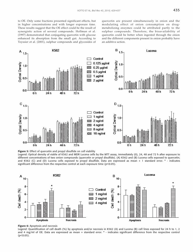
No significant cytotoxic effects were observed in eithercell line treated with quercetin (Fig. 5A,B) or propyl disulfide(Fig. 5C,D).
3.3. Detection of apoptosis/necrosis provoked by OE
In the K562 cells, a significant increase in the percentage ofapoptotic cells at a concentration of 4 mg/ml of OE wasobserved when compared to the control (Fig. 6A). TheLucena cells showed a significant increase in the percentageof necrotic cells at the same concentration (Fig. 6B).
3.4. Antioxidant capacity of OE
A significant decrease in ROS levels was verified in bothcell lines at a concentration of 2 mg/ml in relation torespective controls (Fig. 7A). However, a significantincrease in the ACAP value at a concentration of 2 mg/mlwas only verified in the K562 cells when compared to thecontrol (Fig. 7B).
3.5. DNA damage
The cell lines exhibited significant DNA damage at aconcentration of 2 mg/ml of OE when compared torespective controls, and this effect was similar for both celllines (Fig. 8).
4. DISCUSSION
Based on the potential properties of onion, this work begananalyzing OE effects on MDR and non-MDR erythroleukemiccell lines. The results clearly demonstrate the cytotoxiccapacity of OE. This led us to investigate which fraction orcompounds of the extract is responsible for the cytotoxiceffect. However, neither the fractioned extracts (aqueous,methanolic and ethyl acetate) or the purified compounds(quercetin and propyl disulfide) showed effects comparable
Figure 4: Chromatograms of quercetin and propyl disulfideLegend: Chromatograms of quercetin (A) and propyl disulfide (B). Quercetin standard 1 mg/L (line) and OE (dotted line) (A3); andUV-vis absorbing spectrograms of (A1) quercetin standard and (A2) OE; and chromatogram of standard solution with 0.5 mg/L ofpropyl disulfide (B1), sample analyses (B2) and spectrum of the standard solution in the full scan mode (B3).
435VOTTO ET AL. Biol Res 43, 2010, 429-437
Figure 5: Effect of quercetin and propyl disulfide on cell viabilityLegend: Optical density of viable of K562 and MDR Lucena cells by the MTT assay, immediately (0), 24, 48 and 72 h after exposure todifferent concentrations of two onion compounds (quercetin or propyl disulfide). (A) K562 and (B) Lucena cells exposed to quercetin;and K562 (C) and (D) Lucena cells exposed to propyl disulfide. Data are expressed as mean + 1 standard error. * - indicatessignificant difference from the respective control at each exposure time (p<0.05).
Figure 6: Apoptosis and necrosisLegend: Quantification of cell death (%) by apoptosis and/or necrosis in K562 (A) and Lucena (B) cell lines exposed for 24 h to 1, 2and 4 mg/ml of OE. Data are expressed as mean + standard error. * - indicates significant difference from the respective control(p<0.05).
to OE. Only some fractions presented significant effects, butin higher concentrations and with longer exposure time.These results suggest that the OE effect could be the result ofsynergistic action of several compounds. Hollman et al.(1997) demonstrated that conjugating quercetin with glucoseenhanced its absorption from the small gut. According toTeyssier et al. (2001), sulphur compounds and glycosides of
quercetin are present simultaneously in onion and themodulating effect of onion consumption on drug-metabolizing enzymes could be attributed partly to thesulphur compounds. Therefore, the bioavailability ofquercetin could be better when ingested through the onionand the different components present in onion probably havean additive action.

VOTTO ET AL. Biol Res 43, 2010, 429-437436
Figure 7. ROS and ACAPLegend: Reactive oxygen species (ROS) production (A) and antioxidant capacity against peroxil radicals (ACAP) (B) in K562 andLucena cells exposed for 24 h to 0.5, 1 and 2 mg/ml of OE. Data are expressed as mean + standard error. * - indicates significantdifference from the respective control (p<0.05).
Figure 8: DNA damageLegend: Quantification of DNA damage expressed by Olive tailmoment (OTM) values in K562 (A) and Lucena (B) cell linesexposed for 24 h to 2 mg/ml of OE. Data are expressed as mean+ standard error. * - indicates significant difference from therespective control (p<0.05).
The results demonstrating that OE was cytotoxic in bothK562 and Lucena cell lines, irrespective of MDR phenotype,are very important. Considering that drug resistance, bothprimary and acquired, is a major obstacle to advances incancer chemotherapy, the identification of new substanceswith anti-MDR properties is urgently required (Fernandes etal., 2003). The capacity to overcome resistance mechanismshas been attributed to a few compounds, such as: thetripterpenes, betulinic, oleanolic (OA) and pomolic acids(PA), isolated from Physocarpus intermedius, Licania tomentosaand Chrysobalanus icaco, respectively. The OA was almostequally cytotoxic to Lucena cells overexpressing Pgp incomparison to their K562 sensitive counterpart, and the PAwas highly effective in inhibiting the growth of both celllines (Fernandes et al., 2003). As well, the lupane triterpenelup-28-al-20(29)-en-3-one, a synthetic product, was found tobe a selective anti-leukemia compound and effective not onlyto K562 parental leukemia cells, but also to drug-resistantleukemia cells, producing similar cytotoxic effects (Hata et
al., 2003). Fernandes et al. (2005), using HL-60 cells, showedthat PA induced apoptosis by alteration of the mitochondrialmembrane potential.
In this work the capacity of OE to induce apoptosis inK562 cells was verified. Other compounds of natural originare also capable of inducing apoptosis in K562 or otherleukemic cell lines as HL-60, ML-1, U937 and DS-19 (Jing etal., 1999; Ye et al., 2005; Huang et al., 2005; Sreekanth et al.,2007). However in the present study, an increase in apoptoticcells was verified only in the K562 cells while the Lucenacells presented a significant increase in the percentage ofnecrotic cells. The death of the Lucena cells by necrosismight be explained by the data that show that Pgp, which isover-expressed in MDR cells, may provide resistance toprogrammed cell death (Ruefli and Johnstone, 2003).
As mentioned before, NPTS suppresses the growth ofHL-60 cells through the induction of apoptosis initiated byoxidative stress (Chang et al., 2005), suggesting an oxidantproperty associated with onion. However, based on theresults obtained in this work, an antioxidant capacity for OEwas verified, suggesting that oxidative stress is not involvedin the induction of cell death. Although only the K562 cellshave shown a significant increase in ACAP, the antioxidantcapacity of OE can be argued because Lucena cells present ahigher basal antioxidant capacity, which can explain thiseffect in these cells.
Others authors have also observed the antioxidantcapacity of onion. Sujatha and Srivinas (1995) showed thatthe aqueous extract of Allium cepa inhibited lipidperoxidation (LPO) in human erythrocyte membrane.Similarly, Helen et al. (2000) verified a decrease in the LPOlevels in liver, lung and heart of onion oil and nicotinetreated rats and that this treatment increased activity ofscavenging enzymes, such as catalase and superoxidedismutase (SOD) in all the tissues studied. El-Demerdash etal. (2005) showed that onion juice increased the activity ofglutathione S-transferase antioxidant enzyme (GST) in ratswith induced diabetes, which might be one of the defensemechanism in these animals to detoxify or neutralize thetoxic metabolites generated in liver by diabetes. According toPark et al. (2009), the methanolic extract of onion attenuatesischemia/hypoxia-induced apoptosis in heart-derived H9c2

437VOTTO ET AL. Biol Res 43, 2010, 429-437
cells and in rat hearts, through, at least in part, anantioxidant effect. Nuutila et al. (2003) affirm that thephenolic compounds of Allium plants contribute to theirantioxidant properties. Moreover, it is interesting toremember that the OE utilized in this work containquercetin, the major flavonol present in the onion (Prakash etal., 2007; Bonaccorsi et al., 2008), and that these compoundshave a good correlation with antioxidant capacity (Santas etal., 2008).
The fact that OE did not provoke oxidative stress mayexplain the lack of resistance of the MDR cell line to thistreatment. Trindade et al. (1999) showed that K562 andLucena cells possess different sensitivities to UVA radiation,which causes damage in cells and tissues preferentially byoxidative stress. These authors demonstrated that MDR cellswere very resistant to treatment and also to hydrogenperoxide. However, when the cell lines were exposed to UVBand UVC, which have DNA as preferential cellular target(Beer et al., 1993), the sensitivity was similar in both celllines. Besides, Trindade et al. (2000) utilizing photodynamicaction by methylene blue, which is well known for itscapacity to strongly bind DNA (Floyd et al . , 2004),demonstrated similar sensitivity among these and others celllines. In this work we have shown the same sensitivity inboth cell lines, supported also by similar DNA damage.
Thus, according to the results obtained in this work, it ispossible to suggest that there is a synergistic effect of OEwhen compared to fractioned extracts and compounds. Itsantioxidant capacity suggest that oxidative stress is notinvolved in the induction of cell death, and that the capacityto induce cell death by apoptosis in K562 cells or necrosis inLucena cells might be due to DNA damage. It is important tonote that OE was cytotoxic in both tumoral cell lines,irrespective of MDR phenotype, that permits also to suggestan anti-MDR action of OE.
ACKNOWLEDGEMENTS
This work was supported by the Programa de Pós-graduaçãoem Ciências Fisiológicas - Fisiologia Animal Comparada(FURG). B.S.D. received a fellowship from PBIC program(CNPq). We are thankful to Jorge Alberto Castro Benitez andRobert Tew Boyle for revision of the manuscript.
REFERENCES
AMADO LL, GARCIA ML, RAMOS PB, FREITAS RF, ZAFALON B,FERREIRA JLR, YUNES JS, MONSERRAT JM (2009) A method tomeasure total antioxidant capacity against peroxyl radicals in aquaticorganisms: application to evaluate microcystins toxicity. Sci TotalEnviron 407: 2115-2123.
AUGUSTI KT (1996) Therapeutic values of onion and garlic. J Exp Biol 34:634-640.
BEER JZ, OLVEY KM, MILLER SA, THOMAS DP, GODAR DE (1993)Non-nuclear damage and cell lysis are induced by UVA, but not UVBor UVC, radiation in three strains of L5178Y cells. PhotochemPhotobiol 58: 676-681.
BONACCORSI P, CARISTI C, GARGIULLI C, LEUZZI U (2008) Flavonolglucosides in Allium species: A comparative study by means ofHPLC-DAD-ESI-MS-MS. Food Chem 107: 1668-1673.
BOREK C (2005). Antioxidants and the prevention of hormonallyregulated cancer. Pract med 2: 346-352.
BRAGA F, AYRES-SARAIVA D, GATTASS CR, CAPELLA MAM (2007)Oleanolic acid inhibits the activity of the multidrug resistanceprotein ABCC1 (MRP1) but not of the ABCB1 (P-glycoprotein):Possible use in cancer chemotherapy. Cancer Lett 248: 147-152.
CHANG H-S, YAMATO O, YAMASAKI M, KO M, MAEDE Y (2005)Growth inhibitory effect of alk(en)yl Thiosulfates derived from onionand garlic in human immortalized and tumor cell lines. Cancer Lett223: 47-55.
CORZO-MARTÍNEZ M, CORZO N, VILLAMIEL M (2007) Biologicalproperties of onions and garlic. Trends Food Sci Technol 18: 609-625.
EL-DEMERDASH FM, YOUSEF MI, ABOU EL-NAGA NI (2005)Biochemical study on the hypoglycemic effects of onion and garlic inalloxan-induced diabetic rats. Food and Chemical Toxicol 43: 57-63.
FERNANDES J, CASTILHO RO, DA COSTA MR, WAGNER-SOUZA K,KAPLAN MAC, GATTASS CR (2003) Pentacyclic triterpenes fromChrysobalanaceae species: cytotoxicity on multidrug resistant andsensitive leukemia cell lines. Cancer Lett 190: 165-169.
FERNANDES J , WEINLICH R, CASTILHO RO, KAPLAN MAC,AMARANTE-MENDES GP, GATTASS CR (2005) Pomolic acidtriggers mitochondria-dependent apoptotic cell death in leukemiacell line. Cancer Lett 219: 49-55.
FLOYD RA, SCHNEIDER JR JE, DITTMER DP (2004) Methylene bluephotoinactivation of RNA viruses. Antiviral Res 61: 141-151.
FORD JM, HAIT WN (1990) Pharmacology of drugs that alter multidrugresistance in cancer. Pharmacology 42: 155-199.
FURLONG EB, BAISCH ALM, COLLA E, SOARES LAS (2003) Avaliaçãodo potencial de compostos fenólicos em tecidos vegetais. RevistaVetor 13: 105-114.
GOTTESMAN MM, PASTAN I (1993) Biochemistry of multidrugresistance mediated by the multidrug transporter. Annu RevBiochem 62: 385-427.
HATA K, HORI K, OGASAWARA H, TAKAHASHI S (2003) Anti-leukemia activities of Lup-28-al-20(29)-en-3-one, a lupane triterpene.Toxicol Lett 143: 1-7.
HELEN A, KRISHNAKUMAR K, VIJAYAMMAL PL, AUGUSTI KT (2000)Antioxidant effect of onion oil (Allium cepa. Linn) on the damagesinduced by nicotine in rats as compared to alpha-tocopherol. ToxicolLett 116: 61-68.
HOLLMAN PC, VAN-TRIJP JM, BUYSMAN MN, VAN-DER-GAAG MS,MENGELERS MJ, DEVRIES JH, KATAN MB (1997) Relativebioavailability of the antioxidant .avonoid quercetin from variousfoods in man. FEBS Letters 418: 152-156.
HUANG WW, YANG JS, LIN CF, HO WJ, LEE MR (2005) Pycnogenolinduces differentiation and apoptosis in human promyeloid leukemiaHL-60 cells. Leukemia Res 29: 685-692.
JING Y, NAKAJO S, XIA L, NAKAYA K, FANG Q, WAXMAN S, HAN R(1999) Boswellic acid acetate induces differentiation and apoptosis inleukemia cell lines. Leukemia Res 23: 43-50.
KONCA K, LANKOFF A, BANASIK A, LISOWSKAB H, KUSZEWSKI T,GOZDZ S, KOZA Z, WOJCIK A (2003) A cross-platform publicdomain PC image-analysis program for the comet assay. Mutat Res534: 15-20.
LANKOFF A, BANASIK A, OBE G, DEPERAS M, KUZMINSKI K,TARCZYNSKA M, JURCZAK T, WOJCIK A (2003) Effect ofmicrocystin-LR and cyanobacterial extract from Polish reservoir ofdrinking water on cell cycle progression, mitotic spindle, andapoptosis in CHO-K1 cells. Toxicol Appl Pharmacol 189: 204-213.
NUUTILA AM, PUUPPONEN-PIMIÄ R, AARNI M, OKSMAN-CALDENTEY, K-M (2003) Comparison of antioxidant activities ofonion and garlic extracts by inhibition of lipid peroxidation andradical scavenging activity. Food Chem 81: 485-493.
PARK S, KIM M, LEE DH, LEE SH, BAIK EJ, MOON C, PARK SW, KOEY, OH S, JUNG Y (2009) Methanolic extract of onion (Allium cepa)attenuates ischemia/hypoxia-induced apoptosis in cardiomyocytesvia antioxidant effect. Eur J Nutr 48: 235-242.
PRAKASH D, SINGH BM, UPADHYAY G (2007) Antioxidant and freeradical scavenging activities of phenols fromonion (Allium cepa).Food Chem 102: 1389-1393.
ROLDÁN E, SÁNCHEZ-MORENO C, DE ANCOS B, CANO MP (2008)Characterisation of onion (Allium cepa L.) by-products as foodingredients with antioxidant and antibrowning properties. FoodChem 108: 907-916.
RUEFLI AA, JOHNSTONE RW (2003) A role for P-glycoprotein inregulating cell growth and survival. Clin Appl Immunol Rev 4: 31-47.
RUMJANEK VM, LUCENA M, CAMPOS MM, MARQUES-SILVA VM,MAIA RC (1994) Multidrug resistance in leukemias: the problem andsome approaches to its circumvention. Ciênc Cult 46: 63-69.
SALEHEEN D, ALI SA, YASINZAI MM (2004) Antileishmanial activity ofaqueos onion extract in vitro. Fitoterapia 75: 9-13.
SANTAS J , CARBÓ R, GORDON MH, ALMAJANO MP (2008)
VOTTO ET AL. Biol Res 43, 2010, 429-437438
Comparison of the antioxidant activity of two Spanish onionvarieties. Food Chem 107: 1210-1216.
SEKI T, TSUJI K, HAYATO Y, MORITOMO T, ARIGA T (2000) Garlic andonion oils inhibit proliferation and induce differentiation of HL-60cells. Cancer Lett 160: 29-35.
SINGH NP, MCCOY MT, TICE RR, SCHEIDER EL (1988) A simpletechnique for quantitation of low levels of DNA damage inindividual cells. Exp Cell Res 237: 3-4.
SLIMESTAD R, FOSSEN T, VAGEN IM (2007) Onions: A Source ofUnique Dietary Flavonoids. J Agric Food Chem 55: 10067-10080.
SREEKANTH D, ARUNASREE MK, ROY KR, CHANDRAMOHANREDDY T, REDDY GV, REDDANNA P (2007) Betanin a betacyaninpigment purified from fruits of Opuntia ficus-indica induces apoptosisin human chronic myeloid leukemia Cell line-K562. Phytomedicine14: 739-746.
STEINERT SA, STREIB-MONTEE R, LEATHER JM (1998) DNA damagein mussels at sites in San Diego Bay. Mutat Res Fund Mol Mech Mut399: 65-85.
SUJATHA R, SRIVINAS L (1995) Modulation of lipid peroxidation bydietary components. Toxicol in vitro 9: 231-236.
TEYSSIER C, AMIOT MJ, MONDY N (2001) Effect of onion consumptionby rats on hepatic drug-metabolizing enzymes. Food Chem Toxicol39: 981-7.
TRINDADE GS, CAPELLA MAM, CAPELLA LS, AFFONSO-MITIDIEROR, RUMJANEK VM (1999) Differences in sensitivity to UVC, UVBand UVA radiation of a multidrug-resistant cell line overexpressingP-glycoprotein. Photochem Photobiol 69: 694-699.
TRINDADE GS, FARIAS SLA, RUMJANEK VM, CAPELLA MAM (2000)Methylene blue reverts multidrug resistance: sensitivity of multidrugresistant cells to this dye and its photodynamic action. Cancer Lett14: 161-167.
TSURUO T, LIDA H, OHKOCHI E, TSUKAGOSHI S, SAKURAI Y (1983)Establishment and properties of a vincristine-resistant humanmyelogenous leukemia K562. J Cancer Res 74: 751-758.
WACH A, PYRZYNSKA K, BIESAGA M (2007) Quercetin content in somefood and herbal samples. Food Chem 100: 699-704.
YE C-L, QIAN F, WEI D-Z, LU Y-H, LIU J-W (2005) Induction ofapoptosis in K562 human leukemia cells by 2’,4’-dihydroxy-6’-methoxy-3’,5’-dimethylchalcone. Leukemia Res 29: 887-892.
Copyright © 2022 FDOKUMEN