
TOMMANN TIL AT HAN HATI
92
TOMMANN TIL AT HAN HATI US009931330B2 ( 12 ) United States Patent Zarnitsyn et al . ( 10 ) Patent No .: US 9, 931 , 330 B2 ( 45 ) Date of Patent : * Apr . 3, 2018 ( 58 ) ( 54 ) METHODS AND DEVICES FOR THE TREATMENT OF OCULAR DISEASES IN HUMAN SUBJECTS Field of Classification Search CPC . . A61K 31 / 4439 ; A61K 9/ 0048 ; A61K 47 /26 ; A61K 45 /06 ; A61K 31 / 573 ; ( Continued ) ( 71 ) Applicant : CLEARSIDE BIOMEDICAL , INC ., Alpharetta , GA ( US ) ( 56 ) References Cited U . S . PATENT DOCUMENTS ( 72 ) Inventors : Vladimir Zarnitsyn , Atlanta , GA ( US ) ; Samirkumar Patel , Atlanta , GA ( US ) ; Daniel White , Suwanee , GA ( US ) ; Glenn Noronha , Atlanta , GA ( US ); Brian Burke , Cary , NC ( US ) 2 , 187 , 259 A 2 , 841 , 145 A 1/ 1940 Barnhart 7/ 1958 Epps ( Continued ) @ FOREIGN PATENT DOCUMENTS ( 73 ) Assignee : CLEARSIDE BIOMEDICAL , INC . , Alpharetta , GA ( US ) ?? ( * ) Notice : 2639322 3/ 2009 1706365 12 / 2005 ( Continued ) Subject to any disclaimer , the term of this patent is extended or adjusted under 35 U .S .C . 154 ( b ) by 0 days . This patent is subject to a terminal dis claimer . OTHER PUBLICATIONS Brown , David M ., “ Aflibercept for Treatment of Diabetic Macular Edema , ” Retina Today , Jul . / Aug . 2011 , pp . 59 - 60 .* ( Continued ) @ ( 21 ) Appl . No .: 15 673 , 073 ( 22 ) Filed : Aug . 9 , 2017 Primary Examiner - Aradhana Sasan ( 74 ) Attorney , Agent , or Firm — Cooley LLP ( 65 ) Prior Publication Data US 2017 / 0333416 A1 Nov . 23 , 2017 Related U .S . Application Data ( 63 ) Continuation of application No . 15 / 454 , 636 , filed on Mar . 9, 2017 , which is a continuation of application ( Continued ) ( 57 ) ABSTRACT Methods and devices are provided for targeted non - surgical administration of a drug formulation to the suprachoroidal space ( SCS ) of the eye of a human subject for the treatment of a posterior ocular disorder or a choroidal malady . In one embodiment , the method comprises inserting a hollow microneedle into the eye at an insertion site and infusing a drug formulation through the inserted microneedle and into the suprachoroidal space of the eye , wherein the infused drug formulation flows within the suprachoroidal space away from the insertion site during the infusion . In one embodiment , the fluid drug formulation comprises drug nanoparticles or microparticles . ( 51 ) Int . Ci . A61K 31 / 4439 ( 2006 . 01 ) A61K 47 / 26 ( 2006 . 01 ) ( Continued ) ( 52 ) U .S . Cl . CPC ..... ... A61K 31 / 4439 ( 2013 . 01 ); A61K 9 / 0019 ( 2013 . 01 ); A61K 9/ 0048 ( 2013 . 01 ); ( Continued ) 12 Claims , 40 Drawing Sheets 10 * * * - * * * * * * - . * . * * . - - * * * * * ** * * * * * * * . * * * * * : . . . . . 18 WY 4 . 4 * * * FIG . . . . . . . . . 177 FIS . . L ' - - - VE ) . - * * * * * * * * * * * * * * * * * - * * Y . *
-
Upload
khangminh22 -
Category
Documents
-
view
0 -
download
0
Transcript of TOMMANN TIL AT HAN HATI

TOMMANN TIL AT HAN HATI US009931330B2
( 12 ) United States Patent Zarnitsyn et al .
( 10 ) Patent No . : US 9 , 931 , 330 B2 ( 45 ) Date of Patent : * Apr . 3 , 2018
( 58 ) ( 54 ) METHODS AND DEVICES FOR THE TREATMENT OF OCULAR DISEASES IN HUMAN SUBJECTS
Field of Classification Search CPC . . A61K 31 / 4439 ; A61K 9 / 0048 ; A61K 47 / 26 ;
A61K 45 / 06 ; A61K 31 / 573 ; ( Continued ) ( 71 ) Applicant : CLEARSIDE BIOMEDICAL , INC . ,
Alpharetta , GA ( US ) ( 56 ) References Cited U . S . PATENT DOCUMENTS ( 72 ) Inventors : Vladimir Zarnitsyn , Atlanta , GA ( US ) ;
Samirkumar Patel , Atlanta , GA ( US ) ; Daniel White , Suwanee , GA ( US ) ; Glenn Noronha , Atlanta , GA ( US ) ; Brian Burke , Cary , NC ( US )
2 , 187 , 259 A 2 , 841 , 145 A
1 / 1940 Barnhart 7 / 1958 Epps
( Continued )
@ FOREIGN PATENT DOCUMENTS ( 73 ) Assignee : CLEARSIDE BIOMEDICAL , INC . , Alpharetta , GA ( US )
??
( * ) Notice : 2639322 3 / 2009 1706365 12 / 2005
( Continued ) Subject to any disclaimer , the term of this patent is extended or adjusted under 35 U . S . C . 154 ( b ) by 0 days . This patent is subject to a terminal dis claimer .
OTHER PUBLICATIONS Brown , David M . , “ Aflibercept for Treatment of Diabetic Macular Edema , ” Retina Today , Jul . / Aug . 2011 , pp . 59 - 60 . *
( Continued ) @ ( 21 ) Appl . No . : 15 673 , 073
( 22 ) Filed : Aug . 9 , 2017 Primary Examiner - Aradhana Sasan ( 74 ) Attorney , Agent , or Firm — Cooley LLP ( 65 ) Prior Publication Data
US 2017 / 0333416 A1 Nov . 23 , 2017 Related U . S . Application Data
( 63 ) Continuation of application No . 15 / 454 , 636 , filed on Mar . 9 , 2017 , which is a continuation of application
( Continued )
( 57 ) ABSTRACT Methods and devices are provided for targeted non - surgical administration of a drug formulation to the suprachoroidal space ( SCS ) of the eye of a human subject for the treatment of a posterior ocular disorder or a choroidal malady . In one embodiment , the method comprises inserting a hollow microneedle into the eye at an insertion site and infusing a drug formulation through the inserted microneedle and into the suprachoroidal space of the eye , wherein the infused drug formulation flows within the suprachoroidal space away from the insertion site during the infusion . In one embodiment , the fluid drug formulation comprises drug nanoparticles or microparticles .
( 51 ) Int . Ci . A61K 31 / 4439 ( 2006 . 01 ) A61K 47 / 26 ( 2006 . 01 )
( Continued ) ( 52 ) U . S . Cl .
CPC . . . . . . . . A61K 31 / 4439 ( 2013 . 01 ) ; A61K 9 / 0019 ( 2013 . 01 ) ; A61K 9 / 0048 ( 2013 . 01 ) ; ( Continued ) 12 Claims , 40 Drawing Sheets
10 * * *
- * * * * * * -
. * . * * . - - * * * * * * * *
* * * * * *
.
* * * * *
: . . . . .
18 WY 4 . 4 *
* *
FIG . . . . . . . . . 177 FIS .
. L ' - - - VE ) . -
* * * * * * *
* * * * * * *
* * * - * * Y . *
US 9 , 931 , 330 B2 Page 2
Related U . S . Application Data No . 14 / 441 , 151 , filed as application No . PCT / US2013 / 069156 on Nov . 8 , 2013 , now abandoned .
( 60 ) Provisional application No . 61 / 898 , 926 , filed on Nov . 1 , 2013 , provisional application No . 61 / 873 , 660 , filed on Sep . 4 , 2013 , provisional application No . 61 / 819 , 388 , filed on May 3 , 2013 , provisional application No . 61 / 785 , 229 , filed on Mar . 14 , 2013 , provisional application No . 61 / 773 , 124 , filed on Mar . 5 , 2013 , provisional application No . 61 / 745 , 237 , filed on Dec . 21 , 2012 , provisional application No . 61 / 734 , 872 , filed on Dec . 7 , 2012 , provisional application No . 61 / 724 , 144 , filed on Nov . 8 , 2012 .
( 51 ) Int . CI . A61K 45 / 06 ( 2006 . 01 ) A61K 9 / 00 ( 2006 . 01 ) A61K 4738 ( 2006 . 01 ) A61K 9 / 16 ( 2006 . 01 ) A61K 31 / 573 ( 2006 . 01 ) A61K 47 / 12 ( 2006 . 01 ) A61K 9 / 10 ( 2006 . 01 ) A61K 39 / 00 ( 2006 . 01 ) AGIM 37 / 00 ( 2006 . 01 ) COOK 16 / 22 ( 2006 . 01 )
( 52 ) U . S . CI . ??? . . . . A61K 9 / 10 ( 2013 . 01 ) ; A61K 9 / 16
( 2013 . 01 ) ; A61K 31 / 573 ( 2013 . 01 ) ; A61K 45 / 06 ( 2013 . 01 ) ; A61K 47 / 12 ( 2013 . 01 ) ; A61K
47 / 26 ( 2013 . 01 ) ; A61K 47 / 38 ( 2013 . 01 ) ; AGIM 37 / 0015 ( 2013 . 01 ) ; COOK 16 / 22
( 2013 . 01 ) ; A61K 2039 / 505 ( 2013 . 01 ) ; A61K 2039 / 54 ( 2013 . 01 ) ; A61K 2300 / 00 ( 2013 . 01 ) ;
A61M 2037 / 0023 ( 2013 . 01 ) ; A61M 2037 / 0061 ( 2013 . 01 )
( 58 ) Field of Classification Search CPC . A61K 47 / 38 ; A61K 9 / 10 ; A61K 9 / 16 ; A61K
47 / 12 ; A61K 9 / 0019 ; C07K 16 / 22 , A61M 37 / 0015 ; A61M 2037 / 0023 ; A61M
2037 / 0061 See application file for complete search history .
4 , 826 , 490 A 4 , 826 , 871 A 4 , 889 , 529 A 4 , 941 , 874 A 4 , 966 , 773 A 5 , 015 , 240 A 5 , 066 , 276 A 5 , 098 , 389 A 5 , 137 , 447 A 5 , 164 , 188 A 5 , 172 , 807 A 5 , 181 , 909 A 5 , 273 , 530 A 5 , 279 , 564 A 5 , 295 , 972 A 5 , 300 , 084 A 5 , 312 , 361 A 5 , 354 , 286 A 5 , 364 , 373 A 5 , 364 , 374 A 5 , 364 , 734 A 5 , 397 , 313 A 5 , 399 , 159 A 5 , 407 , 070 A 5 , 443 , 505 A 5 , 538 , 503 A 5 , 547 , 467 A 5 , 632 , 740 A 5 , 658 , 256 A D383 , 049 S 5 , 667 , 491 A 5 , 681 , 825 A 5 , 766 , 242 A 5 , 767 , 079 A 5 , 779 , 668 A 5 , 788 , 679 A 5 , 792 , 099 A 5 , 817 , 075 A 5 , 824 , 072 A 5 , 911 , 223 A 5 , 952 , 378 A 5 , 968 , 022 A 6 , 059 , 111 A 6 , 083 , 199 A 6 , 143 , 329 A 6 , 280 , 470 B1 6 , 299 , 603 B1 6 , 309 , 347 B1 6 , 309 , 374 B1 6 , 319 , 240 B1 6 , 334 , 856 B1 6 , 378 , 526 B1 6 , 387 , 078 B1 6 , 397 , 849 B1 6 , 432 , 090 B1 6 , 503 , 231 B1 6 , 524 , 581 B1 6 , 540 , 725 B1 6 , 551 , 299 B2 6 , 611 , 707 B1 6 , 622 , 864 B1 6 , 743 , 211 B1 6 , 773 , 916 B1 D499 , 153 S 6 , 883 , 222 B2 6 , 918 , 889 B1 6 , 929 , 623 B2 6 , 936 , 053 B1 7 , 025 , 774 B2 7 , 150 , 735 B2 7 , 207 , 965 B2 7 , 207 , 980 B2 7 , 211 , 062 B2 7 , 214 , 212 B2 7 , 226 , 439 B2 7 , 316 , 676 B2 7 , 425 , 207 B2 7 , 435 , 237 B2 7 , 468 , 057 B2
5 / 1989 Byrne et al . 5 / 1989 Gressel et al .
12 / 1989 Haindl 7 / 1990 Sandow et al .
10 / 1990 Gressel et al . 5 / 1991 Soproni et al . 11 / 1991 Wang
3 / 1992 Cappucci 8 / 1992 Hunter
11 / 1992 Wong 12 / 1992 Dragan et al . 1 / 1993 McFarlane
12 / 1993 del Cerro et al . 1 / 1994 Taylor 3 / 1994 Mischenko 4 / 1994 Johnson 5 / 1994 Zadini et al .
10 / 1994 Mesa et al . 11 / 1994 Waskonig et al . 11 / 1994 Morrison et al . 11 / 1994 Morrison et al . 3 / 1995 Gross 3 / 1995 Chin et al . 4 / 1995 Bascos et al . 8 / 1995 Wong et al . 7 / 1996 Henley et al . 8 / 1996 Pliquett et al . 5 / 1997 Koch et al . 8 / 1997 Shields 9 / 1997 Concari et al . 9 / 1997 Pliquett et al .
10 / 1997 Lindqvist et al . 6 / 1998 Wong et al . 6 / 1998 Glaser et al . 7 / 1998 Grabenkort 8 / 1998 Gravlee , Jr . 8 / 1998 DeCamp et al .
10 / 1998 Giungo 10 / 1998 Wong 6 / 1999 Weaver et al . 9 / 1999 Stjernschantz et al .
10 / 1999 Saito 5 / 2000 Davilla et al . 7 / 2000 Thorley et al .
| 11 / 2000 Kim 8 / 2001 Peyman
10 / 2001 Hecker et al . 10 / 2001 Takahashi et al . 10 / 2001 Hecker et al . 11 / 2001 Beck 1 / 2002 Allen et al . 4 / 2002 Bowman et al . 5 / 2002 Gillespie , III 6 / 2002 Bowman et al . 8 / 2002 Brunel 1 / 2003 Prausnitz et al . 2 / 2003 Adamis 4 / 2003 Ponzi 4 / 2003 Miyoshi et al . 8 / 2003 Prausnitz et al . 9 / 2003 Debbs et al . 6 / 2004 Prausnitz et al . 8 / 2004 Thiel et al . 11 / 2004 Kuo 4 / 2005 Landau 7 / 2005 Brunel 8 / 2005 Stone 8 / 2005 Weiss 4 / 2006 Freeman et al .
12 / 2006 Hickle 4 / 2007 Simon 4 / 2007 Christian et al . 5 / 2007 Kwon 5 / 2007 Pommereau et al . 6 / 2007 Prausnitz et al . 1 / 2008 Peyman et al . 9 / 2008 Miller et al .
10 / 2008 Tan 12 / 2008 Ponzi
( 56 ) References Cited U . S . PATENT DOCUMENTS
2 , 939 , 459 A 3 , 376 , 999 A 3 , 477 , 432 A 3 , 739 , 947 A 3 , 762 , 540 A 3 , 788 , 320 A 3 , 892 , 311 A 3 , 964 , 482 A 4 , 226 , 328 A 4 , 377 , 897 A 4 , 383 , 530 A 4 , 417 , 887 A 4 , 501 , 363 A 4 , 564 , 016 A 4 , 601 , 708 A 4 , 615 , 331 A 4 , 689 , 040 A 4 , 708 , 147 A 4 , 717 , 383 A 4 , 736 , 850 A 4 , 755 , 169 A 4 , 795 , 432 A 4 , 804 , 371 A
6 / 1960 Lazarte et al . 4 / 1968 De Hart et al .
11 / 1969 Shaw 6 / 1973 Baumann et al .
10 / 1973 Baumann et al . 1 / 1974 Dye 7 / 1975 Sneider 6 / 1976 Gerstel et al . 10 / 1980 Beddow 3 / 1983 Eichenbaum et al . 5 / 1983 Bruno
11 / 1983 Koshi 2 / 1985 Isbey , Jr . 1 / 1986 Maurice et al . 7 / 1986 Jordan
10 / 1986 Kramann 8 / 1987 Thompson
11 / 1987 Haaga 1 / 1988 Phillips et al . 4 / 1988 Bowman et al . 7 / 1988 Sarnoff et al . 1 / 1989 Karczmer 2 / 1989 Vaillancourt

US 9 , 931 , 330 B2 Page 3
( 56 ) References Cited U . S . PATENT DOCUMENTS
Hoi D590 , 690 S 4 / 2009 Bertini D598 , 543 S 8 / 2009 Vogel et al . 7 , 569 , 035 B1 8 / 2009 Wilmot et al . 7 , 615 , 041 B2 11 / 2009 Sullivan et al . 7 , 678 , 077 B2 3 / 2010 Harris et al . 7 , 678 , 078 B1 . 3 / 2010 Peyman et al . 7 , 722 , 581 B2 5 / 2010 Peyman 7 , 914 , 803 B2 3 / 2011 Chowhan et al . 7 , 918 , 814 B2 4 / 2011 Prausnitz et al . 7 , 918 , 874 B2 4 / 2011 Siegal 7 , 947 , 660 B2 5 / 2011 Clark et al . 7 , 967 , 772 B2 6 / 2011 McKenzie et al . 8 , 003 , 124 B2 8 / 2011 Varner et al . 8 , 114 , 110 B2 2 / 2012 Bednarek et al . 8 , 137 , 312 B2 3 / 2012 Sundar et al . 8 , 172 , 830 B2 5 / 2012 Christian et al . 8 , 173 , 617 B2 5 / 2012 Clark et al . 8 , 192 , 408 B2 6 / 2012 Nazzaro et al . 8 , 197 , 435 B2 6 / 2012 Prausnitz et al . 8 , 197 , 443 B2 6 / 2012 Sundar et al . 8 , 221 , 353 B2 7 / 2012 Cormier et al . 8 , 235 , 967 B2 8 / 2012 Chevallier et al . D667 , 111 S 9 / 2012 Robinson 8 , 287 , 494 B2 10 / 2012 Ma 8 , 303 , 599 B2 11 / 2012 Hess et al . D672 , 506 S 12 / 2012 Szymanski 8 , 323 , 227 B2 12 / 2012 Hamatake et al . 8 , 328 , 772 B2 12 / 2012 Kinast et al . 8 , 337 , 421 B2 12 / 2012 Freeman et al . 8 , 337 , 509 B2 12 / 2012 Schieber et al . 8 , 348 , 924 B2 1 / 2013 Christian et al . 8 , 403 , 941 B23 / 2013 Peterson et al . 8 , 430 , 862 B2 4 / 2013 Peyman et al . 8 , 448 , 786 B2 5 / 2013 Tomes et al . 8 , 460 , 242 B2 6 / 2013 Paques et al . 8 , 506 , 515 B2 8 / 2013 Burns et al . 8 , 529 , 492 B2 9 / 2013 Clauson et al . 8 , 535 , 333 B2 9 / 2013 de Juan , Jr . et al . 8 , 545 , 430 B2 10 / 2013 Silvestrini 8 , 545 , 554 B2 10 / 2013 Novakovic et al . 8 , 562 , 545 B2 10 / 2013 Freeman et al . 8 , 571 , 802 B2 10 / 2013 Robinson et al . 8 , 574 , 214 B2 11 / 2013 Kuhn et al . 8 , 574 , 217 B2 11 / 2013 Peyman 8 , 602 , 959 B1 12 / 2013 Park et al . 8 , 617 , 121 B2 12 / 2013 Lanin et al . 8 , 632 , 589 B2 1 / 2014 H 8 , 636 , 713 B2 1 / 2014 Prausnitz et al . 8 , 652 , 118 B2 2 / 2014 Peyman 8 , 663 , 167 B2 3 / 2014 Bartha 8 , 663 , 303 B2 3 / 2014 Horvath et al . 8 , 668 , 676 B2 3 / 2014 Chang 8 , 685 , 435 B2 4 / 2014 Nivaggioli et al . 8 , 702 , 659 B2 4 / 2014 Lanin et al . 8 , 727 , 117 B2 5 / 2014 Maasarani 8 , 747 , 365 B2 6 / 2014 De Sausmarez Lintell 8 , 795 , 226 B2 8 / 2014 Kuhn et al . 8 , 808 , 225 B2 8 / 2014 Prausnitz et al . 8 , 808 , 242 B2 8 / 2014 Paques et al . D713 , 958 S 9 / 2014 Srinivasan et al . 8 , 821 , 870 B2 9 / 2014 Robinson et al . D715 , 125 S 10 / 2014 Hung 8 , 852 , 137 B210 / 2014 Horvath et al . 8 , 864 , 740 B2 10 / 2014 Schabbach et al . D718 , 602 S 12 / 2014 Musser D719 , 256 S 12 / 2014 Ohashi 8 , 920 , 375 B2 12 / 2014 Gonnelli D726 , 908 S 4 / 2015 Yu et al . D733 , 289 S 6 / 2015 Blanchard et al . D740 , 098 S 10 / 2015 Kuo et al . 9 , 180 , 047 B2 11 / 2015 Andino et al . D750 , 223 S 2 / 2016 Andino et al . 9 , 539 , 139 B2 1 / 2017 Andino et al . 9 , 572 , 800 B2 2 / 2017 Zarnitsyn et al . 9 , 636 , 253 B1 5 / 2017 Andino et al .
9 , 636 , 332 B2 5 / 2017 Zarnitsyn et al . 9 , 770 , 361 B2 9 / 2017 Andino et al .
2001 / 0008961 A1 7 / 2001 Hecker et al . 2001 / 0051798 A1 12 / 2001 Hochman 2002 / 0042594 Al 4 / 2002 Lum et al . 2002 / 0052580 A1 5 / 2002 Ooyauchi 2002 / 0082527 A1 6 / 2002 Liu et al . 2002 / 0082543 A1 6 / 2002 Park et al . 2002 / 0108875 Al 8 / 2002 Feinberg et al . 2002 / 0112981 A1 8 / 2002 Cooper et al . 2002 / 0156413 Al 10 / 2002 Williams et al . 2003 / 0009113 AL 1 / 2003 Olson 2003 / 0083645 Al 5 / 2003 Angel et al . 2003 / 0139729 Al 7 / 2003 Stegmann et al . 2003 / 0171722 A1 9 / 2003 Paques et al . 2004 / 0039253 A1 2 / 2004 Peyman et al . 2004 / 0106904 A1 6 / 2004 Gonnelli et al . 2004 / 0122359 Al 6 / 2004 Wenz et al . 2004 / 0199130 Al 10 / 2004 Chornenky et al . 2004 / 0215347 Al 10 / 2004 Hayes 2004 / 0249404 Al 12 / 2004 Haefliger 2004 / 0265365 A112 / 2004 Daddona et al . 2005 / 0033230 A1 2 / 2005 Alchas et al . 2005 / 0055083 A1 3 / 2005 Carranza et al . 2005 / 0065137 A1 3 / 2005 Jani et al . 2005 / 0089545 Al 4 / 2005 Kuwano et al . 2005 / 0101582 Al 5 / 2005 Lyons et al . 2005 / 0101882 Al 5 / 2005 Leira et al . 2005 / 0101967 A1 5 / 2005 Weber et al . 2005 / 0137525 A1 6 / 2005 Wang et al . 2005 / 0171507 Al 8 / 2005 Christian et al . 2005 / 0209565 Al 9 / 2005 Yuzhakov et al . 2005 / 0244462 AL 11 / 2005 Farooq 2005 / 0244463 AL 11 / 2005 Huang et al . 2005 / 0244469 A1 11 / 2005 Whitcup et al . 2005 / 0245906 A1 11 / 2005 Makower et al . 2005 / 0281862 Al 12 / 2005 Karakelle et al . 2006 / 0013859 Al 1 / 2006 Yamada et al . 2006 / 0032768 A1 2 / 2006 Hamai et al . 2006 / 0036318 A1 2 / 2006 Foulkes 2006 / 0084942 Al 4 / 2006 Kim et al . 2006 / 0086689 Al 4 / 2006 Raju 2006 / 0141049 Al 6 / 2006 Lyons et al . 2006 / 0173418 A1 8 / 2006 Rinaudo et al . 2006 / 0178614 A1 8 / 2006 Nemati 2006 / 0189608 Al 8 / 2006 Bingaman 2006 / 0229562 Al 10 / 2006 Marsh et al . 2006 / 0233858 AL 10 / 2006 Tzekov et al . 2006 / 0259008 A1 11 / 2006 Orilla 2006 / 0271025 AL 11 / 2006 Jones et al . 2007 / 0060927 A1 3 / 2007 Longson et al . 2007 / 0073197 A1 3 / 2007 Prausnitz et al . 2007 / 0082841 A1 4 / 2007 Higuchi et al . 2007 / 0093877 A1 4 / 2007 Beecham et al . 2007 / 0151882 Al 7 / 2007 Cocheteux et al . 2007 / 0178197 A1 8 / 2007 Larue et al . 2007 / 0202186 Al 8 / 2007 Yamamoto et al . 2007 / 0224278 A1 9 / 2007 Lyons et al . 2007 / 0225654 Al 9 / 2007 Hess et al . 2007 / 0233037 Al 10 / 2007 Gifford et al . 2007 / 0260201 A1 11 / 2007 Prausnitz et al . 2007 / 0270745 AL 11 / 2007 Nezhat et al . 2007 / 0270768 A1 11 / 2007 Dacquay et al . 2007 / 0282405 Al 12 / 2007 Wong et al . 2007 / 0287985 Al 12 / 2007 Estes et al . 2008 / 0033351 Al 2 / 2008 Trogden et al . 2008 / 0058704 Al 3 / 2008 Hee et al . 2008 / 0058717 Al 3 / 2008 Spector 2008 / 0071246 A1 3 / 2008 Nazzaro et al . 2008 / 0097346 Al 4 / 2008 Charles 2008 / 0097390 A1 4 / 2008 Dacquay et al . 2008 / 0131484 A1 6 / 2008 Robinson et al . 2008 / 0152694 A1 6 / 2008 Lobl et al . 2008 / 0177239 Al 7 / 2008 Li et al . 2008 / 0228127 A1 9 / 2008 Burns et al . 2009 / 0030381 A1 1 / 2009 Lind et al . 2009 / 0076463 A1 3 / 2009 Attinger 2009 / 0081277 Al 3 / 2009 Robinson et al . 2009 / 0088721 A14 / 2009 Bizemont et al .

US 9 , 931 , 330 B2 Page 4
( 56 ) References Cited U . S . PATENT DOCUMENTS
2009 / 0105749 AL 4 / 2009 De Juan et al . 2009 / 0148527 A1 6 / 2009 Robinson 2009 / 0259180 A1 10 / 2009 Choi 2009 / 0287161 AL 11 / 2009 Traub et al . 2009 / 0312782 AL 12 / 2009 Park 2010 / 0010452 A1 1 / 2010 Paques et al . 2010 / 0012537 A1 1 / 2010 Farrar et al . 2010 / 0015158 Al 1 / 2010 Robinson et al . 2010 / 0057011 A1 3 / 2010 Charles 2010 / 0074957 Al 3 / 2010 Robinson et al . 2010 / 0100054 Al 4 / 2010 Cormier et al . 2010 / 0152646 A1 6 / 2010 Girijavallabhan et al . 2010 / 0152667 A1 6 / 2010 Kietzmann 2010 / 0173866 Al 7 / 2010 Hee et al . 2010 / 0191176 A1 7 / 2010 Ho et al . 2010 / 0211079 Al 8 / 2010 Aramant 2010 / 0256597 Al 10 / 2010 Prausnitz et al . 2010 / 0312120 Al 12 / 2010 Meier 2011 / 0004265 A1 1 / 2011 Wenger et al . 2011 / 0060310 Al 3 / 2011 Prestrelski et al . 2011 / 0112546 Al 5 / 2011 Juan , Jr . et al . 2011 / 0166531 A1 7 / 2011 Stroumpoulis et al . 2011 / 0202012 A1 8 / 2011 Bartlett 2011 / 0213317 A1 9 / 2011 Chen et al . 2011 / 0238075 Al 9 / 2011 Clauson et al . 2011 / 0282298 Al 11 / 2011 Again et al . 2011 / 0306923 A1 12 / 2011 Roy 2012 / 0004245 A1 1 / 2012 May et al . 2012 / 0024987 A1 2 / 2012 Nagele Nacken 2012 / 0029360 A1 2 / 2012 Hendriks et al . 2012 / 0035524 A1 2 / 2012 Silvestrini 2012 / 0078224 A1 3 / 2012 Lerner et al . 2012 / 0083727 Al 4 / 2012 Barnett 2012 / 0095414 Al 4 / 2012 Lanin et al . 2012 / 0095438 A1 4 / 2012 Lanin et al . 2012 / 0101475 A1 4 / 2012 Wilmot et al . 2012 / 0116306 A1 5 / 2012 Heald et al . 2012 / 0123351 A1 5 / 2012 Lanin et al . 2012 / 0123437 Al 5 / 2012 Horvath et al . 2012 / 0123440 Al 5 / 2012 Horvath et al . 2012 / 0123473 Al 5 / 2012 Hernandez 2012 / 0130207 A 5 / 2012 O ’ dea et al . 2012 / 0136318 Al 5 / 2012 Lanin et al . 2012 / 0150128 A1 6 / 2012 Zhao 2012 / 0157880 A1 6 / 2012 Haselby et al . 2012 / 0165723 Al 6 / 2012 Horvath et al . 2012 / 0191064 Al 7 / 2012 Conston et al . 2012 / 0197208 Al 8 / 2012 Bruggemann et al . 2012 / 0226260 A1 9 / 2012 Prausnitz et al . 2012 / 0232522 A1 9 / 2012 Prausnitz et al . 2012 / 0259288 Al 10 / 2012 Wagner et al . 2012 / 0265149 Al 10 / 2012 Lerner et al . 2012 / 0271272 A1 10 / 2012 Hammack et al . 2013 / 0035662 A1 2 / 2013 Decker et al . 2013 / 0041265 Al 2 / 2013 Sostek et al . 2013 / 0060202 Al 3 / 2013 Thorley et al . 2013 / 0072900 A1 3 / 2013 Colantonio 2013 / 0079716 A1 3 / 2013 Thorley et al . 2013 / 0096533 A1 4 / 2013 Freeman et al . 2013 / 0102973 Al 4 / 2013 Thorley et al . 2013 / 0116523 Al 5 / 2013 Jung et al . 2013 / 0138049 A1 5 / 2013 Kemp et al . 2013 / 0140208 Al 6 / 2013 Hemmann 2013 / 0150803 A1 6 / 2013 Shetty et al . 2013 / 0190694 Al 7 / 2013 Barrow - Williams et al . 2013 / 0211335 Al 8 / 2013 Paques et al . 2013 / 0216623 Al 8 / 2013 Yamamoto et al . 2013 / 0218102 Al 8 / 2013 Iwase et al . 2013 / 0218269 A1 8 / 2013 Schachar et al . 2013 / 0237910 Al 9 / 2013 Shetty et al . 2013 / 0237916 A1 9 / 2013 Hanson et al . 2013 / 0245600 A1 9 / 2013 Yamamoto et al . 2013 / 0289545 Al 10 / 2013 Baerveldt et al . 2013 / 0295006 A1 11 / 2013 Christoforidis et al . 2013 / 0331786 A112 / 2013 Hofmann
2013 / 0338612 Al 12 / 2013 Smith et al . 2014 / 0012226 A11 / 2014 Hochman 2014 / 0027326 A1 1 / 2014 Peruzzo 2014 / 0031833 A1 1 / 2014 Novakovic et al . 2014 / 0039391 Al 2 / 2014 Clarke et al . 2014 / 0039413 A1 2 / 2014 Jugl et al . 2014 / 0078854 A1 3 / 2014 Head et al . 2014 / 0094752 A1 4 / 2014 Hiles 2014 / 0102927 A1 4 / 2014 Liversidge 2014 / 0107566 A1 4 / 2014 Prausnitz et al . 2014 / 0114243 Al 4 / 2014 Smith et al . 2014 / 0135716 A1 5 / 2014 Clarke et al . 2014 / 0194834 Al 7 / 2014 Passaglia et al . 2014 / 0200518 A1 7 / 2014 Ekman et al . 2014 / 0224688 A1 8 / 2014 Slemmen et al . 2014 / 0231287 Al 8 / 2014 Tomes et al . 2014 / 0236098 A1 8 / 2014 Mica et al . 2014 / 0243754 A1 8 / 2014 Clarke et al . 2014 / 0249539 A1 9 / 2014 Mica et al . 2014 / 0257207 Al 9 / 2014 Clarke et al . 2014 / 0276482 A1 9 / 2014 Astafieva et al . 2014 / 0296802 Al 10 / 2014 Geiger et al . 2014 / 0309599 Al 10 / 2014 Schaller 2014 / 0323979 Al 10 / 2014 Henley et al . 2014 / 0323985 Al 10 / 2014 Hourmand et al . 2014 / 0330213 Al 11 / 2014 Hourmand et al . 2014 / 0350479 AL 11 / 2014 Hourmand et al . 2014 / 0353190 A112 / 2014 Okihara et al . 2015 / 0013827 A11 / 2015 Kuhn 2015 / 0013835 Al 1 / 2015 Cordes 2015 / 0025474 A1 1 / 2015 Riedel et al . 2015 / 0038905 A1 2 / 2015 Andino et al . 2015 / 0045731 A1 2 / 2015 Gupta et al . 2015 / 0045744 Al 2 / 2015 Gupta et al . 2015 / 0051545 A1 2 / 2015 Henderson et al . 2015 / 0051581 A1 2 / 2015 Andino et al . 2015 / 0129456 Al 5 / 2015 Miller et al . 2015 / 0133415 A1 5 / 2015 Whitcup 2015 / 0209180 A1 7 / 2015 Prausnitz et al . 2015 / 0258120 A1 9 / 2015 Zarnitsyn et al . 2015 / 0320596 Al 11 / 2015 Gifford , III et al . 2016 / 0106584 Al 4 / 2016 Andino et al . 2016 / 0193080 Al 7 / 2016 Hammack et al . 2016 / 0206628 A1 7 / 2016 Zarnitsyn et al . 2016 / 0213662 A1 7 / 2016 Zarnitsyn et al . 2016 / 0310417 Al 10 / 2016 Prausnitz et al . 2017 / 0095369 Al 4 / 2017 Andino et al . 2017 / 0224435 A1 8 / 2017 Godfrey et al . 2017 / 0224534 A1 8 / 2017 Andino et al . 2017 / 0290702 A1 10 / 2017 Yamamoto et al . 2017 / 0340560 Al 11 / 2017 Yamamoto et al .
FOREIGN PATENT DOCUMENTS
CN CN CN CN CN EA EP EP EP
RU
1736474 101052434 A 101351239 A 201356711 Y 101959519 A
006961 1568359 2193821 2307055
2001 - 525826 2009 - 183441 2009 - 531298
2344767 2353393 2428956
WO 92 / 08406 WO 94 / 01124 WO 94 / 12217 WO 96 / 09838 WO 98 / 51348
WO 2000 / 007530 WO 2000 / 007565 WO 2001 / 041685 WO 2003 / 002094 WO 2003 / 024507
2 / 2006 10 / 2007
1 / 2009 12 / 2009
1 / 2011 6 / 2006 8 / 2005 6 / 2010 4 / 2011
12 / 2001 8 / 2009 9 / 2009 1 / 2009 4 / 2009 9 / 2011 5 / 1992 1 / 1994 6 / 1994 4 / 1996 11 / 1998 2 / 2000 2 / 2000 6 / 2001 1 / 2003 3 / 2003
RU RU WO WO wo WO WO WO WO WO WO WO

US 9 , 931 , 330 B2 Page 5
( 56 ) References Cited FOREIGN PATENT DOCUMENTS
WO WO WO WO WO WO WO WO WO WO WO WO WO WO WO WO WO WO WO WO WO WO WO WO WO WO WO WO WO WO WO WO WO WO WO WO WO WO WO WO WO WO WO
WO 2005 / 011741 WO 2005 / 069831 WO 2005 / 072701 WO 2005 / 107845 WO 2006 / 004595 WO 2006 / 042252 WO 2006 / 058189 WO 2006 / 128034 WO 2006 / 138719 WO 2007 / 100745 WO 2007 / 131050 WO 2007 / 150018 WO 2008 / 082637 WO 2009 / 105534 WO 2009 / 114521 WO 2010 / 009034 WO 2010 / 054660 WO 2010 / 132751 WO 2011 / 057065 WO 2011 / 139713 WO 2012 / 051575 WO 2012 / 118498 WO 2012 / 125869 WO 2012 / 125872 WO 2012 / 162459 WO 2013 / 098166 WO 2013 / 151904 WO 2014 / 028285 WO 2014 / 036009 WO 2014 / 074823 WO 2014 / 179698 WO 2014 / 197317 WO 2015 / 015467 WO 2015 / 095772 WO 2015 / 195842 WO 2015 / 196085 WO 2016 / 042162 WO 2016 / 042163 WO 2017 / 120600 WO 2017 / 120601 WO 2017 / 139375 WO 2017 / 190142 WO 2017 / 192565
2 / 2005 8 / 2005 8 / 2005
11 / 2005 1 / 2006 4 / 2006 6 / 2006 11 / 2006 12 / 2006 9 / 2007
11 / 2007 12 / 2007 7 / 2008 8 / 2009 9 / 2009 1 / 2010 5 / 2010
11 / 2010 5 / 2011
11 / 2011 4 / 2012 9 / 2012 9 / 2012 9 / 2012
11 / 2012 7 / 2013
10 / 2013 2 / 2014 3 / 2014 5 / 2014 11 / 2014 12 / 2014 2 / 2015 6 / 2015
12 / 2015 12 / 2015
3 / 2016 3 / 2016 7 / 2017 7 / 2017 8 / 2017
11 / 2017 11 / 2017
International Search Report and Written Opinion for International Application No . PCT / US2013 / 056863 , dated Nov . 26 , 2013 , 8 pages . First Office Action for Chinese Application No . 201380069089 . 0 , dated Nov . 28 , 2016 , 30 pages . Office Action for Eurasian Application No . 201590902 , dated Apr . 4 , 2017 , 2 pages . Supplementary Partial European Search Report for European Appli cation No . 13853777 , dated Jul . 4 , 2016 , 6 pages . Search Report and Written Opinion for Singapore Application No . 112015036378 , dated Jun . 23 , 2016 , 9 pages . Office Action for U . S . Appl . No . 14 / 441 , 151 , dated Sep . 9 , 2016 , 18 pages . International Search Report and Written Opinion for International Application No . PCT / US2013 / 069156 , dated Mar . 10 , 2014 , 11 pages . Office Action for U . S . Appl . No . 15 / 001 , 610 , dated Sep . 8 , 2016 , 12 pages . Office Action for U . S . Appl . No . 15 / 086 , 485 , dated Jul . 28 , 2016 , 9 pages . International Search Report and Written Opinion for International Application No . PCT / US2014 / 040254 , dated Oct . 31 , 2014 , 9 pages . Office Action for Eurasian Application No . 201592109 , dated Apr . 1 , 2016 , 4 pages . International Search Report and Written Opinion for International Application No . PCT / US2014 / 036590 , dated Dec . 10 , 2014 , 10 pages . Office Action for U . S . Appl . No . 14 / 268 , 687 , dated May 19 , 2016 , 6 pages . Office Action for U . S . Appl . No . 14 / 523 , 243 , dated Feb . 27 , 2015 , 14 pages . International Search Report and Written Opinion for International Application No . PCT / US2015 / 036299 , dated Nov . 10 , 2015 , 11 pages . International Search Report and Written Opinion for International Application No . PCT / US2015 / 036715 , dated Jan . 19 , 2016 , 9 pages . Office Action for Canadian Application No . 162010 , dated Aug . 25 , 2015 , 1 page . International Search Report and Written Opinion for International Application No . PCT / US2014 / 071623 , dated Jun . 25 , 2015 , 18 pages .
OTHER PUBLICATIONS Claims filed in copending case U . S . Appl . No . 15 / 454 , 636 on Mar . 9 , 2017 , pp . 1 - 30 . * Office Action for U . S . Appl . No . 11 / 743 , 535 , dated Aug . 19 , 2010 , 7 pages . Office Action for U . S . Appl . No . 11 / 743 , 535 , dated Dec . 29 , 2009 , 6 pages . International Search Report and Written Opinion for International Application No . PCT / US2007 / 068055 , dated Nov . 7 , 2007 , 13 pages . Extended European Search Report for European Application No . 11777924 . 9 , dated Feb . 4 , 2015 , 7 pages . Office Action for Russian Application No . 2012147341 , dated Feb . 26 , 2015 , 8 pages . Office Action for U . S . Appl . No . 12 / 767 , 768 , dated Jun . 10 , 2011 , 5 pages . International Search Report and Written Opinion for International Application No . PCT / US2011 / 033987 , dated Feb . 14 , 2012 , 7 pages . Office Action for U . S . Appl . No . 13 / 447 , 246 , dated Oct . 28 , 2013 , 5 pages . Office Action for U . S . Appl . No . 13 / 453 , 407 , dated Mar . 20 , 2013 , 5 pages . Extended Search Report for European Application No . 13833318 . 2 , dated Apr . 1 , 2016 , 7 pages . Office Action for U . S . Appl . No . 14 / 424 , 685 , dated Jun . 10 , 2016 , 10 pages .
Office Action for Chinese Application No . 200780014501 . 3 , dated Mar . 11 , 2010 , 6 pages . Office Action for Chinese Application No . 200780014501 . 3 , dated Aug . 26 , 2010 , 10 pages . Office Action for European Application No . 07751620 . 1 , dated Sep . 13 , 2013 , 7 pages . Extended European Search Report for European Application No . 07751620 . 1 , dated Jan . 15 , 2013 , 10 pages . Office Action for European Application No . 07751620 . 1 , dated Dec . 11 , 2014 , 5 pages . Invitation pursuant to Article 94 ( 3 ) and Rule 71 ( 1 ) for European Application No . 07751620 . 1 , dated Feb . 29 , 2016 , 3 pages . Office Action for Japanese Application No . 2008 - 556462 , dated Jul . 24 , 2012 , 15 pages . Office Action for India Application No . 3345 / KOLNP / 2008 , dated May 21 , 2015 , 3 pages . Office Action for Singapore Application No . 200805936 - 2 , dated Oct . 15 , 2012 , 7 pages . Search Report and Written Opinion for Singapore Application No . 200805936 - 2 , dated Jun . 8 , 2010 , 13 pages . Supplementary Search Report for Singapore Application No . 200805936 - 2 , dated May 6 , 2011 , 8 pages . Supplementary Search Report for Singapore Application No . 200805936 - 2 , dated May 26 , 2011 , 8 pages . Office Action for U . S . Appl . No . 11 / 709 , 941 , dated Jun . 24 , 2014 , 11 pages . Office Action for U . S . Appl . No . 11 / 709 , 941 , dated Mar . 23 , 2011 , 9 pages . Office Action for U . S . Appl . No . 11 / 709 , 941 , dated Feb . 11 , 2015 , 14 pages .

US 9 , 931 , 330 B2 Page 6
( 56 ) References Cited
OTHER PUBLICATIONS
Office Action for U . S . Appl . No . 11 / 709 , 941 , dated Oct . 27 , 2011 , 8 pages . Office Action for U . S . Appl . No . 11 / 709 , 941 , dated Apr . 12 , 2016 , 25 pages . International Search Report and Written Opinion for International Application No . PCT / US2007 / 004874 , dated Jun . 4 , 2008 , 6 pages . Office Action for Chinese Application No . 201110093644 . 6 , dated Mar . 26 , 2012 , 11 pages . Office Action for Chinese Application No . 201110093644 . 6 , dated Sep . 7 , 2012 , 8 pages . Office Action for Chinese Application No . 201110093644 . 6 , dated Dec . 14 , 2012 , 3 pages . Office Action for U . S . Appl . No . 13 / 842 , 218 , dated Jul . 5 , 2016 , 11 pages . Office Action for U . S . Appl . No . 13 / 842 , 288 , dated Oct . 6 , 2015 , 10 pages . First Office Action for Chinese Application No . 201180060268 . 9 , dated Oct . 10 , 2014 , 9 pages . Second Office Action for Chinese Application No . 201180060268 . 9 , dated Jun . 18 , 2015 , 4 pages . Third Office Action for Chinese Application No . 201180060268 . 9 , dated Feb . 5 , 2016 , 6 pages . Office Action for Japanese Application No . 2013 - 534049 , dated Sep . 1 , 2015 , 11 pages . Office Action for U . S . Appl . No . 13 / 273 , 775 , dated Feb . 12 , 2015 , 13 pages . Office Action for U . S . Appl . No . 13 / 273 , 775 , dated Jul . 3 , 2014 , 12 pages . International Search Report and Written Opinion for International Application No . PCT / US2011 / 056433 , dated Apr . 25 , 2012 , 17 pages Office Action for Chinese Application No . 201510144330 . 2 , dated Apr . 5 , 2016 , 17 pages . Abbott Medical Optics ( HEALON5 @ OVD on http : / / ab bottmedicaloptics . com / products / cataract / ovds / healon5 - viscoelastic ( 2004 ) . Anthem , Medical Policy , Suprachoroidal Injection of a Pharmacologic Agent , Nov . 14 , 2013 , Retrieved from the Internet : < URL : http : / / www . anthem . com / medical policies / policies / mp _ pw _ b076412 . htm > , 3 pages . Beer , P . J . et al . , “ Photographic Evidence of Vitreous Wicks After Intravitreal Injections , " Retina Today , 2 ( 2 ) : 24 - 39 ( Mar . 2007 ) . Berglin , L . C . et al . , “ Tracing of Suprachoroidally Microneedle Injected Labled Drugs and Microbeads in Human , Pig and Rabbit Tissue Using Liquid Nitrogen Snap - Freeze Thaw and Lypholization Techniques , ” Invest Ophthalmol Vis Sci . , 51 : E - Abstract 5330 ( 2010 ) , 2 pages . Careforde Healthcare , B Braun Glass Loss - Of - Resistance Syringes # 332155 — 5cc Glass Loss - Of - Resistance Syringe , Luer Lock Metal Tip , 10 / cs , [ online ] , < http : / / careforde . com / b - braun - glass loss - of - resistance - syringes - 332155 - 5cc - glass - loss of - r . . . > ( 2014 ) , 2 pages . Careforde Healthcare , B Braun Glass Loss - Of - Resistance Syringes # 332158 — 10cc Glass Loss - Of - Resistance Syringe , Luer Slip Metal Tip , 10 / cs , ( 2014 ) , 2 pages . Careforde Healthcare , B Braun Perifix Plastic Loss - Of - Resistance Syringes # 332152 — 8cc Plastic Luer Lock Loss - of - Resistance Syringe , 50 / cs , [ online ] , < http : / / careforde . com / b - braun - perifix plastic - loss - of - resistance - syringes - 332152 - 8cc - plasti . . . > ( 2014 ) , 2 pages . Choy , Y . B . et al . , “ Mucoadhesive microdiscs engineered for oph thalmic drug delivery : effect of particle geometry and fomulation on preocular residence time , " Investigative Ophthalmology & Visual Science , 49 : 4808 - 4815 ( 2008 ) . Doncaster and Bassetlaw Hospitals , NHS Foundation Trust , “ Intravitreal injection of triamcinolone , ” Jul . 2010 , [ Online ] , < URL : http : / / www . dbh . nhs . uk / Library / Patient _ Information _ Leaflets / WPR32110 % 2011T % 20No % 20crops . pdf > , 2 pages .
Edwards , A . et al . , “ Fiber matrix model of sclera and corneal stroma for drug delivery to the eye , ” AICHE Journal , 44 ( 1 ) : 214 - 225 ( 1998 ) . Einmahl , S . et al . , “ Evaluation of a novel biomaterial in the suprachoroidal space of the rabbit eye , ” Invest . Ophthalmol . Vis . Sci . , 43 ( 5 ) : 1533 - 1539 ( 2002 ) . Einmahl , S . et al . , “ Ocular biocompatibility of a poly ( ortho ester ) characterized by autocatalyzed degradation , " J . Biomed . Mater . Res . , 67 ( 1 ) : 44 - 53 ( 2003 ) . “ Epidural , ” Wikipedia [ online ] , retrieved from the internet on Sep . 3 , 2014 , < URL : http : / en . wikipedia . org / wiki / Epidural » , 21 pages . Feldkamp , L . A . et al . , “ Practical cone - beam algorithm , ” J . Opt Soc . Am . A , 1 ( 6 ) : 612 - 619 ( 1984 ) . Geroski , D . H . et al . , “ Drug delivery for posterior segment eye disease , ” Invest Ophthalmol . Vis . Sci . , 41 ( 5 ) : 961 - 964 ( 2000 ) . Gilger , B . C . et al . , “ Treatment of acute posterior uveitis in a porcine model by injection of triamcinolone acetonide into the suprachoroidal space using microneedles , ” Investigative Ophthal mology & Visual Science , 54 ( 4 ) : 2483 - 2492 ( 2013 ) . Hanekamp , S . et al . , “ Inhibition of Corneal and Retinal Angiogenesis by Organic Integrin Antagonists After Intrascleral or Intravitreal Drug Delivery , " Invest Ophthalmol Vis . Sci . , 43 : E - Ab stract 3710 , ARVO ( 2002 ) , 2 pages . Heller , J . , Ocular delivery using poly ( ortho esters ) , Adv . Drug . Deliv . Rev . , 57 ( 14 ) : 2053 - 2062 ( 2005 ) . Hoagan et al . , Chapter Eight , Choroid , In Histology of the Human Eye , 9 pages ( 1971 ) . Jain , A . , “ Pseudo loss of resistance in epidural space localization : A complication of subcutaneous emphysema or simply a faulty tech nique , ” Saudi J . Anaseth , 5 ( 1 ) : 108 - 109 ( 2011 ) ( Abstract ) . Jiang , J . et al . , “ Measurement and Prediction of Lateral Diffusion within Human Sclera , ” Investigative Ophthalmology & Visual Science , 47 ( 7 ) : 3011 - 3016 ( 2006 ) . Jiang , J . et al . , “ Coated Microneedles for Drug Delivery to the Eye , " Investigative Ophthalmology & Visual Science , 48 ( 9 ) : 4038 - 4043 ( 2007 ) . Jiang , J . et al . , “ Intrascleral drug delivery to the eye using hollow microneedles , ” Pharmaceutical Research , 26 ( 2 ) : 395 - 403 ( 2009 ) . Lee , S - B et al . , “ Drug delivery through the sclera : effects of thickness , hydration and sustained release systems , " Experimental Eye Research , 78 : 599 - 607 ( 2004 ) . Lee et al . , “ Thixotropic property in pharmaceutical formulations , ” Journal of Controlled Release ( 2009 ) 136 : 88 - 98 . Lindfield , D . et al . , “ Suprachoroidal Devices in Glaucoma . The Past , Present , and Future of Surgery for Suprachoroidal Drainage . " Cataract & Refractive Surgery Today [ online ] , Oct . 2013 , Retrieved from the Internet : < URL : http : / / bmctoday . net / crstodayeurope / 2013 / 10 / article . asp ? f = suprachoroidal - devices - in - glau . . . > , 3 pages . Loewen , N . , “ The suprachoroidal space in glaucoma surgery , " Jul . 2012 , 4 pages . Maurice , D . , “ Review : Practical Issues in Intravitreal Drug Deliv ery , " J . Ocul . Pharmacol . Ther . , 17 ( 4 ) : 393 - 401 ( 2001 ) . McAllister , D . V . et al . , “ Microfabricated needles for transdermal delivery of macromolecules and nanoparticles : Fabrication methods and transport studies , ” Proc . Nat ' l Acad . Sci USA , 100 ( 24 ) : 13755 13760 ( 2003 ) . Norman , D . , Epidural analgesia using loss of resistance with air versus saline : Does it make a difference ? Should we reevaluate our practice ? , AANA Journal , 71 ( 6 ) : 449 - 453 ( Dec . 2003 ) . Olsen , T . W . et al . , “ Cannulation of the Suprachoroidal Space : A Novel Drug Delivery Methodology to the Posterior Segment , " American J . Opthamology , 142 ( 5 ) : 777 - 787 ( 2006 ) . Olsen , T . , “ Drug Delivery to the Suprachoroidal Space Shows Promise , " Retina Today , pp . 36 - 39 ( Mar . / Apr . 2007 ) . Ozkiris , A . , “ Intravitreal Triamcinolone Acetonide Injection for the Treatment of Posterior Uveitis , " Ocular Immunology and Inflam mation , vol . 14 , Issue 4 , pp . 233 - 238 ( May 2006 ) , Published online : Jul . 8 , 2009 ( Abstract ) . Patel , S . et al . , “ Suprachoroidal Drug Delivery Using Microneedles , " Invest . Ophthalmol . Vis . Sci . , 49 : E - Abstract 5006 ( 2008 ) , 2 pages . Patel , S . et al . , “ Drug Binding to Sclera , ” Invest Ophthalmol Vis Sci . , 50 : E - Abstract 5968 ( 2009 ) , 2 pages .

US 9 , 931 , 330 B2 Page 7
( 56 ) References Cited OTHER PUBLICATIONS
Patel , S . R . et al . , “ Intraocular Pharmacokinetics of Suprachoroidal Drug Delivery Administered Using Hollow Microneedles , ” Invest Ophthalmol Vis Sci . , 51 : E - Abstract 3796 ( 2010 ) , 2 pages . Penkov , M . A . et al . , “ A ten - year experience with usage of the method of supra - choroidal administration of medicinal substances , " Oftalmol . Zh . , 35 ( 5 ) : 281 - 285 ( 1980 ) . Prausnitz , M . R . et al . , “ Permeability of cornea , sclera and con junctiva : A literature analysis for drug delivery to the eye , ” Journal of Pharmaceutical Sciences , 87 ( 12 ) : 1479 - 1488 ( 1998 ) . Prausnitz , M . R . et al . , “ Measurement and prediction of transient transport across sclera for drug delivery to the eye , ” Industrial and Engineering Chemistry Research , 37 ( 8 ) : 2903 - 2907 ( 1998 ) . Prausnitz , M . R . , “ Microneedles for Ocular Drug Delivery , ” Review of Olsen , T . , Drug Delivery to the Suprachoroidal Space Shows Promise , Retina Today , Mar . / Apr . 2007 , p . 39 . Rowe - Rendleman , C . L . et al . , " Prophylactic Intra - Scleral Injection of Steroid Compounds in Rabbit Model of Retinal Neovasculariza tion , " Invest Ophthalmol Vis . Sci . , 43 : E - Abstract 3872 , ARVO ( 2002 ) , 2 pages . Saberski , L . R . et al . , “ Identification of the epidural space : Is loss of resistance to air a safe technique ? A review of the complications related to the use of air , ” Regional Anesthesia , 22 ( 1 ) : 3 - 15 ( 1997 ) . Scott , I . U . et al . , “ Baseline characteristics and response to treatment of participants with hemiretinal compared with branch retinal or central retinal vein occlusion in the standard care vs . corticosteroid for retinal vein occlusion ( SCORE ) , ” Arch . Ophthalmol . , 130 ( 12 ) : 1517 - 1524 ( Dec . 2012 ) . Shuler , R . K . et al . , “ Scleral Permeability of a Small , Single Stranded Oligonucleotide , ” Journal of Ocular Pharmacology and Therapeutics , 20 ( 2 ) : 159 - 168 ( 2004 ) ( Abstract ) . Wang , P . M . et al . , “ Minimally Invasive Extraction of Dermal Interstitial Fluid for Glucose Monitoring Using Microneedles , " Diabetes Technology & Therapeutics , 7 ( 1 ) : 131 - 141 ( 2005 ) . You , X . D . et al . , " Chitosan drug delivery system implanting into suprachoroidal space for perforating ocular injury in rabbits , " International Journal of Ophthalmology , 5 ( 1 ) : 74 - 76 ( 2005 ) [ English Abstract ] . Office Action for Canadian Application No . 2797258 , dated Nov . 21 , 2016 , 3 pages . Examination Report No . 1 for Australian Application No . 2015230874 , dated Jul . 28 , 2017 , 11 pages . Office Action for Japanese Application No . 2016 - 068174 , dated Mar . 1 , 2017 , 8 pages . Office Action for U . S . Appl . No . 14 / 136 , 657 , dated Dec . 16 , 2016 , 7 pages . Office Action for U . S . Appl . No . 14 / 424 , 685 , dated Dec . 12 , 2016 , 15 pages . Examination Report No . 1 for Australian Application No . 2013342275 , dated Sep . 5 , 2017 , 4 pages . Second Office Action for Chinese Application No . 201380069089 . 0 , dated Aug . 22 , 2017 , 10 pages . Notice of Reasons for Rejection for Japanese Application No . 2015 - 540925 , dated Oct . 24 , 2017 , 9 pages . Second Written Opinion for Singapore Application No . 112015036378 , dated Dec . 30 , 2016 , 6 pages . Third Written Opinion for Singapore Application No . 112015036378 , dated Oct . 23 , 2017 , 4 pages . Supplementary European Search Report for European Application No . 14808034 . 4 , dated Jan . 23 , 2017 , 7 pages . Office Action for European Application No . 14808034 . 4 , dated Nov . 8 , 2017 , 4 pages .
Office Action for U . S . Appl . No . 14 / 894 , 161 , dated Dec . 27 , 2016 , 17 pages . Office Action for U . S . Appl . No . 14 / 894 , 161 , dated Sep . 20 , 2017 , 21 pages . Extended Search Report for European Application No . 14791646 . 4 , dated Nov . 21 , 2016 . Office Action for European Application No . 14791646 . 4 , dated Dec . 4 , 2017 , 5 pages . Search Report and Written Opinion for Singapore Application No . 11201509051V , dated Nov . 2 , 2016 , 6 pages . Examination Report for Singapore Application No . 11201509051V , dated Feb . 1 , 2017 , 4 pages . Office Action for U . S . Appl . No . 15 / 383 , 582 , dated May 5 , 2017 , 10 pages . Summons to Attend Oral Proceedings Pursuant to Rule 115 ( 1 ) EPC for European Application No . 07751620 . 1 , mailed Jun . 13 , 2017 , 8 pages . Office Action for U . S . Appl . No . 11 / 709 , 941 , dated Dec . 27 , 2016 . Examination Report for European Application No . 11776049 . 6 , dated Oct . 25 , 2016 . Second Office Action for Chinese Application No . 201510144330 . 2 , dated Dec . 20 , 2016 , 13 pages . Third Office Action for Chinese Application No . 201510144330 . 2 , dated Jun . 28 , 2017 , 3 pages . Office Action for U . S . Appl . No . 14 / 821 , 310 , dated Jul . 14 , 2017 . First Office Action for Chinese Application No . 201610805842 . 3 , dated Jul . 21 , 2017 , 4 pages . Office Action for U . S . Appl . No . 15 / 427 , 823 , dated Apr . 20 , 2017 , 8 pages . Office Action for U . S . Appl . No . 15 / 427 , 823 , dated Sep . 27 , 2017 , 7 pages . International Search Report and Written Opinion for International Application No . PCT / US2017 / 017014 , dated Apr . 27 , 2017 , 13 pages . International Search Report and Written Opinion for International Application No . PCT / US2017 / 012755 , dated Apr . 12 , 2017 , 8 pages . International Search Report and Written Opinion for International Application No . PCT / US2017 / 012757 , dated Apr . 12 , 2017 , 11 pages . International Search Report and Written Opinion for International Application No . PCT / US2017 / 030609 , dated Oct . 6 , 2017 , 12 pages . International Search Report and Written Opinion for International Application No . PCT / US2017 / 030439 , dated Aug . 1 , 2017 , 12 pages . Dinning , W . J . , “ Steroids and the eye - indications and complica tions , ” Postgraduate Medical Journal , vol . 52 , 1976 , pp . 634 - 638 . Gilger , et al . , “ A Novel Bioerodible Deep Scleral Lamellar Cyclosporine Implant for Uveitis , ” Invest Ophthalmol Vis Sci , vol . 47 , Issue 6 , 2006 , pp . 2596 - 2605 . Patel , S . R . et al . , “ Targeted administration into the suprachoroidal spcae using a microneedle for drug delivery to the posterior segment of the eye , ” Investigative Ophthalmology & Visual Science , 53 ( 8 ) : 4433 - 4441 ( Jul . 2012 ) . Extended European Search Report for European Application No . 15808944 . 1 , dated Jan . 19 , 2018 , 14 pages . Cho , S . W . et al . , “ Drug delivery to the suprachoroidal space , ” Chap . 12 in : Ocular Drug Delivery Systems : Barriers and Application of Nanoparticulate Systems , Thassu , D . et al . ( eds . ) , CRC Press , pp . 235 - 258 ( 2012 ) . Patel , S . R . et al . , “ Suprachoroidal drug delivery to the back of the eye using hollow microneedles , ” Pharmaceutical Research , 28 ( 1 ) : 166 - 176 ( 2011 ) , Published online : Sep . 21 , 2010 .
* cited by examiner
US 9 , 931 , 330 B2
0B
* *
* * * * *
*
*
* * * * * * * * * * * * * * * * * * * *
*
*
trofer
* * *
* * * * * * * * * * * * * *
* * *
* *
* un * * * * * * *
time
*
* * * * * * * * * * * * * * * * * * * *
* * * * * * *
*
the
drinin * * * * * * * * * * * *
* * * * * * * * * * * * * * *
* *
* * * *
Yuri
*
The
*
*
fi * * *
* * * * * * * * * * *
* * * * * wdk
* * * *
* Nerine .
*
:
* * * 1146 "
* * * * *
* * * *
Weekeri
* * * * * *
* * * - OXY 45
r ancis
*
-
90
1 -
22
. .
* * * * *
* * F
• he siin
Sheet 1 of 40
si
W Geriausias
AG . 18 .
dr
* * * * *
* * * *
18 - 22
.
* * *
4 272 .
42
_ _ _ " 1 _ 2774 * * * : * :
ini *
+
* * * * * * * * * * * * * * m
* * * *
HTT
.
* * * - 491
U
* * *
* * * * * * * * * * * * * * * * * *
atteft * * * * * * * * * * * * * * * * *
* *
Ervis
* * * * * * * * *
y * *
* * ? " I
??? ?? ?? ??????? ?? ????? ???? ?? ???? ?? ???????? ??????? ?? ?????
*
* * * *
han *
m
* * * *
. + * * * * * * * * * *
*
r
* *
* *
* *
* *
-
"
* * * * *
* * * * *
* * *
d
* * *
* *
*
- ! * *
Apr . 3 , 2018
* * * * * *
* EB * * * *
* * *
2 * * * *
* * * * *
* * * * * * * *
* * * ting * * * * * * * * *
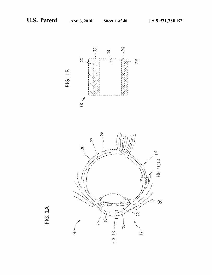
FIG . 1B
FIG . 1A
U . S . Patent

U . S . Patent Apr . 3 , 2018 Sheet 2 of 40 US 9 , 931 , 330 B2
w wwwwwwwwwww . ? W
wowa KW FIG . 1D SAMAHA wwwwwwwwwwwww ?? 1 .
???? ?? ???? ???
. . . . . . . . . . . . . . . XX
1
+ yalama
* - * * * * *
FIG . 1C waardid No ini ma

* * * * * * * * *
* *
?????? ??????? ?????? ???????????????????????? ???? ?????? ????????? ????? ???? ????????? * * * W
wwwwwwwwwww wwwwwwwwwwwwwwwwwwwwwwwwwwwwwwwwwwwwwwwwwwwwwwwwww
FIG , 2
* * * * * *
* * * * * * * * * * * * * * * * * * * * * * * * * *
* * * * * * * * * * * * * * * * * * a * * * www . ceriwww .
www . w
wwwwwwwwww wwwwwwwwwwwwww . varrrr FIG . 3 MediaW
.
* * *
r w . www .
warmwarmervarmen .
US 9 , 931 , 330 B2 Sheet 3 of 40 Apr . 3 , 2018 U . S . Patent

US 9 , 931 , 330 B2 Sheet 4 of 40 Apr . 3 , 2018 U . S . Patent
?????
??? ???? ?? ? ??? 44 : ?? ???
????
????? ????
??? ????? ????
??? ??????? ???? ft ??? ?? ?????? ??
. ??? ??
??? ii ?????? ??? ?? ? ??
??????? ????
??? ???? ?
?? ???
??? ? ??? ? ??? " : 555544
????? ????? ???
??? ???? ? ???? ? ??? ??
??? ? ???? ?
?????? ???? ? ?????
?? .
? ? ????? ... ... ?????
????? ?? ????? ?? ???? ???? ???? ???? ?? ????? ??? ?? ?? ?????? ?? ?? ?? ??
??? ?? ?? ??? ??? ???? ? ???? ??? ?? ????? ?? ???? ????? ?? ?????? . . . . . . ????????? ?? ??? ?? ?? ??? ????
???? ????? * *
?
? ?? ??
??? ?????? ???? ) ?? ???? H ?????? ? * ? - : " : " : ? , ??? - - - - = = : . . . . : : : : .
: : : : : : : - ???? ? ???

er mere om www . com U . S . Patent Apr . 3 , 2018 Sheet 5 of 40 US 9 , 931 , 330 B2
deletina 131
FIG . 6B * *
* * WWW * * * * * * * *
*
* * * * * * * * * * * * * * * *
* * * 1 . 1 . 1111 . 11 . * * * * *
11 * * * * * .
rir * * * * . . .
20 wwwwwwwwwwwwwww
com
. . . . . . . . .
FIG . 6A

US 9 , 931 , 330 B2
1111111111111111111111 aka poy 01
* * * * * * * * * * * * * * * *
* * *
* * * * * * * *
*
Suction
* *
* * *
* * *
* * * * * .
* *
*
*
* * * * * * * * * *
* *
* *
* * *
* * * * * * * * * *
* * *
Terrrrrrrrrrrrrrrr
* * * *
Sheet 6 of 40
* * * * * * * * * * * *
* * * * * * * * *
*
* * * * * * * *
* * * * *
2
5
- 3
* * * *
* * *
* * * * *
* * * * *
Www
* * * * * * * * * * *
* *
"
V
* *
* * * * * * *
* * * * *
* * * * *
* *
i * * * * * *
* * * * *
* * * *
* *
WA * *
* * * *
*
* iireist
.
: : : :
* *
train
* * * * *
:
*
* *
Apr . 3 , 2018
* * * * * * *
* *
*
*
* * * * *
.
onssald Retina Choroid
Sclera . . .
Microneedle Insert
intraocular induce Cannula to
FIG . 7B
FIG . 7A
U . S . Patent

U . S . Patent Apr . 3 , 2018 Sheet 7 of 40 US 9 , 931 , 330 B2
FIG . 8B
wwwwwwwwwwwwwwwwww
Sclera ????? Retina Suprachoidal Space Vitreous
FIG . 8A
* * *
Sciera Choroid Retina Vitreous

U . S . Patent Apr . 3 , 2018 Sheet 8 of 40 US 9 , 931 , 330 B2
FIG . 9C FIG . 9D
FIG . 9A FIG . 9B

U . S . Patent Apr . 3 , 2018 Sheet 9 of 40 US 9 , 931 , 330 B2
. .
. . : . : : . .
FIG . 10B
*
FIG . 10A :
: *
:
.

U . S . Patent Apr . 3 , 2018 Sheet 10 of 40 US 9 , 931 , 330 B2
350 . . .
100 nm Particles * * * * , - r
FIG . 11B wwww * * N *
300
250 Infusion Pressure ( kPa )
200
150
ENS .
193 wnool wroos within 900um wrooor * *
* * *
Wir verilecek
Successful Injections ( % ) S
350
wwwwww Himmmmmmmmmmmm 20 nm Particles
.
FIG . 11A .
* * *
www .
300
250 Infusion Pressure ( kPa )
200
150
*
. . wTool wricos wroo6 wrooor . . " * * *
. . : www
+ . . .
en
8 Successful Injections ( % )

US 9 , 931 , 330 B2
wrooot
wrooote
900um 800um wako 700um
Infusion Pressure ( kPa ) 300
250
200
150
900um h 800um 700um
Infusion Pressure ( kPa )
350
OSE
00€
OSZ
OOC
OST
visitiiviiviisisi D . . . . . . . . . . . . . . . . . . . . . . . . . . . . . . . . . . . . . . . . . . . . . . . . .
. . . . .
Meme
* :
* * * * *
* * *
Sheet 11 of 40
www
. .
. . :
*
: . . * * * * * * *
* . * * * *
:
* .
8 po
* * * *
. :
*
SR
*
*
0 . - . - . - . - .
- . - . - . -
.
0
Successful Injections ( % )
.
* . . * * * * *
Successful Injections ( % )
. . : *
*
* * .
mirixi : . .
.
*
. * * * . *
Apr . 3 , 2018
1
0 .
. . .
. .
0 . . .
. .
. *
.
8
. . . . . . . 0
1000 nm Particles
500 nm Particles
atent
FIG . 11D
FIG . 110

U . S . Patent Apr . 3 , 2018 Sheet 12 of 40 US 9 , 931 , 330 B2
FIG . 12B VV FIG . 12A

U . S . Patent
FIG . 13A
FIG . 13B 36 mmHg IOP
18 mmHg IOP
Apr . 3 , 2018
8
8 * * * * * *
* *
* * * * * * * *
Sitttt * *
* * * * * * * * * * * * * * * * * * * * * * * *
* * * *
* * * * *
+ 63 + 2
* * * * *
* * * * * *
*
*
* * * * * * * * * * * * *
Successful Injections ( % )
* * *
* * * * * * *
* * * * *
. .
he entir * *
w
* * * * * * * * *
Sheet 13 of 40
inter * * * * * * * *
businesses in
* * * * *
700um 800um 900um 1000um
8 pc
* * * *
82 * * * * * * * *
* * * *
ano
* * * * * , * my o
* *
. . . . . . . . . . . . . malig 700um
og 800um 900um me 1000um
200 250 300 Infusion Pressure ( kPa )
* * * * *
* * * * *
* * * *
* * * * * * * *
* * * *
* *
mmmmmmmmmmmmmm ;
0
mention
150 200 250
Infusion Pressure ( kPa )
300
150
US 9 , 931 , 330 B2

U . S . Patent
FIG . 14
FIG . 15
*
* *
*
* * * * * * * * * * * * * * * * *
* *
- -
-
Retina Vitreous
Lens
SCS Fluorescein Injection
1400 1300 1200 1100 1000 900 800 $ . * . . . . * . . * . * . * . 11 . * * * . . . *
Apr . 3 , 2018
10000 * * * * * * * * *
8 * *
8
. .
*
- .
Fluorescent intensity
.
* * * * *
UWS * * *
* * * *
* * *
500 14 * * * * * * * * * * * * * * * * * * * *
Concentration { ng / mL }
Sheet 14 of 40
* * * *
200 100
4
. .
ex * 0
*
*
* wwwwwwwwwwwwwwx 0
20
40
60
80
100
120
140
160
15
20
25
30
0 5 10
- SCS * * Mid - Vitreous
Position
US 9 , 931 , 330 B2

U . S . Patent
FIG . 16
FIG . 17 SCS 500 nm Injection
SCS 20 nm Injection
10000
10000
Apr . 3 , 2018
* * . . . . . . .
. . . . . . . . . . . .
* . * . . * * . . * * * . * * * .
.
.
8 Fluorescence Intensity
Fluorescence Intensity
in .
www .
wwwwwwwwwwwwwwwww
*
*
*
* W
band .
24 : 2
mis
* * * * *
* * *
hirsis
Sheet 15 of 40
the
stopni
turi
stapscesce cose che
cry
. .
ceste . . .
. .
. . . . . . . .
. . . . . . . . .
. . . . . . . .
. . . . . . . . . . . . . *
com . . . . . . . .
. . . . . . . .
. .
0
30
35
40
25
30
5 10 15 20 25
SCS
Time ( days )
Mid - Vitreous
0 5 10 15 20
. . . SCS Time ( days )
. . " . Mid - Vitreous
US 9 , 931 , 330 B2
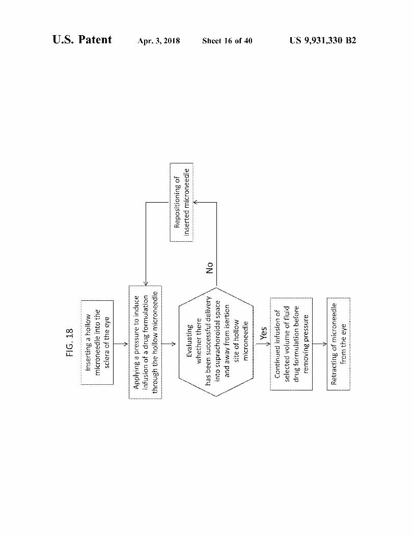
FIG . 18
U . S . Patent
Inserting a hollow microneedle into the sclera of the eye Applying a pressure to induce infusion of a drug formulation through the hollow microneedle
Apr . 3 , 2018
Repositioning of inserted microneedle No
Evaluating whether there has been successful delivery into suprachoroidal space and away from isertion site of hollow microneedle
Sheet 16 of 40
Yes Continued infusion of selected volume of fluid drug formulation before removing pressure
US 9 , 931 , 330 B2
Retracting of microneedle from the eye

FIG . 19A
FIG . 19B Intravitreal injection
U . S . Patent
States
FA
forced in
A .
( left to right for each group )
30nun 7days * 30days % 60days 120days
1000
14 : 19 14 me s tance
suprachoroidal
. . .
500 . . . .
suprachoroidal 11 / 2 = 39 . 8 days
intravitreal 122 13 . 3 days
Apr . 3 , 2018
Vitrine
PRESSEASTEARTEAttacht444tetap tertentu
Sclera Choroid Retina At seg
Lens
o in
WA
chwa !
o
wwwwwwwwwwwwwwwwwwwwwwwwwwwwwwwwwwwwwwwwwwwwwww ????????????????????????????????????????????????????
Wowowieckiego 0
30
90
120
60 Time , days
Suprachoroidal injection
Sheet 17 of 40
2000 1600 TA , Pg
1000 500
. . .
. .
z??ra Choroid
Vitra Ant seg
Lens Optic Ner
or
US 9 , 931 , 330 B2

US 9 , 931 , 330 B2
skep ' owi
090€
OCK
06 wwwwwwwwwwwwwwwwwwwwwwwwwwwwww 0 . 001
0 . 01 0 . 1
wwwwwwwww w
wwwww
IVT
one
900W
*
Lens : Retina
co
box
Sheet 18 of 40
Skep ' awil 06 090€
OZK *
. .
.
.
. . . : : : : : : : : : : :
: : :
: : : : : : : : : : : : : : . .
. .
11
. .
.
. .
Hoogsweglooooooo
Apr . 3 , 2018
SJS
. . .
TA concentration
TAL
. . . . . . . . . . . . .
56
* *
000000
0
000000
plojoyo : sup7
co
U . S . Patent
FIG . 190
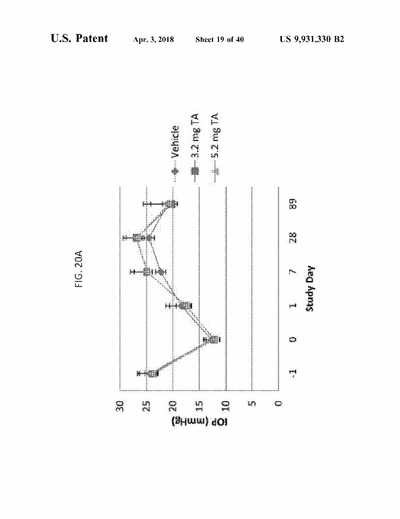
FIG . 20A
U . S . Patent
*
* * * * * * * * * * * * * * * * * * * * * * * * * * * * * * * * * * * * * * * * * * * * * * * * * * * * * * * * * * * * * * * * * * * * * * * * * * * * * * * * * * * * * * * * * * * *
* * * * * * * * * * * * * * * * * * * *
* *
* * * * * * * * * * * * *
Apr . 3 , 2018
( 3Huuuu dÔI th
Vehic Vehicle 3 . 2 mg TA 5 . 2 mg TA
wwwwwwwwwwwwwwwwwwwwwwwwww ww
Sheet 19 of 40
wwwwwwwwwwwwwwwwwwwwwwwwwwwwwwwwwwwwwwwwwwwwwwwwwwwwwwwwwwwwwwwwwwwwwwwwwwwwwwwwwwwwwwwww w wwwwwwwwwwwwwwwwwwwwwwwwwwwwwwwwwwwwwwwwwwwwwwwwwwww
- 1
0
28
89
1 7 Study Day
o
US 9 , 931 , 330 B2
U . S . Patent Apr . 3 , 2018 Sheet 20 of 40 US 9 , 931 , 330 B2
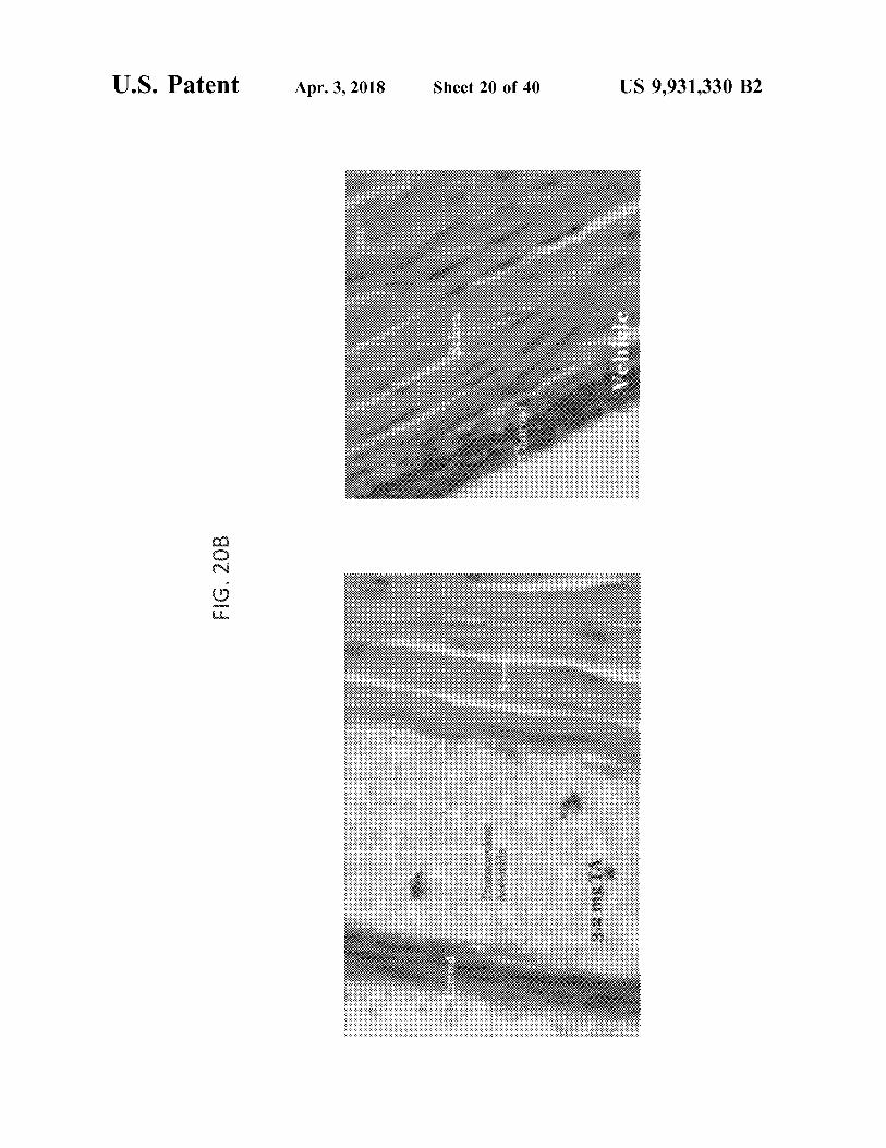
Vehicle
FIG . 20B
32 mg

FIG . 200
atent
Single Dose GLP Toxicology in Rabbits Systemic Exposure , n = 10 rabbits
Apr . 3 , 2018
high dose
Matatatatatatatatatatatatatatatatatatatatatatatatatatatatatatatatatatatatatatatatphalsts that helptetphotphotphotphstrip hp tetphotptpfptptphetptpfptptphp * * * *
low dose
3 . 2 34 6 . 2 mg TA
* * * * * *
* * * *
* * * *
* * * * * * * * * * * * * * * * * * * * * * * * * *
* * *
* * *
* * * *
* *
X
X
XX
* * * * *
* * * *
high dose
* * * * * * * * * * * * * * * * * * * * * * * * * * * * * * * * * * * * * * * * * * * * * * * * * * * * * * * * *
( a ) US® WONOVI
Sheet 21 of 40
musim
caracter high dose ( 5 . 2 mg TA )
low dose
high low dose dose
* *
ca
Wwwwwwwwwwwww
Ministry
* *
low dose ( 3 . 2 mg TA )
* * * * * * * * * * * * * * * * * * * * * * * * * * * * * * * * * * * * * * * * * * * * * * * * * * * * * * * * * * * * * * * * * * * * * * * * * * * * * * * * * * * * * * * * * * * *
* * * * * *
high low dose dase
* * * * * * * * * * * * * *
Stukty 09
:
:
+
+ +
+ +
* * * *
* * *
*
*
*
Day 1 Day 14 Day 28 Day 60
US 9 , 931 , 330 B2

FIG . 20D
U . S . Patent
Rabbit Ocular Tissue Levels After SCS Injection of TA
10000
Sclera / choroid
Apr . 3 , 2018
1000 100
Retina Retina
Concentration ME TA / g tissue
Sheet 22 of 40
ICB
funt
Vitreous
home
0 . 01
Lens
. . . . . . .
. .
0 . 001
0
5
10
15 Time ( days )
20
25
30
US 9 , 931 , 330 B2

FIG . 20E
FIG . 20F
atent
Triamcinolone ( TA ) in the Sclera - Choroid
t
Triamcinolone ( TA ) in the Retina
4000 1
Apr . 3 , 2018
half life 12 days
High Dose TA
High Dose TA
Low Dose TA
9 days
Low Dose TA
c
Mass of TA ( ugl
Mass of TA ( ugl
fingen
Sheet 23 of 40
Whigh dose
low doses
lino
high dose
a
0
wwwwwwwww low dose 5
0
5
20
25
30
0
10
20
25
30
10 15
Time { days }
15 Time ( days )
US 9 , 931 , 330 B2
U . S . Patent
FIG . 21B
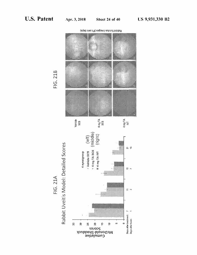
FIG . 21A Rabbit Uveitis Model : Detailed Scores
The
Vehicle
Apr . 3 , 2018
se n
4 eyesigroup
Vehicle scs ( left ) * 4 mg TA SCS ( middle ) * 4 mg TAIVT ( right )
e
Cumulative McDonald - Shadduck
Scores sy
4 mg TA SCS
Rabbit fundus images ( # ' s are days )
Sheet 24 of 40
???
0 m ?? ??r re???? ? Days after toxin : 1
??? ? IT
mat ?
wali hilo
are many
US 9 , 931 , 330 B2

U . S . Patent
FIG . 210
FIG . 21D
Apr . 3 , 2018
.
wive
:
Intraocular Pressure ( IOP )
s
Cumulative Inflammatory Score
( 6Hww
* * *
* * * * * * * * * . * . . . . * .
1
motor Vehicle SCS * 4 mg TA SCS * 4 mg TA IVT 4 6
Sheet 25 of 40
* . * . * . * . * . * . * . * . * . * . * . * . * . * . * *
22 .
??? ??? ?? ? ?? ? ?? ? ?? ? ?? .
so ' z o ' r o ' z o ' z o ' r o ' z o ' o ' rta masina o ' z o ' r o ' r o ' z o ' r as o ' z o ' ringa ' ' o ' r o ' z o ' r o ' r o ' o ' r sioe in so ' z o ' r o ' ' o ' z o ' z o ' so ' rasa ' s
2
0
2
A ( Control ) B ( SCS TA ) C ( VT TA )
Treatment Time days )
US 9 , 931 , 330 B2

U . S . Patent
FIG . 22A
FIG . 22B t Acute Pig Uveitis Model : Scores
.
. . .
Left bars : LPS + vehicle Middle bars : LPS + 2mg TA Right bars : salt solution + vehicle
: : : : : : : :
. . .
: : : : : : : : : :
. . . : : : : :
IIIIII . . . . .
: : : : : :
w wwwwwwwwwwnnnnnnn
* * * * * * * * *
. . IIII
w
No treatment 2 mg SCS 2 mg VVT 0 . 2 mg SCS
( n = 2 ) ( n = 2 ) ( n = 3 ) ( n = 1 )
Apr . 3 , 2018
.
2 mg IVI
. . .
* * *
. . . . . . .
ww
.
mano
* * * *
Mean Cumulative Hackett / McDonald Ocular Scores
O NA ON
. . . . . .
no treatment
Mean Hackett / McDonald Ocular Scores
* * * * * * * * * * * * * * *
.
0 . 2 mg SCS
.
* * * *
*
* * * * * * * * * * * * * * * * * *
* *
Sheet 26 of 40
* * * *
* * *
2 mg SCS
* * * * WS *
* * * * * * *
* * * *
wi
t
- 24
0
24
48
72
0
24
48
72
mi
www
wwwwwwwwwwwwwwww
Time after treatment ( hours )
Time after treatment ( hours )
US 9 , 931 , 330 B2

FIG . 23
U . S . Patent
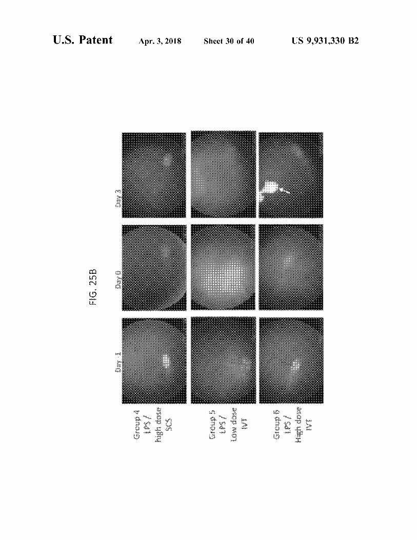
Group 1 ( Negative BSS , Vehicle SCS ) Group 2 ( Positive LPs , Vehicle SCS )
WWW .
Group 3 ( Low dose SCS ) Group 4 ( High dose SCS ) Group 5 ( low dose ( VT ) Group 6 ( High dose ( VT )
Apr . 3 , 2018
Mean SD Cumulative Inflammatory Ocular Scores
? is az ?
Sheet 27 of 40
ww .
www
O
XX
*
GC40 - 1 0
9 9 1 Study Days
2
99 3
US 9 , 931 , 330 B2

FIG . 24
U . S . Patent
Group 1 ( Negative BSS , Vehicle SCS )
??????????????????????
Group 2 ( Positive LPS , Vehicle SCS ) Group 3 ( low dose SCS ) Group 4 ( High dose SCS ) Group 5 ( Low dose IVT )
Apr . 3 , 2018
Group 6 ( High dose IVT )
www
. .
. .
???????????
. .
wi
. * *
. . .
Mean I SD Intraocular Pressure
W
* * * * * * * * * * * Wwwwwwwwww
t
? ? ? ? ?
Sheet 28 of 40
ww
* * * * * * * * * * * * * * * WWW
? ? ? ? ? ? ? ?
6hr
Dayi Day 1 Day 3
Day - 6 Day - 4 Day - 1 Dayo 1hr 3 hr
Study Time
US 9 , 931 , 330 B2

U . S . Patent Apr . 3 , 2018 Sheet 29 of 40 US 9 , 93l . 330 B2
{ G . 25A ?
????? ?
as a ? ????? ? ?????
U . S . Patent Apr . 3 , 2018 Sheet 30 of 40 US 9 , 931 , 330 B2
FIG . 25B
anong UPS / asop uby 9 dnojo IPS / High dose

U . S . Patent Apr . 3 , 2018 Sheet 31 of 40 US 9 , 931 , 330 B2
FIG . 26

U . S . Patent Apr . 3 , 2018 Sheet 32 of 40 US 9 , 931 , 330 B2
FIG . 26

U . S . Patent Apr . 3 , 2018 Sheet 33 of 40 US 9 , 931 , 330 B2
FIG . 26

U . S . Patent
FIG . 27
5 . 0
Group 1 ( Negative BSS , Vehicle SCS ) Group 2 ( Positive LPS , Vehicle SCS ) Group 3 ( Low dose SCS ) Group 4 ( High dose SCS ) Group S ( Low dose IVT ) Group 6 ( High dose IVT )
gr . 2
ê
gr . 3
Apr . 3 , 2018
Mean - SD Histologic Scores
gr . 2
gr . S
gr . 4
?
gr 3
gr .
Sheet 34 of 40
gr . 6
gr . 1
? gr . 1
Ö
Anterior Segment
Posterior Segment
US 9 , 931 , 330 B2

} } G , 28
U . S . Patent
????? 2
3 . 0 -
Group 1 { Negative BSS , Vehic?? $ cs } Group 2 { Pasitive 1PS , Vehicle 5CS } Group 3 ( low dose SCS ) Group 4 { High dose SCS } Group 5 ( Low dose IVT ) Ggu 6 { High dgse T }
Apr . 3 , 2018
Mean + SD cells x 10 , 000 { 1
{ { } . {
* * * * * * * *
r 2
?er ,
3 } .
Sheet 35 of 40
8
6
r ,
4
?? 5 ??? 6
_?
13 . 0
. 1
0 . 0
- -
* *
* * * *
Aqueous Humor
Vitreous Humor
US 9 , 93l . 330 B2

US 9 , 931 , 330 B2
Study Day 95
28
14
1
* * *
* *
ture oppitsisti
4 mg TA IVT
*
* * * * * * * *
* * *
tenaga dan menyet *
*
4 bet
????????????????????????????????????????
* *
Hancon
* * * * * * * * * * * * * * * *
Sheet 36 of 40
IA Concentration in Plasma ( ngmL )
\ 4 mg TA SCS
* * * * * *
Apr . 3 , 2018
????????????????????????? ???????????????????????????????????????
FIG . 29
U . S . Patent

U . S . Patent Apr . 3 , 2018 Sheet 37 of 40 US 9 , 931 , 330 B2
FIG . 30
U . S . Patent
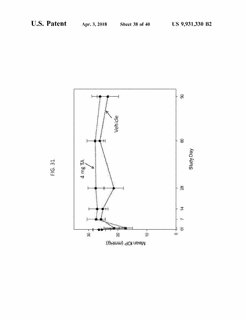
FIG . 31 * *
*
* *
* * * * * * * * * * * * * * * * * *
* * * *
*
* * * * *
4 mg TA
nyewer en
for
Svet
Apr . 3 , 2018
www
* * *
*
finanmaninmiffangenweide
* *
ligne
Vehicle
Mean IOP ( mmHg )
Sheet 38 of 40
0
. . .
0
7
. . . . . . . . . . . . . . . . . . . . . . wie 14 28
90
Study Day
US 9 , 931 , 330 B2

U . S . Patent Apr . 3 , 2018 Sheet 39 of 40 US 9 , 931 , 330 B2
. . . .
.
. .
. . . Study Day FIG . 32 Vehicles 4 mg TA
*
WWW * * * *
montre homme Š CCT ( um )
Š o .

U . S . Patent Apr . 3 , 2018 Apr . 3 , 2018 Sheet 40 of 40 US 9 , 931 , 330 B2
wa
Xe ch? che che c
wwwwwwwwwwwwwwwwwwwwwwwwwwww www 4 mg TA
import Wri h ? che ch?
Study Day r
FIG . 33
wwwwwww iiiiiiiiiiii www . w BZfL imbi 0 * * * * * * * * * * * * * * * * * * * * * * * * * * *
o
TA Concentration in Plasma ( ng / mL )

US 9 , 931 , 330 B2
METHODS AND DEVICES FOR THE maladies associated with vascular abnormalities . The pres TREATMENT OF OCULAR DISEASES IN ent invention addresses these and other needs .
HUMAN SUBJECTS SUMMARY OF THE INVENTION
CROSS REFERENCE TO RELATED APPLICATIONS In one aspect , the present invention relates to non - surgical
ophthalmic therapies in human patients in need of such This application is a continuation of U . S . patent applica treatment , and more particularly to the infusion of a drug
tion Ser . No . 15 / 454 , 636 , filed on Mar . 9 , 2017 , which is a formulation into the suprachoroidal space of the eye for continuation of U . S . patent application Ser . No . 14 / 441 , 151 , 10 targeted , local drug delivery , for the treatment of posterior
ocular disorders , choroidal maladies and other diseases filed May 6 , 2015 , which is a 371 National Stage application associated with vascular abnormalities . based on PCT Application PCT / US2013 / 069156 , which In one aspect of the invention , a method is provided for claims priority from U . S . Provisional Application Ser . No . : treating a posterior ocular disorder in a human subject in 61 / 724 , 144 , filed Nov . 8 , 2012 ; 61 / 734 , 872 , filed Dec . 7 , ! ; 15 need of treatment . In one embodiment , the method com 2012 ; 61 / 745 , 237 , filed Dec . 21 , 2012 ; 61 / 773 , 124 , filed prises non - surgically administering an effective amount of a Mar . 5 , 2013 ; 61 / 785 , 229 , filed Mar . 14 , 2013 ; 61 / 819 , 388 , drug formulation to the suprachoroidal space ( SCS ) of the filed May 3 , 2013 ; 61 / 873 , 660 , filed Sep . 4 , 2013 , and eye of the subject in need of treatment of the posterior ocular 61 / 898 , 926 , filed Nov . 1 , 2013 , all of which are incorporated disorder or choroidal malady . In a further embodiment , upon herein by reference in their entireties for all purposes . 20 administration , the drug formulation flows away from the
insertion site and is substantially localized to the posterior BACKGROUND OF THE INVENTION segment of the eye . In one embodiment , the posterior ocular
disorder is an ocular inflammatory condition such as uveitis , This invention is generally in the field of ophthalmic scleritis , glaucoma , ocular sarcoidosis , optic neuritis , macu
therapies , and more particularly to the use of a microneedle 25 lar edema , diabetic retinopathy , macular degeneration , a for infusion of a fluid drug formulation into ocular tissues for corneal ulcer , an autoimmune disorder , ophthalmic mani targeted , local drug delivery . festations of AIDS , optic nerve degeneration , geographic
The delivery of drug to the eye is extremely difficult , atrophy , choroidal disease or retinitis . The condition in one particularly delivery of macromolecules and delivery to the embodiment is acute . In another embodiment , the condition posterior segment . Many inflammatory and proliferative 30 is chronic . diseases in the posterior region of the eye require long term In another embodiment , the a method is provided for the pharmacological treatment . Examples of such diseases treatment of a choroidal malady , e . g . , ocular neovascular include macular degeneration , diabetic retinopathy , and ization , polypoidal choroidal vasculopathy , choroidal scle uveitis . In addition , many choroidal maladies that are asso rosis , central sirrus choroidopathy , a multi - focal choroidopa ciated with inflammatory responses , proliferation , and neo - 35 thy or a choroidal dystrophy ( e . g . , central gyrate choroidal vascularization require long term pharmacological treat - dystrophy , serpiginous choroidal dystrophy , total central ment . It is difficult to deliver effective doses of drug to the choroidal atrophy ) . In one embodiment , the method com posterior segment using conventional delivery methods such prises non - surgically administering a drug formulation com as topical application , which has poor efficacy , and systemic prising an effective amount of an anti - inflammatory drug , a administration , which often causes significant side effects , 40 vascular endothelial growth factor ( VEGF ) modulator , a and often does not reach the site of infection . ( Geroski & platelet derived growth factor ( PDGF ) modulator , an angio Edelhauser , Invest . Ophthalmol . Vis . Sci . 41 : 961 - 64 ( 2000 ) ) . genesis inhibitor , an immunosuppressive agent , a vascular For example , while eye drops are useful in treating condi - permeability inhibitor , or a combination thereof , to the SCS tions affecting the exterior surface of the eye or tissue ( s ) at of the patient in need of treatment . In a further embodiment , the front of the eye , the eye drops cannot significantly 45 the effective amount of the drug administered to the SCS penetrate the eye , as may be required for the treatment of provides higher efficacy or a greater therapeutic effect of the various retinal diseases and choroidal maladies . drug , compared to the identical drug dose administered
Direct injection into the eye , using conventional needles intravitreally , intracamerally , topically , parenterally or and syringes has been reported to be effective , but requires orally . In even a further embodiment , the patient undergoing professional training and raises concerns about safety ( Mau - 50 treatment via SCS drug therapy was not previously respon rice , J . Ocul . Pharmacol . Ther . 17 : 393 - 401 ( 2001 ) ) . It also sive to a different type of therapy for the same condition . would be desirable to be able to minimize the number and / or In yet another embodiment , a method for decreasing frequency of eye injection treatments needed to deliver subretinal exudation and bleeding in a subject is provided . In therapeutically effective amounts of drug to the ocular tissue a further embodiment , the method comprises non - surgically sites that need it . 55 administering a drug formulation comprising an effective
The suprachoroidal space ( SCS ) of the eye has been amount of an effective amount of an anti - inflammatory drug , studied , and its cannulation described as a possible route for a vascular endothelial growth factor ( VEGF ) modulator , a drug delivery . See , e . g . , Olsen , et al . , American J . Ophthal - platelet derived growth factor ( PDGF ) modulator , an angio mology 142 ( 5 ) : 777 - 87 ( November 2006 ) ; PCT Patent genesis inhibitor , an immunosuppressive agent , a vascular Application Publication No . WO 2007 / 100745 . 60 permeability inhibitor , or a combination thereof , to the SCS
It therefore would be desirable to provide better , safer , of the patient in need of treatment , wherein administration of more effective techniques for the direct delivery of thera - the drug formulation reduces subretinal exudation and peutic agents to posterior segment eye tissues , for example , bleeding experienced by the patient , as compared to the to treat a posterior ocular disorder . It further would be identical dosage of the drug administered intravitreally to desirable to provide better , safer , more effective techniques 65 the patient . for the direct delivery of therapeutic agents to the SCS for In one embodiment , a method for treating a posterior the treatment of choroidal maladies , for example , choroidal ocular disorder or a choroidal malady in a human patient is

US 9 , 931 , 330 B2
771
provided . In a further embodiment , the method comprises thereof . In another embodiment , the drug affects inflamma non - surgically administering an effective amount of a drug tion , neuroprotection , complement inhibition , drusen forma formulation to the suprachoroidal space ( SCS ) of the eye of tion , scar formation , reduction in choriocapillaris or chor the subject in need of treatment of the posterior ocular oidal neocasvularization . In another embodiment , the drug disorder or choroidal malady . In a further embodiment , the 5 formulation comprises microparticles and / or nanoparticles intraocular elimination half life ( t ) of the drug adminis - of the drug . In one embodiment , the drug formulation tered to the SCS is greater than the intraocular ty of the comprises microparticles having a Dso of 1 um or less and / or drug , when administered intravitreally , intracamerally , topi - a Dog of 10 um or less . cally , parenterally or orally . In another embodiment , the As provided above , one aspect of the invention includes mean intraocular maximum concentration ( Cmor ) of the 10 a method for treating a posterior ocular disorder in a human drug , when administered to the SCS via the methods subject in need thereof comprising non - surgically adminis described herein , is greater than the intraocular Cmor of the tering a drug formulation to the SCS of the eye of the human drug , when administered intravitreally , intracamerally , topi - subject , wherein upon administration , the drug formulation cally , parenterally or orally . In another embodiment , the flows away from the insertion site and is substantially mean intraocular area under the curve ( AUCO ) of the drug , 15 localized to the posterior segment . In one embodiment of the when administered to the SCS via the methods described method , the intraocular pressure of the eye remains substan herein , is greater than the intraocular AUCo - of the drug , tially constant during administration of the drug formulation when administered intravitreally , intracamerally , topically , to the SCS . In another embodiment , administration of the parenterally or orally . In yet another embodiment , the drug formulation to the SCS of the eye results in a decreased intraocular time to peak concentration ( tmar ) of the drug , 20 number of side effects , or a reduced severity of one or more when administered to the SCS via the methods described side effects , compared to administration of the same drug herein , is greater than the intraocular tmax of the drug , when dose intravitreally , intracamerally , topically , orally or par the same drug dose is administered intravitreally , intracam - enterally . erally , topically , parenterally or orally . In a further embodi - In one aspect of the invention , the present invention ment , the drug formulation comprises an effective amount of 25 relates to a method for treating a choroidal malady in a an anti - inflammatory drug ( e . g . , a steroid or NSAID ) , a human patient in need of treatment . In one embodiment , the VEGF modulator ( e . g . , VEGF antagonist ) , a platelet derived method comprises non - surgically administering a drug for growth factor ( PDGF ) modulator , an angiogenesis inhibitor , mulation comprising an effective amount of an anti - inflam an immunosuppressive agent , a vascular permeability matory drug , a vascular endothelial growth factor ( VEGF ) inhibitor , or a combination thereof . 30 modulator , a platelet derived growth factor ( PDGF ) modu
In one embodiment , the method for treating a posterior lator , an angiogenesis inhibitor , an immunosuppressive ocular disorder or choroidal malady in a human subject agent or a vascular permeability inhibitor , to the suprachor comprises delivering a drug formulation via a hollow oidal space ( SCS ) of the eye of the patient . In a further microneedle to the SCS of the eye of the human subject in embodiment , the human patient , prior to administration of need of treatment . In a further embodiment , delivering the 35 the drug formulation , was previously treated for the chor drug formulation comprises inserting a hollow microneedle oidal malady and was not properly responsive to the treat into the eye of the human subject at an insertion site , the ment . microneedle having a tip end with an opening ; and infusing In another aspect of the invention , the present invention over a period of time a drug formulation through the inserted relates to a method for treating ocular neovascularization in microneedle and into the SCS space away from the insertion 40 a human patient in need of treatment . In one embodiment , site . The drug formulation administered to the SCS , in one the method comprises non - surgically administering a drug embodiment , flows away from the insertion site and is formulation comprising an effective amount of an anti substantially localized to the posterior segment of the eye , inflammatory drug , a vascular endothelial growth factor thereby increasing the therapeutic efficacy of the dose of the ( VEGF ) modulator ( e . g . , a VEGF antagonist ) , a platelet drug compared to the therapeutic efficacy of the same drug 45 derived growth factor ( PDGF ) modulator ( e . g . , a PDGF dose administered by another means ( e . g . intravitreally , antagonist ) , an angiogenesis inhibitor , an immunosuppres intracamerally , topically , parenterally , and / or orally ) . In sive agent or a vascular permeability inhibitor , to the supra another embodiment , the dose of the drug sufficient to elicit choroidal space ( SCS ) of the eye of the patient . In a further a therapeutic response when administered to the SCS is less embodiment , the ocular neovascularization is a choroidal than the dosage of the drug sufficient to elicit a therapeutic 50 neovascularization . In one embodiment , the human patient response when administered intravitreally , topically , paren - being treated for the ocular neovascularization , prior to terally or orally . In another embodiment , the drug formula administration of the drug formulation , was previously tion is delivered to the SCS by a hollow microneedle treated for the ocular neovascularization and was not prop inserted into the sclera at the equator of the eye or between erly responsive to the treatment . the equator and the limbus of the eye . In a further embodi - 55 The drug formulation delivered by the methods described ment , the hollow microneedle is inserted in the insertion site herein , in one embodiment , comprises an effective amount at a 90 degree angle ( perpendicular ) . of an anti - inflammatory drug , for example a steroidal com
The drug formulation delivered by the methods described pound or a non - steroidal anti - inflammatory drug ( NSAID ) . herein , in one embodiment , comprises an effective amount In another embodiment , the drug delivered to the SCS via of an anti - inflammatory drug , for example a steroid or a 60 the methods described herein is a vascular permeability non - steroidal anti - inflammatory drug ( NSAID ) . In another inhibitor , an angiogenesis inhibitor or a VEGF modulator , embodiment , the drug delivered to the SCS via the methods e . g . , a VEGF antagonist . In one embodiment , the VEGF described herein is a steroid , immunosuppressive , antime - antagonist is a VEGF receptor antagonist or a soluble VEGF tabolite , T - cell inhibitor , alkylating agent , biologic , TNFa receptor . In one embodiment , the drug formulation com antagonist , interleukin antagonist , neuroprotectant , vascular 65 prises drug microparticles having a D50 of 1 um or less endothelial growth factor ( VEGF ) antagonist , platelet and / or a D99 of 10 um or less . In a further embodiment , the derived growth factor ( PDGF ) antagonist , or a combination drug formulation comprises triamcinolone .

US 9 , 931 , 330 B2
In one embodiment of the invention , a method for treating success rate of suprachoroidal delivery of 20 nm particles a choroidal malady or a posterior ocular disorder in a human ( 1A ) , 100 nm particles ( 11B ) , 500 nm particles ( 11C ) , and subject in need thereof is provided comprising non - surgi - 1000 nm particles ( 11D ) into pig eyes . cally administering a drug formulation to the SCS of the eye FIGS . 12A and 12B are fluoroscopic images of a cryo of the human subject , wherein , the intraocular pressure of 5 section of a pig eye after infusion of 20 nm particles ( 12A ) the eye remains substantially constant during administration and 1000 nm particles ( 12B ) in the saggital direction and of the drug formulation to the SCS . In another embodiment , collaged to show spaces both anterior and posterior to the administration of the drug formulation to the SCS of the eye microneedle insertion site . The insets of FIGS . 12A and 12B of the patient in need of treatment of the posterior ocular show magnified views of the microneedle insertion site . disorder or choroidal malady results in a decreased number 10 FIGS . 13A and 13B are graphs showing the effect of the of side effects , or a reduced severity of one or more side intraocular pressure and microneedle length on the success effects , compared to administration of the same drug dose rate of suprachoroidal delivery of 1000 nm particles for a intravitreally , intracamerally , topically , orally or parenter simulated intraocular pressure of 18 mmHg ( 13A ) and 36 ally . In one embodiment , the side effect reduced by the mmHg ( 13B ) . methods described herein is subretinal exudation and / or 15 FIG . 14 is a one - dimensional line of sight scan of rabbit bleeding . eyes taken after injection of sodium fluorescein to the
suprachoroidal space , with the x - axis representing the posi BRIEF DESCRIPTION OF THE DRAWINGS tion in the eye from back ( 0 ) to front ( 160 ) and the y - axis
representing the fluorescent intensity at that position . FIGS . 1A , 1B , 1C , and 1D are cross - sectional illustrations 20 FIG . 15 is a graph showing the rate of clearance of sodium
of the tissue structures of a human eye . The eye as a whole fluorescein from the suprachoroidal space over time . ( A ) , a close - up of the cornea ( 1B ) , and a close - up of the FIG . 16 is a graph showing the rate of clearance of 20 nm sclera and associated tissue in an eye without fluid in the particles from the suprachoroidal space over time . suprachoroidal space ( 1C ) or with fluid in the suprachoroidal FIG . 17 is a graph showing the rate of clearance of 500 nm space ( 1D ) . 25 particles from the suprachoroidal space over time .
FIG . 2 is a cross - sectional view of a microneedle device FIG . 18 is a block diagram of a method for administering comprising a hollow microneedle disposed in an elongated a drug to the eye according to one embodiment . body according to one embodiment . FIG . 19A is a graph showing the amount of triamcinolone
FIG . 3 is a cross - sectional view of the elongated body of ( TA ) retained in the posterior segment of the eye as a the microneedle devices shown in FIG . 2 . 30 function of time after administration . ( circles — SCS injec
FIG . 4 is an illustration of a microneedle device according tion , diamonds intravitreal injection ) . to one embodiment . FIG . 19B are graphs showing the increased retention of
FIG . 5 is an illustration of a microneedle device according triamcinolone ( TA ) in the choroid and retina when admin to one embodiment . istered to the SCS ( bottom ) compared with TA administered
FIGS . 6A and 6B illustrate an embodiment of a process 35 intravitreally ( top ) . for using a hollow microneedle to deliver drug into the FIG . 19C , top , is a graph showing the ratio of the amount suprachoroidal space of an eye , where the process includes of triamcinolone ( TA ) in the lens of the eye to the amount inserting the hollow microneedle into the sclera and infusion of TA in back of the eye ( choroid ) as a function of time after of a fluid drug formulation into the suprachoroidal space . administration . FIG . 19C , bottom , is a graph showing the
FIG . 7A shows a comparison of a hollow microneedle 40 ratio of the amount of TA in the lens of the eye to the amount according to one embodiment as compared to the tip of a of TA in the back of the eye ( retina ) as a function of time conventional 30 gauge hypodermic needle . FIG . 7B shows after administration ( days ) . a schematic illustration of a custom acrylic mold shaped to FIG . 20A is a graph of intraocular pressure ( TOP , mmHg ) fit a whole eye . in rabbit eyes as a function of time after TA administration . FIGS . 8A and 8B are brightfield microscopic images of 45 Rabbits were injected with vehicle , 3 . 2 mg TA or 5 . 2 mg TA
saggital cross sections of a pig eye before and after infusion at study day 0 . of sulforhadamine , respectively . FIG . 20B is a cross sectional image of a rabbit eye
FIGS . 9A , 9B , 9C , and 9D are fluoroscopic images of a following suprachoroidal injection of 3 . 2 mg triamcinolone cryosection of a pig eye with no infusion into the supra - ( left ) or vehicle ( right ) . choroidal space ( 9A ) , a cryosection of a rabbit eye after 50 FIG . 20C are graphs showing the TA plasma concentra infusion of 500 nm fluorescent particles in the axial plan and tion ( ng / mL ) after administration of TA to the SCS of rabbit collaged to form a panoramic view ( 9B ) , a cryosection of a eye , as a function of time after TA administration . pig eye after infusion of 500 nm fluorescent particles in the FIG . 20D is a graph showing the retention of triamcino saggital direction and collaged to show the spaces both lone ( TA ) ( ug TA / g tissue ) in various tissues after adminis anterior and posterior to the microneedle insertion site ( 9C ) , 55 tration to the SCS . The greatest amount of the drug is and a cryosection of a human eye after infusion of 500 nm retained in the tissues in the back of the eye ( choroid , retina ) fluorescent particles in the saggital direction and collaged to with lesser amounts retained in anterior portions of the eye show spaces both anterior and posterior to the microneedle ( lens , vitreous fluid ) . insertion site ( 9D ) . The insets of FIGS . 9B , 9C , and 9D show FIG . 20E is a graph showing the amount of triamcinolone magnified views of the microneedle insertion site . 60 ( TA ) ( ug ) in the sclera and choroid as a function of time after
FIGS . 10A and 10B are microcomputed tomography TA administration . images showing the circumferential spread of 1 um contrast FIG . 20F is a graph showing the amount of triamcinolone particles infused into the suprachoroidal space of a pig eye ( TA ) ( ug ) in the retina as a function of time after TA in a cross - sectional image ( 10A ) and a three - dimensional administration . reconstruction of the cross - sectional images ( 10B ) . 65 FIG . 21A is a graph showing the cumulative McDonald
FIGS . 11A , 11B , 11C , and 11D are graphs showing the Shadduck scores of eyes treated with vehicle ( left ) , 4 mg effect of infusion pressure and microneedle length on the triamcinolone ( TA ) administered to the SCS ( middle ) or 4
US 9 , 931 , 330 B2
mg triamcinolone administered intravitreally ( right ) , as a Group 1 at Day 3 ( Wilcoxon ; P = 0 . 018 ) . Gl = Group 1 ; function of time after treatment , and time after LPS toxin G2 = Group 2 ; G3 = Group 3 ; G4 = Group 4 ; G5 = Group 5 ; administration . The McDonald - Shadduck scores provide a G6 = Group 6 . model of posterior uveitis . FIG . 24 is a graph showing the mean intraocular pressure
FIG . 21B are representative fundus photographs showing 5 in the eve of animals dosed with or without a toxin on Day the effect of triamcinolone ( TA ) administered to the SCS or - 1 and then treated with low or high doses of TA adminis intravitreally in a model of posterior uveitis in NZW rabbits . tered either to the SCS or intravitreally on Day 0 . Mean
FIG . 21C is a graph showing the overall severity of ( + / - SD ) intraocular pressure ( TOP ) in porcine eyes prior to inflammation in NZW rabbits as measured from histology at uveitis induction ( Day - 1 ) , at the time of drug administra the final time point . The following tissues were analyzed : 10 tion ( Day 0 ) with suprachoroidal space ( SCS ) or intravitreal ciliary processes , sclera - choroid , vitreous , retina and optic ( IVT ) injections of 0 . 2 mg ( low dose ) or 2 . 0 mg ( high dose ) nerve ( 0 - 4 scale , max score = 20 ) . triamcinolone acetonide ( TA ) . IOP was measured 1 hr . , 3 hr . , FIG . 21D is a graph showing the intraocular pressure ( mmGh ) in NZW rabbits in response to IVT or SCS TA 6 hr . 1 day , 2 days and 3 days after treatment . a . IOP in administration . 15 Group 1 eyes was significantly higher than Group 2 eyes at
FIG . 22A is a graph showing the mean Hackett / McDonald 1 and 3 hours after treatment injections ( P = 0 . 01 ; 0 . 04 ) . ocular scores of porcine eyes challenged with ( i ) lipopoly - FIGS . 25A - B are wide - field ocular fundus images of eyes saccharide ( LPS ) toxin followed by vehicle ( left ) , ( ii ) LPS from animals dosed with or without a toxin and then treated toxin followed by 2 mg triamcinolone to the SCS ( middle ) , with low or high doses of TA administered either to the SCS or ( iii ) balanced salt solution followed by vehicle . Treatment 20 or intravitreally . Wide - field ocular fundus images were with SCS TA at a dose of 2 mg significantly reduces the imaged at immediately prior to injection with liopolysac ocular inflammatory response in this porcine uveitis model . charide ( LPS ) on Day - 1 , immediately prior to injection
FIG . 22B is a graph showing the mean cumulative Hack with vehicle , 0 . 2 mg ( low dose ) or 2 . 0 mg ( high dose ) of ett / McDonald ocular scores of porcine eyes challenged with trimacinolone acetonide on Day 0 , and at 3 days after ( i ) lipopolysaccharide ( LPS ) toxin followed by vehicle , ( ii ) 25 treatment . Group 1 eyes , which were injected with balanced LPS toxin followed by 2 mg triamcinolone ( TA ) to the SCS , salt solution and vehicle , remained normal in appearance . ( iii ) LPS toxin followed by 2 mg triamcinolone intravit - Substantial cloudiness of the ocular posterior segment devel really , or ( iv ) LPS toxin followed by 0 . 2 mg triamcinolone oped 24 hours after LPS injection in all eyes except in Group to the SCS . A reduction in inflammation was observed 1 eyes . Treatment with low and high dose mg TA into the within 3 days with a dose of TA administered to the SCS that 30 SCS and high dose TA IVT resulted in fundus images near was 10 % of the dose of TA required when administered pre - treatment appearance , while treatment with low dose TA intravitreally IVT resulted in images only slightly improved over vehicle
FIG . 23 is a graph showing the mean ( standard devia - treated eyes . Eyes with 2 . 0 mg TA IVT injections had a solid tion ) cumulative inflammatory ocular score of animals dosed large depot of TA ( Arrow ) visible in the central vitreous . with or without toxin , and then treated with low or high 35 FIG . 26 show the ocular histopathology of eyes three days doses of TA administered either to the SCS or intravitreally . after intravitreal injection of balances salt solution ( BSS ) or The mean inflammatory scores of eyes treated with SCS TA 100 ng of lipopolysaccharide ( LPS ) and 72 hours after SCS were lower than the scores of eyes treated with IVT TA one , or IVT administration of vehicle , 0 . 2 mg TA , or 2 . 0 mg TA . two and three days after treatment . None of the eyes examined had evidence of substantial Mean ( + / - SD ) cumulative inflammatory ocular scores at 40 tissue , structural , or toxicologic changes on histopathology .
uveitis induction ( i . e . , toxin administration ) ( Day - 1 ) , at Ocular histopathology of eyes 3 days after intravitreal ( IVT ) time of drug administration ( Day 0 ) . Eyes were administered injection of balanced salt solution ( BSS ) or 100 ng of suprachoroidal space ( SCS ) or intravitreal ( IVT ) injections lipopolysaccharide ( LPS ) and 72 hours after suprachoroidal of 0 . 2 mg ( low dose ) or 2 . 0 mg ( high dose ) of triamcinolone ( SCS ) or IVT injection of vehicle , 0 . 2 mg triamcinolone acetonide ( TA ) , and ocular scores were calculated 1 , 2 , and 45 acetonide ( low dose TA ) , or 2 . 0 mg of triamcinolone 3 days after treatment . Group 1 mean cumulative inflam - acetonide ( high dose TA ) . Hematoxylin and eosin stain . matory scores were significantly lower than Groups 2 A . Anterior segment of eyes injected with BSS IVT and through 6 at Day 0 ( Wilcoxon ; P < 0 . 028 ) ; b . Group 2 mean vehicle in SCS ( Group 1 ) . Scale bar : 1 mm . cumulative inflammatory scores were significantly higher B . Posterior segment of eyes injected with BSS IVT and than Groups 1 and 3 , 4 , 5 , and 6 at Day 1 ( Wilcoxon ; 50 vehicle in SCS ( Group 1 ) . Scale bar : 200 um . P < 0 . 028 ) ; c . Group 5 mean cumulative inflammatory scores C . Anterior segment of eyes injected with LPS IVT and were significantly higher than Groups 1 , 3 , 4 , and 6 at Day vehicle in SCS ( Group 2 ) . Scale bar : 1 mm . 1 ( Wilcoxon ; P < 0 . 029 ) ; d . Group 6 mean cumulative inflam - D . Posterior segment of eyes injected with LPS IVT and matory scores were significantly higher than Group 1 at Day vehicle in SCS ( Group 2 ) . Scale bar : 200 um . 1 ( Wilcoxon ; P = 0 . 02 ) ; e . Group 2 mean cumulative inflam - 55 E . Anterior segment of eyes injected with LPS IVT and low matory scores were significantly higher than Groups 1 , 3 , 4 , dose TA in SCS ( Group 3 ) . Scale bar : 1 mm . and 6 at Day 2 ( Wilcoxon ; P < 0 . 028 ) ; f . Group 5 mean F . Posterior segment of eyes injected with LPS IVT and low cumulative inflammatory scores were significantly higher dose TA in SCS ( Group 3 ) . Scale bar : 200 um . than Groups 1 and 3 at Day 2 ( Wilcoxon ; P < 0 . 042 ) ; g . Group G . Anterior segment of eyes injected with LPS IVT and high 6 mean cumulative inflammatory scores were significantly 60 dose TA in SCS ( Group 4 ) . Scale bar : 1 mm . higher than Group 1 at Day 2 ( Wilcoxon ; P = 0 . 028 ) ; h . H . Posterior segment of eyes injected with LPS IVT and Group 2 mean cumulative inflammatory scores were sig - high dose TA in SCS ( Group 4 ) . Arrows indicate presence of nificantly higher than Groups 1 , 3 , 4 , 5 , and 6 at Day 3 TA in SCS . Scale bar : 200 um . ( Wilcoxon ; P < 0 . 02 ) ; i . Group 5 mean cumulative inflam - I . Anterior segment of eyes injected with LPS IVT and low matory scores were significantly higher than Groups 1 and 65 dose TA IVT ( Group 5 ) . Scale bar : 1 mm . 6 at Day 3 ( Wilcoxon ; P < 0 . 047 ) ; j . Group 6 mean cumula - J . Posterior segment of eyes injected with LPS IVT and low tive inflammatory scores were significantly higher than dose TA IVT ( Group 5 ) . Scale bar : 200 um .

US 9 , 931 , 330 B2 10
K . Anterior segment of eyes injected with LPS IVT and high non - surgical SCS delivery methods , devices for implement dose TA IVT ( Group 6 ) . Scale bar : 1 mm . ing the methods , and drug formulations for SCS delivery set L . Posterior segment of eyes injected with LPS IVT and high forth herein achieve these desired characteristics . dose TA IVT ( Group 6 ) . Scale bar : 200 um . As used herein , “ non - surgical ” ocular drug delivery meth
FIG . 27 shows the mean ocular histopathologic inflam - 5 ods refer to methods of drug delivery that do not require matory scores of the anterior and posterior segments 4 days general anesthesia and / or retrobulbar anesthesia ( also after intravitreal ( IVT ) injection of balanced salt solution referred to as a retrobulbar block ) . Alternatively or addi ( BSS ) or 100 ng of lipopolysaccharide ( LPS ) and 3 days tionally , a " non - surgical ” ocular drug delivery method is after suprachoroidal ( SCS ) or IVT injection of vehicle , 0 . 2 performed with an instrument having a diameter of 28 gauge mg triamcinolone acetonide ( low dose TA ) , or 2 . 0 mg of 10 or smaller . Alternatively or additionally , “ non - surgical ” ocu triamcinolone acetonide ( high dose TA ) . a . Group 1 mean lar drug delivery methods do not require a guidance mecha histologic inflammatory scores were significantly lower than nism that is typically required for ocular drug delivery via a Groups 2 through 6 ( P < 0 . 04 ) . b . Group 5 mean histologic shunt or cannula . inflammatory scores were significantly higher than Groups 4 The non - surgical posterior ocular disorder and choroidal and 6 ( P < 0 . 04 ) . c . Group 4 mean histologic inflammatory 15 malady treatment methods described herein are particularly scores were significantly lower than Groups 2 , 5 , and 6 useful for the local delivery of drugs to the posterior region ( P < 0 . 04 ) . d . Group 6 mean histologic inflammatory score of the eye , for example the retinochoroidal tissue , macula , are significantly lower than Group 2 ( P = 0 . 018 ) . retinal pigment epithelium ( RPE ) and optic nerve in the
FIG . 28 shows the mean aqueous humor ( AH ) and vitre - posterior segment of the eye . In another embodiment , the ous humor ( VH ) cell counts 3 days after intravitreal ( IVT ) 20 non - surgical methods and microneedles provided herein can injection of balanced salt solution ( BSS ) or 100 ng of be used to target drug delivery to specific posterior ocular lipopolysaccharide ( LPS ) and 72 hours after suprachoroidal tissues or regions within the eye or in neighboring tissue . In ( SCS ) or IVT injection of vehicle , 0 . 2 mg triamcinolone one embodiment , the methods described herein deliver drug acetonide ( low dose TA ) , or 2 . 0 mg of triamcinolone specifically to the sclera , the choroid , the Brach ' s mem acetonide ( high dose TA ) . a . Group 2 mean cell counts were 25 brane , the retinal pigment epithelium , the subretinal space , significantly higher than Groups 1 , 3 , 4 , 5 , and 6 ( P < 0 . 002 ) . the retina , the macula , the optic disk , the optic nerve , the b . Group 5 mean cell counts were significantly higher than ciliary body , the trabecular meshwork , the aqueous humor , Group 1 ( P < 0 . 002 ) . c . Group 6 mean cell counts were the vitreous humor , and / or other ocular tissue or neighboring significantly higher than Group 1 ( P < 0 . 002 ) . d . Group 3 tissue in the eye of a human subject in need of treatment . The mean cell counts were significantly higher than Groups 1 30 methods and microneedles provided herein , in one embodi and 4 ( P < 0 . 048 ) . e . Group 5 mean cell counts were signifi - ment , can be used to target drug delivery to specific posterior cantly higher than Groups 1 and 4 ( P < 0 . 034 ) . ocular tissues or regions within the eye or in neighboring
FIG . 29 is a graph showing triamcinolone ( TA ) concen - tissue . tration in plasma after either SCS or IVT administration . In one embodiment of the methods described herein ,
FIG . 30 are optical Coherence tomography ( OCT ) images 35 non - surgical delivery of a drug , e . g . , an anti - inflammatory of patient number 3 before injection ( left image ) and 56 days drug ( e . g . , triamcinolone ) , a vascular endothelial growth after injection ( right image ) of bevacizumab into the supra factor ( VEGF ) modulator ( e . g . , VEGF antagonist ) , a platelet choroidal space . Decrease in the intraretinal fluid can be derived growth factor ( PDGF ) antagonist to the suprachor observed . oidal space for treatment of a posterior ocular disorder or
FIG . 31 is a graph showing intraocular pressure ( IOP ) 40 choroidal malady , is achieved by inserting a microneedle following SCS administration of 4 mg ( 40 mg / mL ) TA or into the eye of a patient , for example the sclera , and injecting vehicle . or infusing a drug formulation through the inserted
FIG . 32 is a graph showing central corneal thickness on microneedle and into the suprachoroidal space of the eye . In Day 1 and Day 90 following SCS administration of 4 mg ( 40 one embodiment , the effective amount of the drug admin mg / mL ) TA or vehicle . 45 istered to the SCS provides higher therapeutic efficacy of the
FIG . 33 is a graph showing TA concentration in plasma drug , compared to the therapeutic efficacy of the drug when over time following SCS administration of 4 mg ( 40 the identical dosage is administered intravitreally , topically , mg / mL ) TA . intracamerally , parenterally or orally . In one embodiment ,
the microneedle drug delivery methods described herein DETAILED DESCRIPTION OF THE 50 precisely deliver the drug into the SCS for subsequent local
INVENTION delivery to nearby posterior ocular tissues in need of treat ment . The drug may be released into the ocular tissues from
Methods , devices and drug formulations are provided the infused volume ( or , e . g . , from microparticles or nano herein for treating posterior ocular disorders and choroidal particles in the drug formulation ) for an extended period , maladies in human subjects in need thereof . The methods , 55 e . g . , several hours or days or weeks or months , after the devices and formulations provided herein allow for effective non - surgical drug administration has been completed . This posterior segment drug delivery to treat posterior ocular beneficially can provide increased bioavailability of the drug disorders and choroidal maladies , and generally embody the relative , for example , to delivery by topical application of following characteristics : ( 1 ) the methods are non - surgical the drug formulation to ocular tissue surfaces , or increased and thus minimally invasive and safe ; ( 2 ) the drug formu - 60 bioavailability compared to oral , parenteral on intravitreal lations are administered in such a way that they are well administration of the same drug dosage . targeted to the posterior segment of the eye and / or the With the methods and microneedle devices described suprachoroidal space ( SCS ) of the eye while simultaneously herein , the SCS drug delivery methods advantageously limiting drug exposure to the anterior segment or other include precise control of the depth of insertion into the regions of the eye ; ( 3 ) the methods and formulations are 65 ocular tissue , so that the microneedle tip can be placed into capable of delivering drug in a sustained and / or controlled the eye so that the drug formulation flows into the supra manner ; ( 4 ) the methods and devices are user - friendly . The choroidal space and in some embodiments to the posterior
40

US 9 , 931 , 330 B2
ocular tissues surrounding the SCS . In one embodiment , choroid 28 and the retina 27 , collectively referred to as insertion of the microneedle is in the sclera of the eye . In one retinachoroidal tissue . The loose connective tissue , or poten embodiment , drug flow into the SCS is accomplished with tial space , between the choroid 28 and the sclera 20 is out contacting underlying tissues with the microneedle , such referred to as the suprachoroidal space ( SCS ) ( not shown ) . as choroid and retina tissues . 5 FIG . 1B illustrates the cornea 16 , which is composed of the
The methods provided herein , in one embodiment , epithelium 30 , the Bowman ' s layer 32 , the stroma 34 , the achieve delivery of drug to the suprachoroidal space , thereby Descemet ’ s membrane 36 , and the endothelium 38 . FIG . 1C allowing drug access to posterior ocular tissues not obtain - and FIG . ID illustrate the sclera 20 with surrounding Ten able via topical , parenteral , intracameral or intravitreal drug on ' s Capsule 40 or conjunctiva 41 , suprachoroidal space 42 , delivery . Because the methods provided herein deliver drug 10 choroid 28 , and retina 27 , both without and with a fluid in to the posterior ocular tissue for the treatment of a posterior the suprachoroidal space , respectively . ocular disorder or choroidal malady , the suprachoroidal drug As provided throughout , in one embodiment , the methods dose sufficient to achieve a therapeutic response in a human described herein are carried out with a hollow or solid subject treated with the methods provided herein is less than microneedle , for example , a rigid microneedle . As used the intravitreal , topical , parenteral or oral drug dose suffi - 15 herein , the term “ microneedle ” refers to a conduit body cient to elicit the same or substantially the same therapeutic having a base , a shaft , and a tip end suitable for insertion into response . In one embodiment , the SCS delivery methods the sclera and other ocular tissue and has dimensions suit described herein allow for decreased drug dose of the able for minimally invasive insertion and drug formulation posterior ocular disorder treating drug , or the choroidal infusion as described herein . That is , the microneedle has a malady treating drug , compared to the intravitreal , topical , 20 length or effective length that does not exceed about 2000 intracameral parenteral or oral drug dose sufficient to elicit microns and a diameter that does not exceed about 600 the same or substantially the same therapeutic response . In microns . Both the “ length ” and “ effective length ” of the a further embodiment , the suprachoroidal drug dose suffi - microneedle encompass the length of the shaft of the cient to elicit a therapeutic response is 75 % or less , or 50 % microneedle and the bevel height of the microneedle . or less , or 25 % or less than the intravitreal , topical parenteral 25 As used herein , the term “ hollow ” includes a single , or oral drug dose sufficient to elicit a therapeutic response . straight bore through the center of the microneedle , as well The therapeutic response , in one embodiment , is a reduction as multiple bores , bores that follow complex paths through in severity of a symptom / clinical manifestation of the pos - the microneedles , multiple entry and exit points from the terior ocular disorder or the choroidal malady for which the bore ( s ) , and intersecting or networks of bores . That is , a patient is undergoing treatment , or a reduction in number of 30 hollow microneedle has a structure that includes one or more symptom ( s ) / clinical manifestation ( s ) of the posterior ocular continuous pathways from the base of the microneedle to an disorder choroidal malady for which the patient is undergo - exit point ( opening ) in the shaft and / or tip portion of the ing treatment . microneedle distal to the base .
The term “ suprachoroidal space , ” is used interchangeably FIGS . 2 - 5 illustrate exemplary embodiments of with suprachoroidal , SCS , suprachoroid and suprachoroidia , 35 microneedle devices . In one embodiment , illustrated in FIG . and describes the potential space in the region of the eye 2 - 3 , the microneedle device 110 includes a hollow disposed between the sclera and choroid . This region pri - microneedle 114 having a hollow bore 140 through which a marily is composed of closely packed layers of long pig - fluid drug formulation ( not shown ) can be delivered to the mented processes derived from each of the two adjacent eye or through which a biological fluid can be withdrawn tissues ; however , a space can develop in this region as a 40 from the eye . The microneedle includes a proximal portion result of fluid or other material buildup in the suprachoroidal 116 and a tip portion 118 . The microneedle 114 may extend space and the adjacent tissues . Those skilled in the art will from a base comprising , for example , an elongated body 112 appreciate that the suprachoroidal space frequently is having a distal end from which the proximal portion 116 and expanded by fluid buildup because of some disease state in tip portion 118 of the microneedle extends . The elongated the eye or as a result of some trauma or surgical intervention . 45 body may further comprise a means for securing 111 a base In the present description , however , the fluid buildup is portion of the microneedle extending beyond the distal end intentionally created by infusion of a drug formulation into of the base 112 , such as a screw or pin . An exemplary the suprachoroid to create the suprachoroidal space ( which embodiment of the elongated body 112 for securing the is filled with drug formulation ) . Not wishing to be bound by microneedle is illustrated in FIG . 3 , and comprises a cap theory , it is believed that the SCS region serves as a pathway 50 portion 113 and a base portion 115 having a hollow bore 117 for uveoscleral outflow ( i . e . , a natural process of the eye therein . The cap portion 113 and base portion 115 of the moving fluid from one region of the eye to the other through ) elongated body 112 desirably comprise a means for manu and becomes a real space in instances of choroidal detach - ally adjusting the length of needle ( i . e . , the proximal portion ment from the sclera . and tip portion of the microneedle extending from the base
As used herein , " ocular tissue " and " eye ” 10 include both 55 112 ) protruding out of the cap portion of the elongated body . the anterior segment 12 of the eye ( i . e . , the portion of the eye Such means may include , for example , threads 119 allowing in front of the lens ) and the posterior segment 14 of the eye the cap portion 113 to be screwed in and out of the base ( i . e . , the portion of the eye behind the lens ) , as illustrated in portion 115 of the elongated body . In an exemplary embodi FIG . 1A . The anterior segment 12 is bounded by the cornea ment illustrated in FIG . 4 , the base portion 115 of the 16 and the lens 18 , while the posterior segment 14 is 60 elongated body may be operably connected to an actuator bounded by the sclera 20 and the lens 18 . The anterior 120 for controlled infusion of the fluid drug formulation segment 12 is further subdivided into the anterior chamber through the microneedle into the suprachoroidal space . 22 , between the iris 24 and the cornea 16 , and the posterior The microneedle device may further comprise a fluid chamber 26 , between the lens 18 and the iris 24 . The reservoir for containing the drug formulation , e . g . , as a exposed portion of the sclera 20 on the anterior segment 12 65 solution or suspension , and the drug reservoir being in of the eye is protected by a clear membrane referred to as the operable communication with the bore of the microneedle at conjunctiva ( not shown ) . Underlying the sclera 20 is the a location distal to the tip end of the microneedle . The fluid

13 US 9 , 931 , 330 B2
14 reservoir may be integral with the microneedle , integral with in contrast to long conventional needles or cannula which the elongated body , or separate from both the microneedle must approach the suprachoroidal space at a steep angle , and elongated body . taking a longer penetration path through the sclera and other
The microneedle can be formed / constructed of different ocular tissues , increasing the invasiveness of the method , the biocompatible materials , including metals , glasses , semi - 5 size of the needle track and consequently increasing the risk conductor materials , ceramics , or polymers . Examples of of infection and / or vascular rupture . With such long needles , suitable metals include pharmaceutical grade stainless steel , the ability to precisely control insertion depth is diminished gold , titanium , nickel , iron , gold , tin , chromium , copper , and relative to the microneedle approach described herein . alloys thereof . The polymer can be biodegradable or non - The microneedle , in one embodiment , is part of an array biodegradable . Examples of suitable biocompatible , biode - 10 of two or more microneedles such that the method further gradable polymers include polylactides , polyglycolides , includes inserting at least a second microneedle into the polylactide - co - glycolides ( PLGA ) , polyanhydrides , poly - sclera without penetrating across the sclera . In one embodi orthoesters , polyetheresters , polycaprolactones , polyester - ment , where an array of two or more microneedles are amides , poly ( butyric acid ) , poly ( valeric acid ) , polyurethanes inserted into the ocular tissue , the drug formulation of each and copolymers and blends thereof . Representative non - 15 of the two or more microneedles may be identical to or biodegradable polymers include various thermoplastics or different from one another , in drug , formulation , volume / other polymeric structural materials known in the fabrication quantity of drug formulation , or a combination of these of medical devices . Examples include nylons , polyesters , parameters . In one case , different types of drug formulations polycarbonates , polyacrylates , polymers of ethylene - vinyl may be injected via the one or more microneedles . For acetates and other acyl substituted cellulose acetates , non - 20 example , inserting a second hollow microneedle comprising degradable polyurethanes , polystyrenes , polyvinyl chloride , a second drug formulation into the ocular tissue will result polyvinyl fluoride , poly ( vinyl imidazole ) , chlorosulphonate in delivery of the second drug formulation into the ocular polyolefins , polyethylene oxide , blends and copolymers tissue . thereof . Biodegradable microneedles can provide an In another embodiment , the microneedle devices increased level of safety compared to non - biodegradable 25 described herein are adapted to remove substances , such as ones , such that they are essentially harmless even if inad - a fluid , tissue , or molecule sample , from the eye . vertently broken off into the ocular tissue . Those skilled in the art will appreciate , however , that
The microneedle can be fabricated by a variety of meth - other types of microneedles ( e . g . , solid microneedles ) and ods known in the art or as described in the Examples below . other methods of delivering the drug formulation into the In one embodiment , the hollow microneedle is fabricated 30 suprachoroidal space and posterior ocular tissues may be using a laser or similar optical energy source . In one used instead of or in conjunction with the delivery methods example , a microcannula may be cut using a laser to described herein . Non - limiting examples include dissolving , represent the desired microneedle length . The laser may also at least in part , a coating of a drug formulation off of a be use to shape single or multiple tip openings . Single or microneedle ; detaching , at least in part , a coating of a drug multiple cuts may be performed on a single microcannula to 35 formulation ( e . g . , as a substantially intact sleeve or in shape the desired microneedle structure . In one example , the fragments ) off of a microneedle ; breaking or dissolving a microcannula may be made of metal such as stainless steel microneedle off of a base to which the microneedle is and cut using a laser with a wavelength in the infrared region integrally formed or is connected ; or any combination of the light spectrum ( e . g . , from about 0 . 7 to about 300 um ) . thereof . Further refinement may be performed using metal electrop - 40 The microneedle devices described herein also may be olishing techniques familiar to those in the field . In another adapted to use the one or more microneedles as a sensor to embodiment , the microneedle length and optional bevel is detect analytes , electrical activity , and optical or other formed by a physical grinding process , which for example signals . The sensor may include sensors of pressure , tem may include grinding a metal cannula against a moving perature , chemicals , and / or electromagnetic fields ( e . g . , abrasive surface . The fabrication process may further 45 light ) . Biosensors can be located on or within the include precision grinding , micro - bead jet blasting and microneedle , or inside a device in communication with the ultrasonic cleaning to form the shape of the desired precise body tissue via the microneedle . The microneedle biosensor tip of the microneedle . can be any of the four classes of principal transducers :
Further details of possible manufacturing techniques are potentiometric , amperometric , optical , and physiochemical . described , for example , in U . S . Patent Application Publica - 50 In one embodiment , a hollow microneedle is filled with a tion No . 2006 / 0086689 , U . S . Patent Application Publication substance , such as a gel , that has a sensing functionality No . 2006 / 0084942 , U . S . Patent Application Publication No . associated with it . In an application for sensing based on 2005 / 0209565 , U . S . Patent Application Publication No . binding to a substrate or reaction mediated by an enzyme , 2002 / 0082543 , U . S . Pat . No . 6 , 334 , 856 , U . S . Pat . No . the substrate or enzyme can be immobilized in the needle 6 , 611 , 707 , U . S . Pat . No . 6 , 743 , 211 , all of which are incor - 55 interior . In another embodiment , a wave guide can be porated herein by reference in their entireties for all pur - incorporated into the microneedle device to direct light to a poses . specific location , or for detection , for example , using means
The methods and devices provided herein allow for such as a pH dye for color evaluation . Similarly , heat , suprachoroidal drug delivery to be accomplished in a mini - electricity , light , ultrasound or other energy forms may be mally invasive , non - surgical manner , superior to other non - 60 precisely transmitted to directly stimulate , damage , or heal surgical ( e . g . , conventional needle ) and surgical approaches . a specific tissue or for diagnostic purposes . For instance , in one embodiment , the methods provided The microneedle device for non - surgically delivering herein are carried out via the use of one or more drug to the suprachoroidal space of the eye of a human microneedles . In one embodiment , the microneedles are be subject , in one embodiment , comprises a hollow inserted perpendicular , or at an angle from about 80° to 65 microneedle . The device may include an elongated housing about 100° , into the eye , e . g . , into the sclera , reaching the for holding the proximal end of the microneedle . The device suprachoroidal space in a short penetration distance . This is may further include a means for conducting a drug formu
15 US 9 , 931 , 330 B2
16 lation through the microneedle . For example , the means may only a portion of the microneedle that is inserted into the be a flexible or rigid conduit in fluid connection with the ocular tissue and generally has a length that is less than base or proximal end of the microneedle . The means may about 75 % of the total length of the microneedle , less than also include a pump or other devices for creating a pressure about 50 % of the total length of the microneedle , or less than gradient for inducing fluid flow through the device . The 5 about 25 % of the total length of the microneedle . For conduit may in operable connection with a source of the example , in one embodiment the microneedle has a total drug formulation . The source may be any suitable container . effective length between 500 um and 1500 um , wherein the In one embodiment , the source may be in the form of a tip portion has a length that is less than about 400 um , less conventional syringe . The source may be a disposable unit than about 300 um , or less than about 200 um . dose container . 10 In one embodiment , the height of the bevel is about 100
In one embodiment , the microneedle has an effective um to about 500 um . In another embodiment , the height of length of about 50 um to about 2000 um . In another the bevel is about 500 um or less , about 450 um or less , particular embodiment , the microneedle has an effective about 400 um or less or about 350 um or less . In another length of from about 150 um to about 1500 um , or from embodiment , the height of the bevel is from about 200 um about 300 um to about 1250 um , or from about 500 um to 15 to about 500 um , or from about 100 um to about 700 um , or about 1250 um , or from about 500 um to about 1500 um , or from about 200 um to about 700 um . In still other embodi from about 600 um to about 1000 um , or from about 700 um ments , the height of the bevel is from about 500 um to about to about 1000 um . In one embodiment , the effective length 900 um , or from about 500 um to about 800 um , or from of the microneedle is about 600 um , or about 700 um , or about 500 um to about 700 um . In this manner , the arrange about 800 um or about 1000 um . In various embodiments , 20 ment of the bevel can be such that the distal edge is the proximal portion of the microneedle has a maximum sufficiently sharp such as to pierce a target tissue and width or cross - sectional dimension of from about 50 um to penetrate into the vitreous without ( i ) substantially causing 600 um , or from about 50 um to about 400 um , or from about the target tissue to elastically deform or ( ii ) damaging 50 um to about 500 um , or from about 100 um to about 400 internal structures of the eye , e . g . , the lens or retina . um , or from about 200 um to about 600 um , or from about 25 In one embodiment , the microneedle extends from a base . 100 um to about 250 um , with an aperture diameter of about The base may be integral with or separate from the 5 um to about 400 um . In a particular embodiment , the microneedle . The base may be rigid or flexible . The base proximal portion of the microneedle has a maximum width may be substantially planar or it may be curved , for or cross - sectional dimension of about 600 um . Those skilled example , in the shape of the ocular tissue surface at the site in the art will appreciate , however , that in embodiments in 30 of injection or , for example , curved away from the ocular which the tip of the microneedle is beveled that the aperture surface ( e . g . , convex ) so as to minimize contact between the diameter may be greater than the outer diameter of the base and the ocular tissue . Desirably , the base is shaped to proximal portion of the microneedle . The microneedle may provide minimal contact with the surface of the eye at the be fabricated to have an aspect ratio ( width : length ) of about point of insertion . For example , in one embodiment , the base 1 : 1 . 5 to about 1 : 10 . In one embodiment , the aspect ratio of 35 may extend only a minimal distance from the microneedle the microneedle is about 1 : 3 to about 1 : 5 . In another shaft substantially perpendicular . In another embodiment , embodiment , the aspect ratio of the microneedle is about 1 : 4 the base may be shaped so as to elevate the ocular tissue to about 1 : 10 . towards the microneedle so as to counteract the deflection of
The microneedle can have a straight or tapered shaft . In the ocular tissue and facilitate insertion of the microneedle one embodiment , the diameter of the microneedle is greatest 40 into the ocular tissue ( e . g . , the base may extend from the at the base end of the microneedle and tapers to a point at the microneedle toward the tip portion of the microneedle so as end distal the base . The microneedle can also be fabricated to " pinch ” the ocular tissue ) . Some such embodiments may to have a shaft that includes both a straight ( i . e . , untapered ) be based , at least in part , on the devices described in U . S . portion and a tapered ( e . g . , beveled ) portion . In various Pat . No . 6 , 743 , 211 , incorporated herein by reference . embodiments the microneedle has a bevel angle of about 5 45 In a particular embodiment , the microneedle device has a degrees to about 30 degrees , of about 5 degrees to about 25 single microneedle . In one embodiment , illustrated in FIG . degrees , about 5 degrees to about 20 degrees , about 10 5 , the microneedle device 130 includes a convex base 132 degrees to about 20 degrees , and about 10 degrees to about and a hollow microneedle 134 which has a bore 140 through 30 degrees . The microneedles can be formed with shafts that which a fluid drug formulation ( not shown ) can be delivered have a circular cross - section in the perpendicular , or the 50 to the eye or through which a biological fluid can be cross - section can be non - circular . The tip portion of the withdrawn from the eye . The hollow microneedle 134 microneedles can have a variety of configurations . The tip of includes a proximal portion 136 and a tip portion 138 . the microneedle can be symmetrical or asymmetrical about The microneedle may extend from the base of the the longitudinal axis of the shaft . The tips may be beveled , microneedle device at any angle suitable for insertion into tapered , squared - off , or rounded . In various embodiments , 55 the eye . In a particular embodiment , the microneedle the microneedle has a bevel height from about 50 um to 500 extends from the base at an angle of about 90 degrees to um , about 100 um to about 500 um , about 100 um to about provide approximately perpendicular insertion of the 400 um , about 200 um to about 400 um , and about 300 um microneedles into the surface of the eye . In another particu to about 500 um . In particular embodiments , the lar embodiment , the microneedle extends from the base at an microneedle may be designed such that the tip portion of the 60 angle from about 60 to about 110 degrees , or from about 70 microneedle is substantially the only portion of the degrees to about 100 degrees , or from about 80 degrees to microneedle inserted into the ocular tissue ( i . e . , the tip about 90 degrees , or from about 85 degrees to about 95 portion is greater than 75 % of the total length of the degrees . microneedle , greater than 85 % of the total length of the The microneedle device may comprise a means for con microneedle , or greater than about 95 % of the total length of 65 trollably inserting , and optionally retracting , the the microneedle ) . In other particular embodiments , the microneedle into the ocular tissue . In addition , the microneedle may be designed such that the tip portion is microneedle device may include means of controlling the

US 9 , 931 , 330 B2 17
angle at which the at least one microneedle is inserted into In another embodiment , the device includes an array of the ocular tissue ( e . g . , by inserting the at least one two or more microneedles . For example , the device may microneedle into the surface of the ocular tissue at an angle include an array of from 2 to 1000 ( e . g . , from 2 to 100 ) of about 90 degrees ) . microneedles . In one embodiment , a device includes
The depth of microneedle insertion into the ocular tissue 5 between 1 and 10 microneedles . An array of microneedles can be controlled by the length of the microneedle , as well may include a mixture of different microneedles . For as other geometric features of the microneedle . For example , instance , an array may include microneedles having various a flange or other a sudden change in microneedle width can lengths , base portion diameters , tip portion shapes , spacings be used to limit the depth of microneedle insertion . The between microneedles , drug coatings , etc . In embodiments microneedle insertion can also be controlled using a 10 wherein the microneedle device comprises an array of two mechanical micropositioning system involving gears or or more microneedles , the angle at which a single other mechanical components that move the microneedle microneedle extends from the base may be independent into the ocular tissue a controlled distance and , likewise , can from the angle at which another microneedle in the array be operated , for example , in reverse , to retract the extends from the base . microneedle a controlled distance . The depth of insertion 15 The SCS drug delivery methods provided herein allow for can also be controlled by the velocity at which the the delivery of drug formulation over a larger tissue area and microneedle is inserted into the ocular tissue . The retraction to more difficult to target tissue in a single administration as distance can be controlled by elastic recoil of the ocular compared to previously known needle devices . Not wishing tissue into which the microneedle is inserted or by including to be bound by theory , it is believed that upon entering the an elastic element within the microneedle device that pulls 20 SCS the drug formulation flows circumferentially from the the microneedle back a specified distance after the force of insertion site toward the retinochoroidal tissue , macula , and insertion is released . optic nerve in the posterior segment of the eye as well as
The angle of insertion can be directed by positioning the anteriorly toward the uvea and ciliary body . In addition , a microneedle at a first angle relative to the microneedle base portion of the infused drug formulation may remain in the and positioning the base at a second angle relative to the 25 SCS as a depot , or remain in tissue overlying the SCS , for ocular surface . In one embodiment , the first angle can be example the sclera , near the microneedle insertion site , about 90° and the second angle can be about 0° . The angle serving as additional depot of the drug formulation that of insertion can also be directed by having the microneedle subsequently can diffuse into the SCS and into other adja protrude from a device housing through a channel in that cent posterior tissues . housing that is oriented at a specified angle . 30 The microneedle devices and non - surgical methods
One skilled in the art may adapt mechanical systems described herein may be used to deliver drug formulations to known in the art in combination with the disclosure set forth the eye of a human subject , particularly for the treatment , herein and in the Examples below to devise suitable struc - diagnosis , or prevention of a posterior ocular disorder or a tures to controllably drive the microneedle insertion , which choroidal malady . In one embodiment , the drug formulation structures may be manually operable , electromechanically 35 comprises an effective amount of an anti - inflammatory drug , operable , or a combination thereof . an immunosuppressive agent , a VEGF modulator ( e . g . , a
The transport of drug formulation or biological fluid VEGF antagonist ) , an angiogenesis inhibitor ( e . g . , a PDGF through a hollow microneedle can be controlled or moni - antagonist ) or a vascular permeability inhibitor . In a further tored using , for example , one or more valves , pumps , embodiment , the formulation comprises an anti - inflamma sensors , actuators , and microprocessors . For instance , in one 40 tory drug selected from a steroid compound and a non embodiment the microneedle device may include a micro - steroidal anti - inflammatory drug ( NSAID ) . In even a further pump , microvalve , and positioner , with a microprocessor embodiment , the drug formulation is a triamcinolone for programmed to control a pump or valve to control the rate mulation , e . g . , a triamcinolone acetonide formulation . of delivery of a drug formulation through the microneedle The present invention , in one aspect , relates in to the and into the ocular tissue . The flow through a microneedle 45 treatment of a choroidal malady in a human patient in need may be driven by diffusion , capillary action , a mechanical thereof . The method , in one embodiment , comprises non pump , electroosmosis , electrophoresis , convection or other surgically delivering a drug formulation comprising an driving forces . Devices and microneedle designs can be effective amount of a choroidal malady treating drug to the tailored using known pumps and other devices to utilize suprachoroidal space of one or both eyes of the patient in these drivers . In one embodiment , the microneedle device 50 need of treatment . It should be understood that a patient may further include an iontophoretic apparatus , similar to having one eye will undergo treatment in only one eye . that described in U . S . Pat . No . 6 , 319 , 240 to Beck , for In one aspect , the methods and microneedles described enhancing the delivery of the drug formulation to the ocular herein relate to the non - surgical administration of a drug tissue . In another embodiment the microneedle devices can formulation for the treatment of a choroidal malady or further include a flowmeter or other means to monitor flow 55 posterior ocular disorder , wherein the majority of the drug through the microneedles and to coordinate use of the pumps formulation is retained in the SCS in one or both eyes of a and valves . patient in need of treatment of either the choroidal malady
The flow of drug formulation or biological fluid can be or posterior ocular disorder , for a period of time after the regulated using various valves or gates known in the art . The non - surgical treatment method is completed . Without wish valve may be one which can be selectively and repeatedly 60 ing to be bound by theory , drug formulation retention in the opened and closed , or it may be a single - use type , such as a SCS contributes to the sustained release profile of the drug fracturable barrier . Other valves or gates used in the formulations described herein . microneedle devices can be activated thermally , electro - The human subject treated with the methods provided chemically , mechanically , or magnetically to selectively herein may be an adult or a child . A wide range of posterior initiate , modulate , or stop the flow of material through the 65 ocular disorders and disorders and choroidal maladies are microneedles . In one embodiment , the flow is controlled treatable with the methods , devices and drug formulations with a rate - limiting membrane acting as the valve . described herein .

US 9 , 931 , 330 B2 19 20
Examples of posterior ocular disorders amenable for patient in need of such treatment . In one embodiment , the treatment by the methods , devices and drug formulations patient in need of choroidal malady treatment has been described herein include , but are not limited to , uveitis , unresponsive to a previous non - SCS method for treating the glaucoma , macular edema , diabetic macular edema , retin choroidal malady . Examples of choroidal maladies ame opathy , age - related macular degeneration ( for example , wet 5 nable for treatment by the methods , devices and drug AMD or dry AMD ) , scleritis , optic nerve degeneration , formulations described herein include , but are not limited to , geographic atrophy , choroidal disease , ocular sarcoidosis , choroidal neovascularization , polypoidal choroidal vascu optic neuritis , choroidal neovascularization , ocular cancer , lopathy , central sirrus choroidopathy , a multi - focal chor genetic disease ( s ) , autoimmune diseases affecting the pos oidopathy or a choroidal dystrophy ( e . g . , central gyrate terior segment of the eye , retinitis ( e . g . , cytomegalovirus 10 choroidal dystrophy , serpiginous choroidal dystrophy or retinitis ) and corneal ulcers . The posterior ocular disorders total central choroidal atrophy ) . Choroidal maladies are amenable for treatment by the methods , devices , and drug described in further detail below . formulations described herein may be acute or chronic . For example , the ocular disease may be acute or chronic uveitis . In one embodiment , the choroidal malady treating drug is
15 an angiogenesis inhibitor , a vascular permeability inhibitor parasites ; the presence of noninfectious foreign substances or an anti - inflammatory drug . The angiogenesis inhibitor , in in the eye ; autoimmune diseases ; or surgical or traumatic one embodiment , is a vascular endothelial growth factor injury . Disorders caused by pathogenic organisms that can ( VEGF ) modulator or a platelet derived growth factor lead to uveitis or other types of ocular inflammation include , ( PDGF ) modulator . The choroidal malady treatment but are not limited to , toxoplasmosis , toxocariasis , histo - 20 method , in one embodiment , comprises administering the plasmosis , herpes simplex or herpes zoster infection , tuber - drug formulation to the SCS of one or both eyes of the culosis , syphilis , sarcoidosis , Vogt - Koyanagi - Harada syn - patient in need of treatment via a microneedle . In a further drome , Behcet ' s disease , idiopathic retinal vasculitis , Vogt - embodiment , the microneedle is a hollow microneedle hav Koyanagi - Harada Syndrome , acute posterior multifocal ing a tip and an opening , and the drug formulation is infused placoid pigment epitheliopathy ( APMPPE ) , presumed ocu - 25 into the SCS of one or both eyes through the tip of the lar histoplasmosis syndrome ( POHS ) , birdshot chroidopa hollow microneedle . thy , Multiple Sclerosis , sympathetic opthalmia , punctate The method of treating a posterior ocular disorder or inner choroidopathy , pars planitis , or iridocyclitis . Acute choroidal malady in a human subject in need thereof com uveitis occurs suddenly and may last for up to about six prises , in one embodiment , non - surgically administering a weeks . Chronic uveitis is a form of uveitis in which the onset 30 drug formulation to the suprachoroidal space of the eye of of signs and / or symptoms is gradual , and symptoms last the human subject , wherein upon administration , the drug longer than about six weeks . formulation flows away from the insertion site and is sub
Signs of uveitis include ciliary injection , aqueous flare , stantially localized to the posterior segment of the eye . In the accumulation of cells visible on ophthalmic examina one embodiment , the non - surgical methods provided herein tion , such as aqueous cells , retrolental cells , and vitreous 35 allow for longer retention of the drug in the eye , as compared cells , keratic precipitates , and hypema . Symptoms of uveitis to intravitreal , topical , parenteral , intracameral or oral include pain ( such as ciliary spasm ) , redness , photophobia , administration of the same drug dose . increased lacrimation , and decreased vision . Posterior In one embodiment , the suprachoroidal drug dose suffi uveitis affects the posterior or choroid part of the eye . cient to achieve a therapeutic response in a human subject Inflammation of the choroid part of the eye is also often 40 treated with the non - surgical SCS drug delivery method is referred to as choroiditis . Posterior uveitis is may also be less than the intravitreal , parenteral , intracameral , topical , or associated with inflammation that occurs in the retina ( reti oral drug dose sufficient to elicit the identical or substantially nitis ) or in the blood vessels in the posterior segment of the identical therapeutic response . In a further embodiment , the eye ( vasculitis ) . In one embodiment , the methods provided suprachoroidal drug dose is at least 10 percent less than the herein comprise non - surgically administering to a uveitis 45 oral , parenteral or intravitreal dose sufficient to achieve the patient in need thereof , an effective amount of a uveitis identical or substantially identical therapeutic response . In a treating drug to the SCS of the eye of the patient . In a further further embodiment , the suprachoroidal dose is about 10 embodiment , the patient experiences a reduction in the percent to about 25 percent less , or about 10 percent to about severity of the symptoms , after administration of a uveitis 50 percent less than the oral , parenteral , intracameral , topi treating drug to the SCS . 50 cal , or intravitreal dose sufficient to achieve the identical or
In one embodiment , the drug formulation delivered to the substantially identical therapeutic response . Accordingly , in SCS results in the patient experiencing a reduction in one embodiment , the method of treating a posterior ocular inflammation , neuroprotection , complement inhibition , disorder or choroidal malady described herein achieves a drusen formation , scar formation , and / or a reduction in greater therapeutic efficacy than other routes of administra choriocapillaris or choroidal neocasvularization . 55 tion . In one embodiment , the non - surgical method provided
The non - surgical methods described herein are particu - herein comprises inserting a hollow microneedle into the larly useful for the local delivery of drugs to the posterior sclera of the eye of the human subject and infusing a drug region of the eye , for example the retinochoroidal tissue , formulation through the hollow microneedle and into the macula , and optic nerve in the posterior segment of the eye . suprachoroidal space of the eye . As described in more detail In one embodiment , the non - surgical treatment methods and 60 below , the drug formulation , in one embodiment , is a devices described herein may be used in gene - based therapy solution or suspension of the drug . applications . For example , the method , in one embodiment , In one embodiment , where the non - surgical method for comprises administering a drug formulation into the supra treating a posterior ocular disorder or choroidal malady in a choroidal space to deliver select DNA , RNA , or oligonucle - human subject comprises drug delivery to the SCS of one or otides to targeted ocular tissues . 65 both eyes of the patient via a microneedle ( hollow or solid ) , As provided throughout , the methods described herein are the microneedle insertion site is between the equator and the
also amenable for the treatment of a choroidal malady in a limbus of the respective eye .

US 9 , 931 , 330 B2 21
In another embodiment , the insertion site is between In one embodiment , the drug formulation is infused into about 2 mm and about 10 mm posterior to the limbus of the the suprachoroidal space through a hollow microneedle by eye . In one embodiment , the microneedle insertion site is at driving the drug formulation from a source reservoir into the the pars plana . However , in other embodiments the insertion ocular tissue using a pressure gradient ( e . g . , pumping , site is outside the pars plana . In one embodiment , the 5 syringe ) . In other embodiments , the drug formulation is insertion site of the microneedle is at about the equator of the driven from a source reservoir into the ocular tissue using an eye . electric field ( e . g . , iontophoresis ) or another externally
In another embodiment , the insertion site is from 2 to 10 applied energy ( e . g . , ultrasound / acoustic energy ) . mm anterior to the limbus of the eye , for example , about 5 In one embodiment , the amount of drug formulation mm anterior to the limbus . 10 infused into the suprachoroidal space from the non - surgical
In another embodiment , the drug formulation is intro - drug delivery methods described herein is from about 10 uL duced into the SCS at the site of injection ( i . e . , at the tip of to about 200 uL , e . g . , from about 50 uL to about 150 uL . In the microneedle ) and then flows through the SCS away from another embodiment , from about 10 uL to about 500 uL , the site of injection while the injection occurs . In another e . g . , from about 50 uL to about 250 UL , is non - surgically embodiment , the site of injection ( i . e . , at the tip of the 15 administered to the suprachoroidal space . For example , in microneedle ) is anterior to the equator of the eye and at least one embodiment , the non - surgical method comprises insert a portion of the drug formulation flows posterior to the ing a hollow microneedle into the sclera at an insertion site , equator of the eye during the injection ( i . e . , while drug the microneedle having a tip end with an opening , and formulation continues to flow out of the microneedle ) . In infusing the drug formulation through a hollow microneedle another embodiment , the site of injection ( i . e . , at the tip of 20 and into the suprachoroidal space . As provided above , from the microneedle ) is anterior to the equator of the eye and at about 10 uL to about 200 uL , or from about 50 uL to about least a portion of the drug formulation flows near the 150 uL or from about 10 uL to about 500 uL or from about macular during the injection ( i . e . , while drug formulation 50 ul to about 250 uL can be delivered via one or more continues to flow out of the microneedle ) . hollow microneedles described herein .
In one embodiment , the depth of insertion of the 25 In one embodiment , the driving force or pressure infusing microneedle into the ocular tissue is precisely controlled . the drug formulation through the hollow microneedle causes Various methods can be used to control the insertion depth the infused drug formulation to flow within the suprachor of the microneedles described herein . In a particular embodi - cidal space and reach the back of the eye during the ment , the insertion depth is limited by the selected length or administration ( i . e . , during the infusion ) process . This may effective length of the microneedle . The “ effective length " is 30 occur in less than one or two minutes , such as from about 1 that portion available for tissue insertion , i . e . , the length that second to about 100 seconds , e . g . , from about 10 seconds to extends from the base and would be inserted if there were about 30 seconds . In one aspect , the drug formulation flows zero tissue deformation . The “ effective length ” neglects any away from the insertion site during and after infusing the proximal portion of the microneedle that extends into or drug into the SCS . In a further embodiment , the drug flows through the base and thus cannot be inserted in the tissue , 35 circumferentially within the suprachoroidal space during the and includes both the microneedle shaft length and bevel infusion process to a site that is at least 2 . 5 mm away from length . That is , the microneedle may have an effective length the insertion site , or to a site that is at least 5 mm away from approximately equal to the desired penetration depth . In one the insertion site , or to a site that is at least 7 . 5 mm away embodiment , the microneedle is short enough that the tip of from the insertion site , or to a site that is at least 10 mm away the microneedle may be inserted substantially to the base of 40 from the insertion site . In one embodiment , the drug for the sclera ( i . e . , near the interface of the sclera and choroid ) mulation flows circumferentially within the suprachoroidal without completely penetrating across the sclera . In another space from the insertion site toward the back ( posterior embodiment , the tip of the microneedle is inserted through segment ) of the eye ( i . e . , the retinochoroidal tissue , macula , the sclera into the suprachoroidal space without penetrating and optic nerve in the posterior segment of the eye ) . through the choroid . 45 The amount of drug delivered within the SCS also may be
In another embodiment , the microneedle is designed to controlled , in part , by the type of microneedle used and how have a length longer than the desired penetration depth , but it is used . In one embodiment , a hollow microneedle is the microneedle is controllably inserted only part way into inserted into the ocular tissue and progressively retracted the tissue . Partial insertion may be controlled by the from the ocular tissue after insertion to deliver a fluid drug , mechanical properties of the tissue , which bends and 50 where after achieving a certain dosage , the delivery could be dimples during the microneedle insertion process . In this stopped by deactivating the fluid driving force , such as way , as a microneedle is inserted into the tissue , its move - pressure ( e . g . , from a mechanical device such as a syringe ) ment partially elastically deforms the tissue and partially or an electric field , to avoid leakage / uncontrolled deliver of penetrates into the tissue . By controlling the degree to which drug . Desirably , the amount of drug being delivered is the tissue deforms , the depth of microneedle insertion into 55 controlled by driving the fluid drug formulation at a suitable the tissue can be controlled . infusion pressure . In one embodiment , the infusion pressure
In one embodiment , the microneedle is inserted into the may be at least 150 kPa , at least 250 kPa , or at least 300 kPa . eye of the human patient using a rotational / drilling tech - In another embodiment , the infusion pressure is about 150 nique and / or a vibrating action . In this way , the microneedle kPa to about 300 kPa . Suitable infusion pressures may vary can be inserted to a desired depth by , for example , drilling 60 with the particular patient or species . the microneedles a desired number of rotations , which It should be noted that the desired infusion pressure to corresponds to a desired depth into the tissue . See , e . g . , U . S . deliver a suitable amount of drug formulation might be Patent Application Publication No . 2005 / 0137525 , which is influenced by the depth of insertion of the microneedle and incorporated herein by reference , for a description of drilling the composition of the drug formulation . For example , a microneedles . The rotational / drilling technique and / or a 65 greater infusion pressure may be required in embodiments vibrating action may be applied during the insertion step , wherein the drug formulation for delivery into the eye is in retraction step , or both . the form of or includes nanoparticles or microparticles

24
less .
US 9 , 931 , 330 B2 23
encapsulating the active agent or microbubbles . Nanopar angiography , indocyanine green angiography , high resolu ticle or microparticle encapsulation techniques are well tion stereoscopic fundus photography , autofluorescence known in the art . In one embodiment , the drug formulation imaging , ultra - wide field imaging , and various ultrasound is comprised of drug particles in suspension with a D . , of 10 techniques . Thus , the method may further comprise deter um or less . In one embodiment , the drug formulation is 5 mining whether an initial infusion of the fluid drug formu comprised of drug particles in suspension with a Dog of 7 um lation has flowed into the suprachoroidal space of the eye or less . In another embodiment , the drug formulation is and away from the insertion site . If it is determined that an comprised of drug particles in suspension with a D . , of 3 um initial infusion has been successful , a desired volume of the or less . In another embodiment , the drug formulation is fluid drug formulation can be infused and the infusion comprised of drug particles in suspension with a D50 of 5 um 10 discontinued by removing the fluid driving force , such as or less . In one embodiment , the drug formulation is com - pressure , and retracting the microneedle from the eye . If , prised of drug particles in suspension with a Dso 1 um or however , it is determined that the initial infusion of the fluid
drug formulation has been unsuccessful ( i . e . , substantially In one embodiment , the non - surgical method of admin - none of the drug formulation has flowed into the suprachor
istering a drug to the SCS further includes partially retract - 15 oidal space of the eye and away from the insertion site ) , then ing the hollow microneedle after insertion of the the microneedle may be repositioned and the process microneedle into the eye , and before and / or during the repeated until a successful delivery is achieved . infusion of the drug formulation into the suprachoroidal Targeting a drug formulation to the SCS and the posterior space . In a particular embodiment , the partial retraction of ocular tissues allows for high concentrations of the drug to the microneedle occurs prior to the step of infusing the drug 20 be delivered to the choroid / sclera and the retina , with little formulation into the ocular tissue . This insertion / retraction to no drug being delivered to the aqueous humor of the step may form a pocket and beneficially permits the drug anterior chamber . Additionally , the methods provided herein formulation to flow out of the microneedle unimpeded or allow for greater drug retention in the eye compared to other less impeded by ocular tissue at the opening at the tip portion drug delivery methods , for example , a greater amount of of the microneedle . This pocket may be filled with drug 25 drug is retained in the eye when delivered via the methods formulation , but also serves as a conduit through with drug provided herein as compared to the same dose delivered via formulation can flow from the microneedle , through the intracameral , intravitreal , topical , parenteral or oral drug pocket and into the suprachoroidal space . FIG . 6A shows a delivery methods . Accordingly , in one embodiment , the hollow microneedle 130 inserted into the sclera 20 , with intraocular elimination half life ( t ) of the drug when drug formulation 131 temporarily positioned in the hollow 30 delivered via the methods described herein is greater than bore of the microneedle . ( The fluid communication to a the intraocular ty of the drug when the same drug dose is reservoir of the drug formulation is not shown . ) FIG . 6B administered intravitreally , intracamerally , topically , paren shows the microneedle 130 following partial retraction and terally or orally . In another embodiment , the intraocular infusion of the drug formulation 131 into the suprachoroidal Cmor of the drug , when delivered via the methods described space . Arrows show the circumferential flow of the drug 35 herein , is greater than the intraocular Cmor of the drug when formulation through the suprachoroidal space . the same drug dose is administered intravitreally , intracam
In one embodiment , the microneedle infuses a drug for - erally , topically , parenterally or orally . In another embodi mulation through the sclera into the suprachoroidal space for ment , the mean intraocular area under the curve ( AUC ) of controlled ( i . e . , sustained , extended , or modulated over the drug , when administered to the SCS via the methods time ) release of a drug to one or more ocular or neighboring 40 described herein , is greater than the intraocular AUCo . of tissues . This “ controlled release ” or “ sustained release ” or the drug , when administered intravitreally , intracamerally , " extended release " or " modulated release ” is generally more topically , parenterally or orally . In yet another embodiment , prolonged than that obtainable by topical application or the intraocular time to peak concentration ( tmar ) of the drug , intravitreal injection of the drug formulation to the ocular when administered to the SCS via the methods described tissue . In one embodiment , there is a controlled , extended , 45 herein , is greater than the intraocular mor of the drug , when sustained or modulated release of the drug formulation after the same drug dose is administered intravitreally , intracam at least one microneedle is withdrawn from the ocular tissue . erally , topically , parenterally or orally . In a further embodi This delivery method can be particularly advantageous with ment , the drug is an angiogenesis inhibitor , an anti - inflam ocular tissues , where it is desirable for the insertion and matory drug ( e . g . , a steroid or NSAID ) , a VEGF modulator withdrawal process to occur over as short a period as 50 ( e . g . , a VEGF antagonist ) , a PDGF modulator ( e . g . , a PDGF possible to minimize patient discomfort in contrast to antagonist ) , an immunosuppressive agent , or a vascular transdermal microneedle patch applications , where patches permeability inhibitor . In even a further embodiment , the may more likely be worn ( with microneedles inserted ) over drug is triamcinolone , infliximab , mycophenolate , sorafenib , an extended period without patient discomfort . axitinib or nepafenac .
In another aspect , the method of treating a posterior ocular 55 In one embodiment , the intraocular ty of the drug when disorder or choroidal malady by non - surgically administer - administered via the non - surgical SCS drug delivery meth ing a drug to the suprachoroidal space of the eye of a human ods provided herein , is longer than the intraocular t12 of the subject includes monitoring the insertion of the microneedle drug when the identical dose is administered topically , and / or infusion of the fluid drug formulation to ensure intracamerally , intravitreally , orally or parenterally . In a precise delivery of the fluid drug formulation to the SCS 60 further embodiment , the intraocular ty of the drug when ( see , e . g . , FIG . 18 ) . Such monitoring may be achieved using administered via the non - surgical SCS drug delivery meth imaged - guided feedback methods during one or more of ods provided herein , is from about 1 . 1 times to about 10 these steps , non - limiting examples of which include con - times longer , or from about 1 . 25 times to about 10 times ventional microscopy , MRI , X - ray , confocal microscopy , longer , or from about 1 . 5 times to about 10 times longer , or ocular coherence tomography ( e . g . , anterior segment optical 65 about 2 times to about 5 times longer , than the intraocular coherence tomography , Heidelberg retina tomography , spec - t1 / 2 of the drug when the identical dosage is administered tral domain optical coherence tomography ) , fluorescein topically , intracamerally , intravitreally , orally or parenter
US 9 , 931 , 330 B2 25 26
ally . In a further embodiment , the drug is an angiogenesis the SCS , is substantially retained in the SCS over a period inhibitor , an anti - inflammatory drug ( e . g . , a steroid or of time . For example , in one embodiment , about 80 % of the NSAID ) , a VEGF modulator ( e . g . , a VEGF antagonist ) , a drug formulation is retained in the SCS for about 30 min PDGF modulator ( e . g . , a PDGF antagonist ) , an immuno - utes , or about 1 hour , or about 4 hours or about 24 hours or suppressive agent , or a vascular permeability inhibitor . 5 about 48 hours or about 72 hours . In this regard , a depot of
In another embodiment , the intraocular Cmor of the drug , drug is formed in the SCS and / or surrounding tissue , to when delivered via the methods described herein , is greater allow for sustained release of the drug over a period of time . than the intraocular Cmor of the drug when the same drug In one embodiment , the suprachoroidal space , once dose is administered intravitreally , intracamerally , topically , why , loaded with drug ( e . g . , drug microparticles or nanoparticles ) , loa parenterally or orally . In a further embodiment , the intraocu - 10 provides a sustained release of drug to the retina or other lar Cmor of the drug when administered via the non - surgical posterior ocular tissues over a period of time . The targeting SCS drug delivery methods provided herein , is at least 1 . 1 of the drug to the posterior ocular tissues via the methods times greater , or at least 1 . 25 times greater , or at least 1 . 5 times greater , or at least 2 times greater , or at least 5 times described herein allows for a greater therapeutic efficacy in greater , than the intraocular Cmax of the drug when the 15 me the treatment of one or more posterior ocular disorders or identical dose is administered topically , intracamerally , choroidal maladies ( e . g . , PCV ) , as compared to other admin intravitreally , orally or parenterally . In one embodiment , the istration methods of the same drug dose , such as intravitreal , intraocular Cmor of the drug when administered via the intracameral , oral , parenteral and topical delivery of the non - surgical SCS drug delivery methods provided herein , is same drug dose . In a further embodiment , the therapeutic about 1 to about 2 times greater , or about 1 . 25 to about 2 20 effect of the drug delivered to the SCS is achieved with a times greater , or about 1 to about 5 times greater , or about lower dose than the intravitreal , intracameral , topical , par 1 to about 10 times greater , or about 2 to about 5 times enteral or oral dose sufficient to achieve the same therapeutic greater , or about 2 to about 10 times greater , than the effect in the human subject . Additionally , without wishing to intraocular Cmor of the drug when the identical dose is be bound by theory , the lower doses achievable with the administered topically , intracamerally , intravitreally , orally 25 methods provided herein result in reduced number of side or parenterally . In a further embodiment , the drug is an effects of the drug , and / or reduced severity of one or more angiogenesis inhibitor , an anti - inflammatory drug ( e . g . , a side effect ( s ) , compared to higher doses of the drug , or the steroid or NSAID ) , a VEGF modulator ( e . g . , a VEGF same drug dose delivered to the human patient via non antagonist ) , a PDGF modulator ( e . g . , a PDGF antagonist ) , suprachoroidal routes of administration ( e . g . , intravitreal , an immunosuppressive agent or a vascular permeability 30 intracameral , topical , parenteral , oral ) . For example , the inhibitor . In one embodiment , the drug is triamcinolone , methods provided herein provide a reduced number of side infliximab , mycophenolate , methotrexate , sorafenib , axi - effects , or reduced severity of one or more side effects , or tinib or nepafenac . clinical manifestations , as compared to oral , topical , intra
In another embodiment , the mean intraocular area under cameral , parenteral or intravitreal administration of the same the curve ( AUC ) of the drug , when administered to the 35 drug at the same dose . In one embodiment , the side effect or SCS via the methods described herein , is greater than the clinical manifestation that is lessened in the treated patient intraocular AUCo of the drug , when administered intravit is subretinal exudation and / or subretinal bleeding . really , intracamerally , topically , parenterally or orally . In a In one embodiment , the non - surgical suprachoroidal drug further embodiment , the intraocular AUCO - t of the drug delivery methods provided herein result in an increased when administered via the non - surgical SCS drug delivery 40 therapeutic efficacy and / or improved therapeutic response , methods provided herein , is at least 1 . 1 times greater , or at as compared to oral , parenteral and / or intravitreal drug least 1 . 25 times greater , or at least 1 . 5 times greater , or at delivery methods of the identical or similar drug dose . In one least 2 times greater , or at least 5 times greater , than the embodiment , the SCS drug dose sufficient to provide a intraocular AUCo _ of the drug when the identical dose is therapeutic response is about 90 % , or about 75 % , or about administered topically , intracamerally , intravitreally , orally 45 one - half ( e . g . , about one half or less ) the intravitreal , intra or parenterally . In one embodiment , the intraocular AUCO - cameral , topical , oral or parenteral drug dose sufficient to of the drug when administered via the non - surgical SCS provide the same or substantially the same therapeutic drug delivery methods provided herein , is about 1 to about response . In another embodiment , the SCS dose sufficient to 2 times greater , or about 1 . 25 to about 2 times greater , or provide a therapeutic response is about one - fourth the intra about 1 to about 5 times greater , or about 1 to about 10 times 50 vitreal , intracameral , topical , oral or parenteral drug dose greater , or about 2 to about 5 times greater , or about 2 to sufficient to provide the same or substantially the same about 10 times greater , than the intraocular AUCo of the therapeutic response . In yet another embodiment , the SCS drug when the identical dose is administered topically , dose sufficient to provide a therapeutic response is one - tenth intracamerally , intravitreally , orally or parenterally . In a the intravitreal , intracameral , topical , oral or parenteral drug further embodiment , the drug is an angiogenesis inhibitor , an 55 dose sufficient to provide the same or substantially the same anti - inflammatory drug ( e . g . , a steroid or NSAID ) , a VEGF therapeutic response . In one embodiment , the therapeutic modulator ( e . g . , a VEGF antagonist ) , a PDGF modulator response is a decrease in inflammation , as measured by ( e . g . , a PDGF antagonist ) , an immunosuppressive agent or a methods known to those of skill in the art . In another vascular permeability inhibitor . In even a further embodi - embodiment , the therapeutic response is a decrease in num ment , the drug is triamcinolone , infliximab , mycophenolate , 60 ber of ocular lesions , or decrease in ocular lesion size . methotrexate , sorafenib , axitinib or nepafenac . In one embodiment , the total amount of the effective
In one embodiment , the drug formulation comprising the amount of the drug in the drug formulation is about 0 . 05 mg effective amount of the drug ( e . g . , an angiogenesis inhibitor , to about 5 mg . In one embodiment , the total amount of the an anti - inflammatory drug ( e . g . , a steroid or NSAID ) , a drug in the drug formulation is about 0 . 2 mg to about 4 mg . VEGF modulator ( e . g . , a VEGF antagonist ) , a PDGF modu - 65 In another embodiment , the total amount of the drug in the lator ( e . g . , a PDGF antagonist ) , an immunosuppressive drug formulation is about 1 mg to about 4 mg . Drug doses agent or a vascular permeability inhibitor ) , once delivered to can be varied according to methods known to those of skill

US 9 , 931 , 330 B2 27 28
in the art and will vary , for example , based on patient age In one embodiment , the non - surgical administration of an and clinical manifestation of the posterior ocular disorder or effective amount of a drug formulation to the SCS results in choroidal malady . a decreased number of deleterious side effects or clinical
The therapeutic efficacy of the drug formulations deliv manifestations as compared to the number of side effects or ered by the methods described herein and therapeutic 5 clinical manifestations caused by the same drug dose admin response of the human subject can be assayed by standard istered intravitreally , intracamerally , orally or parenterally . means in the art , as known to those of skill in the art . In In another embodiment , the non - surgical administration of general , the therapeutic efficacy of any particular drug can an effective amount of a drug formulation to the SCS results be assessed by measuring the response of the human subject in a decreased number of one or more deleterious side effects after administration of the drug ; a drug with a high thera - 10 or clinical manifestations , as compared to the deleterious peutic efficacy will show a greater amelioration and / or side effects or clinical manifestations caused by the same discontinuation of symptoms than a drug with a lower therapeutic efficacy . In non - limiting examples , the efficacy drug dose administered intravitreally , intracamerally , orally of the drug formulations ( e . g . , an angiogenesis inhibitor , an or parenterally . Examples of side effects and clinical mani anti - inflammatory drug ( e . g . , a steroid or NSAID ) , a VEGF 15 festations that can be reduced or ameliorated include , but are modulator ( e . g . , a VEGF antagonist ) , a PDGF modulator not limited to , inflammation , gastrointestinal side effects ( e . g . , a PDGF antagonist ) , an immunosuppressive agent or a ( e . g . , diarrhea , nausea , gastroenteritis , vomiting , gastroin vascular permeability inhibitor formulation ) provided herein testinal , rectal , and duodenal hemorrhage , hemorrhagic pan can be measured , for example , by observing changes in pain creatitis , large intestine perforation black or bloody stools , intensity , changes in ocular lesions ( size or number ) , 20 and / or coughing up blood ) ; hematologic side effects ( e . g . , intraocular pressure , inflammation ( e . g . , by measuring leucopenia , anemia , pancytopenia and agranulocytosis , changes in the Hackett / McDonald ocular score ) , ocular thrombocytopenia , neutropenia , pure red cell aplasia hypertension , and / or visual acuity . ( PRCA ) , deep venous thrombosis easy bruising ; and / or
In another embodiment , the efficacy of the drug , e . g . , an unusual bleeding from the nose , mouth , vagina , or rectum ) ; angiogenesis inhibitor , an anti - inflammatory drug ( e . g . , a 25 immunologic side effects / clinical manifestations immuno steroid or NSAID ) , a VEGF modulator ( e . g . , a VEGF suppression , immunosuppression resulting in sepsis , oppor antagonist ) , a PDGF modulator ( e . g . , a PDGF antagonist ) , tunistic infections ( herpes simplex virus , herpes zoster , and an immunosuppressive agent or a vascular permeability invasive candidal infections ) , and / or increased infection ) ; inhibitor , can be measured , for example , by observing oncologic side effects / clinical manifestations ( e . g . lym changes in the measurements according to the Hackett / 30 phoma , lymphoproliferative disease and / or non - melanoma McDonald ocular scores , inflammation , visual acuity , and / or skin carcinoma ) ; renal side effects / clinical manifestations edema . In another embodiment , the efficacy of the drug , for ( e . g . dysuria , urgency , urinary tract infections , hematuria , example triamcinolone or mycophenolate , can be measured , kidney tubular necrosis , and / or BK virus - associated neph for example , by observing changes in the measurements ropathy ) ; metabolic side effects / clinical manifestations ( e . g . according to the Hackett / McDonald ocular scores , inflam - 35 edema , hyperphosphatemia , hypokalemia , hyperglycemia , mation , visual acuity , and / or edema . In another embodiment , hyperkalemia . swelling , rapid weight gain , and / or enlarged the therapeutic efficacy of the drug , for example sorafenib thyroid ) ; respiratory side effects / clinical manifestations ( e . g . and / or axitinib , can be measured , for example , by observing respiratory infection , dyspnea , increased cough , primary changes in lesion growth and / or number . In another embodi - tuberculosis dry cough , wheezing , and / or stuffy nose ) ; der ment , the efficacy of the drug , for example infliximab 40 matologic side effects / clinical manifestations ( e . g . acne ; ( Remicade® ) , can be measured , for example , by observing rash , dyshidrotic eczema , papulosquamous psoriatic - like changes in retinal thickness , inflammation , visual acuity , skin eruption rash , blisters , oozing , mouth sores , and / or hair photophobia , typical time between flares , corneal ulceration , loss ) ; musculoskeletal side effects / clinical manifestations and / or edema . In another embodiment , the efficacy of the ( e . g . myopathy and / or muscle pain ) , hepatic side effects / drug , for example nepafenac , can be measured , for example , 45 clinical manifestations ( e . g . hepatoxicity and / or jaundice ) , by observing changes in optical coherence tomography abdominal pain , increased incidence of first trimester preg ( OCT ) measurements of retinal thickness and volume , nancy loss , missed menstrual periods , severe headache , inflammation , visual acuity , pain and / or intraocular pressure . confusion , change in mental status , vision loss , seizure
In another embodiment , the efficacy of the drug , for ( convulsions ) , increased sensitivity to light , dry eye , red eye , example azathiopine , can be measured , for example , by 50 itchy eye , and / or high blood pressure . As provided above , observing changes in visual acuity , macular edema , the reduction or amelioration of the side effect or clinical intraocular pressure , inflammation , and / or measurements on manifestation is a reduction or amelioration , as compared to the SF - 36 Physical Component Score . In another embodi - the severity of the side effect or clinical manifestation prior ment , the efficacy of the drug , for example an anti - inflam - to administration of the drug formulation to the SCS of the matory drug such as an TNF family antagonist , for example , 55 eye of the patient , or a reduction or amelioration of the side TNF - a antagonist , lymphotoxin - a antagonist , lympho effect or clinical manifestation in the patient , as compared to toxin - ß antagonist , CD27L antagonist , CD20L antagonist , the reduction or amelioration experienced upon intravitreal , FASL antagonist , 4 - BBL antagonist , OX40L antagonist , intracameral , parenteral or oral administration of the same TNF - related apoptosis inducing ligand ( TRAIL ) antagonist , drug . a Janus kinase ( JAK ) antagonist , or interleukin antagonist , 60 In one embodiment , the non - surgical administration of an can be measured , for example , by observing changes in effective amount of a drug formulation to the SCS results in inflammation , lesions , cell death , and / or visual acuity . In a decreased number of choroidal malady symptoms , as another embodiment , the therapeutic efficacy of cyclophos compared to the number of symptoms experienced by the phamide can be measured , for example , by observing patient prior to administration of the drug to the SCS , or changes in lesion size and / or number , lesion growth , visual 65 compared to the number of symptoms experienced by the acuity , macular edema , intraocular pressure and / or inflam - patient after treatment with the same drug dose administered mation . intravitreally , intracamerally , orally or parenterally .

US 9 , 931 , 330 B2 29 30
In one embodiment , the non - surgical administration of the tor . In one embodiment , upon administration , the choroidal drug formulation comprising an effective amount of a chor malady treating drug formulation flows away from the oidal malady treating drug to the SCS of one or both eyes of insertion site and is substantially localized to the posterior the patient results in a decreased number of deleterious side segment of the eye . In one embodiment , the non - surgical effects or deleterious clinical manifestations , as compared to 5 methods provided herein allow for longer retention of the the number of deleterious side effects or clinical manifes - drug in the eye , as compared to intravitreal , topical , paren tations caused by the same drug dose administered intrav - teral or oral administration of the same drug dose . itreally , intracamerally , orally , topically or parenterally . In In patients undergoing ocular treatment via shunts or another embodiment , the non - surgical administration of an cannulae , or other surgical methods , a marked increase or effective amount of a drug formulation to the SCS results in 10 decrease in intraocular pressure has been reported after the a decreased severity of a side effect or clinical manifestation treatment method commences . In one embodiment , the in a patient suffering from a choroidal malady . In a further intraocular pressure ( TOP ) of the patient ' s eye undergoing embodiment , the severity of the side effect or clinical treatment for the posterior ocular disorder or choroidal manifestation is decreased , compared to the severity of the maladay , 2 minutes , 10 minutes , 15 minutes or 30 minutes deleterious side effect or clinical manifestation caused by the 15 after suprachoroidal drug administration , is substantially the same drug dose administered intravitreally , intracamerally , same TOP , compared to the TOP of the patient ' s eye prior to orally , topically or parenterally . For example , in one embodi - administration of the posterior ocular disorder or choroidal ment , subretinal exudation is reduced and / or subretinal malady treating drug . In one embodiment , the TOP of the bleeding is reduced in a patient suffering from a choroidal patient ' s eye undergoing treatment for the posterior ocular malady , upon administration of the drug formulation to the 20 disorder or choroidal maladay , 2 minutes , 10 minutes , 15 SCS of the patient ' s eye , as compared to the subretinal minutes or 30 minutes after suprachoroidal drug adminis exudation and / or subretinal bleeding , prior to administration tration , varies by no more than 10 % , compared to the TOP of the drug to the SCS . In a further embodiment , the of the patient ' s eye prior to administration of the posterior subretinal exudation is reduced and / or subretinal bleeding is ocular disorder or choroidal malady treating drug . In one reduced in the patient , compared to the reduction in severity 25 embodiment , the TOP of the patient ' s eye undergoing treat experienced when the same drug is administered intravit ment for the posterior ocular disorder or choroidal maladay , really , intracamerally , orally , topically or parenterally . 2 minutes , 10 minutes , 15 minutes or 30 minutes after
The delivery of the drug formulation to the SCS allows a suprachoroidal drug administration , varies by no more than greater retention of the drug in the posterior ocular tissue , as 20 % , compared to the TOP of the patient ' s eye prior to compared to delivery of the same drug via a topical , intra - 30 administration of the posterior ocular disorder or choroidal vitreal , intracameral , oral or parenteral route . In one embodi - malady treating drug . In one embodiment , the TOP of the ment , the concentration of the drug can be achieved at patient ' s eye undergoing treatment for the posterior ocular greater than 10 ug / g posterior ocular tissue for 28 days or disorder or choroidal maladay , 2 minutes , 10 minutes , 15 longer after a single dose . In another embodiment , the minutes or 30 minutes after suprachoroidal drug adminis concentration of the drug can be achieved at greater than 100 35 tration , varies by no more than 10 % - 30 % , compared to the ug / g posterior ocular tissue for 28 days or longer after a TOP of the patient ' s eye prior to administration of the single dose . In another embodiment , the concentration of the posterior ocular disorder or choroidal malady treating drug . drug can be achieved at greater than 1000 ug / g tissue for In a further embodiment , the effective amount of the pos longer than 28 days after a single dose . It has been found that terior ocular disorder or choroidal malady treating drug more hydrophobic drugs clear slowly from the SCS com - 40 comprises an effective amount of an anti - inflammatory drug , pared to more water soluble drugs . In one embodiment , the a vascular endothelial growth factor ( VEGF ) modulator , a drug formulation administered to the SCS comprises a more platelet derived growth factor ( PDGF ) modulator , an angio hydrophobic drug . genesis inhibitor , an immunosuppressive agent or a vascular
In one embodiment , a method for treating a patient for a permeability inhibitor . choroidal malady is provided , wherein the method com - 45 The choroidal malady amenable for treatment with the prises non - surgically administering a drug formulation com - methods described herein , in one embodiment , is a choroidal prising an effective amount of a choroidal malady treating neovascularization , choroidal sclerosis , polypoidal choroi drug ( e . g . , an angiogenesis inhibitor such as a VEGF modu - dal vasculopathy , central sirrus choroidopathy , a multi - focal lator ) to the SCS of one or both eyes of the patient in need choroidopathy or a choroidal dystrophy . The choroidal dys of treatment , wherein upon administration , the drug formu - 50 trophy , for example , is central gyrate choroidal dystrophy , lation is substantially localized to the posterior segment of serpiginous choroidal dystrophy or total central choroidal the eye . In a further embodiment , the drug formulation is atrophy . In some embodiments , the patient in need of substantially localized to the RPE . In one embodiment , the treatment of the choroidal malady experiences subretinal drug is substantially localized to the macula or the subretinal exudation and bleeding , and the methods provided herein space . One or more of the microneedles described herein , in 55 lessen the subretinal exudation and / or bleeding , compared to one embodiment , are used to carry out the method . the subretinal exudation and / or bleeding experienced by the
The method of treating a choroidal malady in a human patient prior to administration of the drug formulation to the subject in need thereof comprises , in one embodiment , SCS . In another embodiment , the patient in need of treat non - surgically administering a drug formulation comprising ment experiences subretinal exudation and bleeding , and the an effective amount of a choroidal malady treating drug to 60 subretinal exudation and bleeding experienced by the patient the suprachoroidal space of one or both eyes of the human after undergoing one of the non - surgical treatment methods subject . In a further embodiment , the effective amount of the provided herein is less than the subretinal exudation and choroidal malady treating drug comprises an effective bleeding experienced by the patient after intravitreal therapy amount of an anti - inflammatory drug , a vascular endothelial with the same drug at the same dose . growth factor ( VEGF ) modulator , a platelet derived growth 65 In one embodiment , the methods provided herein provide factor ( PDGF ) modulator , an angiogenesis inhibitor , an for effective treatment of a patient who had previously immunosuppressive agent or a vascular permeability inhibi - undergone choroidal malady or posterior ocular disorder

US 9 , 931 , 330 B2 31 32
treatment , but was unresponsive , or not properly responsive angiogenesis inhibitors , VEGF modulators , PDGF modula to the prior treatment . For example , in one embodiment , a tors , anti - inflammatory drugs , vascular permeability inhibi patient undergoing a choroidal malady treatment method or tors , are described in more detail below . posterior ocular disorder treatment method of the present In one embodiment , the patient being treated for PCV invention was previously treated for the same choroidal 5 with one of the non - surgical methods described herein , is malady or posterior ocular disorder , but was unresponsive or also treated for a second ocular disease . In a further embodi not properly responsive . As one of skill in the art will ment , the additional ocular disease is drusen , sickle cell appreciate , a patient unresponsive or not properly responsive retinopathy , central serous chorioretinopathy , typical neo to treatment does not exhibit an improvement in a symptom vascular ( type 1 or 2 ) age related macular degeneration , or improvement in a clinical manifestation of the choroidal 10 melanocytoma of the optic nerve , circumscribed choroidal malady or posterior ocular disorder . In one embodiment , the hemangioma , the tilted disk syndrome , pathological myopia , symptom or clinical manifestation is lesion size , inflamma - choroidal osteoma , retinal microangiopathy . The treatment tion , edema , visual acuity or vitreous haze . of the second ocular disease can be performed with the
In one embodiment , a patient in need of treatment of non - surgical SCS drug delivery methods described herein , polypoidal choroidal vasculopathy is treated with one of the 15 or other methods known in the art , for example , intravitreal non - surgical SCS drug delivery methods provided herein . or topical drug administration . For example , in one embodiment , a patient in need of In another embodiment , the method for treating a chor treatment is administered a drug formulation comprising an oidal malady described herein , i . e . , non - surgical drug deliv effective amount of a PCV treating drug to the SCS of one ery to the SCS of one or both eyes of the patient , is a method or both eyes . In a further embodiment , drug formulation 20 for treating a patient for central serous chorioretinopathy administration is carried out with a microneedle device ( CSC ) ( also known as central serous retinopathy ( CSR ) ) . described herein . In even a further embodiment , the effective CSR is an exudative chorioreditopathy , and is characterized amount of the PCV treating drug comprises an effective by an exudative neurosensory retinal detachment with or amount of an anti - inflammatory drug , a vascular endothelial without an associated detachment of the retinal pigment growth factor ( VEGF ) modulator , a platelet derived growth 25 epithelium ( RPE ) . CSR , in some instances , results in meta factor ( PDGF ) modulator , an angiogenesis inhibitor , an morphopsia and micropsia . In some instances , CSR is char immunosuppressive agent or a vascular permeability inhibi acterized by leakage of fluid under the retina . Additionally , tor . patients with CSR often experience reduced visual acuity . In
PCV is an abnormal choroidal vasculopathy that is one embodiment , a method for treating a patient for CSR is believed to be a variant of type 1 neovascularization , 30 provided , comprising non - surgically administering a drug although it has been proposed that PCV is a distinct vascular formulation comprising an effective amount of a CSR treat abnormality of choroidal vessels ( Imamura et al . ( 2010 ) . ing drug to the SCS of one or both eyes of the patient . The Survey of Ophthalmology , volume 55 , pp . 501 - 515 , incor drug is administered , in one embodiment , with one of the porated by reference herein ) . PCV has been reported to microneedles described herein . In a further embodiment , the occur at a higher frequency in pigmented races , although it 35 CSR treating drug is an anti - inflammatory drug , a vascular has also been reported to be present in Caucasian patients . endothelial growth factor ( VEGF ) modulator ( e . g . , a VEGF ( Imamura et al . ( 2010 ) . Survey of Ophthalmology , volume antagonist ) , a platelet derived growth factor ( PDGF ) modu 55 , pp . 501 - 515 , incorporated by reference herein ) . The lator ( e . g . , a PDGF antagonist ) , an angiogenesis inhibitor , an methods described herein are employable in patients of both immunosuppressive agent or a vascular permeability inhibi pigmented and non - pigmented patients . For example , the 40 tor . In a further embodiment , the patient treated for CSR by patient receiving treatment for PCV , in one embodiment , is one of the methods described herein experiences an increase of African , Hispanic , Middle Eastern or Asian descent . In in visual acuity , compared to the patient ' s visual acuity prior another embodiment , the patient receiving treatment is Cau to undergoing the treatment . In another embodiment , the casian . patient , after undergoing treatment for CSR , experiences a
Clinical manifestations of patients with PCV include 45 decrease in leakage of fluid under the retina , compared to the vascular abnormalities and variably sized serous , serosan - leakage of fluid under the retina experienced by the patient guineous detachments of the neurosensory retina and pig - prior to undergoing treatment via non - surgical SCS drug ment epithelium around the optic nerve or in the central delivery . macula . Subretinal exudation and / or bleeding can also be In yet another embodiment , a method for treating a patient experienced by patients with PCV . In another embodiment , 50 for multi - focal choroiditis ( MFC ) is provided . In one the PCV patient has lipid depositions in the eye . The present embodiment , the MFC treatment method comprises non invention provides for a reduction in occurrence and / or surgically administering a drug formulation comprising an severity of a PCV clinical manifestation experienced by the effective amount of a MFC treating drug to the SCS of one PCV patient treated with the methods described herein , or both eyes of the patient in need of MFC treatment . The compared to the occurrence and / or severity of the clinical 55 drug formulation is administered , in one embodiment , with manifestation prior to treatment . For example , a patient one of the microneedles described herein . In a further receiving treatment for PCV with one of the non - surgical embodiment , the effective amount of the MFC treating drug treatment methods provided herein , experiences a reduction comprises an effective amount of an anti - inflammatory drug , in the occurrence and / or severity of a vascular abnormality , a vascular endothelial growth factor ( VEGF ) modulator as compared to the occurrence and / or severity of the vas - 60 ( e . g . , a VEGF antagonist ) , a platelet derived growth factor cular abnormality manifested prior to undergoing treatment ( PDGF ) modulator ( e . g . , a PDGF antagonist ) , an angiogen with the non - surgical SCS drug delivery method . In another esis inhibitor , an immunosuppressive agent or a vascular embodiment , the severity of subretinal exudation and / or permeability inhibitor . The MFC patient , in one embodi bleeding is reduced in the PCV patient , compared to the ment , is moderately myopic , and in one embodiment , is severity of the subretinal exudation and / or bleeding prior to 65 affected bilaterally . The MFC patient , in some embodiments , undergoing treatment with one of the non - surgical SCS drug presents with symptoms of posterior uveitis including delivery methods described herein . PCV treating drugs , e . g . , decreased visual acuity , floaters , photopsias , as well as with

34 US 9 , 931 , 330 B2
33 anterior segment symptoms such as photophobia . In one amount of an anti - inflammatory drug , a vascular endothelial embodiment , the MFC patient presents with vitreous cell growth factor ( VEGF ) modulator , a platelet derived growth and / or anterior chamber cell . Funduscopic findings in MFC factor ( PDGF ) modulator , an angiogenesis inhibitor , an patients consist of yellow to gray lesions at the level of the immunosuppressive agent or a vascular permeability inhibi retinal pigment epithelium ( RPE ) and choriocapillaris . The 5 tor . In a further embodiment , drug formulation administra lesions range in size from about 50 um to about 1 , 000 um tion is carried out with a microneedle device described and have a distribution in the peripapillary region . The herein . The choroidal dystrophy methods provided herein , in methods provided herein , in one embodiment , reduce the one embodiment , improve a symptom or clinical manifes lesion size and / or number in the patient receiving treatment . tation of the choroidal dystrophy to a greater extent , com Active lesions in some instances are associated with sub - 10 pared to the identical drug administered to the patient via a retinal fluid and fluffy borders . Active disease may be also topical , oral , parenteral , intravitreal or intracameral route . associated with optic nerve head hyperemia and edema , In yet another embodiment , the method for treating a cystoid macular edema and macular and peripapillary chor - choroidal malady described herein , i . e . , a non - surgical SCS oidal neovascularization . drug delivery method described herein , is a method for
In one embodiment , the method for treating a patient for 15 treating a patient for punctuate inner choroidopathy ( PIC ) . MFC comprises non - surgically administering a drug formu - In one embodiment , the method for treating a patient for PIC lation comprising an effective amount of a MFC treating comprises non - surgically administering a drug formulation drug to the SCS of one or both eyes of the patient in need comprising an effective amount of a PIC treating drug to the of treatment . In a further embodiment , the method comprises SCS of one or two eyes of the patient . In a further embodi administration of the drug formulation to the SCS of one or 20 ment , the method comprises drug delivery with one of the both eyes of the patient with one of the microneedles microneedles described herein . In one embodiment , the described herein . For example , a drug formulation is deliv - effective amount of the PIC treating drug is an anti - inflam ered in one embodiment to the SCS of an eye of the patient matory drug , angiogenesis inhibitor , immunosuppressive via a hollow microneedle with a tip and an opening , through agent , VEGF modulator ( e . g . , a VEGF antagonist ) , a PDGF the opening and into the SCS . In a further embodiment , the 25 modulator ( e . g . , a PDGF antagonist ) or vascular permeabil effective amount of the MFC treating drug is an effective ity inhibitor . amount of an anti - inflammatory drug , angiogenesis inhibi - In one embodiment , a PIC patient undergoing one of the tor , VEGF modulator or vascular permeability inhibitor . In SCS treatment methods provided herein experiences an a further embodiment , the patient treated for MFC experi - improved PIC symptom / clinical manifestation , or a ences a decreased number of lesion ( s ) ( e . g . , decreased 30 decreased number of PIC symptoms / clinical manifestations , number of lesion ( s ) in the RPE ) , decreased size of lesion ( s ) compared to the symptoms / clinical manifestations experi ( e . g . , decreased size of lesion ( s ) present in the RPE ) , enced prior to SCS drug administration . In a further embodi decreased amount of subretinal fluid , an increase in visual m ent , the symptom / clinical manifestation is inflammation , acuity , or an attenuation in choroidal neovascularization , blurred vision , photopsia , central and / or peripheral scoto compared to the number of lesion ( s ) , the size of lesion ( s ) , 35 matas or metamorphopsias . In another embodiment , the amount of subretinal fluid , visual acuity and choroidal symptom / clinical manifestation is decreased visual acuity , neovascularization in the eye of the patient , prior to under - bilateral white - yellow chorioretinal lesion ( s ) ( e . g . , from going the MFC treatment method of the invention . In about 100 um to about 200 um in diameter ) at the level of another embodiment , the patient treated for MFC experi - the inner choroid and retinal pigment epithelium . The ences an attenuation of optic nerve head hyperemia and 40 lesion ( s ) typically is not associated with vitritis and typically edema , compared to the optic nerve head hyperemia and does not extend to the midperiphery . PIC lesions progress to edema experienced by the patient prior to undergoing treat - atrophic scars and leave a halo of depigmentation . In one ment via the non - surgical SCS drug delivery method of the embodiment , the PIC patient treated with one of the non invention . surgical SCS drug delivery methods described herein expe
In one embodiment , a patient in need of treatment of 45 riences decreased inflammation , a decreased number of choroidal neovascularization is treated with one of the lesion ( s ) , or decreased size of lesion ( s ) , as compared to the non - surgical SCS drug delivery methods provided herein . inflammation , number of lesion ( s ) , or size of lesion ( s ) For example , in one embodiment , a patient in need of manifested by the patient prior to therapy . In another treatment is administered a drug formulation comprising an embodiment , PIC patient treated with one of the non effective amount of a choroidal neovascularization treating 50 surgical SCS drug delivery methods described herein expe drug to the SCS of one or both eyes . In a further embodi - riences decreased inflammation , a decreased number of ment , drug formulation administration is carried out with a lesion ( s ) , or decreased size of lesion ( s ) , as compared to the microneedle device described herein . In one embodiment , inflammation , number of lesion ( s ) , or size of lesion ( s ) after the effective amount of the choroidal neovascularization intravitreal , oral , topical , parenteral or intracameral drug treating drug is an anti - inflammatory drug , angiogenesis 55 therapy with the same drug dose as administered to the SCS . inhibitor , VEGF modulator , ( e . g . , a VEGF antagonist ) , a In one embodiment , the method for treating a choroidal platelet derived growth factor ( PDGF ) modulator ( e . g . , a malady described herein is a method for treating a patient for PDGF antagonist ) , an angiogenesis inhibitor , an immuno - choroidal dystrophy . In one embodiment , the method for suppressive agent or a vascular permeability inhibitor treating a patient for a choroidal dystrophy comprises non
In one embodiment , a patient in need of treatment of 60 surgically administering a drug formulation comprising an choroidal dystrophy is treated with one of the non - surgical effective amount of a choroidal dystrophy treating drug to SCS drug delivery methods provided herein . For example , in the SCS of one or both eyes of the patient in need of one embodiment , a patient in need of treatment is adminis - treatment . In a further embodiment , the method comprises tered a drug formulation comprising an effective amount of delivering the drug formulation to the SCS of one or both a choroidal dystrophy treating drug to the SCS of one or both 65 eyes of the patient via a hollow microneedle having a tip and eyes . In a further embodiment , the effective amount of the an opening . In a further embodiment , the effective amount of choroidal dystrophy treating drug comprises an effective the choroidal dystrophy treating drug comprises an effective

US 9 , 931 , 330 B2 35 36
amount of an anti - inflammatory drug , a vascular endothelial duced . For example , in one embodiment , the drug delivered growth factor ( VEGF ) modulator ( e . g . , a VEGF antagonist ) , to the suprachoroidal space with the non - surgical methods a platelet derived growth factor ( PDGF ) modulator ( e . g . , a described herein is an antibody or a fragment thereof ( e . g . , PDGF antagonist ) , an angiogenesis inhibitor , an immuno a Fab , Fv or Fc fragment ) . In certain embodiments , the drug suppressive agent or a vascular permeability inhibitor . In 5 is a sub - immunoglobulin antigen - binding molecule , such as one embodiment , the microneedle is inserted into the sclera Fv immunoglobulin fragment , minibody , diabody , and the and the drug formulation is infused into the SCS through the like , as described in U . S . Pat . No . 6 , 773 , 916 , incorporated opening of the inserted microneedle . The choroidal dystro - herein by reference in its entirety for all purposes . In one phy , in one embodiment , is central areolar choroidal dys - embodiment , the drug is a humanized antibody or a fragment trophy ( CACD ) , central gyrate choroidal dystrophy , serpigi - 10 thereof . nous choroidal dystrophy , or total central choroidal atrophy . In one embodiment , the drug is selected from a suitable In a further embodiment , the patient is treated for CACD , oligonucleotide ( e . g . , antisense oligonucleotide agents ) , and the CACD is CACD1 , CACD2 or CACD3 . The CACD , polynucleotide ( e . g . , therapeutic DNA ) , ribozyme , dsRNA , in one embodiment , is with drusen . In another embodiment , siRNA , RNAi , gene therapy vectors , and / or vaccine . In a the CACD is without drusen . 15 further embodiment , the drug is an aptamer ( e . g . , an oligo
The choroidal dystrophy treatment methods provided nucleotide or peptide molecule that binds to a specific target herein , in one embodiment , decrease the number of choroi molecule ) . In another embodiment , the drug formulation dal dystrophy symptom ( s ) experienced by the patient prior delivered via the methods provided herein comprises a small to therapy . In another embodiment , the choroidal dystrophy molecule drug , an endogenous protein or fragment thereof , treatment methods provided herein decrease the severity of 20 or an endogenous peptide or fragment thereof . choroidal dystrophy symptom ( s ) experienced by the patient Representative examples of types of drugs for delivery to prior to therapy . ocular tissues include anti - inflammatory drugs , including ,
In one embodiment of the choroidal malady treatment but not limited to steroids , immunosuppressives , antime methods described herein non - surgical delivery of a drug tabolites , T - cell inhibitors , alkylating agents , biologics , TNF formulation comprising an effective amount of a choroidal 25 antagonists ( e . g . , TNF - a antagonists ) , VEGF antagonists , malady treating drug , for example an effective amount of an and / or non - steroidal anti - inflammatory drugs ( NSAIDs ) . anti - inflammatory drug ( e . g . , a steroidal compound or an Non - limiting examples of specific drugs and classes of drugs NSAID ) , a vascular endothelial growth factor ( VEGF ) that can be delivered to the suprachoroidal space to treat modulator ( e . g . , a VEGF antagonist ) , an immunosuppres - posterior ocular disorders include miotics ( e . g . , pilocarpine , sive agent , an angiogenesis inhibitor ( e . g . , a platelet derived 30 carbachol , physostigmine ) , sympathomimetics ( e . g . , adrena growth factor ( PDGF ) antagonist ) , or a vascular permeabil - line , dipivefrine ) , carbonic anhydrase inhibitors ( e . g . , aceta ity inhibitor , to the SCS of one or both eyes of the patient in zolamide , dorzolamide ) , VEGF antagonists , PDGF antago need of treatment , is achieved by inserting a microneedle nists , NSAIDs , steroids , prostaglandins , anti - microbial into the eye of a patient , and infusing the drug into the SCS compounds , including anti - bacterials and anti - fungals ( e . g . , through the microneedle , or infused into the SCS via a 35 chloramphenicol , chlortetracycline , ciprofloxacin , framyce coating on a solid or hollow microneedle . The solid or tin , fusidic acid , gentamicin , neomycin , norfloxacin , ofloxa hollow microneedle , in one embodiment , is inserted into the cin , polymyxin , propamidine , tetracycline , tobramycin , qui sclera . In one embodiment , a hollow microneedle with a tip nolines ) , aldose reductase inhibitors , anti - inflammatory and / and an opening is inserted into the sclera , and the drug or anti - allergy compounds ( e . g . , steroidal compounds such formulation is injected or infused through the inserted 40 as triamcinolone , betamethasone , clobetasone , dexametha microneedle and into the suprachoroidal space of the eye . In sone , fluorometholone , hydrocortisone , prednisolone and another embodiment , a solid microneedle with an effective non - steroidal compounds such as antazoline , bromfenac , amount of a choroidal malady treating drug coated on the diclofenac , indomethacin , lodoxamide , saprofen , sodium microneedle is inserted into the sclera , and the drug diffuses cromoglycate ) , artificial tear / dry eye therapies , local anes into the SCS of the eye of the patient . 45 thetics ( e . g . , amethocaine , lignocaine , oxbuprocaine ,
The drug formulation delivered to the suprachoroidal proxymetacaine ) , cyclosporine , diclofenac , urogastrone and space of the eye of a human subject for the treatment of a growth factors such as epidermal growth factor , mydriatics posterior ocular disorder or a choroidal malady , may be in and cycloplegics , mitomycin C , and collagenase inhibitors the form of a liquid drug , a liquid solution that includes a and treatments of age - related macular degeneration such as drug in a suitable solvent , or liquid suspension . The liquid 50 pegagtanib sodium , ranibizumab , and bevacizumab . suspension may include microparticles or nanoparticles dis - As provided throughout , in some embodiments , methods persed in a suitable liquid vehicle for infusion . In various of delivering a drug formulation comprising an effective embodiments , the drug is included in a liquid vehicle , in amount of an angiogenesis inhibitor , an anti - inflammatory microparticles or nanoparticles , or in both the vehicle and drug ( e . g . , a steroid or NSAID ) , a VEGF modulator ( e . g . , a particles . The drug formulation is sufficiently fluid to flow 55 VEGF antagonist ) , a PDGF modulator ( e . g . , a PDGF into and within the suprachoroidal space , as well as into the antagonist ) , an immunosuppressive agent or a vascular surrounding posterior ocular tissues . In one embodiment , the permeability inhibitor , to the SCS of an eye of a patient in viscosity of the fluid drug formulation is about 1 cP at 37° need thereof are provided . C . In one embodiment , the angiogenesis inhibitor is an
A wide range of drugs may be formulated for delivery to 60 integrin antagonist , a selectin antagonist , an adhesion mol the suprachoroidal space and posterior ocular tissues with ecule antagonist ( e . g . , antagonist of intercellular adhesion the present microneedle devices and methods . As used molecule ( ICAM ) - 1 , ICAM - 2 , ICAM - 3 , platelet endothelial herein , the term “ drug ” refers to any prophylactic , therapeu adhesion molecule ( PCAM ) , vascular cell adhesion mol tic , or diagnostic agent , i . e . , an ingredient useful for medical ecule ( VCAM ) ) , lymphocyte function - associated antigen 1 applications . The drug may be selected from small mol - 65 ( LFA - 1 ) ) , a basic fibroblast growth factor antagonist , a ecules , proteins , peptides and fragments thereof , which can vascular endothelial growth factor ( VEGF ) modulator , or a be naturally occurring , synthesized or recombinantly pro platelet derived growth factor ( PDGF ) modulator ( e . g . , a
US 9 , 931 , 330 B2 37 38
PDGF antagonist ) . The integrin antagonist delivered via the cartilage - derived angiogenesis inhibitor ( e . g . , peptide tro methods described herein , in one embodiment , is a small ponin I and chrondomodulin I ) , a disintegrin and metallo molecule integrin antagonist , for example , an antagonist proteinase with thrombospondin motif 1 , an interferon ( IFN ) described by Paolillo et al . ( Mini Rev Med Chem , 2009 , ( e . g . , IFN - a , IFN - B , IFN - y ) , a chemokine , e . g . , a chemokine volume 12 , pp . 1439 - 1446 , incorporated by reference in its 5 having the C — X — C motif ( e . g . , CXCL10 , also known as entirety ) , or a leukocyte adhesion - inducing cytokine or interferon gamma - induced protein 10 or small inducible growth factor antagonist ( e . g . , tumor necrosis factor - a cytokine B10 ) , an interleukin cytokine ( e . g . , IL - 4 , IL - 12 , ( TNF - a ) , interleukin - 1B ( IL - 1B ) , monocyte chemotactic IL - 18 ) , prothrombin , antithrombin III fragment , prolactin , protein - 1 ( MCP - 1 ) and a vascular endothelial growth factor the protein encoded by the TNFSF15 gene , osteopontin , ( VEGF ) ) , as described in U . S . Pat . No . 6 , 524 , 581 , incorpo - 10 maspin , canstatin and proliferin - related protein . rated by reference in its entirety herein . In another embodi - In one embodiment , the angiogenesis inhibitor delivered ment , the drug may be a diagnostic agent , such as a contrast via the methods described herein to treat a choroidal malady , agent , known in the art . is an antibody . In a further embodiment , the antibody is a
In one embodiment , the intraocular elimination half life humanized monoclonal antibody . In even a further embodi ( t1 / 2 ) of the angiogenesis inhibitor administered to the SCS 15 ment , the humanized monoclonal antibody is bevacizumab . is greater than the intraocular t , of the angiogenesis inhibi - In one embodiment , the non - surgical treatment methods tor , when the identical dosage of the angiogenesis inhibitor and devices described herein may be used in gene - based is administered intravitreally , intracamerally , topically , par - therapy applications . For example , the method , in one enterally or orally . In another embodiment , the mean embodiment , comprises administering a fluid drug formu intraocular maximum concentration ( Cmar ) of the angiogen - 20 lation into the suprachoroidal space to deliver select DNA , esis inhibitor , when the identical dosage of the angiogenesis RNA , or oligonucleotides to targeted ocular tissues . inhibitor is administered to the SCS via the methods In one embodiment , the drug is useful in treating a described herein , is greater than the intraocular Cmax of the choroidal malady . In a further embodiment , the choroidal angiogenesis inhibitor , when the identical dosage is admin - malady treating drug is a nucleic acid administered to inhibit istered intravitreally , intracamerally , topically , parenterally 25 gene expression . For example , the nucleic acid , in one or orally . In another embodiment , the mean intraocular area embodiment , is a micro - ribonucleic acid ( microRNA ) , a under the curve ( AUC0 - ) of the angiogenesis inhibitor when small interfering RNA ( siRNA ) , a small hairpin RNA administered to the SCS via the methods described herein , ( shRNA ) or a double stranded RNA ( dsRNA ) , that targets a is greater than the intraocular AUC of the angiogenesis gene involved in angiogenesis . In one embodiment , the inhibitor , when the identical dosage of the angiogenesis 30 methods provided herein to treat a choroidal malady com inhibitor is administered intravitreally , intracamerally , topi - prise administering an RNA molecule to the SCS of a patient cally , parenterally or orally . in need thereof . In a further embodiment , the RNA molecule
The angiogenesis inhibitor delivered via the methods and is delivered to the SCS via one of the microneedles devices described herein , in one embodiment , is interferon described herein . In one embodiment , the patient is being gamma 1ß , interferon gamma 1ß ( Actimmune® ) with pir - 35 treated for PCV , and the RNA molecule targets HTRAI , fenidone , ACUHTR028 , aVB5 , aminobenzoate potassium , CFH , elastin or ARMS2 , such that the expression of the amyloid P , ANG1122 , ANG1170 , ANG3062 , ANG3281 , targeted gene is downregulated in the patient , upon admin ANG3298 , ANG4011 , anti - CTGF RNAi , Aplidin , astraga istration of the RNA . In a further embodiment , the targeted lus membranaceus extract with salvia and schisandra chin - gene is CFH , and the RNA molecule targets a polymorphism ensis , atherosclerotic plaque blocker , Azol , AZX100 , BB3 , 40 selected from rs3753394 , rs800292 , rs3753394 , rs6680396 , connective tissue growth factor antibody , CT140 , danazol , rs 1410996 , rs2284664 , rs1329428 , and rs 1065489 . In Esbriet , EXC001 , EXC002 , EXC003 , EXC004 , EXC005 , another embodiment , the patient is being treated for a F647 , FG3019 , Fibrocorin , Follistatin , FT011 , a galectin - 3 choroidal dystrophy , and the RNA molecule targets the inhibitor , GKT137831 , GMCT01 , GMCT02 , GRMD01 , PRPH2 gene . In a further embodiment , the RNA molecule GRMD02 , GRN510 , Heberon Alfa R , interferon a - 2B , 45 targets a mutation in the PRPH2 gene . ITMN520 , JKB119 , JKB121 , JKB122 , KRX168 , LPA1 In one embodiment , the drug delivered to the suprachor receptor antagonist , MGN4220 , MIA2 , microRNA 29a oli oidal space using the non - surgical methods ( e . g . , gonucleotide , MMI0100 , noscapine , PB14050 , PB14419 , microneedle devices and methods ) herein is sirolimus ( Ra PDGFR inhibitor , PF - 06473871 , PGN0052 , Pirespa , Pir - pamycin® , Rapamune® ) . In one embodiment , the non fenex , pirfenidone , plitidepsin , PRM151 , Px102 , PYN17 , 50 surgical drug delivery methods disclosed herein are used in PYN22 with PYN17 , Relivergen , rhPTX2 fusion protein , conjunction with rapamycin to treat , prevent and / or amelio RXI109 , secretin , STX100 , TGF - B Inhibitor , transforming rate a wide range of diseases or disorders including , but not growth factor , B - receptor 2 oligonucleotide , VA999260 or limited to : abdominal neoplasms , acquired immunodefi XV615 . ciency syndrome , acute coronary syndrome , acute lympho
Specific endogenous angiogenesis inhibitors for delivery 55 blastic leukemia , acute myelocytic leukemia , acute non via the methods described herein include endostatin , a 20 lymphoblastic leukemia , adenocarcinoma , adenoma , kDa C - terminal fragment derived from type XVIII collagen , adenomyoepithelioma , adnexal diseases , anaplastic astrocy angiostatin ( a 38 kDa fragment of plasmin ) , or a member of toma , anaplastic large cell lymphoma , anaplastic plasmacy the thrombospondin ( TSP ) family of proteins . In a further toma , anemia , angina pectoris , angioimmunoblastic lymph embodiment , the angiogenesis inhibitor is a TSP - 1 , TSP - 2 , 60 adenopathy with dysproteinemia , angiomyolipoma , arterial TSP - 3 , TSP - 4 and TSP - 5 . Other endogenous angiogenesis occlusive diseases , arteriosclerosis , astrocytoma , atheroscle inhibitors that are amenable for delivery via the choroidal rosis , autoimmune diseases , B - cell lymphomas , blood malady treatment methods described herein include a coagulation disorders , blood protein disorders , bone cancer , soluble VEGF receptor , e . g . , soluble VEGFR - 1 and neuro - bone marrow diseases , brain diseases , brain neoplasms , pilin 1 ( NPR1 ) , angiopoietin - 1 , angiopoietin - 2 , vasostatin , 65 breast beoplasms , bronchial neoplasms , carcinoid syn calreticulin , platelet factor - 4 , a tissue inhibitor of metallo - drome , carcinoid Tumor , carcinoma , squamous cell carci proteinase ( TIMP ) ( e . g . , TIMP1 , TIMP2 , TIMP3 , TIMP4 ) , noma , central nervous system diseases , central nervous

US 9 , 931 , 330 B2 39 40
system neoplasms , choroid diseases , choroid plexus neo - nist is administered intravitreally , intracamerally , topically , plasms , choroidal neovascularization , choroiditis , chronic parenterally or orally . In another embodiment , the mean lymphocytic leukemia , chronic myeloid leukemia , chronic intraocular area under the curve ( AUCO - ) of the PDGF myelomonocytic leukemia , chronic myeloproliferative dis antagonist , when administered to the SCS via the methods orders , chronic neutrophilic leukemia , clear cell renal cell 5 described herein , is greater than the intraocular AUCo . of carcinoma , colonic diseases , colonic neoplasms , colorectal the PDGF antagonist , when the identical dosage of the neoplasms , coronary artery disease , coronary disease , coro - PDGF antagonist is administered intravitreally , intracamer nary Occlusion , coronary restenosis , coronary stenosis , ally , topically , parenterally or orally . coronary thrombosis , cutaneous T - cell lymphoma , diabetes In certain embodiments , the drug delivered to the supra mellitus , digestive system neoplasms , dry eye syndromes , 10 choroidal space using the microneedle devices and methods ear diseases , edema , endocrine gland neoplasms , endocrine disclosed herein is vascular endothelial growth factor system diseases , endometrial neoplasms , Endometrial ( VEGF ) modulator . For example , in one embodiment , the stromal tumors , Ewing ' s sarcoma , exanthema , eye neo - VEGF modulator is a VEGF antagonist . In one embodiment , plasms , fibrosis , follicular lymphoma , gastrointestinal dis the VEGF modulator is a VEGF - receptor kinase antagonist , eases , gastrointestinal neoplasms , genital neoplasms , glio - 15 an anti - VEGF antibody or fragment thereof , an anti - VEGF blastoma , glioma , gliosarcoma , graft vs host disease , receptor antibody , an anti - VEGF aptamer , a small molecule hematologic diseases , hematologic neoplasms , hemorrhagic VEGF antagonist , a thiazolidinedione , a quinoline or a disorders , hemostatic disorders , Hodgkin disease , Hodgkin designed ankyrin repeat protein ( DARPin ) . lymphoma , homologous wasting disease , immunoblastic In one embodiment , the VEGF antagonist delivered via lymphadenopathy , immunologic deficiency syndromes , 20 the non - surgical methods described herein is an antagonist immunoproliferative disorders , infarction , inflammation , of a VEGF receptor ( VEGFR ) , i . e . , a drug that inhibits , intestinal diseases , intestinal neoplasms , ischemia , kidney reduces , or modulates the signaling and / or activity of a cancer , kidney diseases , kidney neoplasms , leukemia , VEGFR . The VEGFR may be a membrane - bound or soluble B - Cell , leukemia , lymphoid , liver cancer , liver diseases , VEGFR . In a further embodiment , the VEGFR is VEGFR - 1 , lung diseases , lymphatic diseases , lymphoblastic lym - 25 VEGFR - 2 or VEGFR - 3 . In one embodiment , the VEGF phoma , lymphoma , macular degeneration , macular edema , antagonist targets the VEGF - C protein . In another embodi melanoma , mouth neoplasms , multiple myeloma , myelodys - ment , the VEGF modulator is an antagonist of a tyrosine plastic syndromes , myelofibrosis , myeloproliferative disor - kinase or a tyrosine kinase receptor . In another embodiment , ders , neuroectodermal tumors , neuroendocrine tumors , neu the VEGF modulator is a modulator of the VEGF - A protein . roepithelioma , neurofibroma , renal cancer , respiratory tract 30 In yet another embodiment , the VEGF antagonist is a diseases , retinal degeneration , retinal diseases , retinal neo - monoclonal antibody . In a further embodiment , the mono plasms , retinoblastoma , rhabdomyosarcoma , thoracic neo - clonal antibody is a humanized monoclonal antibody . plasms , uveitis , vascular diseases , Waldenstrom Macro - In one embodiment , the intraocular elimination half life globulinemia , and wet macular degeneration . In addition , ( t12 ) of the VEGF antagonist administered to the SCS is delivery of rapamycin using the microneedle devices and 35 greater than the intraocular t of the VEGF antagonist , methods disclosed herein may be combined with one or when the identical dosage of the VEGF antagonist is admin more agents listed herein or with other agents known in the istered intravitreally , intracamerally , topically , parenterally art . or orally . In another embodiment , the mean intraocular
In one embodiment , the angiogenesis inhibitor delivered maximum concentration ( Cmar ) of the VEGF antagonist , to the suprachoroidal space using the non - surgical methods 40 when administered to the SCS via the methods described described herein is an antagonist of a member of the platelet herein , is greater than the intraocular Chor of the VEGF derived growth factor ( PDGF ) family , for example , a drug antagonist , when the identical dosage of the VEGF antago that inhibits , reduces or modulates the signaling and / or nist is administered intravitreally , intracamerally , topically , activity of PDGF - receptors ( PDGFR ) . For example , the parenterally or orally . In another embodiment , the mean PDGF antagonist delivered to the suprachoroidal space for 45 intraocular area under the curve ( AUCO - ) of the VEGF the treatment of a choroidal malady , in one embodiment , is antagonist , when administered to the SCS via the methods an anti - PDGF aptamer , an anti - PDGF antibody or fragment described herein , is greater than the intraocular AUCO - of thereof , an anti - PDGFR antibody or fragment thereof , or a the VEGF antagonist , when the identical dosage of the small molecule antagonist . In one embodiment , the PDGF VEGF antagonist is administered intravitreally , intracamer antagonist is an antagonist of the PDGFR - a or PDGFR - B . In 50 ally , topically , parenterally or orally . one embodiment , the PDGF antagonist is the anti - PDGF - B In one embodiment , the non - surgical methods , needles aptamer E10030 , sunitinib , axitinib , sorefenib , imatinib , and / or devices described herein are used to deliver one of the imatinib mesylate , nintedanib , pazopanib HCl , ponatinib , following VEGF modulators to the suprachoroidal space of MK - 2461 , dovitinib , pazopanib , crenolanib , PP - 121 , tela - the eye to treat one or more posterior ocular disorders or tinib , imatinib , KRN 633 , CP 673451 , TSU - 68 , Ki8751 , 55 choroidal maladies described herein : AL8326 , 2C3 anti amuvatinib , tivozanib , masitinib , motesanib diphosphate , body , AT001 antibody , HyBEV , bevacizumab ( Avastin® ) , dovitinib dilactic acid , linifanib ( ABT - 869 ) . ANG3070 , APX003 antibody , APX004 antibody , ponatinib
In one embodiment , the intraocular elimination half life ( AP24534 ) , BDM - E , VGX100 antibody ( VGX100 CIRCA ( 112 ) of the PDGF antagonist administered to the SCS is DIAN ) , VGX200 ( c - fos induced growth factor monoclonal greater than the intraocular t1 / 2 of the PDGF antagonist , 60 antibody ) , VGX300 , COSMIX , DLX903 / 1008 antibody , when the identical dosage of the PDGF antagonist is admin - ENMD2076 , sunitinib malate ( Sutent® ) , INDUS815C , R84 istered intravitreally , intracamerally , topically , parenterally antibody , KDO19 , NM3 , allogenic mesenchymal precursor or orally . In another embodiment , the mean intraocular cells combined with an anti - VEGF antagonist ( e . g . , anti maximum concentration ( Cmax ) of the PDGF antagonist , VEGF antibody ) , MGCD265 , MG516 , VEGF - Receptor when administered to the SCS via the methods described 65 kinase inhibitor , MP0260 , NT503 , anti - DLL4NVEGF bispe herein , is greater than the intraocular Cmax of the PDGF cific antibody , PAN90806 , Palomid 529 , BD0801 antibody , antagonist , when the identical dosage of the PDGF antago - XV615 , lucitanib ( AL3810 , E3810 ) , AMG706 ( motesanib
max

42 US 9 , 931 , 330 B2
41 diphosphate ) , AAV2 - sFLT01 , soluble Flt1 receptor , cedi - prising an effective amount of an anti - inflammatory drug to ranib ( RecentinTM ) , AV - 951 , tivozanib ( KRN - 951 ) , rego the suprachoroidal space of the eye of the patient rafenib ( Stivarga® ) , volasertib ( B16727 ) , CEP11981 , In one embodiment , the drug delivered to the suprachor KH903 , lenvatinib ( E7080 ) , lenvatinib mesylate , teramepro - oidal space of the eye of a human patient via the non - surgical col ( EM1421 ) , ranibizumab ( Lucentis® ) , pazopanib hydro - 5 posterior ocular disorder or choroidal malady treatment chloride ( VotrientTM ) , PF00337210 , PRS050 , SP01 ( cur methods described herein , reduces , inhibits , prevents and / or cumin ) , carboxyamidotriazole orotate , hydroxychloroquine , ameliorates inflammation , i . e . , is an anti - inflammatory drug . linifanib ( ABT869 , RG3635 ) , fluocinolone acetonide ( Ilu - In one embodiment , the drug formulation delivered to the vien® ) , ALG1001 , AGN150998 , DARPin MP0112 , SCS of an eye of a patient in need thereof via the methods AMG386 , ponatinib ( AP24534 ) , AVA101 , nintedanib ( Var - 10 described herein comprises an effective amount of an immu gatefrTM ) , BMS690514 , KH902 , golvatinib ( E7050 ) , nosuppressive agent . For example , in one embodiment , the everolimus ( Afinitor® ) , dovitinib lactate ( TKI258 , immunosuppressive agent is a glucocorticoid , cytokine CHIR258 ) , ORA101 , ORA102 , axitinib ( Inlyta® , inhibitor , cytostatic , alkylating agent , anti - metabolite , folic AG013736 ) , plitidepsin ( Aplidin® ) , PTC299 , aflibercept acid analogue , cytotoxic antibiotic , interferon , opioid , T - cell ( Zaltrap® , Eylea® ) , pegaptanib sodium ( MacugenTM , 15 receptor directed antibody or an IL - 2 receptor directed L1900015 ) , verteporfin ( Visudyne® ) , bucillamine ( Rimatil , antibody . In one embodiment , the immunosuppressive agent Lamin , Brimani , Lamit , Boomiq ) , R3 antibody , AT001 / r84 is an anti - metabolite and the anti - metabolite is a purine antibody , troponin ( BLS0597 ) , EG3306 , vatalanib analog , pyrimidine analogue , folic acid analogue or a protein ( PTK787 ) , Bmab100 , GSK2136773 , Anti - VEGFR Alterase , synthesis inhibitor . In another embodiment , the immunosup Avila , CEP7055 , CLT009 , ESBA903 , HuMax - VEGF anti - 20 pressive agent is an interleukin - 2 inhibitor ( e . g . , basiliximab body , GW654652 , HMPL010 , GEM220 , HYB676 , or daclizumab ) . Other immunosuppressive agents amenable JNJ17029259 , TAK593 , XtendVEGF antibody , Nova21012 , for use with the methods and formulations described herein Nova21013 , CP564959 , Smart Anti - VEGF antibody , include , but are not limited to cyclophosphamide , nitrosou AG028262 , AG13958 , CVX241 , SU14813 , PRS055 , rea , methotrexate , azathioprine , mercaptopurine , fluoroura PG501 , PG545 , PTI101 , TG100948 , ICS283 , XL647 , 25 cil , dactinomycin , anthracycline , mitomycin C , bleomycin , enzastaurin hydrochloride ( LY317615 ) , BC194 , quinolines , mithramycin , muromonab - CD3 , cyclosporine , tacrolimus , COT601M06 . 1 , COT604M06 . 2 , Mabion VEGF , SIR - sirolimus or mycophenolate . In one embodiment , the drug Spheres coupled to anti - VEGF or VEGF - R antibody , Apa - formulation comprises an effective amount mycophenolate . tinib ( YN968D1 ) , and AL3818 . In addition , delivery of a In one embodiment , the intraocular elimination half life VEGF antagonist using the microneedle devices and non - 30 ( 12 ) of the immunosuppressive agent administered to the surgical methods disclosed herein may be combined with SCS is greater than the intraocular t1 / 2 of the immunosup one or more agents listed herein or with other agents known pressive agent , when the identical dosage of the immuno in the art , either in a single or multiple formulations . suppressive agent is administered intravitreally , intracamer
In one embodiment , delivery of a VEGF antagonist to the ally , topically , parenterally or orally . In another embodiment , suprachoroidal space of the eye of a human subject using the 35 the mean intraocular maximum concentration ( Cmax ) of the microneedle devices and methods disclosed herein is used to immunosuppressive agent , when administered to the SCS treat , prevent and / or ameliorate one or more posterior ocular via the methods described herein , is greater than the disorder or choroidal malady . For example , the posterior intraocular Cmor of the immunosuppressive agent , when the ocular disorder , in one embodiment , is a disease of the identical dosage of the immunosuppressive agent is admin retina . In another embodiment , the posterior ocular disorder 40 istered intravitreally , intracamerally , topically , parenterally is a disease of the choroid . In yet another embodiment , the or orally . In another embodiment , the mean intraocular area posterior ocular disorder is an optic nerve disease . In one under the curve ( AUCO - ) of the immunosuppressive agent , embodiment , the posterior ocular disorder or disorder is when administered to the SCS via the methods described selected from macular degeneration , age related macular herein , is greater than the intraocular AUCo - of the immu degeneration , neovascular age - related macular degenera - 45 nosuppressive agent , when the identical dosage of the tion , subfoveal neovascular age related macular degenera - immunosuppressive agent is administered intravitreally , tion , macular edema , macular edema following retinal vein intracamerally , topically , parenterally or orally . occlusion , macular edema with retinal vein occlusion In one embodiment , the drug formulation delivered to the ( RVO ) , diabetic macular edema , macular edema secondary SCS of an eye of a patient in need thereof via the methods to branch retinal vein occlusion , macular edema secondary 50 described herein comprises an effective amount of vascular to central retinal vein occlusion , branch retinal vein occlu - permeability inhibitor . In one embodiment , the vascular sion , central retinal vein occlusion , neovascularization , permeability inhibitor is a vascular endothelial growth factor choroidal neovascularization , subfoveal choroidal neovas - ( VEGF ) antagonist or an angiotensin converting enzyme cularization , visual impairment due to diabetic macular ( ACE ) inhibitor . In a further embodiment , the vascular edema , diabetic retinopathy , dry eye syndrome ( inflamma - 55 permeability inhibitor is an angiotensin converting enzyme tion and corneal tissue damage of dry eye ) , retinal detach ( ACE ) inhibitor and the ACE inhibitor is captopril . ment , retinal disorders , retinitis pigmentosa , retinal vein In one embodiment , the intraocular elimination half life occlusion , branch retinal vein occlusion , central retinal vein ( tv ) of the vascular permeability inhibitor administered to occlusion , myopia , pathological myopia , neurodegenerative the SCS is greater than the intraocular to of the vascular diseases , ocular neovascularization , eye cancer , uveitis , 60 permeability inhibitor , when the identical dosage of the glaucoma , scleritis , ocular sarcoidosis , optic neuritis , cor - vascular permeability inhibitor is administered intravit neal ulcer , ocular autoimmune disorder , or retinitis . really , intracamerally , topically , parenterally or orally . In
In one embodiment , the methods provided herein to treat another embodiment , the mean intraocular maximum con a choroidal malady ( e . g . , choroidal neovascularization , centration ( CM ) of the vascular permeability inhibitor , polyploidal choroidal vasculopathy , central sirrus chor - 65 when administered to the SCS via the methods described oidopathy , multi - focal choroidopathy ) comprise administer - herein , is greater than the intraocular Cmax of the vascular ing to a patient in need thereof , a drug formulation com - permeability inhibitor , when the identical dosage of the

US 9 , 931 , 330 B2 43 44
vascular permeability inhibitor is administered intravit - acid , tolfenamic acid , celecoxib , refecoxib , valdecoxib , really , intracamerally , topically , parenterally or orally . In parecoxib , lumiracoxib , etoricoxib or firocoxib . another embodiment , the mean intraocular area under the Other examples of anti - inflammatory drugs , that can be curve ( AUC ) of the vascular permeability inhibitor , when used in the methods provided herein to treat a posterior administered to the SCS via the methods described herein , 5 ocular disorder or a choroidal malady , choroidal neovascu is greater than the intraocular AUCo of the vascular per larization , or subretinal exudation , include , but are not meability inhibitor , when the identical dosage of the vascu limited to : mycophenolate , remicase , nepafenac , 19AV ago lar permeability inhibitor is administered intravitreally , nist ( s ) , 19GJ agonists , 2MD analogs , 4SC101 , 4SC102 , intracamerally , topically , parenterally or orally . 57 - 57 , 5 - HT2 receptor antagonist , 64G12 , A804598 ,
10 A967079 , AAD2004 , AB1010 , AB224050 , abatacept , eta In one embodiment , the drug is a steroid or a non - steroid racizumab ( AbegrinTM ) , Abevac® , AbGn134 , AbGn168 , anti - inflammatory drug ( NSAID ) . In another embodiment , Abki , ABN912 , ABR215062 , ABR224050 , cyclosporine the anti - inflammatory drug is an antibody or fragment ( Abrammune® ) , docosanol ( behenyl alcohol , Abreva® ) , thereof , an anti - inflammatory peptide ( s ) or an anti - inflam ABS15 , ABS4 , ABS6 , ABT122 , ABT325 , ABT494 , matory aptamer ( s ) . As provided throughout the specifica - 15 ABT874 . ABT963 . ABXIL8 . ABXRB2 . AC430 . Accenetra . tion , the delivery of the anti - inflammatory drug to the lysozyme chloride ( Acdeam® ) , ACE772 , aceclofenac ( Ace suprachoroidal space results in benefits over administration bloc , Acebid , Acenac ) , acetaminophen , chlorzoxazone , ser of the same drug delivered via oral , intravitreal , intracam - rapeptase , tizanidine hydrochloride , betadex , Aceclogesic eral , topical and / or a parenteral route of administration . For Plus , Aceclon , Acecloren , Aceclorism , acecrona , Aceffein , example , in one embodiment , the therapeutic effect of the 20 acemetacin , asprin ( Acenterine ) , Acetal - SP ( Aceclofenac drug delivered to the suprachoroidal space is greater than the combination , ibuprofen , Acetyl - G , acetylsalicylate dl - ly therapeutic effect of the same drug , delivered at the same sine , acetylsalicylic acid , Acicot , Acifine , Acik , Aclocen , dosage , when the drug is delivered via oral , intravitreal , Acloflam - P , Aclomore , Aclon , A - CQ , ACS15 , actarit , topical or parenteral route . In one embodiment , the intraocu Actemra , Acthelea liofilizado , Actifast , Actimab - B , lar elimination half life ( t ) of the anti - inflammatory drug 25 Actiquim , Actirin , Actis PLUS , activated leukocyte cell administered to the SCS is greater than the intraocular t1 / 2 of adhesion molecule antibody , Acular X , AD452 , adalim the anti - inflammatory drug , when the identical dosage of the umab , ADAMTSS inhibitor , ADC1001 , Adco - Diclofenac , anti - inflammatory drug is administered intravitreally , intra - Adco - Indomethacin , Adco - Meloxicam , Adco - Naproxen , camerally , topically , parenterally or orally . In another Adco - Piroxicam , Adcort , Adco - Sulindac , adenosine triphos embodiment , the mean intraocular maximum concentration 30 phate disodium , Adenosine A2a Receptor Agonist , Adimod , ( Cmor ) of the anti - inflammatory drug , when administered to Adinos , Adioct , Adiodol , Adipoplus , adipose derived stem the SCS via the methods described herein , is greater than the and / or regenerative cells , Adizen , Adpep , Advacan , Adva intraocular Cmor of the anti - inflammatory drug , when admin graf , Advel , Adwiflam , AEB071 , Aental , Afenac , Affen Plus , istered intravitreally , intracamerally , topically , parenterally Afiancen , Afinitor , Aflamin , Aflazacort , Aflogen , Afloxan , or orally . In another embodiment , the mean intraocular area 35 AFM15 , AFM16 , AFM17 , AFM23 , Afpred - Dexa , AFX200 , under the curve ( AUCO - t ) of the anti - inflammatory drug , AG011 , Agafen , aganirsen , AGI1096 , Agidex , AGS010 , when administered to the SCS via the methods described Agudol , A - Hydrocort , AIK1 , AIN457 , Airtal , AIT110 , herein , is greater than the intraocular AUCo - of the anti AJM300 , ajulemic acid , AK106 , AL - 24 - 2A1 , AL4 - 1A1 , Ala inflammatory drug , when the identical dosage of the anti - Cort , Alanz , Albumin immune - globulin , alclometasone inflammatory drug is administered intravitreally , intracam - 40 dipropionate , ALD518 , aldesleukin , Aldoderma , alefacept , erally , topically , parenterally or orally . alemtuzumab , AlequelTM , Alergolon , Alergosone , Aletraxon ,
Steroidal compounds that can be administered via the Alfenac , Algason , Algin vek coat , Algioflex , Algirex , methods provided herein include hydrocortisone , hydrocor Algivin Plus , alicaforsen sodium , Alin , Alinia , Aliviodol , tisone - 17 - butyrate , hydrocortisone - 17 - aceponate , hydrocor Aliviosin , alkaline phosphatase , ALKS6931 , allantoin , All tisone - 17 - buteprate , cortisone , tixocortol pivalate , predniso - 45 bupen , Allmol , Allochrysine , allogeneic endothelial cells , lone , methylprednisolone , prednisone , triamcinolone , allogeneic mesenchymal precursor cells , allogeneic mesen triamcinolone acetonide , mometasone , amcinonide , budes chymal stem cells , alminoprofen , alpha 1 antitrypsin , Alpha onide , desonide , fluocinonide , halcinonide , bethamethasone , 7 nicotinic agonists , alpha amylase , alpha chymotrypsin , bethamethasone dipropionate , dexamethasone , fluocor - alpha fetoprotein , alpha linolenic acid , alpha - 1 - antitrypsin , tolone , hydrocortisone - 17 - valerate , halometasone , alclom - 50 a2ß1 integrin inhibitors , Alphacort , Alphafen , alpha - hexi etasone dipropionate , prednicarbate , clobetasone - 17 - bu - dine , alpha - trypsin , Alphintern , Alpinamed mobility omega tyrate , clobetasol - 17 - propionate , fluocortolone caproate , 3 , Alpoxen , AL - Rev 1 , Alterase , ALX0061 , ALX0761 , fluocortolone pivalate , fluprednidene acetate and prednicar - ALXN1007 , ALXN1102 , AM3840 , AM3876 , AMAB , bate . AMAP102 , Amason , Ambene , AmbezimG , amcinonide ,
Specific classes of NSAIDs that can be administered via 55 AME133v , Amecin , Ameloteks , A - Methapred , Amevive , the methods provided herein include , salicylates , propionic AMG108 , AMG139 , AMG162 , AMG181 , AMG191 , acid derivatives , acetic acid derivatives , enolic acid deriva - AMG220 , AMG623 , AMG674 , AMG714 , AMG719 , tives , fenamic acid derivatives and cyclooxygenase - 2 AMG729 , AMG827 , Amidol , amifampridine phosphate , ( COX - 2 ) inhibitors . In one embodiment , the methods pro - diclofenac ( Emifenac® ) , Amimethacin , amiprilose hydro vided herein are used to deliver one or more of the following 60 chloride , Amiprofen , Ammophos , Amoflam , AMP110 , NSAIDs to the SCS of an eye of a patient in need thereof : Ampikyy , Ampion , ampiroxicam , amtolmetin guacil , acetylsalicylic acid , diflunisal , salsalate , ibuprofen , dexibu - AMX256 , AN6415 , ANA004 , ANA506 , Anabu , Anacen , profen , naproxen , fenoprofen , keotoprofen , dexketoprofen , Anaflam , Anaflex ACI , Anaida , anakinra , Analgen Artritis , flurbiprofen , oxaprozin , loxaprofen , indomethacin , tolmetin , Anapan , Anaprox , Anavan , Anax , Anco , andrographis , sulindac , etodolac , ketorolac , diclofenac or nabumetone , 65 Aneol , Anergix , Anervax . RATM ( therapeutic peptide vac piroxicam , meloxicam , tenoxicam , droxicam , lornoxicam or c ine ) , Anflene , ANG797 , Anilixin , Anmerushin , Annexin 1 isoxicam , mefanamic acid , meclofenamic acid , flufenamic peptides , annexin A5 , Anodyne , Ansaid , Anspirin , Antarene ,

US 9 , 931 , 330 B2 45 46
anti BST2 antibody , anti C5a MAb , anti ILT7 antibody , anti Betasonate , Betasone , Betatrinta , Betaval , Betazon , Beta VLA1 antibody , anti - alphall antibody , anti - CD4 802 - 2 , zone , Betesil , Betnecort , Betnesol , Betnovate , Bextra , anti - CD86 monoclonal antibody , anti - chemokine , anti - DC BFPC13 , BFPC18 , BFPC21 , BFPT6864 , BG12 , BG9924 , SIGN , anti - HMGB - 1 MAb , anti - IL - 18 Mab , anti - IL - 1R BI695500 , B1695501 , BIA12 , Big - Joint - D , BIIB023 anti MAb , anti - IL - 1R MAb , anti - IL23 BRISTOL , anti - interleu - 5 body , Bi - ksikam , Bingo , BioBee , Bio - Cartilage , Bio - C kin - 1ß antibody , anti - LIGHT antibody , anti - MIF antibody , Sinkki , Biodexone , Biofenac , Bioreucam , Biosone , Bio anti - MIF antibody , anti - miR181a , antioxidant inflammation sporin , BIRB796 , Bitnoval , Bitvio , Bivigam , BKT140 , modulators , Antiphlamine , AntiRAGE MAb , antithrombin BKTP46 , BL2030 , BL3030 , BL4020 , BL6040 , BL7060 , III , Anti - TIRC - 7 MAb , Anusol - HC , Anyfen , AP105 , BLI1300 , blisibimod , Blokium B12 , Blokium Gesic , AP1089 , AP1189 , AP401 , AP501 , apazone , APD334 , Apen - 10 Blokium , BMS066 , BMS345541 , BMS470539 , tac , APG103 , Apidone , apilimod mesylate , Apitac , Apitoxin , BMS561392 , BMS566419 , BMS582949 , BMS587101 , Apizel , APN inhibitor , apo - azathioprine , Apo - dexametha - BMS817399 , BMS936557 , BMS945429 , BMS - A , BN006 , sone , ApoE mimetics , ApoFasL , apo - Indomethacin , apo BN007 , BNP166 , Bonacort , Bonas , bone marrow stromal mefenamic , apo - methotrexate , apo - nabumetone , Apo cell antigen 2 antibody , Bonflex , Bonifen , Boomiq , Borbit , Napro - NA , apo - Naproxen , aponidin , apo - Phenylbutazone , 15 Bosong , BRO2001 , BR3 - FC , Bradykinin B1 Receptor apo - Piroxicam , apo - Sulin , Apo - Tenoxicam , apo - Tiapro Antagonist , Bredinin , Brexecam , Brexin , Brexodin , briaki fenic , Apranax , apremilast , apricoxib , Aprofen , Aprose , numab , Brimani , briobacept , Bristaflam , Britten , Broben , Aproxen , APX001 antibody , APX007 antibody , APY0201 , brodalumab , Broen - C , bromelains , Bromelin , Bronax , AqvoDex , AQX108 , AQX1125 , AQX131135 , AQX140 , Bropain , Brosiral , Bruace , Brufadol , Brufen , Brugel , Brukil , AQX150 , AQX200 , AQX356 , AQXMN100 , AQXMN106 , 20 Brusil , BT061 , BTI9 , BTK kinase inhibitors , BTT1023 ARA290 , Arava , Arcalyst , Arcoxia , Arechin , Arflur , antibody , BTT1507 , bucillamine , Bucillate , Buco Reigis , ARG098 , ARG301 , arginine aescin , arginine deiminase ( pe bucolome , Budenofalk , budesonide , Budex , Bufect , Bufen gylated ) , ARGX109 antibody , ARGX110 , Arheuma , Aristo con , Bukwang Ketoprofen , Bunide , Bunofen , Busilvex , cort , Aristospan , Ark - AP , ARN4026 , Arofen , Aroff EZ , busulfan , Busulfex , Busulipo , Butartrol , Butarut B12 , Buta Arolef , Arotal , Arpibru , Arpimune , Arpu Shuangxin , 25 sona , Butazolidin , Butesone , Butidiona , BVX10 , BXL628 , ARQ101 , Arrestin SP , Arrox , ARRY162 , ARRY371797 , BYM338 , B - Zone , C1 esterase inhibitor , C243 , c4462 , ARRY614 , ARRY872 , ART621 , Artamin , Arthfree , Artho c5997 , C5aQb , c7198 , c9101 , C9709 , c9787 , CAB101 , Tech , Arthrexin , Arthrispray , Arthrotec , aeterna shark carti cadherin 11 antibody , caerulomycin A , CAL263 , Calcort , lage extract ( ArthrovasTM , NeoretnaTM , PsovascarTM ) Artifit , Calmatel , CAM3001 , Camelid Antibodies , Camlox , Artigo , Artin , Artinor , Artisid , Artoflex , Artren Hipergel , 30 Camola , Campath , Camrox , Camtenam , canakinumab , can Artridol , Artrilase , Artrocaptin , Artrodiet , Artrofen , Artro dida albicans antigen , Candin , cannabidiol , CAP1 . 1 , pan , Artrosil , Artrosilene , Artrotin , Artrox , Artyflam , CAP1 . 2 , CAP2 . 1 , CAP2 . 2 , CAP3 . 1 , CAP3 . 2 , Careram , Car Arzerra , AS604850 , AS605858 , Asacol , ASA - Grindeks , imune , Cariodent , Cartifix , CartiJoint , Cartilago , Cartisafe Asazipam , Aseclo , ASF1096 , ASF1096 , ASK8007 , DN , Cartishine , Cartivit , Cartril - S , Carudol , CaspaCIDe , ASKP1240 , ASLAN003 , Asmo ID , Asonep , ASP015K , 35 CaspaCIDe , Casyn , CAT1004 , CAT1902 , CAT2200 , Cata ASP2408 , ASP2409 , Aspagin , Aspeol , Aspicam , Aspirimex , flam , Cathepsin S inhibitor , Catlep , CB0114 , CB2 agonist , AST120 , astaxanthin , AstroCort , Aszes , AT002 antibody , CC0478765 , CC10004 , CC10015 , CC1088 , CC11050 , AT007 , AT008 antibody , AT008 antibody , AT010 , AT1001 , CC13097 , CC15965 , CC16057 , CC220 , CC292 , CC401 , atacicept , Ataspin , Atepadene , Atgam , ATG - Fresenius , Ath CC5048 , CC509 , CC7085 , CC930 , CCR1 antagonist , CCR6 rofen , ATIO03 , atiprimod , ATL1222 , ATN103 , ATN192 , 40 inhibitor , CCR7 antagonist , CCRL2 antagonist , CCX025 , ATR107 , Atri , Atrmin , Atrosab antibody , ATX3105 , AU801 , CCX354 , CCX634 , CD Diclofenac , CD102 , CD103 anti auranofin , Aurobin , Auropan , Aurothio , aurotioprol , autolo - body , CD103 antibody , CD137 antibody , CD16 antibody , gous adipose derived regenerative cells , Autonec , Avandia , CD18 antibody , CD19 antibody , CDid antibody , CD20 AVE9897 , AVE9940 , Avelox , Avent , AVI3378 , Avloquin , antibody , CD200Fc , CD209 antibody , CD24 , CD3 antibody , AVP1354 , AVP13748 , AVP28225 , AVX002 , Axcel 45 CD30 antibody , CD32A antibody , CD32B antibody , CD4 Diclofenac , Axcel Papain , Axen , AZ17 , AZ175 , Azacortid , antibody , CD40 ligand , CD44 antibody , CD64 antibody , AZA - DR , Azafrine , Azamun , Azanin , Azap , Azapin , Aza - CDC839 , CDC998 , CDIM4 , CDIMO , CDK9 - Inhibitor , pren , Azaprin , Azaram , Azasan , azathioprine , AZD0275 , CDP146 , CDP323 , CDP484 , CDP6038 , CDP870 , AZD0902 , AZD2315 , AZD5672 , AZD6703 , AZD7140 , CDX1135 , CDX301 , CE224535 , Ceanel , Cebedex , Cebutid , AZD8309 , AZD8566 , AZD9056 , Azet , Azintrel , azithromy - 50 Ceclonac , Ceex , CEL2000 , Celact , Celbexx , Celcox , Cele cin , Az - od , Azofit , Azolid , Azoran , Azulene , Azulfidine , biox , Celebrex , Celebrin , Celecox , celecoxib , Celedol , Cele Azulfin , B1 antagonists , Baclonet , BAF312 , BAFF Inhibi - stone , Celevex , Celex , CELG4 , Cell adhesion molecule tor , Bages , Baily S . P . , Baleston , Balsolone , baminercept antagonists , CellCept , Cellmune , Celosti , Celoxib , Celprot , alfa , bardoxolone methyl , baricitinib , Barotase , Basecam , Celudex , cenicriviroc mesylate , cenplace1 - 1 , CEP11004 , basiliximab , Baxmune , Baxo , BAY869766 , BB2827 , 55 CEP37247 , CEP37248 , Cephyr , Ceprofen , Certican , certoli BCX34 , BCX4208 , Becfine , Beclate - C , Beclate - N , Beclo zumab pegol , Cetofenid , Cetoprofeno , cetylpyridinium lab Q , beclomethasone dipropionate , Beclorhin , Becmet - chloride , CF101 , CF402 , CF502 , CG57008 , CGEN15001 , CG , Begita , Begti , belatacept , belimumab , Belosalic , CGEN15021 , CGEN15051 , CGEN15091 , CGEN25017 , Bemetson , Ben , Benevat , Benexam , Benflogin , Benisan , CGEN25068 , CGEN40 , CGEN54 , CGEN768 , CGEN855 , Benlysta , Benlysta , benorilate , Benoson , benoxaprofen , 60 CGI1746 , CG1560 , CG1676 , Cgtx - Peptides , CH1504 , Bentol , benzydamine hydrochloride , Benzymin , Beofenac , CH4051 , CH4446 , chaperonin 10 , chemokine C - C motif Berafen , Berinert , Berlofen , Bertanel , Bestamine , Bestofen , ligand 2 , chemokine C - C motif ligand 2 antibody . Beta Nicip , Betacort , Betacorten G , Betafoam , beta - glucan , chemokine C - C motif ligand 5 antibody , chemokine C - C Betalar , Beta - M , Betamed , Betamesol , betamethasone , motif receptor 2 antibody , chemokine C - C motif receptor 4 betamethasone dipropionate , betamethasone sodium , 65 antibody , chemokine C - X - C motif ligand 10 antibody , betamethasone sodium phosphate , betamethasone valerate , chemokine C - X - C motif ligand 12 aptamer , Chemotaxis Betane , Betanex , Betapanthen , Betapar , Betapred , Betason , Inhibitor , Chillmetacin , chitinase 3 - like 1 , Chlocodemin ,

US 9 , 931 , 330 B2 47 48
Chloquin , chlorhexidine gluconate , chloroquine phosphate , Delphicort , Deltacorsolone prednisolone ( Deltacortril ) , Del choline magnesium trisalicylate , chondroitin sulfate , Chon tafluorene , Deltasolone , Deltasone , Deltastab , Deltonin , droscart , CHR3620 , CHR4432 , CHR5154 , Chrysalin , Demarin , Demisone , Denebola , denileukin diftitox , deno Chuanxinlian , Chymapra , Chymotase , chymotrypsin , Chyt sumab , Denzo , Depocortin , Depo - medrol , Depomethotrex mutrip , CI202 , C1302 , Cicloderm - C , Ciclopren , Cicporal , 5 ate , Depopred , Deposet , Depyrin , Derinase , Dermol , Der Cilamin , Cimzia , cinchophen , cinmetacin , cinnoxicam , molar , Dermonate , Dermosone , Dersone , Desketo , Cinoderm , Cinolone - S , Cinryze , Cipcorlin , cipemastat , desonide , desoxycorticosterone acetate , Deswon , Dexa , Cipol - N , Cipridanol , Cipzen , Citax F , Citogan , Citoken T , Dexabene , Dexacip , Dexacort , dexacortisone , Dexacotisil , Civamide , CJ042794 , CJ14877 , c - Kit monoclonal antibody , dexadic , dexadrin , Dexadron , Dexafar , Dexahil , Dexalab , cladribine , Clafen , Clanza , Claversal , clazakizumab , 10 Dexalaf , Dexalet , Dexalgen , dexallion , dexalocal , Dexalone , Clearoid , Clease , Clevegen , Clevian , Clidol , Clindac , Dexa - M , Dexamecortin , Dexamed , Dexamedis , dexameral , Clinoril , Cliptol , Clobenate , Clobequad , clobetasol butyrate , Dexameta , dexamethasone , dexamethasone acetate , dexam clobetasol propionate , Clodol , clofarabine , Clofen , Clofenal ethasone palmitate , dexamethasone phosphate , dexametha LP , Clolar , Clonac , Clongamma , clonixin lysine , Clotasoce , sone sodium metasulfobenzoate , dexamethasone sodium Clovacort , Clovana , Cloxin , CLT001 , CLT008 , C - MAF 15 phosphate , Dexamine , Dexapanthen , Dexa - S , Dexason , Inhibitor , CMPX1023 , Cnac , CNDO201 , CN11493 , Dexatab , Dexatopic , Dexaval , Dexaven , Dexazolidin , Dexa CNTO136 , CNTO148 , CNTO1959 , Cobefen , CoBenCoD - zona , Dexazone , Dexcor , Dexibu , dexibuprofen , Dexico , erm , Cobix , Cofenac , Cofenac , COG241 , COL179 , colchi Dexifen , Deximune , dexketoprofen , dexketoprofen trometa cine , Colchicum Dispert , Colchimax , Colcibra , Coledes A , mol , Dexmark , Dexomet , Dexon I , Dexonalin , Dexonex , Colesol , Colifoam , Colirest , collagen , type V , Comcort , 20 Dexony , Dexoptifen , Dexpin , Dextan - Plus , dextran sulfate , complement component ( 3b / 4b ) receptor 1 , Complement Dezacor , Dfz , diacerein , Diannexin , Diastone , Dicarol , Component Cls Inhibitors , complement component C3 , Dicasone , Dicknol , Diclo , Diclobon , Diclobonse , Diclobon complement factor 5a receptor antibody , complement factor zox , Diclofast , Diclofen , diclofenac , diclofenac beta - dim 5a receptor antibody , complement factor D antibody , Con - ethylaminoethanol , diclofenac deanol , diclofenac diethyl drosulf , Condrotec , Condrothin , conestat alfa , connective 25 amine , diclofenac epolamine , diclofenac potassium , tissue growth factor antibody , Coolpan , Copaxone , Copiron , diclofenac resinate , diclofenac sodium , Diclogen AGIO , Cordefla , Corhydron , Cort S , Cortan , Cortate , Cort - Dome , Diclogen Plus , Diclokim , Diclomed , Diclo - NA , Diclonac , Cortecetine , Cortef , Corteroid , Corticap , Corticas , Cortic Dicloramin , Dicloran , Dicloreum , Diclorism , Diclotec , DS , corticotropin , Cortiderm , Cortidex , Cortiflam , Cortinet Diclovit , Diclowal , Diclozem , Dico P , Dicofen , Dicoliv , M , Cortinil , Cortipyren B , Cortiran , Cortis , Cortisolu , cor - 30 Dicorsone , Dicron , Dicser , Difena , Diffutab , diflunisal , dil tisone acetate , Cortival , Cortone acetate , Cortopin , Cortoral , mapimod , Dilora , dimethyl sulfone , Dinac , D - Indometha Cortril , Cortypiren , Cosamine , Cosone , cosyntropin , COT cin , Dioxaflex Protect , Dipagesic , Dipenopen , Dipexin , Kinase Inhibitor , Cotilam , Cotrisone , Cotson , Covox , Cox Dipro AS , Diprobeta , Diprobetasone , Diproklenat , Dipro B , COX - 2 / 5 - LO Inhibitors , Coxeton , Coxflam , Coxicam , met , Dipronova , Diprosone , Diprovate , Diproxen , Disarmin , Coxitor , Coxtral , Coxypar , CP195543 , CP412245 , 35 Diser , Disopain , Dispain , Dispercam , Distamine , Dizox , CP424174 , CP461 , CP629933 , CP690550 , CP751871 , DLT303 , DLT404 , DM199 , DM99 , DM19523 , dnaJP1 , CPS12364 , C - quin , CR039 , CR074 , CR106 , CRA102 , DNX02070 , DNX04042 , DNX2000 , DNX4000 , docosanol , CRAC channel inhibitor , CRACM ion channel inhibitor , Docz - 6 , Dolamide , Dolaren , Dolchis , Dolex , Dolflam , Dol Cratisone , CRB15 , CRC4273 , CRC4342 , C - reactive protein fre , Dolgit , Dolmax , Dolmina , Dolo Ketazon , Dolobest , 2 - methoxyethyl phosphorothioate oligonucleotide , 40 Dolobid , Doloc , Dolocam , Dolocartigen , Dolofit , Dolokind , CreaVax - RA , CRH modulators , critic - aid , Crocam , Crohns - Dolomed , Dolonac , Dolonex , Dolotren , Dolozen , Dolquine , vax , Cromoglycic acid , cromolyn sodium , Cronocorteroid , Dom0100 , Dom0400 , Dom0800 , Domet , Dometon , Domi Cronodicasone , CRTX803 , CRx119 , CRx139 , CRx150 , nadol , Dongipap , Donica , Dontisanin , doramapimod , CS502 , CS670 , CS706 , CSFIR Kinase Inhibitors , CSL324 , Dorixina Relax , Dormelox , Dorzine Plus , Doxatar , Doxtran , CSL718 , CSL742 , CT112 , CT1501R , CT200 , CT2008 , 45 DP NEC , DP4577 , DP50 , DP6221 , D - Penamine , DPIVI CT2009 , CT3 , CT335 , CT340 , CT5357 , CT637 , CTP05 , APN Inhibitors , DR1 Inhibitors , DR4 Inhibitors , DRA161 , CTP10 , CT - P13 , CTP17 , Cuprenil , Cuprimine , Cuprindo , DRA162 , Drenex , DRF4848 , DRL15725 , Drossadin , DSP , Cupripen , Curaquin , Cutfen , CWF0808 , CWP271 , CX1020 , Duexis , Duo - Decadron , Duoflex , Duonase , DV1079 , CX1030 , CX1040 , CX5011 , Cx611 , Cx621 , Cx911 , CXC DV1179 , DWJ425 , DWP422 , Dymol , DYN15 , Dynapar , chemokine receptor 4 antibody , CXCL13 antibodies , 50 Dysmen , E5090 , E6070 , Easy Dayz , Ebetrexat , EBI007 , CXCR3 antagonists , CXCR4 antagonist , Cyathus 1104 B , ECO286 , EC0565 , EC0746 , Ecax , echinacea purpurea Cyclo - 2 , Cyclocort , cyclooxygenase - 2 inhibitor , cyclophos extract , EC - Naprosyn , Econac , Ecosprin 300 , Ecosprin 300 , phamide , Cyclorine , Cyclosporin A Prodrug , Cyclosporin Ecridoxan , eculizumab , Edecam , efalizumab , Efcortesol , analogue A , cyclosporine , Cyrevia , Cyrin CLARIS , Effigel , Eflagen , Efridol , EGFR Antibody , EGS21 , elF5A1 CYT007TNFOb , CYTO131L1bQb , CYT0151L170b , 55 siRNA , Ekarzin , elafin , Eldoflam , Elidel , Eliflam , Elisone , CYTO20TNFQL , CYT107 , CYT387 , CYT99007 , cytokine Elmes , Elmetacin , ELND001 , ELND004 , elocalcitol , Elo inhibitors , Cytopan , Cytoreg , CZC24832 , D1927 , D9421C , com , elsibucol , Emanzen , Emcort , Emifen , Emifenac , emor daclizumab , danazol , Danilase , Dantes , Danzen , dapsone , fazone , Empynase , emricasan , Emtor , Enable , Enbrel , Dase - D , Daypro , Daypro Alta , Dayrun , Dazen , DB295 , Enceid , EncorStat , Encortolon , Encorton , Endase , DBTP2 , D - Cort , DD1 , DD3 , DE096 , DE098 , Debio0406 , 60 Endogesic , Endoxan , Enkorten , Ensera , Entocort , Enzylan , Debio0512 , Debio0615 , Debio0618 , Debio1036 , Decaderm , Epanova , Eparang , Epatec , Epicotil , epidermal growth fac Decadrale , Decadron , Decadronal , Decalon , Decan , Deca - tor receptor 2 antibody , epidermal growth factor receptor son , Decdan , Decilone , Declophen , Decopen , Decorex , antibody , Epidixone , Epidron , Epiklin , EPPA1 , epratu Decorten , Dedema , Dedron , Deexa , Defcort , De - flam , zumab , Equio , Erac , Erazon , ERB041 , ERB196 , Erdon , Deflamat , Deflan , Deflanil , Deflaren , Deflaz , deflazacort , 65 EryDex , escherichia coli enterotoxin B subunit , Escin , Defnac , Defnalone , Defnil , Defosalic , Defsure , Defza , E - Selectin Antagonists , Esfenac , ESN603 , esonarimod , Dehydrocortison , Dekort , Delagil , delcasertib , delmitide , Esprofen , estetrol , Estopein , Estrogen Receptor beta agonist ,

US 9 , 931 , 330 B2 49 50
etanercept , etaracizumab , ETC001 , ethanol propolis extract , Hecoria , Hectomitacin , Hefasolon , Helen , Helenil , Hema ET1511 , etiprednol dicloacetate , Etodin , Etodine , Etodol , Max , Hematom , hematopoietic stem cells , Hematrol , Hem etodolac , Etody , etofenamate , Etol Fort , Etolac , Etopin , ner , Hemril , heparinoid , Heptax , HER2 Antibody , Herponil , etoricoxib , Etorix , Etosafe , Etova , Etozox , Etura , Eucob , hESC Derived Dendritic Cells , hESC Derived Hematopoi Eufans , eukaryotic translation initiation factor 5A oligo - 5 etic stem cells , Hespercorbin , Hexacorton , Hexadrol , hexe nucleotide , Eunac , Eurocox , Eurogesic , everolimus , Evino - tidine , Hexoderm , Hexoderm Salic , HF0220 , HF1020 , HFT pon , EVT401 , Exaflam , EXEL9953 , Exicort , Expen , Extra 401 , HG - CSFR ED Fc , Hiberna , high mobility group box 1 Feverlet , Extrapan , Extrauma , Exudase , F16 , F991 , Falcam , antibody , Hiloneed , Hinocam , hirudin , Hirudoid , Hison , Falcol , Falzy , Farbovil , Farcomethacin , Farnerate , Farn - Histamine H4 Receptor Antagonist , Hitenercept , Hizentra , ezone , Farnezone , Farotrin , fas antibody , Fastflam , Fas - 10 HL036 , HL161 , HMPL001 , HMPL004 , HMPL004 , TRACK , Fastum , Fauldmetro , FcgammaRIA antibody , HMPL011 , HMPL342 , HMPL692 , honey bee venom , FE301 , Febrofen , Febrofid , felbinac , Feldene , Feldex , Felo - Hongqiang , Hotemin , HPH116 , HTI101 , HuCAL Antibody , ran , Felxicam , Fenac , Fenacop , Fenadol , Fenaflan , Fenamic , Human adipose mesenchymal stem cells , anti - MHC class II Fenaren , Fenaton , Fenbid , fenbufen , Fengshi Gutong , Feni - monoclonal antibody , Human Immunoglobulin , Human Pla cort , Fenopine , fenoprofen calcium , Fenopron , Fenris , Fen - 15 centa Tissue Hydrolysate , HuMaxCD4 , HuMax - TAC , supp , Fenxicam , fepradinol , Ferovisc , Feverlet , fezaki - Humetone , Humicade , Humira , Huons Betamethasone numab , FG3019 , FHT401 , FHTCT4 , FID114657 , sodium phosphate , Huons dexamethasone sodium phos figitumumab , Filexi , filgrastim , Fillase , Final , Findoxin , phate , Huons Piroxicam , Huons Talniflumate , Hurofen , fingolimod hydrochloride , firategrast , Firdapse , Fisiodar , Huruma , Huvap , HUZAF , HXO2 , Hyalogel , hyaluronate Fivasa , FK778 , Flacoxto , Fladalgin , Flagon , Flamar , Flam - 20 sodium , hyaluronic acid , hyaluronidase , Hyaron , Hycocin , cid , Flamfort , Flamide , Flaminase , Flamirex Gesic , Flanid , Hycort , Hy - Cortisone , hydrocortisone , hydrocortisone Flanzen , Flaren , Flaren , Flash Act , Flavonoid Anti - inflam acetate , hydrocortisone butyrate , hydrocortisone hemisucci matory Molecule , Flebogamma DIF , Flenac , Flex , Flexafen nate , hydrocortisone sodium phosphate , hydrocortisone 400 , Flexi , Flexidol , Flexium , Flexon , Flexono , Flogene , sodium succinate , Hydrocortistab , Hydrocortone , Hydrolin , Flogiatrin B12 , Flogomin , Flogoral , Flogosan , Flogoter , 25 Hydroquine , Hydro - Rx , Hydrosone HIKMA , hydroxychlo Flo - Pred , Flosteron , Flotrip Forte , Flt3 inhibitors , fluaster roquine , hydroxychloroquine sulfate , Hylase Dessau , one , Flucam , Flucinar , fludrocortisone acetate , flufenamate HYMEX , Hypen , HyQ , Hysonate , HZN602 , I . M . 75 , IAP aluminum , flumethasone , Flumidon , flunixin , fluocinolone , Inhibitors , Ibalgin , Ibalgin , Ibex , ibrutinib , IBsolvMIR , Ibu , fluocinolone acetonide , fluocinonide , fluocortolone , Fluo - Ibucon , Ibudolor , Ibufen , Ibuflam , Ibuflex , Ibugesic , Ibu nid , fluorometholone , Flur , flurbiprofen , Fluribec , 30 Hepa , Ibukim , Ibumal , Ibunal , Ibupental , Ibupril , Ibuprof , Flurometholone , Flutal , fluticasone , fluticasone propionate , ibuprofen , Ibuscent , Ibusoft , Ibusuki Penjeong , Ibususpen , Flutizone , Fluzone , FM101 antibody , fms - related tyrosine Ibutard , Ibutop , Ibutop , Ibutrex , IC487892 , ichthammol , kinase 1 antibody , Folitrax , fontolizumab , formic acid , ICRAC Blocker , IDEC131 , IDECCE9 . 1 , Ides , Idicin , Idi Fortecortin , Fospeg , fostamatinib disodium , FP1069 , zone , IDN6556 , Idomethine , IDR1 , Idyl SR , Ifen , igurati FP13XX , FPA008 , FPA031 , FPT025 , FR104 , FR167653 , 35 mod , IK6002 , IKK - beta inhibitor , IL17 Antagonist , IL - 17 Framebin , Frime , Froben , Frolix , FROUNT Inhibitors , Inhibitor , IL - 17RC , IL18 , IL1Hy1 , IL1R1 , IL - 23 Adnectin , Fubifen PAP , Fucole ibuprofen , Fulamotol , Fulpen , Fungi - IL23 Inhibitor , IL23 Receptor Antagonist , IL - 31 mAb , IL - 6 fin , Furotalgin , fusidate sodium , FX002 , FX141L , FX201 , Inhibitor , IL6Qb , Ilacox , Ilaris , ilodecakin , ILV094 , ILV095 , FX300 , FX87L , Galectin modulators , gallium maltolate , Imaxetil , IMD0560 , IMD2560 , Imesel Plus , Iminoral , Gamimune N , Gammagard , Gamma - I . V . , GammaQuin , 40 Immodin , IMMU103 , IMMU106 , Immucept , Immufine , Gamma - Venin , Gamunex , Garzen , Gaspirin , Gattex , Immunex Syrup , immunoglobulin , immunoglobulin G , GBR500 , GBR500 antibody , GBT009 , G - CSF , GEDO301 , Immunoprin , ImmunoRel , Immurin , IMO8400 , IMP731 GED0414 , Gefenec , Gelofen , Genepril , Gengraf , Gen antibody , Implanta , Imunocell , Imuran , Imurek , Imusafe , imune , Geniquin , Genotropin , Genz29155 , Gerbin , Gerbin , Imusporin , Imutrex , IN0701 , Inal , INCB039110 , gevokizumab , GF01564600 , Gilenia , Gilenya , givinostat , 45 INCB18424 , INCB28050 , INCB3284 , INCB3344 , Indexon , GL0050 , GL2045 , glatiramer acetate , Globulin , Glortho Indic , Indo , Indo - A , Indobid , Indo - Bros , Indocaf , Indocarsil , Forte , Glovalox , Glovenin - I , GLPG0259 , GLPG0555 , Indocid , Indocin , Indomehotpas , Indomen , Indomet , Indo GLPG0634 , GLPG0778 , GLPG0974 , Gluco , Glucocerin , metacin , indomethacin , Indomethasone , Indometin , glucosamine , glucosamine hydrochloride , glucosamine sul Indomin , Indopal , Indoron , Indotroxin , INDUS830 , fate , Glucotin , Gludex , Glutilage , GLY079 , GLY145 , Gly - 50 INDUS83030 , Infladase , Inflamac , Inflammasome inhibitor , canic , Glycefort up , Glygesic , Glysopep , GMCSF Antibody , Inflavis , Inflaxen , Inflectra , infliximab , Ingalipt , Inicox dp , GMI1010 , GMI1011 , GMI1043 , GMR321 , GN4001 , Inmecin , Inmunoartro , Innamit , InnoD06006 , INO7997 , Goanna Salve , Goflex , gold sodium thiomalate , golimumab , Inocin , Inoten , Inovan , Inpra , Inside Pap , Insider - P , Instacyl , GP2013 , GPCR modulator , GPR15 Antagonist , GPR183 Instracool , Intafenac , Intaflam , Inteban , Inteban Spansule , antagonist , GPR32 antagonist , GPR83 antagonist , G - protein 55 integrin , alpha 1 antibody , integrin , alpha 2 antibody , Coupled Receptor Antagonists , Graceptor , Graftac , granu Intenurse , interferon alfa , interferon beta - la , interferon locyte colony - stimulating factor antibody , granulocyte - mac gamma , interferon gamma antibody , Interking , interleukin 1 rophage colony - stimulating factor antibody , Gravx , Hyl , interleukin 1 antibody , interleukin 1 receptor antibody , GRC4039 , Grelyse , GS101 , GS9973 , GSC100 , interleukin 1 , beta antibody , interleukin 10 , interleukin 10 GSK1605786 , GSK1827771 , GSK2136525 , GSK2941266 , 60 antibody , interleukin 12 , interleukin 12 antibody , interleukin GSK315234 , GSK681323 , GT146 , GT442 , Gucixiaotong , 13 antibody , interleukin 15 antibody , interleukin 17 anti Gufisera , Gupisone , gusperimus hydrochloride , GW274150 , body , interleukin 17 receptor C , interleukin 18 , interleukin GW3333 , GW406381 , GW856553 , GWB78 , GXP04 , 18 binding protein , interleukin 18 antibody , interleukin 2 Gynestrel , Haloart , halopredone acetate , Haloxin , receptor , alpha antibody , interleukin 20 antibody , Interleukin HANALL , Hanall Soludacortin , Havisco , Hawon Bucil - 65 21 mAb , interleukin 23 aptamer , interleukin 31 antibody , lamin , HB802 , HC31496 , HCQ 200 , HD104 , HD203 , interleukin 34 , Interleukin 6 Inhibitor , interleukin 6 anti HD205 , HDAC inhibitor , HE2500 , HE3177 , HE3413 , body , interleukin 6 receptor antibody , interleukin 7 , inter

52 US 9 , 931 , 330 B2
51 leukin 7 receptor antibody , interleukin 8 , interleukin 8 CSF , MABP1 , macrophage migration inhibitory factor anti antibody , interleukin - 18 antibody , Intidrol , Intradex , body , Maitongna , Majamil prolongatum , major histocom Intragam P , Intragesic , Intraglobin F , Intratect , Inzel , Iomab patibility complex class II DR antibody , major B , IOR - T3 , IP751 , IPH2201 , IPH2301 , IPH24 , IPH33 , histocompatibility complex class II antibody , Malidens , IPI145 , Ipocort , IPP201007 , I - Profen , Iprox , Ipson , Iputon , 5 Malival , mannan - binding lectin , mannan - binding lectin - as IRAK4 Inhibitor , Tremod , Irtonpyson , IRX3 , IRX5183 , sociated serine protease - 2 antibody , MapKap Kinase 2 ISA247 , ISIS104838 , ISIS2302 , ISISCRPRX , Ismafron , Inhibitor , maraviroc , Marlex , masitinib , Maso , MASP2 anti IsoQC inhibitor , Isox , ITF2357 , Iveegam EN , Ivepred , body , MAT304 , Matrix Metalloprotease Inhibitor , mavrili IVIG - SN , IW001 , Izilox , J607Y , J775Y , JAK Inhibitor , mumab , Maxiflam , Maxilase , Maximus , Maxisona , Maxius , JAK3 inhibitor , JAK3 kinase inhibitor , JI3292 , J14135 , Jinan 10 Maxpro , Maxrel , Maxsulid , Maxy12 , Maxy30 , MAXY4 , Lida , JNJ10329670 , JNJ18003414 , JNJ26528398 , Maxy735 , Maxy740 , Mayfenamic , MB11040 , MBPY003b , JNJ27390467 , JNJ28838017 , JNJ31001958 , JNJ38518168 , MCAF5352A , McCam , McRofy , MCS18 , MD707 , MDAM , JNJ39758979 , JNJ40346527 , JNJ7777120 , JNT - Plus , MDcort , MDR06155 , MDT012 , Mebicam , Mebuton , Joflam , Joint Glucosamin , Jointec , Jointstem , Joinup , meclofenamate sodium , Meclophen , Mecox , Medacomb , JPE1375 , JSM10292 , JSM7717 , JSM8757 , JTE051 , 15 Medafen , Medamol , Medesone , MEDI2070 , MEDI5117 , JTE052 , JTE522 , JTE607 , Jusgo , K412 , K832 , Kaflam , MED1541 , MEDI552 , MED1571 , Medicox , Medifen , Medi KAHR101 , KAHR102 , KA19803 , Kalymin , Kam Predsol , solu , Medixon , Mednisol , Medrol , Medrolon , medroxypro Kameton , KANAV071 , Kappaproct , KAR2581 , KAR3000 , gesterone acetate , Mefalgin , mefenamic acid , Mefenix , KAR3166 , KAR4000 , KAR4139 , KAR4141 , KB002 , Mefentan , Meflen , Mefnetra forte , Meftagesic - DT , Meftal , KB003 , KD7332 , KE298 , keliximab , Kemanat , Kemrox , 20 Megakaryocyte Growth and Development Factor , Megas Kenacort , Kenalog , Kenaxir , Kenketsu Venoglobulin - IH , pas , Megaster , megestrol acetate , Meite , Meksun , Melbrex , Keplat , Ketalgipan , Keto Pine , Keto , Ketobos , Ketofan , Melcam , Melcam , Melflam , Melic , Melica , Melix , Melo Ketofen , Ketolgan , Ketonal , Ketoplus Kata Plasma , keto cam , Melocox , Mel - One , Meloprol , Melosteral , Melox , profen , Ketores , Ketorin , ketorolac , ketorolac trometh - Meloxan , Meloxcam , Meloxic , Meloxicam , Meloxifen , amine , Ketoselect , Ketotop , Ketovail , Ketricin , Ketroc , 25 Meloxin , Meloxiv , Melpred , Melpros , Melurjin , Menamin , Ketum , Keyi , Keyven , KF24345 , K - Fenac , K - Fenak , Menisone , Menthomketo , Menthoneurin , Mentocin , Mepa , K - Gesic , Kifadene , Kilcort , Kildrol , KIM127 , Kimotab , Mepharen , meprednisone , Mepresso , Mepsolone , mercap Kinase Inhibitor 4SC , Kinase N , Kincort , Kindorase , topurine , Mervan , Mesadoron , mesalamine , Mesasal , Kineret , Kineto , Kitadol , Kitex , Kitolac , KLK1 Inhibitor , Mesatec , Mesenchymal Precursor Cells , mesenchymal stem Klofen - L , Klotaren , KLS - 40or , KLS - 40ra , KM277 , Kna - 30 cell , Mesipol , Mesren , Mesulan , Mesulid , Metacin , von , Kodolo orabase , Kohakusanin , Koide , Koidexa , Kol - Metadaxan , Metaflex , Metalcaptase , metalloenzyme inhibi bet , Konac , Kondro , Kondromin , Konshien , Kontab , Kor - tors , Metapred , Metax , Metaz , Meted , Metedic , Methacin , dexa , Kosa , Kotase , KPE06001 , KRP107 , KRP203 , Methaderm , Methasone , Methotrax , methotrexate , metho KRX211 , KRX252 , KSB302 , K - Sep , Kv1 . 3 Blocker , Kv1 . 3 trexate sodium , Methpred , Methyl prednisolone acetate , 4SC , Kv1 . 3 inhibitor , KVK702 , Kynol , L156602 , Labizone , 35 methyl salicylate , methyl sulphonyl methane , Methylon , Labohydro , Labopen , Lacoxa , Lamin , Lamit , Lanfetil , Methylpred , methylprednisolone , methylprednisolone laquinimod , larazotide acetate , LAS186323 , LAS187247 , acetate , methylprednisolone sodium succinate , methylpred LAS41002 , Laticort , LBEC0101 , LCP3301 , LCP - Siro , nisolone succinate , Methylprednisolone , Methysol , Met LCP - Tacro , LCSA , LDP392 , Leap - S , Ledercort , Lederfen , indol , Metoart , Metoject , Metolate , Metoral , Metosyn , Lederlon , Lederspan , Lefenine , leflunomide , Leflux , Lefno , 40 Metotab , Metracin , Metrex , metronidazole , Metypred , Lefra , Leftose , Lefumide , Lefunodin , Lefva , lenalidomide , Mevamox , Mevedal , Mevilox , Mevin SR , Mexilal , Mexp lenercept , LentiRA , LE015520 , Leodase , Leukine , Leuko harm , Mext , Mextran , MF280 , M - FasL , MHC class II beta cyte function - associated antigen - 1 antagonist , leukocyte chain peptide , Micar , Miclofen , Miclofenac , Micofenolato immunoglobulin - like receptor , subfamily A , member 4 anti - Mofetil , Micosone , Microdase , microRNA 181a - 2 oligo body , Leukothera , leuprolide acetate , levalbuterol , levom - 45 nucleotide , MIF Inhibitors , MIFQb , MIKA - Ketoprofen , enthol , LFA - 1 Antagonist , LFA451 , LFA703 , LFA878 , Mikametan , milodistim , Miltax , Minafen , Minalfen , Minal LG106 , LG267 Inhibitors , LG688 Inhibitors , LGD5552 , Li fene , Minesulin , Minocort , Mioflex , Miolox , Miprofen , Life , LidaMantle , Lidex , lidocaine , lidocaine hydrochloride , Miridacin , Mirloks , Misoclo , Misofenac , MISTB03 , Lignocaine hydrochloride , LIM0723 , LIM5310 , Limetha MISTB04 , Mitilor , mizoribine , MK0359 , MK0812 , son , Limus , Limustin , Lindac , Linfonex , Linola acute , 50 MK0873 , MK2 Inhibitors , MK50 , MK8457 , MK8808 , Lipcy , lisofylline , Listran , Liver X Receptor modulator , MKC204 , MLN0002 , MLN0415 , MLN1202 , MLN273 , Lizak , LJP1207 , LJP920 , Lobafen , Lobu , Locafluo , Loca - MLN3126 , MLN3701 , MLN3897 , MLNM002 , MM093 , lyn , Locaseptil - Neo , Locpren , Lodine , Lodotra , Lofedic , MM7XX , MN8001 , Mobic , Mobicam , Mobicox , Mobifen Loflam , Lofnac , Lolcam , Lonac , lonazolac calcium , Lopro - Plus , Mobilat , Mobitil , Mocox , Modigraf , Modrasone , fen , Loracort , Lorcam , Lorfenamin , Lorinden Lotio , Lorn - 55 Modulin , Mofecept , Mofetyl , mofezolac sodium , Mofilet , crat , lornoxicam , Lorox , losmapimod , loteprednol etabon - Molace , molgramostim , Molslide , Momekin , Momen Gele , ate , Loteprednol , Lotirac , Low Molecular Ganoderma Moment 100 , Momesone , Momesun , Mometamed , mometa Lucidum Polysaccharide , Loxafen , Loxfenine , Loxicam , sone , mometasone furoate , Monimate , monosodium alpha Loxofen , Loxonal , Loxonin , loxoprofen sodium , Loxoron , luminol , Mopik , MOR103 , MOR104 , MOR105 , MOR208 LP183A1 , LP183A2 , LP204A1 , LPCN1019 , LT1942 , 60 antibody , MORALO22 , Moricam , morniflumate , Mosuolit , LT1964 , LTNS101 , LTNS103 , LTNS106 , LTNS108 , Motoral , Movaxin , Mover , Movex , Movix , Movoxicam , LTS1115 , LTZMP001 , Lubor , lumiracoxib , Lumitect , Mox Forte , Moxen , moxifloxacin hydrochloride , Mozobil , LX2311 , LX2931 , LX2932 , LY2127399 , LY2189102 , MP , MP0210 , MP0270 , MP1000 , MP1031 , MP196 , MP435 , LY2439821 , LY294002 , LY3009104 , LY309887 , MPA , mPGES - 1 inhibitor , MPSS , MRX7EAT , MSL , LY333013 , lymphocyte activation gene 3 antibody , Lym - 65 MT203 , MT204 , mTOR Inhibitor , MTRX1011A , Mucolase , phoglobuline , Lyser , lysine aspirin , Lysobact , Lysoflam , Multicort , MultiStem , muramidase , muramidase , murami Lysozyme hydrochloride , M3000 , M834 , M923 , mAb hG - dase hydrochloride , muromonab - CD3 , Muslax , Muspinil ,

US 9 , 931 , 330 B2 53 54
Mutaze , Muvera , MX68 , Mycept , Mycocell , Mycocept , MD , Oxibut , Oxicam , Oxiklorin , Oximal , Oxynal , oxyphen Mycofenolatmofetil Actavis , Mycofet , Mycofit , Mycolate , butazone , Oxyphenbutazone , ozoralizumab , P13 peptide , Mycoldosa , Mycomun , Myconol , mycophenolate mofetil , P1639 , P21 , P2X7 Antagonists , p38 Alpha Inhibitor , p38 mycophenolate sodium , mycophenolic acid , Mycotil , Antagonist , p38 MAP kinase inhibitor , p38alpha MAP myeloid progenitor cells , Myfenax , Myfetil , Myfortic , 5 Kinase Inhibitor , P7 peptide , P7170 , P979 , PA401 , PA517 , Mygraft , Myochrysine , Myocrisin , Myprodol , Mysone , nab Pabi - dexamethasone , PAC , PAC10649 , paclitaxel , Pain Cyclosporine , Nabentac , nabiximols , Nabton , Nabuco , oxam , Paldon , Palima , pamapimod , Pamatase , Panafcort , Nabucox , Nabuflam , Nabumet , nabumetone , Nabuton , Nac Panafcortelone , Panewin , PanGraf , Panimun Bioral , Pan Plus , Nacta , Nacton , Nadium , Naklofen SR , NAL1207 , mesone , Panodin SR , Panslay , Panzem , Panzem NCD , NAL1216 , NAL1219 , NAL1268 , NAL8202 , Nalfon , Nal - 10 PAP1 , papain , Papirzin , Pappen K Pap , Paptinim - D , paqui gesin S , namilumab , Namsafe , nandrolone , Nanocort , nimod , PAR2 Antagonist , Paracetamol , Paradic , Parafen Nanogam , Nanosomal Tacrolimus , Napageln , Napilac , TAJ , Paramidin , Paranac , Parapar , Parci , parecoxib , Naprelan , Napro , Naprodil , Napronax , Napropal , Naproson , Parixam , Parry - S , Partaject Busulfan , pateclizumab , Pax Naprosyn , Naproval , Naprox , naproxen , naproxen sodium ceed , PB10032 , PB11101 , PB11308 , PB11393 , PB11607 , Naproxin , Naprozen , Narbon , Narexsin , Naril , Nasida , 15 PB11737 , PB12856 , PB14419 , PB14419 , P - Cam , PC131523 , natalizumab , Naxdom , Naxen , Naxin , Nazovel , NC2300 , PC132765 , PC134051 , PC145261 , PC145292 , PC145308 , ND07 , NDC01352 , Nebumetone , NecLipGCSF , Necsulide , PD360324 , PD360324 , PDA001 , PDE4 inhibitor , PDE - IV Necsunim , Nelsid - S , Neo Clobenate , Neo Swiflox FC , Neo Inhibitor , PDL241 antibody , PDL252 , Pediapred , Pefree , coflan , Neo - Drol , Neo - Eblimon , Neo - Hydro , Neoplanta , pegacaristim , Peganix , Peg - Interleukin 12 , pegsunercept , Neoporine , Neopreol , Neoprox , Neoral , Neotrexate , 20 Pegsunercept , PEGylated arginine deiminase , peldesine , Neozen , Nepra , Nestacort , Neumega , Neupogen , Neuprex , pelubiprofen , Penacle , penicillamine , Penostop , Pentalgin , Neurofenac , Neurogesic , Neurolab , Neuroteradol , Neuroxi - Pentasa , Pentaud , pentostatin , Peon , Pepdase , Pepser , Pep cam , Neutalin , neutrazumab , Neuzym , New Panazox , New - tirase , Pepzen , Pepzol , Percutalgine , Periochip , Peroxisome fenstop , NewGam , Newmafen , Newmatal , Newsicam , Proliferator Activated Receptor gamma modulators , Peti NEX1285 , sFcRIIB , Nextomab , NF - kappaB Inhibitor , NF - 25 zene , PF00344600 , PF04171327 , PF04236921 , kB inhibitor , NGD20001 , NHP554B , NHP554P , NI0101 PF04308515 , PF05230905 , PF05280586 , PF251802 , antibody , NI0401 , N10501 antibody , N10701 , N1071 , PF3475952 , PF3491390 , PF3644022 , PF4629991 , NI1201 antibody , N11401 , Nicip , Niconas , Nicool , NiCord , PF4856880 PF5212367 , PF5230896 , PF547659 , Nicox , Niflumate , Nigaz , Nikam , Nilitis , Nimace , Nimaid , PF755616 , PF9184 , PG27 , PG562 , PG760564 , PG8395 , Nimark - P , Nimaz , Nimcet Juicy , Nime , Nimed , Nimepast , 30 PGE3935199 , PGE527667 , PH3 , PH797804 , PHA408 , nimesulide , Nimesulix , Nimesulon , Nimica Plus , Nimkul , Pharmaniaga Mefenamic acid , Pharmaniaga Meloxicam , Nimlin , Nimnat , Nimodol , Nimpidase , Nimsaid - S , Nimser , Pheldin , Phenocept , phenylbutazone , PHY702 , PI3K delta Nimsy - SP , Nimupep , Nimusol , Nimutal , Nimuwin , Nim inhibitor , PI3K Gamma / Delta Inhibitor , PI3K Inhibitor , von - S , Nincort , Niofen , Nipan , Nipent , Nise , Nisolone , Picalm , pidotimod , piketoprofen , Pilelife , Pilopil , Pilovate , Nisopred , Nisoprex , Nisulid , nitazoxanide , Nitcon , nitric 35 pimecrolimus , Pipethanen , Piractam , Pirexyl , Pirobet , Piroc , oxide , Nizhvisal B , Nizon , NL , NMR1947 , NN8209 , Pirocam , Pirofel , Pirogel , Piromed , Pirosol , Pirox , Piroxen , NN8210 , NN8226 , NN8555 , NN8765 , NN8828 , Piroxicam , piroxicam betadex , Piroxifar , Piroxil , Piroxim , NNC014100000100 , NNC051869 , Noak , Nodevex , Nodia , Pixim , Pixykine , PKC Theta Inhibitor , PL3100 , PL5100 Nofenac , Noflagma , Noflam , Noflamen , Noflux , Non - anti - Diclofenac , Placenta Polypeptide , Plaquenil , plerixafor , bacterial Tetracyclines , Nonpiron , Nopain , Normferon , Not - 40 Plocfen , PLR14 , PLR18 , Plutin , PLX3397 , PLX5622 , pel , Notritis , Novacort , Novagent , Novarin , Novigesic , PLX647 , PLX - BMT , pms - Diclofenac , pms - Ibuprofen , pms NOXA12 , NOXD19 , Noxen , Noxon , NP11302a - 3 , Leflunomide , pms - Meloxicam , pms - Piroxicam , pms - Pred NP11342 , NP11387 , NP11390 , NPRCS1 , NPRCS2 , nisolone , pms - Sulfasalazine , pms - Tiaprofenic , PMX53 , NPRCS3 , NPRCS4 , NPRCS5 , NPRCS6 , NPS3 , NPS4 , nPT - PN0615 , PN100 , PN951 , podofilox , POL6326 , Polcortolon , ery , NU3450 , nuclear factor NF - kappa - B p65 subunit oli - 45 Polyderm , Polygam S / D , Polyphlogin , Poncif , Ponstan , Pon gonucleotide , Nucort , Nulojix , Numed - Plus , Nurokind stil Forte , Porine - A Neoral , Potaba , potassium aminobenzo Ortho , Nusone - H , Nutrikemia , Nuvion , NVO alpha , ate , Potencort , Povidone , povidone iodine , pralnacasan , NX001 , Nyclobate , Nyox , Nysa , Obarcort , OC002417 , Prandin , Prebel , Precodil , Precortisyl Forte , Precortyl , Pred OC2286 , ocaratuzumab , OCTSG815 , Oedemase , Oede foam , Predicort , Predicorten , Predilab , Predilone , Predmetil , mase - D , ofatumumab , Ofgyl - O , Ofvista , OHR118 , OKI , 50 Predmix , Predna , Prednesol , Predni , prednicarbate , Predni Okifen , Oksamen , Olai , olokizumab , Omeprose E , Omna - cort , Prednidib , Prednifarma , Prednilasca , prednisolone , cortil , Omneed , Omniclor , Omnigel , Omniwel , onercept , Deltacortril ( prednisolone ) , prednisolone acetate , predniso ON04057 , ONS1210 , ONS1220 , Ontac Plus , Ontak , lone sodium phosphate , prednisolone sodium succinate , ONX0914 , OPC6535 , opebacan , OPN101 , OPN201 , prednisolone sodium succinate , prednisone , prednisone OPN302 , OPN305 , OPN401 , oprelvekin , OPT66 , Optifer , 55 acetate , Prednitop , Prednol - L , Prednox , Predone , Pre Optiflur , OptiMIRA , Orabase Hca , Oradexon , Oraflex , Oral donema , Predsol , Predsolone , Predsone , Predval , Preflam , Fenac , Oralog , Oralpred , Ora - sed , Orasone , orBec , Orbone Prelon , Prenaxol , Prenolone , Preservex , Preservin , Presol . forte , Orel , ORE 10002 , ORE10002 , Orencia , Org214007 , Preson , Prexige , Priliximab , Primacort , Primmuno , Pri Org217993 , Org219517 , Org223119 , Org37663 , Org39141 , mofenac , prinaberel , Privigen , Prixam , Probuxil , Procarne , Org48762 , Org48775 , Orgadrone , Ormoxen , Orofen Plus , 60 Prochymal , Procider - EF , Proctocir , Prodase , Prodel B , Pro Oromylase Biogaran , Orthal Forte , Ortho Flex , Orthoclone dent , Prodent Verde , Proepa , Profecom , Profenac L , Profe OKT3 , Orthofen , Orthoflam , Orthogesic , Orthoglu , Ortho - nid , Profenol , Proflam , Proflex , Progesic Z , proglumetacin , II , Orthomac , Ortho - Plus , Ortinims , Ortofen , Orudis , Oru - proglumetacin maleate , Prograf , Prolase , Prolixan , promet vail , OS2 , Oscart , Osmetone , Ospain , Ossilife , Ostelox , hazine hydrochloride , Promostem , Promune , PronaB , pro Osteluc , Osteocerin , osteopontin , Osteral , otelixizumab , 65 nase , Pronat , Prongs , Pronison , Prontoflam , Propaderm - L , Otipax , Ou Ning , OvaSave , OX40 Ligand Antibody , Oxa , Propodezas , Propolisol , Proponol , propyl nicotinate , Pros Oxagesic CB , Oxalgin DP , oxaprozin , OXCQ , Oxeno , Oxib taloc , Prostapol , Protacin , Protase , Protease Inhibitors , Pro

55 US 9 , 931 , 330 B2
56 tectan , Proteinase Activated Receptor 2 Inhibitor , Protofen , tirose , Seractil , Serafen - P , Serase , Seratid D , Seratiopepti Protrin , Proxalyoc , Proxidol , Proxigel , Proxil , Proxym , dase , Serato - M , Seratoma Forte , Serazyme , Serezon , Sero , Prozym , PRT062070 , PRT2607 , PRTX100 , PRTX200 , Serodase , Serpicam , Serra , serrapeptase , Serratin , Serratio PRX106 , PRX167700 , Prysolone , PS031291 , PS375179 , peptidase , Serrazyme , Servisone , Seven E P , SGI1252 , PS386113 , PS540446 , PS608504 , PS826957 , PS873266 , 5 SGN30 , SGN70 , SGX203 , shark cartilage extract , Sheril , Psorid , PT , PT17 , PTL101 , P - Transfer Factor peptides , Shield , Shifazen , Shifazen - Fort , Shincort , Shincort , Shiosol , PTX3 , Pulminiq , Pulsonid , Purazen , Pursin , PVS40200 , ShK186 , Shuanghuangxiaoyan , S1615 , S1636 , Sigmasporin , PX101 , PX106491 , PX114 , PXS2000 , PXS2076 , Sigmasporin , SIM916 , Simpone , Simulect , Sinacort , Sinal PYM60001 , Pyralvex , Pyranim , pyrazinobutazone , Pyrenol , gia , Sinapol , Sinatrol , Sinsia , siponimod , Sirolim , sirolimus , Pyricam , Pyrodex , Pyroxi - Kid , QAX576 , Qianbobiyan , 10 Siropan , Sirota , Sirova , sirukumab , Sistal Forte , QP11002 , QR440 , qT3 , Quiacort , Quidofil , R107s , SKF105685 , SKF105809 , SKF106615 , SKF86002 , Ski R125224 , R1295 , R132811 , R1487 , R1503 , R1524 , R1628 , nalar , Skynim , Skytrip , SLAM family member 7 antibody , R333 , R348 , R548 , R7277 , R788 , rabeximod , Radix Isa Slo - indo , SM101 , SM201 antibody , SM401 , SMAD family tidis , Radofen , Raipeck , Rambazole , Randazima , Rapacan , member 7 oligonucleotide , SMART Anti - IL - 12 Antibody , Rapamune , Raptiva , Ravax , Rayos , RDEA119 , RDEA436 , 15 SMP114 , SNO030908 , SN0070131 , Sodium aurothio RDP58 , Reactine , Rebif , REC200 , Recartix - DN , receptor malate , sodium chondroitin sulfate , sodium deoxyribonucle for advanced glycation end products antibody , Reclast , otide , sodium gualenate , sodium naproxen , sodium salicy Reclofen , recombinant HSA - TIMP - 2 , recombinant human late , Sodixen , Sofeo , Soleton , Solhidrol , Solicam , Soliky , alkaline Phosphatase , recombinant Interferon Gamma , Soliris , Sol - Melcort , Solomet , Solondo , Solone , Solu - Cort , Recominant human alkaline phosphatase , Reconil , Rectagel 20 Solu - Cortef , Solu - Decortin H , Solufen , Solu - Ket , Solumark , HC , Recticin , Recto Menaderm , Rectos , Redipred , Redolet , Solu - Medrol , Solupred , Somalgen , somatropin , Sonap , Refastin , Regenica , REGN88 , Relafen , Relaxib , Relev , Sone , sonepcizumab , Sonexa , Sonim , Sonim P , Soonil , Relex , Relifen , Relifex , Relitch , Rematof , remestemcel - 1 , Soral , Sorenil , sotrastaurin acetate , SP - 10 , SP600125 , Remesulidum , Remicade® ( infliximab ) , Remsima , Rem Spanidin , SP - Cortil , SPD550 , Spedace , sperm adhesion sima , Remsima , ReN1869 , Renacept , Renfor , Renodapt , 25 molecule 1 , Spictol , spleen tyrosine kinase oligonucleotide , Renodapt - S , Renta , Reosan , Repare - AR , Reparilexin , Sporin , S - prin , SPWF1501 , SQ641 , SQ922 , SR318B , reparixin , Repertaxin , Repisprin , Resochin , Resol , resolvin SR9025 , SRT2104 , SSR150106 , SSR180575 , SSS07 anti E1 , Resurgil , Re - tin - colloid , Retoz , Reumacap , Reumacon , body , ST1959 , STA5326 , stabilin 1 antibody , Stacort , Sta Reumadolor , Reumador , Reumanisal , Reumazin , Reumel , logesic , stanozolol , Staren , Starmelox , Stedex IND - SWIFT , Reumotec , Reuquinol , revamilast , Revascor , Reviroc , Rev - 30 Stelara , Stemin , Stenirol , Sterapred , Steriderm S , Sterio , limid , Revmoksikam , Rewalk , Rexalgan , RG2077 , RG3421 , Sterisone , Steron , stichodactyla helianthus peptide , Stickze RG4934 antibody , RG7416 , RG7624 , Rheila , Rheoma , nol A , Stiefcortil , Stimulan , STNM01 , Store Operated Cal Rheprox , Rheudenolone , Rheufen , Rheugesic , Rheumacid , cium Channel ( SOCC ) Modulator , STP432 , STP900 , Stra Rheumacort , Rheumatrex , Rheumesser , Rheumid , Rheu tasin , Stridimmune , Strigraf , SU Medrol , Subreum , mon , Rheumox , Rheuoxib , Rhewlin , Rhucin , RhuDex , 35 Subuton , Succicort , Succimed , Sulan , Sulcolon , Sul Rhulef , Ribox , Ribunal , Ridaura , rifaximin , rilonacept , fasalazin Heyl , Sulfasalazin , sulfasalazine , Sulfovit , Suli rimacalib , Rimase , Rimate , Rimatil , Rimesid , risedronate dac , Sulide , sulindac , Sulindex , Sulinton , Sulphafine , sodium , Ritamine , Rito , Rituxan , rituximab , RNS60 , Sumilu , SUN597 , Suprafen , Supretic , Supsidine , Surgam , RO1138452 , Ro313948 , RO3244794 , RO5310074 , Surgamine , Surugamu , Suspen , Suton , Suvenyl , Suwei , SW Rob803 , Rocamix , Rocas , Rofeb , rofecoxib , Rofee , Rofe - 40 Dexasone , Syk Family Kinase Inhibitor , Syn1002 , Syna wal , Roficip Plus , Rojepen , Rokam , Rolodiquim , Romacox cran , Synacthen , Synalar C , Synalar , Synavive , Synercort , Fort , Romatim , romazarit , Ronaben , ronacaleret , Ronoxcin , Sypresta , T cell cytokine - inducing surface molecule anti ROR Gamma T Antagonist , ROR gamma t inverse agonists , body , T cell receptor antibody , T5224 , T5226 , TA101 , Rosecin , rosiglitazone , Rosmarinic acid , Rotan , Rotec , TA112 , TA383 , TA5493 , tabalumab , Tacedin , Tacgraf , Rothacin , Roxam , Roxib , Roxicam , Roxopro , Roxygin DT , 45 TACIFc5 , Tacrobell , Tacrograf , Tacrol , tacrolimus , Tade RP54745 , RP178 , RP178M , RP178MN , RPIMN , kinig alpha , Tadolak , TAFA93 , Tafirol Artro , Taizen , RQ00000007 , RQ00000008 , RTA402 , R - Tyflam , Rubicalm , TAK603 , TAK715 , TAK783 , Takfa , Taksta , talarozole , Rubifen , Ruma pap , Rumalef , Rumidol , Rumifen , Talfin , Talmain , talmapimod , Talmea , Talnif , talniflumate , Runomex , rusalatide acetate , ruxolitinib , RWJ445380 , Talos , Talpain , Talumat , Tamalgen , Tamceton , Tamezon , RX10001 , Rycloser MR , Rydol , S1P Receptor Agonists , 50 Tandrilax , tannins , Tannosynt , Tantum , tanzisertib , Tapain S1P Receptor Modulators , S1P1 Agonist , S1P1 receptor beta , Tapoein , Tarenac , tarenflurbil , Tarimus , Tarproxen , agonist , 52474 , 53013 , SA237 , SA6541 , Saaz , S - adenosyl - Tauxib , Tazomust , TBR652 , TC5619 , T - cell , immune regu L - methionine - sulfate - p - toluene sulfonate , Sala , Salazidin , lator 1 , ATPase , H + transporting , lysosomal VO subunit A3 Salazine , Salazopyrin , Salcon , Salicam , salsalate , Sameron , antibody , TCK1 , T - cort , T - Dexa , Tecelac , Tecon , tedu SAN300 , Sanaven , Sandimmun , Sandoglobulin , Sanexon , 55 glutide , Teecort , Tegeline , Tementil , temoporfin , Tencam , SangCya , SAR153191 , SAR302503 , SAR479746 , Sarapep , Tendrone , Tenefuse , Tenfly , tenidap sodium , Tenocam , sargramostim , Sativex , Savantac , Save , Saxizon , Sazo , Tenoflex , Tenoksan , Tenotil , tenoxicam , Tenoxim , Tepadina , SB1578 , SB210396 , SB217969 , SB242235 , SB273005 , Teracort , Teradol , tetomilast , TG0054 , TG1060 , TG20 , SB281832 , SB683698 , SB751689 , SB1087 , SC080036 , TG20 , tgAAC94 , Thi / Th2 Cytokine Synthase Inhibitor , SC12267 , SC409 , Scaflam , SCD ketoprofen , SCIO323 , 60 Th - 17 cell inhibitors , Thalido , thalidomide , Thalomid , The SCI0469 , SD - 15 , SD281 , SDP051 antibody , Sd - rxRNA , misera , Thenil , Therafectin , Therapyace , thiarabine , Thiazo secukinumab , Sedase , Sedilax , Sefdene , Seizyme , SEL113 , lopyrimidines , thioctic acid , thiotepa , THR090717 , Seladin , Selecox , selectin P ligand antibody , Glucocorticoid THR0921 , Threenofen , Thrombate III , Thymic peptide , Receptor Agonist , Selectofen , Selektine , SelK1 antibody , Thymodepressin , Thymogam , Thymoglobulin , Thymoglo Seloxx , Selspot , Selzen , Selzenta , Selzentry , semapimod , 65 buline , Thymoject thymic peptides , thymomodulin , thymo semapimod hydrochloride , semparatide , Semparatide , pentin , thymopolypetides , tiaprofenic acid , tibezonium Senafen , Sendipen , Senterlic , SEP119249 , Sepdase , Sep - iodide , Ticoflex , tilmacoxib , Tilur , T - immune , Timocon ,

57 US 9 , 931 , 330 B2
58 Tiorase , Tissop , TKB662 , TL011 , TLR4 antagonists , TLR8 Zempack , Zempred , Zenapax , Zenas , Zenol , Zenos , Zenox inhibitor , TM120 , TM400 , TMX302 , TNF Alpha inhibitor , one , Zerax , Zerocam , Zerospasm , ZFNs , zinc oxide , Zipsor , TNF alpha - TNF receptor antagonist , TNF antibody , TNF ziralimumab , Zitis , Zix - S , Zocort , Zodixam , Zoftadex , zole receptor superfamily antagonists , TNF TWEAK Bi - Spe dronic acid , Zolfin , Zolterol , Zopyrin , Zoralone , ZORprin , cific , TNF - Kinoid , TNFQb , TNFR1 antagonist , TNR001 , 5 Zortress , ZP1848 , zucapsaicin , Zunovate , Zwitterionic poly TNX100 , TNX224 , TNX336 , TNX558 , tocilizumab , tofaci saccharides , ZY1400 , Zybodies , Zycel , Zyrofen , Zyrogen tinib , Tokuhon happ , TOL101 , TOL102 , Tolectin , Toler - Inhibitors , Zyser , Zytrim , and Zywin - Forte . In addition , the iMab , Tolerostem , Tolindol , toll - like receptor 4 antibody , anti - inflammatory drugs , as listed above , may be combined toll - like receptor antibody , tolmetin sodium , Tongkeeper , with one or more agents listed above or herein or with other Tonmex , Topflame , Topicort , Topleucon , Topnac , Toppin 10 agents known in the art . Ichthammol , toralizumab , Toraren , Torcoxia , Toroxx , Tory , In one embodiment , the anti - inflammatory drug is non Toselac , Totaryl , Touch - med , Touchron , Tovok , Toxic apis , surgically delivered to the suprachoroidal space of the eye Toyolyzom , TP4179 , TPCA1 , TP1526 , TR14035 , Tradil using the microneedle devices and methods disclosed Fort , Traficet - EN , Tramace , tramadol hydrochloride , tranil - herein , and is used to treat , prevent and / or ameliorate a ast , Transimune , Transporina , Tratul , Trexall , Triacort , Tria - 15 posterior ocular disorder in a human patient in need thereof . kort , Trialon , Triam , triamcinolone , triamcinolone acetate , For example , the posterior ocular disorder or disorder triamcinolone acetonide , triamcinolone acetonide acetate , selected from macular degeneration ( e . g . , age related macu triamcinolone hexacetonide , Triamcort , Triamsicort , Tri - lar degeneration , dry age related macular degeneration , anex , Tricin , Tricort , Tricortone , TricOs T , Triderm , Trilac , exudative age - related macular degeneration , geographic Trilisate , Trinocort , Trinolone , Triolex , triptolide , Trisfen , 20 atrophy associated with age related macular degeneration , Trivaris , TRK170 , TRK530 , Trocade , trolamine salicylate , neovascular ( wet ) age - related macular degeneration , neo Trolovol , Trosera , Trosera D , Troycort , TRX1 antibody , vascular maculopathy and age related macular degeneration , TRX4 , Trymoto , Trymoto - A , TT301 , TT302 , TT32 , TT32 , occult with no classic choroidal neovascularization ( CNV ) TT33 , TTI314 , tumor necrosis factor , tumor necrosis factor in age - related macular degeneration , Stargardt ' s disease , 2 - methoxyethyl phosphorothioate oligonucleotide , tumor 25 subfoveal wet age - related macular degeneration , and Vit necrosis factor antibody , tumor necrosis factor kinoid , tumor reomacular Adhesion ( VMA ) associated with neovascular necrosis factor oligonucleotide , tumor necrosis factor recep - age related macular degeneration ) , macular edema , diabetic tor superfamily , member 1B antibody , tumor necrosis factor macular edema , uveitis , scleritis , chorioretinal inflamma receptor superfamily1B oligonucleotide , tumor necrosis fac - tion , chorioretinitis , choroiditis , retinitis , retinochoroiditis , tor superfamily , member 12 antibody , tumor necrosis factor 30 focal chorioretinal inflammation , focal chorioretinitis , focal superfamily , member 4 antibody , tumor protein p53 oligo - choroiditis , focal retinitis , focal retinochoroiditis , dissemi nucleotide , tumour necrosis factor alpha antibody , TuNEX , nated chorioretinal inflammation , disseminated chorioreti TXA127 , TX - RAD , TYK2 inhibitors , Tysabri , ubide nitis , disseminated choroiditis , disseminated retinitis , dis carenone , Ucerase , ulodesine , Ultiflam , Ultrafastin , Ultra - seminated reinochoroiditis , posterior cyclitis , Harada ' s fen , Ultralan , U - Nice - B , Uniplus , Unitrexate , Unizen , 35 disease , chorioretinal scars ( e . g . , macula scars of posterior Uphaxicam , UR13870 , UR5269 , UR67767 , Uremol - HC , pole , solar retinopathy ) , choroidal degeneration ( e . g . , atro Urigon , U - Ritis , ustekinumab , V85546 , Valcib , Valcox , val - phy , sclerosis ) , hereditary choroidal dystrophy ( e . g . , chor decoxib , Valdez , Valdixx , Valdy , Valentac , Valoxib , Valtune , oidermia , choroidal dystrophy , gyrate atrophy ) , choroidal Valus AT , Valz , Valzer , Vamid , Vantal , Vantelin , VAP - 1 hemorrhage and rupture , choroidal detachment , retinal SSAO Inhibitor , vapaliximab , varespladib methyl , Vari - 40 detachment , retinoschisis , hypersentitive retinopathy , retin cosin , Varidase , vascular adhesion protein - 1 antibody , opathy , retinopathy of prematurity , epiretinal membrane , VB110 , VB120 , VB201 , VBY285 , Vectra - P , vedolizumab , peripheral retinal degeneration , hereditary retinal dystrophy , Vefren , VEGFR - 1 Antibody , Veldona , veltuzumab , Vendex - retinitis pigmentosa , retinal hemorrhage , separation of reti ine , Venimmun N , Venoforte , Venoglobulin - IH , Venozel , nal layers , central serous retinopathy , glaucoma , ocular Veral , Verax , vercirnon , vero - dexamethasone , Vero - 45 hypertension , glaucoma suspect , primary open - angle glau Kladribin , Vetazone , VGX1027 , VGX750 , Vibex MTX , coma , primary angle - closure glaucoma , floaters , Leber ' s vidofludimus , Vifenac , Vimovo , Vimultisa , Vincort , Vingraf , hereditary optic neropathy , optic disc drusen , cinflammatory Vioform - HC , Vioxl , Vioxx , Virobron , visilizumab , Viva disorders of the eye , inflammatory lesions in fungal infec globin , Vivalde Plus , Vivian - A , VLST002 , VLST003 , tions , inflammatory lesions , inflammatory pain , inflamma VLST004 , VLST005 , VLST007 , Voalla , voclosporin , 50 tory skin diseases or disorders , Sjogren ' s syndrome , opthal Vokam , Vokmor , Volmax , Volna - K , Voltadol , Voltagesic , mic for Sjogren ' s syndrome . Voltanase , Voltanec , Voltaren , Voltarile , Voltic , Voren , In one embodiment , the drug delivered to the suprachor vorsetuzumab , Votan - SR , VR909 , VRA002 , VRP1008 , oidal space using the non - surgical methods described herein VRS826 , VRS826 , VT111 , VT214 , VT224 , VT310 , VT346 , is an antagonist of a member of the platelet derived growth VT362 , VTX763 , Vurdon , VX30 antibody , VX467 , VXS , 55 factor ( PDGF ) family , for example , a drug that inhibits , VX509 , VX702 , VX740 , VX745 , VX745 , VX850 , W54011 , reduces or modulates the signaling and / or activity of PDGF Walacort , Walix , WC3027 , Wilgraf , Winflam , Winmol , Win - receptors ( PDGFR ) . For example , the PDGF antagonist pred , Winsolve , Wintogeno , WIP901 , Woncox , WSB711 delivered to the suprachoroidal space for the treatment of antibody , WSB712 antibody , WSB735 , WSB961 , one or more posterior ocular disorders or choroidal mala X071NAB , X083NAB , Xantomicin Forte , Xedenol , Xefo , 60 dies , in one embodiment , is an anti - PDGF aptamer , an Xefocam , Xenar , Xepol , X - Flam , Xibra , Xicam , Xicotil , anti - PDGF antibody or fragment thereof , an anti - PDGFR Xifaxan , XL499 , XmAb5483 , XmAb5485 , XmAb5574 , antibody or fragment thereof , or a small molecule antago XmAb5871 , XOMA052 , Xpress , XPro1595 , XtendTNF , nist . In one embodiment , the PDGF antagonist is an antago XToll , Xtra , Xylex - H , Xynofen SR , Yang Shu - IVIG , nist of the PDGFRa or PDGFRB . In one embodiment , the YHB14112 , YM974 , Youfeline , Youfenac , Yuma , Yumerol , 65 PDGF antagonist is the anti - PDGF - B aptamer E10030 , suni Yuroben , YY piroxicam , Z104657A , Zacy , Zaltokin , zalto - tinib , axitinib , sorefenib , imatinib , imatinib mesylate , nint profen , Zap70 Inhibitor , Zeepain , Zeloxim Fort , Zema - Pak , edanib , pazopanib HCl , ponatinib , MK - 2461 , Dovitinib ,

60
ticias
US 9 , 931 , 330 B2 59
pazopanib , crenolanib , PP - 121 , telatinib , imatinib , KRN lone acetonide with ranibizumab , fms - related tyrosine 633 , CP 673451 , TSU - 68 , Ki8751 , amuvatinib , tivozanib , kinase 1 oligonucleotide , fms - related tyrosine kinase 1 oli masitinib , motesanib diphosphate , dovitinib dilactic acid , gonucleotide with kinase insert domain receptor 169 , fos linifanib ( ABT - 869 ) . In one embodiment , the intraocular bretabulin tromethamine , Gamunex , GEM220 , GS101 , elimination half life ( 11 / 2 ) of the PDGF antagonist adminis - 5 GSK933776 , HC31496 , Human n - CoDeR , HYB676 , IBI tered to the SCS is greater than the intraocular t1 / 2 of the 20089 with ranibizumab ( Lucentis® ) , iCo - 008 , Iconl , PDGF antagonist , when administered intravitreally , intrac - I - Gold , Ilaris , Iluvien , Iluvien with Lucentis , immunoglobu amerally , topically , parenterally or orally . In another lins , integrin alpha5betal immunoglobulin fragments , Inte embodiment , the mean intraocular maximum concentration grin inhibitor , IRIS Lutein , I - Sense Ocushield , Isonep , iso ( Cmor ) of the PDGF antagonist , when administered to the 10 propyl unoprostone , JPE1375 , JSM6427 , KH902 , LentiVue , SCS via the methods described herein , is greater than the LFG316 , LP590 , LPO1010AM , Lucentis , Lucentis with intraocular Cmor of the PDGF antagonist , when administered Visudyne , Lutein ekstra , Lutein with myrtillus extract , intravitreally , intracamerally , topically , parenterally or Lutein with zeaxanthin , M200 , M200 with Lucentis , Macu orally . In another embodiment , the mean intraocular area gen , MC1101 , MCT355 , mecamylamine , Microplasmin , under the curve ( AUCO - r ) of the PDGF antagonist , when 15 motexafin lutetium , MP0112 , NADPH oxidase inhibitors , administered to the SCS via the methods described herein , aeterna shark cartilage extract ( ArthrovasTM , NeoretnaTM , is greater than the intraocular AUC of the PDGF antago - PsovascarTM ) , neurotrophin 4 gene , Nova21012 . nist , when administered intravitreally , intracamerally , topi - Nova21013 , NT501 , NT503 , Nutri - Stulln , ocriplasmin , cally , parenterally or orally . OcuXan , Oftan Macula , Optrin , ORA102 with bevacizumab
In one embodiment , the drug non - surgically delivered to 20 ( Avastin® ) , P144 , P17 , Palomid 529 , PAN90806 , Panzem , the suprachoroidal space using devices and methods dis - Panzem , PARP inhibitors , pazopanib hydrochloride , pegap closed herein treats , prevents , and / or ameliorates the poste - tanib sodium , PF4523655 , PG11047 , piribedil , platelet - de rior ocular disorder macular degeneration , or a disease or rived growth factor beta polypeptide aptamer ( pegylated ) , disorder associated with macular degeneration . In one platelet - derived growth factor beta polypeptide aptamer embodiment , the method described herein is used to treat or 25 ( pegylated ) with ranibizumab , PLG101 , PMX20005 , ameliorate age related macular degeneration , dry age related PMX53 , POT4 , PRS055 , PTK787 , ranibizumab , ranibi macular degeneration , exudative age - related macular degen - zumab with triamcinolone acetonide , ranibizumabwith eration , geographic atrophy associated with age related verteporfin , ranibizumab with volociximab , RD27 , Rescula , macular degeneration , neovascular ( wet ) age - related macu - Retaane , retinal pigment epithelial cells , RetinoStat , lar degeneration , neovascular maculopathy and age related 30 RG7417 , RN6G , RT101 , RTU007 , SB267268 , serpin pep macular degeneration , occult with no classic choroidal neo tidase inhibitor , clade F , member 1 gene , shark cartilage vascularization ( CNV ) in age - related macular degeneration , extract , Shefi , SIR1046 , SIR1076 , Sirna027 , sirolimus , Stargardt ' s disease , Subfoveal wet age - related macular SMTD004 , Snelvit , SOD Mimetics , Soliris , sonepcizumab , degeneration or vitreomacular adhesion ( VMA ) associated squalamine lactate , ST602 , StarGen , T2TrpRS , TA106 , with neovascular age related macular degeneration in a 35 talaporfin sodium , Tauroursodeoxycholic acid , TG100801 , human patient in need of treatment . TKI , TLCX99 , TRC093 , TRC105 , Trivastal Retard , TT30 ,
Examples of drugs that treat , prevent and / or ameliorate Ursa , ursodiol , Vangiolux , VAR10200 , vascular endothelial macular degeneration that can be delivered to the suprachor - growth factor antibody , vascular endothelial growth factor oidal space via the devices and methods described herein B , vascular endothelial growth factor kinoid , vascular include , but are not limited to : A0003 , A36 peptide , AAV2 - 40 endothelial growth factor oligonucleotide , VAST Com SFLT01 , ACE041 , ACU02 , ACU3223 , ACU4429 , ADPEDF , pounds , vatalanib , VEGF antagonist ( e . g . , as described aflibercept , AG13958 , aganirsen , AGN150998 , AGN745 , herein ) , verteporfin , Visudyne , Visudyne with Lucentis and AL39324 , AL78898A , AL8309B , ALN - VEG01 , alprostadil , dexamethasone , Visudyne with triamcinolone acetonide , AM1101 , amyloid beta antibody , anecortave acetate , Anti - Vivis , volociximab , Votrient , XV615 , zeaxanthin , ZFP TF , VEGFR - 2 Alterase , Aptocine , APX003 , ARC1905 , 45 zinc - monocysteine and Zybrestat . In one embodiment , one ARC1905 with Lucentis , ATG3 , ATP - binding cassette , sub - or more of the macular degeneration treating drugs family A , member 4 gene , ATXS10 , Avastin with Visudyne , described above is combined with one or more agents listed AVT101 , AVT2 , bertilimumab , bevacizumab with verte above or herein or with other agents known in the art . porfin , bevasiranib sodium , bevasiranib sodium ; with ranibi - In one embodiment , the methods and devices provided zumab , brimonidine tartrate , BVA301 , canakinumab , 50 herein are used to delivery triamcinolone or triamcinolone Cand5 , Cand5 with Lucentis , CERE140 , ciliary neuro - acetonide to the suprachoroidal space of an eye of a human trophic factor , CLT009 , CNTO2476 , collagen monoclonal subject in need of treatment of a posterior ocular disorder or antibody , complement component 5 aptamer ( pegylated ) , choroidal malady . In a further embodiment , the triamcino complement component 5 aptamer ( pegylated ) with ranibi lone or triamcinolone acetonide is delivered for the treat zumab , complement component C3 , complement factor B 55 ment of sympathetic ophthalmia , temporal arteritis , uveitis antibody , complement factor D antibody , copper oxide with and / or other posterior ocular inflammatory conditions . In lutein , vitamin C , vitamin E , and zinc oxide , dalantercept , one embodiment , triamcinolone or triamcinolone acetonide DE109 , bevacizumab , ranibizumab , triamcinolone , triamci - is delivered to the suprachoroidal space of the eye of a nolone acetonide , triamcinolone acetonide with verteporfin , human subject in need of treatment of sympathetic opthal dexamethasone , dexamethasone with ranibizumab and 60 mia with the methods and devices described herein . In verteporfin , disitertide , DNA damage inducible transcript 4 another embodiment triamcinolone or triamcinolone oligonucleotide , E10030 , E10030 with Lucentis , EC400 , acetonide is delivered to the suprachoroidal space of the eye eculizumab , EGP , EHT204 , embryonic stem cells , human in a human subject in need of treatment of temporal arteritis stem cells , endoglin monoclonal antibody , EphB4 RTK with the methods and devices described herein . In yet Inhibitor , EphB4 Soluble Receptor , ESBA1008 , ETX6991 , 65 another embodiment triamcinolone or triamcinolone Evizon , Eyebar , EyePromise Five , Eyevi , Eylea , F200 , acetonide is delivered to the suprachoroidal space of the eye FCFD4514S , fenretinide , fluocinolone acetonide , fluocino - in a human subject in need of treatment of uveitis , with the

US 9 , 931 , 330 B2
methods and devices described herein . In another embodi stimulated by the absorption of infrared light in the range ment , triamcinolone or triamcinolone acetonide is delivered from about 790 nm to about 805 nm . The ICG molecule to the suprachoroidal space of the eye in a human subject in allows for visualization of the choroidal vasculatures . In one need of treatment of one or more posterior ocular inflam - embodiment , a positive diagnosis of a choroidal malady is matory conditions , with the methods and devices described 5 provided when pulsatile polypoidal vessels are visualized in herein . In another embodiment , triamcinolone or triamcino - the macula . In a further embodiment , the choroidal malady lone acetonide is delivered via one of the methods described is PCV . herein . In one embodiment , as provided above , one or more drugs
The triamcinolone composition provided herein , in one provided herein are delivered to the suprachoroidal space of embodiment , is a suspension comprising microparticles or 10 the eye of a patient in need thereof , for the treatment of one nanoparticles of triamcinolone or triamcinolone acetonide . or more posterior ocular disorders or choroidal maladies , The microparticles , in one embodiment , have a Dso of about using the microneedle devices and methods disclosed herein 3 um or less . In a further embodiment , the Dso is about 2 um . treats , prevents , and / or ameliorates fibrosis in the posterior In another embodiment , the D50 is about 2 um or less . In segment of the eye ( e . g . myelofibrosis , fibrosis in diabetic even another embodiment , the D50 is about 1000 nm or less . 15 nephropathy , cystic fibrosis , scarring , and skin fibrosis ) . The microparticles , in one embodiment , have a D99 of about In one embodiment , as provided above , one or more drugs 10 um or less . In another embodiment , the D99 is about 10 provided herein are delivered to the suprachoroidal space of um . In another embodiment , the D99 is about 10 um or less , the eye of a patient in need thereof , for the treatment of or about 9 um or less . choroidal malady , using the microneedle devices and meth
In one embodiment , triamcinolone is present in the com - 20 ods disclosed herein treats , prevents , and / or ameliorates position at from about 1 mg / mL to about 400 mg / mL . In a choroidal dystrophy . further embodiment , triamcinolone is present in the compo - In one embodiment , a drug that treats , prevents and / or sition at from about 2 mg / mL to about 300 mg / mL . In a ameliorates fibrosis is used in conjunction with the devices further embodiment , triamcinolone is present in the compo - and methods described herein and is delivered to the supra sition at from about 5 mg / mL to about 200 mg / mL . In a 25 choroidal space of the eye . In a further embodiment , the drug further embodiment , triamcinolone is present in the compo - is interferon gamma 1b ( Actimmune® ) with pirfenidone , sition at from about 10 mg / mL to about 100 mg / mL . In a ACUHTR028 , AlphaVBeta5 , aminobenzoate potassium , further embodiment , triamcinolone is present in the compo amyloid P , ANG1122 , ANG1170 , ANG3062 , ANG3281 , sition at from about 20 mg / mL to about 75 mg / mL . In a ANG3298 , ANG4011 , Anti - CTGF RNAi , Aplidin , astraga further embodiment , triamcinolone is present in the compo - 30 lus membranaceus extract with salvia and schisandra chin sition at from about 30 mg / mL to about 50 mg / mL . In one ensis , atherosclerotic plaque blocker , Azol , AZX100 , BB3 , embodiment , triamcinolone is present in the composition at connective tissue growth factor antibody , CT140 , danazol , about 10 , about 20 , about 25 , about 30 , about 35 , about 40 , Esbriet , EXC001 , EXC002 , EXC003 , EXC004 , EXC005 , about 45 , about 50 , about 55 about 60 , or about 75 mg / mL . F647 , FG3019 , Fibrocorin , Follistatin , FT011 , Galectin - 3 In one embodiment , triamcinolone is present in the compo - 35 inhibitors , GKT137831 , GMCT01 , GMCT02 , GRMD01 , sition at about 40 mg / mL . GRMD02 , GRN510 , Heberon Alfa R , interferon alfa - 2b ,
In one embodiment , the triamcinolone composition com interferon gamma - lb with pirfenidone , ITMN520 , JKB119 , prises sodium chloride . In another embodiment , the triam - JKB121 , JKB122 , KRX168 , LPA1 receptor antagonist , cinolone composition comprises carboxymethylcellulose MGN4220 , MIA2 , microRNA 29a oligonucleotide , sodium . 40 MMI0100 , noscapine , PB14050 , PB14419 , PDGFR inhibi
In one embodiment , the triamcinolone composition com tor , PF - 06473871 , PGN0052 , Pirespa , Pirfenex , pirfenidone , prises triamcinolone microparticles . In a further embodi plitidepsin , PRM151 , Px102 , PYN17 , PYN22 with PYN17 , ment , the composition comprises polysorbate 80 . In another Relivergen , rhPTX2 Fusion Proteins , RX1109 , secretin , embodiment , the triamcinolone composition comprises one STX100 , TGF - beta Inhibitor , transforming growth factor , or more of CaCl , , MgCl2 , sodium acetate and sodium 45 beta receptor 2 oligonucleotide , VA999260 or XV615 . In citrate . In one embodiment , the composition comprises one embodiment , one or more of the fibrosis treating drugs polysorbate 80 at a w / v % of 0 . 02 % or about 0 . 02 % , 0 . 015 % described above is combined with one or more agents listed or about 0 . 015 % . above or herein or with other agents known in the art .
In one embodiment , the pH of the composition is from In one embodiment , a drug that treats , prevents and / or about 5 . 0 to about 8 . 5 . In a further embodiment , the pH of 50 ameliorates diabetic macular edema is used in conjunction the composition is from about 5 . 5 to about 8 . 0 . In a yet with the devices and methods described herein and is further embodiment , the pH of the composition os from delivered to the suprachoroidal space of the eye . In a further about 6 . 0 to about 7 . 5 embodiment , the drug is AKB9778 , bevasiranib sodium ,
In another aspect , the present invention provides a method Candy , choline fenofibrate , Cortiject , C - raf 2 - methoxyethyl for diagnosing a patient for a choroidal malady . In one 55 phosphorothioate oligonucleotide , DE 109 , dexamethasone , embodiment , the method comprises administering a choroi DNA damage inducible transcript 4 oligonucleotide , dal malady diagnostic agent to the SCS of one or both eyes FOV2304 , iC0007 , KH902 , MP0112 , NCX434 , Optina , of the patient , visualizing the diagnostic agent , and making Ozurdex , PF4523655 , SAR1118 , sirolimus , SK0503 or Tri a determination based on the visualization whether the Lipix . In one embodiment , one or more of the diabetic patient has the choroidal malady . In a further embodiment , 60 macular edema treating drugs described above is combined the diagnostic agent is delivered to the SCS of one or both with one or more agents listed above or herein or with other eyes of the patient via one of the microneedles described agents known in the art . herein . In one embodiment , a drug that treats , prevents and / or
In one embodiment , a method is provided for diagnosing ameliorates macular edema is used in conjunction with the a patient for a choroidal malady . The method comprises 65 devices and methods described herein and is delivered to the administering to the SCS of one or both eyes of the patient , suprachoroidal space of the eye . In a further embodiment , an indocyanine green ( ICG ) molecule . The ICG molecule is the drug is delivered to the suprachoroidal space of a human

US 9 , 931 , 330 B2 63 64
subject in need of treatment of a posterior ocular disorder or of about 10 um or less . The microparticles , in one embodi choroidal malady via a hollow microneedle . In one embodi ment , have a Dso of about 3 um or less . In a further ment , the drug is denufosol tetrasodium , dexamethasone , embodiment , the Dso is about 2 um . In another embodiment , ecallantide , pegaptanib sodium , ranibizumab or triamcino the D50 of the particles in the drug formulation is about 2 um lone . In addition , the drugs delivered to ocular tissues using 5 or less . In another embodiment , the Dso of the particles in the the microneedle devices and methods disclosed herein drug formulation is about 1000 nm or less . In one embodi which treat , prevent , and / or ameliorate macular edema , as ment , the drug formulation comprises microparticles having listed above , may be combined with one or more agents a D99 of about 10 um or less . The microparticles , in one listed above or herein or with other agents known in the art . embodiment , have a D50 of about 3 um or less . In a further In one embodiment , a drug that treats , prevents and / or 10 embodiment , the Dso is about 2 um . In another embodiment , ameliorates ocular hypertension is used in conjunction with the Dso of the particles in the drug formulation is about 2 um the devices and methods described herein and is delivered to or less . In another embodiment , the D50 of the particles in the the suprachoroidal space of the eye . In a further embodi
drug formulation is about 100 nm to about 1000 nm . In one ment , the drug is 2 - MeS - beta gamma - CC12 - ATP , Aceta Diazol , acetazolamide , Aristomol , Arteoptic , AZD4017 , 15 embodiment , the drug lormulation comprises microparticles Betalmic , betaxolol hydrochloride , Betimol , Betoptic S , having a D99 of about 1000 nm to about 10 um . The Brimodin , Brimonal , brimonidine , brimonidine tartrate , microparticles , in one embodiment , have a Dso of about 1 Brinidin , Calte , carteolol hydrochloride , Cosopt , CS088 , um to about 5 um or less . In another embodiment , the drug DE092 , DE 104 , DE111 , dorzolamide , dorzolamide hydro formulation comprises particles having a D99 of about 10 In chloride , Dorzolamide hydrochloride with Timolol maleate , 20 another embodiment , the D99 of the particles in the formu Droptimol , Fortinol , Glaumol , Hypadil , Ismotic , isopropyl lation is less than about 10 or less than about 9 or less than unoprostone , isosorbide , Latalux , latanoprost , Latanoprost about 7 um or less than about 3 um . In a further embodiment , with Timolol maleate , levobunolol hydrochloride , Lotensin , the microparticles or nanoparticles comprise an anti - inflam Mannigen , mannitol , metipranolol , mifepristone , Mikelan , matory drug . In a further embodiment , the anti - inflammatory Minims Metipranolol , Mirol , nipradilol , Nor Tenz , Ocu - 25 drug is triamcinolone . press , olmesartan , Ophtalol , pilocarpine nitrate , Piobaj , Res - Microparticles and nanoparticles may or may not be cula , RU486 , Rysmon TG , SAD448 , Saflutan , Shemol , spherical in shape . “ Microcapsules ” and “ nanocapsules ” are Taflotan , tafluprost , tafluprost with timolol , Thiaboot , Timo defined as microparticles and nanoparticles having an outer comod , timolol , Timolol Actavis , timolol hemihydrate , shell surrounding a core of another material . The core can be timolol maleate , Travast , travoprost , Unilat , Xalacom , Xala - 30 lic liquid , gel , solid , gas , or a combination thereof . In one case , tan or Zomilol . In addition , the drugs delivered to the the microcapsule or nanocapsule may be a “ microbubble ” or suprachoroidal space using the microneedle devices and methods disclosed herein which treat , prevent , and / or ame " nanobubble ” having an outer shell surrounding a core of
gas , wherein the drug is disposed on the surface of the outer liorate ocular hypertension , as listed above , may be com bined with one or more agents listed above or herein or with 35 25 shell , in the outer shell itself , or in the core . ( Microbubbles other agents known in the art . and nanobubles may be respond to acoustic vibrations as
The drug delivered to the suprachoroidal space via the known in the art for diagnosis or to burst the microbubble to non - surgical methods described herein , is present as a drug release its payload at / into a select ocular tissue site . ) “ Micro formulation . The “ drug formulation ” in one embodiment , is spheres ” and “ nanospheres ” can be solid spheres , can be an aqueous solution or suspension , and comprises an effec - 40 porous and include a sponge - like or honeycomb structure tive amount of the drug . Accordingly , in some embodiments , formed by pores or voids in a matrix material or shell , or can the drug formulation is a fluid drug formulation . The “ drug include multiple discrete voids in a matrix material or shell . formulation ” is a formulation of a drug , which typically The microparticles or nanoparticles may further include a includes one or more pharmaceutically acceptable excipient matrix material . The shell or matrix material may be a materials known in the art . The term “ excipient ” refers to 45 polymer , amino acid , saccharride , or other material known any non - active ingredient of the formulation intended to in the art of microencapsulation . facilitate handling , stability , dispersibility , wettability , The drug - containing microparticles or nanoparticles may release kinetics , and / or injection of the drug . In one embodi - be suspended in an aqueous or non - aqueous liquid vehicle . ment , the excipient may include or consist of water or saline . The liquid vehicle may be a pharmaceutically acceptable
In one embodiment , the drug formulation ( e . g . , fluid drug 50 aqueous solution , and optionally may further include a formulation ) includes microparticles or nanoparticles , either surfactant . The microparticles or nanoparticles of drug of which includes at least one drug . Desirably , the micropar - themselves may include an excipient material , such as a ticles or nanoparticles provide for the controlled release of polymer , a polysaccharide , a surfactant , etc . , which are drug into the suprachoroidal space and surrounding posterior known in the art to control the kinetics of drug release from ocular tissue . As used herein , the term “ microparticle ” 55 particles . encompasses microspheres , microcapsules , microparticles , In one embodiment , the drug formulation further includes and beads , having a number average diameter of from about an agent effective to degrade collagen or GAG fibers in the 1 um to about 100 um , for example from about 1 to about sclera , which may enhance penetration / release of the drug 25 um , or from about 1 um to about 7 um . “ Nanoparticles ” into the ocular tissues . This agent may be , for example , an are particles having an average diameter of from about 1 nm 60 enzyme , such a hyaluronidase , a collagenase , or a combi to about 1000 nm . The microparticles , in one embodiment , nation thereof . In a variation of this method , the enzyme is have a D50 of about 3 um or less . In a further embodiment , administered to the ocular tissue in a separate step from , the Dso is about 2 um . In another embodiment , the D50 of the preceding or following — infusion of the drug . The enzyme particles in the drug formulation is about 2 um or less . In and drug are administered at the same site . another embodiment , the D50 of the particles in the drug 65 In another embodiment , the drug formulation is one formulation is about 1000 nm or less . In one embodiment , which undergoes a phase change upon administration . For the drug formulation comprises microparticles having a D99 instance , a liquid drug formulation may be injected through

US 9 , 931 , 330 B2 65 66
hollow microneedles into the suprachoroidal space , where it administrating via IVT one or more of the drugs listed above then gels and the drug diffuses out from the gel for con in combination with one or more drugs disclosed herein trolled release . administered into the suprachoroidal space using the As described above , the drugs delivered to the suprachor - microneedle device described herein .
oidal space via the methods described herein , i . e . , for the 5 treatment of one or more posterior ocular disorders or EXAMPLES choroidal maladies , can be administered with one or more additional drugs . The one or more additional drugs , in one The present invention is further illustrated by reference to embodiment , are present in the same formulation as the first the following Examples . However , it should be noted that posterior ocular disorder treating drug or the choroidal 10 these Examples , like the embodiments described above , are malady treating drug . In another embodiment , the one or illustrative and are not to be construed as restricting the more additional drugs are delivered intravitreally , orally , scope of the invention in any way . topically or parenterally to the human subject in need of Materials and Methods treatment of the posterior Ocular disorder or choroidal Unless otherwise specified , whole rabbit eyes ( Pel - Freez malady . In one embodiment , a VEGF antagonist is delivered 15 Biologicals , Rogers , Ark . ) , pig eyes ( Sioux - Preme Packing , to the suprachoroidal space of the eye of a human subject in Sioux Center , Iowa ) and human eyes ( Georgia Eye Bank , need of treatment of a posterior ocular disorder or choroidal Atlanta , Ga . ) , all with the optic nerve attached , were shipped malady via one of the methods disclosed herein , in conjunc on ice and stored wet at 4° C . for up to 3 days . Prior to use , tion with a PDGF antagonist . The PDGF antagonist is eyes were allowed to come to room temperature and any fat administered , for example , intravitreally , or to the supra - 20 and conjunctiva were removed to expose the sclera . choroidal space . In another embodiment , a PDGF antagonist Unless otherwise specified , hollow microneedles were is delivered to the suprachoroidal space of the eye of a fabricated from borosilicate micropipette tubes ( Sutter human subject via the methods described herein , in con - Instrument , Novato , Calif . ) , as described previously ( J . junction with a VEGF antagonist . The PDGF antagonist and Jiang , et al . , Pharm . Res . 26 : 395 - 403 ( 2009 ) ) . FIG . 7A VEGF antagonist can be administered in the same formu - 25 shows a comparison of the hollow microneedle compared to lation or separate formulations . the tip of a 30 gauge hypodermic needle ( scale = 500 um ) . A
In addition to suprachoroidal delivery , the one or more custom , pen - like device with a threaded cap was fabricated additional drugs delivered to the human subject can be to position the microneedle and allow precise adjustment of delivered via intravitreal ( IVT ) administration ( e . g . , intrav - its length . This device was attached to a micropipette holder itreal injection , intravitreal implant or eye drops ) . Methods 30 ( MMP - KIT , World Precision Instruments , Sarasota , Fla . ) of IVT administration are well known in the art . Examples with tubing that was connected to a carbon dioxide gas of drugs that can be administered via IVT include , but are cylinder for application of infusion pressure . The holder was not limited to : A0003 , A0006 , Acedolone , AdPEDF , afliber - attached to a micromanipulator ( KITE , World Precision cept , AG13958 , aganirsen , AGN208397 , AKB9778 , Instruments ) which was used to control insertion of the AL78898A , amyloid P , Angiogenesis Inhibitor Gene 35 microneedle into the sclera . Therapy , ARC1905 , Aurocort , bevasiranib sodium , brimoni - Carboxylate - modified FluoSpheres® ( Invitrogen , Carls dine , Brimonidine , brimonidine tartrate , bromfenac sodium , bad , Calif . ) were injected as 2 wt % solids suspension of 20 Cand5 , CERE140 , Ciganclor , CLT001 , CLT003 , CLT004 , nm , 100 nm , 500 nm , and 1000 nm diameter particles . CLT005 , complement component 5 aptamer ( pegylated ) , Tween 80 ( Sigma - Aldrich , St . Louis , Mo . ) at a final con complement factor D antibody , Cortiject , C - raf 2 - methoxy - 40 centration of 0 . 5 wt % , was added to the suspension and ethyl phosphorothioate oligonucleotide , cyclosporine , tri - sonicated prior to use . Sulforhodamine B ( Sigma - Aldrich ) amcinolone , DE109 , denufosol tetrasodium , dexametha was dissolved in Hanks ' balanced salt solution ( Mediatech , sone , dexamethasone phosphate , disitertide , DNA damage Manassas , Va . ) to make a sulforhodmine solution of 10 - 4 M . inducible transcript 4 oligonucleotide , E10030 , ecallantide , Barium sulfate particles ( Fisher Scientific , Waltham , Mass . ) EG3306 , Eos013 , ESBA1008 , ESBA105 , Eylea , 45 measuring 1 um in diameter were suspended in balanced salt FCFD4514S , fluocinolone acetonide , fms - related tyrosine solution ( BSS Plus , Alcon , Fort Worth , Tex . ) to form a 1 . 5 kinase 1 oligonucleotide , fomivirsen sodium , fosbretabulin wt % suspension . tromethamine , FOV2301 , FOV2501 , ganciclovir , ganciclo - A custom acrylic mold , shaped to fit a whole eye , was vir sodium , GS101 , GS156 , hyaluronidase , IBI20089 , built to hold the eye steady and used for all experiments iCo007 , Iluvien , INS37217 , Isonep , JSM6427 , Kalbitor , 50 ( FIG . 7B ) . A catheter was inserted through the optic nerve KH902 , lerdelimumab , LFG316 , Lucentis , M200 , Macugen , into the vitreous and connected to a bottle of BSS Plus raised Makyueido , Microplasmin , MK0140 , MP0112 , NCX434 , to a height to generate internal eye pressure ( 18 or 36 mm neurotrophin 4 gene , OC10X , ocriplasmin , ORA102 , Ozur - Hg ) . Suction was applied to a channel within the mold to dex , P144 , P17 , Palomid 529 , pazopanib hydrochloride , hold the external surface of the eye steady during pegaptanib sodium , Plasma Kallikrein Inhibitors , platelet - 55 microneedle insertion and manipulation . Each microneedle derived growth factor beta polypeptide aptamer ( pegylated ) , was pre - filled with a desired volume of the material to be POT4 , PRM167 , PRS055 , QPI1007 , ranibizumab , resvera injected . The microneedle was placed in the device holder at trol , Retilone , retinal pigment epithelium - specific protein 65 a set microneedle length , attached to the micromanipulator kDa gene , Retisert , rod derived cone viability factor , RPE65 and connected to the constant pressure source . Microneedles Gene Therapy , RPGR Gene Therapy , RTP801 , Sd - rxRNA , 60 were then inserted perpendicular to the sclera tissue 5 - 7 mm serpin peptidase inhibitor clade F member 1 gene , Sirna027 , posterior from the limbus . A set pressure was applied to sirolimus , sonepcizumab , SRT501 , STP601 , TG100948 , induce infusion . Thirty Seconds were allowed to see if Trabio , triamcinolone , triamcinolone acetonide , Trivaris , infusion of the solution began . If infusion occurred , the tumor necrosis factor antibody , VEGF / rGel - Op , verteporfin , pressure was stopped immediately upon injection of the Visudyne , Vitrase , Vitrasert , Vitravene , Vitreals , volocix - 65 specified volume . If visual observation of the injected mate imab , Votrient , XG102 , Xibrom , XV615 , and Zybrestat . rial showed localization in the suprachoroidal space , the Accordingly , the methods of the present invention include injection was considered a success . If infusion had not begun
US 9 , 931 , 330 B2 67 68
within that timeframe , then the applied pressure was stopped in vivo have demonstrated suprachoroidal delivery of up to and the needle was retracted . This was considered an unsuc - 100 uL without leakage through these openings ( data not cessful delivery . shown ) .
Eyes to be imaged using microscopy were detached from the set - up within minutes after delivery was completed . The 5 Example 2 . Delivery of Particles to the eyes were placed in acetone or isopentane kept on dry ice or Suprachoroidal Space Using Hollow Microneedles liquid nitrogen , causing the eye to freeze completely within minutes after placement . The frozen eye was removed from Particles with diameters of 500 nm or 1000 nm were the liquid and portions of the eye were hand cut using a razor injected into the suprachoroidal space of rabbit , pig and blade for imaging of injected material . Imaging was per 10 human eyes ex vivo and imaged to evaluate the distribution
and localization of the particles just below the sclera . The formed using a stereo microscope using brightfield and sclera ( 1 ) , choroid ( 2 ) , and retina ( 3 ) were identified in a fluorescence optics ( model SZX12 , Olympus America , Cen fluoroscopic image of a cryosection of a pig eye with no ter Valley , Pa . ) . The portions containing the sclera , choroid infusion into the suprachoroidal space ( FIG . 9A , Scale bar : and retina were placed in Optimal Cutting Temperature 15 500 um ) . Fluoroscopic images of crvosections of a rabbit media ( Sakura Finetek , Torrance , Calif . ) and frozen under eye after injection of 500 nm particles were taken in the axial dry ice or liquid nitrogen . These samples were cryosectioned plane and the images were collaged to form a panoramic 10 - 30 um thick ( Microm Cryo - Star HM 560MV , Walldorf , view ( FIG . 9B , Scale bar : 500 um ) . The spread of the Germany ) and imaged by brightfield and fluorescence fluorescent particles ( which appear as the bright white microscopy ( Nikon E600 , Melville , N . Y . ) to determine the 20 regions in the images ) was observed along the equator of the location of injected material in the eye . Images were col - eye in a thin sheath just below the sclera . A volume of 15 uL laged as necessary using Adobe Photoshop software ( Adobe was injected and , in this particular cross - section taken in the Systems , San Jose , Calif . ) . plane of the insertion site , the injection had spread approxi
Pig eyes used for microcomputed tomography imaging mately 20 mm , which corresponds to about 36 % of the total were not frozen after injection . Instead , pig eyes were 25 circumference of the eye . injected with a barium sulfate suspension and stabilized in a Fluoroscopic images of cryosections of pig and human 30 mm diameter sample tube and scanned in air using a eyes were taken in the sagittal directions so that the images Scanco ?CT40 desktop conebeam system ( Scanco Medical show the anterior of the eye to the right and the posterior of AG , Brüttisellen , Switzerland ) at 30 um isotropic voxel size , the eye to the left ( FIGS . 9C and 9D , respectively ) . These E = 55 kVp , I = 145 us , and integration time = 200 ms . 30 ms 30 images show the ability of microinjected particles ( which Through a convolution backprojection algorithm based on appear bright white ) to spread in the suprachoroidal space
both in the anterior and posterior direction of the eye from techniques from Feldkamp et . al . ( J . Opt . Soc . Am . A - Opt . the injection site . In these experiments , a single microneedle Image Sci . Vis . 1 : 612 - 619 ( 1984 ) ) , raw data were automati delivered 30 uL of a 2 wt % particle suspension into the cally reconstructed to generate 2D grayscale tomograms . z fams . 35 suprachoroidal space of both species . Leakage was observed Global segmentation values ( Gauss sigma , Gauss support , at the vortex vein openings away from the injection site and threshold ) were chosen for the contrast - enhanced region similar to what was observed with sulforhodamine injec similar to what was as well as general eye tissue . Grayscale tomograms were tions . stacked , and 3D binarized images were produced by apply . The insets in these images show magnified views of the ing the optimal segmentation values ( one image for the 40 microneedle insertion site . In each case , the insertion site entire eye and another for the region injected with contrast within the sclera was filled with particles . In the case of the agent ) . These images were overlayed using Scanco image pig ( FIG . 9C ) and human ( FIG . 9D ) , the retina was still processing language to demonstrate the relative 3D position attached and visible , and it was clear that the microneedle of the contrast - enhanced region within the entire eye . had not penetrated to the retina . In the case of the rabbit
45 ( FIG . 9B ) , the retina separated during the cryosectioning Example 1 . Delivery of a Model Compound to the procedure and was not visible . These results confirmed that Suprachoroidal Space Using a Hollow Microneedle a microneedle was able to target the suprachoroidal space of
rabbit , pig , and human eyes to deliver particles up to 1000 Red - fluorescent sulforhodamine B was used as a model nm in diameter . The results further confirmed that these
compound and injected into pig eyes ex vivo using a single 50 particles spread from the injection site circumferentially in hollow microneedle inserted just to the base of the sclera in all directions within the suprachoroidal space . order to target the suprachoroidal space . A brightfield micro Microcomputed tomography ( UCT ) was utilized to image scopic image of the saggital cross section of an untreated pig the circumferential spread and localization of injected mate eye , shown in FIGS . 8A and 8B ( Scale bar : 500 um ) , was rial in the suprachoroidal space in three dimensions using a
55 noninvasive method . After injecting 35 uL of 1 um diameter taken both before and after injection of 35 uL of sulforho barium sulfate contrast agent particles into the suprachoroi damine B . The normal ocular tissue ( FIG . 8A ) can be dal space of a pig eye , cross sectional images showed the distinguished to identify the sclera , choroid , retina , and particles distributed as a thin white strip that circled just vitreous humor . After infusion of the model compound ( FIG . below the outer edge of the eye , i . e . , just below the sclera 8B ) , the sulforhodamine solution can be seen just below the 60 . ( FIG . 10A ) . This profile is characteristic of suprachoroidal sclera and above the choroid in the suprachoroidal space , delivery and similar to the results from fluorescence imag . confirming that the solution was injected and spread within ing . The three - dimensional reconstruction of these cross the suprachoroidal space from the initial injection site . sectional images showed the spread of the particles in the Volumes up to 35 uL were able to be injected without posterior segment of the eye ( FIG . 10B , Scale Bar : 5 mm ) . leakage , but larger volumes leaked out from openings on the 65 The particles spread was approximately 5 mm in radius , surface of the eye where vortex veins would be attached in although asymmetrically distributed around the injection vivo . However , subsequent experiments in pigs and rabbits site , and covered an approximate area of 70 mm ? ( which

US 9 , 931 , 330 B2 69 70
represents 7 % of the surface area of the back of the eye ) . of the sclera , showed no significant dependence on particle This further confirmed the ability of microneedles to spread size , suggesting that overcoming the collagen barrier in the particles over a significant portion of the posterior segment sclera was no longer needed . of the eye by targeting the suprachoroidal space . Not wishing to be bound by any particular theory , the
5 foregoing further suggested that particles of 20 and 100 nm Example 3 . Effect of Operating Parameters on can spread within the sclera as well as the suprachoroidal Particle Delivery to the Suprachoroidal Space space , whereas particles of 500 and 1000 nm should localize
exclusively in the suprachoroidal space . The spread of 20 Particles of 20 , 100 , 500 , and 1000 nm diameter were nm particles ( FIG . 12A ) was compared to the spread of 1000
injected into pig eyes ex vivo using a range of different 10 nm particles ( FIG . 12B ) under identical conditions . As microneedle lengths and infusion pressures to determine the expected , the smaller particles exhibited significant spread success rate of suprachoroidal delivery . An attempted injec in the sclera as well as the suprachoroidal space . In contrast , tion was considered to be either fully successful ( complete the larger particles were relegated primarily to the supra injection of the 25 uL particle suspension into the supra choroidal space and were largely excluded from the sclera . choroidal space ) or fully unsuccessful ( an inability to inject ot 15 This localization of large particles was consistent with the at all ) . No partial injections were observed . The effect of results shown in FIG . 11 .
Thus , 20 and 100 nm particles were reliably injected using infusion pressure and microneedle length on the success rate of suprachoroidal delivery of particles are shown for 20 nm a minimum microneedle length of 800 um and a minimum
pressure of 250 kPa . To deliver 500 and 1000 nm particles , ( FIG . 11A ) , 100 nm ( FIG . 11B ) , 500 nm ( FIG . 11C ) , and 20 20 a minimum microneedle length of 1000 um and a minimum 1000 nm ( FIG . 11D ) particles into pig eyes . pressure of 250 - 300 kPa was required . The success rate increased with greater infusion pressure
and with greater microneedle length ( ANOVA , p < 0 . 05 ) . For Example 4 . Effect of Intraocular Pressure on the 20 nm particles ( FIG . 11A ) , 100 % successful injections Delivery of Particles to the Suprachoroidal Space were achieved using a pressure of 250 kPa at all microneedle 25 lengths . For 100 nm particles ( FIG . 11B ) , the effects of Intraocular Pressure ( TOP ) is the internal pressure within pressure similarly plateaued at 250 kPa and 100 % success the eye that keeps the eye inflated . It provides a back was achieved at all but the shortest microneedle length ( 700 pressure that can counteract the infusion pressure . To evalu um ) . For the larger particles ( 500 and 1000 nm ) ( FIGS . 11C ate the effect of intraocular pressure on particle delivery to and 11D , respectively ) , the effects of pressure generally 30 the suprachoroidal space , 1000 nm particles were injected at plateued at 300 kPa and success rate significantly decreased two different levels of IOP , 18 and 36 mmHg . The effect of for shorter microneedles . Not wishing to be bound by any infusion pressure and microneedle length on the success rate theory , it is believed that short microneedles lengths inject of suprachoroidal delivery of 1000 nm particles at simulated within the sclera , such that particles must be forced through TOP levels of 18 mmHg and 36 mmHg is shown in FIG . a portion of the sclera to reach the suprachoroidal space . 35 13A and FIG . 13B , respectively . The delivery success rate Smaller particles ( 20 and 100 nm ) can more easily force generally increased with an increase in TOP . Notably , at through a portion of the sclera to reach the suprachoroidal normal TOP , no particles were delivered at the lowest space because the spacing of collagen fiber bundles in the infusion pressure ( 150 kPa ) or using the shortest sclera is on the order of 300 nm . Larger particles ( 500 and microneedles ( 700 um ) and only the longest microneedles 1000 nm ) , however , have more difficulty crossing this 40 ( 1000 um ) achieved 100 % success rate at the highest infu anatomical barrier , such that infusion pressure becomes a sion pressure ( 300 kPa ) ( FIG . 13A ) . In contrast , at elevated more important parameter and injection success rate TOP , particles were sometimes delivered at the lowest decreases significantly . infusion pressure and using the shortest microneedles , and a
A statistical comparison of the injection rates of particles 100 % success rate was achieved using both 900 and 1000 of different sizes at different microneedle lengths was made 45 um microneedles at the highest infusion pressure ( FIG . using ANOVA and is summarized in the following table . 13B ) . Significance was considered to be a p < 0 . 05 and indicated by Not wishing to be bound by any theory , it is believed that an asterisk ( * ) . the main effect of elevated TOP is to make the sclera surface
more firm , reducing tissue surface deflection during 50 microneedle insertion and thereby increasing the depth of
500 vs . 20 vs . penetration into sclera for a microneedle of a given length . Length 20 vs . 100 nm 100 vs . 500 nm 1000 nm 1000 nm Although microneedle insertion depth was not measured 700 um 0 . 02 * 0 . 09 0 . 02 directly , these results suggest that microneedle insertion may 800 um 0 . 00 * 0 . 01 * be more effective at elevated TOP because the microneedles
0 . 18 55 insert deeper into the sclera and thereby increase infusion 1000 um success rate .
Microneedle
0 . 02 * 0 . 37
900 um 0 . 03 * 0 . 10 0 . 18 0 . 21
0 . 03 * 0 . 18 0 . 18 0 . 37
The statistical analysis showed that at a microneedle Example 5 . Delivery of Model Compound to length of 700 um , where the most scleral tissue must be Suprachoroidal Space in Live Animal Models traversed to reach the suprachoroidal space , success rate 60 depended strongly on particle size . Using 800 and 900 um The delivery of a fluorescent molecule ( sodium fluores microneedles , particles smaller than the collagen fiber spac - cein ) to the suprachoroidal space was evaluated using rab ing ( 20 and 100 nm ) behaved similarly and particles larger bits according to approved live animal experimental proto than the collagen fiber spacing ( 500 and 1000 nm ) also cols . A one dimensional scan of the eye ( through line of behaved similarly , but there was a significant difference 65 sight ) was taken within the first five minutes after injection between 100 nm and 500 nm particles . The longest to determine the dispersion of the fluorescent molecule in microneedles ( 1000 um ) , which probably reached the base the eye ( FIG . 14 ) . The y - axis indicates the fluorescent

US 9 , 931 , 330 B2
hours .
intensity ( i . e . , the concentration ) and the x - axis represents into the suprachoroidal space using a hollow microneedle or the position in the eye from front ( 160 ) to back ( 0 ) . Thus , the into the vitreous using a standard 30 gauge needle was results illustrate that within the first 5 minutes after injection , the fluorescein had already flowed through the suprachor - On Day 0 , pigmented rabbits were injected with either oidal space to the back of the eye , with some remaining at 5 intravitreal or bilateral suprachoroidal injections of 2 the initial insertion site . mg / 100 ul TA Triesence® ( TA ; Alcon Labs ) . Intravitreal
Similar scans were taken to evaluate the rate of clearance injections were performed using a 30 g needle ( Becton of fluorescein from the suprachoroidal space over time ( FIG . Dickinson ) and suprachoroidal injections were performed 15 ) . The fluorescent intensity was measured in two regions using a 33 g 750 um microneedle . The residual amount of of the eye ( the suprachoroidal space and mid - vitreous 10 TA present in the syringe / needle assembly after injection region ) over time . The results illustrate that the bulk of the was determined by RP - HPLC . Clinical observations , slit material injected remains in the suprachoroidal space with lamp biomicroscopy with McDonald - Shadduck scoring , out passing into the mid - vitreous region and that the material intraocular pressure assessment ( TOP ) , electroretinography substantially cleared the suprachoroidal space within 24
( ERG ) , and systemic exposure were assessed up to 120 days post - dose . Animals were sacrificed at 30 minutes , 7 days , 30
Example 6 . Delivery of Particles to Suprachoroidal days , 60 days , and 120 days for macroscopic observations , Space in Live Animal Models ocular toxicokinetics , and ocular histopathology .
As demonstrated in FIG . 19A , the TA was retained longer Live animal experiments also were conducted to evaluate 20 in the eye after administration to the SCS than after intra
the delivery of particles to the suprachoroidal space . Fluo vitreal administration . On average , the t1 / 2 of intravitreally rescent particles having a diameter of 20 nm and 500 nm administered drug was 13 . 3 days , while the drug delivered were infused into rabbit eyes and the fluorescent intensity to the SCS had a ty of 39 . 8 days . was evaluated to determine the length of time the particles Furthermore , the drug that was delivered to the SCS was remained in two regions of the eye ( the suprachoroidal space 25 targeted to the back of the eye , i . e . , the posterior segment of and mid - vitreous region ) . the eye . FIG . 19B shows that a higher concentration of TA
The smaller particles ( FIG . 16 ) were successfully deliv found in the choroid and retina when the drug was admin ered to the suprachoroidal space and remained in the supra istered to the SCS . SCS administration not only directed the choroidal space for at least 35 days . The larger particles drug to the posterior portion of the eye , but also limited the ( FIG . 17 ) also were successfully delivered to the suprachor - 30 amount of drug found in anterior portions , such as the lens , oidal space and remained in the suprachoroidal space for at least 24 days . Notably , both the smaller and larger particles and in the vitreous fluid . When the drug was administered were well localized as indicated by the low level of fluo intravitreally , the opposite was observed : the highest con rescence in the mid - vitreous region . centrations of the drug were found in the vitreous , with
lower doses being found in the choroid and retina . Higher Example 7 . Triamcinolone Formulations for TA concentrations in the anterior of the eye after intravitreal
Delivery to the Suprachoroidal Space administration are also shown in FIG . 19C . This figures shows that the ratio of TA administered intravitreally was
Triamcinolone is delivered to the suprachoroidal space approximately 1 : 1 for both Lens : Chroroid and Lens : Retina using the methods and devices provided herein . The triam - 40 while the ratio for SCS delivered TA was much lower . cinolone formulation , in one embodiment , is selected from These data show that administration of TA to the SCS one of the following three formulations . instead of intravitreally results a longer half - life of the TA in
Ingredient Formulation A Formulation B Formulation C Formulation D Formulation E 40 mg / mL 40 mg / mL 40 mg / mL 40 mg / mL 40 mg / mL Triamcinolone
acetonide Particle Size D50 : ~ 2 um
D99 : < 10 um 0 . 64 % w / v 0 . 5 % w / v
D50 : ~ 2 um Dgg : < 10 um 0 . 64 % w / v 0 . 5 % w / v
D50 : ~ 2 um D99 : < 10 um 0 . 64 % w / v 0 . 5 % w / v
D50 : ~ 2 um D99 : < 10 um 0 . 64 % w / v 0 . 5 % w / v
D50 : ~ 2 um D99 : < 10 um 0 . 64 % w / v 0 . 5 % w / v
Sodium Chloride Carboxymethylcellulose sodium Polysorbate 80 NaOH / HCI
20 . 015 % w / v 0 . 015 % w / v 0 . 02 % w / v Adjust to pH 6 . 0 - 7 . 5 Adjust to pH 6 . 0 - 7 . 5 Adjust to pH 6 . 0 - 7 . 5
KC1 CaCl2 ( dihydrate ) MgCl2 ( hexahydrate ) Sodium acetate ( trihydrate ) Sodium citrate ( dihydrate )
0 . 02 % w / v Adjust to pH 6 . 0 - 7 . 5 0 . 075 % w / v 0 . 048 % w / v 0 . 030 % w / v 0 . 39 % w / v
0 . 015 % w / v Adjust to pH 6 . 0 - 7 . 5 0 . 075 % w / v 0 . 048 % w / v 0 . 030 % w / v 0 . 39 % w / v
0 . 075 % w / v 0 . 048 % w / v 0 . 030 % w / v 0 . 39 % w / v
0 . 075 % w / v 0 . 048 % w / v 0 . 030 % w / v 0 . 39 % w / v
0 . 075 % w / v 0 . 048 % w / v 0 . 030 % w / v 0 . 39 % w / v
0 . 17 % w / v 0 . 17 % w / v 0 . 17 % w / v 0 . 17 % w / v 0 . 17 % w / v
Example 8 . Comparison of the Total Amount of Triamcinolone Acetonide Delivered Via
Suprachoroidal or Intravitreal Administration
the eye , and better targeting of the TA to the back of they eye . The administration of TA to the SCS using a microneedle also shows a favorable safety profile with no
65 marked inflammation , edema , apoptosis , or necrosis at any time point . Finally , administration of Triesence® into the suprachoroidal space using a microneedle delivered
In this study , the total amount of triamcinolone acetonide ( TA ) delivered into live pigmented rabbit eyes when injected

US 9 , 931 , 330 B2 73 74
approximately the same total dose of TA as a standard intravitreal injection in this model .
TABLE 2
Electroretinography results
5 Example 9 . Suprachoroidal Microinjection of Triamcinolone Acetonide in the New Zealand
White Rabbit Scotopic
OdB OdB
Dose wave A wave B level Right &
left eye Right & left eye ( mg Time # of
TA / eye ) point eyes Protocol Mean SD Mean SD
Baseline 60 11 . 7 - 126 . 4
0 . 7 42 . 5 8 . 7 27 . 1 273 . 7 68 . 6
Day 1 8 60
Time ( ms ) Amplitude ( UV ) Time ( ms ) Amplitude ( UV ) Time ( ms ) Amplitude
11 . 7 - 121
0 . 9 42 . 4 8 . 2 25 . 8 255 . 2 57
In this study , the ocular tolerability and toxicokinetics of suprachoroidal administration of triamcinolone acetonide to ( TA ) using a microneedle in a GLP study in the New Zealand White rabbit . On Day 0 , New Zealand White ( NZW ) rabbits were 15
administered a single bilateral suprachoroidal injection of vehicle , 3 . 2 mg ( 80 uL ) or 5 . 2 mg ( 130 uL ) of TA ( Tries ence® , Alcon labs ) using a 33 g 750 um microneedle . Clinical observations , slit lamp biomicroscopy with McDon - 20 ald - Shadduck scoring , intraocular pressure assessment ( IOP ) , electroretinography ( ERG ) , and systemic exposure were assessed up to 26 weeks post - dose . Animals were sacrificed on Day 1 , Week 13 , and Week 26 for macroscopic observations , ocular toxicokinetics , and ocular histopathol ogy . Results of ophthalmic examinations at 13 weeks post administration are provided in Table 1 , below . Findings 30 absorbed during slit lamp biomicroscopy and fundus exami nation were generally mild and transient in nature , and demonstrated excellent ocular tolerability for up to 13 weeks following suprachoroidal administration . Results of elec - 35 troretinography experiments are provided in Table 2 . There was no administration or TA - related effect on a - wave or b - wave implicit time or amplitude as assessed by scotopic ERG .
Week 13 11 . 7 - 101 . 1
0 . 6 11
41 . 9 8 . 2 248 . 1 60 . 2
( UV ) 3 . 2 Baseline 60 11 . 6
- 133 . 2 0 . 8
24 . 5 42 . 1 285 . 8
8 . 5 59 . 5
Time ( ms ) Amplitude ( UV ) Time ( ms ) Amplitude
Day 1 60 11 . 4 - 132
0 . 8 47 . 5 6 . 7 22 . 2 276 . 5 58 . 7
( UV ) Week 13 20 11 . 7
- 132 . 6 0 . 6 51 . 1 1 . 1 17 . 2 299 . 8 72 . 8
5 . 2 Baseline 60 11 . 7 - 137 . 3
0 . 6 42 . 3 8 . 7 22 . 1 286 . 4 60 . 5
40
Time ( ms ) Amplitude ( UV ) Time ( ms ) Amplitude ( UV ) Time ( ms ) Amplitude ( UV ) Time ( ms ) Amplitude ( UV )
Day 1 60 11 . 4 - 130 . 8
0 . 7 43 . 1 8 . 4 21 . 2 265 . 7 48 . 2 TABLE 1
Results of ophthalmic examinations 45 Week 13 20 12 . 6 - 117 . 1
Meet t 3 . 3 44 . 5
45 . 7 264 . 6
8 . 6 90 . 1 Dose level ( mg TA / eye )
5 . 2 5 . 2 3 . 2 3 . 2 Number of Animals 50
M : 15 F : 15 M : 25 F : 15 M : 25 F : 15 There were no adverse effects related to test article , dose
or method of administration on clinical observations , body weight , or ophthalmic examinations . No effect on IOP was
55 noted in any animal ( FIG . 20A ) . 1 N - I
Conjunctival congestion ( slight , transient ) Conjunctival discharge ( slight , transient ) Corneal Staining ( slight , transient ) Hyperemia ( severe , transient ) Lens Defect *
2 1 1 1 4 2 1 - I
Inflammatory cells and test article were observed in the 60 suprachoroidal space of TA - treated animals on Day 1 but not
Week 13 as assessed by histopathology . Additionally , TA was easily visualized within the suprachoroidal space on day 1 and no adverse effects related to the method of adminis tration or treatments , as assessed by histopathology ( FIG . 20B , Table 3 ) .
- - -
* Anatomic defect unrelated to administration

US 9 , 931 , 330 B2 75
TABLE 3 Histopathology results
1 Dose level ( mg TAeve )
0 5 . 2 Time # animals
point M : 15 F : 15 M : 25 F : 15 M : 25 F : 15 dl Day 1 3 1 1
5
Conjunctival subepithelial inflammatory cells Corneal epithelial thinning Ciliary process edema Dilated choroidal vessels Inflammatory cells in SCS TA in SCS Small area of retinal damage * Conjunctival subepithelial inflammatory cells Corneal epithelial thinning Ciliary process edema Dilated choroidal vessels Inflammatory cells in SCS TA in SCS Small area of retinal damage *
| | | | | an A
| | | | w Ilalaw w | | | | wl | - | | | . 10 + / + | - - | | -
| | | - - | | | Am own
Week 13
|
| | | |
| | | w |
* attributed to needle insertion
25
Plasma and ocular matrixes ( aqueous humor ( AH ) , lens , of TA ( ug ) found in the sclera / choroid over time showed iris / ciliary body ( ICB ) , vitreous humor ( VH ) , sclera / choroid that , by day 30 the amount of TA retained in the sclera / ( SC ) , and retina ) were sampled on Days 1 , 14 , 28 , and 60 . choroid of animals treated with the high dose of TA and Plasma ( LLOQ 0 . 5 ng / mL ) and ocular matrixes ( LLOQ 2 - 15 those treated with the low dose of TA did not significantly ng / mL ) were analyzed using LC - MS / MS , and resulting data 30 ta 30 differ ( FIG . 20E ) . The amount of TA retained in the retina
did not significantly differ by day 15 ( FIG . 20F ) . were assessed for noncompartmental PK parameters . Sys These data suggest that suprachoroidal drug delivery is temic exposure to TA was minimal ( FIG . 20C ) . well tolerated , results in distribution of TA to the sclera / There were no observed adverse effects related to treat choroid and retina , structures that are important targets for
ment . TA in plasma peaked on Day 1 around 12 ng / mL in 35 anti - inflammatory agents in posterior segment disease , and both the high and low TA dose groups . Following SCS TA , - limits TA exposure in the anterior segment . TA was observed ( in decreasing order ) in SC > retina > ICB > VH > lens > AH . TA was observed at high Example 10 . Suprachoroidal Microinjection concentrations in the sclera / choroid and retina , to a lesser Delivers TA to Therapeutically - Relevant Posterior extent in the iris / ciliary body , and was present only at low Ocular Structures and Limits Exposure in the concentrations in the aqueous humor , lens , and vitreous Anterior Segment ( FIG . 20D ) . Specifically , sclera / choroid tissue concentration of TA can be achieved at greater than 100 ug / g tissue for In this study , the ocular and systemic pharmacokinetics longer than 28 days after a single dose . The majority of the ( PK ) of triamcinolone acetonide ( TA ) in the New Zealand dose delivered is retained in the posterior ocular tissue with White rabbit following intravitreal ( IVT ) injection or admin very little drug delivered to the anterior tissues . The levels + istration into the suprachoroidal space ( SCS ) using a hollow of TA in the sclera / choroid were 6 orders of magnitude microneedle were evaluated . different from the levels found in the vitreous . This is the On Day 0 , male rabbits ( 5 per group ) received a single opposite of what one would expect to find after administra - bilateral administration of 4 mg TA ( 100 uL Triesence® tion of the agent intravitreally . The concentration of TA triamcinolone formulation , Alcon Labs ) via SCS injection observed in the posterior portion of the eye provide efficacy using a 33 g 750 um microneedle or an IVT injection using in animal models of posterior inflammation even though a standard 30 g needle . The study design is shown below in there is very little drug concentration in the vitreous . Table 4 .
40
TABLE 4 Study design .
# of Dose administration Schedule
Group animals Dose level Route Dose volume Day of dosing Euthanasia 0 5
5 4 mg TA 4 mg TA
SCS IVT
100 ul 100 uL
day 1 , 14 , 28 , 56 or 91 day 1 , 14 , 28 , 56 or 91 N
Furthermore , the amount of TA retained in the sclera / 65 choroid and the retina delivered to the SCS did not signifi - cantly differ depending on the dose . Comparison of the mass
Clinical observations , body weights , and intraocular pres sure ( IOP ) were assessed up to 13 weeks post - dose . Plasma and ocular matrixes ( aqueous humor ( AH ) , lens , iris / ciliary

US 9 , 931 , 330 B2 77 78
body ( ICB ) , vitreous humor ( VH ) , sclera / choroid ( SC ) , and minimal by either route ( FIG . 29 ) . In addition , SCS TA is retina ) were sampled on Days 1 , 14 , 28 , 56 , and 91 . Plasma absorbed at much greater proportions into the clear / choroid ( LLOQ 0 . 5 ng / mL ) and ocular matrixes ( LLOQ 2 - 15 and retina , while IVT TA distributes throughout the eye , ng / mL ) were analyzed using LC - MS / MS , and resulting data indicating that SCS administration using a microneedle is a were assessed for noncompartmental PK parameters . 5 targeted approach for delivering TA to therapeutically - rel
There were no observed adverse effects related to treat - evant ocular structures of posterior segment disease and ment or method of administration . TA in plasma peaked on limiting anterior segment exposure . Day 1 at 4 ng / mL in both groups , and TA was quantifiable in all ocular matrixes through Day 91 . Following SCS Example 11 . Evaluation of Suprachoroidal injection , Cmar and AUCO - values were greatest in the 10 Microinjection of Triamcinolone Acetonide in a sclera / choroid , followed by the retina , vitreous humor , iris , Model of Posterior Uveitis in New Zealand White lens , aqueous humor , and plasma , in order of decreasing Rabbits values . Following IVT injection , Cmax and AUC0 - 4 values were greatest in the vitreous humor , followed by the iris , In this study , the effects of pretreatment with suprachor retina , lens , sclera / choroid , aqueous humor , and plasma , in 15 oidal or intravitreal triamcinolone acetonide ( TA ) in a sub order of decreasing values . retinal endotoxin - induced model of posterior segment
SCS TA Cmax and AUC ( area under the concentration u veitis in New Zealand White rabbits was evaluated . curve ) was increased in the sclera / choroid ( Cmax : 10 - fold , On Day 1 , female rabbits ( 4 eyes / group ) received a single AUC : 11 - fold ) compared with IVT TA ( Table 5 ) . SCS and unilateral injection of vehicle or 4 mg TA ( Triesence® , 40 IVT TA retina Cmax and AUC were roughly equivalent 20 mg / mL suspension , Alcon Labs ) into the suprachoroidal ( Table 5 ) , but SCS TA peaked more quickly ( Day 1 ) com - space ( SCS ) using a 33 g 750 um microneedle , or a 4 mg TA pared with IVT TA ( Day 14 ) . Exposure to TA was higher in IVT injection using a standard 30 g needle . On Day 6 , each the sclera / choroid following SCS injection compared to IVT animal received a single unilateral subretinal injection of injection . The SCS : IVT Cmax and AUCo - ratios for the lipopolysaccharide ( LPS , 20 - 30 uL , 203 ug / g of 1 % sodium sclera / choroid were 12 . 2 and 11 . 7 , respectively ( Table 5 ) . 25 hylauronate ) to induce ocular inflammation in the treated
IVT TA Cmax and AUC was increased in lens ( Cmori eye . Animals were monitored for 22 days following dose 290 - fold , AUC : 690 - fold ) , aqueous Humor ( Cmor : 250 - fold , administration . Endpoints included body weights , ocular AUC : 63 - fold ) , Iris / ciliary body ( Cmar : 24 - fold , AUC : observations , slit lamp biomicroscopy with McDonald 44 - fold ) and VH ( Cmax : 4 - fold , AUC : 52 - fold ) compared Shadduck scoring and photography , indirect ophthalmos with SCS TA ( Table 5 ) . copy , fundus photography , intraocular pressure ( IOP ) , and
TABLE 5 Pharmacokinetics of IVT vs . SCS treatment .
Matrix Treatment Cmax
( ng / mL ) Tmax ( day )
Clast ( ng / mL )
Tiast AUCO - ( day ) ( ng · day / mL )
NC
SCS : SCS : AUCO . T12 IVT IVT
( ng · day / mL ) ( day ) Cmax AUCO - 4 NA NC 0 . 06 NA NA NC
1 , 190 , 000 0 . 04 0 . 03 NA
Aqueous Humor Iris / Ciliary body Lens
SCS IVT SCS IVT
16 269
50 , 200 50 , 200 1 , 260 , 000
28 14 1 1
16 42
247 247 463 , 000
28 91 91 91
NA 8 , 500
1 , 190 , 000 40 , 900 , 000
0 . 03 Ž Zo
91 0 . 00 0 . 00 91
NC NC NC 91 0 . 18 0 . 04 Vitreous
Humor Retina
9 25
SCS IVT SCS IVT SCS IVT SCS IVT
1 , 870 540 , 000 287 , 000
1 , 640 , 000 907 , 000 781 , 000
2 . 860 . 000 235 , 000
1 14 1 1 1
14 1
14
40 , 300 10
218 , 000 2 , 600
41 , 500 34 . 000 27 , 100
27 , 400 12 , 600 , 000 3 , 460 , 000
77 , 600 , 000 24 , 600 , 000 25 , 100 , 000 101 . 000 . 000
8 , 640 , 000
NA NA NA
85 , 500 , 000 ??
26 , 300 , 000 101 . 000 . 000 10 , 300 , 000
1 . 16 0 . 98 91 91 91 91
NC 21 11 43
12 . 20 Sclera / choroid
11 . 70
" AUCO - t could not be calculated for aqueous humor following SCS injection since there were not at least three measureable concentration values .
The data suggested that both IVT and SCS TA were well tolerated in the albino rabbit and systemic exposure was
histopathology . A summary of the study design is shown below , in Table 6 .
TABLE 6 Study design
Schedule Schedule
Ocular Injections ( OD ) Subretinal
Group # of eyes Formulation Route Dose Day of Volume Dosing
LPS injection Euthanasia +
co 4 . Vehicle TA TA TA
SCS SCS ??
100 uL 100 uL 100 uL
Day 1 Day 1 Day 1
Day 6 Day 6 Day 6
Day 22 Day 22 Day 22 +

US 9 , 931 , 330 B2 79 80
TABLE 6 - continued Study design
Group Toxin Toxin location Treatment Treatment location LPS LPS LPS
Sub - retinal Sub - retinal Sub - retinal
Vehicle TA 4 mg TA 4 mg
suprachoroidal suprachoroidal intravitreal
10
There were no test article - or administration - related Example 13 . Treatment of Acute Posterior Uveitis effects on mortality , body weights , or ocular observations . in a Porcine Model by Injection of Triamcinolone Additionally , no significant increase in TOP was observed Acetonide into the Suprachoroidal Space Using after SCS TA administration ( FIG . 21D ) . Thirteen days Microneedles following LPS injection , eyes that were administered the 15 SCS vehicle displayed greater panuveitis than SCS TA or Use of animals in this study adhered to the ARVO IVT TA eyes ( FIG . 21A ) . SCS administration of TA caused Statement for the Use of Animals in Ophthalmic and Visual a reduction in the overall inflammatory response as com - Research and was approved and monitored by the North pared to control ( FIG . 21A ) . Vitritis , aqueous flare , and Carolina State University Institutional Animal Care and Use cellularity were substantially less severe in both SCS and 20 Committee . Animals were acclimated to the study environ IVT TA groups of eyes compared to SCS vehicle eyes ( FIG . ment for 1 week prior to uveitis induction . 21B ) . Iris vessel dilation and tortuosity was reduced in SCS A total of 20 domestic weanling pigs ( Sus scrofa domes TA animals and reduced to a lesser extent in IVT TA animals ticus ) , male or female , ( 12 - 20 kg ) were used in this study . when compared with the SCS vehicle group . SCS TA caused a significant reduction in inflammatory endpoints when 25 Only the left eye was tested in this study ; the right eye was compared with the vehicle group throughout the study . not injected , examined , or treated . All injections were per There was a marked reduction in inflammation as assessed formed with the pigs anesthetized ( intramuscular Telazol histopathologically in eyes administered either SCS or IVT TA when compared with the vehicle group ( FIG . 21C ) . Ketamine - Xylazine and isoflorane in oxygen via mask ) and
The results of the study showed that SCS administration 30 the eye prepared aseptically ( sterile 5 % betadine solution of 4 mg TA using a hollow microneedle was as effective as followed by irrigation with sterile eyewash ) . Immediately 4 mg IVT TA in reducing the inflammatory response in this following the injections , 1 drop of moxifloxacin ophthalmic subretinal endotoxin - induced model of posterior uveitis in solution ( Vigamox® , Alcon Laboratories , Fort Worth , Tex . ) the albino rabbit . 35 was applied topically .
Example 12 . Treatment of Acute Posterior Uveitis Twenty - four hours prior to SCS or IVT injection of TA or in a Porcine Model by Injection of Triamcinolone vehicle ( Day - 1 ) , 100 ng of lipopolysaccharide ( LPS ; E . coli Acetonide into the Suprachoroidal Space Using Microneedles Versus Triamcinolone Acetonide 055 : B55 ; Sigma , Inc . St . Louis , Mo . ) in 100 ?L BSS
Intravitreal Injection 40 ( Balanced Salt Solution , Alcon Laboratories , Inc . , Fort Worth , Tex . ) , was injected using a 27 gauge needle into the
In this study , the effects of microneedle injection of posterior central vitreous . triamcinolone acetonide ( TA ) into the suprachoroidal space ( SCS ) were compared to the effects of intravitreal ( IVT ) TA Twenty - four hours after the LPS injection ( Day 0 ) , 0 . 2 mg injection in a model of acute posterior uveitis . 45 or 2 . 0 mg of commercially - available TA ( Triesence® ; Alcon
Ten weanling pigs had IVT injection with BSS or lipopo - Laboratories , Inc , Fort Worth , Tex . ) or vehicle was injected lysaccharide ( LPS ) followed 24 hours later with an injection either intravitreally ( 27 gauge needle ) or into the SCS ( 33 of 0 . 2 mg or 2 . 0 mg of TA into the SCS or by IVT . The SCS was accessed using hollow microneedles provided herein . A gauge , 850 um microneedle ) in eyes prepared aseptically measurement of each eye under the Hackett / McDonald 50 ( Table 7 ) . The dose of TA was selected to represent a typical scoring system was performed on - 1 , 0 , and 3 days after therapeutic dose ( e . g . , 2 . 0 mg ) and a dose 10 times less to treatment . Pigs were then euthanized , aqueous and vitreous compare therapeutic effect . humor collected for cell counts and protein levels , and the eyes were processed for histopathology . All injections were made superiorly , approximately 5 - 6
Injection of TA to the SCS using microneedles was 55 mm posterior to the limbus . To help stabilize the eye for SCS associated with a significant reduction in inflammatory injection , a sterile laboratory spatula ( Corning sterile flat end response in the pigs treated . ( FIG . 22A ) . Furthermore , this spatula , Corning Life Sciences , Corning , N . Y . ) was placed in reduction in inflammatory response can be achieved at lower doses when TA is delivered to the SCS than when TA is the inferior conjunctival fornix . To become proficient at the delivered intravitreally . FIG . 22B shows that a reduction in 60 SCS injection technique with microneedles , approximately inflammation was observed with in 3 days with a dose of TA 10 - 15 SCS injections were made in cadaver porcine eyes administered to the SCS that was ten percent of the dose of prior to conducting this study . TA was diluted using vehicle TA required when administered intravitreally . to provide a low dose ( 0 . 2 mg / 100 UL ) or high dose ( 2 . 0 Results from this study suggest that delivery of TA to the SCS provides effective control of inflammation , and may do 65 mi 55 mg / 100 uL ) . The vehicle ( 100 uL ) was also used in the ll so at a significantly lower dose than TA delivered intravit control groups , but without TA . Treatment groups are listed really . in Table 7 .

US 9 , 931 , 330 B2 81
TABLE 7
Treatment groups and study design Treatment 100 uL ( Day - 1 / Day 0 ) Group
Number of animals Examinations
au AWNA BSS IVT / Vehicle SCS 100 ng LPS IVT / Vehicle SCS 100 ng LPS IVT / 0 . 2 mg TA SCS 100 ng LPS IVT / 2 . 0 mg TA SCS 100 ng LPS IVT / 0 . 2 mg TA IVT 100 ng LPS IVT / 2 . 0 mg TA IVT
Ocular inflammatory scores : Days – 1 , 0 , 1 , 2 , & 3 IOP : Days - 6 , - 4 , - 1 , 0 , 1 , 2 , & 3 * ERG , OCT , Photo : Days - 1 , 0 , & 3
Days of the Week / Study Time ( Hours )
M - 192
T - 168
W RF S S M T W RF - 144 - 120 - 96 - 72 - 48 – 24 0 24 48 72 IOP IOP GA GA
LPS TA Start acclimation TOP TOP TOP Exam Exam Exam
GA Euthanasia
BSS - balanced salt solution ; IVT - intravitreal ; SCS — suprachoroidal space ; LPS lipopolysaccharide ; TA — triamcinolone acetanide ; ERG - electroretinography ; OCT - optical coherence tomography ; Photo : ocular fundus photography . * plus 1 , 3 , and 6 hours after treatment injections
Ocular Inflammatory Scores 30 served as the indifferent electrode . A Barraquer eyelid specu A Hackett - McDonald microscopic ocular inflammatory lum was placed to maintain open eyelids and a subdermal
scoring system , modified for use in pigs ( instead of New needle electrode was inserted dorsally as the ground elec Zealand White rabbits ) as described below , was used to trode . ERGs were elicited by brief flashes at 0 . 33 Hz evaluate the ocular anterior segment and anterior vitreous . delivered with a mini - ganzfeld photostimulator ( Roland Scores of the conjunctiva ( congestion , swelling , discharge , 35 Instruments , Wiesbaden , Germany ) at maximal intensity . 0 - 4 ) ; aqueous flare ( 0 - 3 ) ; pupillary light reflex ( 0 - 2 ) ; iris Twenty responses were amplified , filtered , and averaged involvement ( 0 - 4 ) ; cornea ( involvement and area , 0 - 4 ) ; ( Retiport Electrophysiologic Diagnostic Systems , Roland pannus ( vascularization , 0 - 2 ) ; and anterior vitreal cellular Instruments , Wiesbaden , Germany ) . B wave amplitudes infiltrate ( 0 - 4 ) were summed to provide a single inflamma - were recorded from each pig at the designated times . tory score for each animal for each examination . Using a 40 Wide - Field Ocular Fundus Digital Photography portable slit lamp biomicroscope ( Zeiss HSO - 10 , Carl Zeiss On study days – 1 , 0 and 3 , with the animals anesthetized Meditec , Inc . USA ) , ocular inflammatory scores were evalu and pupils dilated with tropicamide 1 % , the ocular fundus ated at Day - 1 ( prior to LPS injection ) , at Day 0 ( prior to was photographed using standardized illumination and focus vehicle or TA injection ) , then at 1 , 2 and 3 days after by a wide - field digital imaging system ( Retcam II , Clarity injection . 45 Medical Systems , Pleasanton , Calif . ) . Intraocular Pressure Optical Coherence Tomography
Intraocular pressure ( TOP ) was measured at - 6 , - 4 , - 1 , 0 , Following wide - field ocular fundus photography on Days 1 , 2 , and 3 days using a Tono Vet Tonometer ( iCare , Finland ) . - 1 , 0 , and 3 , the central retina was imaged with spectral In addition , IOP was measured 1 , 3 , and 6 hours after SCS domain optical coherence tomography ( SD - OCT ) ( Biopti or IVT injections on Day 0 . The measurements were col - 50 gen SDOCT EnVisu Ophthalmic Imaging System , Durham , lected without use of topical anesthetic , per manufacturer N . C . ) using 6 imaging protocols ( including rectangular recommendation . Conditioning of the pigs during acclima - volume scans of 6 , 8 , and 12 mm , and doppler rectangular tion permitted routine ocular examinations and IOP mea - volume ) . The SD - OCT allowed in vivo assessment of retinal surements to be done with minimal manual restraint . The tip pathology and retinal thickness was measured , using internal of the tonometer probe was directed to contact the central 55 calipers , of three representative areas one disc diameter cornea and 6 measurements were made consecutively . After superior to the optic disc then averaged to provide a mean the six measurements , the mean IOP was shown on the value retinal thickness per eye per time period . display providing the IOP that was recorded . Ocular Histopathology Electroretinography ( ERG ) Pigs were euthanized on study day 3 after clinical scoring ,
With the pigs anesthetized on Days - 1 , 0 and 3 , and pupils 60 OCT , ERG , and wide - field ocular fundus photography was dilated with 1 % tropicamide HCL and corneas anesthetized completed . After euthanasia with an overdose of intravenous with 0 . 5 % proparacaine HCl , whole field ERGs were barbiturate , both eyes were removed . Aqueous humor ( AH ) recorded from the left eye prior to injections . All animals was aspirated and a 1 mL sample of vitreous humor ( VH ) were dark adapted for 15 minutes prior to ERG . A monopo - was collected from each eye immediately after euthanasia . lar contact lens electrode ( ERG - jet , La Chaux des Fonds , 65 The globe was then fixed in Davidson ' s solution for 24 Switzerland ) was placed on the cornea to serve as an active hours , followed by alcohol . Central , saggital sections of electrode . A subdermal electrode at the lateral canthus each globe , including the optic nerve , were stained with

83 US 9 , 931 , 330 B2
84 hematoxylin and eosin and examined by light microscopy . accustomed to being handled . On induction of uveitis , the Two pathology - trained observers , masked to the study IOP decreased by time 0 to between 12 and 16 mmHg in groups , graded degree of inflammatory infiltrate of the groups receiving LPS . Following treatment injections , TOP ocular anterior and posterior segments . The grading scale for remained low in all groups through 6 hours post injection , the anterior and posterior ocular segment used was as 5 then returned to baseline . Group 1 eyes , which did not follows : ( = no evidence of cellular infiltrate ; 1 = a few cells receive LPS , had significantly higher TOP 1 and 3 hours infiltrated ( mild ) — focal ; 2 = a few cells infiltrated ( mild ) after treatment injections than Group 2 eyes ( P = 0 . 01 ; 0 . 04 ) . diffuse ; 3 = moderate number of cells infiltrated ; and 4 = high Otherwise , there were no significant differences between the amount of cellular infiltrate . groups ( FIG . 24 ) and there were no acute elevations in TOP Aqueous and Vitreous Humor Inflammatory Cell Counts and 10 noted immediately ( i . e . , 1 , 3 , 6 hours ) after injections . Protein Concentration Electroretinography
After aspiration from the eyes , AH and VH were imme - Scotopic B wave amplitudes were not significantly dif diately placed on ice , transferred to the laboratory , then ferent between any of the groups evaluated at each time frozen at - 80° C . until processing . Samples where thawed at point ( i . e . , Days – 1 , 0 , and 3 ) , except for Group 4 at Day - 1 , room temperature , vortexed , and total cell counts were 15 which was significantly higher than Groups 1 , 3 , 5 , and 6 performed with a hemocytometer . Total protein concentra - ( P < 0 . 007 ) . This pretreatment result was likely a result of tion was measured using the Bradford Assay ( Pierce BCA biologic variation and is not clinically significant . However , Protein Assay Kit , Thermo Scientific Pierce , Rockford , Ill . ) . no evidence of retinal dysfunction ( i . e . , decrease in b wave Data and Statistical Analysis amplitude ) was noted after injections .
For histologic grading , two independent observers 20 Wide - Field Ocular Fundus Digital Photography masked to the treatment group evaluated each eye at each W ide - field ocular fundus images revealed substantial time point and the average resulting scores for each animal cloudiness of the ocular posterior segment 24 hours after were used for analysis . Parametric normally distributed data LPS injection , except in Group 1 , which was injected with ( i . e . , IOP , ERG , retinal thickness , cell counts , protein con - BSS and remained normal in appearance . The cloudiness centration ) were compared by time point for each group 25 observed in the LPS injected eyes was a result of exudative using 1 - way ANOVA models with Tukey - Kramer post - hoc and cellular infiltrate into the vitreous humor . In vehicle analysis . For non - parametric data ( i . e . , clinical scores , his treated eyes ( Group 2 ) , the cloudiness appeared to increase tologic grades ) , Wilcoxon tests were conducted per animal from Days 1 to 3 post - injection . Treatment with 0 . 2 and 2 . 0 by time point . Differences were considered significant at mg TA into the SCS and 2 . 0 mg TA IVT resulted in ocular P < 0 . 05 . Results and probabilities were calculated using 30 fundus images with less vitreal cloudiness and similar to the computerized statistical software ( JMP 10 , SAS Inc . Cary , pre - treatment fundus appearance . However , treatment with N . C . ) . 0 . 2 mg TA IVT resulted in images only slightly improved Results over vehicle treated eyes . Eyes with 2 . 0 mg TA IVT injec
Injections of TA or vehicle into the SCS were accom - tions had a solid large depot of TA visible in the central plished using microneedles without difficulty or adverse 35 vitreous ( FIGS . 25A - B ) . effect . Eyes were examined via slit lamp biomicroscopy and Optical Coherence Tomography indirect ophthalmoscopy following each injection . No evi There was no significant difference in retinal thickness in dence of back - leakage of treatment materials through the any of the groups prior to or following the injections . Overt microneedle scleral perforation or leakage of the white drug retinal pathology was not observed after the induction of suspension into the vitreous was observed following SCS 40 uveitis or treatments , however , cells were observed emanat injection . Intravitreal TA injections were visible as central ing from retinal vasculature . vitreal white depots on indirect ophthalmoscopy . Further Ocular Histopathology more , there was no evidence of injection site or vitreal None of the eyes examined in any group had evidence of hemorrhage following any injections ( SCS or IVT ) . substantial tissue structural or toxicologic changes on his Ocular Inflammatory Scores 45 topathology . However , all eyes , except Group 1 ( BSS intra
Following intravitreal injection of LPS on Day – 1 , cumu - vitreal / vehicle SCS ) , had cellular infiltrate in the anterior lative inflammatory scores elevated to between 6 and 10 in uvea , vitreous , and retina . The cellular infiltrate was pre all groups ( FIG . 23 ) . Scores in eyes injected with LPS were dominantly neutrophils . Group 2 eyes ( LPS intravitreal / significantly higher than in eyes injected with BSS ( P < 0 . 02 ) . vehicle SCS ) had moderate to severe neutrophilic infiltrate Following treatment injections on Day 0 , inflammatory 50 in the iris , iris root and iridocorneal angles . Additionally , scores generally decreased , although 24 hours after treat there was moderate to severe neutrophilic infiltrate in the ment , eyes treated with vehicle ( Group 2 ) had mean scores vitreous body , inner retinal layers , and retinal perivascular significantly higher than the other treatment groups ( P < 0 . 02 ) cuffing of inflammatory cells ( FIG . 26 ) . In Group 3 eyes and eyes treated with IVT 0 . 2 mg TA ( Group 5 ) had mean ( LPS intravitreal / low dose TA SCS ) , there was mild neutro scores that were significantly higher ( P < 0 . 03 ) than Groups 1 , 55 philic infiltrate in the iris , and moderate infiltrate of neutro 3 , 4 and 6 . At 48 and 72 hours after treatment , eyes treated phils in the inner retinal layers and vitreous . The anterior with 0 . 2 mg IVT TA ( Group 4 ) had significantly higher mean segment of Group 4 eyes ( LPS intravitreal / high dose TA scores than eyes treated with SCS TA ( 0 . 2 and 2 . 0 mg ; SCS ) was normal , with only an occasionally observed Groups 3 and 4 ) and vehicle ( Group 1 ) . Eyes treated with inflammation cell . The vitreous had very mild neutrophilic SCS TA ( 0 . 2 and 2 . 0 mg ; Groups 3 and 4 ) and IVT TA ( 2 . 0 60 infiltration and very mild inner retinal cellular infiltrate . TA mg ; Group 6 ) had mean inflammatory scores not signifi - was visible in the SCS space on each eye in Group 4 ( FIG . cantly different than eyes treated with vehicle at each 26 ) indicating that the injection technique indeed delivered examination day ( i . e . , Days 1 , 2 , and 3 ) after treatment ( FIG . TA to the SCS . There was no inflammation or histologic
evidence of toxicity in the SCS as a result of the TA or Intraocular Pressure 65 injection . In Group 5 eyes ( LPS intravitreal / low dose TA
Intraocular pressure ranged from 19 to 24 mmHg during IVT ) , there was mild neutrophilic infiltrate in the anterior acclimation and decreased slightly over time as pigs became uvea and moderate to severe cellular infiltrate in the vitreous
23 ) .

85 US 9 , 931 , 330 B2
86 and moderate infiltrate in the inner retina including moderate intravitreal / low dose TA SCS ) , 5 ( LPS intravitreal / low dose perivascular infiltrate . In Group 6 eyes ( LPS intravitreal / TA IVT ) , and 6 ( LPS intravitreal / high dose TA IVT ) high dose TA SCS ) , there was mild neutrophilic infiltrate in ( P < 0 . 033 ) . the anterior uvea , and moderate vitreal infiltrates , including These data show that delivery of TA to the SCS using mild perivascular infiltrate ( FIG . 26 ) . 5 microneedles was effective and tolerated with an acceptable
Review of ocular histopathologic inflammatory scores safety profile for up to three days after injection in porcine ( FIG . 27 ) of the anterior and posterior segment revealed that eyes . Furthermore , SCS injection of 0 . 2 mg and 2 . 0 mg of Group 1 eyes ( BSS intravitreal / vehicle SCS ) had mean TA was as effective in reducing inflammation in this model histologic inflammatory scores that were significantly lower as 2 . 0 mg TA IVT injection . Mean inflammatory scores , than the other groups ( P < 0 . 04 ) . Eyes in Group 5 ( LPS 10 intravitreal / low dose TA IVT ) had mean histologic inflam vitreal cellular infiltrate OCT scores , and histologic grades
of eyes receiving 0 . 2 mg and 2 . 0 mg of TA in the SCS were matory scores in the anterior segment that were significantly not significantly different from 2 . 0 TA injected IVT . There higher than eyes receiving high dose TA either in the SCS ( Group 4 ) or intravitreally ( Group 6 ) ( P < 0 . 04 ) . Eyes of of was evidence that 0 . 2 mg TA injected in the SCS was as Group 4 ( LPS intravitreal / high dose TA SCS ) had mean 15 ele 5 effective in reducing acute ocular inflammation as was 2 . 0 histologic inflammatory scores in the ocular posterior seg mg TA IVT , while 0 . 2 mg TA IVT was less effective . A ment that were significantly lower than vehicle treated eyes 10 - fold decrease in effective dose when the drug is delivered ( Group 2 ) and eyes treated with IVT TA ( Groups 5 and 6 ) to the SCS may have occurred because of more targeted ( P < 0 . 04 ) . Eyes treated with high dose IVT TA had mean delivery of the TA to the choroid and retina . histologic inflammatory scores that were significantly lower 20 There was no evidence of injection site complications , than vehicle treated eyes ( Group 2 ) ( P = 0 . 018 ) ( FIG . 27 ) . acute elevated intraocular pressure , or retinal toxicity after Aqueous and Vitreous Humor Inflammatory Cell Counts and SCS injections . Acutely elevated TOP was not observed in Protein Concentration this study after SCS injections . Mean aqueous humor ( AH ) cell counts ranged from 2 , 000 Delivery of TA to the SCS provides effective therapy to
cells / ml in Group 1 ( BSS intravitreal / vehicle SCS ) eyes to 25 reduce acute posterior uveitis in a model that is similar in 27 , 800USD 530 cells / ml in Group 2 ( LPS intravitreal / ve - anatomy , size , and retinal vascular pattern to the human eye . hicle SCS ) eyes , which were significantly higher than each There were no adverse effects , increased TOP , or evidence of other group ( P < 0 . 0023 ) . Mean AH cell counts of Groups 5 procedural or acute drug toxicity following injection of TA ( LPS intravitreal / low dose TA IVT ) and 6 ( LPS intravitreal into the SCS in porcine eyes . high dose TA IVT ) were significantly higher than Group 1 30 ( BSS intravitreal / vehicle SCS ) ( P = 0 . 022 ; P = 0 . 021 ) . Mean Example 14 . Suprachoroidal Microinjection of AH cell counts of Groups 3 ( LPS intravitreal / low dose TA Bevacizumab is Well Tolerated in Human Patients SCS ) and 4 ( LPS intravitreal / high dose TA SCS ) were not significantly different than AH cell counts of Group 1 ( BSS The safety and tolerabilty of a single microneedle injec intravitreal / vehicle SCS ) , Group 5 ( LPS intravitreal / low 35 dose TA IVT ) , or 6 ( LPS intravitreal / high dose TA IVT ) tion of bevacizumab into the suprachoroidal space ( SCS )
using a micronoeedle was evaluated . Four adult patients ( FIG . 28 ) . Mean vitreous humor ( VH ) cell counts ranged from 6 , 300 with choroidal neovascularization ( CNV ) , secondary to wet
cells / ml in Group 1 ( BSS intravitreal / vehicle SCS ) eyes to age - related macular degeneration ( AMD ) , were enrolled in age - related macular degeneration ( AMD ) , were enrolled in 55 . 000 + SD 1 . 620 cells / ml in Group 2 ( LPS intravitreal / 40 a phase 1 , single - center , open - label study . Each subject vehicle SCS ) eyes , which was significantly higher than each provided informed consent and was screened for eligibility . other group ( P < 0 . 018 ) . Mean VH cell count of Group 3 ( LPS Following application of topical anesthesia , each patient was intravitreal / low dose TA SCS ) was significantly higher than administered a single unilateral injection of 100 ul bevaci Group 1 ( BSS intravitreal / vehicle SCS ) ( P = 0 . 031 ) and zumab ( Avastin® ) into the SCS using an 850 um 33 gauge Group 4 ( LPS intravitreal / high dose TA SCS ) ( P = 0 . 048 ) . 45 microneedle . The microneedle was inserted into the sclera Mean VH cell count of Group 5 ( LPS intravitreal / low dose approximately 8 - 12 mm posterior to the limbus in the TA IVT ) also was significantly higher than VH cell counts superior temporal quadrant . Treated patients remained in the in Group 1 ( BSS intravitreal / vehicle SCS ) ( P = 0 . 023 ) and clinic for 4 hours for observation and then returned multiple Group 4 ( LPS intravitreal / high dose TA SCS ) ( P = 0 . 032 ) . times for follow - up during a 2 month period . Major safety Mean VH cell count of Group 3 ( LPS intravitreal / low dose 50 examinations included intraocular pressure ( TOP ) , angio TA SCS ) was not significantly different than VH cell counts grams , biomicroscopy , indirect ophthalmoscopy , fundus in Groups 5 ( LPS intravitreal / low dose TA IVT ) and 6 ( LPS photography , optical coherence tomography ( OCT ) , visual intravitreal / high dose TA IVT ) . Furthermore , mean VH cell acuity ( VA ) ( Table 8 ) , and assessment of pain . counts of Group 4 ( LPS intravitreal / high dose TA SCS ) were Four patients were successfully dosed into the SCS which not significantly different than the VH cell counts of Group 55 was confirmed via ophthalmoscope immediately following 1 , the untreated control ( FIG . 28 ) . injection . A moderate level of pain was recorded for the Mean aqueous humor ( AH ) protein concentration ranged administration . There were no unexpected or serious adverse
from 0 . 0 mg / ml in Group 1 ( BSS intravitreal / vehicle SCS ) events related to bevacizumab or the method of administra eyes to 3 . 0USD 3 . 5 mg / ml in Group 6 eyes . There were no tion on ophthalmic examinations . No negative effect on IOP , significant differences in mean aqueous humor protein con - 60 OCT ( FIG . 30 ) or VA was noted in any subject . No patients centration among the groups . Mean vitreous humor ( VH ) required rescue therapy or reinjection during the two months protein concentration ranged from 0 . 0 mg / ml in Group 1 following treatment . The results of the study showed that the ( BSS intravitreal / vehicle SCS ) eyes to 4 . 0USD 0 . 8 mg / ml in SCS can be successfully and safely dosed via the Group 6 ( LPS intravitreal / high dose TA IVT ) eyes . Group 1 microneedle using only topical anethesia . The results of the ( BSS intravitreal / vehicle SCS ) and Group 4 ( LPS intravit - 65 study also demonstrate that 100 uL of bevacizumab can be real / high dose TA SCS ) had significantly lower mean vitre delivered into the SCS without unexpected or serious ous humor protein concentrations than Groups 3 ( LPS adverse events .

28 day post
US 9 , 931 , 330 B2 87 88
TABLE 8 were evaluated for macroscopic observations at necropsy and ocular histopathology . Four animals / sex were given an
Visual Acuity of Patients . additional suprachoroidal administration on Day 90 , and will be followed for an additional 13 weeks .
Patient Screening Pre - does dose 56 day post dose 5 TA injection into the suprachoroidal space using the microneedle was performed successfully in 96 eyes . There 20 / 40 20 / 80 20 / 40
20 / 40 20 / 40 20 / 20 were no administration - or TA - related adverse effects on 20 / 320 20 / 320 20 / 250 clinical observations , body weight , body weight gain , food 20 / 400 20 / 400 20 / 400 consumption , or serum chemistry and hematology .
Findings observed during slit lamp biomicroscopy and fundus examination were generally mild and transient in
Example 15 . Comparison of the Total Amount of nature , and demonstrated excellent ocular tolerability for up Triamcinolone Acetonide Delivered Via to 13 weeks following suprachoroidal administration ( Table
Suprachoroidal or Intravitreal Administration
20 / 40 20 / 20 20 / 250 20 / 400
10
15 9 ) .
Number of Animals
L L
-
| en |
| - | | |
In this study , the total amount of triamcinolone acetonide TABLE 9 ( TA ) delivered into a pig eye when injected into the supra Ophthalmic Examinations choroidal space using a hollow microneedle or into the vitreous using a standard 30 gauge needle was compared . 20 Vehicle 40 mg / mL TA
Whole pig cadaver eyes ( Sioux - Preme Packing ) enucle ated within 24 hours after death were used for all injections . Treatment M : 12 F : 12 M : 12 F : 12 Intravitreal and suprachoroidal injections of TA were per formed using Triesence® ( TA ; Alcon Labs ) . Intravitreal Conjunctival Congestion ( Slight , injections were performed using a 30 g needle ( Becton - 25 Transient )
Conjunctival Discharge ( Slight , - - - 2 Dickinson ) and suprachoroidal injections were performed Transient ) using a hollow microneedle . 1 mL syringes ( Becton - Dick Corneal Staining ( Slight , Transient ) inson ) were loaded with the required amount of TA at each Corneal Scratch ( Trace , Transient )
Fibrin in Vitreous ( Transient ) of the three volumes assessed : 50 , 100 , and 150 uL ( 2 , 4 , and Retina , Abnormal Area ( Small , 6 mg , respectively ) . The residual amount of TA present in 30 Transient ) the syringe / needle assembly after injection was determined by RP - HPLC . The total amount of TA delivered to the eye for each dose volume was determined as the difference in the A decrease in intraocular pressure ( IOP ) related to the total amount loaded into a syringe before injection into the into the injection procedure was observed in both groups at 24 hours n pjg eve versus the residual amount of TA recovered from the 35 following suprachoroidal administration ( FIG . 31 ) . This has syringe / needle assembly after injection . also been observed in studies employing intravitreal injec
Average total dose administered following 50 , 100 and tion . A mild increase in TOP of approximately 2 - 3 mmHg 150 UL TA injected into the suprachoroidal space ranged was observed in the 4 mg TA group when compared with the from 86 - 92 % of the target dose level , while average total vehicle group on Days 7 - 90 , but was not considered adverse dose administered following 50 and 100 uL TA injected into 40 due to the small magnitude of change ( FIG . 31 ) . A decrease the vitreous ranged from 88 - 89 % . Virtually no difference in TOP was observed in the vehicle group on Day 28 , and was observed between the two routes of administration and was investigated , but no contributing factors to this unex needles for each volume . The results of the study showed pected dataset were uncovered . that the target dose level of TA can be consistently delivered On Day 1 a slight decrease in central corneal thickness On Day 1 , a slight decrease in central corneal thickness into the SCS using a microneedle or into the vitreous using 45 ( CCT ) , as measured by pachymetery , was observed in both a 30 g needle . Total amount of TA delivered was similar groups , which resolved by Day 90 . There was no significant between the two administration routes . difference in CCT between treatments ( FIG . 32 ) .
There was no administration - or 4 mg TA - related effect on Example 16 . Suprachoroidal Microinjection of 4 a - wave or b - wave implicit time or amplitude , as assessed by mg Triamcinolone Acetonide in the New Zealand scototopic electroretinography ( ERG ) ( Table 10 ) . White Rabbit 50
TABLE 10
Electroretinography Scotopic
Time a - wave b - wave
Treatment Point Protocol Mean SD Mean SD
In this study , the ocular tolerability and toxicokinetics of . suprachoroidal administration of 4 mg triamcinolone acetonide ( TA ) using a microneedle was determined , in the 55 New Zealand White rabbit as an animal model . On Day 0 , New Zealand White ( NZW ) rabbits ( 4 / sex /
group / terminal time point , 48 total ) were administered a single bilateral suprachoroidal injection of 100 uL of 4 mg TA ( 40 mg / mL ) or vehicle , using a 33 g 750 um 60 Vehicle microneedle . Clinical observations , body weights , food con sumption , slit lamp biomicroscopy with McDonald - Shad duck scoring , indirect ophthalmoscopy , intraocular pressure assessment ( IOP ) , central corneal thickness ( CCT ) , elec troretinography ( ERG ) , serum chemistry and hematology , 65 Triam - and systemic exposure were assessed up to 13 weeks post - cinolone dose . Animals were sacrificed on Day 1 or Week 13 and
Baseline Day 1
Time ( ms ) Amplitude ( UV ) Time ( ms ) Amplitude ( UV ) Time ( ms ) Amplitude ( UV ) Tim Time ( ms ) Amplitude ( UV )
Day 90
12 . 7 0 . 9 116 . 1 22 12 . 9 0 . 9
- 121 . 5 20 . 3 12 . 6 0 . 7
- 115 . 9 19 . 9 12 . 5 0 . 06
- 131 . 6 28 . 2
- 818 39 . 3 7
256 . 6 69 52 . 5 1 . 3 244 . 5 50 . 6 51 . 6 1 290 . 4 53 . 7 43 . 4 8 . 6 257 . 3 33 . 6
Baseline

a - wave b - wave 18
US 9 , 931 , 330 B2 89 90
TABLE 10 - continued of the described invention . All such modifications are intended to be within the scope of the claims appended
Electroretinography hereto . The invention claimed is : Scotopic 5 1 . A method of treating macular edema in a human subject
Time in need thereof , the method comprising , non - surgically administering an effective amount of a
Treatment Point Protocol Mean SD Mean SD drug formulation comprising an anti - inflammatory drug to the suprachoroidal space ( SCS ) of the eye of the
Day 1 Time ( ms ) 12 . 5 0 . 6 51 . 8 1 . 7 Amplitude ( UV ) - 123 . 15 21 . 3 239 . 8 38 . 6 10 human subject in need of treatment of the macular
Day 90 Time ( ms ) 12 . 8 0 . 7 52 . 4 1 . 2 edema ; and Amplitude ( UV ) - 145 . 8 31 . 5 338 . 1 56 . 1 further comprising non - surgically administering a second
drug to the eye of the subject , wherein the second drug FIG . 33 shows the mean TA concentration in plasma , comprises a vascular endothelial growth factor ( VEGF )
USD , as measured in 8 rabbits . Systemic exposure to TA 15 antagonist . 2 . The method of claim 1 , wherein the macular edema is following suprachoroidal administration was minimal with a diabetic macular edema . mean Cmax of 12 ng / mL on Day 1 . 3 . The method of claim 1 , wherein the VEGF antagonist Histopathologically , TA was easily visualized within the is selected from a VEGF - receptor kinase antagonist , an suprachoroidal space on Day 1 , and vehicle was not dis anti - VEGF antibody or fragment thereof , an anti - VEGF
cernible . There were no adverse effects related to the method 20 receptor antibody , an anti - VEGF aptamer , a small molecule of administration or 4 mg TA as assessed by histopathology VEGF antagonist , a thiazolidinedione , a quinoline or a ( Table 11 ) . designed ankyrin repeat protein ( DARPin ) .
4 . The method of claim 1 , wherein the VEGF antagonist TABLE 11 24 is aflibercept .
5 . The method of claim 1 , wherein the anti - inflammatory Histopathological assessments drug is selected from mycophenolate , infliximab , nepafenac , Vehicle 40 mg / mL TA azathioprine , cyclosphosphamide , dexamethasone , diflu
Number of Animals prednate , fluocinolone , fluorometholone , leteprednol , pred nisolone acetate , prednisolone sodium phosphate , rimex
Treatment M : 4 F : 4 M : 4 F : 4 30 olone , triamcinolone , bromfenac , diclofenac , fluibiprofen , Conjunctival Extravasated Lymphocytes ketorolac , adalimumab , etanercept , certolizumab , goti Conjunctival Epithelial Thinning mumab , daclizumab , rituximab , abatacept , basiliximab , Ciliary Process Edema belimumab , anakinra , efalizuma , alefacept , and natali Inflammatory Cells in Stroma Limbus TA in SCS zumab . Small Area of Retinal Damage ( Mild ) 6 . The method of claim 1 , wherein the anti - inflammatory
drug is triamcinolone .
These data provide further evidence that administration 7 . The method of claim 1 , wherein the anti - inflammatory drug is triamcinolone acetonide . into the suprachoroidal space using a microneedle may be a
safe , non - surgical option for ocular drug delivery of drug 40 8 . The method of claim 1 , wherein the second drug is formulation , e . g . , triamcinolone . A single bilateral supra administered to the suprachoroidal space ( SCS ) of the eye of choroidal injection of 4 mg TA ( 40 mg / mL ) using a 33 g 750 the subject . um microneedle was well tolerated in the albino rabbit and 9 . The method of claim 1 , wherein the second drug is
administered intravitreally in a second drug formulation . resulted in limited systemic exposure to TA . 10 . The method of claim 1 , wherein the first drug and Publications , patents and patent applications cited herein 45 are specifically incorporated by reference in their entireties . * second drug are administered to the subject in one dosing While the described invention has been described with session . reference to the specific embodiments thereof it should be 11 . The method of claim 1 , wherein upon administration understood by those skilled in the art that various changes of the drug formulation to the SCS , the drug formulation may be made and equivalents may be substituted without 50 flows away from the insertion site and is substantially departing from the true spirit and scope of the invention . In localized to the posterior segment of the eye . addition , many modifications may be made to adopt a 12 . The method of claim 1 , wherein the macular edema is
macular edema secondary to retinal vein occlusion . particular situation , material , composition of matter , pro cess , process step or steps , to the objective spirit and scope * * * *
?? ?? ??
N - - 1 + 1 AAN ww