Thyrotrophin-releasing hormone receptor 1 and prothyrotrophin-releasing hormone mRNA expression in...
13
EXPERIMENTAL STUDY Thyrotrophin-releasing hormone receptor 1 and prothyrotrophin-releasing hormone mRNA expression in the central nervous system are regulated by suckling in lactating rats A ˚ se-Karine Fjeldheim 1 , Per Ivar Høvring 1 , Ole-Petter Løseth 1 , Per Wiik Johansen 2 , Joel C Glover 3 , Vilborg Matre 4 , Ole Kristoffer Olstad 5 , Sjur Reppe 1 , Jan O Gordeladze 1 , S Ivar Walaas 1 and Kaare M Gautvik 1,5 1 Institute of Basic Medical Science, Department of Biochemistry, University of Oslo, PO Box 1112 Blindern, N-0317 Oslo, Norway, 2 Department of Clinical Pharmacology, Rikshospitalet University Hospital, N-0027 Oslo, Norway, 3 Department of Physiology, University of Oslo, PO Box 1103 Blindern, N-0317 Oslo, Norway, 4 Department of Molecular Biosciences, University of Oslo, PO Box 1041 Blindern, N-0316 Oslo, Norway and 5 Department of Clinical Chemistry, Ulleva ˚l University Hospital, N-0407 Oslo, Norway (Correspondence should be addressed to K M Gautvik, Institute of Basic Medical Science, Department of Biochemistry, University of Oslo, PO Box 1112 Blindern, N-0317 Oslo, Norway; Email: [email protected]) Abstract Background: The accepted function of the hypothalamic peptide, thyrotrophin-releasing hormone (TRH), is to initiate release of thyrotrophin (TSH) from the pituitary. A physiological role for TRH in lactating rats has not yet been established. Methods: Tissues were prepared from random-cycling and lactating rats and analysed using Northern blot, real time RT-PCR and quantitative in situ hybridisation. Results: This study demonstrates that TRH receptor 1 (TRHR1) mRNA expression is up-regulated in the pituitary and in discrete nuclei of the hypothalamus in lactating rats, while proTRH mRNA expression levels are increased only in the hypothalamus. The results were corroborated by quanti- tative in situ analysis of proTRH and TRHR1. Bromocriptine, which reduced prolactin (PRL) concen- trations in plasma of lactating and nursing rats, also counteracted the suckling-induced increase in TRHR1 mRNA expression in the hypothalamus, but had an opposite effect in the pituitary. These changes were confined to the hypothalamus and the amygdala in the brain. Conclusions: The present study shows that the mechanisms of suckling-induced lactation involve region-specific regulation of TRHR1 and proTRH mRNAs in the central nervous system notably at the hypothalamic level. The results demonstrate that continued suckling is critical to maintain plasma prolactin (PRL) levels as well as proTRH and TRHR1 mRNA expression in the hypothalamus. Increased plasma PRL levels may have a positive modulatory role on the proTRH/TRHR1 system during suckling. European Journal of Endocrinology 152 791–803 Introduction Administration of thyrotrophin-releasing hormone (TRH) stimulates release of prolactin (PRL) and thyro- trophin (TSH) from the pituitary gland in humans and animals (1 – 6). TRH is accepted as the physiologi- cal releasing hormone for TSH. In contrast, the role of TRH in controlling PRL secretion is less clear (7 – 9). It is generally accepted that PRL is under major physio- logical inhibitory control from the hypothalamus via dopaminergic pathways, and that a rise in PRL secretion occurs when dopamine release is reduced (8, 10, 11). During pregnancy and parturition in humans there is a progressive and massive increase in PRL secretion, resulting in maintained elevated levels of the hormone in plasma. These changes are associated with initiation of lactation. In lactating women, a substantial increase in circulating PRL con- centration occurs during suckling while TSH is unchanged (1, 2). Also, in rats, suckling has only a small effect on the concentration of TSH in plasma compared with the substantial elevation of PRL observed (9, 12, 13). Therefore, if TRH is involved in PRL secretion and lactation, it probably does not act as a releasing hormone, but as a modulator of synaptic activity. Contrasting results regarding the effect of TRH immunoneutralisation on suckling-induced PRL release have been reported (9, 14). Suckling has been European Journal of Endocrinology (2005) 152 791–803 ISSN 0804-4643 q 2005 Society of the European Journal of Endocrinology DOI: 10.1530/eje.1.01902 Online version via www.eje-online.org
-
Upload
independent -
Category
Documents
-
view
0 -
download
0
Transcript of Thyrotrophin-releasing hormone receptor 1 and prothyrotrophin-releasing hormone mRNA expression in...

EXPERIMENTAL STUDY
Thyrotrophin-releasing hormone receptor 1 andprothyrotrophin-releasing hormone mRNA expressionin the central nervous system are regulated by sucklingin lactating ratsAse-Karine Fjeldheim1, Per Ivar Høvring1, Ole-Petter Løseth1, Per Wiik Johansen2, Joel C Glover3, Vilborg Matre4,Ole Kristoffer Olstad5, Sjur Reppe1, Jan O Gordeladze1, S Ivar Walaas1 and Kaare M Gautvik1,5
1Institute of Basic Medical Science, Department of Biochemistry, University of Oslo, PO Box 1112 Blindern, N-0317 Oslo, Norway, 2Department ofClinical Pharmacology, Rikshospitalet University Hospital, N-0027 Oslo, Norway, 3Department of Physiology, University of Oslo, PO Box 1103Blindern, N-0317 Oslo, Norway, 4Department of Molecular Biosciences, University of Oslo, PO Box 1041 Blindern, N-0316 Oslo, Norway and5Department of Clinical Chemistry, Ulleval University Hospital, N-0407 Oslo, Norway
(Correspondence should be addressed to K M Gautvik, Institute of Basic Medical Science, Department of Biochemistry, University of Oslo, PO Box 1112Blindern, N-0317 Oslo, Norway; Email: [email protected])
Abstract
Background: The accepted function of the hypothalamic peptide, thyrotrophin-releasing hormone(TRH), is to initiate release of thyrotrophin (TSH) from the pituitary. A physiological role for TRHin lactating rats has not yet been established.Methods: Tissues were prepared from random-cycling and lactating rats and analysed using Northernblot, real time RT-PCR and quantitative in situ hybridisation.Results: This study demonstrates that TRH receptor 1 (TRHR1) mRNA expression is up-regulated inthe pituitary and in discrete nuclei of the hypothalamus in lactating rats, while proTRH mRNAexpression levels are increased only in the hypothalamus. The results were corroborated by quanti-tative in situ analysis of proTRH and TRHR1. Bromocriptine, which reduced prolactin (PRL) concen-trations in plasma of lactating and nursing rats, also counteracted the suckling-induced increase inTRHR1 mRNA expression in the hypothalamus, but had an opposite effect in the pituitary. Thesechanges were confined to the hypothalamus and the amygdala in the brain.Conclusions: The present study shows that the mechanisms of suckling-induced lactation involveregion-specific regulation of TRHR1 and proTRH mRNAs in the central nervous system notably atthe hypothalamic level. The results demonstrate that continued suckling is critical to maintainplasma prolactin (PRL) levels as well as proTRH and TRHR1 mRNA expression in the hypothalamus.Increased plasma PRL levels may have a positive modulatory role on the proTRH/TRHR1 systemduring suckling.
European Journal of Endocrinology 152 791–803
Introduction
Administration of thyrotrophin-releasing hormone(TRH) stimulates release of prolactin (PRL) and thyro-trophin (TSH) from the pituitary gland in humansand animals (1–6). TRH is accepted as the physiologi-cal releasing hormone for TSH. In contrast, the role ofTRH in controlling PRL secretion is less clear (7–9). Itis generally accepted that PRL is under major physio-logical inhibitory control from the hypothalamus viadopaminergic pathways, and that a rise in PRLsecretion occurs when dopamine release is reduced(8, 10, 11). During pregnancy and parturition inhumans there is a progressive and massive increase
in PRL secretion, resulting in maintained elevatedlevels of the hormone in plasma. These changes areassociated with initiation of lactation. In lactatingwomen, a substantial increase in circulating PRL con-centration occurs during suckling while TSH isunchanged (1, 2). Also, in rats, suckling has only asmall effect on the concentration of TSH in plasmacompared with the substantial elevation of PRLobserved (9, 12, 13). Therefore, if TRH is involved inPRL secretion and lactation, it probably does not actas a releasing hormone, but as a modulator of synapticactivity. Contrasting results regarding the effect of TRHimmunoneutralisation on suckling-induced PRLrelease have been reported (9, 14). Suckling has been
European Journal of Endocrinology (2005) 152 791–803 ISSN 0804-4643
q 2005 Society of the European Journal of Endocrinology DOI: 10.1530/eje.1.01902
Online version via www.eje-online.org

shown to have a positive effect on proTRH mRNAexpression in the rat paraventricular nucleus (PVN)(15). At present, it is still uncertain if the TRH/TRHreceptor 1 (TRHR1) system is involved in the sucklingreflex. In the present study we wanted to examine ifsuckling leads to changes in the expression of proTRHand/or TRHR1 mRNA in identified regions of the cen-tral nervous system (CNS) and in the pituitary gland,using Northern blot analysis and real time RT-PCRcombined with quantitative in situ hybridisation. Ourworking hypothesis was that possible changes inthe TRH/TRHR1 system would be dependent on theduration and intensity of suckling, and thereforewould be easier to detect after a longer durationof suckling. In an attempt to study the possible influ-ence of a reduced plasma PRL level on theseparameters, we used the dopamine D2 receptor agonist,bromocriptine, which also inhibits suckling-inducedPRL secretion.
Materials and methods
Animals and tissue preparation
Female Wistar rats (Taconic Europe, Ry, Denmark)weighing 290 –340 g, had access to males of provenfertility. The experimental protocol was approved bythe Norwegian National Committee of AnimalResearch prior to the experiments. The female ratswhich were housed individually prior to delivery gavebirth to six or eight pups. Care was taken not to disturbthe mothers, and the litter size was therefore not chan-ged. Normal suckling and nesting behaviour wereobserved in groups II, III and IV. The only access tofood was breast feeding. After a brief (1 min) exposureto CO2-gas, about 2 ml heparinised blood was obtainedby heart puncture before the rats were killed by decapi-tation. The brain and other tissue samples were dis-sected from the mothers immediately after deathusing a carefully designed and standardised protocol(16). The tissues were directly frozen on dry ice or inliquid N2. Horizontal brain sections (15mm) were cuton a cryostat, mounted on silane-treated slides andstored at 280 8C until further processing. The stereo-tactic coordinates of the sections were ascertained inaccordance with the work of Paxinos and Watson(17). Plasma was obtained by centrifugation andfrozen at 220 8C for PRL measurements.
The following groups of rats were used: (a) group I,random-cycling, age-matched females – Northern ana-lysis: n ¼ 6, quantitative in situ hybridisation (ISH):n ¼ 3; (b) group II, mothers nursing their pups for 20days – Northern analysis: n ¼ 9, quantitative ISH:n ¼ 3; (c) group III, mothers nursing their pups for19 days before removal of the pups during the last24 h – Northern analysis: n ¼ 6; (d) group IV, nursingrats received bromocriptine (1.25 mg/kg) twice, by twoseparate subcutaneous injections on day 10 and day 15
after birth, and were otherwise treated as rats in groupII – Northern analysis: n ¼ 5.
Prolactin (PRL) determination
PRL concentrations in plasma were measured by radio-immunoassay (RIA) as described previously (18). Thelower limit of detection was 1.0 ng/ml and intra- andinterassay variations were 8.5% and 11% respectively.
Preparation of total RNA and Northern blotanalysis
Total RNA was isolated according to a modification of apreviously described method (19). Total RNA wasextracted by homogenisation of tissue in guanidinethiocyanate (GTC) buffer consisting of 5 mol/l GTC,25 mmol/l Na-citrate, pH 7.0, 0.5% sodium lauroyl sar-cosine (SLS) and 2-mercaptoethanol. The tissue hom-ogenate was centrifuged in a caesium chloride (CsCl)gradient in a Beckman Ultracentrifuge (BeckmanInstruments GmBH) for 15 h at 36 000 r.p.m. atroom temperature. After centrifugation, the RNA wasdissolved in a buffer consisting of 10 mmol/l Tris (pH7.0), 5 mmol/l EDTA (pH 8.0) and 1% SDS. Proteinswere removed from the RNA solution by extractingwith phenol and then with phenol and CHISAM (chlor-oform:isoamyl alcohol, 24:1) together. After precipi-tation with 1/10 volume 3 mol/l Na-acetate (pH 5.2)and 2.5 –3 £ volume 100% ethanol, and washingwith 70% ethanol, RNA was dissolved in sterile waterand quantitated by a spectrophotometer at 260 nm(4054 UV/Visible Spectrophotometer Ultrospec Plus,LKB-biochem, Amersham Pharmacia Biotech Inc.).
After denaturation (50% formamide, 6% formal-dehyde followed by heating (15 min at 50 8C) and cool-ing on ice), total RNA (20 –40mg) was separated in1.5% agarose gel (6.7% formaldehyde, 20 mmol/l Na-phosphate, pH 7.0) and transferred to a nylon mem-brane (Hybond N þ , Amersham Pharmacia BiotechInc.) using 20 £ SSC (150 mmol/l NaCl, 15 mmol/lNa-citrate, pH 7.0) and a capillary-blotting technique(20). The membrane was UV crosslinked for 2 minusing a UV transilluminator and baked at 80 8C for2 h. The filters were prehybridised, hybridised, washedand stripped according to the manufacturer’s instruc-tions. Radioactively labelled cDNA probes of rat (r)proTRH or rTRHR1 (5 £ 106 c.p.m./ml each) orhuman glyceraldehyde-3-phosphate dehydrogenase(hGAPDH, 1 £ 106 c.p.m./ml) were added to thehybridisation solution. Washing was carried out twicein 2 £ SSPE/0.1% SDS for 15 min at room tempera-ture, once in 1 £ SSPE/0.1% SDS for 20 min at 65 8Cand at least once in 0.1 £ SSPE/0.1% SDS for 15 minat 65 8C. The membranes were exposed to Hyperfilm(Amersham Pharmacia Biotech Inc) at 270 8C for sev-eral days. Variation in loading was corrected for by nor-malisation to hGAPDH mRNA.
792 A Fjeldheim and others EUROPEAN JOURNAL OF ENDOCRINOLOGY (2005) 152
www.eje-online.org

Probe synthesis and labelling for Northernhybridisation
Three different cDNA probes were used: rproTRH,rTRHR1 and hGAPDH. The probes were made byrandom priming and labelled with 32P according tothe manufacturer’s instructions (Oligolabelling Kit,Amersham Pharmacia Biotech Inc.).
The rproTRH cDNA (700 bp) was made by reversetranscriptase-polymerase chain reaction (RT-PCR).The RT reaction used 0.5mg total RNA of a rat hypo-thalamus and 0.5mg Oligo dT15 (Promega). Afterincubation at 70 8C for 10 min the solution wasrapidly cooled on ice. Reaction buffer (First Strand5 £ buffer, Promega), 0.5 mmol/l dNTP and 200 UM-MLV-RT (Promega) were then added to a finalvolume of 20ml before incubation at 42 8C for 1 h.The reaction was stopped at 95 8C for 5 min. Thespecific primers (Gibco BRL) in PCR were: forwardprimer 50-CTGCTGGCTCTGGCTTTGAT-30, reverseprimer 50-CAGAGGTTCGTTGTCCCAG-30. The PCRmixture consisted of 1/10 of the RT mixture, reactionbuffer, 0.1 mmol/l dNTP, 0.2mmol/l of each primerand 2 U AmpliTaq Gold DNA polymerase (PerkinElmer, Hvidovre, Senmark). The reaction mixturewas heated to 95 8C for 3–5 min before addition ofpolymerase (hot-start), followed by 35 cycles of dena-turing at 95 8C for 1 min, annealing at 50 8C for1 min and extension at 72 8C for 1 min. An extraextension at 72 8C for 10 min was used at the end.The PCR products were cloned into the pCRII-TOPOvector according to the manufacturer’s instructions(Invitrogen), and their sequences confirmed.
An rTRHR1 specific cDNA clone (700 bp) (21) show-ing a sequence homology to the rTRHR2 (22, 23) ofless than 30%, was used. The rTRHR1 gave a singlemRNA species of 3.8 kb on all Northern blots, differentfrom the 9.4 kb mRNA reported for the rTRHR2 (22,23). An RT-PCR-based hGAPDH cDNA probe (450 bp)was made by using primers from Clontech (#5405-1).In the RT reaction, 1mg total RNA from a humanTPXM cell line was used. The PCR was performedusing AmpliTaq Gold polymerase (Perkin Elmer)according to the manufacturer’s instructions, modifiedto 35 cycles and an annealing temperature of 60 8C.
Real time RT-PCR for TRH degradationenzyme (TRHDE) and b-actin
Four micrograms total RNA were used for reversetranscription. Twenty microlitres cDNA solutionswere diluted to 100ml with carrier MS2 virus DNA(10mg/ml). Two microlitres cDNA solution wereused in each PCR reaction. Real time RT-PCR wasperformed as described (24). Amplification of cDNAwas performed in triplicate (LightCycler, cat. no.2239264; Roche Diagnostics) which showed a vari-ation of less than 10% for b-actin and TRHDE. The
rat specific primers (Invitrogen Life Technologies)used in the PCR were: b-actin, forward primer50-GCCATCTCTTGCTCGAAGTC-30, reverse primer 50-GCCTACAGCTTGACCACCACA-30; TRHDE, forwardprimer 50-TCTGGAGGAGTAAGGCCAGA-30, reverseprimer 50-TTGGGTGGACGATGTACAGA-30.
In situ hybridisation (ISH)
The ISH was performed by a modified version of theprotocol described by Hoover and Goldman (25) asdescribed by Torp et al. (26). Fixed (4% formaldehyde)15mm sections were rehydrated through graded alco-hols (90%, 80%, 70%, 50%), rinsed in 2 £ SSC(0.3 mol/l NaCl, 0.03 mol/l Na-citrate, pH 7.0) anddigested with Proteinase K (10mmol/l/ml) in0.1 mol/l Tris –HCl and 0.05 mol/l EDTA for 15 minat 37 8C. The sections were then acetylated with0.25% acetic anhydride in 0.1 mol/l triethanolamine(pH 8.0) for 10 min at room temperature, dehydratedthrough graded alcohols (50%, 70%, 80%, 90%) anddried. The sections were incubated in hybridisationsolution (0.01 mol/l Tris –HCl, pH 7.4, 50% forma-mide, 0.3 mol/l NaCl, 0.001 mol/l EDTA, 10% dextransulphate and 1% blocking solution) containing 4 ng(rproTRH, antisense and sense) or 12 ng (rTRHR1,antisense and sense) of digoxigenin-labelled RNAprobe per ml. The riboprobes were made by in vitrotranscription and labelled with digoxigenin (DIG)-UTP according to the manufacturer’s instructions(DIG RNA Labelling Kit, Boehringer Mannheim). Theslides were covered with Parafilm and placed in ahumid chamber for 16–20 h at 55 8C. Followinghybridisation, the sections were rinsed for severaltimes in 2 £ SSC at room temperature (3 £ 5 minand 1 £ 30 min) before they were immersed in 2 £SSC containing 50% formamide at 55 8C for 30 min.The formamide was removed by washing with 2 £SSC at room temperature (2 £ 10 min). UnhybridisedmRNA and RNA probes were removed from the sec-tions using ribonuclease A (RNaseA, 50mg/ml) in0.01 M Tris–HCl, pH 7.4, 0.3 mol/l NaCl and0.001 mol/l EDTA at 37 8C for 30 min. The sectionswere then rinsed in the same solution withoutRNaseA, at 60 8C for 30 min followed by incubatingat room temperature in a washing/blocking solution(2 £ SSC, 0.05% Triton X-100, 1% blocking solution)for 3 h.
The sections were rinsed with 1 £ maleate buffer(0.1 mol/l maleic acid and 0.15 mol/l NaCl, pH 7.5)for 2 £ 5 min and immersed in 1 £ maleate buffercontaining alkaline phosphatase-conjugated DIG anti-body (1:3000), 0.3% Triton X-100 and 1% Blockingsolution for 16–20 h at 4 8C. The sections wererinsed in 1 £ maleate buffer for 2 £ 5 min, once in1 £ Buffer #3 (0.1 mol/l Tris, 0.1 mol/l NaCl,0.05 mol/l MgCl2) for 10 min and then incubatedwith chromogen solution (nitroblue tetrazolium
TRHR1 and proTRH mRNA regulation in lactating rats 793EUROPEAN JOURNAL OF ENDOCRINOLOGY (2005) 152
www.eje-online.org

(NBT), 5-bromo-4-chloro-3-indolyl-phosphate (BCIP)and 2 mmol/l levamisole in 1 £ maleate buffer) at37 8C in the dark for 5 h (rproTRH) or 8 h(rTRHR1). The reaction was stopped by immersingthe sections in a solution consisting of 10 mmol/lTris, pH 7.5, 1 mmol/l EDTA, pH 8.0 and 0.9%NaCl. Sense riboprobes were used as negative controls.The specimens to be compared were given identicaltreatments to compensate for possible non-linearityin, for example, chromophore deposition.
Quantitative ISH of rproTRH mRNA andrTRHR1 mRNA
From three brains of rats from groups I and II, every fifthsection (15mm) from the hypothalamus was hybridisedwith rproTRH or rTRHR1 antisense probes. Controlhybridisations with the same probes in sense orien-tation were carried out for every 49th and 50th sec-tion. Areas of the hypothalamus in which eitherrproTRH mRNA or rTRHR1 mRNA (or both) werehighly expressed were selected for quantification. Typi-cally, 10–16 sections were found to cover the area ofinterest. The analyses were carried out by three personsquantifying blind to the protocol using both manualcell count as well as image processing employing apixel program system (Scion Image, NIH, Frederick,UA, Frederick, UA, USA).
The slides were photographed using a Leica MPS 60camera (Kodak EPY64T film) on a Leica HC micro-scope, scanned using a Nikon Super coolscan 5000and transferred into Photoshop 7.0 (Adobe) for analy-sis. Images to be used for quantification with theScion Image program were first converted to blackand white using the greyscale function. After importingthe images into Scion Image, the background was sub-tracted using the ‘processsubtract background’ func-tion and all grey values in the images converted todifferent densities of either black or white pixels usingthe dither function. This gives a pixilated represen-tation of the original grey scale image having a scalefrom 0 to 256 where 0 represents all white pixels and256 represents all black pixels. The number of blackpixels was counted in each image within a definedarea of interest. This number was in each case dividedby the area of the region, converting it to a pixel densityvalue. From this value, the corresponding value fromthe sense image was subtracted in order to correct forbackground. The corrected value was then multipliedby the corresponding area and the sum of valuesfrom sections throughout the brain region of interestprovided the final number of total ISH signals for onenucleus/region. Hence, variation in pixel densityvalues represents differences both in the numbers ofpositive cells as it relates to the total areas of signal,and in signal intensity in each positive cell, relatingto the intensity of signal per area.
Statistical analysis
The results are presented as mean values^S.E.M. Forexperiments with three or more groups, the Kruskal-Wallis non-parametric test, followed by a Dunn’s Mul-tiple Comparison test were used to calculate the levelsof significance (P values) for n ¼ 3–9 rats per groupusing GraphPad Prism program version 4.00 (Graph-Pad Software Inc., San Diego, CA, USA). To comparethe two groups examined in the ISH analysis, aMann-Whitney test was used to calculate the levels ofsignificance (P values).
Results
Regional effects of suckling on TRHR1 mRNAexpression in hypothalamus
Northern blot analysis from dissected hypothalamishows that rats in group II had a 2.6-fold (P , 0.001)increase in relative values of TRHR1 mRNA comparedwith random-cycling control animals in group I(Fig. 1). In rats subjected to a reduced lactating perioddue to early removal of the pups (group III), theTRHR1 mRNA expression almost returned to that ofthe non-lactating rats (group I).
Groups I and II were also compared using comp-lementary quantitative ISH performed on sections ofhypothalamic regions selected for a high expression ofeither TRHR1 and/or proTRH mRNA, namely thesupraoptic nucleus (SON), the paraventricular nucleus(PVN), the lateral mammilary body (LM) and the lat-
Figure 1 The effects of suckling and bromocriptine treatment onhypothalamic TRHR1 mRNA. Northern blot analyses using 20mghypothalamic total RNA per lane from random-cycling control rats(group I) and from three groups (II, III and IV) of lactating mothers(see Materials and methods) are shown. The results are meanvalues^S.E.M. Group II compared with group I or group III,P , 0.01. Group II compared with group IV, P , 0.05. (A) TRHR1mRNA expression in the four different groups as assessed bysemi-quantitative densitometric scans. (B and C) RepresentativeNorthern blot analysis of TRHR1 and the internal standardGADPH in the four different groups. The films were exposed for10 days (TRHR1) or 2 days (GAPDH) respectively.
794 A Fjeldheim and others EUROPEAN JOURNAL OF ENDOCRINOLOGY (2005) 152
www.eje-online.org

eral preoptic area (LPO), and the substantia nigra, parscompacta (SNC) in the mesencephalon. Quantificationwas carried out by cell count and by expressingimages from the area of interest as pixel density andby using the Scion Image program (NIH, see Materialsand methods). The two approaches gave correspondingresults as shown in Fig. 2. In group II a marked up-regulation was detected in the SON (cell count (A1):2.7-fold, P , 0.05; image processing (A2): 1.4-fold,P ¼ 0.1). Although no statistical difference was foundin the PVN, a small increase could be detected (cellcount (B1): 1.3-fold, P ¼ 0.7; image processing (B2):1.2-fold, P ¼ 0.2). TRHR1 mRNA expression wasmost markedly increased in group II in the LM (cellcount (C1): 4.1-fold, P , 0.05; image processing (C2):3.8-fold, P , 0.05). Typical histological pictures show-ing TRHR1 mRNA-positive neurons in the three differ-ent regions are shown in Fig. 3. Note the more denseareas of TRHR1 mRNA-positive neurons in the SONcompared with the PVN and LM.
TRHR1 mRNA in other CNS regions and in thepituitary
TRHR1 mRNA expression levels were reduced by about50% (P , 0.05) in the amygdala in group II compared
with controls (group I) animals (Table 1). Otherwise, nosignificant changes were found between the experimen-tal groups in the hippocampus, cortex or cerebellum asmeasured by Northern blot analysis. In the thalamusand striatum, TRHR1 mRNA was only faintly present,and no variation was found between the groups (datanot shown).
In the pituitary, suckling resulted in a 3.2-fold(P , 0.01) increase in relative expression levels ofTRHR1 mRNA in rats of group II compared withrandom-cycling females (group I, Fig. 4). Followingreduced suckling stimulus the expression of TRHR1mRNA for rats in group III was reduced, but was still1.6-fold above the non-lactating control group.
Suckling-induced regional effects on proTRHmRNA in the hypothalamus
Results from the hypothalamus show that rats in groupII had a 1.6-fold (P , 0.001) increase in relative valuesof proTRH mRNA compared with group I rats as judgedby Northern blot analysis (Fig. 5). In the absence of aregular suckling stimulus, the level of proTRH mRNAin the hypothalamus was not maintained, but declinedalmost to group I levels after 24 h (group III versusgroup II).
Figure 2 Quantitative in situ hybridisation(ISH) of TRHR1 mRNA in the hypothalamicnuclei, SON, PVN and LM. Results frommanual cell counts (A1, B1 and C1) andfrom image processing giving pixel densities(A2, B2 and C2) in three different hypothala-mic regions taken from rats in groups I and II(see Materials and methods) are shown.The bars represent normalised meanvalues^S.E.M. for sections of hypothalamusof three rats from groups I and II (left y-axis).In addition, the right y-axis in the figuresgives the number of positive cells for themanual count (A1, B1 and C1). Total num-bers of sections per nucleus were 10–16.(A) Supraoptic nucleus (SON). (B) Paraven-tricular nucleus (PVN). (C) Lateral mammil-lary body (LM). *P , 0.05.
TRHR1 and proTRH mRNA regulation in lactating rats 795EUROPEAN JOURNAL OF ENDOCRINOLOGY (2005) 152
www.eje-online.org

Results from the quantitative ISH analysis (Fig. 6)demonstrated that although there was a relativeincrease in proTRH mRNA in the SON of group IIrats, the results did not reach statistical significance(cell count (A1): 1.3-fold, P ¼ 0.35; image processing(A2): 1.5-fold, P ¼ 0.1). The highest proTRH mRNAincreases in group II compared with group I animalswere in the PVN (cell count (B1): 1.7-fold, P ¼ 0.2;image processing (B2): 1.4-fold, P , 0.05) and in theLPO (cell count (C1): 2.4-fold, P , 0.05; image
processing (C2): 1.9-fold, P , 0.05). The SNC in themesencephalon also showed an increase in proTRHmRNA expression in group II compared with group Ianimals (cell count (D1): 1.8-fold, P ¼ 0.1; image pro-cessing (D2): 1.3-fold, P , 0.05).
Typical histological pictures showing proTRHmRNA-positive neurons in the four different regionsare shown in Fig. 7. Note the more dense areas ofproTRH positive neurons in the SON and LPO com-pared with the PVN and SNC.
Figure 3 ISH analysis of TRHR1 mRNA in hori-zontal sections of rat brain. (A) Intensely labelledcells occur in the supraoptic nucleus (SON),next to the optic nerve (opt). The frame shows apart of the labelled cells, which are enlarged in(B). (C) Positive ISH signals in the paraventric-ular nucleus (PVN) on both sides of the thirdventricle (3v). (D) Enlargement of the cells withinthe frame in (C). (E) Positively labelled cells inthe lateral mammillary body (LM). (F) The cellsin the frame in (E) shown in higher magnifi-cation. Scale bars: A, C and E, 240mm; B, Dand F, 63mm.
Table 1 The effects of suckling and bromocriptine treatment (see Materials and methods) on TRHR1 mRNA expression. The mRNAexpression was evaluated by Northern blot analysis and quantified by densitometric scanning. Values for TRHR1 mRNAs are relative tothose of GAPDH and normalised to group I. Mean values^S.E.M. of 3–6 rats (n). For n ¼ 2, the mean values and range are given.
Group
Tissue I II III IV
Amygdala 1.00^0.36 (4) 0.50^0.11* (6) 1.15^0.35 (4) 0.51^0.12* (3)Hippocampus 1.00^0.12 (4) 1.00^0.16 (4) N.D. 0.84^0.26 (4)Cortex 1.00^0.05 (2) 0.92^0.08 (4) N.D. 0.88^0.04 (4)Cerebellum 1.00^0.15 (2) 0.97^0.13 (4) N.D. 0.81^0.18 (2)
* P , 0.05 compared with group I. N.D., not determined.
796 A Fjeldheim and others EUROPEAN JOURNAL OF ENDOCRINOLOGY (2005) 152
www.eje-online.org

ProTRH mRNA in other CNS regions and in thepituitary
No significant changes in proTRH mRNA expressionbetween the groups were observed in thalamus, cortexand cerebellum as measured by Northern blot analysis(data not shown). Due to the low amounts of proTRHmRNA in the pituitary, real time RT-PCR was used tomeasure the proTRH mRNA expression. However, no sig-nificant difference was found between the groups (datanot shown). ProTRH mRNA expression could not bedetected in either the striatum or the hippocampus.
Alterations in TRHR1 and proTRH mRNAexpressions caused by bromocriptinetreatment
Bromocriptine is known to inhibit PRL secretionthrough dopaminergic mechanisms (27). In order toanalyse a possible association between changes inPRL plasma concentrations and TRHR1 and proTRHmRNA expression, we tested if bromocriptine led toalterations in TRHR1 or proTRH mRNA expression inthe pituitary and/or the brain. Plasma PRL concen-trations were elevated about 25-fold (P , 0.001) inthe most suckling intensive group, group II (Table 2).Bromocriptine given to freely nursing rats reducedplasma PRL concentration by 2/3 (P , 0.05) (Table2, group IV) while removal of the pups dramaticallylowered plasma PRL by about 5/6 (group III) compared
with group II. However, the PRL levels were still elev-ated compared with the control group (group I). Inthe hypothalamus, this pharmacological interventiondecreased the suckling-induced TRHR1 and proTRHmRNA expression by 63% and 73% respectively (Figs1 and 5, group IV versus group II). Bromocriptinereduced the proTRH mRNA expression by about 30%(group II versus group IV). Thereby, proTRH mRNAexpression was reduced to the same level as in therandom-cycling control animals (group I). In the pitu-itary, TRHR1 mRNA expression surprisingly showed afurther increase after bromocriptine treatment (6.2-fold higher in group IV compared with group II ani-mals, Fig. 4). This is 15-fold more than the valuesfound in random-cycling females (P , 0.01).
No effect of suckling on TRHDE mRNAexpression
To test if suckling had possible secondary, modulatingeffects on the other parts of the TRH system, we quan-tified the mRNA expression of the specific TRH degra-dation enzyme (TRHDE) in the pituitary andhypothalamus using real time RT-PCR (Table 3). Nosignificant changes in TRHDE mRNA expression wereobserved between the various experimental groups.
Discussion
In the present study, we wanted to analysewhether suckling-induced lactation gave a recognisable
Figure 4 The effects of suckling and bromocriptine treatment onpituitary TRHR1 mRNA. Northern blot analyses using 20mg pitu-itary total RNA per lane from random-cycling control rats (group I)and from three groups (II, III and IV) of lactating mothers (seeMaterials and methods) are shown. The results are meanvalues^S.E.M. Groups II and IV versus group I or group III,P , 0.01. Group III versus group I, P , 0.05. Group IV versusgroup II, P , 0.01. (A) TRHR1 mRNA expression in the fourdifferent groups assessed by semi-quantitative densitometricscans. (B and C) Representative Northern blot analysis of TRHR1and the internal standard GADPH in the four different groups. Thefilms were exposed for 3 days (TRHR1) or 2 days (GAPDH)respectively.
Figure 5 The effects of suckling and bromocriptine treatment onhypothalamic proTRH mRNA. Northern blot analyses using 20mghypothalamic total RNA per lane from random-cycling control rats(group I) and from three groups (II, III and IV) of lactating mothers(see Materials and methods) are shown. The results are meanvalues^S.E.M. Group II compared with group I, P , 0.001. GroupII compared with group III, P , 0.05. (A) proTRH mRNAexpression in the four different groups as assessed by semi-quan-titative densitometric scans. (B and C) Representative Northernblot analysis of proTRH and the internal standard GADPH in thefour different groups. The films were exposed for 4 days (proTRH)or 2 days (GAPDH) respectively.
TRHR1 and proTRH mRNA regulation in lactating rats 797EUROPEAN JOURNAL OF ENDOCRINOLOGY (2005) 152
www.eje-online.org

molecular signature involving the proTRH/TRHR1system in the hypothalamus and other areas of theCNS. Our experiments show for the first time an up-regulation of rat TRHR1 mRNA expression in the hypo-thalamus and in the pituitary during the physiologicalprocess of suckling-induced lactation (Figs 1 and 4). Itis noteworthy that these changes were highly reversibleand critically dependent on the suckling intensity. Themarked difference between TRHR1 and proTRHmRNA expression in groups II and III (Figs 1, 4 and5) was closely associated with the presence/absence ofthe suckling stimulus during the last 24 h. A rapidregulation of proTRH mRNA expression in the hypo-thalamus has previously been described as alreadyoccurring after 30 min of suckling (15) and suckling-induced serum PRL responses occur within 2–15 minin lactating women (2, 15). Nillni and co-workershave reported a five- and sixfold increase in
hypothalamic preproTRH mRNA expression and inserum PRL respectively in lactating rats which hadbeen separated from their pups for 6 h on postnatalday 4, and then reunited with them for 45 min (28).Rats and humans both demonstrate a maximumincrease in serum PRL during suckling after 30 min(2, 15). Thus, rapid neuroendocrine adaptations arefollowed by discrete changes in the neuronal molecularnetwork associated with the suckling–lactation reflex.
Do the enhanced PRL secretion and plasma PRLlevels directly influence proTRH and/or TRHR1mRNA expression? In this study, plasma PRL concen-trations were changed in two ways. In the first, removalof the suckling stimulus led to an approximate 85%reduction in plasma PRL levels. In the second, bromo-criptine given to freely nursing rats which showednormal maternal and grooming behaviour, reducedplasma PRL by about 65% (group IV versus group II,
Figure 6 Quantitative in situ hybridisation (ISH)of proTRH mRNA in the hypothalamic nuclei(SON, PVN and LPO) and in the substantianigra, pars compacta (SNC). Results from man-ual cell count (A1, B1, C1 and D1) and fromimage processing giving pixel densities (A2, B2,C2 and D2) in three different hypothalamicregions and the SNC from rats in groups I and II(see Materials and methods) are shown. Thebars represent normalised mean values^S.E.M.
for sections of hypothalamus of three rats fromgroups I and II (left y-axis). In addition, the righty-axis in the left panel gives the number of posi-tive cells for the manual count (A1, B1 and C1).Total number of sections per nucleus were10–16. (A) Supraoptic nucleus (SON). (B)Paraventricular nucleus (PVN). (C) Lateralpreoptic area (LPO). (D) Substantia nigra, parscompacta (SNC). *P , 0.05.
798 A Fjeldheim and others EUROPEAN JOURNAL OF ENDOCRINOLOGY (2005) 152
www.eje-online.org
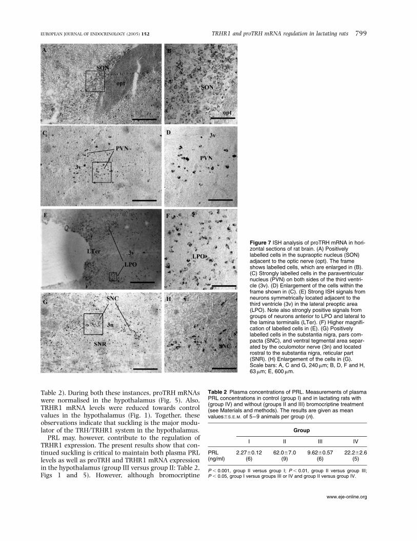
Table 2). During both these instances, proTRH mRNAswere normalised in the hypothalamus (Fig. 5). Also,TRHR1 mRNA levels were reduced towards controlvalues in the hypothalamus (Fig. 1). Together, theseobservations indicate that suckling is the major modu-lator of the TRH/TRHR1 system in the hypothalamus.
PRL may, however, contribute to the regulation ofTRHR1 expression. The present results show that con-tinued suckling is critical to maintain both plasma PRLlevels as well as proTRH and TRHR1 mRNA expressionin the hypothalamus (group III versus group II: Table 2,Figs 1 and 5). However, although bromocriptine
Figure 7 ISH analysis of proTRH mRNA in hori-zontal sections of rat brain. (A) Positivelylabelled cells in the supraoptic nucleus (SON)adjacent to the optic nerve (opt). The frameshows labelled cells, which are enlarged in (B).(C) Strongly labelled cells in the paraventricularnucleus (PVN) on both sides of the third ventri-cle (3v). (D) Enlargement of the cells within theframe shown in (C). (E) Strong ISH signals fromneurons symmetrically located adjacent to thethird ventricle (3v) in the lateral preoptic area(LPO). Note also strongly positive signals fromgroups of neurons anterior to LPO and lateral tothe lamina terminalis (LTer). (F) Higher magnifi-cation of labelled cells in (E). (G) Positivelylabelled cells in the substantia nigra, pars com-pacta (SNC), and ventral tegmental area separ-ated by the oculomotor nerve (3n) and locatedrostral to the substantia nigra, reticular part(SNR). (H) Enlargement of the cells in (G).Scale bars: A, C and G, 240mm; B, D, F and H,63mm; E, 600mm.
Table 2 Plasma concentrations of PRL. Measurements of plasmaPRL concentrations in control (group I) and in lactating rats with(group IV) and without (groups II and III) bromocriptine treatment(see Materials and methods). The results are given as meanvalues^S.E.M. of 5–9 animals per group (n).
Group
I II III IV
PRL(ng/ml)
2.27^0.12(6)
62.0^7.0(9)
9.62^0.57(6)
22.2^2.6(5)
P , 0.001, group II versus group I; P , 0.01, group II versus group III;P , 0.05, group I versus groups III or IV and group II versus group IV.
TRHR1 and proTRH mRNA regulation in lactating rats 799EUROPEAN JOURNAL OF ENDOCRINOLOGY (2005) 152
www.eje-online.org

decreases the PRL concentration in the plasma of lac-tating animals, the hormone concentration does notdecrease as much as in animals in group III. Further-more, the decrease in TRHR1 expression is not as pro-nounced in group IV as in group III, and this can beaccounted for by the higher PRL levels in group IV.
It has been established that the primary structure ofhypothalamic and pituitary PRL is identical. In the pre-sent experiments we cannot differentiate between theeffects of circulating and locally produced hypothala-mic PRL since circulatory hormone has access to hypo-thalamic neurons through the choroid plexi of thebrain ventricles (29). Circulating PRL may also enterthe brain by retrograde blood flow from the anteriorpituitary to the hypothalamus (30, 31). Hence, it isnot possible to determine whether the changes inTRHR1 mRNA expression may be attributed to thehigher levels of plasma PRL in group IV comparedwith group III animals, or are a result of locally syn-thesised PRL or both. Both the long and short form ofthe PRL receptor (PRLR) (32) are present in the hypo-thalamus and in the pituitary. It is possible that hypo-thalamic PRL also acts as an autocrine/paracrinehormone since SON and PVN were found to expresshigh levels of PRLR in lactating, but not in dioestrous,rats (33).
Suckling-initiated reduction in the tonic dopamineinhibition has been suggested not to be the only mech-anism by which PRL secretion is enhanced duringlactation, as summarised by Martinez de la Escaleraand Weiner (34). In this context, the up-regulation ofproTRH/TRHR1 mRNAs in the SON and PVN whichcontain PRL and PRL receptor positive neuronsduring lactation (35, 36) may be part of a local feed-back loop to enhance hypothalamic stimulation ofPRL secretion from the pituitary. Our data also supportthe possibility that a similar mechanism may prevail inthe pituitary where lactotrophs, which contain recep-tors for dopamine and other hypothalamic hormonesknown to affect PRL secretion, show up-regulation ofTRHR1.
Binding of PRL to its receptor leads to receptor dimer-isation and activation of the Jak/STAT pathway,reviewed in Freeman et al. (37). The consensus DNAmotif recognized by STAT1, STAT3 and STAT5 homo-or heterodimers is termed GAS (g-interferon activatedsequence) and consists of the palindromic sequence;
TTCxxxGAA (38). Re-examining the human TRHRpromoter sequence (39), we found one perfect matchof the GAS consensus motif in position 226 to 218relative to the translation start site and several poten-tial binding sites containing only one mismatch.Hence, increased transcription of the TRHR1 genemay occur after PRL activation of the Jak/STAT path-way. It is also of interest that we have previouslydescribed binding of the glucocorticoid receptor to thehuman TRHR promoter (40) as this receptor has beenproposed to interact with STAT to initiate cell- and cyto-kine-specific responses (41, 42). In the pituitary, theanterior pituitary-specific transcription factor Pit-1 isa candidate for regulation of the TRHR gene, since wehave previously demonstrated that Pit-1 binds to thehuman TRHR gene promoter in a cell type-specificmanner (39).
Bromocriptine acts on dopamine D2 receptors whichare also present in the pituitary (43). The detailedmolecular actions of bromocriptine (2-bromo-alpha-ergocryptine), a semi-synthetic ergot alkaloid withlong-term stimulatory effects on dopamine D2 recep-tors, are not known. Since bromocriptine is able topenetrate the blood–brain barrier (44), the drug willhave access to most dopaminergic systems in theCNS. It is clinically used to reduce hyperprolactinaemiaand to inhibit milk secretion, as well as to treat, forexample, Parkinson’s disease and acromegaly (45 –50). TRHR1 mRNA expression is highly up-regulatedin the pituitary after treatment with bromocriptine(Fig. 4). The molecular mechanisms for increasedTRHR1 mRNA levels are unknown, but could be away to compensate for the decrease in plasma PRL con-centration in an effort to make the pituitary moreresponsive to TRH. Maintained plasma PRL concen-trations would also help to sustain milk productionand lactation. It is known that activated TRHR1 signalsthrough the Gaq/11 and a subset of Gas heterotrimericG-proteins (51 –54). It is therefore of interest thatGaq/11-deficient mice where the PRL release axiswas normal, did not display any maternal behaviourincluding nursing (55). We have previously shownthat many signal transduction effects caused by TRHin pituitary adenoma cells (GH cells) in culture, areinhibited or abolished by bromocriptine. These effectscould be explained by inhibition of G-proteinactions (Gi2, Go, Gs and Gq/11) (56, 57). In addition to
Table 3 TRHDE mRNA in rat pituitary gland and hypothalamus quantified by using real time RT-PCR. Values are relative to those ofb-actin and normalised to the control group (group I). The results represent a mean values^S.E.M. of 3–6 rats (n).
Group
Tissue I II III IV
Pituitary 1.00^0.060 (6) 1.20^0.15 (5) 0.87^0.082 (6) 0.95^0.28 (3)Hypothalamus 1.00^0.094 (6) 1.10^0.087 (3) N.D. 0.71^0.17 (6)
N.D., not determined.
800 A Fjeldheim and others EUROPEAN JOURNAL OF ENDOCRINOLOGY (2005) 152
www.eje-online.org

these TRH-inhibitory actions, bromocriptine has alsobeen shown to stimulate the pituitary TRH degradingenzyme (TRHDE), a TRH-specific ectoenzyme respon-sible for the inactivation of TRH (58). This is anothermechanism whereby bromocriptine would be able todecrease the biological effect of TRH at the pituitarylevel. However, no significant change in TRHDEmRNA expression (Table 3) was detected in the hypo-thalamus or in the pituitary between the differentexperimental groups. Thus, the changes in proTRHmRNA expression reported in this study are not influ-enced by, or are secondary to, altered TRH half life.
In this study we examined, in particular, the PVN,SON, LPO and LM, as well as the SNC, which is amesencephalic region enriched in dopaminergic cellslocated adjacent to the hypothalamus. The reason forthis was that neurons in these areas showed a highexpression of mRNA coding for proTRH and/orTRHR1, as documented in histological in situ analyses.The up-regulation of proTRH and/or TRHR1 mRNA inthe different nuclei was mainly a result of more cellsexpressing proTRH/TRHR1 mRNA since the resultsfrom cell-counting paralleled the results from theimage processing.
Our data show that TRHR1 mRNA expression is up-regulated in the SON and the LM during suckling.TRHR1 expression is also slightly increased in thePVN. This is of great interest since oxytocin and vaso-pressin are both produced in the SON and the PVN(59, 60). They are important, for example, in milksecretion, fluid balance and nesting behaviour (61).Intracerebroventricular administration of TRH leadsto increased concentrations of oxytocin and vasopressinin the hypothalamus and in the posterior pituitary innursing rats (62). Noradrenalin and serotonin areknown to control arginine-vasopressin and oxytocinsecretion in the systemic circulation, and have recentlybeen shown to do the same in the PVN and the SON inmouse (63). However, TRH had no effect on noradrena-lin release from neurons of the PVN (64). TRH may stillact by modulating release of, for example, serotoninfrom these nuclei and noradrenalin from the SON.TRH, noradrenalin and serotonin may also act in paral-lel giving an additive or synergetic effect on oxytocinand vasopressin release.
In addition, the nerve cells located in these nuclei areknown to project fibres to other hypothalamic regionsin particular, but also to distinct, extra-hypothalamicbrain areas. Accordingly, TRH may, in relation to nur-sing, modulate a number of CNS functions throughactivation of TRHR1 (65). The present findingsextend previous observations of Uribe, Sanchez and col-leagues (15, 66) that showed that mRNA for proTRHwas up-regulated in the PVN under a number of phys-iological conditions such as lactation and stress associ-ated with freezing.
In our study, TRHR1 mRNA expression was reducedin the amygdala in group II animals (Table 1), but was
normalised 24 h after termination of suckling (groupIII). The nerve cells located in this area are known toproject fibres to other hypothalamic regions and/or todistinct, extra-hypothalamic brain areas, therebymaking it possible to influence a number of brain func-tions associated with motherhood and nursing. Thesepossibilities indicate a functional link between thehypothalamus and the amygdala in the suckling –lacta-tion reflex. In contrast, no significant changes werefound between the experimental groups in the hippo-campus, cortex or cerebellum, indicating that theseareas do not participate in this response. ProTRHmRNA expression did not show any significant changesbetween the groups in the thalamus, cortex or cerebel-lum (data not shown).
Our results indicate that suckling through neuronalactivation stimulates the hypothalamic nuclei toincrease synthesis and release of proTRH and up-regu-lates the TRHR1 system. TRH acting through TRHR1may modulate the synaptic circuitry which is involvedin lactation and may also be important for the groom-ing behaviour of rats. Taken together, the present andearlier results open up an emerging understanding ofthe involvement of TRH and TRHR1 in the physiologyand pharmacology of suckling-induced lactation atthe pituitary and the hypothalamic levels.
Acknowledgements
We thank Professor Egil Haug, Aker Hormone Labora-tory for analysis of rat prolactin. This work was sup-ported by the Norwegian Cancer Society, theNorwegian Research Council (NFR), Anders Jahre Foun-dation for Promotion of Science, Oslo, Norway and TheNovo Nordisk Foundation, Copenhagen, Denmark.
References
1 Gautvik KM, Weintraub BD, Graeber CT, Maloof F, Zuckerman JE& Tashjian AH Jr. Serum prolactin and TSH: effects of nursing andpyroGlu-His-ProNH2 administration in postpartum women. Jour-nal of Clinical Endocrinology and Metabolism 1973 37 135–139.
2 Gautvik KM, Tashjian AH Jr, Kourides IA, Weintraub BD,Graeber CT, Maloof F, Suzuki K & Zuckerman JE. Thyrotropin-releasing hormone is not the sole physiologic mediator of prolac-tin release during suckling. New England Journal of Medicine1974 290 1162–1165.
3 Grosvenor CE, Mena F & Whitworth NS. Evidence that the dopa-minergic prolactin-inhibiting factor mechanism regulates onlythe depletion-transformation phase and not the release phase ofprolactin secretion during suckling in the rat. Endocrinology1980 106 481–485.
4 de Greef WJ, Klootwijk W, Karels B & Visser TJ. Levels of dopamineand thyrotropin-releasing hormone in hypophysial stalk bloodduring an oestrogen-stimulated surge of prolactin in the ovari-ectomized rat. Journal of Endocrinology 1985 105 107– 112.
5 Mori M & Yamada M. Thyroid hormones regulate the amount ofthyrotropin-releasing hormone in the hypothalamic median emi-nence of the rat. Journal of Endocrinology 1987 114 443– 448.
6 Haisenleder DJ, Ortolano GA, Dalkin AC, Yasin M & Marshall JC.Differential actions of thyrotropin (TSH)-releasing hormone
TRHR1 and proTRH mRNA regulation in lactating rats 801EUROPEAN JOURNAL OF ENDOCRINOLOGY (2005) 152
www.eje-online.org

pulses in the expression of prolactin and TSH subunit messengerribonucleic acid in rat pituitary cells in vitro. Endocrinology 1992130 2917– 2923.
7 Aizawa T & Greer MA. Delineation of the hypothalamic area con-trolling thyrotropin secretion in the rat. Endocrinology 1981 1091731–1738.
8 de Greef WJ & Visser TJ. Evidence for the involvement of hypo-thalamic dopamine and thyrotropin-releasing hormone in suck-ling-induced release of prolactin. Journal of Endocrinology 198191 213–223.
9 de Greef WJ, Voogt JL, Visser TJ, Lamberts SW & van der Schoot P.Control of prolactin release induced by suckling. Endocrinology1987 121 316–322.
10 Leblanc H, Lachelin GC, Abu-Fadil S & Yen SS. Effects of dopa-mine infusion on pituitary hormone secretion in humans. Journalof Clinical Endocrinology and Metabolism 1976 43 668– 674.
11 Scanlon MF, Weightman DR, Shale DJ, Mora B, Heath M,Snow MH, Lewis M & Hall R. Dopamine is a physiological regula-tor of thyrotropin (TSH) secretion in normal man. Clinical Endo-crinology 1979 10 7–15.
12 Blake CA. Stimulation of pituitary prolactin and TSH release inlactating and proestrous rats. Endocrinology 1974 94 503–508.
13 Riskind PN, Millard WJ & Martin JB. Evidence that thyrotropin-releasing hormone is not a major prolactin-releasing factorduring suckling in the rat. Endocrinology 1984 115 312–316.
14 Sheward WJ, Fraser HM & Fink G. Effect of immunoneutralizationof thyrotropin-releasing hormone on the release of thyrotropinand prolactin during suckling or in response to electrical stimu-lation of the hypothalamus in the anaesthetized rat. Journal ofEndocrinology 1985 106 113–119.
15 Uribe RM, Redondo JL, Charli JL & Joseph-Bravo P. Suckling andcold stress rapidly and transiently increase TRH mRNA in theparaventricular nucleus. Neuroendocrinology 1993 58 140–145.
16 Walaas SI, Nairn AC & Greengard P. Regional distribution of cal-cium- and cyclic adenosine 30,50-monophosphate-regulated pro-tein phosphorylation systems in mammalian brain. I. Particulatesystems. Journal of Neuroscience 1983 3 291–301.
17 Paxinos G, Watson C. The Rat Brain in Stereotaxic Coordinates.San Diego: Academic Press Inc., 1998.
18 Haug E & Gautvik KM. Radioimmunoassay of rat prolactin and itsuse in measuring prolactin production by cultured pituitary cells.Acta Endocrinologica 1976 82 282–297.
19 Chirgwin JM, Przybyla AE, MacDonald RJ & Rutter WJ. Isolationof biologically active ribonucleic acid from sources enriched inribonuclease. Biochemistry 1979 18 5294–5299.
20 Sambrook J, Fritsch EF & Maniatis T. Molecular Cloning, A LaboratoryManual. New York: Cold Spring Harbor Laboratory Press, 1989.
21 Matre V, Karlsen HE, Wright MS, Lundell I, Fjeldheim A-K,Gabrielsen OS, Larhammar D & Gautvik KM. Molecular cloningof a functional human thyrotropin-releasing hormone receptor.Biochemical and Biophysical Research Communications 1993 195179–185.
22 Cao J, O’Donell D, Vu H, Payza K, Pou C, Godbout C, Jakob A,Pelletier M, Lembo P, Ahmad S & Walker P. Cloning and charac-terization of a cDNA encoding a novel subtype of rat thyrotro-pin-releasing hormone receptor. Journal of Biological Chemistry1998 273 32281–32287.
23 Itadani H, Nakamura T, Itoh J, Iwaasa H, Kanatani A, Borkowski J,Ihara M & Ohta M. Cloning and characterization of a new subtypeof thyrotropin-releasing hormone receptors. Biochemical and Bio-physical Research Communications 1998 250 68–71.
24 Ovstebo R, Foss Haug KB, Lande K & Kierulf P. PCR-based cali-bration curves for studies of quantitative gene expression inhuman monocytes: development and evaluation. Clinical Chem-istry 2003 49 425– 432.
25 Hoover F & Goldman D. Temporally correlated expression ofnAChR genes during development of the mammalian retina.Experimental Eye Research 1992 54 561–571.
26 Torp R, Hoover F, Danbolt NC, Storm-Mathisen J & Ottersen OP.Differential distribution of the glutamate transporters GLT1 and
rEAAC1 in rat cerebral cortex and thalamus: an in situ hybridiz-ation analysis. Anatomy and Embryology 1997 195 317–326.
27 Clark BJ, Fluckiger E, Lowe DM & Vigouret JM. How does bromo-criptine work? Triangle 1978 1 21–32.
28 Nillni EA, Aird F, Seidah NG, Todd RB & Koenig JI. Pre-proTRH(178– 199) and two novel peptides (pFQ7 and pSE14)derived from its processing, which are produced in the paraven-tricular nucleus of the rat hypothalamus, are regulated duringsuckling. Endocrinology 2001 142 896–906.
29 Mangurian LP, Walsh RJ & Posner BI. Prolactin enhancement ofits own uptake at the choroid plexus. Endocrinology 1992 131698– 702.
30 Mezey E & Palkovits M. Two-way transport in the hypothalamo-hypophyseal system. In Frontiers in Neuroendocrinology, pp 1– 29.Eds WF Ganong & L Martini. New York: Raven Press, 1982.
31 Oliver C, Mical RS & Porter JC. Hypothalamic-pituitary vascula-ture: evidence for retrograde blood flow in the pituitary stalk.Endocrinology 1977 101 598–604.
32 Chiu S, Koos RD & Wise PM. Detection of prolactin receptor(PRL-R) mRNA in the rat hypothalamus and pituitary gland.Endocrinology 1992 130 1747–1749.
33 Grattan DR. The actions of prolactin in the brain during preg-nancy and lactation. Progress in Brain Research 2001 133153– 171.
34 Martinez de la Escalera G & Weiner RI. Dissociation of dopaminefrom its receptor as a signal in the pleiotropic hypothalamic regu-lation of prolactin secretion. Endocrine Reviews 1992 13241– 255.
35 Torner L, Maloumby R, Nava G, Aranda J, Clapp C & Neumann ID.In vivo release and gene upregulation of brain prolactin inresponse to physiological stimuli. European Journal of Neuroscience2004 19 1601–1608.
36 Pi X-J & Grattan DR. Differential expression of the two forms ofprolactin receptor mRNA within microdissected hypothalamicnuclei of the rat. Molecular Brain Research 1998 59 1–12.
37 Freeman ME, Kanyicska B, Lerant A & Nagy G. Prolactin: struc-ture, function, and regulation of secretion. Physiological Reviews2000 80 1523–1631.
38 Horseman ND & Yu-Lee LY. Transcriptional regulation by thehelix bundle peptide hormones: growth hormone, prolactin, andhematopoietic cytokines. Endocrine Reviews 1994 15 627–649.
39 Matre V, Hovring PI, Orstavik S, Frengen E, Rian E, Velickovic Z,Murray-McIntosh RP & Gautvik KM. Structural and functionalorganization of the gene encoding the human thyrotropin-releas-ing hormone receptor. Journal of Neurochemistry 1999 72 40– 50.
40 Hovring PI, Matre V, Fjeldheim AK, Loseth OP & Gautvik KM.Transcription of the human thyrotropin-releasing hormonereceptor gene – analysis of basal promoter elements and gluco-corticoid response elements. Biochemical and Biophysical ResearchCommunications 1999 257 829–834.
41 Stoecklin E, Wissler M, Gouilleux F & Groner B. Functional inter-actions between Stat5 and the glucocorticoid receptor. Nature1996 383 726–728.
42 Stoecklin E, Wissler M, Moriggl R & Groner B. Specific DNA bind-ing of Stat5, but not of glucocorticoid receptor, is required fortheir functional cooperation in the regulation of gene transcrip-tion. Molecular and Cellular Biology 1997 17 6708–6716.
43 Johansen PW, Paulssen RH, Bjoro T, Gautvik KM & Gordeladze JO.Distinct guanine nucleotide binding protein alpha-subunit recep-tor coupling in GH cell lines: effects of bromocriptine and hor-mones on effector enzyme modulation. Cellular Physiology andBiochemistry 2001 11 339–352.
44 Renodon A, Boucher JL, Sari MA, Delaforge M, Ouazzani J &Mansuy D. Bromocriptine is a strong inhibitor of brain nitricoxide synthase: possible consequences for the origin of its thera-peutic effects. FEBS Letters 1997 406 33–36.
45 Camanni F, Massara F, Fassio V, Molinatti GM & Muller EE. Effectof five dopaminergic drugs on plasma growth hormone levels inacromegalic subjects. Neuroendocrinology 1975 19 227–240.
802 A Fjeldheim and others EUROPEAN JOURNAL OF ENDOCRINOLOGY (2005) 152
www.eje-online.org

46 Cooke I, Foley M, Lenton E, Preston E, Millar D, Jenkins A,Obiekwe B, McNeilly A, Parsons J & Kennedy G. The treatmentof puerperal lactation with bromocriptine. Postgraduate MedicalJournal 1976 52 (Suppl 1) 75–80.
47 Prysor-Jones RA & Jenkins JS. Effect of bromocriptine, ergotamineand other ergot alkaloids on the hormone secretion and growth ofa rat pituitary tumour. Journal of Endocrinology 1980 86147– 153.
48 Johansen PW, Sand O, Iversen JG, Haug E & Gautvik KM. Effects ofbromocriptine on prolactin release, electrical membrane proper-ties and transmembrane Ca2þ fluxes in cultured rat pituitary ade-noma cells. Acta Endocrinologica 1986 111 185–192.
49 Halse J, Harris AG, Kvistborg A, Kjartansson O, Hanssen E,Smiseth O, Djosland O, Hass G & Jervell J. A randomized studyof SMS 201–995 versus bromocriptine treatment in acromegaly:clinical and biochemical effects. Journal of Clinical Endocrinologyand Metabolism 1990 70 1254–1261.
50 Jaffe CA & Barkan AL. Treatment of acromegaly with dopamineagonists. Endocrinology and Metabolism Clinics of North America1992 21 713– 735.
51 Aragay AM, Katz A & Simon MI. The G alpha q and G alpha 11proteins couple the thyrotropin-releasing hormone receptor tophospholipase C in GH3 rat pituitary cells. Journal of BiologicalChemistry 1992 267 24983–24988.
52 Hsieh KP & Martin TF. Thyrotropin-releasing hormone and gon-adotropin-releasing hormone receptors activate phospholipase Cby coupling to the guanosine triphosphate-binding proteins Gqand G11. Molecular Endocrinology 1992 6 1673–1681.
53 Gautvik KM, Gordeladze JO, Janhsen T, Haug E, Hansson V &Lystad E. Thyroliberin receptor binding and adenylyl cyclase acti-vation in cultured prolactin-producing rat pituitary tumor cells(GH cells). Journal of Biological Chemistry 1983 25810304–10311.
54 Paulssen RH, Paulssen EJ, Gautvik KM & Gordeladze JO. The thyr-oliberin receptor interacts directly with a stimulatory guanine-nucleotide-binding protein in the activation of adenylyl cyclasein GH3 rat pituitary tumour cells. Evidence obtained by the useof antisense RNA inhibition and immunoblocking of the stimu-latory guanine-nucleotide-binding protein. European Journal ofBiochemistry 1992 204 413– 418.
55 Wettschureck N, Moers A, Hamalainen T, Lemberger T, Shcutz G& Offermanns S. Heterotrimeric G proteins of the Gq/11 family arecrucial for the induction of maternal behaviour in mice. Molecularand Cellular Biology 2004 24 8048–8054.
56 Johansen PW, Clausen OP, Haug E, Fossum S & Gautvik KM. Effectsof bromocriptine on cell cycle distribution and cell morphology in
cultured rat pituitary adenoma cells. Acta Endocrinologica 1985110 319–328.
57 Johansen PW, Haug E & Gautvik KM. Effects of bromocriptine onhormone production and cell growth in cultured rat pituitarycells. Acta Endocrinologica 1985 110 200–206.
58 Vargas MA, Bourdais J, Sanchez S, Uriostegui B, Moreno E,Joseph-Bravo P & Charli JL. Multiple hypothalamic factors regu-late pyroglutamyl peptidase II in cultures of adenohypophysealcells: role of the cAMP pathway. Journal of Neuroendocrinology1998 10 199– 206.
59 Cunningham ET Jr & Sawchenko PE. Reflex control of magnocel-lular vasopressin and oxytocin secretion. Trends in Neuroscience1991 14 406– 411.
60 Aguilera G, Lightman SL & Kiss A. Regulation of the hypothala-mic–pituitary –adrenal axis during water deprivation. Endocrin-ology 1993 132 241–248.
61 Lim MM, Wang Z, Olazabal DE, Ren X, Terwilliger EF & Young LJ.Enhanced partner preference in a promiscuous species by manip-ulating the expression of a single gene. Nature 2004 429754–757.
62 Ciosek J & Guzek JW. Thyrotropin-releasing hormone affects theoxytocin, vasopressin and prolactin release in female ratsduring midlactation: relation to suckling. Journal of Physiologyand Pharmacology 1998 49 135–150.
63 Vacher CM, Fretier P, Cremion C, Calas A & Hardin-Pouzet H.Activation by serotonin and noradrenaline of vasopressin andoxytocin expression in the mouse paraventricular and supraopticnuclei. Journal of Neuroscience 2002 22 1513–1522.
64 Brunetti L, Orlando G, Michelotto B, Recinella L & Vacca M.Cocaine- and amphetamine-regulated transcript peptide-(55–102) and thyrotropin releasing hormone inhibit hypothalamicdopamine release. European Journal of Pharmacology 2000 409103–107.
65 Yamada M, Satoh T & Mori M. Mice lacking the thyrotropin-releasing hormone gene: what do they tell us? Thyroid 2003 131111–1121.
66 Sanchez E, Uribe RM, Corkidi G, Zoeller RT, Cisneros M,Zacarias M, Morales-Chapa C, Charli JL & Joseph-Bravo P. Differ-ential responses of thyrotropin-releasing hormone (TRH) neuronsto cold exposure or suckling indicate functional heterogeneity ofthe TRH system in the paraventricular nucleus of the rat hypo-thalamus. Neuroendocrinology 2001 74 407– 422.
Received 21 October 2004
Accepted 4 February 2005
TRHR1 and proTRH mRNA regulation in lactating rats 803EUROPEAN JOURNAL OF ENDOCRINOLOGY (2005) 152
www.eje-online.org