Regression of Nitrosamine-induced Pancreatic Cancers in Hamsters Treated with Luteinizing...
7
1990;50:3716-3721. Cancer Res Bela Szende, Gordan Srkalovic, Kate Groot, et al. Antagonists or Agonists Hamsters Treated with Luteinizing Hormone-releasing Hormone Regression of Nitrosamine-induced Pancreatic Cancers in Updated version http://cancerres.aacrjournals.org/content/50/12/3716 Access the most recent version of this article at: E-mail alerts related to this article or journal. Sign up to receive free email-alerts Subscriptions Reprints and . [email protected] Department at To order reprints of this article or to subscribe to the journal, contact the AACR Publications Permissions . [email protected] Department at To request permission to re-use all or part of this article, contact the AACR Publications on June 4, 2014. © 1990 American Association for Cancer Research. cancerres.aacrjournals.org Downloaded from on June 4, 2014. © 1990 American Association for Cancer Research. cancerres.aacrjournals.org Downloaded from
Transcript of Regression of Nitrosamine-induced Pancreatic Cancers in Hamsters Treated with Luteinizing...
1990;50:3716-3721. Cancer Res Bela Szende, Gordan Srkalovic, Kate Groot, et al. Antagonists or AgonistsHamsters Treated with Luteinizing Hormone-releasing Hormone Regression of Nitrosamine-induced Pancreatic Cancers in
Updated version
http://cancerres.aacrjournals.org/content/50/12/3716
Access the most recent version of this article at:
E-mail alerts related to this article or journal.Sign up to receive free email-alerts
Subscriptions
Reprints and
To order reprints of this article or to subscribe to the journal, contact the AACR Publications
Permissions
To request permission to re-use all or part of this article, contact the AACR Publications
on June 4, 2014. © 1990 American Association for Cancer Research. cancerres.aacrjournals.org Downloaded from on June 4, 2014. © 1990 American Association for Cancer Research. cancerres.aacrjournals.org Downloaded from
(CANCER RESEARCH 50. 3716-3721, June 15, 1990]
Regression of Nitrosamine-induced Pancreatic Cancers in Hamsters Treated withLuteinizing Hormone-releasing Hormone Antagonists or Agonists1
Bela Szende,2 Gordan Srkalovic, Kate Groot, Karoly Lapis, and Andrew V. Schally
Endocrine, Polypeptide, and Cancer institute. Veterans Administration Medical Center, New Orleans, Louisiana 70146 [A. V. S., K. G.J; Section of ExperimentalMedicine, Department of Medicine, Tulane University Medical School, New Orleans, Louisiana 70112 [B. S., G. S., A. V. S.J; and First Institute of Pathology andExperimental Cancer Research, Semmelweis University Medical School, Budapest, H-I085 Hungary [K. L.]
ABSTRACT
Groups of 15 female Syrian golden hamsters with .V-nitr<isol>is(2-oxopropyl)amine-induced pancreatic cancers were treated for 2 mo withmicrocapsules of the luteinizing hormone-releasing hormone (LH-RH)antagonist |Ac-D-Nal(2)1-D-Phe(4Cl)2-r>Pal(3)3,r>Cit6,D-Ala10l LH-RH(SB-75) releasing 8 Mg/day or with the microcapsules of the LH-RHagonist D-tryptophan-6-luteinizing hormone-releasing hormone (o-Trp-6-LH-RH) releasing 8 Mg/dayor 25 Mg/day.Chronic treatment with SB-75 resulted in 70% inhibition of pancreatic tumor weight; D-Trp-6-LH-RH in doses of 8 Mg/dayand 25 Mg/dayproduced 66% and 62% inhibition,respectively. The number of animals with pancreatic tumors was reducedby about 50% in each treated group. I »moronsascites were found inseven control hamsters and in one hamster in each group treated with i>-Trp-6-1.11-RH but not in the group given SB-75. Reduction in serumluteinizing hormone levels and ovarian as well as uterine weights indicatedthat an inhibition of the pituitary-gonadal axis occurred during chronicSB-75 and D-Trp-6-LH-RH treatment. Membrane receptor assaysshowed a significant decrease of the concentration of binding sites forLH-RH in tumor cells after SB-75 or D-Trp-6-LH-RH treatment. Insulin-like growth factor I receptors, but not epidermal growth factor receptors, were down-regulated by D-Trp-6-LH-RH. SB-75 did not influencethe concentration or the binding capacity of insulin-like growth factor Iand epidermal growth factor receptors in the tumor cells. The inhibitory-effect of chronic treatment with SB-75 and D-Trp-6-LH-RH on tumorgrowth was mediated by enhanced apoptosis (programmed cell death)induced by the change in hormonal environment. Apoptosis was alsoproduced in hamsters with /V-nitrosobis(2-oxopropyl)amine-induced pancreatic cancers by acute treatment (3 to 6 days) with high doses of n-Trp-6-LH-RH or SB-75. In view of its potency and an immediatepowerful inhibitory effect, the LH-RH antagonist SB-75 might be considered as a possible new hormonal agent for the treatment of exocrinepancreatic cancer.
INTRODUCTION
Agonist analogues of LH-RH1 and of somatostatin have been
shown to inhibit the growth of various transplantable andinduced, hormone-sensitive experimental animal tumors (1-8).A series of studies were carried out with these peptides on BOP-induced hamster pancreatic carcinoma (1, 5, 6, 9). We alsopointed out that the tumor growth-inhibitory effect of theseanalogues was mediated by an enhanced rate of apoptosis(programmed cell death) (6-10). Recently an increase in survival time of patients with advanced pancreatic cancer after D-Trp-6-LH-RH therapy has been reported (11). Agonistic ana-
Received9/26/89:revised2/19/90.The costs of publication of this article were defrayed in part by the payment
of page charges. This article must therefore be hereby marked advertisement inaccordance with 18 U.S.C. Section 1734 solely to indicate this fact.
1This work was supported by NIH Grant CA 40077. the Medical Research
Services of the Veterans Administration, and the G. Harold and Leila Y. MathersFoundation, all to A. V. S.
2To whom requests for reprints should be addressed, at Veterans Administra
tion Medical Center, 1601 Perdido Street. New Orleans, LA 70146.3The abbreviations used are: LH-RH. luteinizing hormone-releasing hormone:
LH. luteinizing hormone; BOP, ¿V-nitrosobis(2-oxopropyl)amine; EGF. epidermal growth factor: IGF-I. insulin-like growth factor I; D-Trp-6-LH-RH, D-tryptophan-6-luteinizing hormone-releasing hormone: Ac, acetyl; Nal(2), 3-(2-naphthyljalanine: Cit. citrulline (2-amino-5-ureidopentanoic acid); Pal(3), 3-(3-pyridyl)alanine.
logues of LH-RH appear to decrease pancreatic cancer growthby eliminating the stimulatory effect of sex steroids and bydirect effects on tumors (12). However, the agonistic analoguesinduce an initial stimulation of the release of gonadotropin andsex steroids which may cause a temporary flareup of the diseasewith undesirable clinical consequences. The use of new antagonistic analogues of LH-RH (13,14), which cause an immediateinhibition of the pituitary-gonad axis, would avoid the initialstimulatory effects on tumor growth. Studies carried out ontumor cell lines suggest that LH-RH antagonists may also havea direct inhibitory effect on tumor growth (15). This indicatesthe need to investigate the receptors for both LH-RH andgrowth factors in tumors.
In this study, the LH-RH antagonist SB-75 (13), free ofedematogenic effects, was administered to Syrian golden hamsters with BOP-induced carcinomas of the exocrine pancreas.The effect of SB-75 treatment was compared with that of theLH-RH agonist D-Trp-6-LH-RH.
MATERIALS AND METHODS
Animals. Eighty female Syrian golden hamsters (CH:RGH) weighing100 ±10 g were obtained from the National Cancer Institute FrederickCancer Research Facility (Frederick, MD). They were housed two percage at the Animal Research Facility of our institute in an air-conditioned room at 22 ±1°Cand 55 ±5% humidity. The animals were
kept under an automatic 12 h light/12-h darkness schedule and givenRodent Laboratory Chow 50001 (Purina Mills, Inc.) and tap water adlibitum.
Induction of Pancreatic Cancer. The method of Pour et al. (16) wasused with some modifications (5). BOP was purchased from AshStevens (Detroit, MI) and stored at 4°C.The required amount of BOP
was freshly dissolved in 0.9% NaCl solution and given s.c. at a dose of10 mg/kg of body weight once weekly for 6 wk. Eighteen wk later (24wk from the start of the experiment), the surviving 69 hamsters wererandomly divided into 4 groups of 15 and into 3 groups of 3. At thattime the chronic treatment with the peptides was initiated.
Peptides. The LH-RH agonist D-Trp-6-LH-RH (pyro-Glu-His-Trp-Ser-Tyr-D-Trp-Leu-Arg-Pro-Gly-NHj) was synthesized by solid-phase methods and supplied by Debiopharm S. A. (Lausanne, Switzerland). Microcapsule formulation (17) of this peptide in biodegradablepoly(DL-lactide-co-glycolide) was prepared by Dr. P. Orsolini at Cyto-tech S. A. (Martigny, Switzerland) using a phase-separation process.This sustained release formulation in an aliquot of 36 or 12 mgmaintained a continuous liberation of 25 Mg/day or 8 Mg/day of theanalogue for 30 days.
The LH-RH antagonist [Ac-D-Nal(2)',D-Phe(4Cl)2,D-Pal(3)3,D-Cit6,D-Ala'°]LH-RH (SB-75) originally synthesized in our laboratory
(13) was made by Asta Pharma Co., Frankfurt, West Germany, usingsolid-phase methods. Microcapsules of SB-75 in poly(DL-lactide-co-glycolide) for sustained release were prepared also by Dr. P. Orsolini.Batch 1 was designed to release about 8 Mgof SB-75/day for 21 daysfrom an aliquot of 13.3-mg microcapsules. Both types of microcapsuleswere suspended in 0.7 ml of injection vehicle solution containing 2%cellulose and 1% Tween 80 in water. The suspension was mixedthoroughly using a Vortex mixer and injected s.c. through an 18-gaugeneedle.
Experimental Protocol. BOP-lesioned hamsters in groups of 15 re-
3716
on June 4, 2014. © 1990 American Association for Cancer Research. cancerres.aacrjournals.org Downloaded from
LH-RH ANALOGUES IN TREATMENT OF PANCREATIC CANCER
ceived the following treatment: Group 1, injection vehicle only (BOPcontrols); Group 2, SB-75 microcapsules (13.3 mg/animal on Days 0,21, and 42); Group 3, D-Trp-6-LH-RH microcapsules (36 mg/animalon Days 0 and 30); and Group 4, D-Trp-6-LH-RH microcapsules (12mg/animal on Days 0 and 30). Other hamsters in groups of 3 receiveds.c. injections of analogues dissolved in 10% polyvinylpyrrolidone in0.9% NaCl solution as follows: Group 5, 100 Mgof SB-75/animal dailyfor 3 days before the termination of the experiment; Group 6, 50 ng ofD-Trp-6-LH-RH/animal daily, for 3 days before sacrifice; and Group7, 50 n% of D-Trp-6-LH-RH/animal daily for 6 days before sacrifice.The experiment was terminated 8 wk after the first treatment with themicrocapsules or vehicle in Groups 1 to 4 or 4 to 7 days after the firsts.c. injection in Groups 5 to 7, which corresponded in all cases to 32wk from the start of the BOP treatment.
Pathological Procedures. During the 8-wk treatment period, pan-creata and livers of animals that died were processed for histology. Atthe end of the eighth week, the hamsters were exsanguinated undermethoxyflurane (Metofane; Pitmann-Moore, Washington Crossing,NJ) anesthesia. Body weights were measured, and any ascites wasremoved and measured. The average weight of all pancreata as well asthe average weight of the tumorous pancreata (based on histology) ineach group was recorded. Pancreas, liver, uterus, and ovaries wereremoved, cleaned, and weighed. Pancreatic and liver tissue was fixed in7am hon i's fixative (18). Specimens were embedded in Paraplast (Ox
ford Labware, Saint Louis, MO), and step sections 6 urn thick werecut. Sections were stained with hematoxylin/eosin. For the measurement of the apoptotic index, 200 glandular structures were consideredin each tumor. For electron microscopy, 1-mm3 pieces of tumor tissues
of two animals in each group were fixed in 2.5% glutaraldehyde,postfixed in 2% OsO4, and embedded into Polybed (Polysciences,Warrington, PA). Ultrathin sections were cut and stained with uranylacetate and lead citrate and viewed in a JEM 100 B electron microscope(JEOL, Tokyo, Japan).
Receptor Assays. Only tumors weighing more than 0.2 g could beprocessed for receptor measurement. Membrane preparation, radioio-dination of peptides, and receptor binding of D-Trp-6-LH-RH, EGF,and IGF-I were performed as previously described (19-21). Recombinant human IGF-I for receptor assay was kindly provided by Genentech,Inc. (San Francisco, CA). Recombinant human EGF was purchasedfrom Amgen Biologicals (Thousand Oaks, CA). The LIGAND-PCcomputerized curve-fitting program of Munson and Rodbard (22) wasused to determine the types of receptor binding, dissociation constants(A',,),and the maximal binding capacity of receptors (Bmas).
Serum LH and IGF-I Levels. Serum LH and IGF-I levels weredetermined using LH-RP-2 and UKB-487 reference preparations provided by the National Hormone and Pituitary program of NIDDK.
Statistical Analyses. Statistical analyses were performed by the two-sided Student t test and the Fisher exact test.
RESULTS
Body, Ovarian, and Uterine Weights and Liver Pathology. Thebody weights of the animals increased by about 50% during theexperiment. No differences were seen between the control andtreated groups in this respect. A significant decrease in theweight of ovaries and uteri was seen in the treated groups,compared with the control (Table 1).
Serum LH and IGF-I Levels. Serum LH was decreased significantly after the treatment with SB-75. Serum IGF-I increased slightly after D-Trp-6-LH-RH therapy (Table 1).
Tumor Pathology. Five control animals died because of pancreatic cancer during the treatment period. Three of the hamsters treated with SB-75 and 3 treated with 8 pg/day of D-Trp-6-LH-RH also died. In both of these groups, 2 of the 3 hamstershad pancreatic tumors, and one showed preneoplastic lesionssuch as various types of ductal proliferation in the pancreas(23). The livers of all hamsters were cirrhotic, with occasionalcystic dilation of the bile ducts, due to BOP treatment.
At the termination of the experiment, the average weight ofthe pancreata of hamsters treated with BOP increased morethan 6-fold compared with the pancreata in normal hamsters,which was highly significant. The pancreata of the control BOP-treated hamsters weighed 2.17 g in average. The average pancreatic weight of the SB-75-treated animals was 0.53 g. D-Trp-6-LH-RH treatment resulted in an average pancreatic weightof 0.52 g for the 8-pg/day dose and 0.60 g for the 25-Mg/daydose (Table 1). The diameters of the individual nodules variedfrom 2 to 10 mm, but sometimes the nodules were confluent,making it impossible to recognize the size. Because of themultinodularity, the weight of the tumorous pancreas was recorded, on the basis of histological examination, when pancreatic cancers were detected histologically.
Histological examination of the pancreata showed that thenumber of carcinomas in the surviving BOP controls sacrificedat the termination of the experiment was 9. Only 6 carcinomaswere found in the 12 SB-75-treated hamsters, 5 in the 12animals treated with 8 pg/day of D-Trp-6-LH-RH, and 7 in the15 hamsters given 25 /¿g/dayof D-Trp-6-LH-RH. This meansthat altogether 14 pancreatic tumors developed in the 15 BOPcontrol hamsters, but only 8 in the 15 SB-75-treated hamsters.Seven of 15 hamsters in each D-Trp-6-LH-RH-treated grouphad pancreatic tumors, which was significantly lower than inthe control group (Table 1). The pancreata of the remaininganimals showed preneoplastic lesions. The average weight ofthe tumorous pancreata (including both tumor weight and theweight in the rest of the pancreas) in the control group was 2.3g. SB-75 therapy resulted in 70% inhibition of tumor weight.Treatment with o-Trp-6-LH-RH caused 66% and 62% inhibition for doses of 8 pg/day and 25 pg/day, respectively (Table1). Seven of the 14 pancreatic cancers in the control animalsshowed an invasion toward the peritoneum and producedbloody ascites. Only one tumor in the group given 8 pg/day ofD-Trp-6-LH-RH showed the same invasive growth (Table 1).The liver of the control and treated animals showed variousdegrees of atrophie cirrhosis with bile duct proliferation andcysts. In some cases, regardless of pancreatic tumors, 4 to 5 mlof clear ascites were found in the abdominal cavity, apparentlydue to liver cirrhosis. Histologically, the pancreatic tumors wereadenocarcinomas (Fig. 1) with infiltrative ductal and papillarycystic elements as described earlier (5). A scanty fibrotic stromawas seen among the tumorous glandular structures. The mitoticindex was between 1 and 2% in the pancreatic tumors of thecontrol as well as of the treated group.
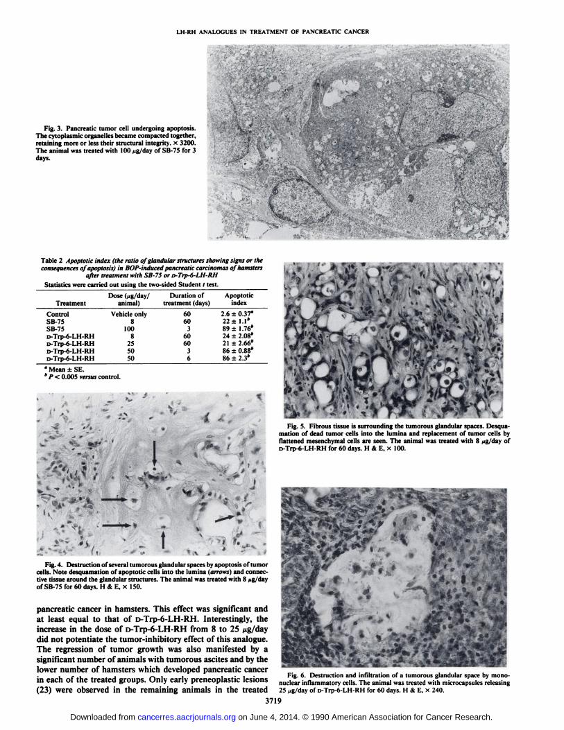
The histological examination of the tumors of animals treatedfor 3 or 6 days with 50 /¿gof D-Trp-6-LH-RH and for 3 dayswith 100 ng of SB-75 showed significant alterations whencompared with the control. The majority of the tumor cellsrevealed the signs of apoptosis. These comprised the shrinkageof tumor cells, formation of apoptotic bodies in the cytoplasm,separation of tumor cells which underwent apoptosis from theneighboring cells, as well as karyopyknosis and karyorrhexis.Several tumor cells showed generalized cytoplasmic swelling(Fig. 2). The cytoplasmic organdÃes become compacted together in several tumor cells, retaining their structural integrity(Fig. 3). The percentage of tumorous glandular structures containing apoptotic cells (apoptotic index) was as high as 80 to90% (Table 2).
In the tumors of the animals treated with SB-75 or with D-Trp-6-LH-RH for 2 mo, the signs of apoptosis and the consequences of the apoptotic process were detected. The latterconsisted of desquamation of dead tumor cells into the glandular spaces and replacement of the tumor cells by presumably
3717
on June 4, 2014. © 1990 American Association for Cancer Research. cancerres.aacrjournals.org Downloaded from

LH-RH ANALOGUES IN TREATMENT OF PANCREATIC CANCER
Table 1 Effect of treatment with antagonist SB-75 and agonist D-Trp-6-LH-RH on body, pancreatic, pancreatic tumor, ovarian, and uterine weights, serum LH andIGF-I levels, number of animals with osciles, and number of animals with pancreatic tumors in Syrian golden hamsters with BOP-induced pancreatic cancers. Each
group consisted of 15 animals.
GroupControl
(BOPBody
wt(g)151±6.3*Pancreas
wt(g)2.17
±0.52Tumorous
pancreas wt(g)2.3
±0.55%
of inhibitionof tumorouspancreatic wtOvarianwt(mg)69
±4Uterine
wt(mg)535
+ 51Serum
LH(ng/ml)0.78
±0.04Serum
IGF-I287.7
±25.29AA°7AT14only)
SB-75 (8 (jg/day) 148 ±5.34 0.53 ±0.09 (a)c 0.7 ±0.13 (b)' 70D-Trp-6-LH-RH 151 ±5.8 0.52 ±0.14 (a) 0.78 ±0.31 (d)c 66
(8 Mg/day)D-Trp-6-LH-RH 145 ±6.5 0.61 ±0.15 (a) 0.88 ±0.26 (d) 62
(25 „g/day)
52 ±5 (a) 453 ±64 0.32 ±0.043 (a) 259.2 ±24.25 0 (e)* 8 (c)
44 ±3(a) 241 ±24(a) 0.64 ±0.068 (d) 284.6 ±20.37 1 (c) 7 (c)
41 ±3(a) 209 ±15(a) 0.65 ±0.046 (d) 331.4 + 36.73 1 (c) 7 (c)
" AA, number of animals with tumorous ascites of 15; AT, number of animals with pancreatic tumors of 15.* Mean ±SE.' Significance versus the control (two-sided Student ( test): (a), P < 0.005; (b), P < 0.01 ; (d), P < 0.05.* Significance versus the control (two-sided Fisher's exact test): (c), P < 0.05.
!-»,<«<Fig. 1. BOP-induced, untreated pancreatic adenocarcinoma of a hamster. Note
the absence of fibrosi v H & E, x 120.
m' -
|'•-; ISFfFig. 2. Apoptotic cells with pyknotic nuclei and swollen cytoplasm (arrows)
in a glandular structure of a pancreatic tumor of an animal treated with 100 ng/day of SB-75 for 3 days. H & E, x 370.
preexisting mesenchymal cells in the tumorous glands (Figs. 4and 5). These glandular structures were surrounded by anincreased amount of fibrous connective tissue (Fig. 5), obviouslya consequence of the regression of tumorous parenchyma. Theconfluence of glandular spaces, the cells in which underwentapoptosis, resulted in the formation of small foci of secondarynecrosis. Accumulation of mononuclear phagocytes in andaround the glandular structures containing apoptotic cells wasfrequently seen (Fig. 6). Calcification of individual apoptoticcells was found in some cases. The ratio of glandular structures
showing the signs or the consequences of apoptosis (apoptoticindex) is given in Table 2. No significant differences were foundbetween the groups treated chronically with SB-75 or the 2doses of D-Trp-6-LH-RH, the apoptotic index being 21 to 24%.Apoptosis was not found in the cells of the nontumorous glandsof the pancreata of hamsters treated with D-Trp-6-LH-RH orSB-75.
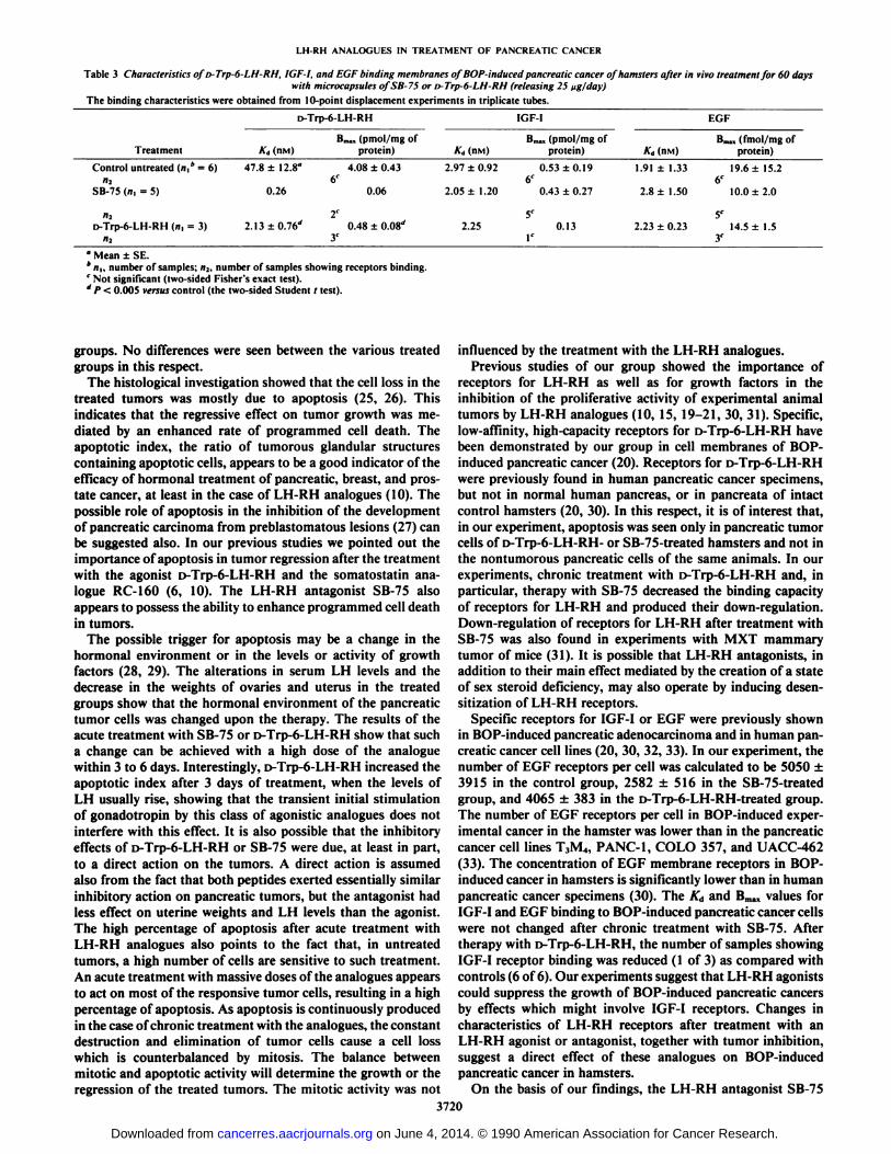
Receptor Assays. The results of receptor assays are shown inTable 3. In control BOP-induced pancreatic cancers, D-Trp-6-LH-RH was bound to one class of noncooperative membrane-binding sites with low affinity (Ad = 47.8 ±12.8 nmol) andhigh capacity (Bma* = 4.08 ±0.43 pmol/mg of membraneprotein). In the group treated with SB-75 for 60 days, receptorsfor LH-RH were found in only 2 of 6 tumors investigated.Chronic treatment with D-Trp-6-LH-RH in a dose of 25 fig/day and particularly with SB-75 decreased the concentration ofreceptors (Bmax)for LH-RH on pancreatic cancer cell membranes. The affinity of D-Trp-6-LH-RH to the binding sites oncancer cell membranes was higher in both treated groups ascompared with the controls.
IGF-I receptors were present in all control tumors. Interaction of 125I-IGF-I with binding sites was consistent with the
presence of a single class of receptors. The binding affinity wasin the nanomolar range (KA = 2.97 ±0.92 nmol), and thebinding capacity was 0.53 ±0.19 pmol/mg of membrane protein. After treatment with D-Trp-6-LH-RH, IGF-I receptorscould be found in only one of 3 investigated tumors. Treatmentwith SB-75 did not change binding characteristics of IGF-Ireceptors.
Analysis of the displacement of labeled EGF by unlabeledEGF suggests that, in control pancreatic cancer cells, labeledpeptide was bound to one class of binding noncooperativemembrane sites with high affinity (KA= 1.91 ±1.33 nmol) andlow capacity (Bmax= 19.6 ±15.2 fmol/mg of membrane protein). Treatment with SB-75 and D-Trp-6-LH-RH did not influence the binding of EGF to the receptors on pancreatic cancercells.
DISCUSSION
We have previously reported that LH-RH agonists inhibitthe growth of experimental pancreatic cancers in rats andhamsters (1, 3, 5, 6, 9). This effect could be due to the inductionof a state of deprivation of sex steroids, although Chester et al.(24) have shown that oophorectomy does not modify pancreaticcarcinogenicity of BOP. Thus, direct effects of LH-RH agonistson pancreatic tumors are possible, as inferred previously (12,20). This study demonstrates for the first time that the LH-RHantagonist SB-75 suppresses the growth of the BOP-induced
3718
on June 4, 2014. © 1990 American Association for Cancer Research. cancerres.aacrjournals.org Downloaded from
LH-RH ANALOGUES IN TREATMENT OF PANCREATIC CANCER
Fig. 3. Pancreatic tumor cell undergoing apoptosis.The cytoplasmic organelles became compacted together,retaining more or less their structural integrity, x 3200.The animal was treated with 100 ¿ig/dayof SB-75 for 3days.
Table 2 Apoptotic index (the ratio of glandular structures showing signs or theconsequences of apoptosis) in BOP-induced pancreatic carcinomas of hamsters
after treatment with SB-75 or D-Trp-6-LH-RHStatistics were carried out using the two-sided Student r test.
TreatmentControlSB-75SB-75D-Trp-6-LH-RHD-Trp-6-LH-RHD-Trp-6-LH-RHD-Trp-6-LH-RHDose(jig/day/ Duration of Apoptotic
animal) treatment (days)indexVehicle
only81008255050606036060362.6 ±0.37°22±1.1*89±1.76*24±2.08*21±2.66*86±0.88*86±2.3*
°Mean ±SE.* P < 0.005 versus control.
Fig. 4. Destruction of several tumorous glandular spaces by apoptosis of tumorcells. Note desquamation of apoptotic cells into the lumina (arrows) and connective tissue around the glandular structures. The animal was treated with 8 ng/dayof SB-75 for 60 days. H & E. x 150.
pancreatic cancer in hamsters. This effect was significant andat least equal to that of D-Trp-6-LH-RH. Interestingly, theincrease in the dose of o-Trp-6-LH-RH from 8 to 25 Mg/daydid not potentiate the tumor-inhibitory effect of this analogue.The regression of tumor growth was also manifested by asignificant number of animals with tumorous ascites and by thelower number of hamsters which developed pancreatic cancerin each of the treated groups. Only early preneoplastic lesions(23) were observed in the remaining animals in the treated
Fig. 5. Fibrous tissue is surrounding the tumorous glandular spaces. Desquamation of dead tumor cells into the lumina and replacement of tumor cells byflattened mesenchymal cells are seen. The animal was treated with 8 fig/day ofD-Trp-6-LH-RH for 60 days. H & E. x 100.
pffi^1 <v-Ì
$•' ' f-::
Fig. 6. Destruction and infiltration of a tumorous glandular space by mono-nuclear inflammatory cells. The animal was treated with microcapsules releasing25 Mg/day of o-Trp-6-LH-RH for 60 days. H & E, x 240.
3719
on June 4, 2014. © 1990 American Association for Cancer Research. cancerres.aacrjournals.org Downloaded from
LH-RH ANALOGUES IN TREATMENT OF PANCREATIC CANCER
Table 3 Characteristics ofi>-Trp-6-LH-RH, IGF-I, and EOF binding membranes oj BOP-induced pancreatic cancer of hamsters after in vivo treatment for 60 dayswith microcapsules ofSB-75 or ¡>Trp-6-LH-RH (releasing 25 fig/day)
The binding characteristics were obtained from 10-point displacement experiments in triplicate tubes.
D-Trp-6-LH-RHTreatmentControl
untreated (n,* = 6)
SB-75 (n, = 5)A'd
1IIM147.8
±12.8°
0.26Bmm,
(pmol/mg ofprotein)4.08
±0.43
0.06Ad
1n\il2.97
±0.92
2.05 ±1.20IGF
IBm.,
(pmol/mg ofprotein)0.53
±0.196r
0.43 ±0.27A'd
(nm)1.91
±1.332.8
±1.50EGFBm.,
( fmol/mg ofprotein)19.6
±15.26r
10.0 ±2.0
5'i»-Trp-6-LH-RH(n, = 3) 2.13 ±0.76''
3'2.25 0.13
5C
2.23 ±0.23 14.5 ±1.53C
' Mean ±SE.*«i,number of samples; n2. number of samples showing receptors binding.f Not significant (two-sided Fisher's exact test).' P < 0.005 versus control (the two-sided Student / test).
groups. No differences were seen between the various treatedgroups in this respect.
The histológica! investigation showed that the cell loss in thetreated tumors was mostly due to apoptosis (25, 26). Thisindicates that the regressive effect on tumor growth was mediated by an enhanced rate of programmed cell death. Theapoptotic index, the ratio of tumorous glandular structurescontaining apoptotic cells, appears to be a good indicator of theefficacy of hormonal treatment of pancreatic, breast, and prostate cancer, at least in the case of LH-RH analogues (10). Thepossible role of apoptosis in the inhibition of the developmentof pancreatic carcinoma from preblastomatous lesions (27) canbe suggested also. In our previous studies we pointed out theimportance of apoptosis in tumor regression after the treatmentwith the agonist D-Trp-6-LH-RH and the somatostatin analogue RC-160 (6, 10). The LH-RH antagonist SB-75 alsoappears to possess the ability to enhance programmed cell deathin tumors.
The possible trigger for apoptosis may be a change in thehormonal environment or in the levels or activity of growthfactors (28, 29). The alterations in serum LH levels and thedecrease in the weights of ovaries and uterus in the treatedgroups show that the hormonal environment of the pancreatictumor cells was changed upon the therapy. The results of theacute treatment with SB-75 or D-Trp-6-LH-RH show that sucha change can be achieved with a high dose of the analoguewithin 3 to 6 days. Interestingly, D-Trp-6-LH-RH increased theapoptotic index after 3 days of treatment, when the levels ofLH usually rise, showing that the transient initial stimulationof gonadotropin by this class of agonistic analogues does notinterfere with this effect. It is also possible that the inhibitoryeffects of D-Trp-6-LH-RH or SB-75 were due, at least in part,to a direct action on the tumors. A direct action is assumedalso from the fact that both peptides exerted essentially similarinhibitory action on pancreatic tumors, but the antagonist hadless effect on uterine weights and LH levels than the agonist.The high percentage of apoptosis after acute treatment withLH-RH analogues also points to the fact that, in untreatedtumors, a high number of cells are sensitive to such treatment.An acute treatment with massive doses of the analogues appearsto act on most of the responsive tumor cells, resulting in a highpercentage of apoptosis. As apoptosis is continuously producedin the case of chronic treatment with the analogues, the constantdestruction and elimination of tumor cells cause a cell losswhich is counterbalanced by mitosis. The balance betweenmitotic and apoptotic activity will determine the growth or theregression of the treated tumors. The mitotic activity was not
influenced by the treatment with the LH-RH analogues.Previous studies of our group showed the importance of
receptors for LH-RH as well as for growth factors in theinhibition of the proliferative activity of experimental animaltumors by LH-RH analogues (10, 15, 19-21, 30, 31). Specific,low-affinity, high-capacity receptors for D-Trp-6-LH-RH havebeen demonstrated by our group in cell membranes of BOP-induced pancreatic cancer (20). Receptors for D-Trp-6-LH-RHwere previously found in human pancreatic cancer specimens,but not in normal human pancreas, or in pancreata of intactcontrol hamsters (20, 30). In this respect, it is of interest that,in our experiment, apoptosis was seen only in pancreatic tumorcells of D-Trp-6-LH-RH- or SB-75-treated hamsters and not inthe nontumorous pancreatic cells of the same animals. In ourexperiments, chronic treatment with D-Trp-6-LH-RH and, inparticular, therapy with SB-75 decreased the binding capacityof receptors for LH-RH and produced their down-regulation.Down-regulation of receptors for LH-RH after treatment withSB-75 was also found in experiments with MXT mammarytumor of mice (31). It is possible that LH-RH antagonists, inaddition to their main effect mediated by the creation of a stateof sex steroid deficiency, may also operate by inducing desen-sitization of LH-RH receptors.
Specific receptors for IGF-I or EGF were previously shownin BOP-induced pancreatic adenocarcinoma and in human pancreatic cancer cell lines (20, 30, 32, 33). In our experiment, thenumber of EGF receptors per cell was calculated to be 5050 ±3915 in the control group, 2582 ±516 in the SB-75-treatedgroup, and 4065 ±383 in the D-Trp-6-LH-RH-treated group.The number of EGF receptors per cell in BOP-induced experimental cancer in the hamster was lower than in the pancreaticcancer cell lines T3M4, PANC-1, COLO 357, and UACC-462(33). The concentration of EGF membrane receptors in BOP-induced cancer in hamsters is significantly lower than in humanpancreatic cancer specimens (30). The Ka and Bmaxvalues forIGF-I and EGF binding to BOP-induced pancreatic cancer cellswere not changed after chronic treatment with SB-75. Aftertherapy with D-Trp-6-LH-RH, the number of samples showingIGF-I receptor binding was reduced (1 of 3) as compared withcontrols (6 of 6). Our experiments suggest that LH-RH agonistscould suppress the growth of BOP-induced pancreatic cancersby effects which might involve IGF-I receptors. Changes incharacteristics of LH-RH receptors after treatment with anLH-RH agonist or antagonist, together with tumor inhibition,suggest a direct effect of these analogues on BOP-inducedpancreatic cancer in hamsters.
On the basis of our findings, the LH-RH antagonist SB-75
3720
on June 4, 2014. © 1990 American Association for Cancer Research. cancerres.aacrjournals.org Downloaded from
LH-RH ANALOGUES IN TREATMENT OF PANCREATIC CANCER
can be considered as a potentially useful agent for the treatmentof cancer of the exocrine pancreas. Its immediate inhibitoryeffects on the pituitary-gonadal axis and its probable directeffects on tumor cells suggest the merit of continued explorationof this potent LH-RH antagonist in the management of pancreatic cancer.
ACKNOWLEDGMENTSWe are grateful to Elvira Monje, Illona Janaky, and Robert O'Brien
for experimental assistance.
REFERENCES
1. Redding. T. W., and Schally, A. V. Inhibition of growth of pancreaticcarcinomas in animal models by analogs of hypothalamic hormones. Proc.Nati. Acad. Sci. USA, 81: 248-252, 1984.
2. Redding, T. W., and Schally, A. V. Inhibition of mammary tumor growth inrats and mice by administration of agonistic analogs of luteinizing hormone-releasing hormone. Proc. Nati. Acad. Sci. USA, SO: 1454-1462. 1983.
3. Zalatnai, A.. Paz-Bouza, J. I., Redding, T. W., and Schally, A. V. Histologiechanges in the rat prostate cancer model after treatment with somatostatinanalogs and D-Trp-6-LH-RH. Prostate. 12: 85-98, 1988.
4. Redding, T. W., and Schally, A. V. Inhibition of growth of the transplantablerat chondrosarcoma by analogs of hypothalamic hormones. Proc. Nati. Acad.Sci. USA, 80: 1078-1082, 1983.
5. Zalatnai, A., and Schally, A. V. Treatment of ,V-nitrosobis(2-oxopro-pyl)amine-induced pancreatic cancer in Syrian golden hamsters with D-Trp-6-LH-RH and somatostatin analogue RC-160. Cancer Res., 49: 1810-1815.1989.
6. Szende, B., Zalatnai, A., and Schally, A. V. Programmed cell death (apop-tosis) in pancreatic cancers of hamsters after treatment with analogs of bothluteinizing hormone-releasing hormone and somatostatin. Proc. Nati. Acad.Sci. USA, 86:1643-1647, 1989.
7. Schally, A. V., Comaru-Schally. A. M.. and Redding. T. W. Antitumor effectsof analogs of hypothalamic hormones in endocrine-dependent cancers. Proc.Soc. Exp. Biol. Med., 175: 259-281, 1984.
8. Schally, A. V., and Comaru-Schally. A. M. Use of luteinizing hormone-releasing hormone analogs in the treatment of hormone-dependent tumors.Semin. Reprod. Endocrinol., 5: 389-398. 1987.
9. Paz-Bouza, J. I., Redding, T. W., and Schally, A. V. Treatment of nitrosa-mine-induced pancreatic tumors in hamsters with analogs of somatostatinand luteinizing hormone-releasing hormone. Proc. Nati. Acad. Sci. USA, 84:1112-1116, 1987.
10. Szende, B., Lapis, K., Redding. T. W., Srkalovic. G., and Schally, A. V.Growth inhibition of MXT mammary carcinoma by enhancing programmedcell death (apoptosis) with analogs of both luteinizing hormone-releasinghormone and somatostatin. Breast Cancer Res. Treat.. 14: 307-314, 1989.
11. González-Barcena, D., Ibarro-Olmos, M. A.. Garcia-Carrasco. F., Gutierez-Samperio. C., Comaru-Schally. A. M.. and Schally, A. V. Influence of D-Trp-6-LH-RH on the survival time in patients with advanced pancreaticcancer. Biomed. Pharmacother., 43: 313-317, 1989.
12. Schally, A. V., Bajusz, S., Redding, T. W., Zalatnai. A., and Comaru-Schally,A. M. Analogs of LH-RH: the present and the future. In: B. Vickery and B.Lunenfeld (eds.), Gn-RH Analogues in Cancer and Human Reproduction,pp. 5-31. Boston: Kluwer Academic Publishers. 1989.
13. Bajusz, S., Kovacs, M., Gazdag. M., Bokser, L., Karashima, T., Csernus, V.
J., Janaky, T., Guoth. J., and Schally, A. V. Highly potent antagonists ofluteinizing hormone-releasing hormone free of edematogenic effects. Proc.Nati. Acad. Sci. USA, 85: 1637-1641, 1988.
14. Bajusz, S., Csernus, V. J., Janaky, T., Bokser, L., Fekete, M., and Schally,A. V. New antagonists of LH-RH. II. Inhibition and potentiation of LH-RHby closely related analogs. Int. J. Peptide Protein Res., 32: 425-435, 1988.
15. Sharoni, T., Bosin, E., Münster,A., Levy, J., and Schally, A. V. Inhibitionof growth of human mammary tumor cells by potent antagonist of luteinizinghormone-releasing hormone. Proc. Nati. Acad. Sci. USA, 86: 1648-1651,1989.
16. Pour, P.. Salmasi, S. J., and Runge, R. G. Selective induction of pancreaticductular tumors by single doses of A'-nitrosobis(2-oxopropyl)amine in Syriangolden hamster. Cancer Lett., 4: 317-323. 1978.
17. Schally, A. V. The use of LH-RH analogs in gynecology and tumor therapy.In: General Gynecology, Vol. 6. Carnforth, England: Parthenon Publishing.1989.
18. Zamboni. L.. and DeMartino. C. Buffered picric acid-formaldehyde: a newrapid fixative for electron microscopy. J. Cell Biol., 35:148A, 1967.
19. Fekete, M., Wittliff, J. L.. and Schally. A. V. Characteristics and distributionof receptors for D-Trp-6-luteinizing hormone-releasing hormone, somatostatin, epidermal growth factor, and sex-steroids in 500 biopsy samples ofhuman breast cancer. J. Clin. Lab. Anal., 3: 137-147, 1989.
20. Fekete, M.. Zalatnai, A., and Schally. A. V. Presence of membrane bindingsites of D-Trp-6-luteinizing hormone-releasing hormone in experimentalpancreatic cancer. Cancer Lett., 45: 87-91. 1989.
21. Srkalovic. G.. Szende, B.. Redding. T. W., Groot. K., and Schally. A. V.Receptors for D-Trp-6-luteinizing hormone-releasing hormone, somatostatin. and insulin-like Growth Factor I in MXT mouse mammary carcinoma.Proc. Soc. Exp. Biol. Med., 192: 209-218, 1989.
22. Munson, P. J., and Rodbard, S. LIGAND: a versatile computerized approachfor characterization of ligand-binding systems. Anal. Biochem., 707: 220-239, 1980.
23. Pour, P., Althoff. J., and Takahashi, M. Early lesions of pancreatic ductalcarcinoma in the hamster model. Am. J. Pathol.. 88: 291-308. 1977.
24. Chester, J. F., Nicholson, R. I., Lever, J. V., Turnbull, A. R., and Britton.D. C. Oophorectomy has no effect on experimental pancreatic carcinogenesisin the Syrian hamster. Br. J. Cancer, 59: 696-697. 1989.
25. Kerr, J. F. R., Wyllie, A. H.. and Currie, A. R. Apoptosis: basic biologicalphenomenon with wide-ranging implications in tissue kinetics. Br. J. Cancer,20:239-257. 1972.
26. Wyllie. A. H. The biology of cell death in tumors. Anticancer Res., 5: 131-136. 1985.
27. Bursch. W.. Lauer. B.. Timmermann-Trosiener, I., Barthel, G.. Schuppler,J., and Schulte-Hermann. R. Controlled cell death (apoptosis) of normal andputative preneoplastic cells in rat liver following withdrawal of tumor promoters. Carcinogenesis (Lond.), 5:453-458. 1984.
28. Gouston, A. S., Leof, E. B., Shipley, G. S., and Moses. H. L. Growth factorsand cancer. Cancer Res., 46: 1015-1029. 1986.
29. Haas. R. For whom the cell tolls: a portrait of physiological cell death. J.NIHRes., Õ.-91-94, 1989.
30. Fekete. M.. Zalatnai, A., Comaru-Schally. A. M., and Schally, A. V. Membrane receptors for peptides in experimental and human pancreatic cancers.Pancreas, 4: 521-528, 1989.
31. Szende. B., Srkalovic. G.. Groot. K., Lapis, K.. and Schally. A. V. Growthinhibition of MXT mouse mammary tumor by the LH-RH antagonist SB-75. J. Nati. Cancer Inst., 82: 513-517, 1990.
32. Liebow. C.. Reilly. C., Serrano, M., and Schally. A. V. Somatostatin analogues inhibit growth of pancreatic cancer by stimulating tyrosine phospha-tase. Proc. Nati. Acad. Sci. USA, 86: 2003-2007, 1989.
33. Korc, M., Meltzer, P., and Trent, J. Enhanced expression of epidermalgrowth factor receptor with alterations of chromosome 7 in human pancreaticcancer. Proc. Nati. Acad. Sci. USA, 83: 5141-5144. 1986.
3721
on June 4, 2014. © 1990 American Association for Cancer Research. cancerres.aacrjournals.org Downloaded from