Thymoquinone up-regulates PTEN expression and induces apoptosis in doxorubicin-resistant human...
20
Thymoquinone up-regulates PTEN expression and induces apoptosis in doxorubicin-resistant human breast cancer cells El-Shaimaa A. Arafa 1 , Qianzheng Zhu 1 , Zubair I. Shah 3 , Gulzar Wani 1 , Bassant M. Barakat 1 , Ira Racoma 1 , Mohamed A. El-Mahdy 1,* , and Altaf A. Wani 1,2,3,4,* 1 Department of Radiology, The Ohio State University, Columbus, OH 43210, USA 2 Department of Molecular and Cellular Biochemistry, The Ohio State University, Columbus, OH 43210, USA 3 James Cancer Hospital and Solove Research Institute, The Ohio State University, Columbus, OH 43210, USA 4 DNA Research Chair, King Saud University, Riyadh, Saudi Arabia Abstract The use of innocuous naturally occurring compounds to overcome drug resistance and cancer recalcitrance is now in the forefront of cancer research. Thymoquinone (TQ) is a bioactive constituent of the volatile oil derived from seeds of Nigella sativa Linn. TQ has shown promising anti-carcinogenic and anti-tumor activities through different mechanisms. However, the effect of TQ on cell signaling and survival pathways in resistant cancer cells has not been fully delineated. Here, we report that TQ greatly inhibits doxorubicin-resistant human breast cancer MCF-7/DOX cell proliferation. TQ treatment increased cellular levels of PTEN proteins, resulting in a substantial decrease of phosphorylated Akt, a known regulator of cell survival. The PTEN expression was accompanied with elevation of PTEN mRNA. TQ arrested MCF-7/DOX cells at G2/M phase and increased cellular levels of p53 and p21 proteins. Flow cytometric analysis and agarose gel electrophoresis revealed a significant increase in Sub-G1 cell population and appearance of DNA ladders following TQ treatment, indicating cellular apoptosis. TQ-induced apoptosis was associated with disrupted mitochondrial membrane potential and activation of caspases and PARP cleavage in MCF-7/DOX cells. Moreover, TQ treatment increased Bax/Bcl2 ratio via up-regulating Bax and down-regulating Bcl2 proteins. More importantly, PTEN silencing by target specific siRNA enabled the suppression of TQ-induced apoptosis resulting in increased cell survival. Our results reveal that up-regulation of the key upstream signaling factor, PTEN, in MCF-7/DOX cells inhibited Akt phosphorylation, which ultimately causes increase in their regulatory p53 levels affecting the induction of G2/M cell cycle arrest and apoptosis. Overall results provide mechanistic insights for understanding the molecular basis and utility of the anti- tumor activity of TQ. © 2010 Elsevier B.V. All rights reserved. * To whom correspondence should be addressed: Altaf A. Wani ([email protected]) or Mohamed A. El-Mahdy (Mohamed.el- [email protected]), 460 W 12 th avenue, Columbus, OH 43210. Fax: 614-292-9102. Publisher's Disclaimer: This is a PDF file of an unedited manuscript that has been accepted for publication. As a service to our customers we are providing this early version of the manuscript. The manuscript will undergo copyediting, typesetting, and review of the resulting proof before it is published in its final citable form. Please note that during the production process errors may be discovered which could affect the content, and all legal disclaimers that apply to the journal pertain. Conflict of interest statement The authors declare that they have no competing interests. NIH Public Access Author Manuscript Mutat Res. Author manuscript; available in PMC 2012 January 10. Published in final edited form as: Mutat Res. 2011 January 10; 706(1-2): 28–35. doi:10.1016/j.mrfmmm.2010.10.007. NIH-PA Author Manuscript NIH-PA Author Manuscript NIH-PA Author Manuscript
Transcript of Thymoquinone up-regulates PTEN expression and induces apoptosis in doxorubicin-resistant human...
Thymoquinone up-regulates PTEN expression and inducesapoptosis in doxorubicin-resistant human breast cancer cells
El-Shaimaa A. Arafa1, Qianzheng Zhu1, Zubair I. Shah3, Gulzar Wani1, Bassant M.Barakat1, Ira Racoma1, Mohamed A. El-Mahdy1,*, and Altaf A. Wani1,2,3,4,*1Department of Radiology, The Ohio State University, Columbus, OH 43210, USA2Department of Molecular and Cellular Biochemistry, The Ohio State University, Columbus, OH43210, USA3James Cancer Hospital and Solove Research Institute, The Ohio State University, Columbus,OH 43210, USA4DNA Research Chair, King Saud University, Riyadh, Saudi Arabia
AbstractThe use of innocuous naturally occurring compounds to overcome drug resistance and cancerrecalcitrance is now in the forefront of cancer research. Thymoquinone (TQ) is a bioactiveconstituent of the volatile oil derived from seeds of Nigella sativa Linn. TQ has shown promisinganti-carcinogenic and anti-tumor activities through different mechanisms. However, the effect ofTQ on cell signaling and survival pathways in resistant cancer cells has not been fully delineated.Here, we report that TQ greatly inhibits doxorubicin-resistant human breast cancer MCF-7/DOXcell proliferation. TQ treatment increased cellular levels of PTEN proteins, resulting in asubstantial decrease of phosphorylated Akt, a known regulator of cell survival. The PTENexpression was accompanied with elevation of PTEN mRNA. TQ arrested MCF-7/DOX cells atG2/M phase and increased cellular levels of p53 and p21 proteins. Flow cytometric analysis andagarose gel electrophoresis revealed a significant increase in Sub-G1 cell population andappearance of DNA ladders following TQ treatment, indicating cellular apoptosis. TQ-inducedapoptosis was associated with disrupted mitochondrial membrane potential and activation ofcaspases and PARP cleavage in MCF-7/DOX cells. Moreover, TQ treatment increased Bax/Bcl2ratio via up-regulating Bax and down-regulating Bcl2 proteins. More importantly, PTEN silencingby target specific siRNA enabled the suppression of TQ-induced apoptosis resulting in increasedcell survival. Our results reveal that up-regulation of the key upstream signaling factor, PTEN, inMCF-7/DOX cells inhibited Akt phosphorylation, which ultimately causes increase in theirregulatory p53 levels affecting the induction of G2/M cell cycle arrest and apoptosis. Overallresults provide mechanistic insights for understanding the molecular basis and utility of the anti-tumor activity of TQ.
© 2010 Elsevier B.V. All rights reserved.*To whom correspondence should be addressed: Altaf A. Wani ([email protected]) or Mohamed A. El-Mahdy ([email protected]), 460 W 12th avenue, Columbus, OH 43210. Fax: 614-292-9102.Publisher's Disclaimer: This is a PDF file of an unedited manuscript that has been accepted for publication. As a service to ourcustomers we are providing this early version of the manuscript. The manuscript will undergo copyediting, typesetting, and review ofthe resulting proof before it is published in its final citable form. Please note that during the production process errors may bediscovered which could affect the content, and all legal disclaimers that apply to the journal pertain.Conflict of interest statementThe authors declare that they have no competing interests.
NIH Public AccessAuthor ManuscriptMutat Res. Author manuscript; available in PMC 2012 January 10.
Published in final edited form as:Mutat Res. 2011 January 10; 706(1-2): 28–35. doi:10.1016/j.mrfmmm.2010.10.007.
NIH
-PA Author Manuscript
NIH
-PA Author Manuscript
NIH
-PA Author Manuscript
KeywordsPTEN tumor suppressor; breast cancer; thymoquinone; apoptosis; cell signaling
1. IntroductionBreast cancer is one of the most common human malignancies and the second leading causeof cancer-related deaths in women [1]. Breast cancer patients show initial response tochemotherapeutic treatment; however, the majority of women eventually experience cancertreatment recalcitrance and emergence of drug resistant cells [2,3]. Doxorubicin, one of theclinically most important anti-neoplastic agents, possesses a wide spectrum of anti-canceractivity against various solid tumors, including breast cancer. However, the development ofdoxorubicin resistance limits its use in treating breast cancer patients [4]. Severalmechanisms are responsible for doxorubicin resistance, including altered drug efflux, drugdetoxification, enhanced DNA repair and disruption in apoptotic signaling pathways. Drugresistance is also associated with activation of signaling pathways such as thephosphatidylinositol-3 kinase (PI3K)/Akt or suppression of tumor suppressor genes,including PTEN [5].
The PI3K/Akt pathway is a pivotal signaling pathway, which controls cell growth, survival,proliferation and tumorigenesis [6,7]. PI3K-activated (phosphorylated) Akt promotes cellsurvival by inhibiting apoptosis through its ability to phosphorylate/inactivate downstreamtargets of apoptotic machinery, such as pro-apoptotic Bcl2 family member BAD andGSK-3β [8,9].The (PI3K)/Akt pathway is regulated by several critical upstream factors, e.g.,tumor suppressor PTEN [10]. The tumor suppressor gene PTEN is one of the most commontargets of mutation in human cancers, with a mutation frequency approaching that of p53.PTEN mutations have been found in glioblastomas, malignant melanomas, carcinoma of theprostate, breast, kidney, urinary bladder, uterus and other cancers, mostly in advanced stagesof tumor progression [11,12]. Genetic mutation of PTEN resulted in increased Akt activityin many types of tumor [13]. On the other hand, overexpression of PTEN in cancer cellscarrying mutant or deletion type PTEN was reported to inhibit cell proliferation andtumorigenicity via induction of cell cycle arrest and apoptosis [14]. PTEN exerts a tightregulatory control over the PI3K pathway and downstream functions, including activation ofprotein kinase B/Akt, cell survival, cell proliferation and cell arrest [14,15]. It has beenreported that PTEN and p53 functions are mechanistically linked through MDM2phosphorylation, which modulates the ubiquitination of p53 by the activation of PI3K/Aktpathway [16,17]. p53 functions as a transcription factor by binding to a p53-specific DNAsequence in responsive genes, which would increase the synthesis of p21, the mostimportant checkpoint proteins involved in cell arrest at both G1 and G2/M.
Inhibition of PI3K/Akt pathway has been targeted as a strategy for drug development [18]and recently has drawn considerable attention for combination therapy alongside the use ofnaturally occurring innocuous dietary agents to achieve greater efficacy for drug-resistantcancer cells [19–21]. For instance, some of the dietary phytochemicals have been shown todownregulate PI3K/Akt pathway, thereby sensitizing drug-resistant cancer cells for low-dose chemotherapeutic drug-induced apoptosis. Dietary phytochemical thymoquinone (TQ)is the main active ingredient of the volatile oil of Nigella sativa Linn, known as the blackseed. The black seed is an annual plant that has been widely used in the Indian subcontinent,Arabian countries and Europe for culinary and medicinal purposes [22,23]. Recently,Several clinical studies were conducted in humans receiving oral Nigella Sativa extract orblack seeds for up to 8 weeks to evaluate the anti-inflammatory, antihypertensive, orantioxidant effects [24,25]. Most of the known biological activities of the seed have been
Arafa et al. Page 2
Mutat Res. Author manuscript; available in PMC 2012 January 10.
NIH
-PA Author Manuscript
NIH
-PA Author Manuscript
NIH
-PA Author Manuscript
attributed to the active ingredient TQ. TQ has been shown to be safe on a wide variety ofnormal cells. The selective cytotoxicity of TQ for human cancer cells compared to primarymouse keratinocytes [23], mouse normal kidney cells [26], non-malignant fibroblasts [27]and normal human lung fibroblasts [28] has been reported in several studies. TQ has beenused for decades as anti-oxidant, anti-inflammatory, and anti-neoplastic agent [29,30].Previous studies have shown that TQ exhibits inhibitory effects on cell proliferation of manytypes of cancer cells [31] including non-Dox resistant breast cancer cells (MCF-7)[26,32,33]. TQ induces cell death and inhibits tumor growth by suppressing NF-κB, Aktactivation, and extracellular signal-regulated kinase signaling pathways as well asangiogenesis [34,35]. We have previously reported that TQ induced apoptosis throughactivation of caspase pathway and mitochondrial events in p53-null myeloblastic leukemiaHL-60 cells [36].
To better understand the molecular mechanism by which TQ exerts its anti-neoplasticeffects, we examined the anti-proliferative effects of TQ in doxorubicin-resistant humanbreast cancer cells. In this study, we investigated the potential mechanism by which TQ mayregulate cell proliferation and apoptosis in doxorubicin-resistant human breast cancerMCF-7/DOX cells. We provide evidence that TQ transcriptionally upregulate PTEN, whichled to phosphorylation of Akt and induction p53 protein and its transcriptional target p21,thus induces G2/M phase arrest and apoptosis in doxorubicin-resistant MCF-7/DOX cells.These observations provide a strong rationale towards designing TQ-based combinationaltherapeutic strategies for treating human breast cancer.
2. Materials and methods2.1. Cell Lines and cultures
Doxorubicin-resistant human breast adenocarcinoma cell line, MCF-7/DOX, was generouslyprovided by Dr. S A Salama (University of Texas-medical branch, TX). The cells weregrown in DMEM medium supplemented with 10% fetal bovine serum, 50 IU/ml penicillin,50 mg/ml streptomycin at 37°C in a humidified 5% CO2 atmosphere.
2.2. Chemicals and reagentsTQ (Fig. 1) was prepared as a 10 mM stock solution in DMSO, and appropriate workingdilutions were prepared with the cell culture medium immediately prior to the experiments.TQ, MTT [3-(4,5-dimethylthiazol-2-yl)-2,5-diphenyltetrazolium bromide], Hoechst dye andother chemicals were purchased from Sigma-Aldrich (St. Louis, MO). Cell culture supplieswere obtained from Life Technologies (Grand Island, NY). Antibodies against phospho-Akt[p-Akt (Ser473)], PARP, caspase-7, caspase-8, caspase-9, PTEN, Fas L, Bcl-2, Bax, andSignalSilence PTEN siRNA kit were purchased from Cell Signaling Technology (Danvers,MA). Antibodies against p53, p21 and β-actin were purchased from Neomarkers (Fremont,CA) and Santa Cruz Biotechnology (Santa Cruz, CA), respectively. Antibody againstγH2AX was purchased from Millipore (Billerica, MA). Horseradish peroxidase (HRP)-conjugated secondary antibodies, protease inhibitor cocktail tablets were from Roche(Indianapolis, IN). Chemiluminescence substrate kit was obtained from Pierce (Rockford,IL). DC Bio-Rad protein quantitation reagents were from Bio-Rad (Hercules, CA). Thecaspase inhibitor, z-VAD-FMK, was purchased from R&D systems (Minneapolis, MN). TheMitoCapture apoptosis detection kit was made by Calbiochem (La Jolla, CA). TrizolReagent, Invitrogen SuperScript III first strand-synthesis system and PTEN primers for RT-PCR were obtained from Invitrogen Corporation (Carlsbad, CA).
Arafa et al. Page 3
Mutat Res. Author manuscript; available in PMC 2012 January 10.
NIH
-PA Author Manuscript
NIH
-PA Author Manuscript
NIH
-PA Author Manuscript
2.3. MTT assay for cell growth and cytotoxicityThe MTT assay measures the living cells’ ability to uptake and convert soluble MTT intoformazan crystals as described previously [37]. Exponentially growing MCF-7/DOX cellswere seeded in 96-well plates at an initial density of 5×10³/well, treated with differentconcentration of TQ and maintained in culture for indicated times and then phenol-freemedium containing MTT (0.2 mg/ml) was added to the cultures for an additional 2 hours.The medium was replaced with acidified isopropanol (0.04 N HCl in isopropanol) and theplates were incubated at room temperature for 1 hour. The colorimetric absorbance of thesamples was determined by Spectramax M5 microplate reader (Molecular Devices, CA).Cellular proliferation was expressed as a percentage of cell viability of TQ-treated cellsrelative to untreated controls.
2.4. DNA fragmentation analysis for detecting apoptosisExponentially growing MCF-7/DOX cells were treated with increasing concentrations ofTQ and maintained in culture. After indicated time periods, all adherent and floating cellswere recovered, centrifuged, washed once with PBS, resuspended in lysis buffer [20 mMEDTA, 100 mM Tris-HCl (pH 8.0), 1% SDS] containing 100 µg/ml RNase and incubated at55°C overnight. Proteinase K (100 µg/ml, final concentration) was added to each sampleand incubated at 37°C for 2 hours. DNA was precipitated with 2.5 volume of 100% ethanoland dissolved in TE pH 8. The DNA samples were separated on a 2% agarose gel, stainedwith ethidium bromide and visualized under UV light.
2.5. Flow cytometric analysis of cell cycle and apoptosisCell cycle distribution was determined as previously described [21]. TQ-treated cells werecollected, washed with ice-cold PBS and fixed with 70% ice-cold ethanol overnight at−20°C. The cells were centrifuged at 1500 rpm for 5 minutes, uniformly resuspended in amix of propidium iodide (50 µg/ml) and RNase A (100 µg /ml), and incubated at 37°C for40 minutes. The cells were pelleted, washed and resuspended in PBS to a final concentrationof 1×10⁶/ml. The cell cycle distribution was analyzed using BD FACS Calibur (BDBiosciences, San Jose, CA).
2.6. Hoechst stainingThe nuclear morphology of MCF-7/DOX cells was analyzed according to Lee et al, 1996[38]. Cells, either TQ-treated or untreated, were fixed with formaldehyde and stained withHoechst 33342 at 6.15 µg/ml for 15 min at room temperature. Cells were washed with andresuspended in PBS. The nuclear morphology of cells was observed by fluorescencemicroscopy. Cells with condensed or fragmented nuclei were considered apoptotic cells.
2.7. ImmunofluorescenceThe immunofluorescence double labeling was performed according to the methodestablished in our laboratory [39]. Briefly, TQ-treated cells at different time points werewashed twice with cold PBS, permeabilized with 0.5% Triton X-100/PBS for 8 minutes onice and then fixed with 2% paraformaldehyde in 0.5% Triton X-100 at 40C for 30 min. Afterfixation, the coverslips were rinsed twice with cold PBS and blocked with 20% normal goatserum (NGS) in 0.1%Triton X-100/PBS washing buffer at room temperature for 2 h.Primary rabbit anti-γH2AX antibody (1:200 dilution) and goat anti-mouse FITC secondaryantibody (1:200 dilution) were prepared in washing buffer containing 5% NGS and layeredon the coverslips for 1 h at room temperature. Following each antibody incubation step, thecells were washed with 0.1% Tween-20 in PBS 4 times for 5 min each. Fluorescence imageswere captured with a Nikon Fluorescence Microscope E80i (Nikon, Tokyo, Japan) equippedwith SPOT analysis software.
Arafa et al. Page 4
Mutat Res. Author manuscript; available in PMC 2012 January 10.
NIH
-PA Author Manuscript
NIH
-PA Author Manuscript
NIH
-PA Author Manuscript
2.8. Western blot analysisThe MCF-7/DOX cells were grown to 50% confluency and treated with TQ for the indicatedtime periods. The cells were then harvested, washed with PBS and lysed by boiling for 5minutes in lysis buffer (10% Glycerol, 2% SDS in 62 mM Tris-HCl, pH 6.8) containing acocktail of protease inhibitors. Equal amounts of total protein, as determined by DC proteinassay, were separated on an 8–16% polyacrylamide gel and electrophoretically transferred toa PVDF membrane. After blocking with 5% non-fat dry milk in TBST buffer, the PVDFmembranes were incubated with a specific primary antibody at 4°C overnight, washed threetimes, 10 min/each time with TBST buffer, and incubated again with an appropriate HRP-conjugated secondary antibody at 37°C for 1 hour. The membranes were washed with TBSTbuffer and examined by chemiluminescence detection.
2.9. Mitochondrial membrane potentialMitochondrial membrane potential was examined using the MitoCapture mitochondrialapoptosis detection kit, according to the manufacturer’s procedure. Cells were centrifuged at600 g for 5 min. Cell pellets were resuspended in 1 ml of diluted MitoCapture solution,incubated at 37 °C for 15–20 min and then centrifuged again for 5 min. Pellets wereresuspended in 1 ml pre-warmed incubation buffer and examined immediately byfluorescence microscopy.
2.10. Small RNA interference (RNAi) of PTEN expressionThe SignalSilence PTEN siRNA kit was used to silence PTEN expression according to themanufacturer’s protocol. Briefly, MCF-7/DOX cells were seeded in 12-well plates andgrown for 24 hours to ~50% confluency. Before transfection, the medium was replaced with500 µl of an antibiotic-free medium. To transfect siRNA, 2 µl of transfection reagent wasadded to 100 µl of serum-free medium in a sterile microfuge tube, followed by the additionof control or 3 µl PTEN siRNA (10 µM) to a final 50 nM concentration. This siRNA-transfection reagent mixture was incubated for 5 minutes at room temperature and thenapplied to cultured cells. After 48 hours of incubation, the medium was refreshed and thecells were treated with or without TQ for additional 48 hours. Finally, the cells werecollected for Western blotting analysis.
2.11. Quantitative reverse transcription-polymerase chain reaction (RT-PCR)MCF-7/DOX cells were treated with 50 µM TQ and maintained in culture for the indicatedtimes. The cells were washed with PBS and total cellular RNA from various cell sampleswas isolated by using Trizol Reagent. The RT-PCR was performed by using the InvitrogenSuperScript III first strand-synthesis system as instructed by the manufacturer. Briefly, 1 µgtotal RNA, 2.5 µM Oligo(dT)20 and 500 µM dNTP were incubated in a 10 µl reactionvolume at 65 °C for 5 min followed by chilling on ice for 1 min. RT buffer [5 mM MgCl2,10 mM DTT, RNaseOUT (40 U/µl)] and SuperScript III RT (200 U/µl) were added to afinal 20 µl volume for cDNA synthesis. For real-time RT-PCR, we used Power SYBRGreen PCR Master Mix from Applied Biosystems (Foster City, CA) and the reactions wererun on Roche Light cycler 480 (Applied Science, Indianapolis, IN). The following primerswere used: GAPDH forward, 5´-GTGAAGCAGGCATC-3´; reverse, 5´-CGAAGGTGGAAGAG-3´ and PTEN forward, 5´-CTTCTCTTTTTTTTCTGTCC-3´;reverse, 5´-AAGGATGATGAGAATTT CAAGC A-3´. Real-time PCR was performed at 50cycles (15s at 95°C, 56 s at 56°C and 45 s at 68°C). A melting point dissociation curvegenerated by the instrument was used to confirm the single product. Relative RNA levelsexpressed in arbitrary units were calculated using comparative method based on ΔCt andΔΔCt values and normalized to GAPDH (endogenous control). All samples were run intriplicate.
Arafa et al. Page 5
Mutat Res. Author manuscript; available in PMC 2012 January 10.
NIH
-PA Author Manuscript
NIH
-PA Author Manuscript
NIH
-PA Author Manuscript
2.12. Statistical analysisGraphPad InStat software, version 3.06 (GraphPad, San Diego, CA), was used to computestatistical data. Data are expressed as mean ± SE of three to six independent experiments.Statistical comparisons were performed using unpaired Student’s t-test test. The 0.05 levelof probability was used as the criterion of significance.
3. Results3.1. TQ inhibits the proliferation of MCF-7/DOX cells
MCF-7/DOX is a doxorubicin-resistant variant of human breast adenocarcinoma MCF-7 cellline [40,41]. We first examined the anti-proliferative effect of TQ on MCF-7/DOX cells. Asmeasured by the MTT assay, the MCF-7/DOX cell proliferation following 12, 24 and 48hours of exposure to 25, 50 or 100 µM TQ showed significant growth inhibition in TQ-treated cells compared to non-treated controls. The proliferation of TQ-treated cellsdecreased as a function of both TQ concentration and exposure time. For example, 12, 24and 48 hours post-treatment with 100 µM TQ, cellular proliferation was approximately 70%,50% and 35%, respectively (Fig. 2).
3.2. TQ induces apoptosis in MCF-7/DOX cellsWe next examined the cellular apoptosis by agarose gel electrophoresis of DNA degradedby internucleosomal DNA fragmentation. Treatment of MCF-7/DOX cells with 50 and 100µM TQ for 48 hours induced a dose-dependent DNA fragmentation, exhibiting a typicalDNA ladder feature of apoptosis (Figure 3A). Consistent with DNA fragmentation,morphological changes e.g., fragmented nuclei and apoptotic bodies were seen when cellswere stained with Hoechst dye and examined by fluorescence microscopy (Fig. 3B). TQ-induced apoptosis was further confirmed by flow cytometric assay, which showed a 25.6%sub-G1 population in TQ-treated cells as compared with 1.3% in untreated controls (Fig.3C).
2.13. TQ disrupts mitochondrial membrane potential and activates caspase pathwayWe further examined the changes in mitochondria membrane potential in TQ-treatedMCF-7/DOX cells. The results from fluorescence microscopy showed that MCF-7/DOXcells lost their mitochondrial membrane potential following TQ treatment (Fig. 4A). Innormal cells, the MitoCapture reagent accumulated and aggregated in the mitochondria,giving off a bright red fluorescence. In apoptotic cells, the MitoCapture reagent did notaggregate in the mitochondria, due to altered mitochondrial membrane potential, and thusremained in its monomeric form, generating a green fluorescence. About 25% of cells lostmembrane potential, approximately the same as that of apoptotic cells examined by Hoechststaining (data not shown). These results indicate that loss of membrane potential occurs inthe early stage of TQ-induced apoptosis in MCF-7/DOX cells.
3.4. TQ treatment modulates pro-apoptotic and anti-apoptotic proteins in MCF-7/DOX cellsBax/Bcl2 ratio represents a critical balance of regulatory pro-apoptotic and anti-apoptoticproteins in normal living cells. The increase in Bax/Bcl2 ratio leads to the release ofCytochrome c from the mitochondria, a decisive event in the apoptotic pathway. It is shownthat TQ induces both p53-dependent and independent apoptosis by diminishing Bcl2 proteinlevel [36,42]. We next determined whether the Bax/Bcl2 ratio changes upon the treatment ofMCF-7/DOX cells with TQ. As seen in Fig. 4D, TQ treatment of MCF-7/DOX cells resultedin moderate decrease in Bcl2 and a significant increased level of Bax protein level. Theseresults suggest that TQ induced-apoptosis is associated with a significant decrease of theBcl2/Bax ratio in MCF-7/DOX cells.
Arafa et al. Page 6
Mutat Res. Author manuscript; available in PMC 2012 January 10.
NIH
-PA Author Manuscript
NIH
-PA Author Manuscript
NIH
-PA Author Manuscript
3.5. Caspase activity is involved in TQ-induced apoptosisTo understand activation of the caspase cascade during TQ-induced apoptosis in MCF-7/DOX cells, we investigated various caspase-specific cleavages in TQ-treated cells. TQtreatment caused a dose–dependent proteolytic cleavage of caspases 3, 7 and 9, but not ofcaspase 8, as revealed by the appearance of the cleaved protein species (Fig. 4C). TQ alsoinduced dose-dependent proteolytic cleavage of PARP, a known substrate of caspase-3.Treatment of cells with the general caspases inhibitor, z-VAD-FMK, markedly preventedproteolytic cleavage of PARP and caspases, in addition to attenuated TQ-induced DNAfragmentation (Fig. 4B). These results indicate the involvement of the caspase pathway inTQ-induced apoptosis.
3.6. TQ treatment arrests MCF-7/DOX Cells in G2/M phase and induces DNA damageTo test whether the anti-proliferative effect of TQ was related to cell cycle arrest,asynchronous MCF-7/DOX cells were treated with TQ and the cycle progression wasexamined by the flow cytometry (Fig. 5A). In untreated control, the percentage of cells inG1, S and G2/M phases were found to be 67%, 17.6% and 15%, respectively. TQ treatmentresulted in a pronounced G2/M arrest in a time-dependent manner. The percentage of G2/Mphase cell population was found to be 32% following TQ treatment. Consistent with thisobservation, TQ treatment reduced the levels of Cdc25C and cyclin B1, while the p21 levelwas elevated at 12 hours and thereafter (Fig. 5B). To check whether TQ causes DNAdamage in MCF-7/DOX cells, we performed the analysis of γH2AX activation as a sensitiveindicator of direct or indirect damage induction by TQ. Cells were treated with TQ (100µM) for various periods or exposed to UV irradiation (50 J) to serving as a positivereference control for γH2AX activation. As compared to significant γH2AX activation byhigh dose UV irradiation, a modest time-dependent increase in the phosphorylation ofH2AX was observed following TQ treatment (Fig. 5D). In addition, immunofluorescentanalysis confirmed the results of Western Blots as γH2AX-containing nuclear foci wereevident in TQ-treated MCF-7/DOX cells (Fig. 5D).
3.7. PTEN and its downstream substrates are upregulated by TQ treatmentTo investigate whether regulation of PI3K/Akt survival pathway is involved in TQ-inducedapoptosis, we first analyzed the cellular level of PTEN and other regulatory proteins byWestern blotting. The results showed a time-dependent increase of PTEN in MCF-7/DOXcells treated with TQ, as compared with that in untreated cells (Fig. 6A). The p-Akt leveldecreased while p53 level increased slightly upon TQ treatment. We further probed whetherTQ treatment increases PTEN at transcription level using real-time RT-PCR (Fig. 6C). TQtreatment induced an increase in PTEN mRNA by 1.8, 2.0, 3.8, 5.9 and 7.9-fold after 1, 2, 4,8 and 24 hours, respectively. Taken together, this data suggested that regulation of PTEN/Akt pathway is intimately associated with TQ-induced growth inhibition, apoptosis and G2/M arrest.
3.8. TQ-induced apoptosis in MCF-7/DOX cells was blocked by knocking-down PTENTo assess the contribution of PTEN to the anti-proliferative effect of TQ in MCF-7/DOXcells, we tested the effect of siRNA-mediated PTEN silencing on cellular apoptosis andproliferation in TQ-treated MCF-7/DOX cells. The cells were transfected with PTEN siRNAbefore TQ treatment and the PTEN knockdown was confirmed by Western blotting (Fig.7A). TQ-treated cells exhibited both PARP cleavage and decreased cell survival (Figs 7Aand B). However, in siRNA-treated cells, TQ-induced PARP cleavage and cell death weredramatically blocked, indicating that PTEN knockdown overrode the effect of TQ andprevented TQ-treated cells from undergoing apoptosis. We surmised that upregulation of
Arafa et al. Page 7
Mutat Res. Author manuscript; available in PMC 2012 January 10.
NIH
-PA Author Manuscript
NIH
-PA Author Manuscript
NIH
-PA Author Manuscript
PTEN by TQ eliminates survival signals mediated by p-Akt and consequently rendersMCF-7/DOX cells susceptible to TQ-induced apoptosis.
4. DiscussionAcquired and de novo resistance remain a major clinical challenge in cancer treatment,restricting the successful use chemotherapeutic agents. The dose escalation necessary toovercome even a small increase in cellular resistance can cause severe cytotoxicity tonormal tissues. Innocuous natural compounds are being increasingly explored to achievegreater efficacy for drug-resistant cancer cells. For instance, we reported that the dietaryphytochemical tangeretin downregulates PIK3/Akt pathway and sensitizes cisplatin-resistanthuman ovarian cancer cells to low-dose cisplatin-induced cell death [21]. In the currentstudy, we examined the effectiveness of TQ on growth inhibition and apoptosis indoxorubicin-resistant human breast adenocarcinoma MCF-7/DOX cells. We have found thatTQ treatment greatly inhibits the proliferation of MCF-7/DOX cells. Our results show thatthe inhibitory effect of TQ on cell proliferation is associated with DNA damage, G2/Mphase arrest, inhibition of PI3K/Akt pathway, and upregulation of PTEN expression.
Multiple factors contribute to development of drug resistance of cancer cells, includingreduction of intracellular drug accumulation, increase in DNA damage repair, constitutiveactivation of PI3K/Akt signaling, activation of the Ras and MAPK pathways, dysfunction ofthe tumor suppressor gene p53 [43]. In acquiring resistance to chemotherapeutic drugdoxorubicin, MCF-7/DOX cells exhibit a considerable dysregulation of the miRNAomeprofile and epigenetic changes in DNA methylation and histone modification [40,41]. Thesechanges confer a characteristic gene expression profile that is consistent with the cancerousphenotype of the MCF-7/DOX cells. For instance, anti-apoptotic proteins BCL6, NOTCH1and K-RAS were upregulated, whereas PTEN, BRCA1 and RB1 were shown to bedownregulated in MCF-7/DOX cells. In our study, TQ was able to upregulate and therebyrestore the normal function of the very upstream factor, PTEN, in MCF-7/DOX cells. Theupregulated PTEN can function towards cell cycle arrest and apoptosis through severalregulatory axes of the signaling pathway. It has been shown that PTEN over-expressionsubdues constitutive Akt activation and sensitizes receptor mediated drug-induced apoptosisin cancer cells, and suppresses tumor formation in animal models [10,44]. PTEN also exertspositive regulation of p53 level and function [45,46]. For instance, PTEN has been shown toinhibit PI3K/Akt signaling that promotes the nuclear translocation of MDM2, the majorregulator of p53. The ability of PTEN to restrain MDM2 in the cytoplasm gives rise to thecellular contents and transactivation of the p53, promoting p21 induction and cell cyclearrest. Consistent with these observations, we observed a decrease in p-Akt but an increasein p53 and p21 level in TQ-treated MCF-7/DOX cells (Figs. 5 and 6). Additionally, weobserved a G2/M arrest in TQ-treated cells. Perhaps, such an effect is due to DNA damagingeffect of TQ, as it might be metabolized to reactive species and increase oxidative stress[47]. Our observation that TQ induced DNA damage (Fig. 5C&D) came in accordance withRoepke et al., 2007 and Gurung et al., 2010 who reported TQ-induced damage inOsteosarcoma cell and in glioblastoma cells, respectively [28,48]. It is worthy to note thatDNA damage causes a decrease in Cyclin B1 expression, and such a repression isconsidered as one of the mechanisms by which the p53 inhibits G2/M transition [49].Accordingly, we have shown in Fig. 5B that TQ caused Cyclin B1 repression.
Generally, two pathways are involved in apoptosis: the extrinsic pathway, activated by cellsurface death receptors, and the intrinsic or mitochondrial pathway, which is usuallyactivated by DNA damage and is controlled by the tumor suppressor gene p53 [50]. In ourstudy, TQ-induced apoptosis was completely abolished by the general caspase inhibitor, z-VAD-FMK, indicating that the apoptosis in MCF-7/DOX cells is mediated through a
Arafa et al. Page 8
Mutat Res. Author manuscript; available in PMC 2012 January 10.
NIH
-PA Author Manuscript
NIH
-PA Author Manuscript
NIH
-PA Author Manuscript
caspase-dependent mechanism. Furthermore, the caspase-3, -7, -9, but not caspase-8, wereactivated upon TQ treatment, and, PARP was cleaved in a TQ dose-dependent manner.Therefore, TQ-induced apoptosis in MCF-7/DOX cells occurs through the intrinsic pathway,possibly triggered by DNA damage caused by TQ induced reactive species and oxidativestress. To support this view, TQ was shown to disrupt mitochondrial membrane potential inMCF-7/DOX cells and change the Bax/Bcl2 ratio by increasing Bax while decreasing Bcl2expression. It has been known that the Bax/Bcl2 ratio plays an important role in the releaseof Cytochrome c from mitochondria, which is critical event in caspase inactivation. It wasalso reported that TQ-treatment abolishes Bcl2 in a p53-dependent manner [42]. In ourexperiments, both p53 and its downstream target p21 were increased by TQ treatment. Wesurmise that TQ-induced apoptosis is also regulated by p53 in MCF-7/DOX cells.
Our results showed for the first time that TQ induces apoptosis in doxorubicin-resistantbreast cancer cells through up-regulation of PTEN at transcription level. The up-regulatedPTEN, in turn, inhibited the PI3K/Akt pathway and induced p53 and p21 protein expression,thereby inducing G2/M cell cycle arrest and apoptosis. How TQ exerts its regulatory effectson PTEN transcription, however, remains largely to be established. A recent study revealedthat dietary phytochemicals were able to inhibit the activity of DNA methyltransferase(s)and reactivate epigenetically silenced genes [51]. It would be interesting to learn whetherTQ treatment affects cellular gene expression profile by resetting DNA methylation andhistone modifications, which could be altered during the development of drug resistance. Intime, answers to these questions could open promising new avenues, via modulation ofepigenetic traits of cancer cells, as potential chemotherapy or chemoprevention targets.
AcknowledgmentsWe thank Dr. S A Salama (University of Texas medical branch, Galveston, TX) for providing doxorubicin-resistantbreast cancer cell line. This work is supported by Public Health Service NIH Grants ES2388, ES12991 andCA93413 to AAW.
References1. Hsu YL, Uen YH, Chen Y, Liang HL, Kuo PL. Tricetin, a dietary flavonoid, inhibits proliferation of
human breast adenocarcinoma mcf-7 cells by blocking cell cycle progression and inducingapoptosis, J. Agric. Food Chem 2009;57:8688–8695.
2. Mellor HR, Callaghan R. Resistance to chemotherapy in cancer: a complex and integrated cellularresponse. Pharmacology 2008;81:275–300. [PubMed: 18259091]
3. Come SE, Buzdar AU, Ingle JN, Johnston SR, Brodie AM, Coombes RC, Miller WR, Pritchard KI,Winer EP, Zujewski JA, Goss PE. Endocrine and targeted manipulation of breast cancer: summarystatement for the Sixth Cambridge Conference. Cancer 2008;112:673–678. [PubMed: 18072254]
4. Rusetskaya NV, Lukyanova NY, Chekhun VF. Molecular profile and cell cycle in MCF-7 andMCF-7/Dox cells exposed to conventional and liposomal forms of doxorubicin. Exp. Oncol2009;31:140–143. [PubMed: 19783968]
5. Coley HM. Mechanisms and strategies to overcome chemotherapy resistance in metastatic breastcancer. Cancer Treat. Rev 2008;34:378–390. [PubMed: 18367336]
6. Franke TF, Hornik CP, Segev L, Shostak GA, Sugimoto C. PI3K/Akt and apoptosis: size matters.Oncogene 2003;22:8983–8998. [PubMed: 14663477]
7. Franke TF, Yang SI, Chan TO, Datta K, Kazlauskas A, Morrison DK, Kaplan DR, Tsichlis PN. Theprotein kinase encoded by the Akt proto-oncogene is a target of the PDGF-activatedphosphatidylinositol 3-kinase. Cell 1995;81:727–736. [PubMed: 7774014]
8. Franke TF, Cantley LC. Apoptosis. A Bad kinase makes good. Nature 1997;390:116–117.[PubMed: 9367147]
9. Pap M, Cooper GM. Role of glycogen synthase kinase-3 in the phosphatidylinositol 3-Kinase/Aktcell survival pathway. J. Biol. Chem 1998;273:19929–19932. [PubMed: 9685326]
Arafa et al. Page 9
Mutat Res. Author manuscript; available in PMC 2012 January 10.
NIH
-PA Author Manuscript
NIH
-PA Author Manuscript
NIH
-PA Author Manuscript
10. Cantley LC, Neel BG. New insights into tumor suppression: PTEN suppresses tumor formation byrestraining the phosphoinositide 3-kinase/AKT pathway. Proc. Natl. Acad. Sci. U. S. A1999;96:4240–4245. [PubMed: 10200246]
11. Ren Y, Wu J. Simultaneous suppression of Erk and Akt/PKB activation by a Gab1 pleckstrinhomology (PH) domain decoy. Anticancer Res 2003;23:3231–3236. [PubMed: 12926057]
12. Hlobilkova A, Knillova J, Svachova M, Skypalova P, Krystof V, Kolar Z. Tumour suppressorPTEN regulates cell cycle and protein kinase B/Akt pathway in breast cancer cells. Anticancer Res2006;26:1015–1022. [PubMed: 16619501]
13. Kurose K, Zhou XP, Araki T, Cannistra SA, Maher ER, Eng C. Frequent loss of PTEN expressionis linked to elevated phosphorylated Akt levels, but not associated with p27 and cyclin D1expression, in primary epithelial ovarian carcinomas. Am. J. Pathol 2001;158:2097–2106.[PubMed: 11395387]
14. Chung JH, Eng C. Nuclear-cytoplasmic partitioning of phosphatase and tensin homologue deletedon chromosome 10 (PTEN) differentially regulates the cell cycle and apoptosis. Cancer Res2005;65:8096–8100. [PubMed: 16166282]
15. Parsons R. Human cancer, PTEN and the PI-3 kinase pathway. Semin. Cell Dev. Biol2004;15:171–176. [PubMed: 15209376]
16. Freeman DJ, Li AG, Wei G, Li HH, Kertesz N, Lesche R, Whale AD, Martinez-Diaz H, RozengurtN, Cardiff RD, Liu X, Wu H. PTEN tumor suppressor regulates p53 protein levels and activitythrough phosphatase-dependent and -independent mechanisms. Cancer Cell 2003;3:117–130.[PubMed: 12620407]
17. Tang Y, Eng C. PTEN autoregulates its expression by stabilization of p53 in a phosphatase-independent manner. Cancer Res 2006;66:736–742. [PubMed: 16424003]
18. Workman P, Clarke PA, Raynaud FI, van Montfort RL. Drugging the PI3 kinome: from chemicaltools to drugs in the clinic. Cancer Res 2010;70:2146–2157. [PubMed: 20179189]
19. Sarkar FH, Adsule S, Padhye S, Kulkarni S, Li Y. The role of genistein and synthetic derivatives ofisoflavone in cancer prevention and therapy. Mini. Rev. Med. Chem 2006;6:401–407. [PubMed:16613577]
20. Li Y, Ahmed F, Ali S, Philip PA, Kucuk O, Sarkar FH. Inactivation of nuclear factor kappaB bysoy isoflavone genistein contributes to increased apoptosis induced by chemotherapeutic agents inhuman cancer cells. Cancer Res 2005;65:6934–6942. [PubMed: 16061678]
21. Arafa ES, Zhu Q, Barakat BM, Wani G, Zhao Q, El-Mahdy MA, Wani AA. Tangeretin SensitizesCisplatin-Resistant Human Ovarian Cancer Cells through Downregulation of Phosphoinositide 3-Kinase/Akt Signaling Pathway. Cancer Res. 2009
22. Ali BH, Blunden G. Pharmacological and toxicological properties of Nigella sativa. Phytother. Res2003;17:299–305. [PubMed: 12722128]
23. Gali-Muhtasib HU, bou Kheir WG, Kheir LA, Darwiche N, Crooks PA. Molecular pathway forthymoquinone-induced cell-cycle arrest and apoptosis in neoplastic keratinocytes. AnticancerDrugs 2004;15:389–399. [PubMed: 15057144]
24. Al-Jenoobi FI, Al-Thukair AA, Abbas FA, Ansari MJ, Alkharfy KM, Al-Mohizea AM, Al-Suwayeh SA, Jamil S. Effect of black seed on dextromethorphan O- and N-demethylation inhuman liver microsomes and healthy human subjects. Drug Metab Lett 2010;4:51–55. [PubMed:20201775]
25. Dehkordi FR, Kamkhah AF. Antihypertensive effect of Nigella sativa seed extract in patients withmild hypertension. Fundam. Clin. Pharmacol 2008;22:447–452. [PubMed: 18705755]
26. Shoieb AM, Elgayyar M, Dudrick PS, Bell JL, Tithof PK. In vitro inhibition of growth andinduction of apoptosis in cancer cell lines by thymoquinone. Int. J. Oncol 2003;22:107–113.[PubMed: 12469192]
27. Effenberger K, Breyer S, Schobert R. Terpene conjugates of the Nigella sativa seed-oil constituentthymoquinone with enhanced efficacy in cancer cells. Chem. Biodivers 2010;7:129–139.[PubMed: 20087986]
28. Gurung RL, Lim SN, Khaw AK, Soon JF, Shenoy K, Mohamed AS, Jayapal M, Sethu S, Baskar R,Hande MP. Thymoquinone induces telomere shortening, DNA damage and apoptosis in humanglioblastoma cells. PLoS. One 2010;5:e12124. [PubMed: 20711342]
Arafa et al. Page 10
Mutat Res. Author manuscript; available in PMC 2012 January 10.
NIH
-PA Author Manuscript
NIH
-PA Author Manuscript
NIH
-PA Author Manuscript
29. Trang NT, Wanner MJ, Phuong V l, Koomen GJ, Dung NX. Thymoquinone from Eupatoriumayapana. Planta Med 1993;59:99. [PubMed: 17230346]
30. Hosseinzadeh H, Parvardeh S. Anticonvulsant effects of thymoquinone, the major constituent ofNigella sativa seeds, in mice. Phytomedicine 2004;11:56–64. [PubMed: 14971722]
31. Gali-Muhtasib H, Roessner A, Schneider-Stock R. Thymoquinone: a promising anti-cancer drugfrom natural sources. Int. J. Biochem. Cell Biol 2006;38:1249–1253. [PubMed: 16314136]
32. Ravindran J, Nair HB, Sung B, Prasad S, Tekmal RR, Aggarwal BB. Thymoquinone poly (lactide-coglycolide) nanoparticles exhibit enhanced anti-proliferative, anti-inflammatory, andchemosensitization potential. Biochem. Pharmacol 2010;79:1640–1647. [PubMed: 20105430]
33. Effenberger-Neidnicht K, Schobert R. Combinatorial effects of thymoquinone on the anti-canceractivity of doxorubicin. Cancer Chemother. Pharmacol. 2010
34. Sethi G, Ahn KS, Aggarwal BB. Targeting nuclear factor-kappa B activation pathway bythymoquinone: role in suppression of antiapoptotic gene products and enhancement of apoptosis.Mol Cancer Res 2008;6:1059–1070. [PubMed: 18567808]
35. Yi T, Cho SG, Yi Z, Pang X, Rodriguez M, Wang Y, Sethi G, Aggarwal BB, Liu M.Thymoquinone inhibits tumor angiogenesis and tumor growth through suppressing AKT andextracellular signal-regulated kinase signaling pathways. Mol Cancer Ther 2008;7:1789–1796.[PubMed: 18644991]
36. El-Mahdy MA, Zhu Q, Wang QE, Wani G, Wani AA. Thymoquinone induces apoptosis throughactivation of caspase-8 and mitochondrial events in p53-null myeloblastic leukemia HL-60 cells.Int. J. Cancer 2005;117:409–417. [PubMed: 15906362]
37. Scudiero DA, Shoemaker RH, Paull KD, Monks A, Tierney S, Nofziger TH, Currens MJ, Seniff D,Boyd MR. Evaluation of a soluble tetrazolium/formazan assay for cell growth and drug sensitivityin culture using human and other tumor cell lines. Cancer Res 1988;48:4827–4833. [PubMed:3409223]
38. Lee E, Miyaguchi F, Inoue M, Kariya K, Sasaki H. A novel DNA cleaving agent, 2,2'-bis(2-aminoethyl)-4,4'-bithiazole, induces thymocyte apoptosis. Biochem. Mol. Biol. Int 1996;40:151–157. [PubMed: 8886281]
39. El-Mahdy MA, Zhu Q, Wang QE, Wani G, Praetorius-Ibba M, Wani AA. Cullin 4A-mediatedproteolysis of DDB2 protein at DNA damage sites regulates in vivo lesion recognition by XPC. J.Biol. Chem 2006;281:13404–13411. [PubMed: 16527807]
40. Chekhun VF, Lukyanova NY, Kovalchuk O, Tryndyak VP, Pogribny IP. Epigenetic profiling ofmultidrug-resistant human MCF-7 breast adenocarcinoma cells reveals novel hyper- andhypomethylated targets. Mol. Cancer Ther 2007;6:1089–1098. [PubMed: 17363502]
41. Kovalchuk O, Filkowski J, Meservy J, Ilnytskyy Y, Tryndyak VP, Chekhun VF, Pogribny IP.Involvement of microRNA-451 in resistance of the MCF-7 breast cancer cells to chemotherapeuticdrug doxorubicin. Mol. Cancer Ther 2008;7:2152–2159. [PubMed: 18645025]
42. Gali-Muhtasib H, ab-Assaf M, Boltze C, Al-Hmaira J, Hartig R, Roessner A, Schneider-Stock R.Thymoquinone extracted from black seed triggers apoptotic cell death in human colorectal cancercells via a p53-dependent mechanism. Int. J. Oncol 2004;25:857–866. [PubMed: 15375533]
43. Siddik ZH. Cisplatin: mode of cytotoxic action and molecular basis of resistance. Oncogene2003;22:7265–7279. [PubMed: 14576837]
44. Yuan XJ, Whang YE. PTEN sensitizes prostate cancer cells to death receptor-mediated and drug-induced apoptosis through a FADD-dependent pathway. Oncogene 2002;21:319–327. [PubMed:11803475]
45. Mayo LD, Dixon JE, Durden DL, Tonks NK, Donner DB. PTEN protects p53 from Mdm2 andsensitizes cancer cells to chemotherapy. J. Biol. Chem 2002;277:5484–5489. [PubMed: 11729185]
46. Zhou M, Gu L, Findley HW, Jiang R, Woods WG. PTEN reverses MDM2-mediated chemotherapyresistance by interacting with p53 in acute lymphoblastic leukemia cells. Cancer Res2003;63:6357–6362. [PubMed: 14559824]
47. Khader M, Bresgen N, Eckl PM. In vitro toxicological properties of thymoquinone. Food Chem.Toxicol 2009;47:129–133. [PubMed: 19010375]
Arafa et al. Page 11
Mutat Res. Author manuscript; available in PMC 2012 January 10.
NIH
-PA Author Manuscript
NIH
-PA Author Manuscript
NIH
-PA Author Manuscript
48. Roepke M, Diestel A, Bajbouj K, Walluscheck D, Schonfeld P, Roessner A, Schneider-Stock R,Gali-Muhtasib H. Lack of p53 augments thymoquinone-induced apoptosis and caspase activationin human osteosarcoma cells. Cancer Biol. Ther 2007;6:160–169. [PubMed: 17218778]
49. Porter LA, Cukier IH, Lee JM. Nuclear localization of cyclin B1 regulates DNA damage-inducedapoptosis. Blood 2003;101:1928–1933. [PubMed: 12424202]
50. Nicholson DW. From bench to clinic with apoptosis-based therapeutic agents. Nature2000;407:810–816. [PubMed: 11048733]
51. Paluszczak J, Krajka-Kuzniak V, Baer-Dubowska W. The effect of dietary polyphenols on theepigenetic regulation of gene expression in MCF7 breast cancer cells. Toxicol. Lett 2010;192:119–125. [PubMed: 19840838]
Arafa et al. Page 12
Mutat Res. Author manuscript; available in PMC 2012 January 10.
NIH
-PA Author Manuscript
NIH
-PA Author Manuscript
NIH
-PA Author Manuscript
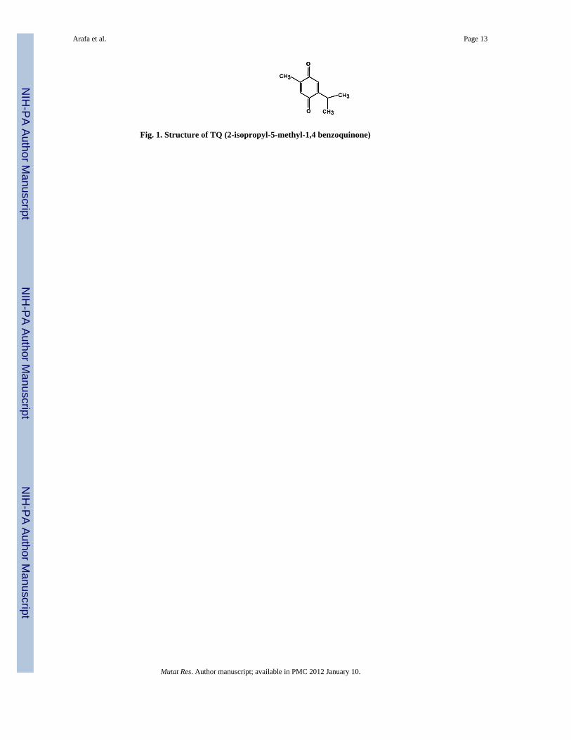
Fig. 1. Structure of TQ (2-isopropyl-5-methyl-1,4 benzoquinone)
Arafa et al. Page 13
Mutat Res. Author manuscript; available in PMC 2012 January 10.
NIH
-PA Author Manuscript
NIH
-PA Author Manuscript
NIH
-PA Author Manuscript
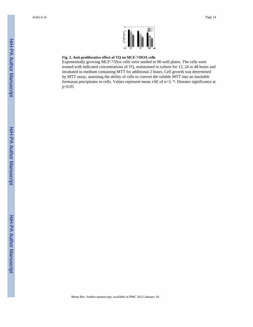
Fig. 2. Anti-proliferative effect of TQ on MCF-7/DOX cellsExponentially growing MCF-7/Dox cells were seeded in 96-well plates. The cells weretreated with indicated concentrations of TQ, maintained in culture for 12, 24 or 48 hours andincubated in medium containing MTT for additional 2 hours. Cell growth was determinedby MTT assay, assessing the ability of cells to convert the soluble MTT into an insolubleformazan precipitates in cells. Values represent mean ±SE of n=3. *: Denotes significance atp<0.05
Arafa et al. Page 14
Mutat Res. Author manuscript; available in PMC 2012 January 10.
NIH
-PA Author Manuscript
NIH
-PA Author Manuscript
NIH
-PA Author Manuscript
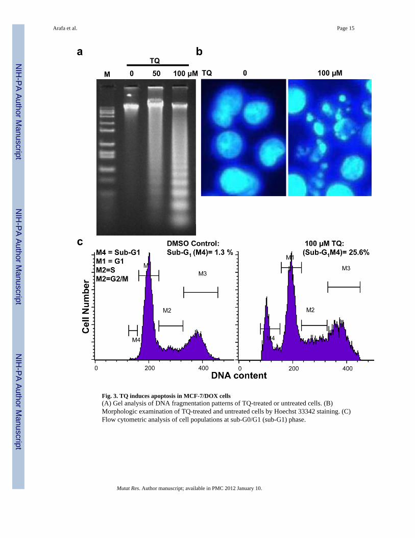
Fig. 3. TQ induces apoptosis in MCF-7/DOX cells(A) Gel analysis of DNA fragmentation patterns of TQ-treated or untreated cells. (B)Morphologic examination of TQ-treated and untreated cells by Hoechst 33342 staining. (C)Flow cytometric analysis of cell populations at sub-G0/G1 (sub-G1) phase.
Arafa et al. Page 15
Mutat Res. Author manuscript; available in PMC 2012 January 10.
NIH
-PA Author Manuscript
NIH
-PA Author Manuscript
NIH
-PA Author Manuscript
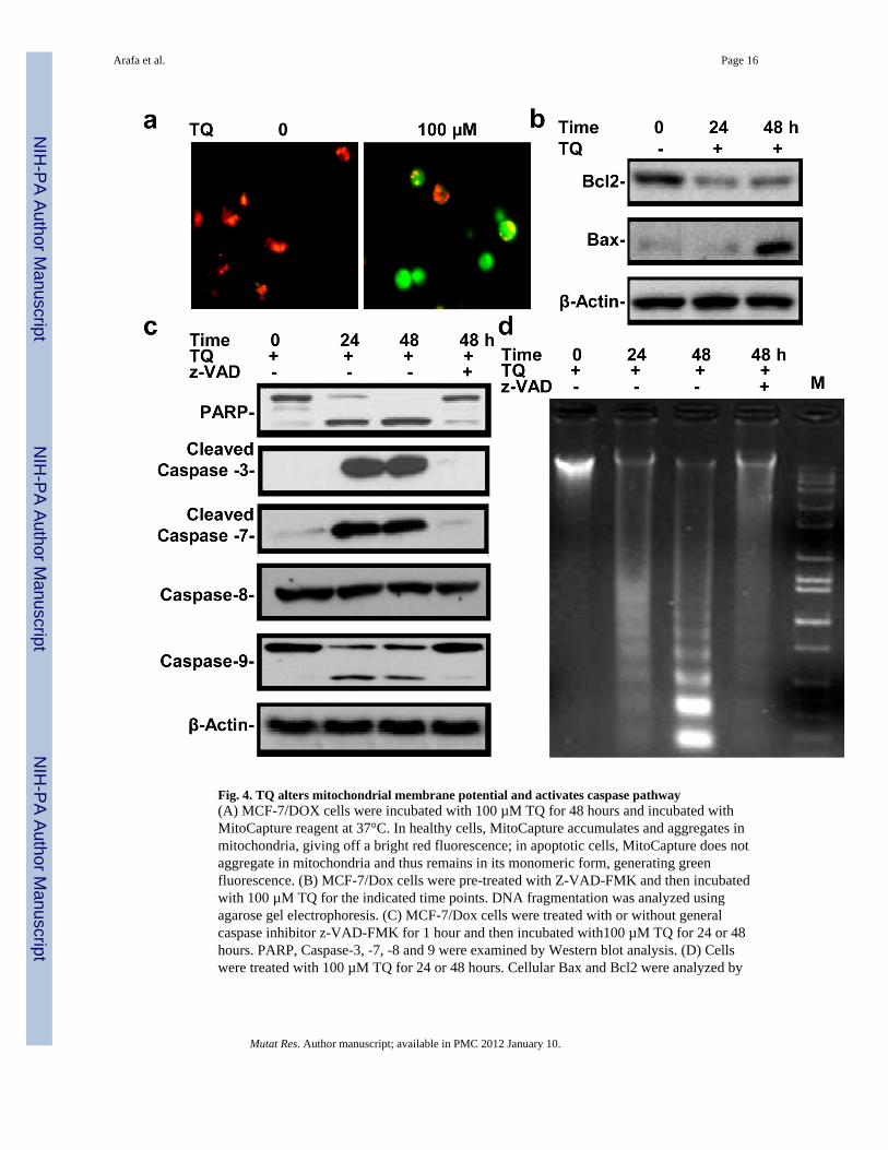
Fig. 4. TQ alters mitochondrial membrane potential and activates caspase pathway(A) MCF-7/DOX cells were incubated with 100 µM TQ for 48 hours and incubated withMitoCapture reagent at 37°C. In healthy cells, MitoCapture accumulates and aggregates inmitochondria, giving off a bright red fluorescence; in apoptotic cells, MitoCapture does notaggregate in mitochondria and thus remains in its monomeric form, generating greenfluorescence. (B) MCF-7/Dox cells were pre-treated with Z-VAD-FMK and then incubatedwith 100 µM TQ for the indicated time points. DNA fragmentation was analyzed usingagarose gel electrophoresis. (C) MCF-7/Dox cells were treated with or without generalcaspase inhibitor z-VAD-FMK for 1 hour and then incubated with100 µM TQ for 24 or 48hours. PARP, Caspase-3, -7, -8 and 9 were examined by Western blot analysis. (D) Cellswere treated with 100 µM TQ for 24 or 48 hours. Cellular Bax and Bcl2 were analyzed by
Arafa et al. Page 16
Mutat Res. Author manuscript; available in PMC 2012 January 10.
NIH
-PA Author Manuscript
NIH
-PA Author Manuscript
NIH
-PA Author Manuscript

Western blotting and the Bcl2/Bax ratio was calculated. Values represent mean ±SE of n=3.*: Denotes significance at p<0.05
Arafa et al. Page 17
Mutat Res. Author manuscript; available in PMC 2012 January 10.
NIH
-PA Author Manuscript
NIH
-PA Author Manuscript
NIH
-PA Author Manuscript
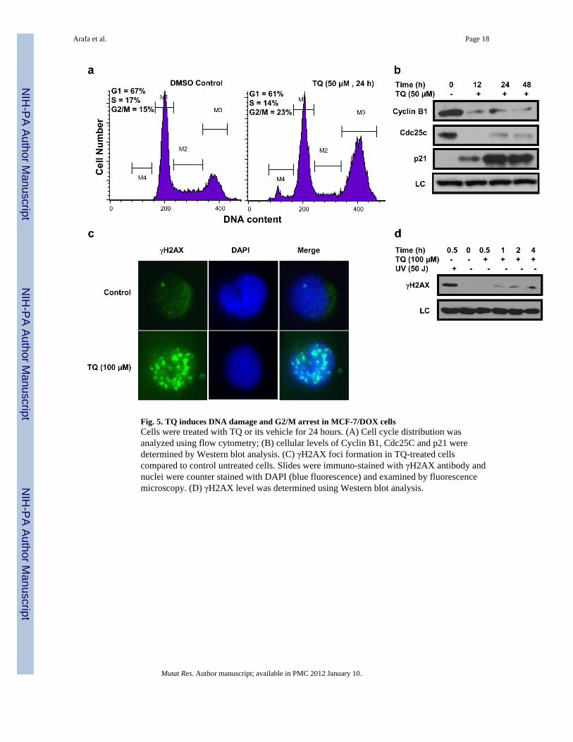
Fig. 5. TQ induces DNA damage and G2/M arrest in MCF-7/DOX cellsCells were treated with TQ or its vehicle for 24 hours. (A) Cell cycle distribution wasanalyzed using flow cytometry; (B) cellular levels of Cyclin B1, Cdc25C and p21 weredetermined by Western blot analysis. (C) γH2AX foci formation in TQ-treated cellscompared to control untreated cells. Slides were immuno-stained with γH2AX antibody andnuclei were counter stained with DAPI (blue fluorescence) and examined by fluorescencemicroscopy. (D) γH2AX level was determined using Western blot analysis.
Arafa et al. Page 18
Mutat Res. Author manuscript; available in PMC 2012 January 10.
NIH
-PA Author Manuscript
NIH
-PA Author Manuscript
NIH
-PA Author Manuscript
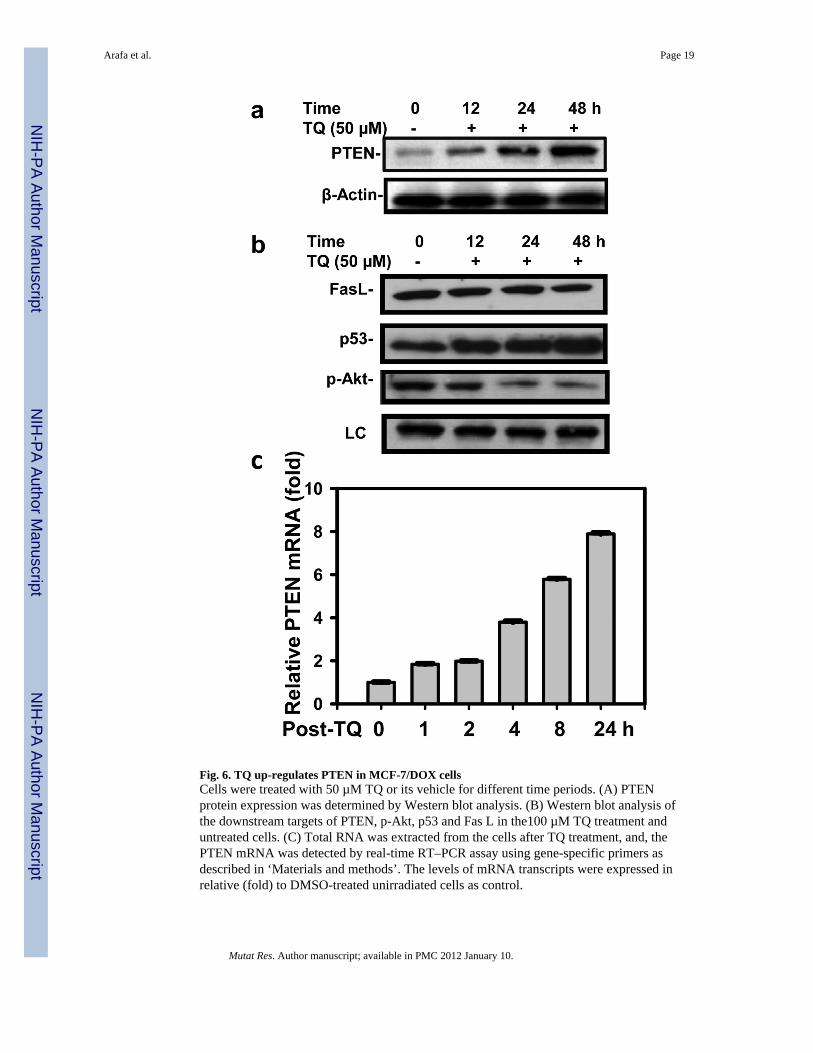
Fig. 6. TQ up-regulates PTEN in MCF-7/DOX cellsCells were treated with 50 µM TQ or its vehicle for different time periods. (A) PTENprotein expression was determined by Western blot analysis. (B) Western blot analysis ofthe downstream targets of PTEN, p-Akt, p53 and Fas L in the100 µM TQ treatment anduntreated cells. (C) Total RNA was extracted from the cells after TQ treatment, and, thePTEN mRNA was detected by real-time RT–PCR assay using gene-specific primers asdescribed in ‘Materials and methods’. The levels of mRNA transcripts were expressed inrelative (fold) to DMSO-treated unirradiated cells as control.
Arafa et al. Page 19
Mutat Res. Author manuscript; available in PMC 2012 January 10.
NIH
-PA Author Manuscript
NIH
-PA Author Manuscript
NIH
-PA Author Manuscript
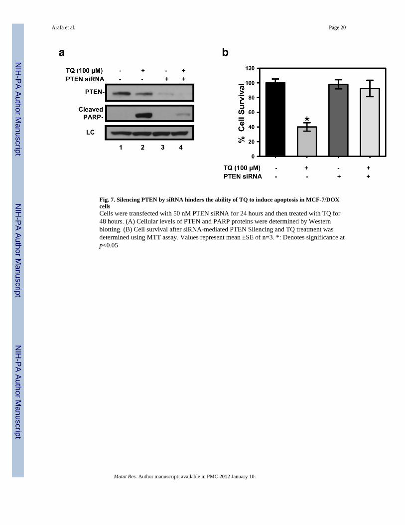
Fig. 7. Silencing PTEN by siRNA hinders the ability of TQ to induce apoptosis in MCF-7/DOXcellsCells were transfected with 50 nM PTEN siRNA for 24 hours and then treated with TQ for48 hours. (A) Cellular levels of PTEN and PARP proteins were determined by Westernblotting. (B) Cell survival after siRNA-mediated PTEN Silencing and TQ treatment wasdetermined using MTT assay. Values represent mean ±SE of n=3. *: Denotes significance atp<0.05
Arafa et al. Page 20
Mutat Res. Author manuscript; available in PMC 2012 January 10.
NIH
-PA Author Manuscript
NIH
-PA Author Manuscript
NIH
-PA Author Manuscript