Three-dimensional cartilage tissue engineering using adult stem cells from osteoarthritis patients
11
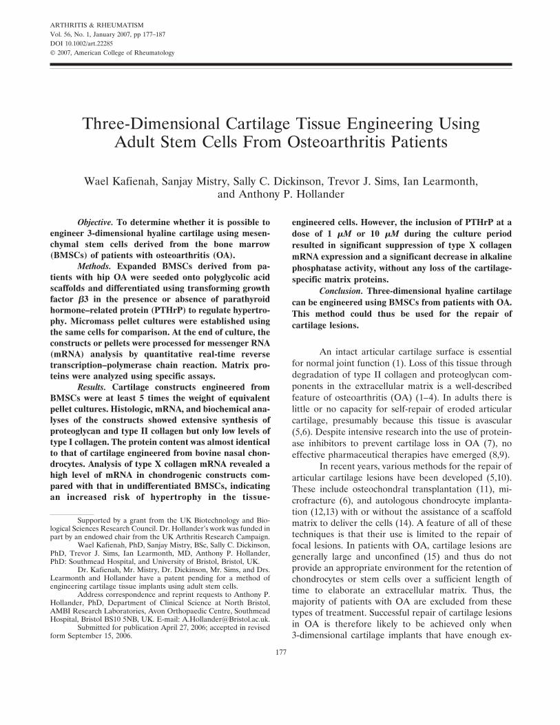
ARTHRITIS & RHEUMATISM Vol. 56, No. 1, January 2007, pp 177–187 DOI 10.1002/art.22285 © 2007, American College of Rheumatology Three-Dimensional Cartilage Tissue Engineering Using Adult Stem Cells From Osteoarthritis Patients Wael Kafienah, Sanjay Mistry, Sally C. Dickinson, Trevor J. Sims, Ian Learmonth, and Anthony P. Hollander Objective. To determine whether it is possible to engineer 3-dimensional hyaline cartilage using mesen- chymal stem cells derived from the bone marrow (BMSCs) of patients with osteoarthritis (OA). Methods. Expanded BMSCs derived from pa- tients with hip OA were seeded onto polyglycolic acid scaffolds and differentiated using transforming growth factor 3 in the presence or absence of parathyroid hormone–related protein (PTHrP) to regulate hypertro- phy. Micromass pellet cultures were established using the same cells for comparison. At the end of culture, the constructs or pellets were processed for messenger RNA (mRNA) analysis by quantitative real-time reverse transcription–polymerase chain reaction. Matrix pro- teins were analyzed using specific assays. Results. Cartilage constructs engineered from BMSCs were at least 5 times the weight of equivalent pellet cultures. Histologic, mRNA, and biochemical ana- lyses of the constructs showed extensive synthesis of proteoglycan and type II collagen but only low levels of type I collagen. The protein content was almost identical to that of cartilage engineered from bovine nasal chon- drocytes. Analysis of type X collagen mRNA revealed a high level of mRNA in chondrogenic constructs com- pared with that in undifferentiated BMSCs, indicating an increased risk of hypertrophy in the tissue- engineered cells. However, the inclusion of PTHrP at a dose of 1 M or 10 M during the culture period resulted in significant suppression of type X collagen mRNA expression and a significant decrease in alkaline phosphatase activity, without any loss of the cartilage- specific matrix proteins. Conclusion. Three-dimensional hyaline cartilage can be engineered using BMSCs from patients with OA. This method could thus be used for the repair of cartilage lesions. An intact articular cartilage surface is essential for normal joint function (1). Loss of this tissue through degradation of type II collagen and proteoglycan com- ponents in the extracellular matrix is a well-described feature of osteoarthritis (OA) (1–4). In adults there is little or no capacity for self-repair of eroded articular cartilage, presumably because this tissue is avascular (5,6). Despite intensive research into the use of protein- ase inhibitors to prevent cartilage loss in OA (7), no effective pharmaceutical therapies have emerged (8,9). In recent years, various methods for the repair of articular cartilage lesions have been developed (5,10). These include osteochondral transplantation (11), mi- crofracture (6), and autologous chondrocyte implanta- tion (12,13) with or without the assistance of a scaffold matrix to deliver the cells (14). A feature of all of these techniques is that their use is limited to the repair of focal lesions. In patients with OA, cartilage lesions are generally large and unconfined (15) and thus do not provide an appropriate environment for the retention of chondrocytes or stem cells over a sufficient length of time to elaborate an extracellular matrix. Thus, the majority of patients with OA are excluded from these types of treatment. Successful repair of cartilage lesions in OA is therefore likely to be achieved only when 3-dimensional cartilage implants that have enough ex- Supported by a grant from the UK Biotechnology and Bio- logical Sciences Research Council. Dr. Hollander’s work was funded in part by an endowed chair from the UK Arthritis Research Campaign. Wael Kafienah, PhD, Sanjay Mistry, BSc, Sally C. Dickinson, PhD, Trevor J. Sims, Ian Learmonth, MD, Anthony P. Hollander, PhD: Southmead Hospital, and University of Bristol, Bristol, UK. Dr. Kafienah, Mr. Mistry, Dr. Dickinson, Mr. Sims, and Drs. Learmonth and Hollander have a patent pending for a method of engineering cartilage tissue implants using adult stem cells. Address correspondence and reprint requests to Anthony P. Hollander, PhD, Department of Clinical Science at North Bristol, AMBI Research Laboratories, Avon Orthopaedic Centre, Southmead Hospital, Bristol BS10 5NB, UK. E-mail: [email protected]. Submitted for publication April 27, 2006; accepted in revised form September 15, 2006. 177
-
Upload
independent -
Category
Documents
-
view
1 -
download
0
Transcript of Three-dimensional cartilage tissue engineering using adult stem cells from osteoarthritis patients
ARTHRITIS & RHEUMATISMVol. 56, No. 1, January 2007, pp 177–187DOI 10.1002/art.22285© 2007, American College of Rheumatology
Three-Dimensional Cartilage Tissue Engineering UsingAdult Stem Cells From Osteoarthritis Patients
Wael Kafienah, Sanjay Mistry, Sally C. Dickinson, Trevor J. Sims, Ian Learmonth,and Anthony P. Hollander
Objective. To determine whether it is possible toengineer 3-dimensional hyaline cartilage using mesen-chymal stem cells derived from the bone marrow(BMSCs) of patients with osteoarthritis (OA).
Methods. Expanded BMSCs derived from pa-tients with hip OA were seeded onto polyglycolic acidscaffolds and differentiated using transforming growthfactor �3 in the presence or absence of parathyroidhormone–related protein (PTHrP) to regulate hypertro-phy. Micromass pellet cultures were established usingthe same cells for comparison. At the end of culture, theconstructs or pellets were processed for messenger RNA(mRNA) analysis by quantitative real-time reversetranscription–polymerase chain reaction. Matrix pro-teins were analyzed using specific assays.
Results. Cartilage constructs engineered fromBMSCs were at least 5 times the weight of equivalentpellet cultures. Histologic, mRNA, and biochemical ana-lyses of the constructs showed extensive synthesis ofproteoglycan and type II collagen but only low levels oftype I collagen. The protein content was almost identicalto that of cartilage engineered from bovine nasal chon-drocytes. Analysis of type X collagen mRNA revealed ahigh level of mRNA in chondrogenic constructs com-pared with that in undifferentiated BMSCs, indicatingan increased risk of hypertrophy in the tissue-
engineered cells. However, the inclusion of PTHrP at adose of 1 �M or 10 �M during the culture periodresulted in significant suppression of type X collagenmRNA expression and a significant decrease in alkalinephosphatase activity, without any loss of the cartilage-specific matrix proteins.
Conclusion. Three-dimensional hyaline cartilagecan be engineered using BMSCs from patients with OA.This method could thus be used for the repair ofcartilage lesions.
An intact articular cartilage surface is essentialfor normal joint function (1). Loss of this tissue throughdegradation of type II collagen and proteoglycan com-ponents in the extracellular matrix is a well-describedfeature of osteoarthritis (OA) (1–4). In adults there islittle or no capacity for self-repair of eroded articularcartilage, presumably because this tissue is avascular(5,6). Despite intensive research into the use of protein-ase inhibitors to prevent cartilage loss in OA (7), noeffective pharmaceutical therapies have emerged (8,9).
In recent years, various methods for the repair ofarticular cartilage lesions have been developed (5,10).These include osteochondral transplantation (11), mi-crofracture (6), and autologous chondrocyte implanta-tion (12,13) with or without the assistance of a scaffoldmatrix to deliver the cells (14). A feature of all of thesetechniques is that their use is limited to the repair offocal lesions. In patients with OA, cartilage lesions aregenerally large and unconfined (15) and thus do notprovide an appropriate environment for the retention ofchondrocytes or stem cells over a sufficient length oftime to elaborate an extracellular matrix. Thus, themajority of patients with OA are excluded from thesetypes of treatment. Successful repair of cartilage lesionsin OA is therefore likely to be achieved only when3-dimensional cartilage implants that have enough ex-
Supported by a grant from the UK Biotechnology and Bio-logical Sciences Research Council. Dr. Hollander’s work was funded inpart by an endowed chair from the UK Arthritis Research Campaign.
Wael Kafienah, PhD, Sanjay Mistry, BSc, Sally C. Dickinson,PhD, Trevor J. Sims, Ian Learmonth, MD, Anthony P. Hollander,PhD: Southmead Hospital, and University of Bristol, Bristol, UK.
Dr. Kafienah, Mr. Mistry, Dr. Dickinson, Mr. Sims, and Drs.Learmonth and Hollander have a patent pending for a method ofengineering cartilage tissue implants using adult stem cells.
Address correspondence and reprint requests to Anthony P.Hollander, PhD, Department of Clinical Science at North Bristol,AMBI Research Laboratories, Avon Orthopaedic Centre, SouthmeadHospital, Bristol BS10 5NB, UK. E-mail: [email protected].
Submitted for publication April 27, 2006; accepted in revisedform September 15, 2006.
177
tracellular matrix for fixation within the joint can begenerated.
Cartilage tissue engineering provides a potentialmethod for the production of 3-dimensional implants(16,17). Effective engineering protocols have alreadybeen developed in which chondrocytes, usually fromyoung animals, are seeded onto biodegradable scaffoldsand cultured in a bioreactor (18,19). Generating3-dimensional cartilage using adult human chondrocytesis far more challenging and, in the case of older OApatients, is probably impossible in the clinic setting,because of the lack of autologous donor tissue. Onestudy demonstrated that OA chondrocytes can generatea cartilage-like matrix when grown on hyaluronic acidscaffolds (20), indicating the potential utility of thesecells in OA. However, the cells synthesized significantlyless collagen than was generated by chondrocytes fromindividuals without arthritis, suggesting that there maybe a need for additional up-regulation of chondrogenesisif OA chondrocytes are ever to be used in a clinicalsetting.
This has led several groups of investigators toexplore the use of mesenchymal stem cells (MSCs) forthe generation of autologous chondrocytes (21). MSCsare multipotent cells with a self-renewing capacity(22,23). Many studies have utilized adherent bone mar-row MSCs (BMSCs) cultured as small micromass pelletsand stimulated with transforming growth factor �(TGF�) to drive chondrogenesis (24,25). From thesestudies there is strong histologic evidence that underthese conditions, the BMSCs become chondrocytes andsynthesize both type II collagen and proteoglycan. How-ever, micromass pellets were designed for use in exper-imental models, and the amount of extracellular matrixproduced by the pellets is too small to be of practicalvalue for implantation (26). Furthermore, there is clearevidence that BMSCs stimulated with TGF� expresstype X collagen, an early marker of hypertrophy that isnormally absent from hyaline cartilage (27).
It is not yet known whether BMSCs derived fromOA patients have the capacity to become chondrocytesand generate hyaline cartilage. Most studies have uti-lized BMSCs from animals or healthy human donors(23–25,28). Nevertheless, one study investigated OABMSCs cultured as pellets, and the results demonstrateda reduced chondrogenic capacity of these cells (29). Thepurpose of the present study was therefore to determinewhether OA BMSCs can be used for tissue engineeringof 3-dimensional hyaline cartilage.
PATIENTS AND METHODS
Patients. Bone marrow plugs were collected as trabec-ular bone biopsy specimens from the femoral heads of 23patients with OA who were undergoing hip arthroplasty atSouthmead Hospital (Bristol, UK), of whom 52% were menand 48% were women. The mean age of the 23 patients was65.8 years (range 42–90 years). The study was carried out in fullaccordance with local ethics guidelines, and all of the patientsgave their informed consent.
Isolation and characterization of BMSCs. Cells wereisolated from the bone marrow plugs by washing in expansionmedium consisting of low-glucose Dulbecco’s modified Eagle’smedium (DMEM) (Sigma, St. Louis, MO) supplemented with10% fetal bovine serum (FBS), 1% (volume/volume) Glu-tamax (1�; Invitrogen, Carlsbad, CA), and 1% (v/v) penicillin(100 units/ml)–streptomycin (100 �g/ml) (Invitrogen). Theserum batch was selected to promote the growth and differen-tiation of MSCs (30).
The cell suspension was separated from any bone inthe sample using a 19-gauge needle. The cells were centrifugedat 1,500 revolutions per minute for 5 minutes and thesupernatant/fat was removed. The resulting cell pellet wasresuspended in medium and then plated at a seeding density of1.5–2.0 � 105 nucleated cells per cm2. The expansion mediumwas supplemented with 1 ng/ml fibroblast growth factor 2(FGF-2) (PeproTech, London, UK) to enhance BMSC prolif-eration and differentiation (31,32). These flasks were incu-bated at 37°C in a humidified atmosphere of 5% CO2 and 95%air. The first medium change was after 4 days, and the mediumwas then changed every other day until adherent cells reached90% confluence and were ready for passaging. The cells werecharacterized for stem cell surface markers and multilineagepotential, as described previously (30).
Micromass pellet cultures using OA BMSCs. Ex-panded BMSCs were trypsinized and cultured in micromasspellets as described previously (24), with slight modification.Briefly, 500,000 cells were placed in a 15-ml conical polypro-pylene tube and resuspended in 1.0 ml chondrogenic differen-tiation medium consisting of DMEM containing 4.5 gm/literglucose supplemented with 10 ng/ml of TGF�3 (R&D Systems,Abingdon, UK), 1 mM sodium pyruvate (Sigma), 50 �g/mlascorbic acid-2-phosphate (Sigma), 1 � 10�7M dexamethasone(Sigma), 1% insulin–transferrin–selenium (Invitrogen), and1% (v/v) penicillin (100 units/ml)–streptomycin (100 �g/ml)(Invitrogen). Cells were centrifuged at 1,500 rpm for 5 minutesat 20°C. The pellets were maintained in culture with 1 pellet/tube and 1.0 ml chondrogenic medium/tube. Medium waschanged every 2–3 days. After the first week the medium wasfurther supplemented with 50 �g/ml insulin (Sigma) until theend of culture. Chondrogenic pellets were harvested at 21 daysfor messenger RNA (mRNA), matrix proteins, and histologicanalyses.
Tissue engineering using OA BMSCs. Polyglycolic acid(PGA) scaffolds (a kind gift from Dr. James Huckle, Smith &Nephew, York, UK) were produced as 5-mm (diameter) � 2-mm (thick) discs according to an established method (33). Thescaffolds were presoaked in 100 �g/ml human fibronectin(Sigma) in phosphate buffered saline (PBS) to support BMSCadherence to PGA fibers. BMSCs from passage 2 or passage 3were trypsinized and suspended in 30 �l of expansion medium.
178 KAFIENAH ET AL
The suspension was loaded drop-wise onto the scaffold intissue-culture wells precoated with 1% (weight/volume) aga-rose (Sigma) to prevent cell adherence to plastic. After incu-bation for 4 hours, the scaffolds were turned over and incu-bated for a further 4 hours to allow for even distribution ofcells across the scaffold.
The constructs were maintained in chondrogenic dif-ferentiation medium, as described above for micromass pelletcultures. After the first week, the medium was further supple-mented with 50 �g/ml insulin (PeproTech) until the end ofculture. Human recombinant parathyroid hormone–relatedprotein (PTHrP) (Sigma) was included in the differentiationmedium at 1 �M or 10 �M, as appropriate. The medium waschanged 3 times a week. The constructs were incubated at37°C, in 5% CO2 on a rotating platform at 50 rpm for 35 days.Harvested samples were digested with collagenase to releasethe cells, which were stored at �70°C for subsequent RNAextraction or alkaline phosphatase activity assay. Other sam-ples were stored at �20°C prior to quantitative biochemicalanalysis (see below). In some experiments, cartilage was engi-neered using bovine nasal chondrocytes (BNCs) that wereisolated as described previously (34).
Histologic and immunohistochemical analyses of mi-cromass pellets and engineered cartilage. Micromass pelletsand mature cartilage engineered from stem cells were frozen inOCT embedding matrix (BDH Chemicals, Poole, UK). Full-depth sections (thickness 7 �m) were cut with a cryostat andfixed in 4% (w/v) paraformaldehyde (Sigma) in PBS, pH 7.6.Some sections were stained with hematoxylin and eosin or0.1% (w/v) Safranin O (both from Sigma) to evaluate thedistribution of matrix and proteoglycan, respectively. Othersections were immunostained with monoclonal antibodiesagainst types I and II collagen (Southern Biotechnology,Birmingham, AL) and type X collagen (a kind gift from Dr.Alvin Kwan, Cardiff University, UK), as previously described(34,35). Biotinylated secondary antibodies were detected witha peroxidase-labeled biotin–streptavidin complex (VectastainElite kit; Vector Laboratories, Peterborough, UK) with diami-nobenzidine substrate (Vector Laboratories). Natural cartilageand tendon were used as positive controls for detection of typeII collagen and type I collagen, respectively. Normal goatserum was used as a negative control, and all sections werecounterstained with hematoxylin (Vector Laboratories).
Quantitative biochemical analyses of engineered car-tilage. Dry weights of the constructs were determined afterfreeze-drying. The samples were then solubilized with trypsinand processed for complete biochemical analysis as recentlydescribed (36). Briefly, samples were digested with 2 mg/mlTPCK-treated bovine pancreatic trypsin containing 1 mM iodo-acetamide, 1 mM EDTA, and 10 �g/ml pepstatin A (all fromSigma). An initial incubation for 15 hours at 37°C with 250 �ltrypsin was followed by a further 2-hour incubation at 65°Cafter the addition of a further 250 �l of the freshly preparedproteinase. All samples were boiled for 15 minutes at the endof incubation to destroy any remaining enzyme activity. Theextracts were assayed by inhibition enzyme-linked immunosor-bent assay (ELISA) using a mouse IgG monoclonal antibodyto denatured type II collagen, COL2-3/4m, as previously de-scribed (2). Peptide CB11B (CGKVGPSGAP[OH]GEDGRP-[OH]GPP[OH]GPQY) was synthesized using 9-fluorenyl-methoxycarbonyl chemistry (a gift from Dr. A. Moir, Kreb’s
Institute, Sheffield University, UK) and was used as a standardin all of the immunoassays. The extracts were also assayed byinhibition ELISA using a rabbit antipeptide antibody to type Icollagen, as previously described (36). Peptide SFLPQPPQwas synthesized using 9-fluorenyl-methoxycarbonyl chemistry(also from Dr. A. Moir) and was used as a standard in all of theimmunoassays. Proteoglycan in the digests was measured bythe detection of sulfated glycosaminoglycan using colorimetricassay with dimethylmethylene blue (Aldrich, Gillingham, UK)as previously described (37).
Alkaline phosphatase activity. Cells in engineeredcartilage constructs were assayed for alkaline phosphataseactivity after collagenase digestion of the extracellular matrix,as described previously (38). Although it is theoretically pos-sible that the collagenase digestion could itself influencealkaline phosphatase activity, this is unlikely to affect theinterpretation of our results, since all samples were extractedunder identical conditions and assayed as soon as possible aftercompletion of the extraction process. Briefly, the cells werelysed with 0.1 ml of 25 mM sodium carbonate (pH 10.3), 0.1%(v/v) Triton X-100. After 2 minutes, each sample was treatedwith 0.2 ml of 15 mM p-nitrophenyl phosphate (di-tri salt;Sigma) in 250 mM sodium carbonate (pH 10.3), 1.5 mMMgCl2. Lysates were then incubated at 37°C for 2 hours. Afterthe incubation period, 0.1-ml aliquots were transferred to a96-well microtiter plate and the absorbance was read at 405nm. An ascending series of p-nitrophenol (25–500 �M) pre-pared in the incubation buffer enabled quantification of prod-uct formation.
RNA isolation and reverse transcription. RNA wasextracted from cell cultures using the GenElute MammalianTotal RNA kit (Sigma), according to the manufacturer’sinstructions. Reverse transcription (RT) was carried out usingthe Superscript II system (Invitrogen). Total RNA (2 �g) wasreverse transcribed in a 20-�l reaction volume containingSuperscript II (200 units), random primers (25 �M), and dNTP(0.5 mM each) at 42°C for 50 minutes.
Primer design. The coding sequences for human typeX, type II, and type I collagens (accession no. NM_000493,NM_001844, and NM_000088, respectively) were used todesign primers using the online software, Primer3 (WhiteheadInstitute for Biomedical Research, Cambridge, MA). Theprimers span intronic junctions to avoid the amplification ofgenomic sequences. They were also checked for the amplifica-tion of potential pseudogenes. A BLAST search against allknown sequences confirmed specificity. Published primers forthe housekeeping gene �-actin (39) were used as a referencefor normalization in all RT–polymerase chain reactions (RT-PCRs). In preliminary studies (results not shown) we foundthat this gene was expressed more stably than GAPDH orribosomal RNA for a range of differentiated progeny of stemcells. All of our primers were specifically designed not tocoamplify processed pseudogenes in contaminating genomicDNA and they all generated the correct sizes of the PCRfragments with no nonspecific products, thus confirming thespecificity of the real-time RT-PCR (results not shown).Details of the primers used in the study are as follows: for typeX collagen �, forward GACACAGTTCTTCATTCCCTACACand reverse GCAACCCTGGCTCTCCTT; for type II collagen�1 (A�B), forward CAACACTGCCAACGTCCAGAT andreverse CTGCTTCGTCCAGATAGGCAAT; for type I colla-
CARTILAGE TISSUE ENGINEERING USING STEM CELLS FROM OA PATIENTS 179
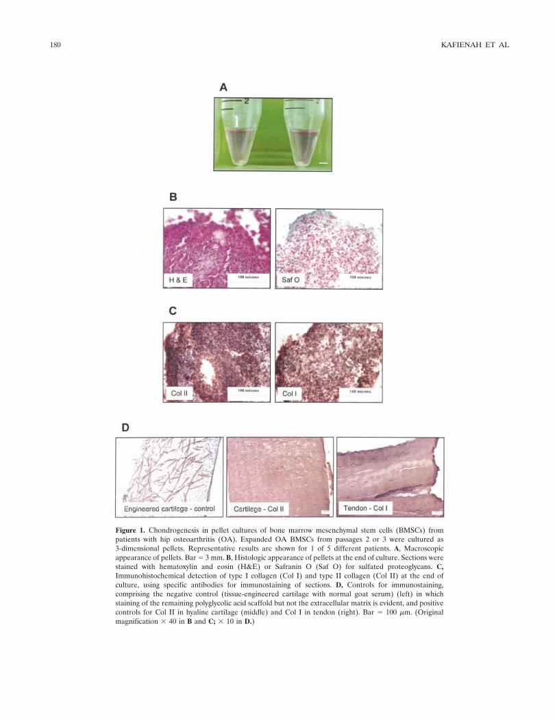
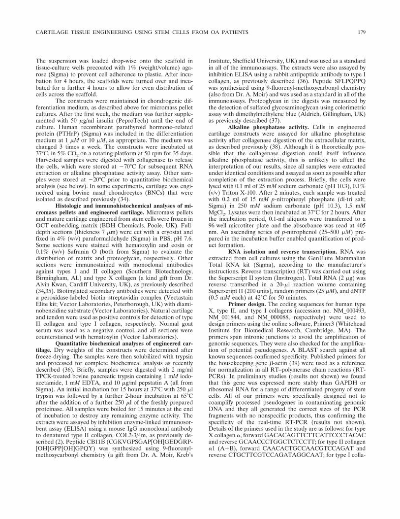
Figure 1. Chondrogenesis in pellet cultures of bone marrow mesenchymal stem cells (BMSCs) frompatients with hip osteoarthritis (OA). Expanded OA BMSCs from passages 2 or 3 were cultured as3-dimensional pellets. Representative results are shown for 1 of 5 different patients. A, Macroscopicappearance of pellets. Bar � 3 mm. B, Histologic appearance of pellets at the end of culture. Sections werestained with hematoxylin and eosin (H&E) or Safranin O (Saf O) for sulfated proteoglycans. C,Immunohistochemical detection of type I collagen (Col I) and type II collagen (Col II) at the end ofculture, using specific antibodies for immunostaining of sections. D, Controls for immunostaining,comprising the negative control (tissue-engineered cartilage with normal goat serum) (left) in whichstaining of the remaining polyglycolic acid scaffold but not the extracellular matrix is evident, and positivecontrols for Col II in hyaline cartilage (middle) and Col I in tendon (right). Bar � 100 �m. (Originalmagnification � 40 in B and C; � 10 in D.)
180 KAFIENAH ET AL
gen �1, forward AGGGCCAAGACGAAGACATC and re-verse CAACACTGCCAACGTCCAGAT; and for �-actin,forward GACAGGATGCAGAAGGAGATTACT and re-verse TGATCCACATCTGCTGGAAGGT.
Quantitative real-time RT-PCR. Quantitative real-time RT-PCR was performed in a 25-�l reaction volumecontaining 12.5 �l of the SYBR Green PCR master mix(Sigma), 5 �l of the RT reaction mixture, and 300 nM eachprimer using the Smart Cycler II System (Cepheid, Sunnyvale,CA). For the �-actin gene, the RT reaction mixture was diluted100 times. The amplification program consisted of initialdenaturation at 95°C for 2 minutes followed by 40 cycles of95°C for 15 seconds, annealing at 58°C for 30 seconds, andextension at 72°C for 15 seconds. After amplification, meltanalysis was performed by heating the reaction mixture from60°C to 95°C at a rate of 0.2°C/second. The threshold cycle (Ct)value for each gene of interest was measured for each RTsample. The Ct value for �-actin was used as an endogenousreference for normalization. Real-time RT-PCR assays weredone in duplicate or triplicate, with each set of assays repeated2–4 times.
Statistical analysis. Comparison of differences be-tween individual groups was performed using the 2-tailedMann-Whitney U test. For multiple comparisons, the groupswere compared by analysis of variance using the nonpara-metric Kruskal-Wallis test. When significant variance wasdemonstrated, differences between individual groups werethen determined using the 2-tailed Mann-Whitney U test withDunn’s post hoc correction. In all analyses, P values less than0.05 were considered significant.
RESULTS
Phenotype of isolated OA BMSCs. A well-characterized population of stem cells was used for the3-dimensional engineering of cartilage tissue. We havepreviously described this cell population as being posi-tive for CD105, CD106, CD49a, CD117, STRO-1, andbone morphogenetic protein receptor type 1A and neg-ative for CD34 (30). We have also shown the populationto be multipotential, in that it consistently is able todifferentiate into adipogenic, chondrogenic, and osteo-genic lineages (30). In the present study there was nosignificant variation in the extent of stem cell differen-tiation among the patient samples (n � 23), betweenmale and female patients, or between different agegroups (results not shown). Therefore, we were able toconfirm that the BMSC population used was consistentlymultipotent, as expected for stem cells that have beenexpanded with the use of FGF-2 (30–32).
Chondrocyte formation from OA BMSCs. In ourinitial experiments, we cultured OA BMSCs withTGF�3 in high-density pellets. Under these conditions,the pellets grew into cartilage-like nodules that werevisible to the naked eye (Figure 1A). At the histologic
level, these nodules contained a large number of cells aswell as an extracellular matrix that stained consistentlyfor proteoglycan, although this was largely in a pericel-lular location (Figure 1B). Similarly, there was stainingfor types I and II collagen throughout the pellets andthis was most intense around the cells (Figure 1C). Theextent of immunostaining for type I collagen was rela-tively similar to that for type II collagen, suggesting thatthe pellet tissue was likely to be predominantly fibrocar-tilage rather than hyaline cartilage. Negative and posi-tive controls for the immunostaining are shown in Figure1D.
Cartilage tissue engineering using OA BMSCs.We were able to successfully engineer 3-dimensionalcartilage using a carefully ordered sequence of signals, asdescribed above. First, the OA BMSCs were expandedin 10% FBS and 1 ng/ml FGF-2. Second, the expandedcells were seeded onto PGA scaffolds that had beenprecoated with fibronectin. Third, the cells were cul-tured on a gently rotating platform for 1 week with 10ng/ml TGF�3 in differentiation medium. Fourth, thecells were cultured for a further 4 weeks on the rotatingplatform in differentiation medium with 50 �g/ml insulinas well as 10 ng/ml TGF�3.
Under these carefully defined conditions, wewere able to generate a white, shiny tissue that resem-bled hyaline cartilage at a macroscopic level (Figure2A). On histologic analysis, these cartilage constructswere found to contain an extracellular matrix thatstained extensively for proteoglycan, moderately for typeII collagen, and weakly for type I collagen (Figure 2B).At higher magnification, it was possible to observerounded cells, some pericellular staining for type Xcollagen, and staining for type II collagen (strongly) andtype I collagen (weakly) in the interterritorial matrix.
Biochemical findings in engineered cartilage. Weundertook an extensive quantitative analysis of the en-gineered cartilage using a series of well-validated andspecific assays (36). For comparison, micromass pelletcultures were also analyzed. Despite the sensitive natureof our assays (36), we had to use a minimum of 500,000cells per micromass pellet, and at the end of culture wehad to combine 3 of these pellets in order to generateenough extracellular matrix for quantitative analysis. Fortissue engineering, we were able to use as few as 300,000cells per scaffold and to analyze one sample at a time.Cultures were maintained in TGF�3 for up to 35 daysprior to analysis. However, in cultures with the micro-mass pellets, there was evidence of some loss of matrixbeyond 21 days, and therefore the pellet cultures were
CARTILAGE TISSUE ENGINEERING USING STEM CELLS FROM OA PATIENTS 181
stopped at this optimal time point. After 35 days inculture, the mean dry weight was 0.08 mg, the type II
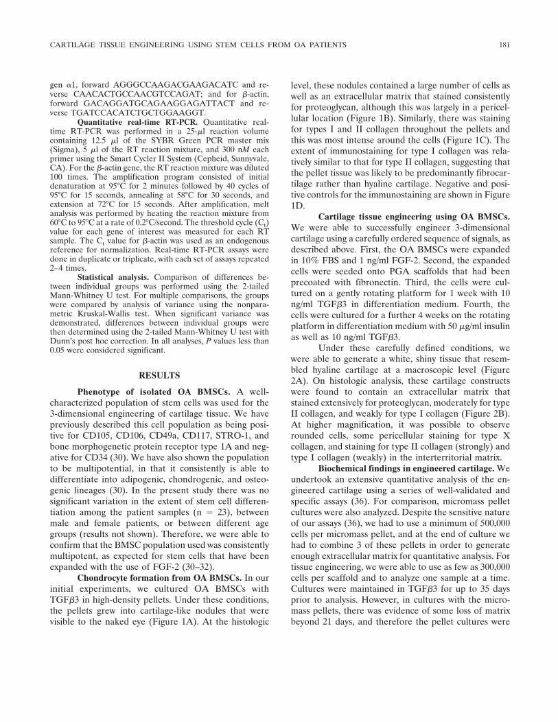
collagen content was 0.4%, and proteoglycan contentwas 0.75%, whereas the type I collagen content wasundetectable; the equivalent findings after 21 days inpellet cultures were a mean 0.23 mg dry weight, 0.57%type II collagen, 1.14% proteoglycan, and 0.26% type Icollagen. The dry weights of pellets and engineeredtissue were calculated after freeze-drying. In the case ofengineered tissue, the weight of any remaining PGAscaffold was determined after enzymatic digestion of theextracellular matrix, and this was subtracted from thetotal dry weight.
Using these calculations, we determined that theextracellular matrix of engineered tissue was at least 5times that of pellet cultures, and this difference wassignificant (Table 1). Furthermore, the engineered car-tilage contained significantly more proteoglycan andtype II collagen than was observed in micromass pelletcultures. There was also a slightly higher type I collagencontent in engineered cartilage, although this was still�10% of the type II collagen content in engineeredcartilage.
We previously demonstrated that the best resultsfrom cartilage tissue engineering could be achievedusing BNCs (34). We therefore compared the results ofcartilage engineering using human OA BMSCs withthose using BNCs. There was no significant difference inthe content of type II collagen, type I collagen, orproteoglycan between the 2 cell types (Figure 3), indi-cating that chondrocytes derived from OA BMSCs areas effective as BNCs.
Inhibition of hypertrophy by PTHrP. In prelimi-nary experiments, we observed that OA BMSCs culturedwith TGF�3 in micromass pellets displayed increasedexpression of type X collagen, indicating that these cells
Figure 2. Cartilage tissue engineering from BMSCs. Expanded OABMSCs from passages 2 or 3 were used to engineer cartilage onpolyglycolic acid scaffolds. Representative results are shown for 1 of 8different patients. A, Macroscopic appearance of engineered cartilage.Bar � 3 mm. B, Histologic appearance of engineered cartilage at theend of culture, in sections stained with H&E or Saf O for sulfatedproteoglycans and immunostained for Col II and Col I using specificantibodies. Bar � 100 �m. C, Histologic appearance of engineeredcartilage at the end of culture, in sections stained with H&E andimmunostained for types X, II, and I collagens using specific antibod-ies; arrows indicate staining for Col X limited to the pericellularregion. See Figure 1 for other definitions. (Original magnification � 10in B; � 40 in C.)
Table 1. Biochemical analysis of cartilage extracellular matrix
ParameterPellet culture
(n � 10)Tissue-engineeredcartilage (n � 15)
Number of cells seeded* 500,000 300,000Dry weight, mean � SEM mg 0.23 � 0.03 1.25 � 0.77†Type II collagen, mean �
SEM % of dry weight‡0.57 � 0.07 17.21 � 2.88†
Proteoglycan, mean � SEM %of dry weight§
1.14 � 0.13 29.96 � 3.45†
Type I collagen, mean � SEM% of dry weight‡
0.26 � 0.11 1.76 � 0.25†
* Minimum number of cells required for accurate quantification usingbiochemical assays.† P � 0.0001 versus pellet culture, by Mann-Whitney U test.‡ Determined by specific immunoassay after selective extraction ofpeptide epitopes.§ Determined by dimethylmethylene blue colorimetric assay of glycos-aminoglycans.
182 KAFIENAH ET AL
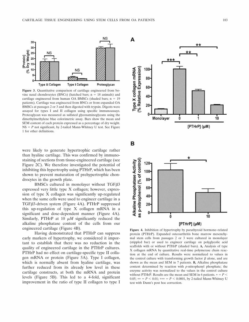
were likely to generate hypertrophic cartilage ratherthan hyaline cartilage. This was confirmed by immuno-staining of sections from tissue-engineered cartilage (seeFigure 2C). We therefore investigated the potential ofinhibiting this hypertrophy using PTHrP, which has beenshown to prevent maturation of prehypertrophic chon-drocytes in the growth plate.
BMSCs cultured in monolayer without TGF�3expressed very little type X collagen; however, expres-sion of type X collagen was significantly up-regulatedwhen the same cells were used to engineer cartilage in aTGF�3-driven system (Figure 4A). PTHrP suppressedthis up-regulation of type X collagen mRNA in asignificant and dose-dependent manner (Figure 4A).Similarly, PTHrP at 10 �M significantly reduced thealkaline phosphatase content of the cells from ourengineered cartilage (Figure 4B).
Having demonstrated that PTHrP can suppressearly markers of hypertrophy, we considered it impor-tant to establish that there was no reduction in thequality of engineered cartilage in the PTHrP cultures.PTHrP had no effect on cartilage-specific type II colla-gen mRNA or protein (Figure 5A). Type I collagen,which is normally absent from hyaline cartilage, wasfurther reduced from its already low level in thesecartilage constructs, at both the mRNA and proteinlevels (Figure 5B). This led to a 4-fold, significantimprovement in the ratio of type II collagen to type I
Figure 4. Inhibition of hypertrophy by parathyroid hormone–relatedprotein (PTHrP). Expanded osteoarthritis bone marrow mesenchy-mal stem cells from passages 2 or 3 were cultured in monolayer(stippled bar) or used to engineer cartilage on polyglycolic acidscaffolds with or without PTHrP (shaded bars). A, Analysis of typeX collagen mRNA by quantitative real-time polymerase chain reac-tion at the end of culture. Results were normalized to values inthe control culture with transforming growth factor � alone, and areshown as the mean and SEM in 7 patients. B, Alkaline phosphatasecontent determined by reaction with p-nitrophenyl phosphate; theenzyme activity was normalized to the values in the control culturewithout PTHrP. Results are the mean and SEM in 6 patients. � � P �0.05; �� � P � 0.01; ��� � P � 0.0001, by 2-tailed Mann-Whitney Utest with Dunn’s post hoc correction.
Figure 3. Quantitative comparison of cartilage engineered from bo-vine nasal chondrocytes (BNCs) (hatched bars; n � 18 animals) andcartilage engineered from human OA BMSCs (shaded bars; n � 19patients). Cartilage was engineered from BNCs or from expanded OABMSCs at passages 2 or 3 and then digested with trypsin. Digests wereassayed for types I and II collagen using specific immunoassays.Proteoglycan was measured as sulfated glycosaminoglycans using thedimethylmethylene blue colorimetric assay. Bars show the mean andSEM content of each protein expressed as a percentage of dry weight.NS � P not significant, by 2-tailed Mann-Whitney U test. See Figure1 for other definitions.
CARTILAGE TISSUE ENGINEERING USING STEM CELLS FROM OA PATIENTS 183
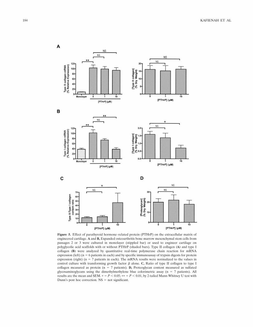
Figure 5. Effect of parathyroid hormone–related protein (PTHrP) on the extracellular matrix ofengineered cartilage. A and B, Expanded osteoarthritis bone marrow mesenchymal stem cells frompassages 2 or 3 were cultured in monolayer (stippled bar) or used to engineer cartilage onpolyglycolic acid scaffolds with or without PTHrP (shaded bars). Type II collagen (A) and type Icollagen (B) were analyzed by quantitative real-time polymerase chain reaction for mRNAexpression (left) (n � 6 patients in each) and by specific immunoassay of trypsin digests for proteinexpression (right) (n � 7 patients in each). The mRNA results were normalized to the values incontrol culture with transforming growth factor � alone. C, Ratio of type II collagen to type Icollagen measured as protein (n � 7 patients). D, Proteoglycan content measured as sulfatedglycosaminoglycans using the dimethylmethylene blue colorimetric assay (n � 7 patients). Allresults are the mean and SEM. � � P � 0.05; �� � P � 0.01, by 2-tailed Mann-Whitney U test withDunn’s post hoc correction. NS � not significant.
184 KAFIENAH ET AL
collagen (Figure 5C), whereas no effect of PTHrP on theproteoglycan content was observed (Figure 5D).
DISCUSSION
The present study is the first to demonstrate thefeasibility of tissue engineering of hyaline cartilage fromOA BMSCs. Biochemically, the cartilage quality wascomparable with that achieved using the best availablecell source, namely BNCs (34). Furthermore, we wereable to show that the tendency of BMSCs to becomehypertrophic can be down-regulated using PTHrP.These findings suggest that it will be feasible to developa method of cartilage repair in OA patients using theirown BMSCs to generate 3-dimensional cartilage im-plants.
This study investigated the use of stem cellsderived from patients with hip OA who were undergoingarthroplasty at a large orthopedic referral center in theUK. These patients are likely to be typical of individualswho might benefit from cartilage implantation. Wedemonstrated that despite the poor capacity of the cellsto produce any extracellular matrix in micromass pelletcultures, the cells could be directed to produce cartilagethrough the use of a series of specific molecular signals,applied in appropriate order. We cannot be certain thatthis set of conditions is the best possible for cartilageformation, but our results demonstrate that the se-quence of signals described herein can be used success-fully to generate hyaline cartilage.
First, the adherent mesenchymal cells must bedriven to proliferate so that their cell number can beexpanded within a reasonable time frame. Murphy et al(29) found that the proliferation rate of OA BMSCscultured in 10% serum was reduced compared with thatof control cells. In the present study we used 1 ng/mlFGF-2 in addition to serum. This growth factor has beenpreviously shown to enhance the proliferation of normalBMSCs (31,32). More recently, we found that prolifer-ation of OA BMSCs is enhanced by FGF-2 and that themechanism is dependent on the stem cell nucleolarprotein nucleostemin (30). This suggests that the re-duced proliferative capacity identified by Murphy et alcan be overcome by using this growth factor.
The second molecular signal used was fibronec-tin, coated onto the PGA scaffolds in order to enhanceadhesion of the OA BMSCs. Fibronectin has beenpreviously shown to promote the adhesion of normalmesenchymal cells (40), and our experiments revealedthe same effect on BMSCs derived from OA patients.The third molecular signal was TGF�3. There is exten-
sive evidence indicating that growth factors of the TGFsuperfamily promote chondrogenesis in micromass pel-let cultures of normal human or animal BMSCs (23–25,28), and our results showed that TFG�3 is effective atdriving chondrogenesis in BMSCs from OA patients.The fourth signal was 50 �g/ml insulin, which was addedto the tissue-engineering cultures 1 week after the startof differentiation by TGF�, to promote the formation ofextracellular matrix by the differentiated cells (41).
Finally, as a fifth molecular signal, we investi-gated the use of PTHrP. Previous studies (23,27) haveshown that the TGF�s promote the formation of hyper-trophic chondrocytes, as shown by the up-regulation oftype X collagen mRNA. It is also possible that dexa-methasone, which was included in our culture medium,can contribute to hypertrophy. PTHrP is known todown-regulate the maturation of prehypertrophic chon-drocytes in the growth plate (42), and we thereforeconsidered it logical to investigate the effects of PTHrPin our tissue-engineering cultures. Not only did PTHrPdown-regulate the early hypertrophic markers, it alsoenhanced the biochemical quality of our extracellularmatrix, as shown by the down-regulation of type Icollagen and the maintenance of both type II collagenand proteoglycan.
We were able to generate cartilage that was ofthe highest quality, comparable with that of cartilagegenerated using BNCs, which we have previously shownto be an excellent source of chondrocytes for cartilageengineering (34). However, this engineered cartilagehad a lower collagen content than that found in naturaltissue, a feature that is true of all cartilage engineeredin vitro (18,19,34,41,43). Although there is growingevidence that even very immature cartilage constructscan mature into natural hyaline cartilage once implantedwithin the joint (44,45), it would nevertheless be prefer-able to engineer fully matured tissue in vitro prior toimplantation. It is, at present, unclear whether suchmaturation can be achieved in vitro.
Our findings support and build on the work ofother investigators who have shown that normal BMSCscan be used to generate chondrocytes (21,23–26,28).Murphy et al (29) described the poor capacity of OABMSCs to proliferate and to form chondrocytes inmicromass pellet cultures. We were able to overcomethis reduced potential of the OA-derived cells by testingthe range of molecular signals as described above and bythe use of a PGA scaffold. Li et al (26) described theimportance of using scaffolds to generate cartilage witha sufficient volume and mass to be implanted. However,in their studies, the histologic results suggested that the
CARTILAGE TISSUE ENGINEERING USING STEM CELLS FROM OA PATIENTS 185
cartilage quality was no better than that achieved usingmicromass pellet cultures. The reason that we were ableto successfully generate constructs of enhanced quality ispresumably because of the use of the specific molecularsignals in conjunction with the use of a biomaterialscaffold.
Our conclusion that OA BMSCs can be used togenerate relatively mature cartilage implants opens upthe possibility of developing a cartilage therapy utilizingautologous stem cells. The use of autologous cells hasseveral advantages. It avoids the risk of immune systemrejection or the need for immunosuppression that wouldbe required for donor cells. It also avoids the risk ofdisease transmission from donor to patient. There iscurrently intensive research into the use of embryonicstem cells (22,46) as well as other cells to generatechondrocytes. Albeit this is of scientific importance, it iscurrently unclear whether embryonic cell lines will everbe used in the clinical setting. Apart from the concernsof some patients regarding ethics, there is an inherentrisk of teratoma formation as well as the potential forimmune system rejection that must be managed (22).
Autologous stem cells provide an attractive op-tion for patients and clinicians. However, it must also berecognized that autologous therapies are expensive,requiring growth of cells and tissue over several weeks inspecialized ultraclean rooms. Therefore, it will be im-portant to develop our tissue-engineering protocol sothat it can be undertaken in the shortest possible time inorder to reduce costs. We also need to develop methodsof attaching the cartilage implants to the subchondralbone and of promoting integration of the implant withsurrounding tissue. Despite these challenges, our find-ings represent a step forward in the development of anautologous cartilage replacement therapy.
ACKNOWLEDGMENTS
We are grateful to Ms Maureen Lee (University ofBristol) for her assistance in providing patient samples, and toDr. Alvin Kwan (Cardiff University) for kindly providing uswith the anti–type X collagen antibody.
AUTHOR CONTRIBUTIONS
Dr. Hollander had full access to all of the data in thestudy and takes responsibility for the integrity of the data andthe accuracy of the data analysis.Study design. Dr. Kafienah, Mr. Mistry, and Dr. Hollander.Acquisition of data. Dr. Kafienah, Mr. Mistry, Dr. Dickinson,Mr. Sims, and Drs. Learmonth and Hollander.Analysis and interpretation of data. Dr. Kafienah, Mr. Mistry,Dr. Dickinson, Mr. Sims, and Drs. Learmonth and Hollander.
Manuscript preparation. Dr. Kafienah, Mr. Mistry, Dr. Dick-inson, Mr. Sims, and Drs. Learmonth and Hollander.Statistical analysis. Mr. Mistry and Dr. Hollander.
REFERENCES
1. Poole AR. Cartilage in health and disease. In: Koopman WJ,editor. Arthritis and allied conditions: a textbook of rheumatology.13th ed. Baltimore: Williams & Wilkins; 1997. p. 255–308.
2. Hollander AP, Heathfield TF, Webber C, Iwata Y, Bourne R,Rorabeck C, et al. Increased damage to type II collagen inosteoarthritic articular cartilage detected by a new immunoassay.J Clin Invest 1994;93:1722–32.
3. Hollander AP, Pidoux I, Reiner A, Rorabeck C, Bourne R, PooleAR. Damage to type II collagen in aging and osteoarthritis startsat the articular surface, originates around chondrocytes, andextends into the cartilage with progressive degeneration. J ClinInvest 1995;96:2859–69.
4. Caterson B, Flannery CR, Hughes CE, Little CB. Mechanisms ofproteoglycan metabolism that lead to cartilage destruction in thepathogenesis of arthritis. Drugs Today (Barc) 1999;35:397–402.
5. Hunziker EB. Articular cartilage repair: are the intrinsic biologicalconstraints undermining this process insuperable? OsteoarthritisCartilage 1999;7:15–28.
6. Steadman JR, Rodkey WG, Rodrigo JJ. Microfracture: surgicaltechnique and rehabilitation to treat chondral defects. Clin OrthopRelat Res 2001;391 Suppl:S362–9.
7. Cawston TE. Blocking cartilage destruction with metalloprotein-ase inhibitors: a valid therapeutic target? Ann Rheum Dis 1993;52:769–70.
8. Greenwald RA. Thirty-six years in the clinic without an MMPinhibitor: what hath collagenase wrought? Ann N Y Acad Sci1999;878:413–9.
9. Close DR. Matrix metalloproteinase inhibitors in rheumatic dis-eases. Ann Rheum Dis 2001;60 Suppl 3:iii62–7.
10. Smith GD, Knutsen G, Richardson JB. A clinical review ofcartilage repair techniques. J Bone Joint Surg Br 2005;87:445–9.
11. Hangody L, Feczko P, Bartha L, Bodo G, Kish G. Mosaicplasty forthe treatment of articular defects of the knee and ankle. ClinOrthop Relat Res 2001;391 Suppl:S328–36.
12. Peterson L, Minas T, Brittberg M, Nilsson A, Sjogren-Jansson E,Lindahl A. Two- to 9-year outcome after autologous chondrocytetransplantation of the knee. Clin Orthop Relat Res 2000;374:212–34.
13. Minas T, Peterson L. Advanced techniques in autologous chon-drocyte transplantation. Clin Sports Med 1999;18:13–44.
14. Trattnig S, Ba-Ssalamah A, Pinker K, Plank C, Vecsei V, MarlovitsS. Matrix-based autologous chondrocyte implantation for cartilagerepair: noninvasive monitoring by high-resolution magnetic reso-nance imaging. Magn Reson Imaging 2005;23:779–87.
15. Pritzker KP, Gay S, Jimenez SA, Ostergaard K, Pelletier JP,Revell PA, et al. Osteoarthritis cartilage histopathology: gradingand staging. Osteoarthritis Cartilage 2006;14:13–29.
16. Langer R, Vacanti JP. Tissue engineering. Science 1993;260:920–6.
17. Langer R. 1994 Whitaker lecture: polymers for drug delivery andtissue engineering. Ann Biomed Eng 1995;23:101–11.
18. Freed LE, Hollander AP, Martin I, Barry JR, Langer R, Vunjak-Novakovic G. Chondrogenesis in a cell-polymer bioreactor system.Exp Cell Res 1998;240:58–65.
19. Riesle J, Hollander AP, Langer R, Freed LE, Vunjak-NovakovicG. Collagen in tissue engineered cartilage; types, structure andcrosslinks. J Cell Biochem 1998;71:313–27.
20. Tallheden T, Bengtsson C, Brantsing C, Sjogren-Jansson E, Carls-son L, Peterson L, et al. Proliferation and differentiation potential
186 KAFIENAH ET AL
of chondrocytes from osteoarthritic patients. Arthritis Res Ther2005;7:R560–8.
21. Raghunath J, Salacinski HJ, Sales KM, Butler PE, Seifalian AM.Advancing cartilage tissue engineering: the application of stem celltechnology. Curr Opin Biotechnol 2005;16:503–9.
22. Vats A, Bielby RC, Tolley NS, Nerem R, Polak JM. Stem cells.Lancet 2005;366:592–602.
23. Martin I, Shastri VP, Padera RF, Yang J, Mackay AJ, Langer R,et al. Selective differentiation of mammalian bone marrow stromalcells cultured on three-dimensional polymer foams. J BiomedMater Res 2001;55:229–35.
24. Yoo JU, Barthel TS, Nishimura K, Solchaga L, Caplan AI,Goldberg VM, et al. The chondrogenic potential of humanbone-marrow-derived mesenchymal progenitor cells. J Bone JointSurg Am 1998;80:1745–57.
25. Johnstone B, Hering TM, Caplan AI, Goldberg VM, Yoo JU. Invitro chondrogenesis of bone marrow-derived mesenchymal pro-genitor cells. Exp Cell Res 1998;238:265–72.
26. Li WJ, Tuli R, Okafor C, Derfoul A, Danielson KG, Hall DJ, et al.A three-dimensional nanofibrous scaffold for cartilage tissueengineering using human mesenchymal stem cells. Biomaterials2005;26:599–609.
27. Cheng H, Jiang W, Phillips FM, Haydon RC, Peng Y, Zhou L, etal. Osteogenic activity of the fourteen types of human bonemorphogenetic proteins. J Bone Joint Surg Am 2003;85-A:1544–52.
28. Majumdar MK, Banks V, Peluso DP, Morris EA. Isolation,characterization, and chondrogenic potential of human bone mar-row-derived multipotential stromal cells. J Cell Physiol 2000;185:98–106.
29. Murphy JM, Dixon K, Beck S, Fabian D, Feldman A, Barry F.Reduced chondrogenic and adipogenic activity of mesenchymalstem cells from patients with advanced osteoarthritis. ArthritisRheum 2002;46:704–13.
30. Kafienah W, Mistry S, Williams C, Hollander AP. Nucleostemin isa marker of proliferating stromal stem cells in adult human bonemarrow. Stem Cells 2006;24:1113–20.
31. Martin I, Muraglia A, Campanile G, Cancedda R, Quarto R.Fibroblast growth factor-2 supports ex vivo expansion and main-tenance of osteogenic precursors from human bone marrow.Endocrinology 1997;138:4456–62.
32. Solchaga LA, Penick K, Porter JD, Goldberg VM, Caplan AI,Welter JF. FGF-2 enhances the mitotic and chondrogenic poten-tials of human adult bone marrow-derived mesenchymal stemcells. J Cell Physiol 2005;203:398–409.
33. Freed LE, Vunjak-Novakovic G, Biron RJ, Eagles DB, LesnoyDC, Barlow SK, et al. Biodegradable polymer scaffolds for tissueengineering. Biotechnology (NY) 1994;12:689–93.
34. Kafienah W, Jakob M, Demarteau O, Frazer A, Barker MD,Martin I, et al. Three dimensional tissue engineering of hyalinecartilage: comparison of adult nasal and articular chondrocytes.Tissue Eng 2002;8:817–26.
35. Stephens M, Kwan AP, Bayliss MT, Archer CW. Human articularsurface chondrocytes initiate alkaline phosphatase and type Xcollagen synthesis in suspension culture. J Cell Sci 1992;103:1111–6.
36. Dickinson SC, Sims TJ, Pittarello L, Soranzo C, Pavesio A,Hollander AP. Quantitative outcome measures of cartilage repairin patients treated by tissue engineering. Tissue Eng 2005;11:277–87.
37. Handley CJ, Buttle DJ. Assay of proteoglycan degradation. Meth-ods Enzymol 1995;248:47–58.
38. Yarram SJ, Tasman C, Gidley J, Clare M, Sandy JR, Mansell JP.Epidermal growth factor and calcitriol synergistically induce os-teoblast maturation. Mol Cell Endocrinol 2004;220:9–20.
39. Raff T, van der Giet M, Endemann D, Wiederholt T, Paul M.Design and testing of �-actin primers for RT-PCR that do notco-amplify processed pseudogenes. Biotechniques 1997;23:456–60.
40. Cool SM, Nurcombe V. Substrate induction of osteogenesis frommarrow-derived mesenchymal precursors. Stem Cells Dev 2005;14:632–42.
41. Blunk T, Sieminski AL, Gooch KJ, Courter DL, Hollander AP,Nahir AM, et al. Differential effects of growth factors on tissue-engineered cartilage. Tissue Eng 2002;8:73–84.
42. Stott NS, Chuong CM. Dual action of sonic hedgehog on chon-drocyte hypertrophy: retrovirus mediated ectopic sonic hedgehogexpression in limb bud micromass culture induces novel cartilagenodules that are positive for alkaline phosphatase and type Xcollagen. J Cell Sci 1997;110(Pt 21):2691–701.
43. Marijnissen WJ, van Osch GJ, Aigner J, Verwoerd-Verhoef HL,Verhaar JA. Tissue-engineered cartilage using serially passagedarticular chondrocytes: chondrocytes in alginate, combined in vivowith a synthetic (E210) or biologic biodegradable carrier (DBM).Biomaterials 2000;21:571–80.
44. Hollander AP, Dickinson SC, Sims TJ, Soranzo C, Pavesio A.Quantitative analysis of repair tissue biopsies following chondro-cyte implantation. Novartis Found Symp 2003;249:218–29.
45. Hollander AP, Dickinson SC, Sims TJ, Brun P, Cortivo R, Kon E,et al. Maturation of tissue engineered cartilage implanted ininjured and osteoarthritic human knees. Tissue Eng 2006;12:1787–98.
46. Vats A, Bielby RC, Tolley N, Dickinson SC, Boccacini AR,Hollander AP, et al. Chondrogenic differentiation of humanembryonic stem cells: the effect of the micro-environment. TissueEng 2006;12:1687–97.
CARTILAGE TISSUE ENGINEERING USING STEM CELLS FROM OA PATIENTS 187