Therapy with 125I-labelled internalized and non-internalized monoclonal antibodies in nude mice with...
12
Nucl. Med. Viol.Vol. 20, No. 2, pp. 133-144, 1993 Printed in Great Britain. All rights reserved 0883-2897/93 $6.00 + 0.00 Copyright 0 1993 Pergamon Press Ltd Therapy with 1251-labelledInternalized and Non-internalized Monoclonal Antibodies in Nude Mice with Human Colon Carcinoma Xenografts EVA FORSSELL ARONSSON’*, JAKOBiNA GRETARSDbTTIR’, LARS JACOBSSON’, TOM BACK’, SVEN HERTZMAN’, STURE LINDEGREN3, BORJE KARLSSON3, LEIF LINDHOLM3, STIG HOLMBERG*, LARSOLOF HAFSTRijM2 and SdREN MATTSSON4 Departments of ‘Radiation Physics and ‘Surgery, University of Giiteborg, Sahlgrenska Hospital, S-413 45 Goteborg, jPharmacia CanAg, P.O. Box 12136, S-40242 Giiteborg and 4Department of Radiation Physics. Lund University, Malmii General Hospital, S-214 01 Malmii, Sweden (Received 6 August 1992) The therapeutic effects of ‘251-labelled (18-97 MBq) monoclonal antibodies (MAb) C-242, C-2 15 and S-S. 1 were studied in nude mice with human colorectal adenocarcinoma tumours. The antibodies were administered 2 or lo-16 days after implantation of the tumour cells. The monoclonal antibody C-242 was internalized into the tumour cells, C-21 5 was internalized to a lower degree while S-S. 1 (unspecific MAb) was not internalized at all. No enhanced therapeutic effect of ‘2s1-C-242 was observed, as a result of Auger electrons, compared with ‘25I-C-215and “‘1-S-S. 1 Introduction Several monoclonal antibodies (MAb) have been pro- duced against the human colorectal adenocarcinoma cell line Colo-205 (Lindholm et al., 1983; Larson et al., 1988), and epitopes have been characterized or identified (Larson et al., 1988; Lindholm et al., 1985; Mansson et al., 1985; Nilsson et al., 1985). In previ- ous studies, the biokinetics of some of these mono- clonal antibodies have been followed in nude mice implanted with Colo-205 tumours (Forssell Aronsson et al., 1991) and in some patients (Forssell Aronsson et al., 1992). The tumour/(normal tissue) concentra- tion ratios obtained were moderate. In nude mice the mean tumour/blood concentration ratio has been found to be between 0.5 and 4 and the tumour/muscle concentration ratio between 3 and 10. Similar values were obtained in the patients. For effective tumour therapy with p-emitting radionuclides, such as 13’1 or 9oY higher ratios are required. Therapeutic studies in animals, mainly using 13’1- labelled monoclonal antibodies, have demonstrated increased survival and effects on tumour growth (e.g. Redwood et al., 1984; Zalcberg et al., 1984; Jones *All correspondence should be addressed to: Eva Forssell Aronsson, Department of Radiation Physics, Sahlgrenska Hospital, S-41 3 45 Giiteborg, Sweden. et al., 1985; Cheung et al., 1986; Yoneda et al., 1988; Senekowitsch et al., 1989) although complete tumour remission and cure was not frequent. Hitherto, several clinical studies have been performed showing only limited success of therapy after i.v. or i.p. injection of i3’I- or 9oY-labelled monoclonal or poly- clonal antibodies (e.g. Larson, 1985; Epenetos et al., 1987; Baum et al., 1988; Stewart et al., 1989; Vriesendorp, 1989; Bergh et al., 1990; Order, 1990). When radionuclides emitting cascades of low- energy electrons, such as Auger or Coster-Kronig electrons (e.g. “Br, ‘*‘I and “‘Tl), are incorporated into or distributed near DNA, the radiobiological effectiveness is higher than when the radionuclide is distributed within the cytoplasm or outside the cell (Burki et al., 1973; Hofer et al., 1975; Chan et al., 1976; Commerford et al., 1980; Kassis et al., 1983; Sundell-Bergman et al., 1985; Younis and Watt, 1989). This property may be utilized in radioimmuno- therapy if the radionuclide is internalized into the tumour cell and transported into the cell nucleus, provided that no or minimal internalization occurs in normal cells. Some in vitro studies have shown specific cellular damage after incubation of tumour cells with 1251-labelledmonoclonal antibodies (Boven et al., 1986; Lindmo et al., 1985; Sugiyama et al., 1988; Woo et al., 1989). This specific radiotoxicity of Auger-electron-emitting radionuclides, together with 133
-
Upload
independent -
Category
Documents
-
view
2 -
download
0
Transcript of Therapy with 125I-labelled internalized and non-internalized monoclonal antibodies in nude mice with...

Nucl. Med. Viol. Vol. 20, No. 2, pp. 133-144, 1993 Printed in Great Britain. All rights reserved
0883-2897/93 $6.00 + 0.00 Copyright 0 1993 Pergamon Press Ltd
Therapy with 1251-labelled Internalized and Non-internalized Monoclonal Antibodies
in Nude Mice with Human Colon Carcinoma Xenografts
EVA FORSSELL ARONSSON’*, JAKOBiNA GRETARSDbTTIR’, LARS JACOBSSON’, TOM BACK’, SVEN HERTZMAN’,
STURE LINDEGREN3, BORJE KARLSSON3, LEIF LINDHOLM3, STIG HOLMBERG*, LARSOLOF HAFSTRijM2 and SdREN MATTSSON4
Departments of ‘Radiation Physics and ‘Surgery, University of Giiteborg, Sahlgrenska Hospital, S-413 45 Goteborg, jPharmacia CanAg, P.O. Box 12136, S-40242 Giiteborg and 4Department of Radiation
Physics. Lund University, Malmii General Hospital, S-214 01 Malmii, Sweden
(Received 6 August 1992)
The therapeutic effects of ‘251-labelled (18-97 MBq) monoclonal antibodies (MAb) C-242, C-2 15 and S-S. 1 were studied in nude mice with human colorectal adenocarcinoma tumours. The antibodies were administered 2 or lo-16 days after implantation of the tumour cells. The monoclonal antibody C-242 was internalized into the tumour cells, C-21 5 was internalized to a lower degree while S-S. 1 (unspecific MAb) was not internalized at all. No enhanced therapeutic effect of ‘2s1-C-242 was observed, as a result of Auger electrons, compared with ‘25I-C-215 and “‘1-S-S. 1
Introduction
Several monoclonal antibodies (MAb) have been pro- duced against the human colorectal adenocarcinoma cell line Colo-205 (Lindholm et al., 1983; Larson et al., 1988), and epitopes have been characterized or identified (Larson et al., 1988; Lindholm et al., 1985; Mansson et al., 1985; Nilsson et al., 1985). In previ- ous studies, the biokinetics of some of these mono- clonal antibodies have been followed in nude mice implanted with Colo-205 tumours (Forssell Aronsson et al., 1991) and in some patients (Forssell Aronsson et al., 1992). The tumour/(normal tissue) concentra- tion ratios obtained were moderate. In nude mice the mean tumour/blood concentration ratio has been found to be between 0.5 and 4 and the tumour/muscle concentration ratio between 3 and 10. Similar values were obtained in the patients. For effective tumour therapy with p-emitting radionuclides, such as 13’1 or 9oY higher ratios are required.
Therapeutic studies in animals, mainly using 13’1- labelled monoclonal antibodies, have demonstrated increased survival and effects on tumour growth (e.g. Redwood et al., 1984; Zalcberg et al., 1984; Jones
*All correspondence should be addressed to: Eva Forssell Aronsson, Department of Radiation Physics, Sahlgrenska Hospital, S-41 3 45 Giiteborg, Sweden.
et al., 1985; Cheung et al., 1986; Yoneda et al., 1988; Senekowitsch et al., 1989) although complete tumour remission and cure was not frequent. Hitherto, several clinical studies have been performed showing only limited success of therapy after i.v. or i.p. injection of i3’I- or 9oY-labelled monoclonal or poly- clonal antibodies (e.g. Larson, 1985; Epenetos et al.,
1987; Baum et al., 1988; Stewart et al., 1989; Vriesendorp, 1989; Bergh et al., 1990; Order, 1990).
When radionuclides emitting cascades of low- energy electrons, such as Auger or Coster-Kronig electrons (e.g. “Br, ‘*‘I and “‘Tl), are incorporated into or distributed near DNA, the radiobiological effectiveness is higher than when the radionuclide is distributed within the cytoplasm or outside the cell (Burki et al., 1973; Hofer et al., 1975; Chan et al., 1976; Commerford et al., 1980; Kassis et al., 1983; Sundell-Bergman et al., 1985; Younis and Watt, 1989). This property may be utilized in radioimmuno- therapy if the radionuclide is internalized into the tumour cell and transported into the cell nucleus, provided that no or minimal internalization occurs in normal cells. Some in vitro studies have shown specific cellular damage after incubation of tumour cells with 1251-labelled monoclonal antibodies (Boven et al., 1986; Lindmo et al., 1985; Sugiyama et al., 1988; Woo et al., 1989). This specific radiotoxicity of Auger-electron-emitting radionuclides, together with
133

134 EVA FORSSELL ARONWN et al.
the property of the monoclonal antibodies of binding to tumour cells and being internalized into the tumour cells, may give high absorbed doses in the tumour nuclei with tolerable absorbed doses in normal tissues, even if the tumour/(normal tissue) concentration ratios are moderate.
In vitro studies (reported in this article) show that one of the monoclonal antibodies directed against Colo-205 cells, C-242, is rapidly internalized into the Colo-205 cells. The monoclonal antibody C-215 also binds to Colo-205 cells, but is only internalized to a minor extent compared with C-242. When given in large amounts, unlabelled C-215 has been shown to promote lysis of Colo-205 cells (Larson et al., 1988; Johansson et al., 1991). The monoclonal antibody S-S.1 does not bind to the tumour cells (Johansson et al., 1991).
The aim of this study was to investigate the thera- peutic effects of different amounts of ‘251-labelled monoclonal antibodies on established and non- established tumours and, in particular, to investigate whether the therapeutic effect of the internalized antibody C-242 is enhanced compared with the two other antibodies, C-215 and S-S.1, all labelled with ‘251. An enhancement of the radiation effect of ‘25I-C- 242 compared with that expected from the macro- scopic absorbed dose in the tumour should indicate the effect of Auger electrons from 12’1 distributed in the cell nucleus.
Materials and Methods
Monoclonal antibodies
The human adenocarcinoma cell line, Colo-205, was obtained from the American Type Culture Collection (ATCC, Md, U.S.A.). The cells were cultured as a monolayer using standard medium from Iscove’s modified Dulbecco’s medium (Gibco, U.K.) supplemented with 2 mM L-glutamine, 36 mg/mL L-asparagine, 116 mg/mL L-arginine_HCl, 50 mM /?-mercapto-ethanol, 10 mg/L folic acid, 110.1 mg/L sodium pyruvate and 10% (v/v) foetal calf serum in a 5% CO2 humidified air atmosphere at 37°C.
The in vitro production of the monoclonal anti- bodies C-215 and C-242, directed against Colo-205, has been described previously (Forssell Aronsson et al., 1991). C-215, of isotype IgG2a, is directed against a peptide determinant of a protein epitope and C-242, of isotype IgGl, against a sialylated carbohydrate epitope, both on Colo-205 cells. They have shown good binding properties in vitro to the Colo-205 cells and no binding to normal nude mouse tissue (Johansson, unpublished data).
The monoclonal antibody S-S.1 of isotype IgG2a (ATCC, Md, U.S.A.), raised against sheep erythro- cytes, shows no binding to Colo-205 cells (Johansson et al., 1991) and was considered as unspecific in this study. S-S.1 was produced in vitro, in the same way as C-21 5 and C-242.
The monoclonal antibodies were labelled with 125I (Amersham, U.K.), using the stationary-phase chloramide Iodogen (Pierce Chemical Company, Ill., U.S.A.) (Fraker and Speck, 1978). The labelling procedure is described elsewhere (Forssell Aronsson et al., 1991). In this study, however, it was scaled up to 100 pg Iodogen, 500-1000 pg MAb and 500-1000 MBq rz51 and the reaction was performed for 2 min at room temperature. The immunoreactivity of iodine-labelled C-215 (Gretarsdottir et al., 1992) and C-242 (Lindegren, unpublished data) was essentially preserved during the labelling procedure.
Chromatographic analysis of the solutions of labelled antibodies was performed with size exclusion HPLC (high performance liquid chromatography) using two columns (9.4 mm i.d. x 25 cm, Zorbax Bio Series GF-250, DuPont Scandinavia AB, Sweden) connected in series, with 0.1 M phosphate buffer containing 0.05% sodium azide (NaN,) at pH 7.2 as the mobile phase. The activity in the collected fractions was measured in a gamma counter. Corrections were made for the background.
Endocytosis assay
Single-cell suspensions of Colo-205 cells were pre- pared. The cells were trypsinized, extensively washed and resuspended in standard medium. Multiple samples of single-cell suspensions of lo6 Colo-205 cells were made up in 5 mL test-tubes. ‘25I-C-242 or ‘25I-C-2l5 was added to the samples, giving a final volume of 250 PL in each test-tube, and the mixture incubated for 1 h at 0°C. The suspensions were centrifuged and the cells were washed in ice-cold PBS (0.02 mol/L sodium phosphate buffer in 0.15 mol/L NaCl solution, pH 7.2) to remove unbound MAb. The cells (with surface-bound MAb) were resuspended in a standard medium and incubated at 37°C for 20, 40, 60, 80, 100, 120, 180 and 240min. The total amount of cell-bound MAb was determined by radio- activity measurements of cells not incubated at 37°C. The samples were centrifuged and the amount of radionuclides released from the cells was determined by radioactivity measurements of the supernatant. Surface-bound MAb was removed by acid washing in 0.2 mol/L glycin-HCl buffer (pH 1.5) containing NaCl (0.5 mol/L) and pepsin (2 mg/mL). The intemal- ized MAb was quantified by determining the radio- activity retained by the acid-washed cells. The amount of remaining surface-bound antibody was calculated by subtracting the released and the internalized ‘25I activity from the total amount of cell-bound ‘25I activity. The radioactivity was measured using a y counter, and corrections were made for the background.
Tumours and animals
Female athymic nude mice (BALB/c), aged between 4 and 8 weeks and fed ad libitum with water and pellets, were implanted subcutaneously with 2 x lo6 Colo-205 cells (under ether anaesthesia) on two sites

Therapy with ‘ZSI-labelled MAb in nude mice 135
Table I. Data on the experimental design. The treated mice were injected either 2 days (“non-established turnours”) or IO-16 days (“established turnours”) after tumour cell implantation with I*% labelled monoclonal antibodies C-242, C-215 or S-S. I or with unlabelled C-242 or C-215. The untreated mice did not receive any iniection. Animal weinht refers to the weight at the time of iniection
Treatment
Injected Injected activity amount (MBq) of Injected mean MAb volume
(SEM) (fig) (mL)
Animal weight
(8) Number mean of
(SEW animals
Non -esrcrblished tutnours C-242
“‘I-C-242 25.3 (0.9) “‘I-C-242 54.4 (2. If
C-215 ‘*51-c-21 5 22.8 (0.2) ‘251-C-21 5 46.4 (0.3) Established ~U~OIUS
C-242 C-242
‘25I-C-242 19.9 (0.2) ‘z51-C-242 46.9 (2.0)
90 0.10 20.8 (0.6) 40 0.07 20.3 (0.2) 80 0.15 20.9 (0.3) 60 0.07 14.5 (0.6) 30 0.07 14.6 (0.9) 60 0.15 14.8 (0.4)
90 0.15 19.0 (0.7) 150 0.15 16.2 (0.7) 45 0.07 19.2 (0.8) 90 0.15 18.4 (0.6)
6 6
‘*‘l-C-242 96.6 i4.8j 150 0.25 18.5 io.4j 6 C-215 60 0.07 22.5 (0.5) 6
‘*51-c-2 15 18.6 (0.4) 30 0.07 23.4 (0.3) 6 ‘*W-215 39.7 (0.2) 60 0.15 21.6(0.6) 6 ‘251-S-S. I 18.2 (0.3) 40 0.06 23.1 (0.8) 5 “51-S-S. I 40.4(H) 80 0.35/0.12 23.4 (0.7) IO Unrreared ~urnours
NOIK - 20.2 (0.5) 43
on the back. The weights of the animals are given in Table 1.
Experimental design
The mice were injected with MAb either 2 days (“non-established tumours”) or lo-16 days, (“estab- lished tumours”) after tumour cell implantation. Treatment was performed with unlabelled or lz51- labelled MAb, except for S-S.1 where no unlabelled MAb were administered. The animals were injected in the tail vein with either unlabelled monoclonal anti- bodies dissolved in 0.1 mL 0.02 M phosphate buffer or with labelled monoclonal antibodies in 0.1-0.3 mL PBS-BSA (0.02 M phosphate buffer in a 0.15 M NaCl solution, pH 7.2 containing 1% bovine serum albumin). The activity of the injected solutions was measured with the aid of an ionization chamber (CRC-IORB, Capintech, U.S.A.). Stable iodide (100 mg KI/L) was given in the drinking water to block the thyroid injection. The experimental groups are listed in Table 1.
Whole-body content
The co,ntent of ‘25I in the whole body was measured with the aid of an ionization chamber immediately after injection and several times thereafter during a period of up to 22 days after injection. The whole- body content at various times after injection was expressed as the fraction of the activity immediately after injection and given as per cent of the injected activity (%IA). The biological half-time of “‘1 in the whole body during a time interval was calculated for each experimental group using a least-squares fit of a mono-exponential function.
Tumour volume measurements
At various times after injection, in vivo estimation of the tumour volume was made using the formula (Carlsson et al., 1983)
a x6* V=-
2 (1)
where a is the larger “diameter” of the tumour and b the perpendicular “diameter”, measured with a sliding calliper.
At the end of the experiment, the mice were sacrificed under ether anaesthesia and tissues were collected and weighed. To check the method of tumour volume determination, the final volumes were compared with the actual tumour weights after dissection of the animals.
In studies where the labelled antibodies were injected late (established tumours) only tumours with volumes greater than 150 mm3 were considered. The tumour growth for established tumours was presented as the relative tumour volume RV(t), defined as the ratio between the volume determined t days after injection V(t), and the volume deter- mined at the time of injection V(0):
RV(I) = z. Concentration of ‘251 in tissue
In a separate experiment, mice were sacrificed as described above at various times after injection of i25I-C-242 to obtain the concentration of ‘*‘I in tissue (especially tumour). The “‘1 content in these tissue samples and in tissue samples from the therapy experiment was measured in a y-counter equipped with a NaI(T1) crystal and a multi-channel pulse- height analyser. A calibration factor between the ionization chamber and the y-counter was obtained. Corrections were always made for the background and for radioactive decay.
The uptake (or content) of 125I in tissue, U(tissue), was expressed as the fraction of the injected activity per unit mass of the tissue, given as %/g.
Tissue (TI)-to-blood (B) concentration ratios were calculated:
U(tissue) TI/B=----.
U(blood)
Dosimetric measurements
The absorbed doses from “‘1 were measured with cylindrical 1 mm (dia) x 6 mm thermoluminiscent dosimeters, TLD (TLD-100, Harshaw, Ohio, U.S.A. or LiF Microrod, Teledyne Isotopes, N.J., U.S.A.), placed in thin-walled (0.2 mm) plastic tubes. The TL dosimeters were calibrated against WSr (1.3 Gy). Since the response per unit absorbed dose of LiF is about 25% higher for ‘25I than for ?+, corrections for the energy dependence were made according to Fowler and Attix (1966) using a factor of 0.80.
136 EVA FORSSELL ARONSON et al.
Measurements were made both in mice and in a phantom. In a separate experiment, TL dosimeters in the plastic tubes were implanted subcutaneously on the back in two mice at the time of injection and were removed when the mice were sacrificed. In four other mice, TL dosimeters in plastic tubes were placed in the abdomen and in the tumours after death. The phantom consisted of a plastic cast of an average- sized tumour-bearing mouse (total weight x 19 g). The “body” (12.7 mL) and the two “turnours” (0.9 and 1.1 mL respectively) could be filled separately with a radionuclide solution and each tumour con- tained a central plastic tube in which the TL dosimeter could be placed. During dosimetric measurements compartments were first filled with water. The body compartment was then filled with 12’1 and absorbed dose was measured for 24 h with TL dosimeters centrally placed in the tubes. One tumour compart- ment was then filled with a “‘1 solution and the measurement repeated. Finally, the other tumour compartment was filled with a “‘1 solution and the procedure was repeated. These measurements give the absorbed dose in the tumour from photons emitted from “‘1 distributed either in one tumour, in the other tumour or in the rest of the body.
Dosimetric calculations
The calculation of the absorbed dose was based on the MIRD (Medical Internal Radiation Dose Committee) method using absorbed fractions (Loevinger and Berman, 1976; Ellett and Humes, 1971; Weber et al., 1989). The absorbed dose in the tumour was calculated using
D(tumour) = D,(tu - tu)
+ D,(tu +- tu) + D,(tu + body) (4)
where the first term represents the absorbed dose from electrons (e) and the second term the absorbed dose from photons (p) (i.e. y-radiation and x-rays), emitted from ‘25I in the tumour, while the third term gives the absorbed dose from photons emitted from ‘25I distributed in the remainder of the body. Data from Weber et al. (1989) were used to determine the equilibrium dose constants. When calculating the first term, it was assumed that all electrons were absorbed within the turnout-. The cumulated activity per gram tumour tissue was estimated from the tumour uptake vs time curve obtained in this study and a previous study (Forssell Aronsson et al., 1991) and was used in the calculations of both the first and the second terms. A mono- or bi-exponential function was fitted to the tumour uptake vs time curve, and the integra- tion was performed from t = 0.5 d to take into account the non-instantaneous uptake with maximum values at about 1 day after injection. The absorbed fraction in the second term was estimated from Ellett and Humes (1971). When calculating the contribution from the third term the cumulated activity in the whole body was calculated from the whole-body
Table 2. Results from size exclusion HPLC of the injection solutions. The fraction of ‘25I bound to IgG-sized and iodide-sized molecules is
&en as mean (range)
MAb
‘25I-C-242 ‘251-C-21 5 ‘%3-s.1
Fraction of ‘*‘I bound to
IgG-sized molecules
W)
88 (81-96) 98 (97-98) 56 (51-62)
Fraction of ‘251 bound to iodide-sized Number of molecules ‘*‘I-MAb
W) solutions
6 (2-8) 5 I (I-I) 2
21 (2l?-221 2
activity measurements, including the activity in the tumours. The absorbed fraction was then estimated from the phantom measurements.
When relating the therapeutic effect to the absorbed dose, the total absorbed dose in the tumour was cal- culated up to the time when the effect was determined.
Statistical analysis
Student’s t-test was used to determine the statistical significance of differences between experimental groups. Probabilities of more than 95% (P < 0.05) were considered significant.
Chromatography
Results
Results from exclusion chromatography of the injected solutions are given in Table 2. The fraction of “‘1 bound to IgG-sized molecules was above 80% for C-242 and C-215 but lower for S-S.1.
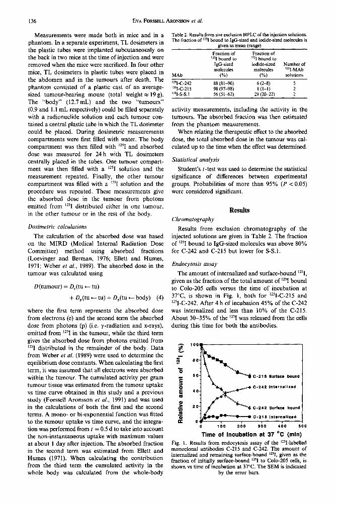
Endocytosis assay
The amount of internalized and surface-bound “‘1, given as the fraction of the total amount of 125I bound to Colo-205 cells versus the time of incubation at 37°C is shown in Fig. I, both for ‘25I-C-215 and ‘25I-C-242. After 4 h of incubation 45% of the C-242 was internalized and less than 10% of the C-215. About 30-35% of the lz51 was released from the cells during this time for both the antibodies.
-I 0 100 200 300 400 500
Time of incubation at 37 “C (mln)
Fig. 1. Results from endocytosis assay of the i2~I-labelled monoclonal antibodies C-215 and C-242. The amount of internalized and remaining surface-bound ‘*% given as the fraction of initially surface-bound lz51 to Colo-205 cells, is shown vs time of incubation at 37°C. The SEM is indicated
by the error bars.

Therapy with ‘251-labelled MAb in nude mice 137
Tumour volume measurements
The correlation coefficient between measured tumour volume and actual tumour weight was high, R = 0.93 (data not shown). The relation between the tumour volume (unit: mm3) and the tumour weight (unit: mg) for tumours larger than 150 mm3 was:
weight = 0.83 x volume.
Growth of untreated turnours
(5)
The tumour growth in untreated mice could be expressed:
volume = 50 x e” I” (6)
when fitting an exponential function to the measured tumour volume. The unit of the volume is mm3 and t is the number of days after tumour cell implantation.
The fitting of an exponential function to the estimated relative tumour volume for tumours larger than 150 mm3 1 O-16 days after implantation in untreated mice resulted in:
R V = 1 .OO x e”.‘25’ (7)
where t is the number of days after the initialization of tumour volume measurements on the established tumours (IO-16 days after tumour cell implantation).
Therapeutic effects on non-established tumours
To determine whether unlabelled antibodies had any cytotoxic effect on non-established tumours, the ratio between tumour volume for animals given MAb ( VhlAb) and tumour volume for untreated mice
( VLntrenWd) was investigated as a function of time after implantation (Fig. 2). The tumour volumes were statistically significantly smaller after treatment with 60 ~1 g C-2 15 than the untreated tumours. No statistic- ally significant difference was obtained between tumours treated with 90 pg C-242 and untreated ones.
To compare the tumour growth after treatment with ‘251-labelled MAb the ratio between tumour volume after injection of “‘1-MAb, V,25,, and tumour
0.5
i
,..,._+A~+~ c-215 (60 Ml)
0.0 I I I I I I I a
0 5 10 15 20 25 30 35 40
Time after implantation (d)
Fig. 2. Therapeutic effect of unlabelled MAb on non- established tumours. The ratio between tumour volume in mice injected with unlabelled MAb, V,,,Ab, and the tumour volume in untreated mice, VUntlrald, is shown vs time after tumour cell implantation. The injected mice were given 60 pg C-215 or 90 pg C-242 2 days after tumour cell
implantation. The SEM is indicated by the error bars.
volume after injection of unlabelled MAb, VMAb,
is shown as a function of time after tumour cell implantation (Fig. 3). Beyond about 10 days after implantation, statistically significant therapeutic effects were observed after injections of 54 MBq ‘*‘I-C-242, 23 MBq or 46 MBq ‘25I-C-2l5.
Therapeutic effects on established tumours
To determine whether unlabelled antibodies had any cytotoxic effect on established tumours, the ratio between relative tumour volume for animals given
MAb, R I’MA,,, and relative tumour volume for un- treated mice, R VUntreated, was studied as a function of time after injection (Fig. 4). There was no statistically significant difference, IO days after injection, between the untreated tumours and those treated with 6Opg unlabelled C-21 5 or 90 pg or 150 /lg unlabelled C-242.
To compare the tumour growth after treatment with ‘251-labelled MAb the ratio between the relative volume after injection of ‘251-MAb, RVlz5,, and the relative tumour volume in untreated mice, R VUntreated,
was investigated as a function of time after injection (Fig 5). IO-12 days after the injection of 47MBq,
(a) 1.6,
1.4 1
1.0
0.8
0.6
0.4
“’ l-C-242
o.oLI 0 5 10 15 20 25 30
Time after implantation (d)
(b) 1.6,
1.4- 1*6 I-C-21 5
1.2-
2 10
>= .: \ 0.8-
z 0.6-
>' 0.4-
0.2-
0.0? ! I I I I 0 5 10 15 20 25 30
Time after implantation (d)
Fig. 3. Therapeutic effect of ‘251-labelled MAb on non- established tumours. The ratio between tumour volumes in mice injected with different amounts of activity of (a) ‘25I-C-242 and (b) ‘25I-C-215, V,,,,, and tumour volume in mice injected with unlabelled (a) C-242 and (b) C-21 5, V M.&S is shown vs time after tumour cell implantation. The mice were injected 2 days after tumour cell implantation.
The SEM is indicated by the error bars.
138 EVA FORSELL AROWQN et al.
0.0 1 0 I I I I I I
0 2 4 6 6 10 12 14 16
Time after injection (d)
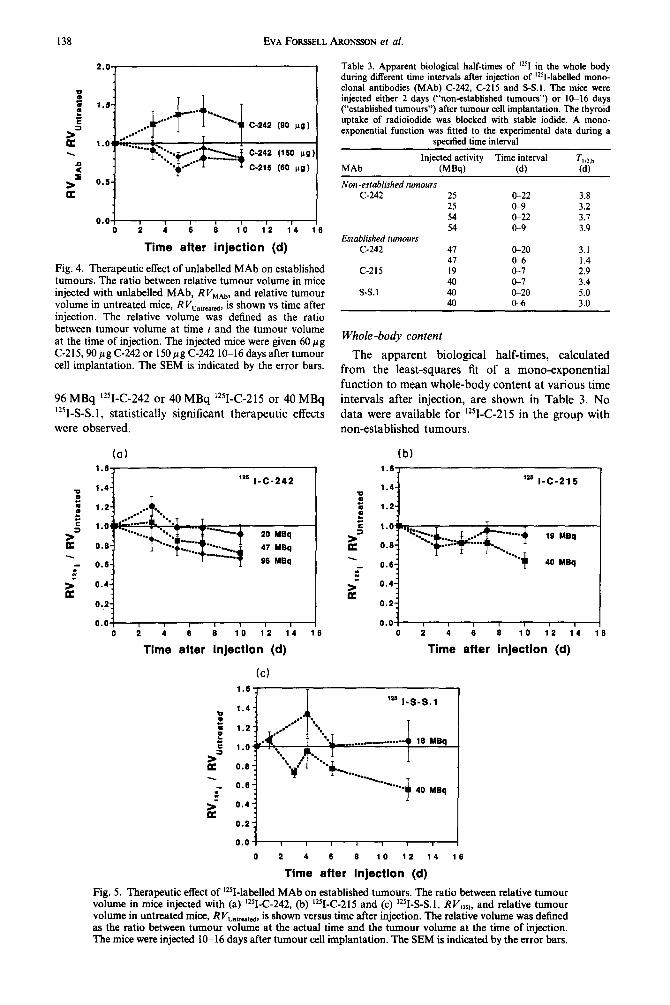
Fig. 4. Therapeutic effect of unlabelled MAb on established tumours. The ratio between relative tumour volume in mice . injected wtth unlabelled MAb, RI’s,‘,,,,, and relative tumour volume in untreated mice, RVuIUntreatcd, is shown vs time after injection. The relative volume was defined as the ratio between tumour volume at time t and the tumour volume at the time of injection. The injected mice were given 60 pg C-215,90 pg C-242 or 150 pg C-242 lo-16 days after tumour cell implantation. The SEM is indicated by the error bars.
96 MBq %C-242 or 40 MBq ‘*‘I-C-215 or 40 MBq
125I-S-S. 1, statistically significant therapeutic effects were observed.
(a)
‘*s 1-C-242
-...__ 20 MBq ----..._
#
47 MBq --.-......
96 MBq
0.0 I I I I 1 I I 0 2 4 6 6 10 12 14 16
Time after injection (d)
Table 3. Apparent biological half-times of ‘25I in the whole body during different time intervals after injection of ‘2’1-labelled mono- clonal antibodies (MAb) C-242, C-215 and S-S.1. The mice were injected either 2 days (“non-established tumours”) or IO-16 days (“established turnouts”) after tumour cell implantation. The thyroid uptake of radioiodide was blocked with stable iodide. A mono- exponential function was fitted to the experimental data during a
specified time interval
Injected activity Time interval T li2.b MAb (MW (d) (d)
Non -established turnours C-242 25 CL22 3.8
2.5 I%9 3.2 54 &22 3.1 54 &9 3.9
Established tumours C-242 41 l&20 3.1
47 &6 1.4 C-215 19 o-7 2.9
40 Is7 3.4 S-S.1 40 ck20 5.0
40 o-6 3.0
Whole-body content
The apparent biological half-times, calculated
from the least-squares fit of a mono-exponential
function to mean whole-body content at various time intervals after injection, are shown in Table 3. No
data were available for ‘2sI-C-215 in the group with non-established tumours.
lb) I.”
l.4- ‘*s I-C-21 5
1.2-
19 YBq
0.6: 40 MBq
0.4:
0.2:
0.0 I I I I I I I 0 2 4 6 0 10 12 14 16
Time after injection (d)
(cl 1.6, I
o.oT 0 2 4 6 0 10 12 14 16
Tlme after injection (d)
Fig. 5. Therapeutic effect of 12sI-labelled MAb on established tumours. The ratio between relative tumour volume in mice injected with (a) ‘*‘I-C-242, (b) 125I-C-215 and (c) ‘*WL% 1, RI’,,,,, and relative tumour volume in untreated mice, RVuntrrstcd, is shown versus time after injection. The relative volume was defined as the ratio between tumour volume at the actual time and the tumour volume at the time of injection. The mice were injected lo-16 days after tumour cell implantation. The SEM is indicated by the error bars.
Tab
le
4. C
onte
nt
of
12’1
in
bloo
d (%
/g)
and
tissu
e/bl
ood
conc
entr
atio
n ra
tios,
gi
ven
as m
ean
(SE
M),
at
var
ious
tim
es
afte
r in
ject
ion
of ‘
zSI-
labc
lled
mon
oclo
nal
antib
odie
s C
-242
, C
-215
(b
oth
spec
ific
) an
d S-
S. I
(uns
peci
fic)
. T
he
mic
e w
ere
inje
cted
ei
ther
2
days
(“
non-
esta
blis
hed
turn
ours
”)
or
lOPi
da
ys
(“es
tabl
ishe
d tu
rnou
rs”)
af
ter
turn
our
cell
impl
anta
tion.
T
he
num
ber
of m
ice,
n,
and
tu
rnou
rs,
T,
are
give
n fo
r ea
ch
grou
p
MA
b
Inje
cted
ac
tivity
(MB
@
Non
-es
tabl
ishe
d tu
mou
rs
C-2
42
25
54
c-21
5
23
46
Esta
blish
ed
twno
un
C-24
2 20
38
38
38
40
54
43
97
C-21
5 19
40
s-s.
I 18
40
Tim
e
(d)
27
28
28
28
10 1 3 6 10
10
12
10
IO
IO
12
12
Blo
od
(W
0.04
8 (0
.005
) 0.
055
(0.0
05)
0.14
(0
.04)
0.
071
(0.0
57)
0.40
7 (0
.009
)
11 (1)
1.9
(0
.2)
0.78
(0
.03)
0.
32
(0.0
2)
0.41
2 (0
.007
) 0.
50
(0.19
) 0.
23
(0.0
1) 0.
90
(0.31
) 1.8
(0
.3)
1.1
(0.1)
I .4
3 (0
.06)
Kid
ney/
bloo
d L
iver
/blo
od
Lun
g/bl
ood
Mus
cle/
bloo
d Sp
leen
/blo
od
Tum
our/
bloo
d nl
T
0.65
(0.
03)
0.47
(0.
03)
0.59
(0.
03)
0.91
(0.
44)
0.38
(0.
01)
0.27
(0.
01)
0.24
(0.
02)
0.24
(0.
01)
0.45
(0.
02)
0.31
(0.
03)
0.3;
:0,,
0.38
(0.
03)
0.33
(0.
01)
ND
N
D
0.54
(0
.02)
0.
46
(0.0
2)
0.44
(0
.03)
0.
71
(0.3
7)
0.34
(0
.01)
0.29
(0
.01)
0.29
(0
.01)
0.
26
(0.0
2)
0.37
(0
.01)
0.30
3 (0
.004
) N
D
0.29
(0.
01)
0.45
(0.
04)
0.34
(0.
01)
ND
N
D
I .24
(0.0
6)
I .28
(0.0
9)
I .34
(0.
08)
0.95
(0.
03)
0.44
(0.
05)
1.14
(0.
05)
0.96
(0.
06)
0.43
(0.
02)
I .9 (
0.2)
I .3
(0.
6)
0.53
(0.
20)
3.6
(2.3
)
0.66
(0.
04)
0.28
(0.
02)
0.36
4 (0
.004
) 0.
314
(0.0
05)
0.11
(0.
01)
0.23
(0.
02)
0.36
(0.
02)
0.11
2 (0
.005
) 0.
25 (
0.02
) 0.
46 (
0.02
) 0.
148
(0.0
06)
0.32
(0.
02)
0.70
(0.
04)
0.30
(0.
01)
0.38
(0.
02)
0.52
6 (0
.005
) 0.
204
(0.0
09)
0.38
(0.
01)
0.5:
(:02
) 0.
23?(
:006
) 0.
5:(:
02)
0.97
(0.
10)
0.44
(0.
08)
1.1
(0.2
) 0.
80 (
0.04
) 0.
37 (
0.02
) I .0
2 (0
.08)
N
D
ND
N
D
ND
N
D
ND
0.67
(0.
02)
0.62
(0.
04)
0.94
(0.
03)
1.55
(0.
04)
0.65
(0.
03)
0.47
(0.
02)
1.32
(0.
04)
I. 19
(0.
03)
I .08
(0.0
6)
1.06
(0.
06)
0.90
(0.
05)
0.93
(0.
03)
I .4 (
0.2)
0.
96 (
0.04
) 0.
332
(0.0
09)
0.25
7 fO
.005
)
6110
S/
IO
6/11
214
315
316
316
316
6/l
I 31
6 61
11
5/10
61
12
6112
S/
IO
6/12

140 EVA FORSSELL A~otsso~ et al.
Concentration of lz51 in tissue Dosimetry and therapeutic eflects of radiation
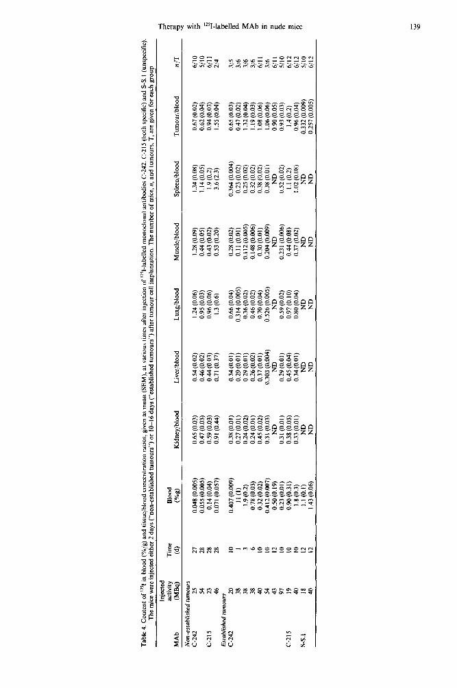
The blood content and tissue/blood concentration ratios at various times after injection are given in Table 4. The tumour/blood concentration ratios are high for both C-215 and C-242, indicating specific binding to the tumour cells, while the ratios for the unspecific S-S.1 are low, suggesting that the anti- bodies are only distributed in the extracellular fluid in the tumour. The (normal tissue)/blood concentration ratios are low, except for some values for lung, muscle and spleen late after injection.
The results of the dosimetric calculations are pre- sented in Table 5, both as the total absorbed dose in the tumour and as the different terms in equation (4). More than 60% of the absorbed dose in the tumour results from the electrons from “‘1 within the tumour. About 20-30% is from “‘1 distributed outside the tumour.
Dosimetric measurements
From the TLD measurement in the phantom, the absorbed dose (from photons) in tumour per unit cumulated activity was found to be 2.6 mGy/MBqh from ‘*‘I homogeneously distributed within the tumour (x 1 g) and 0.18 mGy/MBqh from ‘*‘I homo- geneously distributed in the rest of the body. From the TLD measurements in dead mice the correspond- ing values were found to be 1.4 mGy/MBqh for tumours weighing about 0.4 g, 0.78 mGy/MBqh for a tumour weighing about 0.2 g and 0.17 mGy/MBqh from i25I distributed in the rest of the body. The in vitro TLD measurements in mice resulted in values of 0.25 mGy/MBqh from “‘1 distributed in the whole body to dosimeters subcutaneously situated on the back.
The relation between absorbed dose in the tumour and therapeutic effect is illustrated in Fig. 6. Figure 6(a) shows the results for non-established tumours 9-10 days after injection. The ratio between tumour volume after the injection of “‘I-C-215 or “‘I-C-242 and the tumour volume after injection of unlabelled monoclonal antibodies is given versus the estimated absorbed dose in the tumour. Figure 6(b) shows the results for established tumours lo-12 days after injection. The ratio between the relative tumour volume after injection of “‘I-C-215, ‘251-C-242 or ‘251-S-S.l and the relative tumour volume for the untreated mice is given versus the estimated absorbed dose in the tumour.
From Fig. 6 it can be seen that for absorbed doses in tumours above 2 Gy, ‘*‘I-MAb exhibited statistically significant therapeutic effects both for non-established and established tumours and for all antibodies studied. There was no enhanced therapeutic effect of lz51-C-242 compared with ‘25I-C-2l 5 and ‘25I-S-S. 1 when relating
Table 5. Estimated absorbed dose to tumour at various times after injection of ‘2SI-labelled mono- clonal antibodies (MAb). D,(tu + tu) is the absorbed dose from electrons and D,(tu + tu) is the absorbed dose from photons emitted from the tumour from photons emitted from
12’1 in the tumour. D,(tu + body) is the absorbed dose to 12SI distributed in the remainder of the body. &, is the sum of these absorbed doses
MAb
Injected Time after activity injection D&u c tu) D,(tu t tu) D,(tu + body) (MW (d) (GY) (GY) (GY)
Non-esfablished turnours C-242 25
54
C-215 23
46
Established turnours C-242 20
41
91
C-215 19
40
s-s. 1 18
40
9 20
6 9
20 _I
IO
20 I
IO 20
IO
10
IO
IO
I IO
6 12
6 12
0.90 0.99 I .I9 1.94 2.13 1.24 1.51 I .93 2.51 3.08 3.93
0.04 0.04 0.08 0.08 0.09 0.05 0.07 0.08 0.11 0.13 0.17
0.42 I .36 0.49 1.53 0.76 2.62 0.90 2.93 1.06 3.29 0.42 1.71 0.48 2.05 0.53 2.54 0.86 3.48 0.97 4.18 I .06 5.16
0.65 0.04 0.30 1.00 0.71 0.04 0.34 I .09 1.54 0.09 0.71 2.34 I .68 0.09 0.80 2.51 3.18 0.17 1.45 4.80 3.44 0.19 I .66 5.29 I .Ol 0.07 0.34 I .42 I .23 0.08 0.39 I .70 2.15 0.14 0.73 3.03 2.64 0.17 0.83 3.64 0.56 0.04 0.24 0.84 0.74 0.05 0.34 1.13 I .24 0.08 0.54 1.85 1.47 0.10 0.67 2.24

Therapy with ‘2sI-labelled MAb in nude mice 141
(a)
9-10 d
:: Z--
0.6
- i 0.4 c-2, 6
0.2 -3
0.0: I I 4 I # I 0 1 2 3 4 6 6 7
Absorbed dose in tumour (Gy)
(b) 1.6-
1.4- 10-12 d
1.2-
1.0.
3 0.8-
0.6:
0.4;
0.2 1
0.0: I t I I I I
0 1 2 3 4 5 6 7
Absorbed dose in tumour (Gy)
Fig. 6. Therapeutic effect of ‘2SI-labelled MAb versus ab- sorbed dose in tumour for (a) non-established tumours and (b) established tumours. The therapeutic effect is expressed as (a) the ratio between tumour volume in mice injected with %MAb and mice injected with unlabeiled MAb 9-10 days after injection and (b) the ratio between relative tumour volume in mice injected with ‘*?-MAb and untreated mice
IO--t2 days after injection.
the therapeutic effect to the absorbed dose in the tumour.
Discussion
The purpose of this study was to investigate whether the effect of Auger electrons could be utilized for therapy with radiolabelled monoclonal antibodies. The antibodies were labelled with “‘1 using a conven- tional labelling procedure (Iodogen). In general, this method works quite well, but in this study problems arose with one of the antibodies. The results from HPLC of the injected solutions show that the amount of lz51 bound to IgG-sized molecules was much less for S-S.1 than for C-215 and C-242 (Table 2). This might be explained by S-S.1 being more sensitive to the iodine labelling procedure and thus being damaged to a higher degree than C-215 and C-242. Since S-S.1 was used as an unspecific antibody and no binding to the tumour was expected, this possible damage would probably only lead to a shorter biological half-time in the mice. Fortunately this
presented no problems in this study, since the thera- peutic effects are related to the absorbed dose, which
was estimated from the actual biokinetic data. The
antibodies C-2 15 and C-245 seemed to be intact after the radiolabelling, and the tumour/blood concen- tration ratios were high enough to indicate specific uptake in the tumour. The ratios were similar to those obtained in earlier experiments (Forssell Aronsson et al., 1991; Gretarsdottir et al., 1992).
In order to obtain an effect from Auger electrons,
it is necessary that the antibodies be internalized into the tumour cells. ‘25 I- C-21 5 was internalized to a very low degree; less than 10% of the initially surface- bound ‘251-C-215. For ‘2sI-C-242, 45% of the initially surface-bound “‘I-C-242 was internalized after 4 h.
In relation to the amount of surface-bound ‘2sI-C-242 at a certain time, the internalization rate of ‘25I-C-242
was fairly constant (1 %/min). This would indicate that all surface-bound C-242 is bound in such a way that it should be internalized.
The therapeutic effect was studied through measure-
ments of tumour growth. For established tumours it was important to be able to relate results to the
tumour volume at the onset of the therapy. It was also important to choose tumours with a similar growth rate. For two reasons, only tumours with
volumes larger than 150 mm3 were considered. Firstly, the relative uncertainty in volume measure- ment is larger for very small tumours and secondly. the relative tumour growth was faster for the smallest
untreated turnours. Other studies have shown that unlabelled IgG
antibodies inhibit the growth of human tumours in nude mice, while antibodies of other subclasses were less effective (Kirch and Hammerling, 1981; Young
and Hakomori, 1981; Herlyn and Koprowski, 1982; Larson er al., 1988). In particular, antibodies of the
isotype IgG2a suppress growth of murine tumours (Herlyn and Koprowski, 1982). Larson et al. (1988) and Johansson et al. (1991) have previously shown that C-2 15 promotes the lysis of Colo-205 cells in vitro and of non-established Colo-205 tumours in nude mice, but for established Colo-205 tumours C-2 15 did not exhibit any significant tumour growth suppression.
The difference in cytotoxicity between unlabelled
C-215 and C-242 in this study is illustrated in Fig. 2 for non-established tumours and in Fig. 4 for estab- lished tumours. C-2 15 exhibited statistically significant cytotoxicity of the non-established tumours while C-242 showed no effect, There was no statistically
significant therapeutic effect on established tumours 10 days after the administration of 60 ng unlabelled C-21 5 or of either of the two doses of unlabelled C-242. As a consequence of these results, the mice treated with unlabelled C-2 15 or C-242 were used as controls for the experiments on the effects of “‘I- MAb on non-established turnours. For the investi- gations on the therapeutic effects of ‘*‘I-MAb on established tumours, the untreated mice were chosen as controls to obtain a larger control group.

142 EVA FOIWELL ARONWN et al.
The tumour growth was related to the absorbed dose in the turnout-. It is difficult to estimate the uncertainty in the calculated absorbed dose in the tumour. However, the precision of the dose calcula- tion does not affect the estimation of any enhanced therapeutic effect from the Auger electrons, since this was made by comparisons. An error affects the total absorbed dose in tumour to roughly the same degree for all monoclonal antibodies.
The absorbed dose in the tumour was calculated from the amount of injected “‘1 and the measured biokinetics in this study and in a previous study (Forssell Aronsson et al., 1991) together with results from the TLD measurements. The determining factor in the estimation of the absorbed dose is the cumulated activity in the body and, above all, in the tumour. The calculations for the mice showed that about 75% of the total absorbed dose is due to ‘25I in the tumour. The precision is thus mainly dependent on the estimation of the cumulated activity in the tumour. The cumulated activity was based on a mean uptake curve from, in general, lo-15 mice at each regis- tration time. Out of an observed CV of 40% for the tumour uptake, the uncertainty in the mean cumu- lated activity in the tumour for an experimental group of mice in this study can be estimated to about 20%.
The estimation of cumulated activity in non- established tumours may be more uncertain, especially early after injection. In performing the calculations it was assumed that the concentration of radionuclide was the same as in established tumours. This may lead to an uncertainty in the calculation of the absorbed dose in non-established tumours. This error would probably be similar for both antibodies.
The TLD measurements were necessary to deter- mine the absorbed dose in the tumour due to photons, per unit cumulated activity from lz51 distributed in the rest of the body. The results obtained in the dead mice and in the phantom were similar. For live mice the value was higher for the subcutaneously placed dosimeters, probably because they were located closer to the tumours. The contribution from the penetrating radiation from the body was overestimated by 5-7% since the activity within the tumours was included, and underestimated by 2-3% since the TL dosimeters were placed in a plastic tube. However, the net overestimation was only a few per cent of the third term in equation (4) and thus very small compared with the total absorbed dose in the tumour. The assumption that ‘25I was homogeneously distributed in the body was supported by the measured biokinetic data given in Table 4.
The precision in the calculation of absorbed dose in the tumour, due to photons, per unit cumulated activity from lz51 distributed in the turnout-, is of less importance because of its very small contribution to the total absorbed dose in the tumour. The measured value was lower in mice than in the phantom. The tumours in these mice were smaller (0.2-0.5 g) than
in the phantom (Z 1.0 g) and for that reason the dosimeters were not totally covered with tissue.
Since it is not known whether there is a time delay between radiation damage and effects on tumour growth for this tumour, the absorbed dose was calculated up to the time when the therapeutic effect was determined. By studying the therapeutic effect at a time not too early after injection, the influence of this factor ought to be minimized.
The absorbed doses obtained in tumours were low compared with the doses required for complete tumour regression. The tumour growth rate was only reduced. This dose level was chosen consciously to increase the possibility of observing any enhanced therapeutic effect from Auger electrons.
Provided that there were no synergistic effects between radiation damage and other effects from the monoclonal antibody, and assuming that the cyto- toxicity of iodine-labelled antibody and unlabelled was the same, the therapeutic effect on the tumour from the radiation of “‘1 only, is given in Fig. 6. For established tumours, neither ‘25I-C-242 nor ‘25I-C-21 5 gave a greater therapeutic effect than ‘251-S-S.l. For the non-established tumours there was no difference between ‘*‘I-C-242 and ‘25I-C-215. The results pre- sented in Fig. 6 thus do not show any signs of an enhanced therapeutic effect of Auger electrons from “‘1 when conjugated to the internalized antibody C-242. Assuming that internalization occurs to the same extent in vivo as in vitro, this indicates that ‘251 does not come as close as necessary to the DNA in the cell nucleus or does not accumulate in the cell nucleus. A further step towards the use of “‘1 for tumour therapy would be to find a method of transporting internalized ‘*‘I to the DNA and see that the ‘25I remains there for a sufficiently long time.
Acknowledgements-This work was supported by grants from the Swedish Medical Research Council (B92-17x- 07184-08A) and the Kine Gustaf V Jubilee Clinic Cancer Research Foundation in?Xteborg, Sweden.
References
Baum R. P., Lorenz M., Senekowitsch R., Chatal J. F., Saccavini J. C. et al. (1988) Clinical exoerience in cancer diagnosis with radiolabeledmonoclonai antibodies in 200 patients and initial attempts at radioimmunotherapy. In Radiolabeled Monoclonal Antibodies for Imaging and Therapy (Edited by Srivastava S. C.), pp. 613-651. Plenum Press, New York.
Bergh J., Nilsson S., Liljedahl. C., Sivolapenko G., Maripuu E.. Stavrou D. and Eoenetos A. A. (1990) In uivo imaging and treatment of human brain tumours u&zing the radio- labelled monoclonal antibody MUC 2-63. Anticancer Res. 10, 655-660.
Boven E., Lindmo T., Mitchell J. B. and Bunn P. A. Jr (1986) Selective cytotoxicity of ‘2sI-labeled monoclonal antibody TlOl in human malignant T cell lines. Bfood 67, 429-435.
Burki H. J., Roots R., Feinendegen L. E. and Bond V. P. (1973) Inactivation of mammalian cells after disintegra- tions of 3H or lzsI in cell DNA at - 196°C. Int. J. Radiat. Biol. 24, 363-315.
Therapy with ‘251-labelled MAb in nude mice 143
Carlsson G., Gullberg B. and Hafstrom L. (1983) Estima- tion of liver tumor volume using different formulas-an experimental study in rats. Cancer Res. Clin. Oncol. 105, 20-23.
Chan P. C., Lisco E., Lisco H. and Adelstein S. J. (1976) The radiotoxicity of iodine-125 in mammalian cells. II. A comparative study on cell survival and cytogenetic responses to ‘*%JdR, 13’IUdR, and ‘HTdR. Radiat. Res. 67; 3322343.
Cheung N.-K. V., Landmeier B., Neely J., Nelson A. D., Abramowskv C.. Ellerv S.. Adams RB. and Miraldi F. (I 986) Complete’ tumor ablation with iodine 13 1 -radio- labeled disialoganglioside G,-specific monoclonal anti- body against human neuroblastoma xenografted in nude mice. J. Natl. Cancer Inst. 71, 7399745.
Commerford S. L., Bond V., Cronkite E. and Reincke U. (1980) Radiotoxicity of intranuclear ‘251 atoms not bound to DNA. Int. J. Radiat. Biol. 37, 547-554.
Ellett W. H. and Humes R. M. (1971) Absorbed fractions for small volumes containing photon-emitting radio- activity. In Medical Internal Radiation Dose (MIRD), Pamphlet 8. Society of Nuclear Medicine, New York.
Eoenetos A. A.. Munro A. J.. Stewart S et al. (1987) ‘Antibody-guided irradiation of advanced ovarian cancer with intraperitoneally administered radiolabeled mono- clonal antibodies. J. Clin. Oncol. 5, 1890-1899.
Forssell Aronsson E., Grttarsdottir J., Jacobsson L., Mattsson S., Holmberg S. B., Hafstriim L.-O., Karlsson B. and Lindholm L. (1991) Comparison of seven iodine-labelled monoclonal antibodies in nude mice with human colon carcinoma xenografts. Acfa Oncol. 30, 3855393.
Forssell Aronsson E., Gritarsdbttir J., Jacobsson L., Mattsson S., Holmberg S., Hafstriim L.-O., Karlsson B. and Lindholm L. (1992) Pharmacokinetics and dosimetry of the radioiodine-labelled monoclonal antibodies C-215 and C-245 studied in two patients with adenocarcinoma. In Proceedings of the Fifrh International Radiopharmaceu- tical Dosimetry Symposium Oak Ridge Associated Uni- versities, Oak Ridge, Tenn., U.S.A. (Edited by Schlafke-Stelson A. T. and Watson E. E.) CONF-910529.
Fowler J. F. and Attix F. H. (1966) Solid state integrating dosimeters. In Radiafion Dosimetry (Edited by Attix F. H. and Roesch W. C.), 2nd Edn, Vol. II, pp. 269-281. Academic Press, New York.
Fraker P. J. and Soeck J. C. Jr (1978) Protein and cell membrane iodinations with a sparingly soluble chloramide 1,3,4,6-tetrachloro-3a, 6a-diphenylglycoluril. Biochem. Biophys. Res. Commun. 80, 8499857.
Gretarsdottir J., Forssell Aronsson E., Jacobsson L., Hafsteinsdottir 0.. Holmberg S. B., Hafstriim L., Lindegren S.. Karlsson B., Lindholm L. and Mattsson S. (1992) Comoarison of the biodistribution of “Se or ‘j’1 labellkd moioclonal antibodies in nude mice. Nucl. Med. Biol. 19, 719-726.
Herlyn D. and Koprowski H. (1982) IgG2a monoclonal antibodies inhibit human tumor growth through inter- action with effector cells. Proc. NatI Acad. Sci. U.S.A. 79, 4X-4765.
Hofer K. G., Harris C. R. and Smith J. M. (1975) Radio- toxicity of intracellular 67Ga, ‘? and )H. Nuclear versus cytoplasmic radiation effects in murine L1210 leukaemia. ht. J. Radiat. Biol. 28, 225-241.
Johansson C., Segrtn S. and Lindholm L. (1991) Tumour- growth suppression in nude mice by a murine monoclonal antibody: factors hampering successful therapy. Inf. J. Cancer 48, 297-304.
Jones D. H., Goldman A., Gordon I., Pritchard J., Gregory B. J. and Kemshead J. T. (1985) Therapeutic application of a radiolabelled monoclonal antibody in nude mice xenografted with human neuroblastoma: tumoricidal effects and distribution studies. Im. J. Cancer 35, 715-720.
Kassis A. I., Adelstein S. J., Haydock C. and Sastry K. S. R. (1983) Thallium-201: an experimental and a theoretical radiobiological approach to dosimetry. J. Nucl. Med. 24, 1164-l 175.
Kirch M. E. and Hammerling U. (1981) Immunotherapy of murine leukemias by monoclonal antibody. I. Effect of passively administered antibody on growth of transplanted tumor cells. J. Immunol. 127, 805-810.
Larson L. N., Johansson C., Lindholm L. and Holmgren J. (1988) Mouse monoclonal antibodies for experimental immunotherapy promotes killing of tumor cells. Int. J. Cancer 42, 8777882.
Larson S. M. (1985) Radiolabeled monoclonal anti-tumor antibodies in diagnosis and therapy. J. Nucl. Med. 26, 5388545.
Lindholm L., Holmgren J., Svennerholm L., Fredman P., Nilsson O., Persson B., Myrvold H. and Lagerglrd T. (1983) Monoclonal antibodies against gastrointestinal tumor-associated antigens isolated as monosialoganglio- sides. Int. Arch. Allergy Appl. Immunol. 71, 178-181.
Lindholm L., Johansson C., Jansson E.-L., Hallberg C. and Nilsson 0. (1985) An immunoradiometric assay (IRMA) for the CA-50 antigen. In Tumor Marker Antigens (Edited by Holmgren J.), pp. 1233133. Studentlitteratur. Lund, Sweden.
Lindmo T., Boven E., Mitchell J. B., Morstyn G. and Bunn P. A. Jr (1985) Specific killing of human melanoma cells by ‘2iI-labeled 9.2.27 monoclonal antibody. Cancer Res. 45, 5080&5087.
Loevinger R. and Berman M. (1976) A revised schema for calculating the absorbed dose from biologically distributed radionuclides. Medical Internal Radiation Dose (MIRD), Pamphlet I. Society of Nuclear Medicine. New York.
Mansson J.-E., Fredman P., Nilsson O., Lindholm L., Holmgren J. and Svennerholm, L. (1985) Chemical structure of carcinoma ganglioside antigens defined by monoclonal antibody C-50 and some allied gangliosides of human pancreatic adenocarcinoma. Biochim. Biophys. Acta 834, 110-l 17.
Nilsson 0.. Mansson J.-E., Lindholm L., Holmgren J. and Svennerholm L. (1985) Sialosyllactotetraosylceramide, a novel ganglioside antigen detected in human carcinomas bv a monocional antibodv. FEBS L&t. 182. 398-402.
Order S. E. (1990) Presidential address: systemic radio- therapy-the new frontier. Int. J. Radial. Oncol. Biol. Phys. 18, 981-992.
Redwood W. R., Tom T. D. and Strand M. (1984) Specificity, efficacy and toxicity of radioimmunotherapy in erythroleukemic mice. Cancer Res. 44, 5681-5687.
Senekowitsch R., Reidel G., MBllenstPdt S., Kriegel H. and Pabst H.-W. (1989) Curative radioimmunotherapy of human mammary carcinoma xenografts with iodine- 131-labeled monoclonal antibodies, J. Nucl. Med. 30, 53 1-537.
Stewart J. S. W., Hird V., Snook D., Sullivan M., Hooker G., Courtenay-Luck N., Sivolapenko G., Griffiths M., Myers M. J., Lambert H. E., Munro A. J. and Epenetos A. A. (1989) Intraperitoneal radioimmunotherapy for ovarian cancer: Pharmacokinetics, toxicity, and efficacy of I-131 labeled monoclonal antibodies. Int. J. Radial. Oncol. Biol. Phys. 16, 405-413.
Sugiyama Y., Chen F.-A., Takita H. and Bankert R. B. (1988) Selective growth inhibition of human lung cancer cell lines bearing a surface glycoprotein gp160 by 12’1- labeled anti-gp160 monoclonal antibody. Cancer Res. 48, 2768-2773.
Sundell-Bergman S., Bergman R. and Johanson K. J. (1985) Chromosome damage by decay of 3H and ‘251 incorpor- ated into DNA of Chinese hamster cells. Mufat. Res. 149, 2577263.
Vriesendorp H. M., Herpst J. M., Leichner P. K., Klein J. L. and Order S. E. (1989) Polyclonal WYttrium labeled

144 EVA FORETELL ARONSSON et al.
antiferritin for refractory Hodgkin’s disease. ht. J. “‘I-labelled monoclonal antibody. Br. J. Cancer 58, Radiat. Oncol. Biol. Phys. 17, 8155821. 292-295.
Weber D. A., Eckerman K. F., Dillman L. T. and Ryman J. C. (1989) MIRD: Radionuclide Data and Decay Schemes. The Society of Nuclear Medicine, New York.
Young W. W. Jr and Hakomori S.-I. (1981) Therapy of mouse lymphoma with monoclonal antibodies and glyco- lipid: selection of low antigen variants in uiuo. Science 211, 487-489.
Woo D. V., Li D., Mattis J. A. and Steplewski Z. (1989) Selective chromosomal damage and cytotoxicity of 1251- labeled monoclonal antibody 17-la in human cancer cells. Cancer Rex 49, 2952-2958.
Yoneda S., Fujisawa M., Watanabe J., Okabe T., Takaku F., Homma T. and Yoshida K. (1988) Radioimmuno- therapy of transplanted small cell lung cancer with
Younis A.-R. S. and Watt D. E. (1989) The quality of ionising radiations emitted by radionuclides incorporated into mammalian cells. Phys. Med. Biol. 34, 821-834.
Zalcberg J. R., Thompson C. H., Lichtenstein M. and McKenzie I. F. C. (1984) Tumor immunotherapy in the mouse with the use of I-131-labeled monoclonal antibodies. J. Nat1 Cancer Inst. 72, 679-704.
![Noninvasive Molecular Imaging of MYC mRNA Expression in Human Breast Cancer Xenografts with a [ 99m Tc]Peptide−Peptide Nucleic Acid−Peptide Chimera](https://static.fdokumen.com/doc/165x107/63214cddbc33ec48b20e4a4a/noninvasive-molecular-imaging-of-myc-mrna-expression-in-human-breast-cancer-xenografts.jpg)