99m Tc-besilesomab (Scintimun®) in peripheral osteomyelitis: comparison with 99m Tc-labelled white...
12
ORIGINAL ARTICLE 99m Tc-besilesomab (Scintimun®) in peripheral osteomyelitis: comparison with 99m Tc-labelled white blood cells Wolf S. Richter & Velimir Ivancevic & Johannes Meller & Otto Lang & Dominique Le Guludec & István Szilvazi & Holger Amthauer & Florence Chossat & Amel Dahmane & Carsten Schwenke & Alberto Signore Received: 23 August 2010 / Accepted: 4 January 2011 / Published online: 15 February 2011 # The Author(s) 2011. This article is published with open access at Springerlink.com Abstract Purpose The diagnosis of osteomyelitis is a challenge for diagnostic imaging. Nuclear medicine procedures including white blood cell imaging have been successfully used for the identification of bone infections. This multinational, phase III clinical study in 22 European centres was undertaken to compare anti-granulocyte imaging using the murine IgG antibody besilesomab (Scintimun®) with 99m Tc-labelled white blood cells in patients with peripheral osteomyelitis. Methods A total of 119 patients with suspected osteomy- elitis of the peripheral skeleton received 99m Tc-besilesomab and 99m Tc-hexamethylpropyleneamine oxime (HMPAO)- labelled white blood cells (WBCs) in random order 2–4 days apart. Planar images were acquired at 4 and 24 h after injection. All scintigraphic images were interpreted in an off-site blinded read by three experienced physicians specialized in nuclear medicine, followed by a fourth blinded reader for adjudication. In addition, clinical follow-up information was collected and a final diagnosis was provided by the investigators and an independent truth panel. Safety data including levels of human anti-mouse antibodies (HAMA) and vital signs were recorded. W. S. Richter Pharmtrace klinische Entwicklung GmbH, Alt-Moabit 59-61, 10555 Berlin, Germany W. S. Richter : H. Amthauer Clinics for Radiology and Nuclear Medicine, University Clinics Magdeburg, Leipziger Strasse 44, 39120 Magdeburg, Germany V. Ivancevic Nuclear Medicine Celle, Siemensplatz 4, 29223 Celle, Germany J. Meller Department of Nuclear Medicine, University Medicine (UMG) Göttingen, Robert-Koch-Str. 40, 37075 Göttingen, Germany O. Lang Department of Nuclear Medicine, Charles University, 3rd Medical Faculty, UH Kralovske Vinohrady, Šrobarova 50, 100 34 Prague, Czech Republic D. Le Guludec CHU Bichat-Claude Bernard, Service de Médecine Nucléaire, 46 rue Henri Huchard, 75877 Paris Cedex 18, France I. Szilvazi Department of Nuclear Medicine, Országos Gyógyintézeti Központ, Szabolcs u. 33, 1135 Budapest, Hungary F. Chossat : A. Dahmane IBA/CIS bio international, BP 32, 91192 Gif sur Yvette Cedex, France C. Schwenke SCOSSIS, Zeltinger Strasse 58 G, 13465 Berlin, Germany A. Signore (*) Nuclear Medicine Unit, “Sapienza” University of Rome, 2nd Faculty of Medicine, Ospedale S. Andrea, Via di Grottarossa 1035, 00189 Rome, Italy e-mail: [email protected] Eur J Nucl Med Mol Imaging (2011) 38:899–910 DOI 10.1007/s00259-011-1731-2
-
Upload
independent -
Category
Documents
-
view
1 -
download
0
Transcript of 99m Tc-besilesomab (Scintimun®) in peripheral osteomyelitis: comparison with 99m Tc-labelled white...
ORIGINAL ARTICLE
99mTc-besilesomab (Scintimun®) in peripheral osteomyelitis:comparison with 99mTc-labelled white blood cells
Wolf S. Richter & Velimir Ivancevic & Johannes Meller & Otto Lang &
Dominique Le Guludec & István Szilvazi & Holger Amthauer & Florence Chossat &Amel Dahmane & Carsten Schwenke & Alberto Signore
Received: 23 August 2010 /Accepted: 4 January 2011 /Published online: 15 February 2011# The Author(s) 2011. This article is published with open access at Springerlink.com
AbstractPurpose The diagnosis of osteomyelitis is a challenge fordiagnostic imaging. Nuclear medicine procedures includingwhite blood cell imaging have been successfully used for theidentification of bone infections. This multinational, phase IIIclinical study in 22 European centres was undertaken tocompare anti-granulocyte imaging using the murine IgGantibody besilesomab (Scintimun®) with 99mTc-labelledwhite blood cells in patients with peripheral osteomyelitis.Methods A total of 119 patients with suspected osteomy-elitis of the peripheral skeleton received 99mTc-besilesomab
and 99mTc-hexamethylpropyleneamine oxime (HMPAO)-labelled white blood cells (WBCs) in random order2–4 days apart. Planar images were acquired at 4 and24 h after injection. All scintigraphic images wereinterpreted in an off-site blinded read by three experiencedphysicians specialized in nuclear medicine, followed by afourth blinded reader for adjudication. In addition, clinicalfollow-up information was collected and a final diagnosiswas provided by the investigators and an independent truthpanel. Safety data including levels of human anti-mouseantibodies (HAMA) and vital signs were recorded.
W. S. RichterPharmtrace klinische Entwicklung GmbH,Alt-Moabit 59-61,10555 Berlin, Germany
W. S. Richter :H. AmthauerClinics for Radiology and Nuclear Medicine,University Clinics Magdeburg,Leipziger Strasse 44,39120 Magdeburg, Germany
V. IvancevicNuclear Medicine Celle,Siemensplatz 4,29223 Celle, Germany
J. MellerDepartment of Nuclear Medicine,University Medicine (UMG) Göttingen,Robert-Koch-Str. 40,37075 Göttingen, Germany
O. LangDepartment of Nuclear Medicine, Charles University,3rd Medical Faculty, UH Kralovske Vinohrady,Šrobarova 50,100 34 Prague, Czech Republic
D. Le GuludecCHU Bichat-Claude Bernard, Service de Médecine Nucléaire,46 rue Henri Huchard,75877 Paris Cedex 18, France
I. SzilvaziDepartment of Nuclear Medicine,Országos Gyógyintézeti Központ,Szabolcs u. 33,1135 Budapest, Hungary
F. Chossat :A. DahmaneIBA/CIS bio international,BP 32,91192 Gif sur Yvette Cedex, France
C. SchwenkeSCOSSIS,Zeltinger Strasse 58 G,13465 Berlin, Germany
A. Signore (*)Nuclear Medicine Unit, “Sapienza” University of Rome,2nd Faculty of Medicine,Ospedale S. Andrea, Via di Grottarossa 1035,00189 Rome, Italye-mail: [email protected]
Eur J Nucl Med Mol Imaging (2011) 38:899–910DOI 10.1007/s00259-011-1731-2

Results The agreement in diagnosis across all three readersbetween Scintimun® and 99mTc-HMPAO-labelled WBCswas 0.83 (lower limit of the 95% confidence interval 0.8).Using the final diagnosis of the local investigator as areference, Scintimun® had higher sensitivity than 99mTc-HMPAO-labelled WBCs (74.8 vs 59.0%) at slightly lowerspecificity (71.8 vs 79.5%, respectively). All parametersrelated to patient safety (laboratory data, vital signs) did notprovide evidence of an elevated risk associated with the useof Scintimun® except for two cases of transient hypoten-sion. HAMA were detected in 16 of 116 patients after scan(13.8%).Conclusion Scintimun® imaging is accurate, efficaciousand safe in the diagnosis of peripheral bone infectionsand provides comparable information to 99mTc-HMPAO-labelled WBCs.
Keywords Osteomyelitis . Scintimun® .WBC . Imaging .
Nuclear medicine
Introduction
The diagnosis of osteomyelitis is a challenge for physiciansand diagnostic imaging because of a substantial heteroge-neity in its possible clinical presentations. Diagnosis isusually based on a combination of clinical, laboratory andimaging findings. Whereas clinical and laboratory dataprovide general evidence of an inflammatory process,diagnostic imaging is typically used to detect and documentosteomyelitic changes in specific bones or joints.
There is not one single optimal imaging method forpatients with suspected osteomyelitis. Instead, the mostappropriate imaging modality has to be selected on thebasis of the individual strengths and limitations of eachimaging approach and depends also on patient-specificfactors such as disease history, comorbidity and the locationof the suspected disease process. The imaging modalitiesmost widely used in suspected osteomyelitis include X-ray,conventional bone scintigraphy, CT and MRI. Whileconventional X-ray may show characteristic changes inlater osteomyelitis, early diagnosis is frequently missed,particularly when the inflammatory process is limited to themedullary space of the bone as, e.g. in early haematogenousosteomyelitis [1]. In addition, X-ray diagnosis is consideredto be neither sensitive nor specific in the case of suspectedprosthetic joint infection [2].
CTmay provide useful high-definition images, particularlyfor peripheral osteomyelitis, whereas MRI has emerged as apromising method for its superior soft tissue contrast and thehigh anatomical resolution. Limitations of CT and MRI arerelated to the presence of artefacts in patients after jointreplacement and to difficulties regarding the differentiation
between osteomyelitis and other diseases with similarsymptoms, e.g. the differentiation between diabetic foot andCharcot neuropathic osteoarthropathy [3–5].
Conventional bone scintigraphy almost always identifiesosteomyelitic lesions but is also positive in a wide varietyof non-osteomyelitic conditions and, therefore, suffers froma limited specificity. Imaging of white blood cells (WBCs)with scintigraphic methods is used as an adjunct tool todistinguish between infection and sterile inflammation andother disorders in areas that are abnormal on bone scans.The combination of conventional bone scanning and WBCscintigraphy has proven to be a clinically useful approachin patients with suspected osteomyelitis [6].
Different scintigraphic methods are available for WBCimaging. In vitro labelling of WBCs with 111In or 99mTc isregarded as the gold standard; however, this approach iscumbersome as it requires in vitro separation of WBCs,their radioactive labelling and re-injection of the labelledWBCs into the patient [7–9]. In vitro labelling of WBCs isalso associated with infection risks for personnel andpatients and a high radiation burden to technicians incharge of the labelling [6]. The recent availability of aclosed system device for WBC labelling1 has greatlyreduced these problems, but in vivo labelling of WBCsstill represents an attractive alternative to direct in vitrolabelling, particularly because it reduces preparation time.The first approach regarding in vivo labelling was themurine IgG1k antibody BW 250/183 (99mTc-besilesomab,Scintimun®) which recognizes the nonspecific cross-reacting antigen 95 (NCA-95; also referred to as CD66band CEACAM8) in the cytoplasm and on the cellmembranes of granulocytes and granulocyte precursor cells.Radiolabelled besilesomab has been used since 1992 inSwitzerland, Hungary, the Czech Republic and Sweden onthe basis of local marketing approvals and in Germany (onthe basis of individual prescription). Since 1992, anestimated 100,000 patients have been diagnosed withScintimun® in these countries. In January 2010, Scintimun®was granted a marketing authorization for all Europeancountries by the European Medicines Agency (EMEA) fordetermining the location of infection in peripheral bone inadults with suspected osteomyelitis.
This article reports the results of the recent phase IIIclinical trial comparing Scintimun® with 99mTc-hexame-thylpropyleneamine oxime (HMPAO)-labelled WBCs inperipheral osteomyelitis. The trial was conducted with theprimary aim of evaluating diagnostic concordance betweenthe two techniques and as secondary aims to evaluate thesensitivity, specificity and image quality of Scintimun®and 99mTc-HMPAO-labelled WBCs in acute and chronicperipheral osteomyelitis.
1 http://www.leukokit.com/home.htm
900 Eur J Nucl Med Mol Imaging (2011) 38:899–910

Materials and methods
Study design
This study was a randomized, open-label, intraindividualcomparison, multicentre phase III clinical trial with 22study centres in Europe (see Annex for a listing of all studycentres). Patients with suspected or documented osteomy-elitis were assigned to receive two scintigraphic examina-tions in random order: one scintigraphy with 99mTc-besilesomab (Scintimun®) and one scintigraphy with99mTc-HMPAO-labelled WBCs. The minimum acceptableinterval between the two examinations was 2 days, and themaximum acceptable interval was 4 days.
Study procedures included planar scintigraphic imagingat 4 and 24 h after injection of each radiopharmaceutical,the documentation of vital signs and adverse events, thedetermination of serum levels of human anti-mouse anti-bodies (HAMA), and the determination of laboratory values[complete blood count including differential blood count,alanine aminotransferase (ALT), aspartate aminotransferase(AST), gamma-glutamyltranspeptidase (GT), alkaline phos-phatase, total bilirubin, conjugated bilirubin, creatinine,non-fasting glucose, chloride, potassium, sodium, totalprotein, albumin, alkaline reserve, urea, calcium, C-reactive protein (CRP) including the coagulation profile(prothrombin time and activated partial thromboplastintime)]. The primary efficacy variable was the agreementrate of Scintimun® and 99mTc-HMPAO-labelled WBCswith regard to the diagnosis of infection or sterileinflammation, based on the evaluation of three blindedand independent readers in a central image analysis.Secondary efficacy variables included image quality andsensitivity/specificity/accuracy of the reader’s diagnosesusing the final assessment of the local investigator as thereference. The final assessment of the investigator wasobtained at 1 month after scintigraphic imaging and tookinto account all available clinical information includingdata on follow-up and the results of all imaging procedures.
In addition, an independent truth panel reviewed themedical history and the clinical findings of the patients andgave a final assessment on the presence of infection orsterile inflammation and the affected body regions. Thetruth panel was provided with all relevant clinical informa-tion, the results of all diagnostic imaging procedures relatedto infection (except information about the results ofimaging with Scintimun® and 99mTc-HMPAO-labelledWBCs) and information about the further follow-up of thepatients up to 12 months after scintigraphy (whereavailable). The truth panel was composed of three physi-cians (one nuclear medicine specialist, one radiologist andone orthopaedist). All decisions of the truth panel werederived by consensus. Based on the assessment of the truth
panel, the sensitivity, specificity and accuracy of Scinti-mun® and 99mTc-HMPAO-labelled WBCs were calculatedas an additional secondary analysis.
The protocol was reviewed and approved by the local and/or central independent Ethics Committees and the responsibleregulatory authorities of each study centre. The study wasconducted in accordance with all local legal and regulatoryrequirements, the ethical principles that have their origin in theDeclaration of Helsinki, and the International Conference onHarmonisation (ICH) guideline E6 (Good Clinical Practice).Patients were only included in the study after having providedinformed consent in writing.
Patients
Male or female patients aged 18 or older could be recruited,provided they presented with suspected or documentedosteomyelitis (acute, subacute, chronic) of the peripheralskeleton. Patients with loosening of joint prosthesis andpatients with diabetic foot were accepted for the study. Allpatients had to present with at least one of the followingsigns or symptoms: localized pain, non-healing skinulceration, fever above 37.8°C for at least 3 days, leukocytecount in excess of the upper normal limit, erythrocytesedimentation rate in excess of the upper normal limit,radiographic findings suggestive of osteomyelitis, or posi-tive blood or wound cultures.
Patients were not allowed to enter the study if they werepregnant or breast-feeding, had a history of allergy to mouseproteins, had a history of idiosyncratic reactions to any drug,had a positive HAMA test prior to first study radiopharma-ceutical administration, suffered from hereditary fructoseintolerance, had suffered from a severe disease or hadundergone surgery (except for orthopaedic reasons) within4 weeks prior to first study radiopharmaceutical administra-tion, or had a leukocyte count below 4×10⁹/l. Furtherexclusion criteria included the use of non-steroidal anti-inflammatory drugs and corticosteroids within 3 days priorto first study radiopharmaceutical administration as well asthe intake of cancer chemotherapy, immunosuppressivedrugs, and immunomodulators within 4 weeks prior to studyentry. Patients were not allowed to participate in anotherstudy within 1 month prior to screening. No other nuclearmedicine diagnostic procedure was accepted within 2 daysprior to first study radiopharmaceutical administration.
Scintimun® and 99mTc-HMPAO-labelled WBCs
Scintimun® (IBA/CIS bio international, Saclay, France) wassupplied as a kit containing two types of vials (vial 1 and vial2). Reconstitution and quality control of the final solutionwere done according to the procedure described in theSummary of Product Characteristics (SmPC). The radio-
Eur J Nucl Med Mol Imaging (2011) 38:899–910 901

activity dose of Scintimun® was 700–900 MBq (19–24 mCi) per injection (patient) as proposed by therespective guideline of the German Society of NuclearMedicine. Radiochemical purity (RCP) of Scintimun®was checked prior to injection in all but two patients ofthe per protocol (PP) population. RCP was above 95% in116 patients and RCP was below 95% in 1 patient(91.9%). The mean RCP was 97.7%, standard deviation1.7%, and the median 97.8%.
Ex vivo labelling of WBCs with 99mTc was performedusing Ceretec™ (GE Healthcare, Buc, France). Prepara-tion, labelling and quality control of 99mTc-HMPAO-labelled WBCs had to follow the procedures described inthe SmPC of Ceretec™. Individual trial sites were allowedto deviate from these procedures if validated proceduresfor WBC labelling were in place at that site for routinediagnostic studies. The radioactivity dose of 99mTc-HMPAO-labelled WBCs was 250–400 MBq (7–11 mCi)per injection (patient). Labelling efficiency (LE) wasassessed in 117 patients of the PP population (2 patientswith missing values); the mean value of LE was 61.1%(standard deviation 15.5%, median 61.5%). According tothe recent guidelines of the European Association ofNuclear Medicine (EANM) for labelling of leukocyteswith 99mTc-HMPAO, LE is expected to be between 40 and80% [7].
Scintigraphy
Planar scintigraphic images including anterior and posteriorviews of the affected body regions (ipsilateral and contra-lateral) were acquired at 4 and 24 h after each radiotraceradministration. Whole-body acquisitions were accepted.The investigator was free to acquire additional views (e.g.lateral views) or to perform single photon emissioncomputed tomography (SPECT). SPECT/CT was neverperformed. During all acquisitions with both tracers,imaging had to follow the same procedures and the sameviews were to be obtained. All planar images availablewere processed for the central blinded read.
A large field of view gamma camera equipped with low-energy high-resolution collimator had to be used. If countrates were low on 24-h images, centres were free to use alow-energy all-purpose (LEAP) collimator for late imaging.For both tracers and all time points, images were to beacquired with either a minimum of 800 kcounts per view oran acquisition time of 15 min. The pulse height analyser ofthe gamma camera had to be centred at 140 keV with anenergy window of 15–20%. Patients had to be studied withthe same camera/computer system during Scintimun® and99mTc-HMPAO-labelled WBC studies. All images weresubjected to a quality assurance procedure at the blindedread core lab to assure consistent quality standards.
Laboratory assessments and safety parameters
Laboratory assessments were obtained before and 24 hafter injection of each tracer and included serumchemistry, haematology, inflammation parameters andcoagulation profile. A final laboratory assessment wasobtained after 1 month. The presence of HAMA in serumwas assessed at screening and at days 30 and 90 afterScintimun® scintigraphy using the commercial HAMA-ELISA kit from medac (Hamburg, Germany). Vital signs(blood pressure and heart rate) were assessed before andafter each injection (at 5 min, 4 h and 24 h); adverseevents were recorded from the first tracer injection until1 month after injection.
Central (blinded) image analysis
The blinded image analysis was performed in threesessions. During the first session, all patients were shownonce to each of three blinded readers. Readers saw eitherthe Scintimun® images or the 99mTc-HMPAO-labelledWBC images (4- and 24-h images). The readers had toidentify areas of infection/inflammation by drawing appro-priate regions of interest (ROIs). Furthermore, readers hadto assess body segments contralateral to each identifiedinfectious or sterile inflammatory lesion. These contralater-al body sides were expected to be normal (unaffected byinfection or inflammation) and were required for thecalculation of the agreement rate in normal regions andfor the calculation of specificity. Further assessmentsincluded the level of diagnostic confidence, image qualityand the final diagnosis.
The second session was conducted in the same way asthe first session; however, the second available image sethad to be assessed by the reader (e.g. if Scintimun® imageswere presented during the first session, 99mTc-HMPAO-labelled WBC images were presented during the secondsession). The time interval between reading sessions 1 and2 was at least 3 weeks to avoid information carryover.
During the third session, a fourth blinded readerevaluated all image pairs (Scintimun® and 99mTc-HMPAO-labelled WBCs) of each patient with the ROIsdrawn by the three blinded readers. The fourth reader had todecide if, for each reader, the sites identified by ROIs onScintimun® and 99mTc-HMPAO-labelled WBC imagesrepresented the identical area of infection or inflammation.In the same way, this fourth reader had to assess thecontralateral body side for identity of assessments.
All blinded readers were certified in nuclear medicineand had experience in scintigraphy of infection or inflam-mation. The readers were independent and did notparticipate in the clinical part of the study. During theirassessments, all readers were blinded with regard to all
902 Eur J Nucl Med Mol Imaging (2011) 38:899–910
clinical information; the first three readers were alsoblinded with regard to the tracer.
Statistics
Analyses were performed in the all subjects examined(ASE) population for safety and in the PP population forefficacy. The ASE population consisted of all randomizedpatients who received at least one of the two studytreatments. The PP population consisted of all patientswho completed all study procedures including imaging with99mTc-HMPAO-labelled WBCs and Scintimun® withoutmajor protocol violations. The agreement rate was analysedusing a modified adjusted χ2 test to cover clustered dataand multiple measurements per cluster [8]. The limit ofclinical relevance was set to 0.7, and agreement betweenboth methods was concluded when the 95% confidenceinterval (CI) for the agreement rate was positioned above0.7. For further analyses of efficacy, summary statistics,frequency counts and CIs were computed, as appropriate.
The primary efficacy variable was the agreement ratebetween Scintimun® and 99mTc-HMPAO-labelled WBCscalculated as an average across the results of the threeblinded readers. For each reader, the agreement rate wascalculated after adjudication by the fourth reader taking intoconsideration all sites identified as affected by infection orsterile inflammation together with the respective segmentson the contralateral body side (which were expected to beunaffected by infection or inflammation):(number of agreedaffected sites + number of agreed not affected sites)/(totalnumber of all affected and not affected sites)
Continuous data were compared between treatmentgroups by t test, and categorical data by χ2 tests. Theimage quality was compared between the treatment groupsby multivariate regression analysis based on generalizedestimation equations taking into account the multiplemeasurements of each image by the three readers. Inde-pendence was used as working correlation matrix with acumulative logit function as link function. Statisticalsignificance was concluded with two-sided p values below0.05. All calculations were performed with SAS 9.2 (SASInstitute, Cary, NC, USA).
Results
A total of 141 patients were screened for the study. Ofthese, 121 received both study radiopharmaceuticals, 2patients received Scintimun® only, 7 patients dropped outbefore any study radiopharmaceutical administration and 11patients were screening failures. The ASE population forsafety consisted of 123 patients who received at least one ofthe two study radiopharmaceuticals: 61 in the treatment
sequence “Scintimun® first” and 62 patients in thetreatment sequence “99mTc-HMPAO-labelled WBCs first”.
The PP population for efficacy consisted of 120 patientswho received both study radiopharmaceuticals and com-pleted all required study procedures including scintigraphicimaging without major protocol violations: 59 patients inthe treatment sequence Scintimun® first and 61 patients inthe sequence 99mTc-HMPAO-labelled WBCs first. Onepatient from the PP population was excluded from theanalyses of agreement rates because he was not assessed byone of the three blinded readers. All results reported hererefer to 119 patients of the PP population who wereassessed by all blinded readers. Details about these 119patients are summarized in Table 1.
The primary efficacy variable of the study was theagreement rate between Scintimun® and 99mTc-HMPAO-labelled WBCs after adjudication by the fourth reader. Theagreement rate across all readers was 0.83 with a lowerlimit of the 95% CI at 0.8. The lower limit of the 95% CIwas clearly above the predefined level of 0.7 (p<0.0001)and, therefore, the primary objective of study AG-PH3 wasmet and a clinically relevant and statistically significantagreement between Scintimun® and 99mTc-HMPAO-labelledWBCs demonstrated. Results for the agreement rate byreader are summarized in Table 2.
In a secondary analysis, the sensitivity and specificity ofboth tracers were calculated using the final diagnosis of thelocal investigator at the 1-month follow-up as the reference.For this assessment, the local investigator had to make use ofall available clinical information (including Scintimun® and99mTc-HMPAO-labelled WBC imaging). The investigatorassessment was compared with the results of the blindedread in a patient-based manner, i.e. patients were regarded aspositive if at least one lesion was identified in the blindedread (irrespective of the location of the lesion) or negative ifno lesion was identified in the blinded read. At the 1-monthfollow-up, the local investigator rated 73 patients assuffering from infection or sterile inflammation, while 39were rated as normal. Seven patients were excluded fromthis analysis because the final assessment of the localinvestigator was missing. Using the final diagnosis of thelocal investigator as the reference, Scintimun® had highersensitivity than 99mTc-HMPAO-labelled WBCs (74.8 vs59.0%) and slightly lower specificity (71.8 vs 79.5%) (seeTable 3 for detailed results).
An additional patient-based analysis was performedusing a truth panel assessment as the reference. The truthpanel was provided with all relevant clinical data including12 months of follow-up information (where available).Information about the results of imaging with Scintimun®and 99mTc-HMPAO-labelled WBCs was not provided to thetruth panel. The truth panel was able to establish a finaldiagnosis in 74 patients: 41 patients were assessed as
Eur J Nucl Med Mol Imaging (2011) 38:899–910 903
positive (suffering from infection or sterile inflammation)and 33 were assessed as negative. Also from this analysis,Scintimun® showed higher sensitivity than 99mTc-HMPAO-labelled WBCs (75.6 vs 62.6%) and slightly lowerspecificity (68.7 vs 75.8%, respectively).
Microbiological cultures were obtained in 17 patients of thePP population. Of these 17 patients, 7 had infection as provenby a positive microbiological culture. With Scintimun®, allthree blinded readers correctly identified six of these patientsandmissed one (sensitivity across readers 85.7%).With 99mTc-HMPAO-labelled WBCs, two readers correctly identifiedfour positive patients and one reader correctly identified threepositive patients (sensitivity across readers 61.9%).
This phase III study was primarily designed to assess theagreement between Scintimun® and 99mTc-HMPAO-labelledWBCs regarding infection or inflammation without distinc-tion between those two conditions. In clinical practice,however, a differentiation between infection and sterileinflammation is required because of the different ensuingtherapeutic consequences. To address this aspect, ananalysis was performed in the subgroup of patients inwhom the local investigator was able to make adistinction between sterile inflammation and infectionon the basis of the 1-month follow-up. This assessmentof the local investigator at 1 month was compared to hisassessment obtained directly after each of the twoimaging procedures. In total, 34 patients were classifiedby the local investigator as having an infection and 31 as
having a sterile inflammation. The image interpretationcriteria for infection were based on the increase ofactivity in suspected areas or the increase of sizeshowing uptake with time, by comparing images at4 and 24 h (Figs. 1 and 2). In this subgroup, Scintimun®had a sensitivity of 47% for correct identification ofinfection, a specificity of 77% and an overall accuracy of62%. The respective values for 99mTc-HMPAO-labelledWBCs were 44% for sensitivity, 87% for specificity and65% for overall accuracy. The difference between the tworadiopharmaceuticals was statistically not significant.
Acute vs chronic infection or inflammation
In 95 patients clinical symptoms started earlier than 6 weeksbefore inclusion into the study. These patients wereclassified as suffering from “chronic” disease [9]. In theremaining 24 patients, the onset of clinical symptoms waswithin 6 weeks before inclusion and these patients wereclassified as suffering from “acute” disease. The agree-ment rate between Scintimun® and 99mTc-HMPAO-labelled WBCs after adjudication by the fourth readerwas 0.79 in the chronic group and 0.80 in the acute group.Taking the final diagnosis of the investigator as thereference, Scintimun® exhibited a significantly highersensitivity than 99mTc-HMPAO-labelled WBCs in patientswith chronic disease. Further details of this analysis aresummarized in Table 4.
Scintimun® first WBCs first p valuea
Total number 59 60
Men 38 (64%) 35 (58%) 0.4963
Ageb 62 ± 13 61 ± 16 0.7140
Heightb 170 ± 10 169 ± 13 0.5120
Weightb 84 ± 15 81 ± 18 0.3477
Clinical symptoms
Localized pain 10 (17%) 7 (12%) 0.4103
Non-healing skin ulceration 39 (66%) 47 (78%) 0.1362
Fever >37.8°C for at least 3 days 57 (97%) 57 (95%) 0.6616
Other clinical symptoms 44 (75%) 50 (83%) 0.2410
Biological signs of infectionc 28 (47%) 21 (35%) 0.1674
Imaging findings suggestive of infectiond 28 (47%) 33 (55%) 0.4105
Loosening of joint prosthesis 29 (49%) 33 (55%) 0.5232
Diabetic foot 13 (22%) 12 (20%) 0.7854
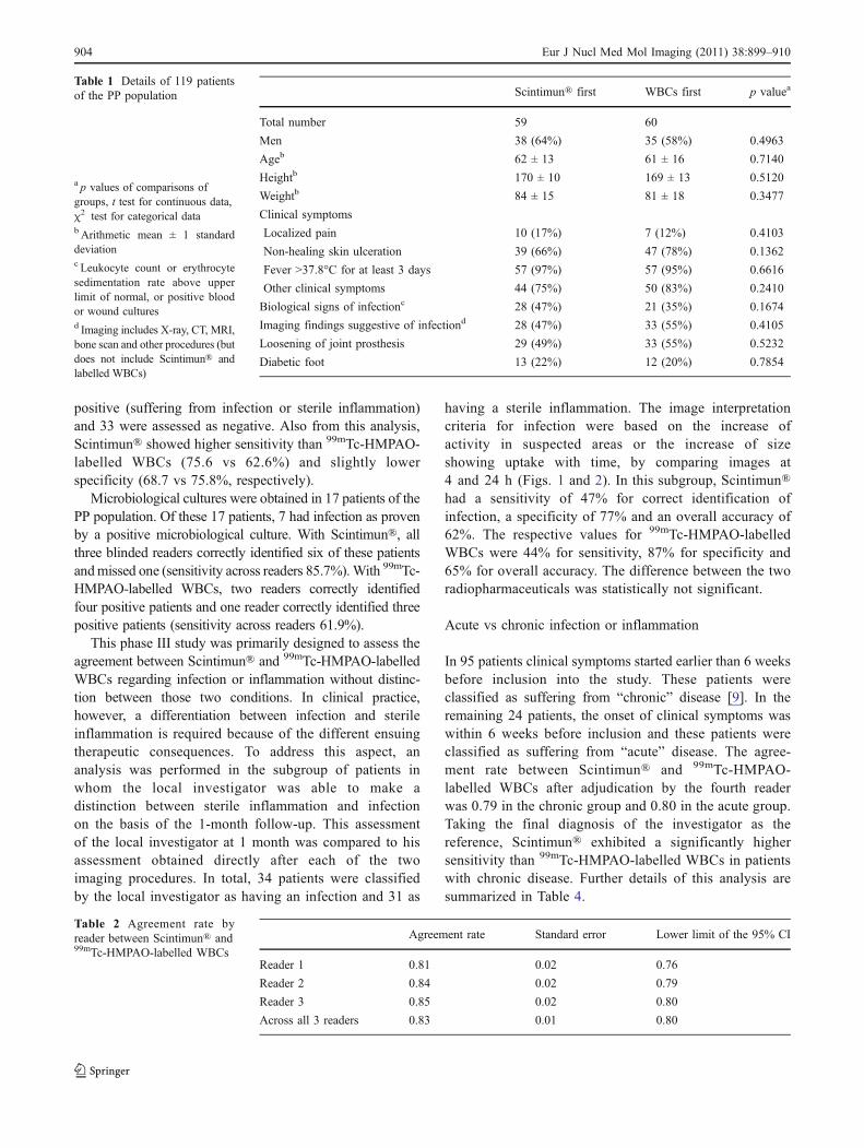
Table 1 Details of 119 patientsof the PP population
a p values of comparisons ofgroups, t test for continuous data,χ2 test for categorical datab Arithmetic mean ± 1 standarddeviationc Leukocyte count or erythrocytesedimentation rate above upperlimit of normal, or positive bloodor wound culturesd Imaging includes X-ray, CT, MRI,bone scan and other procedures (butdoes not include Scintimun® andlabelled WBCs)
Agreement rate Standard error Lower limit of the 95% CI
Reader 1 0.81 0.02 0.76
Reader 2 0.84 0.02 0.79
Reader 3 0.85 0.02 0.80
Across all 3 readers 0.83 0.01 0.80
Table 2 Agreement rate byreader between Scintimun® and99mTc-HMPAO-labelled WBCs
904 Eur J Nucl Med Mol Imaging (2011) 38:899–910

Image quality
During the blinded read, each reader also had to assess thequality of images on a 4-point scale as poor, moderate,good or excellent. The results are summarized in Table 5.The cumulated frequency of good and excellent images was
267 (75%) for Scintimun® and 199 (56%) for 99mTc-HMPAO-labelled WBCs. The difference in image qualitybetween both tracers was significant (χ2 test based on amultinomial regression, p<0.0001), although differentamounts of radioactivity were injected for the two typesof scans.
Fig. 1 A 40-year-old patientpresenting with pain in the areaof the left tibia. His historyincluded a motorcycle accidentalmost 4 years earlier with se-vere trauma of the left bodyside. WBC imaging was per-formed to rule out osteomyelitis.The Scintimun® study showedincreased uptake in the upperinner part of the left tibia con-sistent with material infection.The labelled WBC studyshowed low uptake at 4 h butrather normal images at 22 h. Atsurgery 4 months after imagingno evidence of infection wasfound. The truth panel rated thispatient as abnormal (sufferingfrom infection or inflammation)
Scintimun® 99mTc-HMPAO-labelled WBCs
Sensitivity 95% CI Sensitivity 95% CI
Reader 1 0.770 0.674; 0.867 0.703 0.598; 0.808
Reader 2 0.757 0.658; 0.855 0.514 0.399; 0.628
Reader 3 0.716 0.613; 0.820 0.554 0.440; 0.668
Across readers 0.748 0.666; 0.829 0.590 0.497; 0.683
Specificity 95% CI Specificity 95% CI
Reader 1 0.641 0.575; 0.861 0.667 0.517; 0.817
Reader 2 0.795 0.667; 0.923 0.897 0.801; 0.994
Reader 3 0.718 0.575; 0.861 0.821 0.699; 0.943
Across readers 0.718 0.594; 0.842 0.795 0.703; 0.887
Table 3 Sensitivity and speci-ficity of Scintimun® and 99mTc-HMPAO-labelled WBCs usingthe final diagnosis of the localinvestigator at the 1-monthfollow-up as the reference
Eur J Nucl Med Mol Imaging (2011) 38:899–910 905
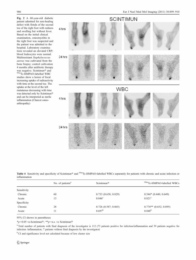
Fig. 2 A 60-year-old diabeticpatient admitted for non-healingdefect with fistula of the secondtoe of the right foot with rednessand swelling but without fever.Based on the initial clinicalexamination, osteomyelitis inthe right foot was suspected andthe patient was admitted to thehospital. Laboratory examina-tions revealed an elevated CRP;blood leukocytes were normal.Multiresistant Staphylococcusaureus was cultivated from thebone biopsy; control cultivation4 months after antibiotic therapywas negative. Scintimun® and99mTc-HMPAO-labelled WBCstudies show a lesion of focalincreasing uptake of radioactivitywith time at the second toe. Theuptake at the level of the leftmetatarsus decreasing with timewas detected only by Scintimun®and can be interpreted as sterileinflammation (Charcot osteo-arthropathy)
Table 4 Sensitivity and specificity of Scintimun® and 99mTc-HMPAO-labelled WBCs separately for patients with chronic and acute infection orinflammation
No. of patientsa Scintimun® 99mTc-HMPAO-labelled WBCs
Sensitivity
Chronic 60 0.733 (0.638; 0.829) 0.544* (0.440; 0.649)
Acute 13 0.846c 0.821c
Specificity
Chronic 28 0.726 (0.587; 0.865) 0.774** (0.652; 0.895)
Acute 11 0.697b 0.848b
95% CI shown in parentheses
*p<0.05 vs.Scintimun®; **p=n.s. vs Scintimun®a Total number of patients with final diagnosis of the investigator is 112 (73 patients positive for infection/inflammation and 39 patients negative forinfection /inflammation; 7 patients without final diagnosis by the investigator)b CI and significance level not calculated because of low cluster size
906 Eur J Nucl Med Mol Imaging (2011) 38:899–910

Adverse events
Amongst the 123 patients of the ASE population whoreceived at least one of the two study radiopharmaceuticals,24 patients (19%) reported a total of 31 adverse events. Ofthese 31 adverse events, 14 (45%) were related to infectiousor inflammatory disease and 2 (6%) were assessed aspossibly or probably related to Scintimun® and concerned 2cases of mild and transient hypotension. Only one patientexperienced a serious adverse event during study participa-tion. This patient suffered from necrosis at the level of themetatarsus and was hospitalized for transmetatarsal ampu-tation after completion of scintigraphic imaging. His healthcondition deteriorated during hospitalization, a gastroscopyrevealed haemorrhage of a duodenal ulcer and he finallydied from cardiopulmonary failure. This serious adverseevent was assessed by the investigator as unrelated to studyradiopharmaceuticals.
Laboratory assessments, vital signs and HAMA
Assessment of laboratory parameters was comparable betweenthe two study groups (Scintimun® first and 99mTc-HMPAO-labelled WBCs first). The majority of abnormal laboratoryvalues was already abnormal before the first injection ofstudy radiopharmaceutical and was related to the inflamma-tory process. Mean values did not change from baseline afteradministration of Scintimun® or 99mTc-HMPAO-labelledWBCs. No effect of Scintimun® or 99mTc-HMPAO-labelledWBCs on vital signs was detected except in three patients inwhom blood pressure decreased after injection of Scintimun®.Two of these cases of hypotension were assessed as possiblyor probably related to Scintimun®.
Blood samples for HAMAwere collected at screening andtwice after injection of Scintimun® (at 1 month and at3 months). Four patients presented with HAMA levels above40 μg/l at screening and were not allowed to enter the study(screening failures). Seven patients had no HAMA assess-ment during follow-up. Consequently, 116 patients had atleast one HAMA assessment after administration ofScintimun®. Only 16 of 116 patients (13.8%) became positivewith HAMA levels >40 μg/l after Scintimun® administration.
Discussion
The results of this phase III trial show that Scintimun®imaging is accurate, efficacious and safe in diagnosinginfection of the peripheral skeleton and provides compara-ble information to 99mTc-HMPAO-labelled WBCs. In thisstudy, Scintimun® was more sensitive than 99mTc-HMPAO-labelled WBCs in patients with microbiologically proveninfection of the bone and in patients with chronicosteomyelitis.
The basis of the Scintimun® mechanism of action is itsnanomolar affinity to the NCA-95 (CEACAM8, CD66b)antigen, which is found on granulocytes and mature bonemarrow cells of the granulocytic lineage [10, 11]. Thesecells are present in major amounts in inflammatory andinfectious lesions and are also found in the haematopoieticbone marrow. The typical uptake pattern of Scintimun® inhumans, therefore, includes focal uptake at sites of infectionor inflammation as well as a staining of the haematopoieticbone marrow.
Since its introduction into clinical practice in a limitednumber of European countries almost 20 years ago, thespecific accumulation of Scintimun® at sites of infection orinflammation has been studied in various clinical indica-tions. The majority of these studies have been conducted inpatients with osteomyelitis. Other indications includeendocarditis [12, 13], inflammatory bowel disease [14–17], lung infection [18], the detection of perioperativeseptic foci [19] and fever of unknown origin [20–22].
The most important clinical indication for Scintimun®imaging is osteomyelitis [23]. In this context, osteomyeliticlesions of the peripheral skeleton and of the central skeletonhave to be considered separately. In the peripheral skeleton,osteomyelitic foci are characterized by an increased uptakeof Scintimun®. In the central skeleton the typical finding ofosteomyelitis is a focally decreased uptake (cold spot)which is based on the displacement of normal bone marrowby the inflammatory process. The finding of a cold spot inthe haematopoietic bone marrow of the central skeleton isnot specific for osteomyelitis but can also be caused byother processes (e.g. metastases, haemangioma). Inherently,therefore, a reduced specificity of Scintimun® has to be
Image quality Scintimun® 99mTc-HMPAO-labelled WBCs
Excellent 40 (11%) 13 (4%)
Good 227 (64%) 186 (52%)
Moderate 71 (20%) 110 (31%)
Poor 13 (4%) 41 (11%)
Not evaluable 6 (2%) 7 (2%)
Total no. of assessments 357 (100%)a 357 (100%)
Table 5 Image quality asassessed by the three readers
a Total number of assessments is357: 119 patients assessed by 3readers
Eur J Nucl Med Mol Imaging (2011) 38:899–910 907

expected in the central skeleton as has already beenreported for 99mTc-HMPAO-labelled WBCs. This consider-ation is confirmed by data from Guhlmann et al. whostudied 36 patients with suspected chronic osteomyelitis ofthe peripheral skeleton and 15 patients with suspectedchronic osteomyelitis of the central skeleton by combinedScintimun® and conventional bone scintigraphy [24].Sensitivity was comparable in the central and peripheralskeleton (85 vs 91%, respectively), while specificity waslower in the central skeleton (60 central vs 85% peripheral).
In peripheral osteomyelitis, the results of publishedstudies show that Scintimun® has high sensitivity andspecificity. Typically, reported values of sensitivity andspecificity in osteomyelitis range between 70 and 90% [18,24–28]. Reuland et al. [28] studied Scintimun® in 106patients with suspected peripheral bone infection aftersurgery. The final diagnosis was obtained by microbiolog-ical culture and clinical follow-up information. Sensitivitywas highest in the lower leg (100%), followed by thigh(85%), knee (70%) and hip (69%). Specificity rangedbetween 83 (knee) and 100% (lower leg). The highsensitivity of Scintimun® has been confirmed by Peltier etal. [18] in a small series of eight patients with suspectedperipheral osteomyelitis who underwent bone biopsy forfinal confirmation of disease. All eight patients withosteomyelitis were correctly identified (sensitivity 100%).
Only very limited data are available about an intra-individual comparison of Scintimun® against alternativescintigraphic procedures for imaging of WBCs. Thiscomparison is required to ultimately assess the diagnosticvalue of Scintimun® against competing methods. Thisstudy was primarily designed to provide data about thecomparison of Scintimun® with the accepted gold standardfor imaging of infection (99mTc-HMPAO-labelled WBCs).
The results of this trial show that Scintimun® and 99mTc-HMPAO-labelled WBCs provide comparable clinical infor-mation in osteomyelitis. This is the first larger series ofpatients studied intraindividually with both methods andallows for the first time a direct comparison betweenScintimun® and 99mTc-HMPAO-labelled WBCs.
It is important to mention again that it was not theprimary aim of this study to evaluate sensitivity andspecificity of the two radiopharmaceuticals. Nevertheless,sensitivity and specificity were calculated for comparativereasons. For the calculation of sensitivity and specificity, atruth panel decision was used as a surrogate for the truegold standard (bone biopsy showing granulocytic accumu-lation). The resulting values of sensitivity and specificitywere slightly lower for both radiopharmaceuticals in thistrial than the respective values in the published literature.The reason for the lower values is most probably explainedby the very strict and controlled environment of the blindedimage evaluation that was used in this trial. All blinded
readers were completely unaware of any clinical informa-tion related to the patients. The lack of complementaryinformation represents an artificial situation with limitedclinical relevance. It is assumed that imaging withScintimun® and 99mTc-HMPAO-labelled WBCs in a clin-ical environment will have a greater contribution to medicaldecision-making than implied by the sensitivity andspecificity values obtained in this trial. This assumption isin line with results from an earlier phase III trial in 775patients where Scintimun® was rated by the investigator tosupply additional information which was not provided byother diagnostic methods in 31.5% of patients, to assure orconfirm a diagnosis already suspected by other diagnosticmethods in another 35.7% of patients, and positivelyinfluenced the treatment strategy in 35% of patients [29].
Besides providing overall evidence for the good agreementbetween Scintimun® and 99mTc-HMPAO-labelled WBCs, thisstudy also points towards specific differences between thetwo tracers. A subanalysis of patients with acute and chronicosteomyelitis shows that Scintimun® detects chronic osteo-myelitis with higher sensitivity than 99mTc-HMPAO-labelledWBCs. The high sensitivity of Scintimun® in this trial inchronic osteomyelitis is in line with comparable values fromthe literature. Kaim et al. [25] studied 24 patients withchronic post-traumatic osteomyelitis and reported a sensitivityof 84% and a specificity of 72% of combined Scintimun®imaging and conventional bone scintigraphy. Slightly lowervalues were published by Boubaker et al. [30] for patientswith subacute or chronic infection of hip prosthesis. Theirstudy involved 57 patients and yielded a sensitivity of 67%and a specificity of 75% using microbiological examinationsand clinical follow-up as the reference.
The higher sensitivity of Scintimun® compared to99mTc-HMPAO-labelled WBCs in chronic osteomyelitiscan be explained by two factors: the first factor is relatedto the higher image quality of Scintimun® which allowsdiscrete increases of tracer uptake to be detected moreeasily. Chronic osteomyelitis is frequently characterized bya lower disease activity and a lower level of granulocyticaccumulation than acute osteomyelitis. A slightly increaseduptake in chronic lesions is probably detected more easilyin high-quality images. The second factor is related to thepresumed uptake mechanism of Scintimun® in osteomye-litic lesions. Uptake of Scintimun® is supposed to involvetwo components which occur in parallel: (1) targeting ofNCA-95 on circulating granulocytes in the bloodstreamwith subsequent migration of Scintimun®-labelled granulo-cytes to the inflammatory lesion and (2) transport of freeScintimun® with the bloodstream to sites of infection withsubsequent sequestration to the extravascular space due toan increased capillary permeability, followed by specificbinding to granulocytes which are present in the extravas-cular space. The latter mechanism is suggested to be more
908 Eur J Nucl Med Mol Imaging (2011) 38:899–910
important in the clinical situation. Ex vivo labelledWBCs, in contrast to Scintimun®, accumulate in osteo-myelitic lesions only on the basis of the first mechanism(migration of labelled cells into the lesion). It isconceivable that the second mechanism (sequestrationof the free Scintimun® antibody to sites of infection orinflammation with subsequent binding to granulocytes)contributes to a relevant degree to the accumulation ofScintimun® in chronic lesions. This difference in uptakemechanism may also explain the higher sensitivity ofScintimun® in patients with microbiologically confirmedosteomyelitis.
For clinical purposes, the differentiation between infectionand sterile inflammation is of particular relevance for therapydecisions. In clinical practice, an increase in uptake or sizeover time is typically interpreted as infection, whereas adecrease in uptake or size is assumed to represent sterileinflammation or bone marrow activity. In this study, Scinti-mun® and 99mTc-HMPAO-labelled WBCs could be com-pared with regard to their ability to differentiate infectionfrom sterile inflammation in a subgroup of 65 patients. Inthese patients, the local investigator was able to obtain a finaldiagnosis on the basis of follow-up information. In these 65patients, Scintimun® and 99mTc-HMPAO-labelled WBCswere equally effective in differentiating between infectionand sterile inflammation.
Finally, this study provides the first systematic dataabout the development of HAMA after injection ofScintimun®. Of 116 patients, 16 developed HAMA afterScintimun® injection and may have a potential risk ofhypersensitivity at re-exposure. According to the SmPC 2 ofScintimun®, determination of HAMA levels is requestedbefore injection of Scintimun® and re-administration toHAMA-positive patients is contraindicated.
With the exception of two cases of mild and transienthypotension, no other parameters related to patient safety(laboratory data, vital signs) provided evidence of anelevated risk associated with the use of Scintimun® Thisis in line with the available safety profile which is basedon some 100,000 injections and does not identify specificsafety risks.
Besides Scintimun® and labelled WBCs, other nuclearmedicine methods have also been used for the diagnosis ofosteomyelitis. The murine Fab’ fragment sulesomab targetsNCA-90 (CEACAM6, CD66c) on granulocytes and hasshown to be of value in osteomyelitis. However, affinity togranulocytes is lower than the respective values of Scinti-mun® [31]. Recently, 18F-fluorodeoxyglucose (FDG) inconjunction with positron emission tomography (PET) has
been used for infection or inflammation imaging. Availableresults are promising; however, the increased uptake of 18F-FDG is due to an increased glucose metabolism and in thisregard unspecific for infection or inflammation.
In summary, Scintimun® is a promising, efficacious andsafe tool for diagnosis of osteomyelitis in the peripheralskeleton.
Conflicts of interest Wolf S. Richter is the managing director ofPharmtrace, a CRO that provides contract research services to IBA/CIS bio international (marketing authorization holder of Scintimun®),Carsten Schwenke provides statistical services to IBA/CIS biointernational, Florence Chossat and Amel Dahmane are employeesof IBA/CIS bio international. All other authors do not have a conflictof interest.
Open Access This article is distributed under the terms of theCreative Commons Attribution Noncommercial License which per-mits any noncommercial use, distribution, and reproduction in anymedium, provided the original author(s) and source are credited.
Annex: participating sites and principal investigators
Prof. Michèle Allard (CHU Pellegrin, Bordeaux, France);Prof. Dr. H. Amthauer (Charité, Berlin, Germany); Dr.Magdolna Bakos (Békés Megyei Képviselotestület, PándyKálmán Kórház, Gyula, Hungary); Prof. Hatem Boulahdour(CHRU Jean Minjoz, Besançon, France); Prof. LászlóGaluska (Debreceni Egyetem Orvos-és Egészségtudományi,Debrecen, Hungary); Prof. Dominique Le Guludec (CHUBichat-Claude Bernard, Paris, France); Dr. John M. H. deKlerk (Meander Medical Center, Amersfoort, The Nether-lands); Dr. Otakar Kraft (UH Ostrava, Ostrava, CzechRepublic); Dr. Otto Lang (Charles University, 3rd MedicalFaculty, UH Kralovske Vinohrady, Prague, Czech Republic);Dr. Endre Lenart (Bács-Kiskun Megyei ÖnkormányzatKórháza, Kecskemet, Hungary); Dr. Isabelle Morelec (CHCivils Lyon Sud, Pierre-Benite, France); Dr. MiroslavMyslivecek (UH Olomouc, Olomouc, Czech Republic); Prof.Christoph H. J. Reiners (Universitätsklinikum Würzburg,Würzburg, Germany); Prof. Sven N. Reske (Universitätskli-nikum Ulm, Ulm, Germany); Dr. Frederik Smit (RijnlandZiekenhuis, Galeiderdorp, The Netherlands); Prof. IstvánSzilvazi (Országos Gyógyintézeti Központ, Budapest, Hun-gary); Dr. Sarolta Takacs (Budai Irgalmasrendi Hospital,Budapest, Hungary); Dr. Adrienn Tombacz (Heves County,Markhot Ferenc Hospital, Eger, Hungary); Prof. Dr. J. FredVerzijlbergen (St. Antonius Ziekenhuis, Nieuwegein, TheNetherlands); Dr. Jaroslav Vizda (UH Hradec Kralove,Hradec Kralove, Czech Republic); Dr. Petr Vlcek (UHMotol,Prague 5, Czech Republic); Dr. Anthonie Zwijnenburg(Spaarne Ziekenhuis Hoofdorp, The Netherlands).
2 http://www.ema.europa.eu/docs/en_GB/document_library/EPAR_-_Product_Information/human/001045/WC500075575.pdf
Eur J Nucl Med Mol Imaging (2011) 38:899–910 909
References
1. Concia E, Prandini N, Massari L, Ghisellini F, Consoli V,Menichetti F, et al. Osteomyelitis: clinical update for practicalguidelines. Nucl Med Commun 2006;27:645–60.
2. Love C, Marwin SE, Palestro CJ. Nuclear medicine and theinfected joint replacement. Semin Nucl Med 2009;39:66–78.
3. Chatha DS, Cunningham PM, Schweitzer ME. MR imaging of thediabetic foot: diagnostic challenges. Radiol Clin North Am2005;43:747–59. ix.
4. Gnanasegaran G, Chicklore S, Vijayanathan S, O’Doherty MJ,Fogelman I. Diabetes and bone: advantages and limitations ofradiological, radionuclide and hybrid techniques in the assessmentof diabetic foot. Minerva Endocrinol 2009;34:237–54.
5. RozzanigoU, Tagliani A,Vittorini E, Pacchioni R, Brivio LR, CaudanaR. Role of magnetic resonance imaging in the evaluation of diabeticfoot with suspected osteomyelitis. Radiol Med 2009;114:121–32.
6. Rojas-Burke J. Health officials reacting to infection mishaps. JNucl Med 1992;33:13N–4N. 27N.
7. de Vries EF, Roca M, Jamar F, Israel O, Signore A. Guidelines forthe labelling of leucocytes with (99m)Tc-HMPAO. Inflammation/infection Taskgroup of the European Association of NuclearMedicine. Eur J Nucl Med Mol Imaging 2010;37:842–8.
8. Schwenke C, Busse R. Analysis of differences in proportions fromclustered data with multiple measurements in diagnostic studies.Methods Inf Med 2007;46:548–52.
9. Termaat MF, Raijmakers PG, Scholten HJ, Bakker FC, Patka P,Haarman HJ. The accuracy of diagnostic imaging for theassessment of chronic osteomyelitis: a systematic review andmeta-analysis. J Bone Joint Surg Am 2005;87:2464–71.
10. Steinsträsser A, Berberich R, Kuhlmann L, Zabori S, Schwarz A.Binding of the monoclonal antibody BW 250/183 to humangranulocytes. Nuklearmedizin 1992;31:57–63.
11. Bosslet K, Steinsträsser A, Schwarz A, Harthus HP, Lüben G,Kuhlmann L, et al. Quantitative considerations supporting theirrelevance of circulating serum CEA for the immunoscintigraphicvisualization of CEA expressing carcinomas. Eur J Nucl Med1988;14:523–8.
12. Morguet AJ, Munz DL, Ivancević V, Werner GS, Sandrock D,Bökemeier M, et al. Immunoscintigraphy using technetium-99m-labeled anti-NCA-95 antigranulocyte antibodies as an adjunct toechocardiography in subacute infective endocarditis. J Am CollCardiol 1994;23:1171–8.
13. Morguet AJ, Munz DL, Ivancević V, Werner GS, Kreuzer H. Theclinical importance of scintigraphy with the murine monoclonalantigranulocyte antibody BW 250/183 for the diagnosis of prosthesis-related endocarditis. Dtsch Med Wochenschr 1995;120:861–6.
14. Segarra I, Roca M, Baliellas C, Vilar L, Ricart Y, Mora J, et al.Granulocyte-specific monoclonal antibody technetium-99m-BW250/183 and indium-111 oxine-labelled leucocyte scintigraphy ininflammatory bowel disease. Eur J Nucl Med 1991;18:715–9.
15. Papos M, Nagy F, Narai G, Rajtar M, Szantai G, Lang J, et al.Anti-granulocyte immunoscintigraphy and [99mTc]hexamethyl-propyleneamine-oxime-labeled leukocyte scintigraphy in inflam-matory bowel disease. Dig Dis Sci 1996;41:412–20.
16. Almers S, Granerus G, Franzén L, Ström M. Technetium-99mscintigraphy: more accurate assessment of ulcerative colitis with
exametazime-labelled leucocytes than with antigranulocyte anti-bodies. Eur J Nucl Med 1996;23:247–55.
17. Tarján Z, Tóth G, Györke T, Mester A, Karlinger K, Makó EK.Ultrasound in Crohn’s disease of the small bowel. Eur J Radiol2000;35:176–82.
18. Peltier P, Potel G, Lovat E, Baron D, Chatal JF. Detection of lung andbone infection with anti-granulocyte monoclonal antibody BW 250/183 radiolabelled with 99Tcm. NuclMed Commun 1993;14:766–74.
19. Kroiss A, Sporn P, Auinger C, Redl E, Böck F, Dinstl K, et al.Immunoscintigraphy for detection of inflammatory perioperativefoci. Acta Med Austriaca 1993;20:45–9.
20. Becker W, Dölkemeyer U, Gramatzki M, Schneider MU, ScheeleJ, Wolf F. Use of immunoscintigraphy in the diagnosis of fever ofunknown origin. Eur J Nucl Med 1993;20:1078–83.
21. Meller J, Ivancevic V, Conrad M, Gratz S, Munz DL, Becker W.Clinical value of immunoscintigraphy in patients with fever ofunknown origin. J Nucl Med 1998;39:1248–53.
22. Gratz S, Behr TM, Herrmann A, Meller J, Conrad M, Zappel H, etal. Immunoscintigraphy (BW 250/183) in neonates and infantswith fever of unknown origin. Nucl Med Commun 1998;19:1037–45.
23. Graute V, Feist M, Lehner S, Haug A, Müller PE, Bartenstein P, etal. Detection of low-grade prosthetic joint infections using (99m)Tc-antigranulocyte SPECT/CT: initial clinical results. Eur J NuclMed Mol Imaging 2010;37:1751–9.
24. Guhlmann A, Brecht-Krauss D, Suger G, Glatting G, Kotzerke J,Kinzl L, et al. Fluorine-18-FDG PET and technetium-99mantigranulocyte antibody scintigraphy in chronic osteomyelitis. JNucl Med 1998;39:2145–52.
25. Kaim A, Maurer T, Ochsner P, Jundt G, Kirsch E, Mueller-BrandJ. Chronic complicated osteomyelitis of the appendicular skeleton:diagnosis with technetium-99m labelled monoclonal antigranulo-cyte antibody-immunoscintigraphy. Eur J Nucl Med 1997;24:732–8.
26. Kroiss A, Böck F, Perneczky G, Auinger C, Weidlich G,Kleinpeter G, et al. Immunoscintigraphy for the detection ofinflammation foci in bone and joint diseases. Wien KlinWochenschr 1990;102:713–7.
27. Hotze AL, Briele B, Overbeck B, Kropp J, Gruenwald F,Mekkawy MA, et al. Technetium-99m-labeled anti-granulocyteantibodies in suspected bone infections. J Nucl Med1992;33:526–31.
28. Reuland P, Winker KH, Heuchert T, Ruck P, Müller-Schauen-burg W, Weller S, et al. Detection of infection in postoperativeorthopedic patients with technetium-99m-labeled monoclonalantibodies against granulocytes. J Nucl Med 1991;32:2209–14.
29. Steinsträsser A, Oberhausen E. Granulocyte labelling kit BW 250/183–results of the European multicenter trial. Nuklearmedizin1996;35:1–11.
30. Boubaker A, Delaloye AB, Blanc CH, Dutoit M, Leyvraz PF,Delaloye B. Immunoscintigraphy with antigranulocyte monoclo-nal antibodies for the diagnosis of septic loosening of hipprostheses. Eur J Nucl Med 1995;22:139–47.
31. Becker W, Bair J, Behr T, Repp R, Streckenbach H, Beck H, et al.Detection of soft-tissue infections and osteomyelitis using atechnetium-99m-labeled anti-granulocyte monoclonal antibodyfragment. J Nucl Med 1994;35:1436–43.
910 Eur J Nucl Med Mol Imaging (2011) 38:899–910


![[TC 025] Alvenaria estrutural_2020 - dcc@ufpr](https://static.fdokumen.com/doc/165x107/631358cfaca2b42b580d255f/tc-025-alvenaria-estrutural2020-dccufpr.jpg)


![Noninvasive Molecular Imaging of MYC mRNA Expression in Human Breast Cancer Xenografts with a [ 99m Tc]Peptide−Peptide Nucleic Acid−Peptide Chimera](https://static.fdokumen.com/doc/165x107/63214cddbc33ec48b20e4a4a/noninvasive-molecular-imaging-of-myc-mrna-expression-in-human-breast-cancer-xenografts.jpg)