Immunoscintigraphy of small-cell lung cancer xenografts with anti neural cell adhesion molecule...
8
British Journal of Cancer (1996) 73, 439-446 © 1996 Stockton Press All rights reserved 0007-0920/96 $12.00 Immunoscintigraphy of small-cell lung cancer xenografts with anti neural cell adhesion molecule monoclonal antibody, 123C3: improvement of tumour uptake by internalisation HB Kwal'2, J Wesseling', AHM Verhoeven', N van Zandwijk2 and J Hilkens' Departments of 'Tumour Biology and 2Medical Oncology, The Netherlands Cancer Institute (Antoni van Leeuwenhoekhuis), Plesmanlaan 121, 1066 CX Amsterdam, The Netherlands. Summary The efficacy of three murine monoclonal antibodies (MAbs) for immunoscintigraphy of small-cell lung cancer (SCLC) xenografts was studied in a Balb/c nu/nu mouse model. These MAbs, 123C3, 123A8 and MOCl91, belong to cluster 1 of anti-SCLC MAbs and bind to the neural cell adhesion molecule (NCAM) with similar affinity. After intraperitoneal injection of these MAbs, labelled with 1251, the highest uptake in tumour tissue was obtained with MAb 123C3. Seven days after administration of this MAb 13.9% of the injected dose per gram of tumour tissue was retained in the tumour. The corresponding tumour to tissue ratios ranged from 3.97 for blood to 31.03 for colon. The imaging results and the tumour uptake were less favourable for the two other MAbs, 123A8 and MOCl91 (fractions of injected dose respectively 6.7% and 9.2%), although affinity, biological activity after labelling and uptake in non-tumour tissues were very similar for all three MAbs. These results may be explained by the differences in the interaction between the MAbs and the tumour cells. MAb 123C3 is internalised into tumour cells, whereas both other anti-NCAM MAbs are not. Internalisation into NCI H69 cells was demonstrated in vitro by a radioimmunoassay, confocal laser scanning microscopy and electron microscopy. The internalised fraction of MAb 123C3 was 22.3% after 24 h, whereas this fraction was only 7.5% for MAb 123A8. Although the internalised radiolabelled MAbs are usually degraded and dehalogenated intracellularly, the retained radioactivity is high. Apparently, intracellular degradation of radiolabelled MAb 123C3 and subsequent secretion of radioactive iodine did not prevent the accumulation of intracellular radioactivity. In conclusion, accumulation and retention of radioactivity in the tumour tissue, due to internalisation of radiolabelled MAbs, may improve the results of immunoscintigraphy. Keywords: immunoscintigraphy; small-cell lung cancer; neural cell adhesion molecule; monoclonal antibody; internalisation Clinically, lung cancer is divided into small-cell lung cancer (SCLC) and non-small-cell lung cancer (NSCLC). SCLC accounts for about 25% of the cases and is associated with the worst prognosis of all lung cancers (Yesner, 1985). For the prognosis and treatment of this type of tumour it is very important to determine the initial stage of the tumour (Ihde, 1985). Unfortunately, standard staging procedure is very laborious and inaccurate. In a recent meta-analysis, the best non-invasive diagnostic technique, computerised tomography (CT), achieved an accuracy of only 0.80 for the detection of intrathoracic lesions (Dales et al., 1990). There is clearly a need for new techniques with a higher sensitivity and specificity to replace the multitude of diagnostic procedures used in the initial staging (Ihde, 1985). Monoclonal antibodies (MAbs) have greatly increased the sensitivity in detecting bone marrow metastases in SCLC (Ledermann et al., 1994). Using radiolabelled MAbs in patients with SCLC (Nelp et al., 1990) distant metastases can be detected by immunoscintigraphy in 10% of patients with limited disease, when staged by conventional methods. This finding suggests that immunoscintigraphy has a higher sensitivity than the standard staging procedure. Monoclonal antibodies (MAbs) raised against SCLC are categorised in clusters by the antigen recognised, according to an international workshop (Beverley et al., 1988). The MAbs belonging to cluster 1 bind to the neural cell adhesion molecule (NCAM) (Moolenaar et al., 1990). Of the existing NCAM isoforms, NCAM-140 and NCAM-180 are the most important in SCLC (Moolenaar et al., 1990) and all three cluster 1 MAbs used in this study recognise these isoforms (Beverley et al., 1988; Hida et al., 1991). As NCAM is expressed by all SCLCs, it seems a suitable target for immunoscintigraphy with these MAbs. The three MAbs investigated in this study showed similar affinities for NCAM and were used for radioimmunodetection in a mouse SCLC xenograft model. We demonstrated that one of the MAbs that is internalised by the SCLC cells shows a significantly higher uptake in tumour tissue. Materials and methods Cell lines The NCI H69 cell line (hereafter referred to as H69 cells), derived from a small cell lung carcinoma, was kindly provided by Dr D Carney (Gazdar et al., 1980) and was grown in Dulbecco's modified Eagle medium supplemented with 1 mm glutamine, 10% fetal calf serum and antibiotics. This cell line expresses high levels of NCAM when grown in vitro and as xenografts in nude mice (Rygaard et al., 1992). Monoclonal antibodies From a panel of nine cluster 1 MAbs, MAbs 123C3, 123A8 and MOCl91 were selected for this study, on the basis of their affinity for NCAM and biological activity after labelling with radioactive iodine (Beverley et al., 1988; Moolenaar et al., 1990). MAbs 123C3 and 123A8 are IgGI antibodies raised at our institute against a membrane fraction of a fresh SCLC specimen. Both MAbs recognise epitopes on the protein backbone of NCAM close to the attachment site of the polysialic acid side-chains (Gerardy-Shahn and Eckhardt, 1994) and bind to all NCAM isoforms (Moolenaar et al., 1990). The tissue distribution of MAb 123C3 in human tissues was described previously (Schol et al., 1988). MAb 123A8 showed a very similar tissue distribution (DJ Schol and Ph C Hageman, unpublished data). MAb MOC191 is an Correspondence: HB Kwa, Department of Pulmonology, OLVG Hospital, le Oosterparkstraat 279, 1091 HM Amsterdam, The Netherlands Received 16 May 1995; revised 8 September 1995; accepted 13 September 1995
-
Upload
independent -
Category
Documents
-
view
1 -
download
0
Transcript of Immunoscintigraphy of small-cell lung cancer xenografts with anti neural cell adhesion molecule...
British Journal of Cancer (1996) 73, 439-446© 1996 Stockton Press All rights reserved 0007-0920/96 $12.00
Immunoscintigraphy of small-cell lung cancer xenografts with anti neuralcell adhesion molecule monoclonal antibody, 123C3: improvement oftumour uptake by internalisation
HB Kwal'2, J Wesseling', AHM Verhoeven', N van Zandwijk2 and J Hilkens'
Departments of 'Tumour Biology and 2Medical Oncology, The Netherlands Cancer Institute (Antoni van Leeuwenhoekhuis),Plesmanlaan 121, 1066 CX Amsterdam, The Netherlands.
Summary The efficacy of three murine monoclonal antibodies (MAbs) for immunoscintigraphy of small-celllung cancer (SCLC) xenografts was studied in a Balb/c nu/nu mouse model. These MAbs, 123C3, 123A8 andMOCl91, belong to cluster 1 of anti-SCLC MAbs and bind to the neural cell adhesion molecule (NCAM) withsimilar affinity. After intraperitoneal injection of these MAbs, labelled with 1251, the highest uptake in tumourtissue was obtained with MAb 123C3. Seven days after administration of this MAb 13.9% of the injected doseper gram of tumour tissue was retained in the tumour. The corresponding tumour to tissue ratios ranged from3.97 for blood to 31.03 for colon. The imaging results and the tumour uptake were less favourable for the twoother MAbs, 123A8 and MOCl91 (fractions of injected dose respectively 6.7% and 9.2%), although affinity,biological activity after labelling and uptake in non-tumour tissues were very similar for all three MAbs. Theseresults may be explained by the differences in the interaction between the MAbs and the tumour cells. MAb123C3 is internalised into tumour cells, whereas both other anti-NCAM MAbs are not. Internalisation intoNCI H69 cells was demonstrated in vitro by a radioimmunoassay, confocal laser scanning microscopy andelectron microscopy. The internalised fraction of MAb 123C3 was 22.3% after 24 h, whereas this fraction wasonly 7.5% for MAb 123A8. Although the internalised radiolabelled MAbs are usually degraded anddehalogenated intracellularly, the retained radioactivity is high. Apparently, intracellular degradation ofradiolabelled MAb 123C3 and subsequent secretion of radioactive iodine did not prevent the accumulation ofintracellular radioactivity. In conclusion, accumulation and retention of radioactivity in the tumour tissue, dueto internalisation of radiolabelled MAbs, may improve the results of immunoscintigraphy.
Keywords: immunoscintigraphy; small-cell lung cancer; neural cell adhesion molecule; monoclonal antibody;internalisation
Clinically, lung cancer is divided into small-cell lung cancer(SCLC) and non-small-cell lung cancer (NSCLC). SCLCaccounts for about 25% of the cases and is associated withthe worst prognosis of all lung cancers (Yesner, 1985). Forthe prognosis and treatment of this type of tumour it is veryimportant to determine the initial stage of the tumour (Ihde,1985). Unfortunately, standard staging procedure is verylaborious and inaccurate. In a recent meta-analysis, the bestnon-invasive diagnostic technique, computerised tomography(CT), achieved an accuracy of only 0.80 for the detection ofintrathoracic lesions (Dales et al., 1990). There is clearly aneed for new techniques with a higher sensitivity andspecificity to replace the multitude of diagnostic proceduresused in the initial staging (Ihde, 1985). Monoclonalantibodies (MAbs) have greatly increased the sensitivity indetecting bone marrow metastases in SCLC (Ledermann etal., 1994). Using radiolabelled MAbs in patients with SCLC(Nelp et al., 1990) distant metastases can be detected byimmunoscintigraphy in 10% of patients with limited disease,when staged by conventional methods. This finding suggeststhat immunoscintigraphy has a higher sensitivity than thestandard staging procedure.
Monoclonal antibodies (MAbs) raised against SCLC arecategorised in clusters by the antigen recognised, according toan international workshop (Beverley et al., 1988). The MAbsbelonging to cluster 1 bind to the neural cell adhesionmolecule (NCAM) (Moolenaar et al., 1990). Of the existingNCAM isoforms, NCAM-140 and NCAM-180 are the mostimportant in SCLC (Moolenaar et al., 1990) and all threecluster 1 MAbs used in this study recognise these isoforms
(Beverley et al., 1988; Hida et al., 1991). As NCAM isexpressed by all SCLCs, it seems a suitable target forimmunoscintigraphy with these MAbs. The three MAbsinvestigated in this study showed similar affinities for NCAMand were used for radioimmunodetection in a mouse SCLCxenograft model. We demonstrated that one of the MAbsthat is internalised by the SCLC cells shows a significantlyhigher uptake in tumour tissue.
Materials and methods
Cell lines
The NCI H69 cell line (hereafter referred to as H69 cells),derived from a small cell lung carcinoma, was kindlyprovided by Dr D Carney (Gazdar et al., 1980) and wasgrown in Dulbecco's modified Eagle medium supplementedwith 1 mm glutamine, 10% fetal calf serum and antibiotics.This cell line expresses high levels of NCAM when grown invitro and as xenografts in nude mice (Rygaard et al., 1992).
Monoclonal antibodies
From a panel of nine cluster 1 MAbs, MAbs 123C3, 123A8and MOCl91 were selected for this study, on the basis oftheir affinity for NCAM and biological activity after labellingwith radioactive iodine (Beverley et al., 1988; Moolenaar etal., 1990). MAbs 123C3 and 123A8 are IgGI antibodiesraised at our institute against a membrane fraction of a freshSCLC specimen. Both MAbs recognise epitopes on theprotein backbone of NCAM close to the attachment site ofthe polysialic acid side-chains (Gerardy-Shahn and Eckhardt,1994) and bind to all NCAM isoforms (Moolenaar et al.,1990). The tissue distribution of MAb 123C3 in humantissues was described previously (Schol et al., 1988). MAb123A8 showed a very similar tissue distribution (DJ Scholand Ph C Hageman, unpublished data). MAb MOC191 is an
Correspondence: HB Kwa, Department of Pulmonology, OLVGHospital, le Oosterparkstraat 279, 1091 HM Amsterdam, TheNetherlandsReceived 16 May 1995; revised 8 September 1995; accepted 13September 1995
Immunoscintigraphy of SCLC xenografts with anti-NCAM MAb 123C3HB Kwa et al
440IgG2a antibody and was kindly provided by Dr LF de Ley,University Hospital of Groningen. The epitope recognised byMAb MOC191 is probably located at the third immunoglo-bulin loop (Hida et al., 1991; Gerardy-Shahn and Eckhardt,1994). MAb M6/1, and IgGI MAb raised against melanomacells, detects a high molecular weight proteoglycan and didnot bind to H69 cells in vitro. All MAbs were affinity purifiedfrom ascitic fluid by protein-A-Sephadex column chromato-graphy and were eluted with a citrate buffer (pH 4.5) (Joneset al., 1985).
Antibody labelling
The MAbs were labelled with 1251 (Amersham) using thechloramine-T method (Hunter and Greenwood, 1962). MAb(50 jug) was labelled with 50 1iCi of 1251I. Free iodine wasremoved from the labelled MAbs by ion-exchange columnchromatography (Dowex G25). For immunofluorescenceexperiments the MAbs were labelled with fluoresceinisothiocyanate (FITC) (The and Feltkamp, 1970). Briefly,1 mg of FITC in dimethylsulphoxide (DMSO) was added to1 mg of MAb solution in 0.1 M sodium carbonate buffer(pH 9.5) and incubated overnight at 4°C. Free FITC wasremoved over a Dowex G25 column.
Determination of immunoreactivity and in vitro affinityThe immunoreactivity of the MAbs after labelling with 1251,was assessed according to the method described by Lindmo etal. (1984) with slight modifications. Serial dilutions of H69cells in a volume of 200 ul of medium, starting at a cellconcentration of 25 x 106 ml', were incubated with anequivolume of 1251-labelled MAb in phosphate-bufferedsaline (PBS) at a concentration of 50 ng ml-' for 4 h at4°C. After centrifugation of the cell suspension 200,l ofsupernatant was taken and the fraction with and without thepellet was measured separately in a gamma counter todetermine the amount of bound and free radiolabelledantibody. From the results the immunoreactive fractionafter iodination was calculated and the immunoreactivefraction was used to determine the affinity using theScatchard method (Lindmo et al., 1984). Briefly, 200 pl of acell suspension in medium containing 2.5 x 10⁶ H69 cells wasincubated for 4 h at 4°C with 200 ,Il of a serial dilution of theradiolabelled MAb in PBS starting at a concentration of1 ,g ml-'. The amount of bound and free radioactivity wasdetermined in the same way as described above. Aftercorrection for the immunoreactive fraction the associationconstant Ka and the number of binding sites per cell werecalculated.
H69 xenograft model
Human xenografts of H69 cells were established in Balb/c-nu/nu mice by subcutaneous injection of a cell suspension in PBScontaining 106 cells from in vitro cultures. Within 3 to 4weeks the tumours reached a volume of 150- 500 mm3(diameters between 5 mm and 10 mm), suitable for in vivostudies.
Immunoscintigraphy and biolocalisation
For imaging purposes, groups of five tumour-bearing micewere injected with radiolabelled MAbs 123C3, 123A8 orMOC191 and a group of three mice received an injection ofradiolabelled MAb M6/1. A dose varying between 50 and100 jug of MAb labelled with 50 juCi 1251 was injectedintraperitoneally in each mouse. No potassium iodide wasgiven to the animals to block the iodine uptake in the thyroidgland. Images were made on days 2, 4 and 7 afteradministration of the radiolabelled MAb. From the imagesthe counts from the tumour area and the background areused for quantification of the tumour to background ratio.The animals were killed after the last image had been made,and the tissue samples were collected. Wet tissue weight was
determined and the retained radioactivity in the samples wasmeasured in a gamma counter. Percentages of injected doseper g of tissue and tumour to tissue ratios for the sampleswere calculated from these results.
Internalisation assay
A cell suspension containing 1 x 10⁶ H69 cells in 200 jul ofmedium was incubated with 10 -20 ,ug of each MAb in 200 ylof PBS, labelled with 50 ,uCi 1251, at 4°C for 60 min. Afterremoving unbound antibodies by washing the cells threetimes in PBS the bound radioactivity was determined in agamma counter. Subsequently, the cells were incubated at0°C or 37°C for various time periods and washed three timesin PBS. The antibodies still present on the cell surface wereremoved by incubation of the cells with a buffer containing0.1 M glycine hydrochloric acid and 0.1 M acetic acid (pH 3)for 5 min at 0°C (modified from Matzku et al., 1986). Afterwashing the cells three times in PBS, the remainingradioactivity was measured. The fraction of internalisedantibody was calculated from the remaining radioactivitydivided by the initially bound radioactivity.
Immunofluorescence
To determine the internalisation of MAb 123C3 byimmunofluorescence, 1 x 10⁶ H69 cells in 1 ml of mediumwere incubated with 20 ,ug FITC-labelled MAb 123C3 for120 min at 37°C. Subsequently, the cells were washed withPBS and divided into two fractions. One fraction did notreceive any additional treatment and from the other fractionthe surface-bound antibody was removed by treatment withlow-pH buffer as described above to improve visibility of theintracellular fraction. Then, the cells were attached to a glassslide coated with poly-L-lysin and fixed with 4% parafor-maldehyde for 10 min. As control, the same experiment wasdone with FITC-labelled MAb 123A8 and with MAb 123C3in the presence of 50 mM 2-deoxy-D-glucose and 0.01%sodium azide to block energy-dependent processes, includinginternalisation. The cells were imaged with a confocal laserscanning fluorescence microscope (CLSM) with tomographicslices of 1 /im thickness.
Electron microscopy
To investigate the internalisation process at the ultrastructur-al level, a suspension of 1 x 10⁸ H69 cells in 1 ml wasincubated with 1 mg of MAb 123C3 for 24 h. UnboundMAb was removed by washing the cells three times in PBS.The cells were fixed with 4% paraformaldehyde in 0.1 Mphosphate buffer for 60 min at room temperature andembedded in 10% gelatine. After impregnation with 20%PVP-10 and 1.8 M sucrose for 120 min, the cells were frozenin liquid nitrogen and 60 nm sections were cut. Subsequently,the sections were incubated with a 1: 40 dilution of rat- anti-mouse immunoglobulin [RAM/Ig (Nordic)] for 30 min,followed by incubation a 1:40 dilution of a conjugate ofgoat anti-rat immunoglobulin with gold particles [GAR/G10(Amersham)] for 20 min. After washing, the sections werecovered with 1.5% methylcellulose and 0.3% uranylacetate.The sections were then examined with a Philips CM10electron microscope.
Internalisation and degradationTo investigate the fate of the radiolabelled antibody afterinternalisation, 2.5 ,ug MAb 123C3 or 123A8 in PBS, labelledwith 25 ,uCi '251, were incubated with 1.4 x 10⁶ H69 cells in0.5 ml of culture medium at 4°C for 1 h. After removal of theunbound MAbs, the total amount of radioactivity in the cellsuspension was determined and the cells were cultured at37°C. After several periods the culture medium was collectedand the cell suspensions were washed twice in PBS. Theamount of radioactivity in the culture medium and the cell-associated radioactivity were determined in a gamma counter.
Immunoscintigraphy of SCLC xenografts with antl-NCAM MAb 123C3HB Kwa et a!
Cell-surface bound and intracellular radioactivity weredetermined by measuring the cell-associated radioactivitybefore and after treatment with low-pH buffer (see above).The amount of free '25I in the culture supernatant wasdertermined by adding 0.3 ml of 10% trichloroacetic acid(TCA) and counting the radioactivity in the supernatant aftercentrifugation.
Results
We determined the immunoreactive fraction of a panel ofnine cluster 1 MAbs after labelling with '25I according to themethod described by Lindmo et al. (1984). The affinity of theMAbs for NCAM was determined by Scatchard assay. Forthese assays, H69 SCLC cells, expressing high levels ofNCAM, were used (Rygaard et al., 1992). MAbs 123C3,123A8 and MOC191, which showed the highest immuno-reactivity and affinity, were selected for further study. Theimmunoreactivity of these MAbs were respectively 0.94, 0.68and 0.85 and the association constants (Ka), corrected for theimmunoreactive fraction, were respectively 1.04, 0.43 and1.16.109 M-l. The results of 3-5 experiments were shown.From the results of the Scatchard analysis we calculated thatfor each MAb approximately the same numbers of antibodybinding sites were available per H69 cell (5 x 10⁶ per cell).
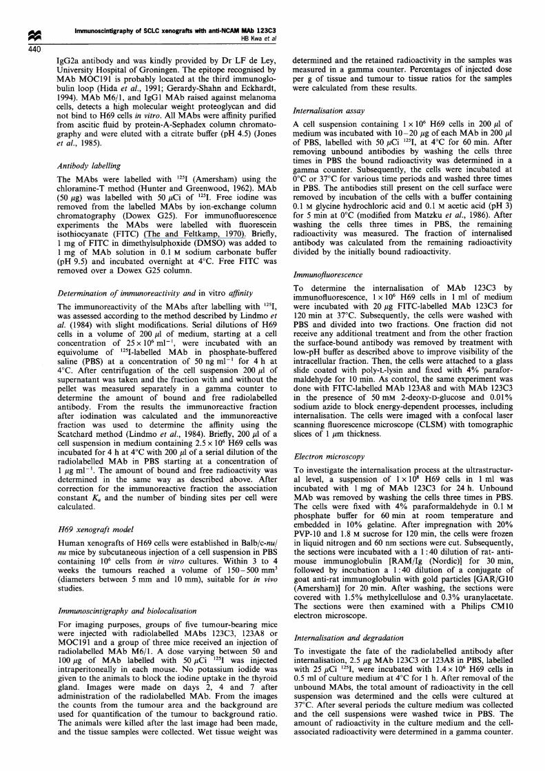
All three selected MAbs and the control MAb, M6/1,labelled with 1251, were injected intraperitoneally into micebearing H69 xenografts and after 2, 4 and 7 days scintigramswere made. The images made on days 2 and 4 showed a highbackground and a relatively low tumour uptake, whereas theimages made on day 7 had a relatively low background andhigher tumour uptake and were judged to be optimal (Figure1). These results were confirmed by quantifying the counts
from the images. The mean tumour to background ratioobtained from the images made with MAb 123C3 showed anincrease from 1.03 on day 2 to 1.99 on day 7, whereas forMAbs MOC191 and 123A8 the values remained constant at1.13 and 1.05. Images made with radiolabelled MAb 123C3showed a higher tumour uptake, resulting in a more distinctlocalisation of the tumour than the images produced withMAbs MOC191 and 123A8. The larger tumours showed ahigher total radioactive count than the smaller ones. Imagesmade with M6/1, the control antibody, showed no tumour atall, and a higher background activity than the cluster 1MAbs. The thyroid gland was not blocked in order tofacilitate the orientation of the scan and was clearly visible onall scintigraphic images.On day 7 the mice were dissected and the radioactivity in
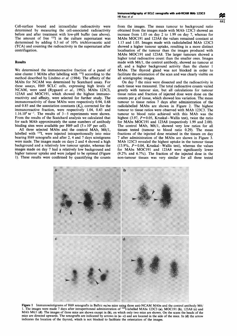
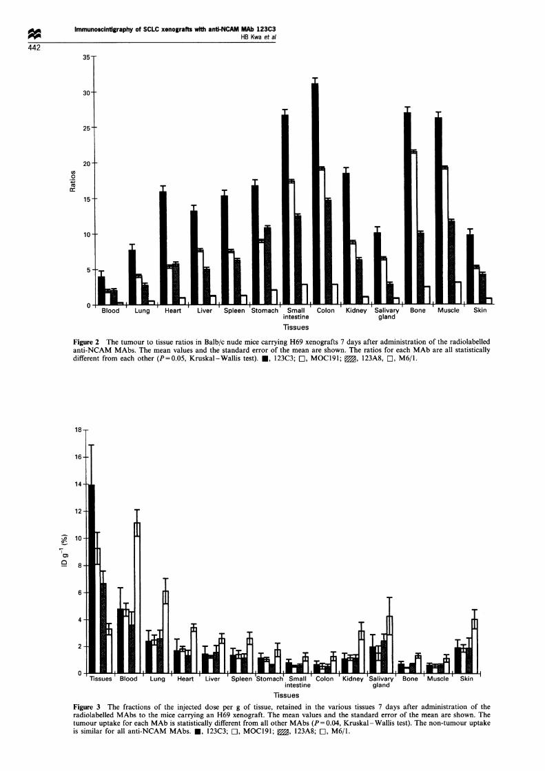
each tissue was measured. The total radioactive counts variedgreatly with tumour size, but all calculations for tumourtissue ratios and fraction of injected dose were done on thecounts per g of tissue, which showed less variation. The meantumour to tissue ratios 7 days after administration of theradiolabelled MAbs are shown in Figure 2. The highesttumour to tissue ratios were observed with MAb 123C3. Thetumour to blood ratio achieved with this MAb was thehighest (3.97, P=0.05, Kruskal-Wallis test), twice the ratiofor MAbs MOC191 and 123A8 (respectively 1.99 and 2.00).The control MAb, M6/1, showed very low ratios for alltissues tested (tumour to blood ratio 0.29). The meanfractions of the injected dose retained in the tissues on day7 after administration of the MAbs are shown in Figure 3.MAb 123C3 revealed the highest uptake in the tumour tissue(13.9%, P=0.04, Kruskal-Wallis test), whereas the valuesfor MAbs MOC191 and 123A8 were significantly lower(9.2% and 6.7%). The fraction of the injected dose in thenon-tumour tissues was very similar for all three tested
Figure 1 Immunoscintigrams of H69 xenografts in Balb/c nu/nu mice usin5g three anti-NCAM MAbs and the control antibody M6/1. The images were made 7 days after intraperitoneal administration of 12 I-labelled MAbs 123C3 (a), MOC191 (b), 123A8 (c) andMAb M6/1 (d). The images of three mice are shown except in (b), on which only two mice are shown. On the scans the heads of themice are directed upwards. The xenografts are indicated by arrows in (a-c) and are located in the side of the mice. In (d) the arrowindicates the location of the thyroid, which is not blocked to facilitate the orientation of the images.
Immunoscintigraphy of SCLC xenografts with anti-NCAM MAb 123C3HB Kwa et al
442
Co04)
intestine gland
Tissues
Figure 2 The tumour to tissue ratios in Balb/c nude mice carrying H69 xenografts 7 days after administration of the radiolabelledanti-NCAM MAbs. The mean values and the standard error of the mean are shown. The ratios for each MAb are all statisticallydifferent from each other (P=0.05, Kruskal-Wallis test). *, 123C3; E, MOCl91; 0, 123A8, E, M6/1.
18-
16-
14 -
T1
IT
_11
T
ssues Blood Lung Heart Liver Spleen Stomach Small Colon Kidney Salivary Bone Muscle Skinintestine gland
Tissues
Figure 3 The fractions of the injected dose per g of tissue, retained in the various tissues 7 days after administration of theradiolabelled MAbs to the mice carrying an H69 xenograft. The mean values and the standard error of the mean are shown. Thetumour uptake for each MAb is statistically different from all other MAbs (P = 0.04, Kruskal -Wallis test). The non-tumour uptakeis similar for all anti-NCAM MAbs. *, 123C3; O, MOCl91; 0, 123A8; E, M6/1.
12
10 -
8-
0,-
CD
6
4-
2
6-i4.. 6. --. . .-- .-- . . .-_ _JE --L..u
a
a-Moff-in -1 ILELINA t
Immunoscintigraphy of SCLC xenografts with anti-NCAM MAb 123C3HB Kwa et a!
443
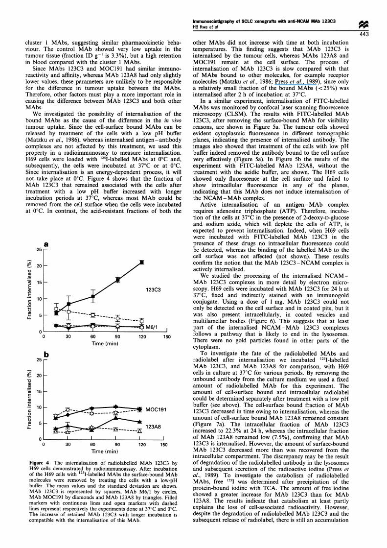
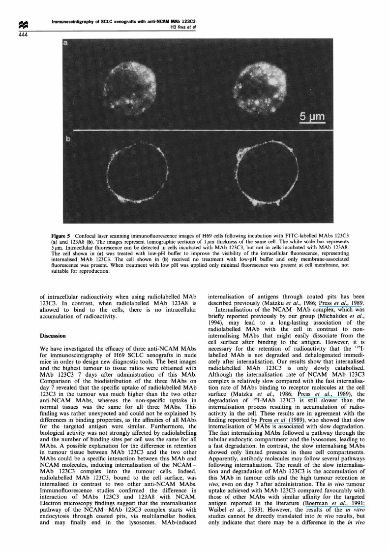
,ter 1 MAbs, suggesting similar pharmacokinetic beha- other MAbs did not increase with time at both incubationir. The control MAb showed very low uptake in the temperatures. This finding suggests that MAb 123C3 iskour tissue (fraction ID g-' is 3.3%), but a high retention internalised by the tumour cells, whereas MAbs 123A8 and)lood compared with the cluster 1 MAbs. MOC191 remain at the cell surface. The process ofsince MAbs 123C3 and MOC191 had similar immuno- internalisation of MAb 123C3 is slow compared with thatXivity and affinity, whereas MAb 123A8 had only slightly of MAbs bound to other molecules, for example receptorer values, these parameters are unlikely to be responsible molecules (Matzku et al., 1986; Press et al., 1989), since onlythe difference in tumour uptake between the MAbs. a relatively small fraction of the bound MAbs (<25%) wasrefore, other factors must play a more important role in internalised after 2 h of incubation at 37°C.sing the difference between MAb 123C3 and both other In a similar experiment, internalisation of FITC-labelledLbs. MAbs was monitored by confocal laser scanning fluorescenceVe investigated the possibility of internalisation of the microscopy (CLSM). The results with FITC-labelled MAbind MAbs as the cause of the difference in the in vivo 123C3, after removing the surface-bound MAb for visibilityour uptake. Since the cell-surface bound MAbs can be reasons, are shown in Figure 5a. The tumour cells showedased by treatment of the cells with a low pH buffer evident cytoplasmic fluorescence in different tomographicttzku et al., 1986), whereas internalised antigen-antibody planes, indicating the presence of internalised antibody. Theiplexes are not affected by this treatment, we used this images also showed that treatment of the cells with low pHperty in a radioimmunossay to measure internalisation. buffer indeed removed the antibody bound to the cell surface) cells were loaded with 1251-labelled MAbs at 0°C and, very effectively (Figure 5a). In Figure Sb the results of thesequently, the cells were incubated at 37°C or at 0°C. experiment with FITC-labelled MAb 123A8, without thece internalisation is an energy-dependent process, it will treatment with the acidic buffer, are shown. The H69 cellstake place at 0°C. Figure 4 shows that the fraction of showed only fluorescence at the cell surface and failed to
Lb 123C3 that remained associated with the cells after show intracellular fluorescence in any of the planes,.tment with a low pH buffer increased with longer indicating that this MAb does not induce internalisation ofibation periods at 37°C, whereas most MAb could be the NCAM-MAb complex.toved from the cell surface when the cells were incubated Active internalisation of an antigen-MAb complex)'C. In contrast, the acid-resistant fractions of both the requires adenosine triphosphate (ATP). Therefore, incuba-
tion of the cells at 37°C in the presence of 2-deoxy-D-glucoseand sodium azide, which will deplete the cells of ATP, isexpected to prevent internalisation. Indeed, when H69 cellswere incubated with FITC-labelled MAb 123C3 in the
a presence of these drugs no intracellular fluorescence could25 - be detected, whereas the binding of the labelled MAb to the
cell surface was not affected (not shown). These resultsconfirm the notion that the MAb 123C3 -NCAM complex isactively internalised.We studied the processing of the internalised NCAM-
MAb 1 23C3 complexes in more detail by electron micro-123C3 scopy. H69 cells were incubated with MAb 123C3 for 24 h at
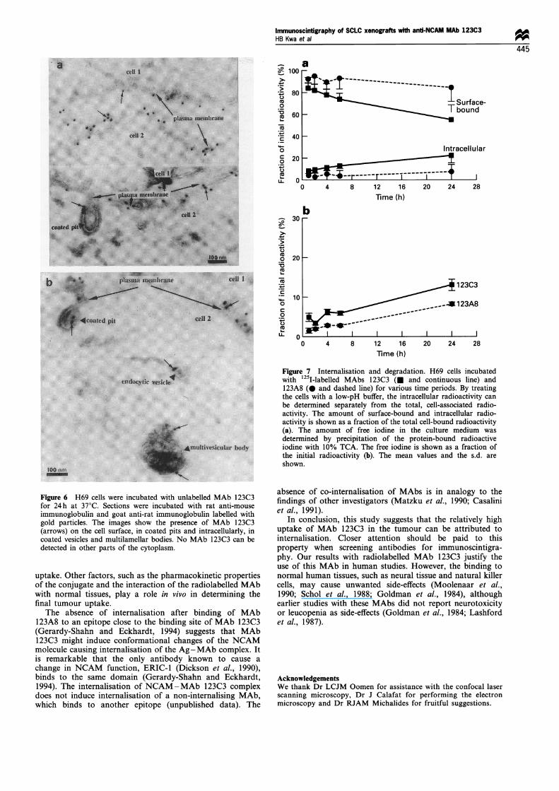
37°C, fixed and indirectly stained with an immunogoldconjugate. Using a dose of 1 mg, MAb 123C3 could notonly be detected on the cell surface and in coated pits, but itwas also present intracellularly, in coated vesicles andmultilamellar bodies (Figure 6). This suggests that at least
M6/1 part of the internalised NCAM-MAb 123C3 complexes0 6 so 1 follows a pathway that is likely to end in the lysosomes.0 30 60 90 120 150 There were no gold particles found in other parts of the
Time (min) cytoplasm.
b To investigate the fate of the radiolabelled MAbs and25 - radiolabel after internalisation we incubated 1251-labelled
MAb 123C3, and MAb 123A8 for comparison, with H6920 cells in culture at 37°C for various periods. By removing the
unbound antibody from the culture medium we used a fixedamount of radiolabelled MAb for this experiment. The
15- amount of cell-surface bound and intracellular radiolabelcould be determined separately after treatment with a low pH
10 M0C191buffer (see above). The cell-surface bound fraction of MAb
-------a ___ OCl91123C3 decreased in time owing to internalisation, whereas theamount of cell-surface bound MAb 123A8 remained constant
5123A8 (Figure 7a). The intracellular fraction of MAb 123C3
-----~~~~~fi~--- ]a increased to 22.3% at 24 h, whereas the intracellular fraction0 of MAb 123A8 remained low (7.5%), confirming that MAb0 30 60 90 120 150 123C3 is internalised. However, the amount of surface-bound
Time (min) MAb 123C3 decreased more than was recovered from theintracellular compartment. The discrepancy may be the result
gure 4 The internalisation of radiolabelled MAb 123C3 by of degradation of the radiolabelled antibody in the lysosomes69 cells demonstrated by radioimmunoassay. After incubation and subsequent secretion of the radioactive iodine (Press etthe H69 cells with 125I-labelled MAbs the surface-bound MAb al., 1989). To investigate the catabolism of radiolabelledDlecules were removed by treating the cells with a low-pH MAbs, free 1251 was determined after precipitation of theiffer. The mean values and the standard deviation are shown. protein-bound iodine with TCA. The amount of free iodine
Ab 123C3 is represented by squares, MAb M6/1 by circles, sAb MOC191 by diamonds and MAb 123A8 by triangles. Filled howed a greater increase for MAb 123C3 than for MAb
irkers with continuous lines and open markers with dashed 123A8. The results indicate that catabolism at least partly
es represent respectively the experiments done at 37°C and 0°C. explains the loss of cell-associated radioactivity. However,
ie increase of retained MAb 123C3 with longer incubation is despite the degradation of radiolabelled MAb 123C3 and thempatible with the internalisation of this MAb. subsequent release of radiolabel, there is still an accumulation
clusviou
tumin b
SreaclowforThecauMA
boutumrele,(MacowprolH6Csub:SincnotMAtreaincuremat C
V
0
U-
:._
co
c
G)
0
U-
FiE
of
bu,
lini
Th
coi
20
15
10 _
5 _
Immunoscintigraphy of SCLC xenografts with anti-NCAM MAb 123C3HB Kwa et al44
444
Figure 5 Confocal laser scanning immunofluorescence images of H69 cells following incubation with FITC-labelled MAbs 123C3(a) and 123A8 (b). The images represent tomographic sections of 1 gim thickness of the same cell. The white scale bar represents5 /tm. Intracellular fluorescence can be detected in cells incubated with MAb 123C3, but not in cells incubated with MAb 123A8.The cell shown in (a) was treated with low-pH buffer to improve the visibility of the intracellular fluorescence, representinginternalised MAb 123C3. The cell shown in (b) received no treatment with low-pH buffer and only membrane-associatedfluorescence was present. When treatment with low pH was applied only minimal fluorescence was present at cell membrane, notsuitable for reproduction.
of intracellular radioactivity when using radiolabelled MAb123C3. In contrast, when radiolabelled MAb 123A8 isallowed to bind to the cells, there is no intracellularaccumulation of radioactivity.
Discussion
We have investigated the efficacy of three anti-NCAM MAbsfor immunoscintigraphy of H69 SCLC xenografts in nudemice in order to design new diagnostic tools. The best imagesand the highest tumour to tissue ratios were obtained withMAb 123C3 7 days after administration of this MAb.Comparison of the biodistribution of the three MAbs onday 7 revealed that the specific uptake of radiolabelled MAb123C3 in the tumour was much higher than the two otheranti-NCAM MAbs, whereas the non-specific uptake innormal tissues was the same for all three MAbs. Thisfinding was rather unexpected and could not be explained bydifferences in binding properties, as the affinities of all MAbsfor the targeted antigen were similar. Furthermore, thebiological activity was not strongly affected by radiolabellingand the number of binding sites per cell was the same for allMAbs. A possible explanation for the difference in retentionin tumour tissue between MAb 123C3 and the two otherMAbs could be a specific interaction between this MAb andNCAM molecules, inducing internalisation of the NCAM-MAb 123C3 complex into the tumour cells. Indeed,radiolabelled MAb 123C3, bound to the cell surface, wasinternalised in contrast to two other anti-NCAM MAbs.Immunofluorescence studies confirmed the difference ininteraction of MAbs 123C3 and 123A8 with NCAM.Electron microscopy findings suggest that the internalisationpathway of the NCAM-MAb 123C3 complex starts withendocytosis through coated pits, via multilamellar bodies,and may finally end in the lysosomes. MAb-induced
internalisation of antigens through coated pits has beendescribed previously (Matzku et al., 1986; Press et al., 1989.
Internalisation of the NCAM-MAb complex, which wasbriefly reported previously by our group (Michalides et al.,1994), may lead to a long-lasting association of theradiolabelled MAb with the cell in contrast to non-internalising MAbs that might easily dissociate from thecell surface after binding to the antigen. However, it isnecessary for the retention of radioactivity that the 1251_labelled MAb is not degraded and dehalogenated immedi-ately after internalisation. Our results show that internalisedradiolabelled MAb 123C3 is only slowly catabolised.Although the internalisation rate of NCAM-MAb 123C3complex is relatively slow compared with the fast internalisa-tion rate of MAbs binding to receptor molecules at the cellsurface (Matzku et al., 1986; Press et al., 1989), thedegradation of 1251-MAb 123C3 is still slower than theinternalisation process resulting in accumulation of radio-activity in the cell. These results are in agreement with thefinding reported by Press et al. (1989), who showed that slowinternalisation of MAbs is associated with slow degradation.The fast internalising MAbs followed a pathway through thetubular endocytic compartment and the lysosomes, leading toa fast degradation. In contrast, the slow internalising MAbsshowed only limited presence in these cell compartments.Apparently, antibody molecules may follow several pathwaysfollowing internalisation. The result of the slow internalisa-tion and degradation of MAb 123C3 is the accumulation ofthis MAb in tumour cells and the high tumour retention invivo, even on day 7 after administration. The in vivo tumouruptake achieved with MAb 123C3 compared favourably withthose of other MAbs with similar affinity for the targetedantigen reported in the literature (Boerman et al., 1991;Waibel et al., 1993). However, the results of the in vitrostudies cannot be directly translated into in vivo results, butonly indicate that there may be a difference in the in vivo
Immunoscintigraphy of SCLC xenografts with anti-NCAM MAb 123C3HB Kwa et al
445
O Aa
Surface-T bound
0 4 8 12 16Time (h)
20 24 28
123C3
_ __-123A8
0 4 8 12 16 20 24 28Time (h)
Figure 6 H69 cells were incubated with unlabelled MAb 123C3for 24h at 37°C. Sections were incubated with rat anti-mouseimmunoglobulin and goat anti-rat immunoglobulin labelled withgold particles. The images show the presence of MAb 123C3(arrows) on the cell surface, in coated pits and intracellularly, incoated vesicles and multilamellar bodies. No MAb 123C3 can bedetected in other parts of the cytoplasm.
uptake. Other factors, such as the pharmacokinetic propertiesof the conjugate and the interaction of the radiolabelled MAbwith normal tissues, play a role in vivo in determining thefinal tumour uptake.
The absence of internalisation after binding of MAb123A8 to an epitope close to the binding site of MAb 123C3(Gerardy-Shahn and Eckhardt, 1994) suggests that MAb123C3 might induce conformational changes of the NCAMmolecule causing internalisation of the Ag-MAb complex. Itis remarkable that the only antibody known to cause a
change in NCAM function, ERIC-1 (Dickson et al., 1990),binds to the same domain (Gerardy-Shahn and Eckhardt,1994). The internalisation of NCAM-MAb 123C3 complexdoes not induce internalisation of a non-internalising MAb,which binds to another epitope (unpublished data). The
Figure 7 Internalisation and degradation. H69 cells incubatedwith 125I-labelled MAbs 123C3 (M and continuous line) and123A8 (0 and dashed line) for various time periods. By treatingthe cells with a low-pH buffer, the intracellular radioactivity can
be determined separately from the total, cell-associated radio-activity. The amount of surface-bound and intracellular radio-activity is shown as a fraction of the total cell-bound radioactivity(a). The amount of free iodine in the culture medium wasdetermined by precipitation of the protein-bound radioactiveiodine with 10% TCA. The free iodine is shown as a fraction ofthe initial radioactivity (b). The mean values and the s.d. areshown.
absence of co-internalisation of MAbs is in analogy to thefindings of other investigators (Matzku et al., 1990; Casaliniet al., 1991).
In conclusion, this study suggests that the relatively highuptake of MAb 123C3 in the tumour can be attributed tointernalisation. Closer attention should be paid to thisproperty when screening antibodies for immunoscintigra-phy. Our results with radiolabelled MAb 123C3 justify theuse of this MAb in human studies. However, the binding tonormal human tissues, such as neural tissue and natural killercells, may cause unwanted side-effects (Moolenaar et al.,1990; Schol et al., 1988; Goldman et al., 1984), althoughearlier studies with these MAbs did not report neurotoxicityor leucopenia as side-effects (Goldman et al., 1984; Lashfordet al., 1987).
AcknowledgementsWe thank Dr LCJM Oomen for assistance with the confocal laserscanning microscopy, Dr J Calafat for performing the electronmicroscopy and Dr RJAM Michalides for fruitful suggestions.
Il 10-
400
0i.-
Cu4I-
b^a 30
L-l
C.,C2
o 20CuL-
.' 100
cJ00Cu
A-
.
Immunoscintigraphy of SCLC xenografts with anti-NCAM MAb 123C3HB Kwa et a!
446References
BEVERLEY PCL, SOUHAMI RL AND BOBROW L. (1988). Results ofthe central data analysis. Lung Cancer, 4, 15 - 36.
BOERMAN OC, MIJNHEERE EP, BROERS JLV, VOOIJS GP ANDRAMAEKERS FCS. (1991). Biodistribution of a monoclonalantibody (RNL-1) against the neural cell adhesion molecule(NCAM) in athymic mice bearing human small-cell lung cancerxenografts. Int. J. Cancer, 48, 457-462.
CASALINI P, MEZZANZANICA D, CANEVARI S, DELLA TORRE G,MIOTTI S, COLNAGHI MI AND MATZKU S. (1991). Use ofcombination of monoclonal antibodies directed against threedistinct epitopes of a tumor-associated antigen: analysis of cellbinding and internalization. Int. J. Cancer, 48, 284-290.
DALES RE, STARK RM AND RAMAN S. (1990). Computedtomography to stage lung cancer. Approaching a controversyusing meta-analysis. Am. Rev. Resp. Dis., 141, 1096 -1101.
DICKSON G, PECK D, MOORE SE, BARTON H AND WALSH FS.(1990). Enhanced myogenesis in NCAM-transfected mousemyoblasts. Nature, 344, 348-351.
GAZDAR AF, CARNEY DN, RUSSEL EK, SIMS HL, BAYLIN SB,BUNN PA, GUCCION JG AND MINNA JD. (1980). Establishmentof continuous, clonable cultures of small-cell carcinoma of thelung which have amine precursor uptake and decarboxylation cellproperties. Cancer Res., 40, 3502-3507.
GERARDY-SHAHN R AND ECKHARDT M. (1994). Hot spots ofantigenicity in the neural cell adhesion molecule NCAM. Int. J.Cancer, 49, (suppl. 8), 38-42.
GOLDMAN A, VIVIAN G, GORDON I, PRITCHARD J AND KEMS-HEAD J. (1984). Immunolocalization of neuroblastoma usingradiolabeled monoclonal antibody UJ13A. J. Pediatr., 105, 252-256.
HIDA T, KOIKE K, SEKIDO Y, NISHIDA K, SIGIURA T, ARIYOSHI Y,TAKAHASHI T AND UEDA R. (1991). Epitope analysis of cluster 1and NK cell-related monoclonal antibodies. Br. J. Cancer, 63(suppl.), 24-28.
HUNTER WM AND GREENWOOD FC. (1962). Preparation of iodine-131-labelled growth hormone of high specific radioactivity.Nature, 194, 495-496.
IHDE DC. (1985). Staging evaluation and prognostic factors in small-cell lung cancer. In Lung Cancer, Aisner J (ed). pp. 241-268.Churchill Livingstone: New York.
JONES DH, GOLDMAN A, GORDON I, PRITCHARD J, GREGORY BJAND KEMSHEAD JT. (1985). Therapeutic application of aradiolabelled antibody in nude mice xenografted with humanneuroblastoma: tumoricidal effects and distribution studies. Int.J. Cancer, 35, 715-720.
LASHFORD L, JONES D, PRITCHARD J, GORDON I, BREATNACH FAND KEMSHEAD JT. (1987). Therapeutic application ofradiolabeled monoclonal antibody UJ1 3A in children withdisseminated neuroblastoma. NCI Monogr., 3, 53 - 57.
LEDERMANN JA, PASINI F, OLABIRAN Y AND PELOSI G. (1994).Detection of the neural cell adhesion molecule (NCAM) in serumof patients with small-cell lung cancer (SCLC) with 'limited' or'extensive' disease, and bone-marrow infiltration. Int. J. Cancer, 8(suppl.), 49 - 52.
LINDMO T, BOVAN E, CUTTITA F, FEDORKO J AND BUNN PA Jr.(1984). Determination of immunoreactive fraction of radiola-beled monoclonal antibodies by linear extrapolation to binding atinfinite antigen excess. J. Immunol. Methods, 72, 77- 79.
MATZKU S, BROCKER EB, BRUGGEN J, DIPPOLD WG AND TILGENw. (1986). Modes of binding and internalization of monoclonalantibodies to human melanoma cell lines. Cancer Res., 46, 3848 -3854.
MATZKU S, MOLDENHAUER G, KALTHOFF H, CANEVARI S,COLNAGHI M, SCHUHMACHER J AND BIHL H. (1990). Anti-body transport and internalization into tumours. Br. J. Cancer, 62(suppl. X), 1-5.
MICHALIDES R, KWA B, SPRINGALL D, VAN ZANDWIJK N,KOOPMAN J, HILKENS J AND MOOI W. (1994). NCAM andlung cancer. Int. J. Cancer, 8 (suppl.), 34- 37.
MOOLENAAR CEC, MULLER EJ, SCHOL DJ, FIGDOR CG, BOCK E,BITTER-SUERMANN D AND MICHALIDES RJAM. (1990).Expression of neural cell adhesion molecule related sialoglyco-protein in small cell lung cancer and neuroblastoma cell lines H69and CHP-212. Cancer Res., 50, 1102- 1106.
NELP WB, GRIEP RG, SALK D, ABRAMS P, SUPPERS V ANDHANLEY M. (1990). Successful staging of small cell lung cancer(SCLC) with monoclonal antibody. Eur. J. Nucl. Med., 16, SI 99.
PRESS OW, FARR AG, BORROZ KI, ANDERSON SK AND MARTINPJ. (1989). Endocytosis and degradation of monoclonal anti-bodies targeting human B-cell malignancies. Cancer Res., 49,4906-4912.
RYGAARD K, M0LLER C, BOCK E AND SPANG-THOMSEN M.(1992). Expression of cadherin and NCAM in human small celllung cancer cell lines and xenografts. Br. J. Cancer, 65, 573 - 577.
SCHOL DJ, MOOI WJ, VAN DER GUGTEN AA, WAGENAAR SJScAND HILGERS J. (1988). Monoclonal antibody 123C3, identifyingsmall cell carninoma phenotype in lung tumors, recognizesmainly, but not exclusively, endocrine and neuron-supportingnormal tissues. Int. J. Cancer, 43 (suppl.2), 34-40.
THE TH AND FELTKAMP TEW. (1970). Conjugation of fluoresceinisothiocyanate to antibodies. Immunology, 18, 865-881.
WAIBEL R, MANNHART M, O'HARA CJ, BROCKLEHURST C,ZANGEMEISTER-WITTKE U, SCHENKER T, LEHMANN HP,WEBER E AND STAHEL RA. (1993). Monoclonal antibodySEN7 recognizes a new epitope on the neural cell adhesionmolecule present on small cell lung cancer but not onlymphocytes. Cancer Res., 53, 2840-2845.
YESNER R. (1985). Classification of lung cancer histology. N. Engl.J. Med., 312, 652-653.