The Underlying Physiology of Arterial Pulse Wave Morphology ...
11
495 Available at http://pvamu.edu/aam Appl. Appl. Math. ISSN: 1932-9466 Vol. 8, Issue 2 (December 2013), pp. 495 – 505 Applications and Applied Mathematics: An International Journal (AAM) The Underlying Physiology of Arterial Pulse Wave Morphology in Spatial Domain Nzerem Francis Egenti Department of Mathematics & Statistics University of Port Harcourt Nigeria [email protected] Alozie Harrieth Nkechi State Education Management Board Zone 2 Owerri, Nigeria Received: February 5, 2013; Accepted: August 28, 2013 Abstract Cardio-vascular events are among the world’s leading causes of morbidity and mortality. Most postulates suppose that culinary delights can be implicated in incidences of cardio-vascular diseases. This school of thought holds well in many respects. Much as the truistic value of the said school is acknowledged, we conceived of physiological disposition as an endogenous dominant factor in the events being considered, whereas culinary measures constitute an exogenous contributory factor. In this work we aimed at studying the effects of distance (stature) on pulse waveforms. Certain elements of our study showed that pulse wavelength was dominant in prescribing cardio-vascular physiology. Keywords: Mathematical analysis; soliton; left ventricle; hemodynamics; pulse amplification; physiology AMS-MSC 2010 No.: 93A30, 74F10 1. Introduction The world is riddled with nauseating pathologies with attendant tolls on human life. The quality of life derives from sound and delectable health. Unfortunately, the deleteriousness of diseases brings much to bear on the wellbeing of animated life, especially of humans. The World Health Organization’s (2004) release of the leading causes of human mortality showed that
-
Upload
khangminh22 -
Category
Documents
-
view
1 -
download
0
Transcript of The Underlying Physiology of Arterial Pulse Wave Morphology ...
495
Available at http://pvamu.edu/aam
Appl. Appl. Math.
ISSN: 1932-9466
Vol. 8, Issue 2 (December 2013), pp. 495 – 505
Applications and Applied Mathematics:
An International Journal (AAM)
The Underlying Physiology of Arterial Pulse Wave
Morphology in Spatial Domain
Nzerem Francis Egenti Department of Mathematics & Statistics
University of Port Harcourt Nigeria
Alozie Harrieth Nkechi State Education Management Board
Zone 2 Owerri, Nigeria
Received: February 5, 2013; Accepted: August 28, 2013
Abstract Cardio-vascular events are among the world’s leading causes of morbidity and mortality. Most postulates suppose that culinary delights can be implicated in incidences of cardio-vascular diseases. This school of thought holds well in many respects. Much as the truistic value of the said school is acknowledged, we conceived of physiological disposition as an endogenous dominant factor in the events being considered, whereas culinary measures constitute an exogenous contributory factor. In this work we aimed at studying the effects of distance (stature) on pulse waveforms. Certain elements of our study showed that pulse wavelength was dominant in prescribing cardio-vascular physiology. Keywords: Mathematical analysis; soliton; left ventricle; hemodynamics; pulse amplification;
physiology AMS-MSC 2010 No.: 93A30, 74F10
1. Introduction
The world is riddled with nauseating pathologies with attendant tolls on human life. The quality of life derives from sound and delectable health. Unfortunately, the deleteriousness of diseases brings much to bear on the wellbeing of animated life, especially of humans. The World Health Organization’s (2004) release of the leading causes of human mortality showed that
496 Nzerem Francis Egenti and Alozie Harrieth Nkechi
cardiovascular diseases are ranked first in frequency (with 29.34 per 100 deaths). Cardiovascular diseases affect the heart and the blood vessels. In assessing the quality of life, experts agree that auxology, the study of stature, is one of the key factors in determining overall health. It was opined by Samaras and Elrick (2002) that tallness is associated with cardiovascular wellbeing. Shortness was said to have the conflict of being associated with longevity and with associated diseases. In this work, pulse waveform was studied in relation to distance with the aim of determining its effect on cardiovascular physiology. Our spatial domain is the arterial length occupied by the pulse wave, which is apparently dependent on the stature of an individual subject.
In a study on cardiovascular system, hemodynamics is of essential. Hemodynamics as proposed by McDonald (1974) is defined as the physical study of flowing blood (fluid) and the entire solid structures (arteries) through which it flows. Thus, fluid-structure interaction (FSI) maintains the integrity of arterial flow. The germ of hemodynamics, however, is the dynamics of the heart’s left ventricle (LV), which ejects blood into the aorta during the systolic phase. Pulse is created in the event of aortic effusion into the arterial network. In essence, PW is a component of cardiovascular system. Many literatures such as Crepeau and Sorine (2005), Laleg et al. (2007), Fu and IL` Ichev (2010) and Duan et al. (1997) agree that the Korteweg de-Vries (KdV) equation holds well for arterial pulse waves. In Crepeau and Sorine (2005), the goal was to interpret pulse wave characteristics and to provide basis for the estimations from non-invasive measurements. Laleg et al. (2007) decomposed pressure into a travelling wave which is evident during systolic phase and a windkessel flow during the diastolic phase. Many important physiological interests are the associated pulse wave (PW). O’Rourke and Pauca (2001) showed that pulse wave analysis (PWA) is a choice descriptor of physiological states. Wei et al. (1989) used it in explaining the resonance of the organs with the heart. Yu-Cheng et al. (2004) studied the harmonic stability of the arterial pulse.
An aspect of physiological interest that may not have received adequate attention is the morphological dependence of waveforms on the distance occupied by solitary waves. Neither the literatures we have presented here nor any other work, as much as we know at present, dealt with the mathematical theory of pulse wave morphology in spatial domain, especially in relation to human stature. This work supplies the omission.
In Section 2.0 some vital elements of FSI are treated. The equation relating pressure to shear modulus is derived. Section 3.0 discusses the emergence of arterial pulse waves in the event of left ventricular ejection. Korteweg de-Vries (KdV) equation, which holds well for arterial pulse waves, was derived. Consequently, two soliton-solutions (2SS), which are characteristic of the KdV equation, were obtained. They are enough to describe the ‘peaking’ and ‘steepening’ wave phenomena. In section 4.0, a physiological analysis of the resulting pressure waveforms was made. In section 5.0, some conclusions were drawn based on the analysis. 2. Equations of Fluid-Structure Interaction Here we consider the flow inside deformable domains (arteries) and the resulting motion of the domains. Figure 2.1 shows the nondeformed arterial configuration. Define the arterial reference domain (t) by (Suncica et al. (2005)),

AAM: Intern. J., Vol. 8, Issue 2 (December 2013) 497
(t)=xℝ3: x = (rcos, rsin, z), r < R + (z, t), 0 < z < L, (2.1)
Figure 2.1. Reference domain
and define the lateral boundary by
=x=(R (z) cos, R(z) sin, z) ℝ3: (0, 2), z(0, L), (2.2) where R and r are the tube’s outer and inner radii respectively, L the tube’s length, =R/L, the radial displacement from the reference state; Suppose the flow in (t) is fully developed and axisymmetric, with no angular velocity. The following, describing equations of flow in Eulerian formulation, apply:
01
22
2
2
2
r
p
r
v
r
v
rz
v
r
v
z
vv
r
vv
t
v rrrrrz
rr
r
, (2.3)
01
2
2
2
2
z
p
r
v
rz
v
r
v
z
vv
r
vv
t
v zzzzz
zr
r
. (2.4)
The continuity equation representing the incompressibility condition, div v = 0 is
0
r
v
z
v
r
v rzr
, (2.5)
where vr, vz are the radial and longitudinal components of the fluid respectively, μ the viscosity of the fluid, p the pressure and the density. The evolution of the response of the lateral boundary to the fluid flow is described by the Navier’s equations given by (Quarteroni et al. (2000)):
z
zrzw
z
v
z
v
R
Eh
t
vh
2
2
22
2
1 in (0, L), (2.6)
where, h is the arterial wall thickness, E is the Young modulus of elasticity, R is the arterial reference radius at rest, w is the arterial volumetric mass, σ is the Poisson ratio, is the term due to the external forces, including the stress from the fluid. It is noteworthy that this model is based on a Lagrangian description of the motion of the elastic wall, referred to a material domain (0), corresponding to the rest state, where vr = vz = 0.
z
)(zR
Ω L
0

498 Nzerem Francis Egenti and Alozie Harrieth Nkechi
The cylinder, together with its boundary, deforms as a result of the fluid-wall interaction between the fluid occupying the domain and the cylinder’s boundary, as shown in Figure 2.2.
Figure 2.2. Wall displacement
We assume that the lateral wall allows only radial displacements: (t) = r = R +η (z, t)(0, L). Thus, the radial contact force given by Suncica et al. (2005) in the form is
2
2
2
2
22)()()(
1
)()(
th
zGh
R
EhF wr
, (2.7)
where E() is the Young’s modulus of elasticity, h() is the wall thickness, is the Timoshenko shear correction factor (see Reisman (1988)), G is the shear modulus and is the Poisson ratio. (If the material is incompressible, then = 0.5). In equation (2.7), the right-hand members are described as follows: the first term is the elastic response function, the second term is related to the radial pre-stress state of the vessel and the third term is the inertia term that is proportional to the radial acceleration of the vessel wall. The fluid velocity at the deformed interface (R+η, z, t) must be equal to the Lagrangean velocity of the membrane. Since only the radial displacements are non-zero we have
),(),,( tz
ttzRvr
on (0, L) ℝ+ , (2.8)
0),,(
t
StzRvz
on (0, L) ℝ+ . (2.9)
The continuity of velocity along the longitudinal displacement from the reference state Sε is
assumed zero, and thus t
S
= 0. The radial contact force is equal to the radial displacement
component of the stress exerted by the fluid to the membrane. Thus,
-Fr =[(p – pref) I - 2D (v)] ner = r
vpp r
ref
2I , (2.10)
where pref is the pressure at the reference domain, n is the unit normal at the deformed structure, D (v) is the deviatoric part of an axially symmetric vector/tensor-valued function zzrr evevv given by
εS
ΩR
AAM: Intern. J., Vol. 8, Issue 2 (December 2013) 499
z
v
r
v
z
vr
v
r
v
z
v
r
v
vD
zzr
r
zrt
02
1
00
2
10
)(
.
The cylinder’s initial condition, when filled with fluid, under reference pressure pref is such that
0
t
S
tS
, and v = 0 on × 0. (2.11)
The integrity of flow is the temporal dynamic pressure prescribed at both ends of the cylinder. We, therefore, apply the following boundary (inlet/outlet) conditions as given in Suncica et al. (2005):
R)0()(2
)(,0 0
2
zonptpv
pv refz
r
, (2.12)
R)()(2
)(,0
2
Lzonptpv
pv refLz
r
, (2.13)
z
S
= = 0 for z = 0, S = = 0 for z = L and tℝ +, (2.14)
where the pressure drop is assumed to be A(t) = pL(t) – p0(t) ),0(C0 .
We summarize the fluid-structure interaction problem as follows:
Equations (2.3), (2.4), (2.5) in Ωε ,
Equations (2.8), (2.9) on Σε× ℝ + ,
Equation (2.10) on Σε× ℝ + , (2.15)
Equations (2.12), (2.13) on (∂Ωε∩ z = 0, L × ℝ + ,
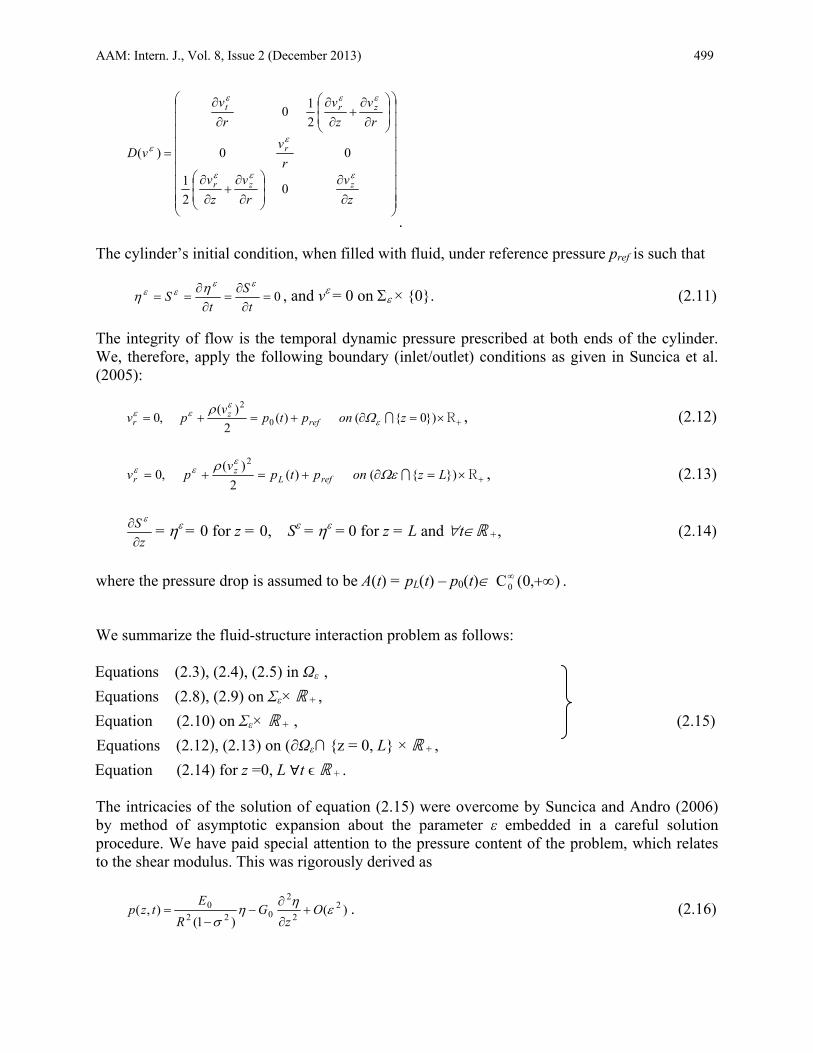
Equation (2.14) for z =0, L ∀t ϵ ℝ + . The intricacies of the solution of equation (2.15) were overcome by Suncica and Andro (2006) by method of asymptotic expansion about the parameter ε embedded in a careful solution procedure. We have paid special attention to the pressure content of the problem, which relates to the shear modulus. This was rigorously derived as
)()1(
),( 22
2
0220
Oz
GR
Etzp
. (2.16)

500 Nzerem Francis Egenti and Alozie Harrieth Nkechi
The shear modulus is required mainly in the determination of the wave speeds; it also describes the response to the shear wave. When shear waves are considered, shear modulus is of great importance. Here, the assumption of negligible shear modulus and material incompressibility implies that G0 = 0 and σ = 0.5 respectively. Hence,
RO
A
A
R
E
R
Ep
)0(1
3
4
3
4 020 , (2.17)
where A(0) is the cross-sectional area at x corresponding to zero pressure.
3. Equations of Pressure Waves In the preceding section, the governing equations of FSI were presented. The integrity of pressure waves is the arterial pressure. Equation (2.17) is the expression of pressure that is required to drive pulse waves in an arterial segment. Such waves are presented in this section. Assume an elastic artery and incompressible fluid flow. The governing Navier-Stokes equations as presented by Crepeau and Sorine (2006) are
AT + QZ = 0, (3.1)
02
A
QP
A
A
QQ ZT
, (3.2)
where A (T, Z) = R2 (T, Z) is the cross-sectional area of the vessel, Q (T, Z) is the blood flow volume, P (T, Z) is the blood pressure, with T and Z being temporal and spatial dimensions respectively, is the blood density, ν is a coefficient of the blood viscosity and the quantities AT, PZ and QZ are gradients associated with their respective subscripts. Yomosa (1987) described the motion of the arterial wall by
so
oeTT
o
oow
R
hPPA
A
Rh
)( , (3.3)
where w is the wall density, Pe is the pressure outside the tube, ho is the mean thickness of the wall, Ro is the mean radius and s is the extending stress in the tangential direction. Since the wall is elastic, the local compliance of the vessel is such that
,
2
)(
o
os A
AAE (3.4)
where A0 is the arterial cross-sectional area at rest, and E is the coefficient of elasticity. The non-dimensional representations of equations (3.1)-(3.4) are
at + qz= 0, (3.5)

AAM: Intern. J., Vol. 8, Issue 2 (December 2013) 501
a
qpa
a
qq z
z
t
1)1(
1
2
, (3.6)
5att + a = p, (3.7)
where )(00cA
, is the typical wavelength of the wave propagated in the tube and co the
Moens-Korteweg sound wave velocity in the fluid-filled tube.
When equations (3.5), (3.6) and (3.7) are expressed as an asymptotic expansion at the second order of ε, and noting that the coefficients of series must vanish, we have the following KdV equation:
021 TTTTToZ PdPPdPdP , (3.8)
with 3221
2,
1
2
3,
1
o
oow
oooo
c
Rhd
cAd
cd
.
Let P = Ps be a soliton solution, then we put equation (3.8) in the form
.03
3
21
t
Pd
t
PPdd
z
P ssso
s (3.9)
With the following transformation: Γ = t – doz, = d2z and sPd
du
2
1
6 , we get
u +6uuΓ+uΓΓΓ=0. (3.10) The two soliton solution (2SS) of equation (3.10) is (see Hereman and Zhuang (1994)):
u(x,t)
22
21
1
2222
12212
22
1
2tanh
2coth
2sec
2cos
2
kk
hkechkkk , (3.11)
where iiii tx 3 ; i being an arbitrary constant (i = 1,2), k is the wave number.
4. Physiological Analysis Waves furnish a wealth of information about the matter in which they propagate. A model of arterial wave propagation widens our knowledge of the cardiovascular system and provides a clue to the diagnosis of pathological conditions. It, therefore, helps in the prediction of the outcome of medical interventions. The solution of the wave problem given by equation (3.11) describes the ‘peaking’ and ‘steepening’ phenomena that are typical of solitary waves. Such

502 Nzerem Francis Egenti and Alozie Harrieth Nkechi
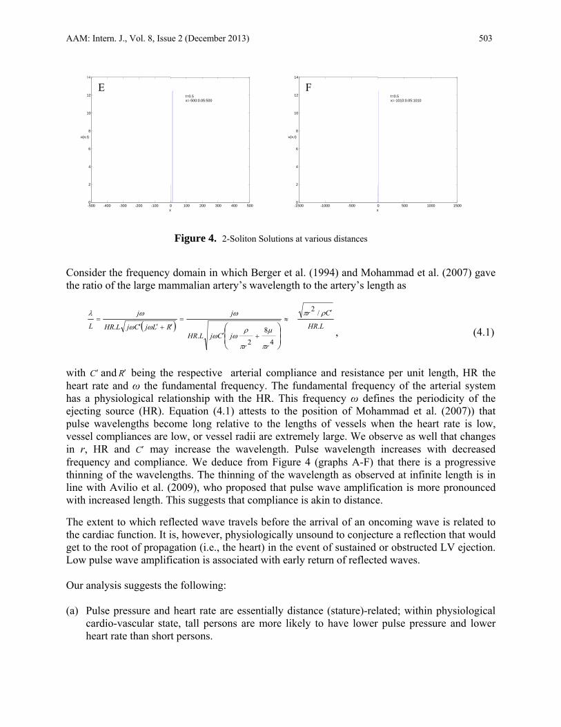
pulse waves propagated through the arterial bed are an estimator of the systolic phase of blood pressure. A usual phenomenon in wave motion is reflection. Waves are reflected at various arterial sites which may include points of bifurcation, areas of changes in arterial geometry, peripheral and terminal regions. However, it is customary to integrate multiple reflected waves as a single wave. The sum of the reflected waves and the forward ejected wave forms the final profile of the pressure pulse wave. The issue that is of clinical importance is the return of the reflected waves. This brings to concern the implication of proximity or otherwise of an organ to the heart. In this work the behavior of pulse wave was studied in relation to the distance travelled. In doing this, distance was varied at a fixed time mark. The understanding of the waveform at various distances occupied by the wave would provide a clue to its relationship with arterial length (or height) so that a physiological condition may be analyzed. 4.1. Distance Effects The waveforms of equation (3.11) at various distances are shown in the graphs of Figure 4. The graphs (A-F) represent six 2SS at various distances. They reveal distance effects on solitons. Each successive increase in distance caused the shorter wave to plummet, as seen in Graphs A-F (Figure 4.2). In graph F the shorter wave was almost encapsulated by the taller one. There is successive diminishing of the wavelength.
A
-2 -1.5 -1 -0.5 0 0.5 1 1.5 20
2
4
6
8
10
12
x
u(x,t)
t=0.05x=-2:0.05:2 B
-5 -4 -3 -2 -1 0 1 2 3 4 50
2
4
6
8
10
12
x
u(x,t)
t=0.05x=-5:0.05: 5
C
-10 -8 -6 -4 -2 0 2 4 6 8 100
2
4
6
8
10
12
x
u(x,t)
t=0.05 x=-10:0.05:10
D
-40 -30 -20 -10 0 10 20 30 400
2
4
6
8
10
12
x
u(x,t)
t=0.05x=-40:0.05:40
AAM: Intern. J., Vol. 8, Issue 2 (December 2013) 503
Figure 4. 2-Soliton Solutions at various distances
Consider the frequency domain in which Berger et al. (1994) and Mohammad et al. (2007) gave the ratio of the large mammalian artery’s wavelength to the artery’s length as
LHR
Cr
rr
jCjLHR
j
RLjCjLHR
j
L .
/2
4
8
2.
.
, (4.1)
with C and R being the respective arterial compliance and resistance per unit length, HR the heart rate and ω the fundamental frequency. The fundamental frequency of the arterial system has a physiological relationship with the HR. This frequency ω defines the periodicity of the ejecting source (HR). Equation (4.1) attests to the position of Mohammad et al. (2007)) that pulse wavelengths become long relative to the lengths of vessels when the heart rate is low, vessel compliances are low, or vessel radii are extremely large. We observe as well that changes in r, HR and C may increase the wavelength. Pulse wavelength increases with decreased frequency and compliance. We deduce from Figure 4 (graphs A-F) that there is a progressive thinning of the wavelengths. The thinning of the wavelength as observed at infinite length is in line with Avilio et al. (2009), who proposed that pulse wave amplification is more pronounced with increased length. This suggests that compliance is akin to distance.
The extent to which reflected wave travels before the arrival of an oncoming wave is related to the cardiac function. It is, however, physiologically unsound to conjecture a reflection that would get to the root of propagation (i.e., the heart) in the event of sustained or obstructed LV ejection. Low pulse wave amplification is associated with early return of reflected waves. Our analysis suggests the following: (a) Pulse pressure and heart rate are essentially distance (stature)-related; within physiological
cardio-vascular state, tall persons are more likely to have lower pulse pressure and lower heart rate than short persons.
E
-500 -400 -300 -200 -100 0 100 200 300 400 5000
2
4
6
8
10
12
14
x
u(x,t)
t=0.5x=-500:0.05:500
F
-1500 -1000 -500 0 500 1000 15000
2
4
6
8
10
12
14
x
u(x,t)
t=0.5 x=-1010:0.05:1010

504 Nzerem Francis Egenti and Alozie Harrieth Nkechi
(b) Short statures are likely to experience more shortened return time of reflected waves, and thus lower pulse amplification, than tall statures. (Shortened return time of reflected waves to the aorta in early systole, even at physiological wave velocity, may burden the left ventricle with importunate requests for more pulsatile effort.)
(c) At an infinitely long distance pulse wavelength gets thinner. This is in line with the
transmission theory, as contained in Berger et al. (1994), which supposes finite wavelength. 5. Summary and Conclusion Arterial pulse wave is a phenomenon of choice in prescribing physiological states. In particular, Yu-Cheng et al. (2004) contended that waveforms are a key factor in explaining the overall pathologies and prognosis. This work considered pulse waveforms in the light of stature, with the view of gaining an insight into its influence on cardiovascular events. The integrity of the blood flow is the FSI. We presented the FSI in mathematical terms. In pursuance of the essence of this work, the distance effects of the 2SS obtained from the wave problem were analyzed. It was seen that pulse wavelength is phenomenal in describing some cardiovascular parameters such as HR and arterial compliance. The decrease in pulse wavelength, as seen in increased distance, is a mark of arterial compliance and physiological HR. HR, compliance and pulse amplification are modulators of cardiovascular state. Subsisting long pulse wavelength, which keeps pulse amplification wanting, resides in short arterial distance. As earlier remarked (in section 4.0), long pulse wavelength is a mark of stifled physiological HR and/or arterial compliance. We are inclined to conclude that short arterial length, marked by short stature at maturity, can be a cardiovascular liability.
REFERENCES
Avolio, Alberto P., Van Bortel, Luc M., Boutouyrie, Pierre., Cockcroft, John R., McEniery,
Carmel M., Protogerou, Athanase D., Roman, Mary J., Safar, Michel E., Segers, Patrick and Smulyan, Harold (2009). Role of Pulse Pressure Amplification in Arterial Hypertension, Seventh International Workshop on Structure and Function of the Vascular System, Hypertension, Vol. 54.
Berger, D. S., Li J. K. and Noordergraaf, A. (1994). Differential effects of wave reflections and peripheral resistance on aortic blood pressure: a model-based study, Am J Physiol Heart Circ Physiol, 266.
Crepeau, Emmanuelle and Sorine, Michel (2006). A reduced model of pulsatile flow in an arterial compartment, Chaos, Solutions & Fractals, doi:10.1016/j.chaos.2006.03.096.
Duan, Wen-shan, Wang, Ben-ren, and Wei, Rong-jue (1997). Reflection and transmission of nonlinear blood waves due to arterial branching, Physical Review E 55, No. 2.
Emmanuelle, Crepeau and Sorine, Michel (2005). Identifiability of a reduced model of pulsatile flow in an arterial compartment, CDC-ECC’05.44th IEEE Conference, Doi: 10.1109/CDC.2005.1582270.

AAM: Intern. J., Vol. 8, Issue 2 (December 2013) 505
Fu, Y. B. and IL’Ichev, A. T. (2010). Solitary Waves in fluid-filled tubes: existence, persistence and the role of axial displacement, IMA Journal of Applied Mathematics, Vol.75.
Hereman, Willy and Zhuang, Wuning (1994). Symbolic Computation of Solitons via Hirota’s Bilinear Method http://inside.mines.edu/~whereman/papers/Hereman-Zhuang-Hirota-Method-Preprint-1994.pdf, retrieved 24 May, 2013.
McDonald, D.A. (1974). Blood flow in arteries (2nd Ed.), UK: Arnold. O’Rourke, Michael F., Alfredo, Pauca and Jiang Xiong-Jing J. (2001). Pulse Wave Analysis, Br
J Clin Pharmacol, Vol 51, No. 6. Quarteroni, Alfio., Tuveri, Massimiliano and Veneziani, Alessandro (2000). Computational
vascular fluid dynamics: Problems, models and methods. Comput. Vis. Sci.,Vol.2. Quick, C. M., Berger, D.S. and Noordergraaf, A. (1998). Apparent arterial compliance. Am J
Physiol Heart Circ Physiol., Vol. 274. Reismann, H. (1988).Elastic Plates: Theory and Applications, Wiley, NY. Samaras, T., Elrick, H. (2002). Height, Body Size and Longevity: Is smaller better for the human
body? The Western Journal of Medicine, Vol. 173, No. 3. Suncica Canic, Daniel, Lamponi, Andro, Mikelic, and Josip, Tambaca (2005). Self-Consistent
Effective Equations Modeling Blood Flow in Medium-to Large Compliant Arteries, Multiscale modeling and Simulation, Vol.3.
Suncica, Canic and Andro, Mikelic. (In press). Effective Equations Modeling the Flow of a Viscous Incompressible Fluid through a Long Elastic Tube Arising in the Study of Blood Flow through Small Arteries, SIAM J. Appl. Dyn. Systems, doi. 10.1137/S1111111102411286.
Taous-Merien, Laleg, Crespeau, Emmanuelle, Sorine, Mickel (2006). Seperation of arterial pressure into solitary waves and windkessel flow, Modeling and Control in Biomedical Systems, Volume 6, Part 1.
Wei-Kung, Wang, Lo YY, Chieng Y, Yuh-Ying, Lin, W, and Hsu, TL. (1989). Resonance of organs with the heart, In Biomedical Engineering: an International Symposium (Young W.J., Editor), pp. 259-268, Washington: Hemisphere Publishing Corp.
World Health Organization (2004) Annex Table 2: Deaths by Cause, Sex and Mortality Stratum in WHO Regions, Estimates for 2002 (pdf). The World Health Report-Changing History, http://www.who.int/whr/2004/annex/topic/en/annex_2_en.pdf, Retrieved, 2012-11-01.
Yomosa, S. (1987). Solitary waves in large blood vessels, J Phys Soc Jpn, Vol. 56. Yu-Cheng Mohammad, Mohiuddin W., Glen, Laine A. and Christopher, Quick M. (2007).
Increase in pulse wavelength causes the systemic arterial tree to degenerate into a classical windkessel, AJP – Heart, Vol. 293, No 2.
Yu-Cheng, Kuo, Lo, S.H. and Wei-Kung, Wang. Harmonic variations of arterial pulse during dying process in rats. In: http://www.dtic.mil/dtic/tr/fulltext/u2/a412480.pdf, Retrieved on 6 July, 2012.
Yu-Cheng, Kuo, Tai-Yuan, Chiu, Ming-Yie, Jan, Jian-Guo, Bau, Sai-Ping, Li, Wei-Kung,Wang, Wang, Yuh-Ying and Lin, W. (2004). Losing harmonic stability of arterial pulse in terminally ill patients, Blood Press Monit. Vol. 9, No. 5.
Yuh-Ying, Lin W., Ming-Yie, Jan, Ching-Show, Shyu, Chi-Ang, Chiang and Wei-Kung, Wang (2004). The natural frequencies of the arterial system and their relation to the heart rate, IEEE Trans. Biomed. Eng., Vol. 51.