The Turkish Journal of - Ear Nose and Throat - DergiPark
39
E-ISSN 2602-4837 The Turkish Journal of Ear Nose and Throat Volume 31, Number 1 / March 2021
-
Upload
khangminh22 -
Category
Documents
-
view
0 -
download
0
Transcript of The Turkish Journal of - Ear Nose and Throat - DergiPark
E-ISSN 2602-4837
The Turkish Journal of
Ear Nose and Throat
Volume 31, Number 1 / March 2021
i
The Turkish Journal of Ear Nose and ThroatE-ISSN 2602-4837
Volume 31, Number 1 / March 2021
DİZİNLER / INDEXING AND ABSTRACTINGTÜBİTAK-ULAKBİM TR Index
ii
The Turkish Journal of Ear Nose and ThroatE-ISSN 2602-4837
Volume 31, Number 1 / March 2021
OWNERProf. Dr. Tufan TÜKEK
Istanbul University, Istanbul Faculty of Medicine, Istanbul, Turkey
RESPONSIBLE MANAGER Prof. Dr. Bülent BAYRAKTAR
Istanbul University, Istanbul Faculty of Medicine, Istanbul, Turkey
CORRESPONDENCE ADDRESS Istanbul University, Istanbul Faculty of Medicine Deanery, Publication Commission
Turgut Özal Caddesi 34093 Çapa, Fatih, Istanbul, TurkeyPhone: +90 (212) 414 21 61
E-mail: [email protected]://dergipark.org.tr/tr/pub/trent
https://iupress.istanbul.edu.tr/tr/journal/tr-ent/home
PUBLISHER Istanbul University Press
Istanbul University Central Campus,34452 Beyazit, Fatih / Istanbul, Turkey
Phone: +90 (212) 440 00 00
Dergide yer alan yazılardan ve aktarılan görüşlerden yazarlar sorumludur.Authors bear responsibility for the content of their published articles.
Yayın dili İngilizce’dir. The publication language of the journal is English.
Mart, Haziran, Eylül ve Aralık aylarında, yılda dört sayı olarak yayımlanan uluslararası, hakemli, açık erişimli ve bilimsel bir dergidir.This is a scholarly, international, peer-reviewed and open-access journal published quarterly in March, June, September and December.
Publication Type: Periodical
iii
The Turkish Journal of Ear Nose and ThroatE-ISSN 2602-4837
Volume 31, Number 1 / March 2021
EDITOR-IN-CHIEFProf. İsmet ASLAN – Istanbul University, Faculty of Medicine, Department of Otorhinolaryngology, Istanbul, Turkey – [email protected]
CO-EDITOR-IN-CHIEF Prof. Kadir Serkan ORHAN – Istanbul University, Faculty of Medicine, Department of Otorhinolaryngology, Istanbul, Turkey – [email protected]. Prof. Bora BAŞARAN – Istanbul University, Faculty of Medicine, Department of Otorhinolaryngology, Istanbul, Turkey – [email protected]
SECTION EDITORSOtology & Neurootology Section EditorsAssoc. Prof. Beldan POLAT – Istanbul University, Faculty of Medicine, Department of Otorhinolaryngology, Istanbul, Turkey – [email protected]. Prof. Mehmet ÇELİK – Istanbul University, Faculty of Medicine, Department of Otorhinolaryngology, Istanbul, Turkey – [email protected] Dr. Said SÖNMEZ – Van Training and Research Hospital, Department of Otorhinolaryngology, Van, Turkey – dr.saidsonmez@ gmail.com
Rhinology Section EditorsProf. Nesil KELEŞ – Istanbul University, Faculty of Medicine, Department of Otorhinolaryngology, Istanbul, Turkey – [email protected]. Prof. Şenol ÇOMOĞLU – Istanbul University, Faculty of Medicine, Department of Otorhinolaryngology, Istanbul, Turkey – [email protected]. Prof. Levent AYDEMİR – Istanbul University, Faculty of Medicine, Department of Otorhinolaryngology, Istanbul, Turkey – [email protected]
Head&Neck Section EditorsAssoc. Prof. Murat ULUSAN – Istanbul University, Faculty of Medicine, Department of Otorhinolaryngology, Istanbul, Turkey – [email protected]. Prof. Cömert ŞEN – Istanbul University, Faculty of Medicine, Department of Otorhinolaryngology, Istanbul, Turkey – [email protected]
General ORL Section EditorsAssoc. Prof. Selçuk GÜNEŞ – Memorial Hospital, Department of Otorhinolaryngology, Istanbul, Turkey – [email protected]. Prof. Burak KARABULUT – İstanbul Kartal Dr. Lutfi Kırdar City Hospital, Department of Otorhinolaryngology, Istanbul, Turkey – [email protected]
Facial Plastic Surgery Section EditorsProf. Yusufhan SÜOĞLU – Istanbul University, Faculty of Medicine, Department of Otorhinolaryngology, Istanbul, Turkey – [email protected]. Deniz KANLIADA – Special Clinic, Department of Otorhinolaryngology, London, England – [email protected]. Caner KESİMLİ – Istinye University, Faculty of Medicine, Department of Otorhinolaryngology, Istanbul, Turkey – [email protected]
Laryngology Section EditorsAssist. Prof. Necati ENVER – Marmara University, Faculty of Medicine, Department of Otorhinolaryngology, Istanbul, Turkey – [email protected]. Assist. Can DORUK – Tekirdağ City Hospital, Department of Otorhinolaryngology, Istanbul, Turkey – [email protected]
PUBLICITY MANAGERAssoc. Prof. Mehmet ÇELİK – Istanbul University, Faculty of Medicine, Department of Otorhinolaryngology, Istanbul, Turkey – [email protected]
LANGUAGE EDITORSElizabeth Mary EARL – Istanbul University, Department of Foreign Languages, Istanbul, Turkey – [email protected] James NEWSON – Istanbul University, Department of Foreign Languages, Istanbul, Turkey – [email protected]
EDITORIAL ASSISTANTBirgül TAŞTEMIR – İstanbul University, Istanbul, Turkey – [email protected]
EDITORIAL MANAGEMENT BOARD
iv
The Turkish Journal of Ear Nose and ThroatE-ISSN 2602-4837
Volume 31, Number 1 / March 2021
Prof. Shakeel SAEED – University Collage London Hospital, Department of Otorhinolaryngology, London, United-Kingdom – [email protected]. Vedat TOPSAKAL – Universitair Ziekenhuis Brussel, Department of Otorhinolaryngology, Brussels, Belgium – [email protected]. Prof. Barış KARAKULLUKÇU – Netherlands Cancer Institute, Department of Otorhinolaryngology, Amsterdam, Netherlands – [email protected]. Cüneyt ALPER – University of Pitssburgh, Department of Otorhinolaryngology, Pitssburg, United-States – [email protected]. Prof. Sancak YÜKSEL – Texas University, Department of Otorhinolaryngology, Texas, United-States – [email protected]. Mehmet MANİSALI – St. George’s Hospital, Department of Otorhinolaryngology, London, United-Kingdom – [email protected]. Emre Vural – UAMS Health, Arkansas, Department of Otorhinolaryngology, United-States – [email protected]. Jaydip RAY, Sheffield Children Hospital, Department of Otorhinolaryngology, Sheffield, United-Kingdom – [email protected]. Ulugbek KHASANOV – Tashkent Medical Academy, Department of Otorhinolaryngology, Tashkent, Uzbekistan – [email protected] Abdulhaliq EMİN – Hawler Medical Universit (HMU) Collage of Medicine, Department of Otorhinolaryngology, Erbil, Iraq – [email protected]. Yahya GÜLDİKEN – İstanbul University, Faculty of Medicine, Department of Otorhinolaryngology, Istanbul, Turkey – [email protected]. Çağatay OYSU – Marmara University, Faculty of Medicine, Department of Otorhinolaryngology, Istanbul, Turkey – [email protected]. Orhan ÖZTURAN – Bezm-İ Âlem Vakıf University, Faculty of Medicine, Department of Otorhinolaryngology, Istanbul, Turkey – [email protected]. Arif ULUBİL – Private Practice, Istanbul, Turkey – [email protected]. Murat ÜNAL – Liv Ulus Hospital, Department of Otorhinolaryngology Istanbul, Turkey – [email protected]. Bayram VEYSELLER – Private Practice, Istanbul, Turkey – [email protected]. Kemal DEĞER – İstanbul University, Faculty of Medicine, Department of Otorhinolaryngology, Istanbul, Turkey – [email protected]. Erkan KIYAK – İstanbul University, Faculty of Medicine, Department of Otorhinolaryngology, Istanbul, Turkey – [email protected]. Enis Alpin GÜNERİ – Dokuz Eylul University, Faculty of Medicine, Department of Otorhinolaryngology, İzmir, Turkey – [email protected]
EDITORIAL ADVISORY BOARD
v
The Turkish Journal of Ear Nose and ThroatE-ISSN 2602-4837
Volume 31, Number 1 / March 2021
CONTENTS
RESEARCH ARTICLES1 Ultrasonographic and Cytological Diagnostic Difficulties of Follicular-Variant Papillary Thyroid Carcinoma Orhan Asya, Ali Yumuşakhuylu, Yavuz Gündoğdu, Cemal Aydın Gündoğmuş, Çağatay Oysu
6 A Comparison of Pain and Operation Time in Children Undergoing a Tonsillectomy using Different Energy Doses of Bipolar Cautery Ayhan Kars, Fatih Bingöl, Korhan Kılıç
10 Effect of Septoplasty on Objective and Subjective Parameters of Osas Treatment Ela Araz Server, Engin Acıoğlu, Özgür Yiğit, Ecem Sevim Akı, Nihal Seden, Ahmet Görkem Yasak
16 Surgery for Submandibular Gland Diseases: 14-years of Experience of a Single Tertiary Center Göksel Turhal, Arin Öztürk, Hakan Ceylan, Nurullah Serdar Akyıldız, Kerem Öztürk
CASE REPORT20 Tuberculous Granuloma of Thyroid Gland Mimicking Metastatic Thyroid Carcinoma: A Case Report and Review of Literature Ahmet Görgel, Şenol Fatih Elbir, Erkan Karataş, Arzu Avcı

1
ABSTRACT
Objective: The Nuclear properties of both follicular-variant papillary thyroid carcinoma and conventional variant of papillary thyroid carcinoma are the same, but some diagnostic difficulties exist with the follicular variant. In the present study, we aimed to define the reasons for this diagnostic difficulty and raise awareness of this problem once more. Materials and Methods: In our study, we retrospectively reviewed the ultrasonographic findings and fine-needle aspiration biopsies of 104 patients whose histopathology had been surgically proven as being either the conventional or follicular-variant papillary thyroid carcinoma in a hospital between January 2012 and December 2018.Results: A highly suspicious sonographic pattern occurred in 56% of the conventional type, whereas only 21% of the follicular variant resulted in a suspicious sonographic pattern. The fine-needle aspiration biopsy of the conventional papillary carcinoma was consistent with malignancy or was suspected of malignancy in 80% of the cases, whereas this percentage was 58% for the follicular variant.Conclusion: Thus, the follicular-variant of papillary thyroid carcinoma has a higher correlation to benign sonographic features and a higher rate of false negative results via cytological examination in comparison to the conventional variant. Radiologists, pathologists, and clinicians must be aware of this situation and demonstrate care in the evaluation of nodules that appear benign.
Keywords: Papillary thyroid carcinoma, ultrasound, follicular variant, aspiration biopsy
Ultrasonographic and Cytological Diagnostic Difficulties of Follicular-Variant Papillary Thyroid CarcinomaOrhan Asya1 , Ali Yumuşakhuylu2 , Yavuz Gündoğdu3 , Cemal Aydın Gündoğmuş4 , Çağatay Oysu2
1Malazgirt State Hospital, Department of Otorhinolaryngology, Muş, Turkey2Marmara University Pendik Training and Research Hospital, Department of Otorhinolaryngology, İstanbul, Turkey3İzzet Baysal State Hospital, Department of Otorhinolaryngology, Bolu, Turkey4Yüksekova State Hospital, Department of Otorhinolaryngology, Hakkari, Turkey
ORCID ID: O.A. 0000-0003-0366-3099; A.Y. 0000-0002-8421-211X; Y.G. 0000-0003-3662-829X; C.A.G. 0000-0001-8662-994X; Ç.O. 0000-0002-6756-8456
Citation: Asya O, Yumusakhuylu A, Gundogdu Y, Gundogmus CA, Oysu C. Ultrasonographic and cytological diagnostic difficulties of follicular-variant papillary thyroid carcinoma. Tr-ENT 2021;31(1):1-5. https://doi.org/10.26650/Tr-ENT.2021.64325
RESEARCH ARTICLEDOI: 10.26650/Tr-ENT.2021.64325
The Turkish Journal of Ear Nose and Throat 2021;31(1):1-5
Corresponding Author: Orhan Asya E-mail: [email protected]
Submitted: 04.02.2021 • Accepted: 04.06.2021
This work is licensed under Creative Commons Attribution-NonCommercial 4.0 International License.
INTRODUCTION
Papillary thyroid carcinoma (PTC), the most common malignant tumor of the thyroid, has 15 histological subtypes according to the 2017 thyroid tumor classification by the World Health Organization (1). Among these subtypes, the conventional type of PTC is the most common, followed by the follicular-variant papillary thyroid carcinoma (FVPTC). Pseudo-inclusions, grooves and a ground-glass appearance are nuclear properties of PTC. In addition to these nuclear features, the formation of the papillary structures is seen in the conventional type of PTC (2). Nuclear features are common in all PTC subtypes, and what differs among these subtypes is the predominant histological pattern, which is the papillary formation in the conventional
type and not the papillary formation in other types of PTC. The follicular architectural pattern is the predominant histological pattern of FVPTC (3, 4).
The ultrasonographic (US) character of thyroid nodules and the cytological character of aspirated material are important for the follow-up and management of thyroid nodules. To avoid unnecessary biopsies, US criteria must be established. The US features of malignant thyroid nodules include solid hypoechoic nodules, microcalcifications, extrathyroidal extensions, irregular borders, and a nodule orientation that is taller than it is wide (5, 6). Although the conventional type of PTC demonstrates these malignant US findings and the diagnosis of malignant nodules and decisions regarding biopsy are easier, FVPTC
The Turkish Journal of Ear Nose and Throat
2
displays relatively benign sonographic features, which is why the decision to perform a biopsy is more difficult (7). Similar to US difficulties, some cytological difficulties also occur in decision-making for FVPTC.
The diagnosis of FVPTC is difficult compared to the conventional type of PTC via a fine-needle aspiration biopsy (FNAB). This is because of the properties of FVPTC that overlap with other follicular lesions (8). Moreover, the characteristic distribution of tumoral cells in the nodule may be a reason for this, for example, the location of the nuclear features beneath tumoral capsule and the multifocal rather than diffuse presentation of the nuclear properties of FVPTC (9). Furthermore, during FNAB, the center of the nodule is usually targeted (10), which may lead to procurement of the sample from areas that show a subtle presentation of the nuclear properties (9). In this study, we compared the US features of the nodules according to the estimated risk of malignancy and the sensitivity of the FNAB for the conventional type of PTC and FVPTC.
MATERIALS AND METHODS
We retrospectively reviewed the ultrasonic findings and FNABs of 104 patients whose histopathology had been surgically proven as being either conventional PTC or FVPTC at a tertiary medical center hospital between January 2012 and December 2018. The US features of the nodules were categorized as being high suspicion, intermediate suspicion, low suspicion, very low suspicion, and benign according to the nodule’s sonographic pattern and risk of malignancy, as stated in the 2015 American Thyroid Association Guidelines (11). The Bethesda system was used to classify the FNAB results to report thyroid cytopathology (12). In 5 patients, FVPTC and conventional PTC co-existed; therefore, these cases were excluded. All
66 of the patients with conventional PTC underwent a total thyroidectomy, whereas 35 patients with FVPTC underwent a total thyroidectomy, and 3 patients with FVPTC underwent a lobectomy.
Data were analyzed using the Statistical Program for Social Sciences (SPSS for IBM, 17.0). Conformity of the variables to normal distribution was examined using the Kolmogorov-Smirnov test and histogram graphics. The mean, standard deviation, and median values were used while presenting descriptive analyses. Categorical variables were compared using the Pearson Chi-Square Test. The Mann Whitney U Test was used to evaluate nonparametric variables between two groups. The effect of FNAB and sonographic findings on the groups were examined using binary logistic regression analysis. The level of significance was accepted as p˂0.005 for all analyses.
The study was approved by a tertiary medical center hospital Ethics Committee for Clinical Research on May 5, 2017, with numbered 09.2017.371. Informed consent was not obtained from patients because of the retrospective design of the study.
RESULTS
One hundred and four patients were included in the study. Among the patients, 70 (67.3%) were female, and 34 (32.7%) were male. The average age of the patients was 42.7 years. Whereas 66 (63.5%) patients had a conventional PTC pathology, 38 (36.5%) patients had an FVPTC pathology. There was no significant difference between groups in terms of age and gender as shown in Table 1.
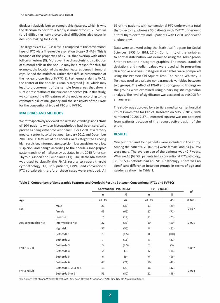
Table 1: Comparison of Sonographic Features and Cytologic Results Between Conventional PTCs and FVPTCs
Conventionel PTC (n=66) FVPTC (n=38)p¹
n % n %
Age 42±15 42 44±15 45 0.468²
Sexmale 23 (35) 11 (29)
0.537female 43 (65) 27 (71)
ATA sonographic risk
Low risk 7 (11) 11 (29)
0.001İntermediate risk 22 (33) 19 (50)
High risk 37 (56) 8 (21)
FNAB result
Bethesda 1 1 (1.5) 0 (0.0)
0.037
Bethesda 2 7 (11) 8 (21)
Bethesda 3 3 (4.5) 2 (5)
Bethesda 4 2 (3) 6 (16)
Bethesda 5 6 (9) 6 (16)
Bethesda 6 47 (71) 16 (42)
FNAB resultBethesda 1, 2, 3 or 4 13 (20) 16 (42)
0.014Bethesda 5 or 6 53 (80) 22 (58)
¹Chi-Square Test, ²Mann Whitney U Test, ATA: American Thyroid Association, FNAB: Fine Needle Aspiration Biopsy
Asya et al. Follicular-Variant Papillary Thyroid Carcinoma
3
Patients with conventional PTC were grouped according to their sonographic features: 37 (56%) were categorized as high suspicion, 22 (33.3%) as intermediate suspicion, and 7 (10.7%) as low suspicion as shown in Table 1. None of the patients with conventional PTC showed a very low or benign sonographic pattern. Patients with FVPTC were also grouped according to their sonographic features: 8 (21%) were categorized as high suspicion, 19 (50%) as intermediate suspicion, and 11 (29%) as low suspicion as shown in Table 1. None of the patients with FVPTC had very low or benign sonographic patterns. While high-risk nodules were detected in 56% of the conventional variant cases, this was present in 21% of the follicular-variant cases (p=0.001).
The FNAB results of patients with conventional PTC were evaluated: 47 (71.2%) had Bethesda 6 cytology, 6 (9.1%) had Bethesda 5 cytology, 2 (3%) had Bethesda 4 cytology, 3 (4.5%) had Bethesda 3 cytology, 7 (10.7%) had Bethesda 2 cytology, and 1 (1.5%) had Bethesda 1 cytology as shown in Table 1. The FNAB results of the patients with FVPTC were also evaluated: 16 (42.1%) had Bethesda 6 cytology, 6 (15.8%) had Bethesda 5 cytology, 6 (15.8%) had Bethesda 4 cytology, 2 (5.3%) had Bethesda 3 cytology, 8 (21%) had Bethesda 2 cytology, and none had Bethesda 1 cytology as shown in Table 1. In 80% of the conventional PTC cases, malignancy or the suspicion of malignancy was detected by the FNAB, whereas this rate was 58% for FVPTC cases (p=0.014).
It is known that the follicular architectural pattern is the predominant histological pattern of FVPTC,and we analyzed whether or not this could affect the FNAB result. While Bethesda 4 (follicular neoplasm or suspicious for follicular neoplasm) cytology was reported in 3% of FNAB of conventional PTC cases, this rate was approximately 16% in the follicular variant (p=0.037).
Logistic regression analysis was performed to show the effect of risk factors on PTC. We found that high risk nodules increases the possibility of conventional variant papillary thyroid carcinoma by 7,268 times compared to low risk nodules as shown in Table 2 (p=0.001 95% confidence interval). We also found that nodules with Bethesda 5 or 6 cytology increases the possibility of conventional variant papillary thyroid carcinoma by 2,965 times compared to nodules with Bethesda 1,2,3 or 4 cytology as shown in Table 3 (p=0.016 95% confidence interval).
DISCUSSION
In our study, FVPTC clearly differs from conventional PTC when comparing the sonographic patterns of the nodules and the FNAB results. More than half of the patients with conventional PTC had border irregularity, microcalcification, or a taller-than-wide shape with a hypoechoic solid nodule, which places them in a high-risk category. However, only 21% of those with FVPTC had such a sonographic pattern. The relative paucity of border irregularity, microcalcification, or a taller-than-wide shape that we observed in FVPTC may be attributed to the propensity of these lesions to grow parallel to the normal tissue plane rather than infiltratively across the normal tissue (5, 7, 13). Nearly one-third of the patients with FVPTC in our study had hyperechoic or isoechoic solid nodules without any other suspicious patterns, just like the nodules with multinodular goiter. This percentage was about 10% of the patients with conventional PTC in our study. The relatively frequent hyperechogenicity or isoechogenicity in FVPTC compared to conventional PTC might be related to the abundance of follicles and the lesser degree of cellularity in FVPTC (14). Hypoechogenicity of the nodule, which suggests a malignant finding, was thought to be due to the high cellularity of the nodule. The follicular neoplasms contain multiple follicular structures and less cellularity compared to conventional PTC. From this point, via US, they resemble a multinodular goiter (14).
Table 2: Correlation Between Sonographic Risk of the Thyroid Nodule and Conventional PTC
β estimate Standard error P value Exp(B) Odds ratio95% Confidence Interval for EXP(B)
Lower Upper
Low risk nodule 0.002
İntermediate risk nodule 0.599 0.576 0.299 1.820 0.588 5.627
High risk nodule 1.983 0.621 0.001 7.268 2.151 24.553
Constant -0.452 0.483 0.350 0.636
Binary Logistic Regression
Table 3: Correlation Between FNAB and Conventional PTC
β estimate Standard error P value Exp(B) Odds ratio95% Confidence Interval for EXP(B)
Lower Upper
FNAB result (Bethesda 5 ve 6) 1.087 0.451 0.016 2.965 1.224 7.182
Constant -0.208 0.373 0.578 0.812
Binary Logistic Regression

The Turkish Journal of Ear Nose and Throat
4
In contrast to the high incidence of suspicious nodules via sonograph for malignancy in conventional PTC, a lower incidence of suspicious nodules occurs in FVPTC. From this perspective, the absence of suspicious malignant features via a sonograph cannot guarantee that the thyroid nodules are benign (13, 15). The tumoral nodules of FVPTC are usually iso- or hyperechoic, noncalcified, round (width greater than anteroposterior dimension) nodules with regular borders (15).
The FNABs of the patients were analyzed retrospectively. Benign cytological findings (Bethesda 2) were obtained in 7 cases of the 66 conventional PTCs. From these 7 cases, in 4 cases, the biopsies were taken from a dominant nodule, and incidental micropapillary carcinoma was detected in another nodule in the thyroidectomy specimen. Thus, only 4.8% (3 out of 62) of patients with Bethesda 2 cytology were found to have a final pathology of conventional PTC. Similarly,there were 8 cases of Bethesda 2 cytology out of the 38 FVPTC patients, and of these, 2 cases had micropapillary carcinoma in a nodule other than the nodule that was aspirated for cytopathologic examination. Therefore, although the biopsy was taken from a pathological nodule, 16.7% (6 out of 36) of FVPTC patients showed preoperative Bethesda 2 cytology. Among the pathological nodules where a biopsy was taken, in 85.5% (53 out of 62) of conventional PTC cases, a preoperative cytological examination resulted in Bethesda 5 or 6, whereas this percentage was 61.1% (22 out of 36) for the FVPTC cases.
Kim et al. (15) reported that the diagnosis of Bethesda 3 in FVPTC was higher than that in conventional PTC (46% vs 19%). In our study, this percentage was 5.3% vs 4.5%, respectively, whereas an important difference exists in the Bethesda 4 category. Among all cases of FVPTC, 15.8% were reported as Bethesda 4, whereas about 3% were reported for conventional PTCs. Kim et al. (15) reported that only 1 of the 35 FVPTC cases was diagnosed as FVPTC. In our study, no case was reported as FVPTC based on the FNAB.
The FNAB is highly sensitive in the diagnosis of PTC (16). Although the diagnosis of conventional PTC is not problematic in most cases, difficulties exist with the cytological diagnosis of FVPTC in a substantial number of cases. The presence of follicular architecture along with nuclear properties of papillary carcinoma allows us to make the cytological diagnosis of FVPTC (4). Two main reasons exist for diagnostic difficulties with FVPTC upon a cytological examination. First, FVPTC has cytomorphological properties that overlap with follicular lesions due to the presence of abundant colloid and monolayer sheets of follicular cells (17). Second, FVPTC contains very few of the nuclear properties that are characteristic of papillary carcinoma. For this reason, these nuclear properties are often missed on examination (9).
Moreover, these sparse nuclear changes may be subcapsularly located (9), which creates an interesting problem because the center of the nodule is usually targeted (18). These factors
contribute to the false negative results of FVPTC using FNABs. Therefore, FNABs may be misdiagnosed as an adenomatous nodule or follicular neoplasia. These problems may lead to wrong decisions in the decision-making process. Because nearly 16% of our cases with FVPTC had follicular neoplasia on an FNAB and nearly 16% showed benign cytology on an FNAB, a more careful evaluation of nodules that appear benign is considered mandatory.
In the case of a highly suspicious nodule via a sonograph, although a cytological examination is inconsistent with malignancy or suspicion for malignancy, we usually repeat the FNAB within a short period of time. In doing so, we aim to avoid missing the patients at risk. However, this is not the case for benign nodules. To overcome this problem, during an FNAB of the nodules that appear more benign, the number of aspirations may be more than usual. A careful examination of the entire nodule must be done together with the central part, and the subcapsular location of the nodule may be aspirated in case of suspicion.
CONCLUSION
In conclusion, some difficulties exist regarding the diagnosis of FVPTC based on both sonographic and cytological examinations. The FVPTC showed a higher correlation with benign sonographic features and a higher rate of false negative results based on a cytological examination. Radiologists, pathologists, and clinicians must be aware of this difficulty and demonstrate care in the evaluation of nodules that appear benign.
Ethics Committee Approval: The study was approved by a tertiary medical center hospital Ethics Committee for Clinical Research on May 5, 2017, with numbered 09.2017.371. Informed consent was not obtained from patients because of the retrospective design of the study.
Peer Review: Externally peer-reviewed.
Author Contributions: Conception/Design of Study- O.A., A.C.Y., Y.G., C.A.G., Ç.O.; Data Acquisition- O.A., Y.G.; Data Analysis/Interpretation- O.A., A.C.Y., Y.G., C.A.G., Ç.O.; Drafting Manuscript- O.A., A.C.Y., Ç.O.; Critical Revision of Manuscript- O.A., A.C.Y., Y.G., C.A.G., Ç.O.; Final Approval and Accountability- O.A., A.C.Y., Y.G., C.A.G., Ç.O.
Conflict of Interest: Authors declared no conflict of interest.
Financial Disclosure: Authors declared no financial support.
REFERENCES
1. Lloyd RV, Osamura RY, Klöppel G, Rosai J, Bosman FT, Jaffe ES, et al. WHO classification of tumours of endocrine organs: International Agency for Research on Cancer; 2017.
2. Rosai J, Carcangiu M, Delellis R. Tumors of the thyroid gland In: Rosai J, Sobin LH (eds) Atlas of Tumor Pathology, vol. 5. Armed Forces Institute of Pathology, New York. 1992:161-82.
3. Tielens ET, Sherman SI, Hruban RH, Ladenson PW. Follicular variant of papillary thyroid carcinoma. A clinicopathologic study. Cancer 1994;73(2):424-31.
Asya et al. Follicular-Variant Papillary Thyroid Carcinoma
5
4. Mesonero CE, Jugle JE, Wilbur DC, Nayar R. Fine-needle aspiration of the macrofollicular and microfollicular subtypes of the follicular variant of papillary carcinoma of the thyroid. Cancer Cytopathology: Interdisciplinary International Journal of the American Cancer Society 1998;84(4):235-44.
5. Frates MC, Benson CB, Charboneau JW, Cibas ES, Clark OH, Coleman BG, et al. Management of thyroid nodules detected at US: Society of Radiologists in Ultrasound consensus conference statement. Radiology 2005;237(3):794-800.
6. Frates MC, Benson CB, Doubilet PM, Cibas ES, Marqusee E. Can color Doppler sonography aid in the prediction of malignancy of thyroid nodules? Journal of Ultrasound in Medicine 2003;22(2):127-31.
7. Yoon JH, Kim E-K, Hong SW, Kwak JY, Kim MJ. Sonographic features of the follicular variant of papillary thyroid carcinoma. Journal of Ultrasound in Medicine 2008;27(10):1431-7.
8. Martínez-Parra D, Fernández JC, Hierro-Guilmain CC, Pérez JS, Pérez-Guillermo M. Follicular variant of papillary carcinoma of the thyroid: to what extent is fine-needle aspiration reliable? Diagnostic Cytopathology 1996;15(1):12-6.
9. Baloch ZW, Gupta PK, Yu GH, Sack MJ, LiVolsi VA. Follicular variant of papillary carcinoma: cytologic and histologic correlation. American Journal of Clinical Pathology 1999;111(2):216-22.
10. Kini SR. Guids to Clinical Aspiration Biopsy. Thyroid. 1996.11. Haugen BR, Alexander EK, Bible KC, Doherty GM, Mandel
SJ, Nikiforov YE, et al. 2015 American Thyroid Association management guidelines for adult patients with thyroid nodules and differentiated thyroid cancer: the American Thyroid Association guidelines task force on thyroid nodules and differentiated thyroid cancer. Thyroid 2016;26(1):1-133.
12. Cibas ES, Ali SZ. The Bethesda system for reporting thyroid cytopathology. Thyroid 2009;19(11):1159-65.
13. Moon W-J, Jung SL, Lee JH, Na DG, Baek J-H, Lee YH, et al. Benign and malignant thyroid nodules: US differentiation—multicenter retrospective study. Radiology 2008;247(3):762-70.
14. Jeh S-k, Jung SL, Kim BS, Lee YS. Evaluating the degree of conformity of papillary carcinoma and follicular carcinoma to the reported ultrasonographic findings of malignant thyroid tumor. Korean Journal of Radiology 2007;8(3):192-7.
15. Kim DS, Kim J-h, Na DG, Park S-H, Kim E, Chang K-H, et al. Sonographic features of follicular variant papillary thyroid carcinomas in comparison with conventional papillary thyroid carcinomas. Journal of Ultrasound in Medicine 2009;28(12):1685-92.
16. Caraway NP, Sneige N, Samaan NA. Diagnostic pitfalls in thyroid fine-needle aspiration: a review of 394 cases. Diagnostic Cytopathology 1993;9(3):345-50.
17. BALOCH ZW, SACK MJ, YU GH, LIVOLSI VA, GUPTA PK. Fine-needle aspiration of thyroid: an institutional experience. Thyroid 1998;8(7):565-9.
18. Silverman JF, West RL, Larkin EW, Park HK, Finley JL, Swanson MS, et al. The role of fine-needle aspiration biopsy in the rapid diagnosis and management of thyroid neoplasm. Cancer 1986;57(6):1164-70.
6
ABSTRACT
Objective: The aim of the study is to compare the operative time and degree of post-operative pain in children who underwent a tonsillectomy using different energy doses of bipolar cautery and to specify the most appropriate energy dose.Materials and Methods: Patients included in the study were allocated to three groups with 20 in each and each group underwent the operation with 20, 30 and 40 watt energy doses of bipolar cautery. Operative time was recorded for each patient. The Wong-Baker FACES pain rating Scale (WBS) was used in the post-operative period for each patient and the pain severity of the patients was evaluated at post-operative 30th min, 1st, 6th, 24th hours and 10th day. The operative times and pain severity of the patients were compared thereafter.Results: A significant difference was not detected between the groups in which 20, 30 and 40 watt bipolar cautery was used (p>0.05). A significant difference was not detected between groups with regard to pain scores at post-operative 30th min, 1st, 6th, 24th hour and 10th day (p>0.05).Conclusion: We have compared groups with regard to post-operative pain and operative time according to energy dose of bipolar cautery and detected no statistically significant difference in tonsillectomies conducted with different energy doses. Therefore we consider that energy dose should be as low as possible in tonsillectomies conducted with bipolar cautery.
Keywords: Bipolar cautery, child, operation time, postoperative pain, tonsillectomy
A Comparison of Pain and Operation Time in Children Undergoing a Tonsillectomy using Different Energy Doses of Bipolar CauteryAyhan Kars1 , Fatih Bingöl2 , Korhan Kılıç3
1Kastamonu University, Faculty of Medicine, Department of Otorhinolaryngology, Head and Neck Surgery, Kastamonu, Turkey2Nigde Omer Halisdemir University Training and Research Hospital, Clinic of Otorhinolaryngology, Nigde, Turkey 3Ataturk University, Faculty of Medicine, Department of Otorhinolaryngology, Head and Neck Surgery, Erzurum, Turkey
ORCID ID: A.K. 0000-0003-4580-315X; F.B. 0000-0001-8260-0349; K.K. 0000-0001-6048-034X
Citation: Kars A, Bingol F, Kilic K. A comparison of pain and operation time in children undergoing a tonsillectomy using different energy doses of bipolar cautery. Tr-ENT 2021;31(1):6-9. https://doi.org/10.26650/Tr-ENT.2021.30092
RESEARCH ARTICLEDOI: 10.26650/Tr-ENT.2021.30092
The Turkish Journal of Ear Nose and Throat 2021;31(1):6-9
Corresponding Author: Ayhan Kars E-mail: [email protected]
Submitted: 22.01.2021 • Revision Requested: 31.05.2021 • Last Revision Received: 04.06.2021 • Accepted: 06.06.2021
This work is licensed under Creative Commons Attribution-NonCommercial 4.0 International License.
INTRODUCTION
The tonsillectomy is one of the most common surgeries performed in otorhinolaryngology clinics, especially in children with recurrent tonsillitis and tonsil hypertrophy (1-3). Although there are different surgical techniques for carrying out a tonsillectomy, the determinant factor on which technique to perform is the incidence of complications that may occur during and after the surgery (1). Cold dissection, monopolar and bipolar cautery dissection, harmonic scalpel tonsillectomy and coblation tonsillectomy are the methods used in tonsillectomy (2). The common purpose of all these techniques is to reduce the complications of surgery, shorten the operation time, and increase the comfort and safety of the patient (4). Among these methods, bipolar cautery is the most commonly used
(1). Bipolar cautery is an important technique in terms of less intraoperative blood loss and shorter operation time (5, 6).
Postoperative pain is the most worrying problem in patients undergoing tonsillectomy (2). Pains usually lasting longer than 1 week can be seen (3). There are pharmacological and surgical approaches for pain reduction.(2) Bipolar cautery can cause tissue damage due to high heat that can reach 400-600°C (2, 3). Therefore, it is important to perform the operation with a low energy dose. There are conflicting results in terms of postoperative pain in previous studies conducted using low and high energy (3).
The aim of this study was to compare the operation time and the degree of postoperative pain in children having undergone
Kars et al. Tonsillectomy with Bipolar Cautery
7
tonsillectomy using different energy doses of bipolar cautery and to determine the most appropriate energy dose.
PATIENTS AND METHODS
For this prospectively planned study, approval was obtained from Ataturk University Faculty of Medicine Clinical Research Ethics Committee with number of B.30.2.ATA.0.01.00 / 58. 60 children aged between 3-15 years who were evaluated in Erzurum Regional Training and Research Hospital in Otorhinolaryngology Clinic and who decided to undergo tonsillectomy between the dates of February 2018 – May 2018 were included in the study. Written informed consent was obtained from the parents of the patients for the operation.
Patients with known bleeding disorder, chronic disease, suspected hematological malignancy, peritonsillar abscess history, acute upper respiratory tract infection, drug allergy and acute tonsillitis were excluded from the study. Patients were questioned as to their use of aspirin. Preoperative whole blood count, prothrombin time (PT) and activated thromboplastin time (aPTT) were tested routinely. The patients were divided into three groups of 20 patients and the groups were operated on using 20, 30, and 40 watts of bipolar cautery (Covidien ValleyLab Force Fx Electrosurgical Generator, Instant ResponseTM Technology, USA), respectively. The operations were performed under endotracheal general anesthesia. 3-5 mg/kg sodium thiopental and 0.6 mg/kg rocuronium bromide were used for anesthesia induction. Anaesthesia was maintained with 1-2% sevoflurane after intubation. Intravenoz (iv) 1 mg/kg methylprednisolone was given prior to surgery.
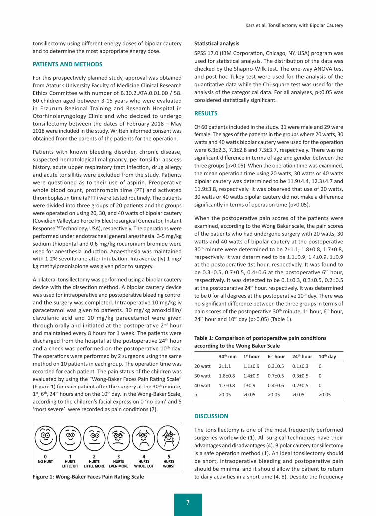
A bilateral tonsillectomy was performed using a bipolar cautery device with the dissection method. A bipolar cautery device was used for intraoperative and postoperative bleeding control and the surgery was completed. Intraoperative 10 mg/kg iv paracetamol was given to patients. 30 mg/kg amoxicillin/clavulanic acid and 10 mg/kg paracetamol were given through orally and initiated at the postoperative 2nd hour and maintained every 8 hours for 1 week. The patients were discharged from the hospital at the postoperative 24th hour and a check was performed on the postoperative 10th day. The operations were performed by 2 surgeons using the same method on 10 patients in each group. The operation time was recorded for each patient. The pain status of the children was evaluated by using the “Wong-Baker Faces Pain Rating Scale” (Figure 1) for each patient after the surgery at the 30th minute, 1st, 6th, 24th hours and on the 10th day. In the Wong-Baker Scale, according to the children’s facial expression 0 ‘no pain’ and 5 ‘most severe’ were recorded as pain conditions (7).
Statistical analysis
SPSS 17.0 (IBM Corporation, Chicago, NY, USA) program was used for statistical analysis. The distribution of the data was checked by the Shapiro-Wilk test. The one-way ANOVA test and post hoc Tukey test were used for the analysis of the quantitative data while the Chi-square test was used for the analysis of the categorical data. For all analyses, p<0.05 was considered statistically significant.
RESULTS
Of 60 patients included in the study, 31 were male and 29 were female. The ages of the patients in the groups where 20 watts, 30 watts and 40 watts bipolar cautery were used for the operation were 6.3±2.3, 7.3±2.8 and 7.5±3.7, respectively. There was no significant difference in terms of age and gender between the three groups (p>0.05). When the operation time was examined, the mean operation time using 20 watts, 30 watts or 40 watts bipolar cautery was determined to be 11.9±4.4, 12.3±4.7 and 11.9±3.8, respectively. It was observed that use of 20 watts, 30 watts or 40 watts bipolar cautery did not make a difference significantly in terms of operation time (p>0.05).
When the postoperative pain scores of the patients were examined, according to the Wong Baker scale, the pain scores of the patients who had undergone surgery with 20 watts, 30 watts and 40 watts of bipolar cautery at the postoperative 30th minute were determined to be 2±1.1, 1.8±0.8, 1.7±0.8, respectively. It was determined to be 1.1±0.9, 1.4±0.9, 1±0.9 at the postoperative 1st hour, respectively. It was found to be 0.3±0.5, 0.7±0.5, 0.4±0.6 at the postoperative 6th hour, respectively. It was detected to be 0.1±0.3, 0.3±0.5, 0.2±0.5 at the postoperative 24th hour, respectively. It was determined to be 0 for all degrees at the postoperative 10th day. There was no significant difference between the three groups in terms of pain scores of the postoperative 30th minute, 1st hour, 6th hour, 24th hour and 10th day (p>0.05) (Table 1).
DISCUSSION
The tonsillectomy is one of the most frequently performed surgeries worldwide (1). All surgical techniques have their advantages and disadvantages (4). Bipolar cautery tonsillectomy is a safe operation method (1). An ideal tonsilectomy should be short, intraoperative bleeding and postoperative pain should be minimal and it should allow the patient to return to daily activities in a short time (4, 8). Despite the frequency Figure 1: Wong-Baker Faces Pain Rating Scale
Table 1: Comparison of postoperative pain conditions according to the Wong Baker Scale
30th min 1st hour 6th hour 24th hour 10th day
20 watt 2±1.1 1.1±0.9 0.3±0.5 0.1±0.3 0
30 watt 1.8±0.8 1.4±0.9 0.7±0.5 0.3±0.5 0
40 watt 1.7±0.8 1±0.9 0.4±0.6 0.2±0.5 0
p >0.05 >0.05 >0.05 >0.05 >0.05
The Turkish Journal of Ear Nose and Throat
8
of tonsillectomies, the ideal technique has not been found yet (9). The risk of bleeding increases due to vasodilator effect of anesthetic gases. For this reason, short operation time is important in terms of providing the use of less amount of anesthetic drugs and decreasing the morbidity rate in children (1, 10). Appropriate and rapid intraoperative bleeding control reduces the operation time. When the studies in the literature were examined, the operation time was observed to be shorter in the cauterization methods when compared to the cold techniques (1). Weimert et al. reported the mean operation time in unilateral tonsillectomy by monopolar cauterization and cold dissection tonsillectomy method to be 2.5 and 6 minutes, respectively (11). In our patients, the mean operation time in bilateral tonsillectomy was determined as 12.3 on average.
Pain after a tonsillectomy is an important problem (12).
Pharmacological and surgical approaches are important in reducing pain (2). Cold dissection tonsillectomy is the most commonly used surgical technique in combination with traditional and bipolar cautery method (4, 13). Although there is less tissue damage in cold techniques when compared to other electronic methods, the results of studies on this issue are still controversial (2). In some studies, it has been indicated that cold dissection and bipolar cautery dissection are not different in terms of postoperative pain (11, 14-16). In other studies, it has been revealed that there is less pain in the cold dissection method (9, 17). On the contrary, there is also a study indicating that there is less pain in bipolar cautery dissection (18). For this reason, there is no method that can be said to be certainly more advantageous in terms of postoperative pain. In this study, the Wong-Baker scale was used to evaluate postoperative pain. Previously, this scale was also used in the evaluation of pain after tonsillectomies in children (13).
The use of bipolar cautery is important in terms of reducing intraoperative and postoperative bleeding (1). In some studies, the risk of postoperative bleeding was indicated to be higher in hot methods. On the contrary, there are also studies demonstrating that bipolar cautery method is more effective and safer than cold techniques (13). The most common serious complication after tonsillectomy is late-term bleeding and it is seen in 2-4% of patients (4). No early- and late-term bleeding was observed in any of our patients. The sample size selected in this study was insufficient to compare bleeding rates between the groups.
The applied diathermic energy dose is calculated in watts (1). In tonsillectomies, the cauterization dose applied is between 6-50 watts for performing surgical procedures and providing hemostasis (19). A high dose of bipolar cautery is especially important in terms of pain, delayed wound healing, changes in the sensitive branches of glossopharyngeal and vagal nerve and tissue damage (20). The tonsillectomy is usually performed due to recurrent tonsillitis, and in recurrent tonsillitis, fibrosis occurs in the tissues, which was true for our patients. In addition, the vascular structures providing nutrition to the tissues are damaged due to the fact that 400-600°C energy is applied to the tissues with bipolar cautery (1). In this regard, the British Association of Otorhinolaryngology and Head &
Neck Surgery has recommended the use of as low energy as possible for dissection and hemostasis during a tonsillectomy (21). It is recommended to use a low energy dose, however, the time when energy is applied is also important because if the time increases, more thermal energy and electrical energy are transferred to the tissues, and as a result, more tissue damage occurs.(1) According to our study, since the use of different energy doses did not differ in terms of postoperative pain and operation time, the dose of energy used should be as low as possible. Results supporting our results have also been found in the study conducted by Hyun Chang and J.Hun Hah (2).
In this study, we compared the bipolar cautery method in terms of pain and operation time according to the applied energy dose. In conclusion, we determined that there was no statistically significant difference in terms of operation time and postoperative pain in tonsillectomies using different energy doses. Therefore, we believe that the dose of energy used should be as low as possible in tonsillectomies performed using bipolar cautery.
Ethics Committee Approval: For this prospectively planned study, approval was obtained from Ataturk University Faculty of Medicine Clinical Research Ethics Committee with number of B.30.2.ATA.0.01.00/58.
Peer Review: Externally peer-reviewed.
Informed Consent: Written consent was obtained from the participants.
Author Contributions: Conception/Design of Study- A.K., F.B.; Data Acquisition- A.K., F.B.; Data Analysis/Interpretation- A.K., F.B., K.K.; Drafting Manuscript- A.K., F.B., K.K.; Critical Revision of Manuscript- A.K., F.B., K.K.; Final Approval and Accountability- A.K., F.B., K.K.
Conflict of Interest: Authors declared no conflict of interest.
Financial Disclosure: Authors declared no financial support.
REFERENCES
1. Soy FK, Dündar R, Yazici H, Kulduk E, Aslan M, Sakarya EU. Bipolar cautery tonsillectomy using different energy doses: Pain and Bleeding. Int J Pediatr Otorhinolaryngol 2014;78:402-6.
2. Chang H, Hah JH. Comparison of post-tonsillectomy pain with two different types of bipolar forceps: Low temperature quantum molecular resonance device versus high temperature conventional electrocautery. Acta Oto-Laryngologica 2012;132:130-3.
3. Arbin L, Enlund M, Knutsson J. Post-tonsillectomy pain after using bipolar diathermy scissors or the harmonic scalpel: a randomised blinded study. Eur Arch Otorhinolaryngol 2017;274:2281-5.
4. Ozkırış M. Comparison of three techniques in pediatric tonsillectomy. Eur Arch Otorhinolaryngol 2012;269:1497-501.
5. Shirley WP, ALW, Wiatrak BJ, Pharyngitis and adenotonsillar disease, in: 6th edition, in: Flint PW, Haughey BH, Niparko JK, Richardson MA, Lund VJ, Robbins KT, Thomas JR (Eds.), Cummings Otolaryngology-Head and Neck Surgery: Head and Neck Surgery, vol. 3, Elsevier Health Sciences, 2010,p.2796.
6. G NA, Evaluation and management of pediatric obstructive sleep apnea, in: 6th edition, in: Flint PW, Haughey BH, Niparko JK, Richardson MA, Lund VJ, Robbins KT, Lesperance MM, Thomas JR (Eds.), Cummings Otolaryngology-Head and Neck Surgery: Head and Neck Surgery, vol. 3, Elsevier Health Sciences, 2015, p.2862.
https://www.ncbi.nlm.nih.gov/pubmed/?term=Sakarya%20EU%5BAuthor%5D&cauthor=true&cauthor_uid=24424292
Kars et al. Tonsillectomy with Bipolar Cautery
9
7. Wong DL, Baker CM. Pain in children: comparison of assessment scales. Pediatr Nurs 1988;14:9-17.
8. Collison PJ, Weiner R. Harmonic scalpel versus conventional tonsillectomy: a double-blind clinical trial. Ear Nose Throat J 2004;83:707-10.
9. Silveira H, Soares JS, Lima HA. Tonsillectomy: cold dissection versus bipolar electrodissection. Int J Pediatr Otorhinolaryngol 2003;67:345-51.
10. Guida RA, Mattucci KF. Tonsillectomy and adenoidectomy: an inpatient or outpatient procedure? Laryngoscope 1990;100:491-3.
11. Weimert TA, Babyak JW, Richter HJ. Electrodissection tonsillectomy. Arch. Otolaryngol. Head Neck Surg1990;116:186-8.
12. Warnock FF, Lander J. Pain progression, intensity and outcomes following tonsillectomy. Pain 1998;75:37-45.
13. Dadgarnia MH, Aghaei MA, Atighechi S, Behniafard N, Vahidi MR, Meybodian M, et al. The comparison of bleeding and pain after tonsillectomy in bipolar electrocautery vs cold dissection. Int J Pediatr Otorhinolaryngol 2016;89:38-41.
14. Heyden HV, Schäfer E, Jecker P, Gosepath J, Mann WJ. Tonsillectomy technique: bipolar scissors vs raspatory: results of a case control study in 138 patients. HNO 2007;55:684-9.
15. Pang YT. Pediatric tonsillectomy: bipolar electrodissection and dissection/ snare compared. J. Laryngol. Otol 1995;109:733-6.
16. Raut V, Bhat N, Kinsella J, Toner JG, Sinnathuray AR, Stevenson M. Bipolar scissors versus cold dissection tonsillectomy: a prospective, randomized, multi-unit study. Laryngoscope 2001;111:2178-82.
17. Atallah N, Kumar M, Hilali A, Hickey S. Post-operative pain in tonsillectomy: bipolar electrodissection technique vs dissection ligation technique. A double-blind randomized prospective trial. J Laryngol Otol 2000;114:667-70.
18. Kirazli T, Bilgen C, Midilli R, Ogüt F, Uyar M, Kedek A. Bipolar electrodissection tonsillectomy in children. Eur Arch Otorhinolaryngol 2005;262:716-8.
19. Lowe D, Cromwell DA, Lewsey JD, Copley LP, Brown P, Yung M, et al. Diathermy power settings as a risk factor for hemorrhage after tonsillectomy. Otolaryngol. Head Neck Surg 2009;140:23-8.
20. Tosun F. Tonsillektomi Ve Adenoidektomi Komplikasyonları. Turkiye Klinikleri J Surg Med Sci 2005;1:26-9.
21. Interim Guidance on the Use of Diathermy in Tonsillectomy, 2005 Available at: http://www.nice.org.uk (accessed 20.04.05).
https://www.ncbi.nlm.nih.gov/pubmed/?term=Silveira%20H%5BAuthor%5D&cauthor=true&cauthor_uid=12663105
https://www.ncbi.nlm.nih.gov/pubmed/?term=Mattucci%20KF%5BAuthor%5D&cauthor=true&cauthor_uid=2329905
https://www.ncbi.nlm.nih.gov/pubmed/?term=Gosepath%20J%5BAuthor%5D&cauthor=true&cauthor_uid=17136555
10
ABSTRACT
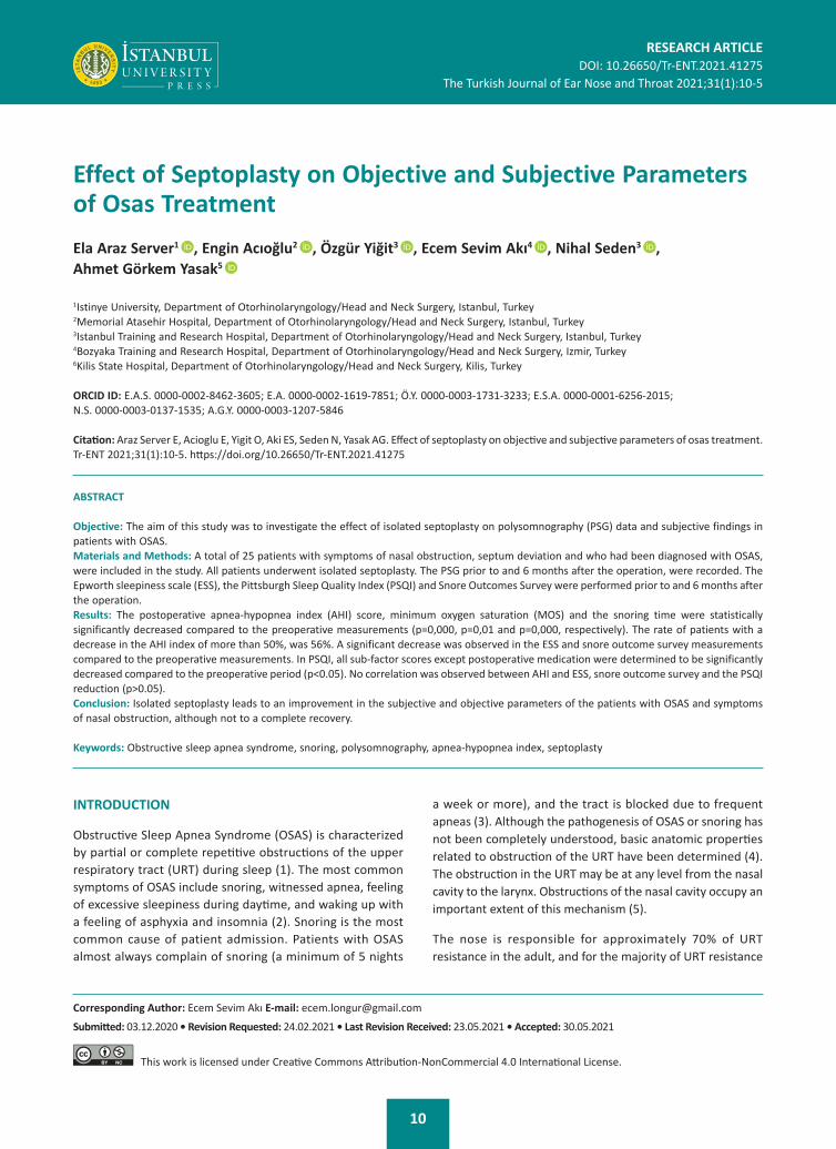
Objective: The aim of this study was to investigate the effect of isolated septoplasty on polysomnography (PSG) data and subjective findings in patients with OSAS.Materials and Methods: A total of 25 patients with symptoms of nasal obstruction, septum deviation and who had been diagnosed with OSAS, were included in the study. All patients underwent isolated septoplasty. The PSG prior to and 6 months after the operation, were recorded. The Epworth sleepiness scale (ESS), the Pittsburgh Sleep Quality Index (PSQI) and Snore Outcomes Survey were performed prior to and 6 months after the operation. Results: The postoperative apnea-hypopnea index (AHI) score, minimum oxygen saturation (MOS) and the snoring time were statistically significantly decreased compared to the preoperative measurements (p=0,000, p=0,01 and p=0,000, respectively). The rate of patients with a decrease in the AHI index of more than 50%, was 56%. A significant decrease was observed in the ESS and snore outcome survey measurements compared to the preoperative measurements. In PSQI, all sub-factor scores except postoperative medication were determined to be significantly decreased compared to the preoperative period (p<0.05). No correlation was observed between AHI and ESS, snore outcome survey and the PSQI reduction (p>0.05).Conclusion: Isolated septoplasty leads to an improvement in the subjective and objective parameters of the patients with OSAS and symptoms of nasal obstruction, although not to a complete recovery.
Keywords: Obstructive sleep apnea syndrome, snoring, polysomnography, apnea-hypopnea index, septoplasty
Effect of Septoplasty on Objective and Subjective Parameters of Osas TreatmentEla Araz Server1 , Engin Acıoğlu2 , Özgür Yiğit3 , Ecem Sevim Akı4 , Nihal Seden3 ,Ahmet Görkem Yasak5
1Istinye University, Department of Otorhinolaryngology/Head and Neck Surgery, Istanbul, Turkey2Memorial Atasehir Hospital, Department of Otorhinolaryngology/Head and Neck Surgery, Istanbul, Turkey 3Istanbul Training and Research Hospital, Department of Otorhinolaryngology/Head and Neck Surgery, Istanbul, Turkey 4Bozyaka Training and Research Hospital, Department of Otorhinolaryngology/Head and Neck Surgery, Izmir, Turkey6Kilis State Hospital, Department of Otorhinolaryngology/Head and Neck Surgery, Kilis, Turkey
ORCID ID: E.A.S. 0000-0002-8462-3605; E.A. 0000-0002-1619-7851; Ö.Y. 0000-0003-1731-3233; E.S.A. 0000-0001-6256-2015;N.S. 0000-0003-0137-1535; A.G.Y. 0000-0003-1207-5846
Citation: Araz Server E, Acioglu E, Yigit O, Aki ES, Seden N, Yasak AG. Effect of septoplasty on objective and subjective parameters of osas treatment. Tr-ENT 2021;31(1):10-5. https://doi.org/10.26650/Tr-ENT.2021.41275
RESEARCH ARTICLEDOI: 10.26650/Tr-ENT.2021.41275
The Turkish Journal of Ear Nose and Throat 2021;31(1):10-5
Corresponding Author: Ecem Sevim Akı E-mail: [email protected]
Submitted: 03.12.2020 • Revision Requested: 24.02.2021 • Last Revision Received: 23.05.2021 • Accepted: 30.05.2021
This work is licensed under Creative Commons Attribution-NonCommercial 4.0 International License.
INTRODUCTION
Obstructive Sleep Apnea Syndrome (OSAS) is characterized by partial or complete repetitive obstructions of the upper respiratory tract (URT) during sleep (1). The most common symptoms of OSAS include snoring, witnessed apnea, feeling of excessive sleepiness during daytime, and waking up with a feeling of asphyxia and insomnia (2). Snoring is the most common cause of patient admission. Patients with OSAS almost always complain of snoring (a minimum of 5 nights
a week or more), and the tract is blocked due to frequent apneas (3). Although the pathogenesis of OSAS or snoring has not been completely understood, basic anatomic properties related to obstruction of the URT have been determined (4). The obstruction in the URT may be at any level from the nasal cavity to the larynx. Obstructions of the nasal cavity occupy an important extent of this mechanism (5).
The nose is responsible for approximately 70% of URT resistance in the adult, and for the majority of URT resistance
Araz Server et al. Is Septoplasty enough for OSAS treatment?
11
during awakeness (6). It is known that nasal obstruction related to mechanical (such as septal deviation or nasal polyps) or inflammatory/vasomotor (such as acute or chronic rhinitis) causes contribute to OSAS and snoring. Many studies have demonstrated the importance of correction of these pathologies via medical or surgical methods in the treatment of OSAS and snoring. Among these studies, some have reported no improvement in the objective parameters of OSAS measured by Polysomnography (PSG) despite the improvements in subjective parameters such as the Epworth S leepiness S cale (ESS), snoring and quality of life; on the other hand, some studies have reported improvement in the objective parameters as well (7-9). In most of these studies, nasal surgeries were studied. However, there are many different pathologies which lead to nasal obstruction and many surgical methods to treat these pathologies, such as septoplasty, septorhinoplasty, turbinoplasty, valve surgery, functional sinus surgery.
Nasal septum deviation is the most frequent pathology of nasal obstruction and the most frequent surgery performed for this pathology is septoplasty. The number of studies investigating the effect of isolated septoplasty on the PSG measurements and subjective findings in patients with OSAS is limited. In our study, we aimed to investigate the effect of isolated septoplasty on snoring, quality of sleep and PSG measure parameters in patients with OSAS and nasal obstruction, and to assess whether the changes observed in subjective parameters were correlated with those observed in PSG measurements or not.
MATERIALS AND METHODS
Ethical approval was obtained from the local committee. Patients included in the study were informed and written informed consents were obtained from each. Patient selection was made upon investigation in the sleep laboratory. All patients underwent a complete ear-nose-throat examination. The patients were asked to answer the question whether they had nasal obstruction or not. Those who answered this question as ‘yes’, those who were found to have nasal septum deviation and those with an apnea-hypo pnea index (AHI) of >5 were included in the study if they were between 18 and 60 years of age.
Those with no nasal obstruction, septum deviation, those who had additional nasal pathologies such as sinusitis, nasal polyp, concha hypertrophy or nasal valve failure, those who required an additional surgical procedure other than septoplasty and those with a systemic disease were excluded from the study. Patients with malocclusion, retrognathia, tongue base hypertrophy, grade 2 tonsil hypertrophy, laryngeal pathology, and obstruction sites identified in Muller’s maneuver were also excluded from the study.
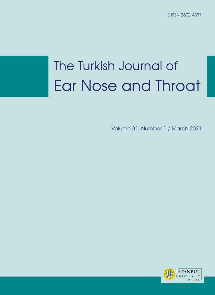
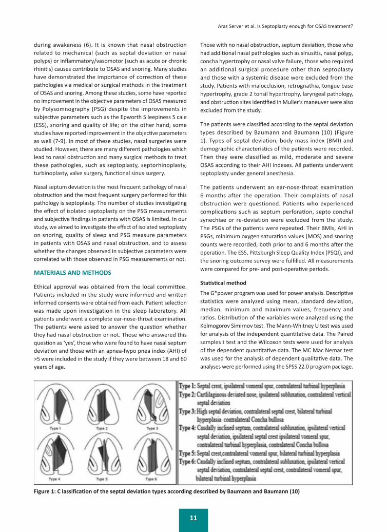
The patients were classified according to the septal deviation types described by Baumann and Baumann (10) (Figure 1). Types of septal deviation, body mass index (BMI) and demographic characteristics of the patients were recorded. Then they were classified as mild, moderate and severe OSAS according to their AHI indexes. All patients underwent septoplasty under general anesthesia.
The patients underwent an ear-nose-throat examination 6 months after the operation. Their complaints of nasal obstruction were questioned. Patients who experienced complications such as septum perforation, septo conchal synechiae or re-deviation were excluded from the study. The PSGs of the patients were repeated. Their BMIs, AHI in PSGs, minimum oxygen saturation values (MOS) and snoring counts were recorded, both prior to and 6 months after the operation. The ESS, Pittsburgh Sleep Quality Index (PSQI), and the snoring outcome survey were fulfilled. All measurements were compared for pre- and post-operative periods.
Statistical method
The G*power program was used for power analysis. Descriptive statistics were analyzed using mean, standard deviation, median, minimum and maximum values, frequency and ratios. Distribution of the variables were analyzed using the Kolmogorov Simirnov test. The Mann-Whitney U test was used for analysis of the independent quantitative data. The Paired samples t test and the Wilcoxon tests were used for analysis of the dependent quantitative data. The MC Mac Nemar test was used for the analysis of dependent qualitative data. The analyses were performed using the SPSS 22.0 program package.
Figure 1: C lassification of the septal deviation types according described by Baumann and Baumann (10)
The Turkish Journal of Ear Nose and Throat
12
RESULTS
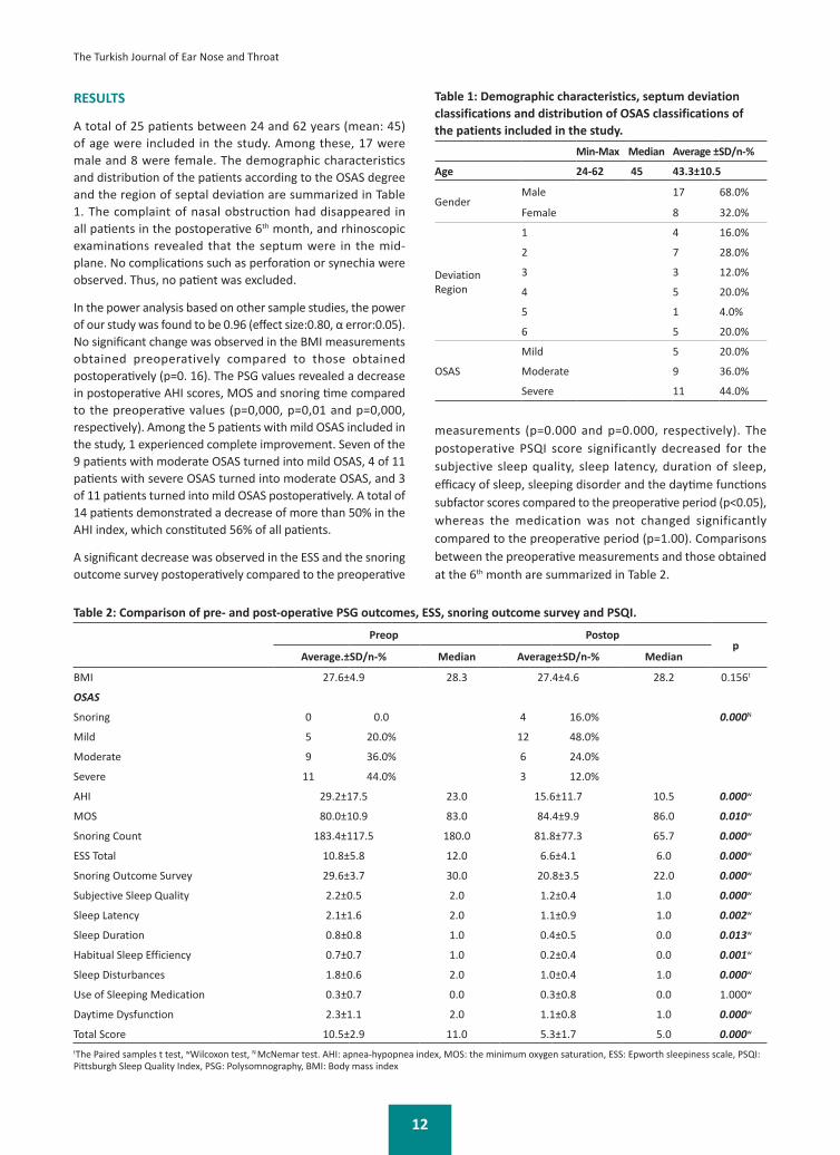
A total of 25 patients between 24 and 62 years (mean: 45) of age were included in the study. Among these, 17 were male and 8 were female. The demographic characteristics and distribution of the patients according to the OSAS degree and the region of septal deviation are summarized in Table 1. The complaint of nasal obstruction had disappeared in all patients in the postoperative 6th month, and rhinoscopic examinations revealed that the septum were in the mid-plane. No complications such as perforation or synechia were observed. Thus, no patient was excluded.
In the power analysis based on other sample studies, the power of our study was found to be 0.96 (effect size:0.80, α error:0.05). No significant change was observed in the BMI measurements obtained preoperatively compared to those obtained postoperatively (p=0. 16). The PSG values revealed a decrease in postoperative AHI scores, MOS and snoring time compared to the preoperative values (p=0,000, p=0,01 and p=0,000, respectively). Among the 5 patients with mild OSAS included in the study, 1 experienced complete improvement. Seven of the 9 patients with moderate OSAS turned into mild OSAS, 4 of 11 patients with severe OSAS turned into moderate OSAS, and 3 of 11 patients turned into mild OSAS postoperatively. A total of 14 patients demonstrated a decrease of more than 50% in the AHI index, which constituted 56% of all patients.
A significant decrease was observed in the ESS and the snoring outcome survey postoperatively compared to the preoperative
measurements (p=0.000 and p=0.000, respectively). The postoperative PSQI score significantly decreased for the subjective sleep quality, sleep latency, duration of sleep, efficacy of sleep, sleeping disorder and the daytime functions subfactor scores compared to the preoperative period (p<0.05), whereas the medication was not changed significantly compared to the preoperative period (p=1.00). Comparisons between the preoperative measurements and those obtained at the 6th month are summarized in Table 2.
Table 1: Demographic characteristics, septum deviation classifications and distribution of OSAS classifications of the patients included in the study.
Min-Max Median Average ±SD/n-%
Age 24-62 45 43.3±10.5
GenderMale 17 68.0%
Female 8 32.0%
Deviation Region
1 4 16.0%
2 7 28.0%
3 3 12.0%
4 5 20.0%
5 1 4.0%
6 5 20.0%
OSAS
Mild 5 20.0%
Moderate 9 36.0%
Severe 11 44.0%
Table 2: Comparison of pre- and post-operative PSG outcomes, ESS, snoring outcome survey and PSQI.
Preop Postop
pAverage.±SD/n-% Median Average±SD/n-% Median
BMI 27.6±4.9 28.3 27.4±4.6 28.2 0.156t
OSAS
Snoring 0 0.0 4 16.0% 0.000N
Mild 5 20.0% 12 48.0%
Moderate 9 36.0% 6 24.0%
Severe 11 44.0% 3 12.0%
AHI 29.2±17.5 23.0 15.6±11.7 10.5 0.000w
MOS 80.0±10.9 83.0 84.4±9.9 86.0 0.010w
Snoring Count 183.4±117.5 180.0 81.8±77.3 65.7 0.000w
ESS Total 10.8±5.8 12.0 6.6±4.1 6.0 0.000w
Snoring Outcome Survey 29.6±3.7 30.0 20.8±3.5 22.0 0.000w
Subjective Sleep Quality 2.2±0.5 2.0 1.2±0.4 1.0 0.000w
Sleep Latency 2.1±1.6 2.0 1.1±0.9 1.0 0.002w
Sleep Duration 0.8±0.8 1.0 0.4±0.5 0.0 0.013w
Habitual Sleep Efficiency 0.7±0.7 1.0 0.2±0.4 0.0 0.001w
Sleep Disturbances 1.8±0.6 2.0 1.0±0.4 1.0 0.000w
Use of Sleeping Medication 0.3±0.7 0.0 0.3±0.8 0.0 1.000w
Daytime Dysfunction 2.3±1.1 2.0 1.1±0.8 1.0 0.000w
Total Score 10.5±2.9 11.0 5.3±1.7 5.0 0.000w
tThe Paired samples t test, wWilcoxon test, N McNemar test. AHI: apnea-hypopnea index, MOS: the minimum oxygen saturation, ESS: Epworth sleepiness scale, PSQI: Pittsburgh Sleep Quality Index, PSG: Polysomnography, BMI: Body mass index
Araz Server et al. Is Septoplasty enough for OSAS treatment?
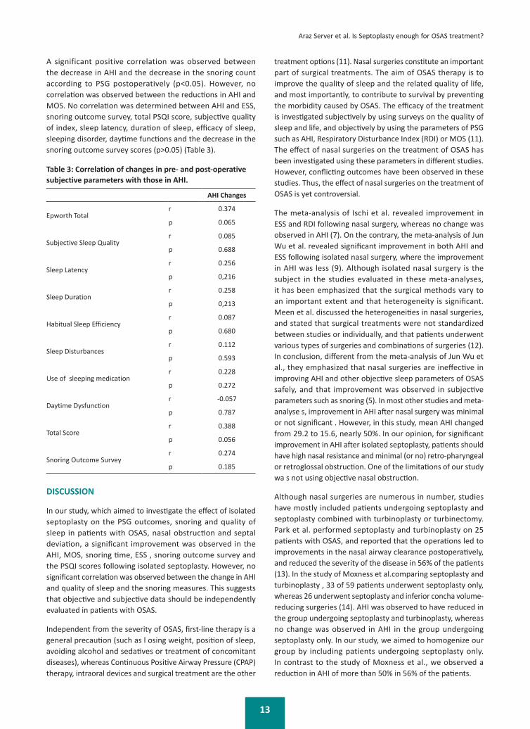
13
A significant positive correlation was observed between the decrease in AHI and the decrease in the snoring count according to PSG postoperatively (p<0.05). However, no correlation was observed between the reductions in AHI and MOS. No correlation was determined between AHI and ESS, snoring outcome survey, total PSQI score, subjective quality of index, sleep latency, duration of sleep, efficacy of sleep, sleeping disorder, daytime functions and the decrease in the snoring outcome survey scores (p>0.05) (Table 3).
DISCUSSION
In our study, which aimed to investigate the effect of isolated septoplasty on the PSG outcomes, snoring and quality of sleep in patients with OSAS, nasal obstruction and septal deviation, a significant improvement was observed in the AHI, MOS, snoring time, ESS , snoring outcome survey and the PSQI scores following isolated septoplasty. However, no significant correlation was observed between the change in AHI and quality of sleep and the snoring measures. This suggests that objective and subjective data should be independently evaluated in patients with OSAS.
Independent from the severity of OSAS, first-line therapy is a general precaution (such as l osing weight, position of sleep, avoiding alcohol and sedatives or treatment of concomitant diseases), whereas Continuous Positive Airway Pressure (CPAP) therapy, intraoral devices and surgical treatment are the other
treatment options (11). Nasal surgeries constitute an important part of surgical treatments. The aim of OSAS therapy is to improve the quality of sleep and the related quality of life, and most importantly, to contribute to survival by preventing the morbidity caused by OSAS. The efficacy of the treatment is investigated subjectively by using surveys on the quality of sleep and life, and objectively by using the parameters of PSG such as AHI, Respiratory Disturbance Index (RDI) or MOS (11). The effect of nasal surgeries on the treatment of OSAS has been investigated using these parameters in different studies. However, conflicting outcomes have been observed in these studies. Thus, the effect of nasal surgeries on the treatment of OSAS is yet controversial.
The meta-analysis of Ischi et al. revealed improvement in ESS and RDI following nasal surgery, whereas no change was observed in AHI (7). On the contrary, the meta-analysis of Jun Wu et al. revealed significant improvement in both AHI and ESS following isolated nasal surgery, where the improvement in AHI was less (9). Although isolated nasal surgery is the subject in the studies evaluated in these meta-analyses, it has been emphasized that the surgical methods vary to an important extent and that heterogeneity is significant. Meen et al. discussed the heterogeneities in nasal surgeries, and stated that surgical treatments were not standardized between studies or individually, and that patients underwent various types of surgeries and combinations of surgeries (12). In conclusion, different from the meta-analysis of Jun Wu et al., they emphasized that nasal surgeries are ineffective in improving AHI and other objective sleep parameters of OSAS safely, and that improvement was observed in subjective parameters such as snoring (5). In most other studies and meta-analyse s, improvement in AHI after nasal surgery was minimal or not significant . However, in this study, mean AHI changed from 29.2 to 15.6, nearly 50%. In our opinion, for significant improvement in AHI after isolated septoplasty, patients should have high nasal resistance and minimal (or no) retro-pharyngeal or retroglossal obstruction. One of the limitations of our study wa s not using objective nasal obstruction.
Although nasal surgeries are numerous in number, studies have mostly included patients undergoing septoplasty and septoplasty combined with turbinoplasty or turbinectomy. Park et al. performed septoplasty and turbinoplasty on 25 patients with OSAS, and reported that the operations led to improvements in the nasal airway clearance postoperatively, and reduced the severity of the disease in 56% of the patients (13). In the study of Moxness et al.comparing septoplasty and turbinoplasty , 33 of 59 patients underwent septoplasty only, whereas 26 underwent septoplasty and inferior concha volume-reducing surgeries (14). AHI was observed to have reduced in the group undergoing septoplasty and turbinoplasty, whereas no change was observed in AHI in the group undergoing septoplasty only. In our study, we aimed to homogenize our group by including patients undergoing septoplasty only. In contrast to the study of Moxness et al., we observed a reduction in AHI of more than 50% in 56% of the patients.
Table 3: Correlation of changes in pre- and post-operative subjective parameters with those in AHI.
AHI Changes
Epworth Totalr 0.374
p 0.065
Subjective Sleep Quality r 0.085
p 0.688
Sleep Latencyr 0.256
p 0,216
Sleep Durationr 0.258
p 0,213
Habitual Sleep Efficiency r 0.087
p 0.680
Sleep Disturbances r 0.112
p 0.593
Use of sleeping medication r 0.228
p 0.272
Daytime Dysfunction r -0.057
p 0.787
Total Scorer 0.388
p 0.056
Snoring Outcome Survey r 0.274
p 0.185
The Turkish Journal of Ear Nose and Throat
14
In the literature, success in surgical treatment of OSAS is generally accepted as a reduction in the AHI of more than 50% or a final AHI of less than 20/h. However, a reduction in AHI would lead to a decrease in the mortality and morbidity of the disease and an increase in the quality of life of the patient (11). Therefore, the success of nasal surgeries should be evaluated not only by the PSG findings, but improvement in CPAP use, psychological situations such as depression, daytime sleepiness, quality of sleep and snoring should be taken into account as well.
Although conflicting outcomes of PSG have been reported in studies demonstrating the effect of nasal surgeries on OSAS, many studies are in agreement for its positive effects on ESS, snoring and quality of life (15-17). ESS is the most frequently used subjective survey in patients with OSAS. Improvement has been observed in ESS following nasal surgeries in most of the conducted studies (7-9). In our study, too, a significant improvement was observed in ESS. Similar to our study, Lorente et al. showed recovery of AHI and ESS after septoplasty in 34 OSAS patients (18). However, in our study, no correlation was observed between this improvement in ESS and the improvement in AHI.
One of the most contentious subjective findings is snoring. Bury et al. emphasized that nasal surgeries may not provide a precise solution for snoring, although they may lead to an improvement, and that palatal pathologies, which play an important role in the pathogenesis of snoring, should be considered as well (15). In the study of Wu et al. investigating the effect of nasal surgeries on subjective findings, a significant reduction was observed in the visual analog scale of nasal obstruction, ESS, snoring outcome survey, bedroom partner survey and the Sino-Nasal Outcome Test scores (16). Likewise, it was observed in the study of Kalaycik et al. that Nasal Obstruction Symptom Evaluation scale (NOSE), ESS, and Snore Symptom Inventory (SSI) scores improved in patients with habitual snoring and nasal obstruction following septoplasty (17). However, no correlation was observed between score and the degree of septum deviation . In our study, we used snoring time and the snoring outcome survey in PSG to evaluate the effect of septoplasty both objectively and subjectively. We demonstrated an objective and subjective improvement following septoplasty. However, while we did not observe any patients with a complete recovery, as emphasized in the review of Bury et al., we did observe an improvement in snoring.
Quality of sleep is an important parameter for patients with OSAS, which impairs the quality of life. The “Pittsburgh Sleep Quality Index (PSQI)”, which is a reliable and consistent survey widely used in determining the quality of sleep within the last 1 month, has been used in many patient populations (19). In the study of Stapleton et al., demonstrating the effect of nasal surgery on the quality of sleep, it was found that PSQI better correlated with the Sleep Quality Likert Scores than to ESS (20). They reported that a secondary improvement would be observed in the quality of sleep following nasal surgeries, and
that the degree of improvement in the quality of sleep would correlate with the severity of preoperative nasal obstruction and the degree of improvement in the obstruction by surgery. In our study, we observed a significant improvement in the sleep quality of patients with symptoms of nasal obstruction following septoplasty as well.
In studies evaluating subjective parameters, surveys grading nasal obstruction such as NOSE or the Sino-Nasal Outcome Index were compared to the subjective parameters of OSAS, and significant correlations were determined (16, 17, 19). In our study, we aimed to investigate the correlation between improvements in the objective and subjective parameters of OSAS, and thus, we did not plan an additional survey for nasal obstruction. One of the limitations of our study is not using these surveys evaluating nasal obstruction. The other limitations include small sample size, impossibility of statistical evaluation of the septal deviation site due to the insufficient number of cases, and lack of comparison for evaluation of the deviation and obstruction findings by degrees.
Our study is important since it provides a homogeneous distribution of patients undergoing isolated septoplasty , due to nasal obstruction symptoms. Furthermore, our study is the first to evaluate OSAS with both objective and subjective parameters, and to demonstrate that the improvement in subjective parameters is independent from AHI.
CONCLUSION
Our study demonstrated that isolated septoplasty provided positive effects on PSG findings such as AHI, MOS or snoring time, and ESS, PSQI and the snoring outcome survey in patients with OSAS describing nasal obstruction and diagnosed to have septal deviation. However, the improvements in subjective parameters do not correlate to the improvements in AHI. Therefore, objective and subjective parameters should be independently evaluated for the efficacy of septoplasty in patients with OSAS.
Ethics Committee Approval: Ethical approval was obtained from the Istanbul Training and Research Hospital Clinical Research Ethics Committee (2011-KAEK-50).
Peer Review: Externally peer-reviewed.
Informed Consent: Patients included in the study were informed and written informed consents were obtained from each.
Author Contributions: Conception/Design of Study- E.A.S., E.A., Ö.Y., E.S.A., N.S., A.G.Y.; Data Acquisition- E.A.S., E.A., Ö.Y., E.S.A., N.S., A.G.Y.; Data Analysis/Interpretation- E.A.S., E.A., Ö.Y., E.S.A., N.S., A.G.Y.; Drafting Manuscript- E.A.S., E.A., Ö.Y., E.S.A., N.S., A.G.Y.; Critical Revision of Manuscript- E.A.S., E.A., Ö.Y., E.S.A., N.S., A.G.Y.; Final Approval and Accountability- E.A.S., E.A., Ö.Y., E.S.A., N.S., A.G.Y.
Conflict of Interest: Authors declared no conflict of interest.
Financial Disclosure: Authors declared no financial support.
Araz Server et al. Is Septoplasty enough for OSAS treatment?
15
REFERENCES
1. Gaudette E, Kimoff RJ. Pathophysiology of OSA. Eur Respir Mon 2010;50:31-50.
2. Malow BA. Approach to the patient with disordered sleep In: Kryger MH, Roth T, Dement WC, eds. Principles and Practice of Sleep Medicine. 4th Ed. Philadelphia, WB Saunders 2005;589-93.
3. Schlosshan D, Elliott MW. Clinical presentation and diagnosis of the obstructive sleep apnea hypopnea syndrome. Thorax 2004;59:347-52.
4. Schwab RJ, Remmers JE, Kuna ST. Chapter 101 - Anatomy And Physiology of Upper Airway Obstruction. In: Kryger MH, Roth T, Dement WC (eds). Principles and Practice of Sleep Medicine, 5th ed. Missouri: Elsevier Saunders 2011:1153-71.
5. Gharibeh T, Mehra R. Obstructive sleep apnea syndrome: natural history, diagnosis, and emerging treatment options. Nat Sci Sleep 2010;2:233-55.
6. Woodson BT. Sleep Medicine and Surgery. In: James Snow, P. Ashley Wackym, ed. Ballenger’s Otolaryngology, Head and Neck Surgery, centennial ed. New York : People’s Medical Publishing House. BC Decker Inc. 2009:983-96.
7. Ishii L, Roxbury C, Godoy A, et al. Does nasal surgery improve OSA in patients with nasal obstruction and OSA? A meta-analysis. Otolaryngol Head Neck Surg 2015;153:326-33.
8. Li HY, Wang PC, Chen YP, et al. Critical appraisal and meta-analysis of nasal surgery for obstructive sleep apnea. Am J Rhinol Allergy 2011;25:45-9.
9. Wu J, Zhao G, Li Y, Zang H, Wang T, Wang D, Han D. Apnea-hypopnea index decreased significantly after nasal surgery for obstructive sleep apnea: A meta-analysis. Medicine (Baltimore). 2017;96(5):e6008
10. Baumann I, Baumann H. A new classification of septal deviations. Rhinology 2007;45:220-3.
11. Mickelson SA. Nasal Surgery for Obstructive Sleep Apnea Syndrome. Otolaryngol Clin North Am 2016;49(6):1373-81.
12. Meen EK, Chandra RK. The role of the nose in sleep disordered breathing . Am J Rhinol Allergy 2013;27(3):213-20.
13. Park CY, Hong JH, Lee JH, et al. Clinical effect of surgical correction for nasal pathology on the treatment of obstructive sleep apnea syndrome. PloSOne 2014;9:e9876.
14. Moxness MH, Nordgård S. An observational cohort study of the effects of septoplasty with or without inferior turbinate reduction in patients with obstructive sleep apnea. BMC Ear Nose Throat Disord 2014;14:11.
15. Bury SB, Singh A. The role of nasal treatments in snoring obstructive sleep apnoea . Curr Opin Otolaryngol Head Neck Surg 2015;23:39-46.
16. Wu J, Zang HR, Wang T, Zhou B, Ye JY, Li YC, Han DM. Evaluation of the subjective efficacy of nasal surgery. J Laryngol Otol 2017;131(1):37-43.
17. Ertugay CK, Toros SZ, Karaca CT, Kulekci S, Verim A, Ertugay OC, Naiboglu B. Is septoplasty effective on habitual snoring in patients with nasal obstruction ?Eur Arch Otorhinolaryngol. 2015;272(7):1687-91.
18. Lorente J, Jurado MJ, Romero O, Quesada P, Quesada JL, Sagalés T. Effects Of Functional Septoplasty in obstructive sleep apnea syndrome. Med Clin (Barc). 2005;125(8):290-2.
19. Buysse DJ, Hall ML, Strollo PJ, et al. Relationships between the Pittsburgh Sleep Quality Index (PSQI), Epworth Sleepiness Scale (ESS), and clinical/polysomnographic measures in a community sample. J Clin Sleep Med 2008;4:563-71.
20. Stapleton AL, Chang YF, Soose RJ, Gillman GS. The impact of nasal surgery on sleep quality: a prospective outcomes study. Otolaryngol Head Neck Surg 2014;151(5):868-73.

16
ABSTRACT
Objective: Surgical removal is a valid option in the treatment of submandibular gland diseases but there are still operative and diagnostic pitfalls. We aimed to review and investigate the long-term clinical, surgical, and pathologic outcomes of our cases that had undergone submandibular gland removal. Materials and Methods: Retrospective analysis was conducted on patients who underwent submandibular gland excision via the transcervical approach with minimal skin incision between January 2007 and April 2020. Demographic data, preoperative FNA cytology results, clinical findings, surgical procedure, postoperative histopathological diagnosis, and postoperative morbidity were reviewed and analyzed. Results: 66 patients (41.7%) had sialolithiasis, 47 patients (29.7%) had chronic sialadenitis. Pleomorphic adenoma was found in 30 of the 44 patients that been operated on for submandibular mass. Gland derived carcinoma was found in 9 patients. The sensitivity of FNA biopsy in our case series was 83.3% (10/12), specificity was 78.1% (25/32), positive predictive value was 71.4% (10/14), and the negative predictive value was 96.1% (25/26). Permanent facial nerve marginal mandibular branch palsy was found in two patients (1.2%). Conclusion: The transcervical approach to the submandibular gland offers safe and reliable removal of the submandibular gland with a minimal risk of complications. FNA cytology was found to be useful in the preoperative evaluation of the submandibular masses.
Keywords: Sialolithiasis, submandibular gland, submandibular gland excision, submandibular gland neoplasm
Surgery for Submandibular Gland Diseases: 14-years of Experience of a Single Tertiary CenterGöksel Turhal1 , Arın Öztürk2 , Hakan Ceylan1 , Nurullah Serdar Akyıldız1 , Kerem Öztürk1
1Ege University School of Medicine, Department of Otolaryngology, Izmir, Turkey2Çermik State Hospital, Department of Otolaryngology, Diyarbakır, Turkey
ORCID ID: G.T. 0000-0003-0020-1921; A.O. 0000-0002-3321-7033; H.C. 0000-0002-3822-1867; N.S.A. 0000-0002-7472-5499; K.O. 0000-0003-4608-6105
Citation: Turhal G, Ozturk A, Ceylan H, Akyildiz NS, Ozturk K. Surgery for submandibular gland diseases: 14-years of experience of a single tertiary center. Tr-ENT 2021;31(1):16-9. https://doi.org/10.26650/Tr-ENT.2021.27247
RESEARCH ARTICLEDOI: 10.26650/Tr-ENT.2021.27247
The Turkish Journal of Ear Nose and Throat 2021;31(1):16-9
Corresponding Author: Hakan Ceylan E-mail: [email protected]
Submitted: 27.05.2021 • Accepted: 04.06.2021
This work is licensed under Creative Commons Attribution-NonCommercial 4.0 International License.
INTRODUCTION
Surgical excision of the submandibular gland is indicated in patients with submandibular neoplasm or inflammatory diseases. Ten percent of the salivary gland neoplasms are located in the submandibular gland, and there is a higher malignancy rate than other major salivary glands (1, 2). Fine needle aspiration (FNA) biopsy is a minimally invasive procedure and provides diagnostic information before the operation. Since high-grade and locally advanced malignant submandibular masses may require more extensive surgery or a multi-modality treatment, FNA biopsy may affect the type or the timing of the surgical treatment (3). However, many studies reported different outcomes in the diagnostic accuracy, and there are controversies on the clinical significance of FNA biopsy in the diagnosis of salivary gland masses (4, 5).
Chronic sialadenitis and sialolithiasis are mostly seen in the submandibular glands, and surgical excision is considered a standard and safe method for patients not respondent to conservative treatments and minimally invasive techniques (6, 7). However, submandibular gland surgery carries a risk of operative complications like damage to the facial nerve’s mandibular branch, hypoglossal nerve, and lingual nerve. Smaller incisions and meticulous dissection are usually preferred, but there is a considerable risk of postoperative complications like salivary fistula, wound infection, scar formation, and disease recurrence (8).
Surgical management is the most valid method in the treatment of submandibular gland diseases. However, there are still operative and diagnostic pitfalls that might lead to significant complications. Our study aimed to review our 14 years of experience in submandibular gland surgery and investigate the clinical, surgical and pathological outcomes of our cases.
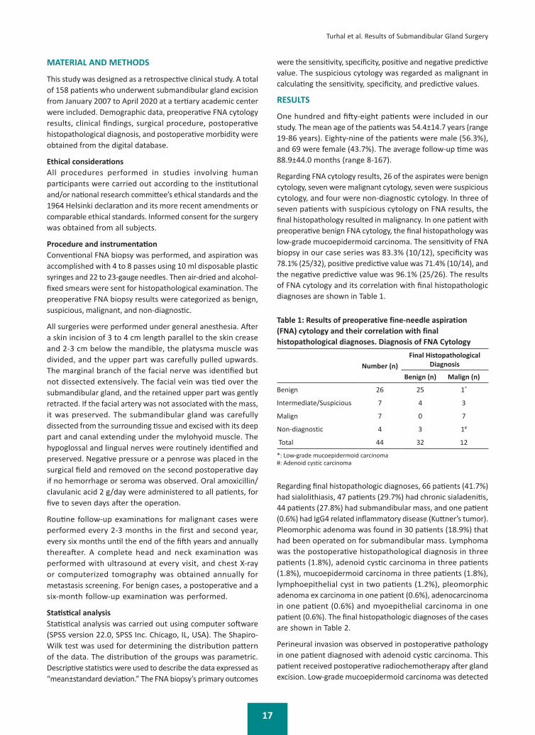
Turhal et al. Results of Submandibular Gland Surgery
17
MATERIAL AND METHODS
This study was designed as a retrospective clinical study. A total of 158 patients who underwent submandibular gland excision from January 2007 to April 2020 at a tertiary academic center were included. Demographic data, preoperative FNA cytology results, clinical findings, surgical procedure, postoperative histopathological diagnosis, and postoperative morbidity were obtained from the digital database.
Ethical considerationsAll procedures performed in studies involving human participants were carried out according to the institutional and/or national research committee’s ethical standards and the 1964 Helsinki declaration and its more recent amendments or comparable ethical standards. Informed consent for the surgery was obtained from all subjects.
Procedure and instrumentationConventional FNA biopsy was performed, and aspiration was accomplished with 4 to 8 passes using 10 ml disposable plastic syringes and 22 to 23-gauge needles. Then air-dried and alcohol-fixed smears were sent for histopathological examination. The preoperative FNA biopsy results were categorized as benign, suspicious, malignant, and non-diagnostic.
All surgeries were performed under general anesthesia. After a skin incision of 3 to 4 cm length parallel to the skin crease and 2-3 cm below the mandible, the platysma muscle was divided, and the upper part was carefully pulled upwards. The marginal branch of the facial nerve was identified but not dissected extensively. The facial vein was tied over the submandibular gland, and the retained upper part was gently retracted. If the facial artery was not associated with the mass, it was preserved. The submandibular gland was carefully dissected from the surrounding tissue and excised with its deep part and canal extending under the mylohyoid muscle. The hypoglossal and lingual nerves were routinely identified and preserved. Negative pressure or a penrose was placed in the surgical field and removed on the second postoperative day if no hemorrhage or seroma was observed. Oral amoxicillin/clavulanic acid 2 g/day were administered to all patients, for five to seven days after the operation.
Routine follow-up examinations for malignant cases were performed every 2-3 months in the first and second year, every six months until the end of the fifth years and annually thereafter. A complete head and neck examination was performed with ultrasound at every visit, and chest X-ray or computerized tomography was obtained annually for metastasis screening. For benign cases, a postoperative and a six-month follow-up examination was performed.
Statistical analysisStatistical analysis was carried out using computer software (SPSS version 22.0, SPSS Inc. Chicago, IL, USA). The Shapiro-Wilk test was used for determining the distribution pattern of the data. The distribution of the groups was parametric. Descriptive statistics were used to describe the data expressed as “mean±standard deviation.” The FNA biopsy’s primary outcomes
were the sensitivity, specificity, positive and negative predictive value. The suspicious cytology was regarded as malignant in calculating the sensitivity, specificity, and predictive values.
RESULTS
One hundred and fifty-eight patients were included in our study. The mean age of the patients was 54.4±14.7 years (range 19-86 years). Eighty-nine of the patients were male (56.3%), and 69 were female (43.7%). The average follow-up time was 88.9±44.0 months (range 8-167).
Regarding FNA cytology results, 26 of the aspirates were benign cytology, seven were malignant cytology, seven were suspicious cytology, and four were non-diagnostic cytology. In three of seven patients with suspicious cytology on FNA results, the final histopathology resulted in malignancy. In one patient with preoperative benign FNA cytology, the final histopathology was low-grade mucoepidermoid carcinoma. The sensitivity of FNA biopsy in our case series was 83.3% (10/12), specificity was 78.1% (25/32), positive predictive value was 71.4% (10/14), and the negative predictive value was 96.1% (25/26). The results of FNA cytology and its correlation with final histopathologic diagnoses are shown in Table 1.
Regarding final histopathologic diagnoses, 66 patients (41.7%) had sialolithiasis, 47 patients (29.7%) had chronic sialadenitis, 44 patients (27.8%) had submandibular mass, and one patient (0.6%) had IgG4 related inflammatory disease (Kuttner’s tumor). Pleomorphic adenoma was found in 30 patients (18.9%) that had been operated on for submandibular mass. Lymphoma was the postoperative histopathological diagnosis in three patients (1.8%), adenoid cystic carcinoma in three patients (1.8%), mucoepidermoid carcinoma in three patients (1.8%), lymphoepithelial cyst in two patients (1.2%), pleomorphic adenoma ex carcinoma in one patient (0.6%), adenocarcinoma in one patient (0.6%) and myoepithelial carcinoma in one patient (0.6%). The final histopathologic diagnoses of the cases are shown in Table 2.
Perineural invasion was observed in postoperative pathology in one patient diagnosed with adenoid cystic carcinoma. This patient received postoperative radiochemotherapy after gland excision. Low-grade mucoepidermoid carcinoma was detected
Table 1: Results of preoperative fine-needle aspiration (FNA) cytology and their correlation with final histopathological diagnoses. Diagnosis of FNA Cytology
Number (n)Final Histopathological
Diagnosis
Benign (n) Malign (n)
Benign 26 25 1*
Intermediate/Suspicious 7 4 3
Malign 7 0 7
Non-diagnostic 4 3 1#
Total 44 32 12
*: Low-grade mucoepidermoid carcinoma #: Adenoid cystic carcinoma

The Turkish Journal of Ear Nose and Throat
18
in one, and gland excision was performed. For two patients diagnosed with high-grade mucoepidermoid carcinoma, we performed supraomohyoid neck dissection on both of them in the third postoperative week in addition to the gland excision. In these two patients, lymph node metastasis was not detected in the initial histopathology.
None of the nine patients diagnosed with submandibular gland-derived carcinoma had clinical or pathological lymph node metastasis, and no local recurrence or disease-related mortality was observed during follow-up. In the seventh-year follow-up of the patient diagnosed with adenoid cystic carcinoma with perineural invasion, no recurrence was observed, and the follow-up continued.
Lingual or hypoglossal nerve damage was not observed in any patient. Transient marginal mandibular nerve palsy was observed in 14 patients (8.8%). Only two patients diagnosed with malignancy (1.2%) developed permanent facial nerve marginal mandibular branch palsy. Major arterial hemorrhage and mortality due to operation in the early postoperative period were not detected in any patients. In seven of the 113 patients diagnosed with submandibular sialadenitis and sialolithiasis, purulent discharge from the incision was observed in the early postoperative time. Also, in four patients diagnosed with submandibular sialadenitis and sialolithiasis, seroma formation was observed. All of these 11 patients responded well to the intravenous antibiotic treatment and daily sterile dressings. No additional revision surgery was performed in this patient group. Also, none of the patients developed a saliva fistula at the operation site. None of any patients developed hypertrophic or severely undesired scar formation and underwent scar revision surgery in the postoperative follow-up period.
DISCUSSION
Salivary gland disorders and their treatment is one of the most frequently encountered diseases in an otolaryngologist’s
practice. Since surgery is indicated in most submandibular gland pathologies, excision of the gland is performed in many clinics by both senior and junior surgeons. Our current study represented our clinic’s experience of 158 patients who had undergone submandibular gland excision due to different indications. The conventional lateral surgical approach to the submandibular gland has remained the most preferred and standard procedure for over nearly three decades (9), and we were able to review the long-term outcomes of a large group of homogenous patients that we had operated on for 14 years in our clinic.
In the last two decades, minimally invasive surgical procedures have gained importance with better cosmetic outcomes and reduced morbidity in the otolaryngology practice. The smaller incisions parallel to the skin crease are preferred by many (10). On the other hand, intraoral or submental approaches (11, 12) or retroauricular hairline incisions (13) are described to reduce the conventional procedure’s cosmetic morbidity. Also, an endoscope assisted submandibular gland excision procedure was reported to be feasible (14). We represent the results of submandibular gland excision via the transcervical approach with the minimal skin incision. Submandibular gland excision is associated with the risk of permanent nerve palsy, reported in around 7% of cases (8). Paralysis of the hypoglossal and lingual nerve is rare (15, 16). In our study, only two patients (1.2%) developed permanent paralysis of the mandibular nerve. There was no patient with permanent lingual or hypoglossal nerve paralysis. Our case series’ low nerve paralysis rate was due to the careful identification and meticulous dissection of the facial vein and mandibular branch. We think that optimum surgical view can be achieved with minimal cervical skin incision without harming the marginal mandibular nerve. The submandibular gland can also be removed safely from lingual and hypoglossal nerves with meticulous dissection and gentle retraction of the gland.
In our study, symptomatic sialolithiasis and sialadenitis was the most common indication for submandibular gland resection, similar to previous studies (1, 6, 7, 16). Diagnostic or interventional sialendoscopy can be an option for some cases, but it carries some limitations (17). After the excision of an inflamed submandibular gland, patients can develop a seroma, salivary fistula, or surgical site infection. Also, severe adhesions due to chronic inflammation can be challenging during surgery. We observed surgical site infection and purulent discharge in seven patients and seroma in four patients. None of the patients developed salivary fistula, or undesired scar formation in long term follow-up. We observed that a transcervical approach with minimal skin incision offered safe removal of the inflamed submandibular gland and resulted in less morbidity and favorable cosmetic outcome.
Twelve (27.2%) of the 44 patients who underwent excision due to a mass in the submandibular gland were diagnosed with malignancy. Nine (20.4%) of these patients had a diagnosis of primary submandibular gland-derived carcinoma. None of these nine patients had lymph node metastasis. Perineural invasion was detected in a patient diagnosed with adenoid cystic carcinoma. Adenoid cystic carcinoma is reported as the most common pathological diagnosis, followed by mucoepidermoid
Table 2: Final Histopathologic Diagnoses of the Cases
Histopathological Diagnosis Number (158) %
Inflammatory 114 72.1
Sialolithiasis 66 41.7
Chronic Sialadenitis 47 27.8
IgG4 related disease 1 0.6
Benign 32 20.2
Pleomorphic adenoma 30 18.9
Lymphoepithelial cyst 2 1.2
Malignant 12 7.5
Lymphoma 3 1.8
Adenoid cystic Carcinoma 3 1.8
Mucoepidermoid carcinoma 3 1.8
Pleomorphic adenoma ex carcinoma 1 0.6
Adenocarcinoma 1 0.6
Myoepithelial carcinoma 1 0.6
Turhal et al. Results of Submandibular Gland Surgery
19
carcinoma (18, 19). The prognosis of adenoid cystic carcinoma varies, and its long-term management can be challenging due to the perineural invasion or distant metastasis. In our patient group, all three patients diagnosed with adenoid cystic carcinoma had early stage diseases without distant or occult metastases, and complete removal of the tumor with the submandibular gland was successful in disease control. Early-stage low-grade mucoepidermoid carcinoma and the other submandibular gland derived carcinomas were treated successfully with submandibular gland excision. We performed selective neck dissection in two of our patients diagnosed with high-grade mucoepidermoid carcinoma. No lymph node metastasis was present at the postoperative histopathology, and disease recurrence was not observed in their third and fifth-year controls. No mortality due to the primary disease was found.
FNA biopsy was performed on patients diagnosed with submandibular mass. The FNA cytology resulted in malign or suspicious in ten of twelve patients, and FNA’s sensitivity in submandibular gland malignancy was 83.3% with 71.4% (10/14) positive predictive value. Our results were similar to our studies (4, 20). In one patient, who was later diagnosed with low-grade mucoepidermoid carcinoma, the preoperative FNA cytology was benign. Also, non-diagnostic cytology was reported in an adenoid cystic carcinoma case. The most common benign pathology was pleomorphic adenoma in our case series, similar to the literature (1, 6, 7, 16). FNA cytology specificity was 78.1%, with a high negative predictive value (96.1%). FNA cytology had favorable accuracy in detecting or excluding malignancy in diagnosing a submandibular gland neoplasm, but low patient number meant making a certain conclusion was difficult on its clinic role. Also, since the final histopathological examination always plays a crucial role in selecting the treatment modality, excision of the submandibular originated masses has to be considered as the gold standard procedure. Overall, as a diagnostic tool during the preoperative planning, FNA cytology is a reliable method and mandatory in the evaluation of a submandibular mass.
CONCLUSION
The transcervical approach to the submandibular gland with minimal skin incision offers safe and reliable removal of the submandibular gland in inflammatory diseases and neoplastic tumors. It also has good postoperative outcomes with a minimal risk of permanent nerve injury. FNA cytology was found to be useful in the preoperative evaluation of the submandibular masses.
Ethics Committee Approval: Ethical Statement: Date: 21.01.2021 Decision No: 21-1.1T6 Institution: Ege University Faculty of Medicine Research Ethics Committees.
Peer Review: Externally peer-reviewed.
Informed consent: Informed consent for the surgery was obtained from all subjects.
Author Contributions: Conception/Design of Study- G.T.; Data Acquisition- H.C.; Data Analysis/Interpretation- A.O.; Drafting Manuscript- K.O.; Critical Revision of Manuscript- N.S.A.; Final Approval and Accountability- A.O.
Conflict of Interest: Authors declared no conflict of interest.
Financial Disclosure: Authors declared no financial support.
REFERENCES
1. Spiro RH. Salivary neoplasms: overview of a 35-year experience with 2,807 patients. Head Neck Surg 1986;8(3):177-84.
2. Pinkston JA, Cole P. Incidence rates of salivary gland tumors: results from a population-based study. Otolaryngol Head Neck Surg 1999;120(6):834-40.
3. Frable MA, Frable WJ. Fine-needle aspiration biopsy of salivary glands. Laryngoscope 1991;101(3):245-9.
4. Feinstein AJ, Alonso J, Yang SE, St. John M. Diagnostic accuracy of fine-needle aspiration for parotid and submandibular gland lesions. Otolaryngol Head Neck Surg 2016;155(3):431-6.
5. Schmidt RL, Hall BJ, Wilson AR, Layfield LJ. A systematic review and meta-analysis of the diagnostic accuracy of fine- needle aspiration cytology for parotid gland lesions. Am J Clin Path 2011;136:45-59.
6. Lustmann J, Regev E, Melamed Y. Sialolithiasis. A survey on 245 patients and a review of the literature. Int J Oral Maxillofac Surg 1990;19(3):135-8.
7. Marchal F, Dulguerov P. Sialolithiasis management: the state of the art. Arch Otolaryngol Head Neck Surg 2003;129(9):951-6.
8. Hernando M, Echarri RM, Taha M, Martin-Fragueiro L, Hernando A, Mayor GP. Surgical complications of submandibular gland excision. Acta Otorrinolaringol Esp 2012;63(1):42-6.
9. Beahm DD, Peleaz L, Nuss DW, Schaitkin B, Sedlmayr JC, Rivera-Serrano CM, Zanation AM, Walvekar RR. Surgical approaches to the submandibular gland: a review of literature. Int J Surg 2009;7(6):503-9.
10. Hussain A, Murray DP. Modified submandibular sialoadenectomy. Ear Nose Throat J. 2004;83(11):768, 770-1.
11. Hong KH, Kim YK. Intraoral removal of the submandibular gland: a new surgical approach. Otolaryngol Head Neck Surg 2000;122(6):798-802.
12. Roh JL, Yoon YH, Park C. Removal of nonthyroidal neck masses with improved cosmetic outcomes in skin-lifting surgery. Otolaryngol Head Neck Surg 2006;135(3):463-8.
13. Roh JL. Retroauricular hairline incision for removal of upper neck masses. Laryngoscope 2005;115(12):2161-6.
14. Baek CH, Jeong HS. Endoscope-assisted submandibular sialadenectomy: a new minimally invasive approach to the submandibular gland. Am J Otolaryngol 2006;27(5):306-9.
15. Ichimura K, Nibu KI, Tanaka T. Nerve paralysis after surgery in the submandibular triangle: review of University of Tokyo Hospital experience. Head Neck 1997;19(1):48-53.
16. Preuss SF, Klussmann JP, Wittekindt C, Drebber U, Beutner D, Guntinas-Lichius O. Submandibular gland excision: 15 years of experience. J Oral Maxillofac Surg 2007;65(5):953-7.
17. Marchal F, Barki G, Dulguerov P, Disant F, Becker M, Lehmann W. Submandibular diagnostic and interventional sialendoscopy: new procedure for ductal disorders. Ann Otol Rhinol Laryngol 2002;111(1):27-35.
18. Bhattacharyya N. Survival and prognosis for cancer of the submandibular gland. J Oral Maxillofac Surg 2004;62(4):427-30.
19. Aro K, Tarkkanen J, Saat R, Saarilahti K, Mäkitie A, Atula T. Submandibular gland cancer: Specific features and treatment considerations. Head Neck 2018;40(1):154-62.
20. Singh Nanda KD, Mehta A, Nanda J. Fine-needle aspiration cytology: a reliable tool in the diagnosis of salivary gland lesions. J Oral Pathol Med 2012;41(1):106-12.
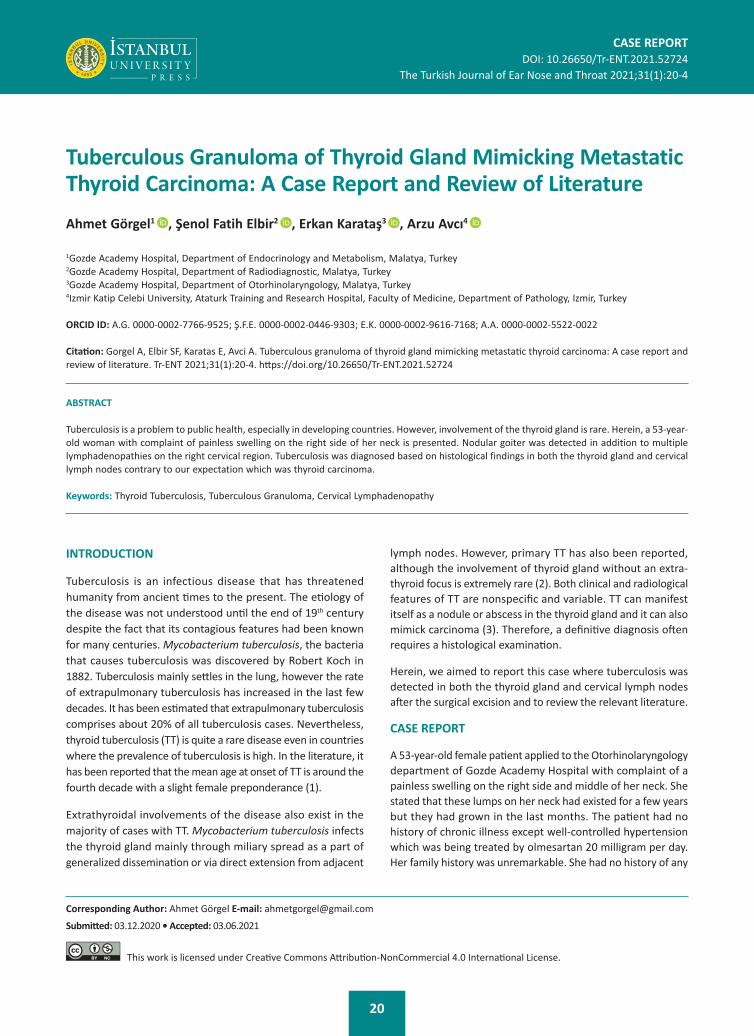
20
ABSTRACT
Tuberculosis is a problem to public health, especially in developing countries. However, involvement of the thyroid gland is rare. Herein, a 53-year-old woman with complaint of painless swelling on the right side of her neck is presented. Nodular goiter was detected in addition to multiple lymphadenopathies on the right cervical region. Tuberculosis was diagnosed based on histological findings in both the thyroid gland and cervical lymph nodes contrary to our expectation which was thyroid carcinoma.
Keywords: Thyroid Tuberculosis, Tuberculous Granuloma, Cervical Lymphadenopathy
Tuberculous Granuloma of Thyroid Gland Mimicking Metastatic Thyroid Carcinoma: A Case Report and Review of LiteratureAhmet Görgel1 , Şenol Fatih Elbir2 , Erkan Karataş3 , Arzu Avcı4
1Gozde Academy Hospital, Department of Endocrinology and Metabolism, Malatya, Turkey2Gozde Academy Hospital, Department of Radiodiagnostic, Malatya, Turkey3Gozde Academy Hospital, Department of Otorhinolaryngology, Malatya, Turkey4Izmir Katip Celebi University, Ataturk Training and Research Hospital, Faculty of Medicine, Department of Pathology, Izmir, Turkey
ORCID ID: A.G. 0000-0002-7766-9525; Ş.F.E. 0000-0002-0446-9303; E.K. 0000-0002-9616-7168; A.A. 0000-0002-5522-0022
Citation: Gorgel A, Elbir SF, Karatas E, Avci A. Tuberculous granuloma of thyroid gland mimicking metastatic thyroid carcinoma: A case report and review of literature. Tr-ENT 2021;31(1):20-4. https://doi.org/10.26650/Tr-ENT.2021.52724
CASE REPORTDOI: 10.26650/Tr-ENT.2021.52724
The Turkish Journal of Ear Nose and Throat 2021;31(1):20-4
Corresponding Author: Ahmet Görgel E-mail: [email protected]
Submitted: 03.12.2020 • Accepted: 03.06.2021
This work is licensed under Creative Commons Attribution-NonCommercial 4.0 International License.
INTRODUCTION
Tuberculosis is an infectious disease that has threatened humanity from ancient times to the present. The etiology of the disease was not understood until the end of 19th century despite the fact that its contagious features had been known for many centuries. Mycobacterium tuberculosis, the bacteria that causes tuberculosis was discovered by Robert Koch in 1882. Tuberculosis mainly settles in the lung, however the rate of extrapulmonary tuberculosis has increased in the last few decades. It has been estimated that extrapulmonary tuberculosis comprises about 20% of all tuberculosis cases. Nevertheless, thyroid tuberculosis (TT) is quite a rare disease even in countries where the prevalence of tuberculosis is high. In the literature, it has been reported that the mean age at onset of TT is around the fourth decade with a slight female preponderance (1).
Extrathyroidal involvements of the disease also exist in the majority of cases with TT. Mycobacterium tuberculosis infects the thyroid gland mainly through miliary spread as a part of generalized dissemination or via direct extension from adjacent
lymph nodes. However, primary TT has also been reported, although the involvement of thyroid gland without an extra-thyroid focus is extremely rare (2). Both clinical and radiological features of TT are nonspecific and variable. TT can manifest itself as a nodule or abscess in the thyroid gland and it can also mimick carcinoma (3). Therefore, a definitive diagnosis often requires a histological examination.
Herein, we aimed to report this case where tuberculosis was detected in both the thyroid gland and cervical lymph nodes after the surgical excision and to review the relevant literature.
CASE REPORT
A 53-year-old female patient applied to the Otorhinolaryngology department of Gozde Academy Hospital with complaint of a painless swelling on the right side and middle of her neck. She stated that these lumps on her neck had existed for a few years but they had grown in the last months. The patient had no history of chronic illness except well-controlled hypertension which was being treated by olmesartan 20 milligram per day. Her family history was unremarkable. She had no history of any
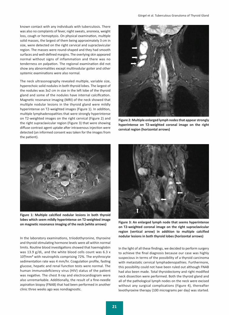
Görgel et al. Tuberculous Granuloma of Thyroid Gland
21
known contact with any individuals with tuberculosis. There was also no complaints of fever, night sweats, anorexia, weight loss, cough or hemoptysis. On physical examination, multiple solid masses, the largest of them being approximately 3 cm in size, were detected on the right cervical and supraclavicular region. The masses were round-shaped and they had smooth surfaces and well-defined margins. The overlying skin appeared normal without signs of inflammation and there was no tenderness on palpation. The regional examination did not show any abnormalities except multinodular goiter and other systemic examinations were also normal.
The neck ultrasonography revealed multiple, variable size, hyperechoic solid nodules in both thyroid lobes. The largest of the nodules was 3x2 cm in size in the left lobe of the thyroid gland and some of the nodules have internal calcification. Magnetic resonance imaging (MRI) of the neck showed that multiple nodular lesions in the thyroid gland were mildly hyperintense on T2-weighted images (Figure 1). In addition, multiple lymphadenopathies that were strongly hyperintense on T2-weighted images on the right cervical (Figure 2) and the right supraclavicular region (Figure 3) that were showing diffuse contrast agent uptake after intravenous injection were detected (an informed consent was taken for the images from the patient).
In the laboratory examinations, triiodothyronine, thyroxine and thyroid stimulating hormone levels were all within normal limits. Routine blood investigations showed that haemoglobin was 13.9 g/dL, and the white blood cells count was 6.3 x 10³/mm³ with neutrophils comprising 72%. The erythrocyte sedimentation rate was 4 mm/hr. Coagulation profile, fasting glucose, hepatic and renal function tests were normal. The human immunodeficiency virus (HIV) status of the patient was negative. The chest X-ray and electrocardiogram were also unremarkable. Additionally, the result of a fine-needle aspiration biopsy (FNAB) that had been performed in another clinic three weeks ago was nondiagnostic.
In the light of all these findings, we decided to perform surgery to achieve the final diagnosis because our case was highly suspecious in terms of the possibility of a thyroid carcinoma with metastatic cervical lymphadenopathies. Furthermore, this possibility could not have been ruled out although FNAB had also been made. Total thyroidectomy and right modified neck dissection were performed. Both the thyroid gland and all of the pathological lymph nodes on the neck were excised without any surgical complications (Figure 4), thereafter levothyroxine therapy (100 micrograms per day) was started.
Figure 2: Multiple enlarged lymph nodes that appear strongly hyperintense on T2-weighted coronal image on the right cervical region (horizontal arrows)
Figure 1: Multiple calcified nodular lesions in both thyroid lobes which seem mildly hyperintense on T2-weighted image on magnetic resonance imaging of the neck (white arrows) Figure 3: An enlarged lymph node that seems hyperintense
on T2-weighted coronal image on the right supraclavicular region (vertical arrow) in addition to multiple calcified nodular lesions in both thyroid lobes (horizontal arrows)
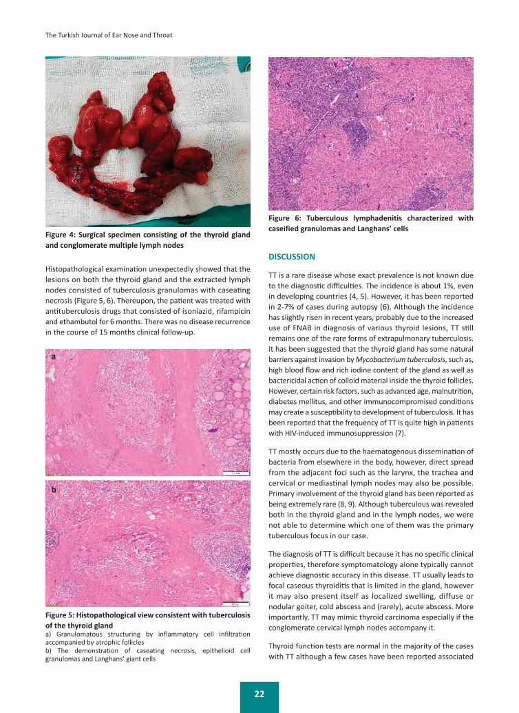
The Turkish Journal of Ear Nose and Throat
22
Histopathological examination unexpectedly showed that the lesions on both the thyroid gland and the extracted lymph nodes consisted of tuberculosis granulomas with caseating necrosis (Figure 5, 6). Thereupon, the patient was treated with antituberculosis drugs that consisted of isoniazid, rifampicin and ethambutol for 6 months. There was no disease recurrence in the course of 15 months clinical follow-up.
DISCUSSION
TT is a rare disease whose exact prevalence is not known due to the diagnostic difficulties. The incidence is about 1%, even in developing countries (4, 5). However, it has been reported in 2-7% of cases during autopsy (6). Although the incidence has slightly risen in recent years, probably due to the increased use of FNAB in diagnosis of various thyroid lesions, TT still remains one of the rare forms of extrapulmonary tuberculosis. It has been suggested that the thyroid gland has some natural barriers against invasion by Mycobacterium tuberculosis, such as, high blood flow and rich iodine content of the gland as well as bactericidal action of colloid material inside the thyroid follicles. However, certain risk factors, such as advanced age, malnutrition, diabetes mellitus, and other immunocompromised conditions may create a susceptibility to development of tuberculosis. It has been reported that the frequency of TT is quite high in patients with HIV-induced immunosuppression (7).
TT mostly occurs due to the haematogenous dissemination of bacteria from elsewhere in the body, however, direct spread from the adjacent foci such as the larynx, the trachea and cervical or mediastinal lymph nodes may also be possible. Primary involvement of the thyroid gland has been reported as being extremely rare (8, 9). Although tuberculous was revealed both in the thyroid gland and in the lymph nodes, we were not able to determine which one of them was the primary tuberculous focus in our case.
The diagnosis of TT is difficult because it has no specific clinical properties, therefore symptomatology alone typically cannot achieve diagnostic accuracy in this disease. TT usually leads to focal caseous thyroiditis that is limited in the gland, however it may also present itself as localized swelling, diffuse or nodular goiter, cold abscess and (rarely), acute abscess. More importantly, TT may mimic thyroid carcinoma especially if the conglomerate cervical lymph nodes accompany it.
Thyroid function tests are normal in the majority of the cases with TT although a few cases have been reported associated
Figure 4: Surgical specimen consisting of the thyroid gland and conglomerate multiple lymph nodes
Figure 6: Tuberculous lymphadenitis characterized with caseified granulomas and Langhans’ cells
Figure 5: Histopathological view consistent with tuberculosis of the thyroid glanda) Granulomatous structuring by inflammatory cell infiltration accompanied by atrophic folliclesb) The demonstration of caseating necrosis, epithelioid cell granulomas and Langhans’ giant cells
a
b
Görgel et al. Tuberculous Granuloma of Thyroid Gland
23
with thyrotoxicosis (9, 10). Thyrotoxicosis may also be a transient period preceding hypothyroidism, as in the course of subacute thyroiditis. Persistent hypothyroidism generally develops as a result of the inflammatory destruction of the thyroid gland and subsequently the replacement of thyroid follicles with fibrous tissue.
Imaging techniques are not very helpful in diagnosis either. Neck ultrasounds may reveal round heterogeneously hypoechoic unique or multiple lesions or anechoic lesion, irregular borders of nodules, and regional adenopathy (11). Contrary to what is mentioned above, multiple hyperechoic nodules were present in our case. Computed tomography findings of TT are also mostly non-specific. Madhusudhan et al. (12) have reported that the tuberculous lesion in the thyroid gland of their case showed an intermediate signal on T2-weighted images on MRI, the signal intensity was higher than the normal glandular parenchyma. Likewise, the MRI images of our case were similar to theirs. However, this appearance in the thyroid gland is not specific, and thyroid carcinoma can also have a similar feature (13).
FNAB is an effective and inexpensive method to diagnose both TT and other thyroid lesions. Acid-fast bacilli staining and culture from fine needle aspiration material are among the most reliable diagnostic methods. However, it is reported in the literature that the acid-fast bacilli are not always demonstrable in the lesion (14). Hence, the diagnosis of TT is frequently delayed and may represent an incidental finding at pathological examination (15). Some authors adopt the definitive histological and bacteriological evidence as the diagnostic essential criterion (16, 17). Histopathological features include caseating necrosis, epithelioid cell granulomas and Langhans’ giant cells. Furthermore, epithelioid granulomas may be seen in the course of not only TT but also other granulomatous inflammatory processes such as sarcoidosis, subacute thyroiditis, fungal infections, syphilis, thyroid neoplasia, granulomatous vasculitis, and foreign body reaction. However, the demonstration of acid-fast bacilli and the presence of caseating necrosis are pathognomonic for tuberculous thyroiditis. In this case, we preferred surgery without repeating FNAB because the findings were highly suspicious in point of malignancy. However, the findings may have been misleading as also seen in our case. Nevertheless, we considered that the operation was not a completely unnecessary attempt for the patient. We could have missed the presence of the tuberculosis or a possible malignancy even if FNAB had been repeated before surgery. TT may mistakenly be thought to be thyroid carcinoma. Moreover, TT might also coexist with thyroid carcinoma (18, 19). Treatment of TT is primarily based on antituberculous medications and where the response to antituberculous therapy is satisfactory. A thyroidectomy should be performed on large masses, TT cases coexisting with thyroid carcinoma or mimicking malignant lesions (20).
As a consequence, TT is a rare entity and it may resemble thyroid carcinoma in the first evaluation. Therefore, it should be taken into account in the differential diagnosis of thyroid
lesions and a careful approach should be adopted to avoid unnecessary surgical intervention.
Peer Review: Externally peer-reviewed.
Informed Consent: Informed consent was taken for the images from the patient.
Author Contributions: Conception/Design of Study- E.K., A.G.; Data Acquisition- Ş.F.E., A.A.; Data Analysis/Interpretation- A.G., Ş.F.E.; Drafting Manuscript- A.G., Ş.F.E.; Critical Revision of Manuscript- E.K., A.A.; Final Approval and Accountability- A.G., E.K., Ş.F.E., A.A.
Conflict of Interest: Authors declared no conflict of interest.
Financial Disclosure: Authors declared no financial support.
REFERENCES
1. Bulbuloglu E, Ciralik H, Okur E, Ozdemir G, Ezberci, F, Cetinkaya A. Tuberculosis of the Thyroid Gland: Review of the Literature. World J Surg 2006;30:149-55.
2. Akbulut S, Sogutcu N, Arikanoglu Z, Bakir S, Ulku A, Yagmur Y. Thyroid tuberculosis in southeastern Turkey: is this the resurgence of a stubborn disease? World J Surg 2011;35:1847-52.
3. Balasarkar D, Dhareswar J, Satoskar RR, Awsare N, Mahey R, Kumar V. Primary thyroid tuberculosis. Available from: URL: https://www.bhj.org.in/journal/special_issue_tb/SP_13.HTM
4. Mondal A, Patra DK. Efficacy of fine needle aspiration cytology in the diagnosis of tuberculosis of the thyroid gland: a study of 18 cases. J Laryngol Otol 1995;109:36-8.
5. Ozekinci S, Mizrak B, Saruhan G, Senturk S. Histopathologic diagnosis of thyroid tuberculosis. Thyroid 2009;19(9):983-6.
6. Simkus A. Thyroid tuberculosis. Medicina (Kaunas) 2004;40:201-4.7. Basílio-De-Oliveira CA. Infectious and neoplastic disorders of the
thyroid in AIDS patients: an autopsy study. Braz J Infect Dis 2000; 4(2):67-75.
8. Pazaitou K, Chrisoulidou A, Ginikopoulou E, Angel J, Destouni C, Vainas I. Primary tuberculosis of the thyroid gland: report of three cases. Thyroid 2002;12(12):1137-40.
9. Dhar R, Rana M, Bhardwaj S. Primary Tubercular Granulomatous Thyroiditis, Presenting as Thyroid Nodule with Hyperthyroidism, Pyrexia of Unknown Origin and Severe Anemia. J Assoc Physicians India 2018;66(5):94-5.
10. Kapoor VK, Subramani K, Das SK, Mukhopadhyay AK, Chattopadhyay, TK. Tuberculosis of the thyroid gland associated with thyrotoxicosis. Postgrad Med J 1985;61:339-40.
11. Oueslati I, Sakka I, Ismail O, Akrout I, Marghli A, Chihaoui M. Tuberculosis of the Thyroid Gland Presented as a Rapid Enlargement of a Preexisting Goiter. Case Reports in Endocrinology 2018;10:1-4.
12. Madhusudhan KS, Seith A, Khadgawat R, Das P, Mathur S. Tuberculosis of the thyroid gland: magnetic resonance imaging appearances. Singapore Med J 2009;50(7):235-8.
13. Gotway MB, Higgins CB. MR imaging of the thyroid and parathyroid glands. Magn Reson Imaging Clin N Am 2000;8(1):163-82, ix.
14. Chaurasia JK, Garg C, Agarwal A, Naim M. Tubercular thyroiditis with multinodular goitre with adenomatous hyperplasia: a rare coexistence. BMJ Case Rep 2013; pii: bcr2013200861. doi: 10.1136/bcr-2013-200861.
https://www.ncbi.nlm.nih.gov/pubmed/?term=Ozekinci%20S%5BAuthor%5D&cauthor=true&cauthor_uid=19678750
https://www.ncbi.nlm.nih.gov/pubmed/?term=Pazaitou%20K%5BAuthor%5D&cauthor=true&cauthor_uid=12593728
https://www.ncbi.nlm.nih.gov/pubmed/?term=Destouni%20C%5BAuthor%5D&cauthor=true&cauthor_uid=12593728

The Turkish Journal of Ear Nose and Throat
24
15. Jiang Y-D, Gao J-Q, Chen S, Deng X-Y. Thyroid tuberculosis: two cases report and review of the literature. Int J Clin Exp Med 2016;9(11):22477-85.
16. Sanehi S, Dravid C, Chaudhary N, Rai AK. Primary thyroid tuberculosis. Indian J Otolaryngol Head Neck Surg 2007;59:154-6.
17. Chandanwale SS, Buch AC, Vimal SS, Sachdeva P. Thyroid tuberculosis: presenting symptom of mediastinal tuberculous lymphadenitis-an unusual case. Indian J Tuberc 2014;61:84-7.
18. Meng L, Hu S, Huang L, Xu C. Papillary thyroid cancer coexisting with thyroid tuberculosis: A case report. Oncol Lett 2014;7(5): 1563-5.
19. Errami N, Benjellounb A, Hemmaouia B, Nadoura K, Benariba F. Association of tuberculous thyroiditis and papillary carcinoma of the thyroid: a rare coincidence. Pan Afr Med J 2014;19:118. doi: 10.11604/pamj.2014.19.118.4805.
20. Zendah I, Daghfous H, Ben Mrad S, Tritar F. Primary tuberculosis of the thyroid gland. Hormones (Athens) 2008;7:330-3.
The Turkish Journal of Ear Nose and ThroatINSTRUCTIONS TO AUTHORS
AIM AND SCOPE
The Turkish Journal of Ear Nose and Throat (Tr-ENT) aims to contribute to the literature by publishing high quality original articles, case clinical reports, surgical techniques and invited reviews focusing on key subjects and contemporary developments in the field. The scope of the journal includes otology, neurootology, rhinology, head and neck, general ORL, facial plastic surgery and laryngology. The journal welcomes articles from other disciplines as well, provided that these are related to the major subject area. The target audience of the journal consists of academicians, researchers, professionals, students, related professional and academic bodies and institutions.
The editorial and publication processes of the journal are shaped in accordance with the guidelines of the International Committee of Medical Journal Editors (ICMJE), World Association of Medical Editors (WAME), Council of Science Editors (CSE), Committee on Publication Ethics (COPE), European Association of Science Editors (EASE), and National Information Standards Organization (NISO). The journal is in conformity with the Principles of Transparency and Best Practice in Scholarly Publishing (doaj.org/bestpractice).
The Turkish Journal of Ear Nose and Throat is currently indexed in TUBITAK ULAKBIM TR Index.
Processing and publication are free of charge with the journal. No fees are requested from the authors at any point throughout the evaluation and publication process.
All expenses of the journal are covered by the İstanbul University.
INSTRUCTION TO AUTHORS
The Turkish Journal of Ear Nose and Throat (Tr-ENT) is an international, scientific, open acces and peer-reviewed publication of Istanbul University. It is published quarterly in March, June, September and December. The publication language of the journal is English. The journal started off in 1990 under the editorship of Professor Behbut Cevanşir. Since then it has been a significant source of knowledge in the field of otorhinolaryngology for researchers, residents and specialists. As of year 2018 Volume 28 Issue 1, the title of the journal was changed from “Kulak Burun Boğaz İhtisas Dergisi” (ISSN: 1300-7475, E-ISNN: 2147-67569) to “The Turkish Journal of Ear Nose and Throat” and the journal decided to publish articles only in English with a frequency of four issues per year. The Turkish Journal of Ear Nose and Throat aims to contribute to the literature by publishing high quality original articles, case reports, surgical techniques and invited reviews focusing on key subjects and contemporary developments in the field. The scope of the journal includes otology, neurootology, rhinology, head and neck, general ORL, facial plastic surgery and laryngology. The journal welcomes articles from other disciplines as well, provided that these are related to the major subject area. The target audience of the journal consists of academicians, researchers, professionals, students, related professional and academic bodies and institutions. Former title of the journal: Kulak Burun Boğaz İhtisas Dergisi (P-ISNN: 1300-7475 / E-ISNN: 2147-6756)
POLICIES
Publication PolicyThe subjects covered in the manuscripts submitted to the Journal for publication must be in accordance with the aim and scope of the journal. The journal gives priority to original research papers submitted for publication.
Only those manuscripts approved by its every individual author and that were not published before in or sent to another journal, are accepted for evaluation.
Submitted manuscripts that pass preliminary control are scanned for plagiarism using iThenticate software. After plagiarism check, the eligible ones are evaluated by editor-in-chief for their originality, methodology, the importance of the subject covered and compliance with the journal scope.
Short presentations that took place in scientific meetings can be referred if indicated in the article. The editor hands over the papers matching the formal rules to at least two national/international referees for evaluation and gives green light for publication upon modification by the authors in accordance with the referees’ claims. Changing the name of an author (omission, addition or order) in papers submitted to the Journal requires written permission of all declared authors. Refused manuscripts and graphics are not returned to the author.
Open Access StatementThe Turkish Journal of Ear Nose and Throat (Tr-ENT) is an open access journal which means that all content is freely available without charge to the user or his/her institution. Except for commercial purposes, users are allowed to read, download, copy, print, search, or link to the full texts of the articles in this journal without asking prior permission from the publisher or the author.
The Turkish Journal of Ear Nose and ThroatINSTRUCTIONS TO AUTHORS
The articles in The Turkish Journal of Ear Nose and Throat (Tr-ENT) are open access articles licensed under the terms of the Creative Commons Attribution-NonCommercial 4.0 International (CC BY-NC 4.0) license (https://creativecommons.org/licenses/by-nc/4.0/deed.en)
Article Processing Charge
All expenses of the journal are covered by the Istanbul University. Processing and publication are free of charge with the journal. There is no article processing charges or submission fees for any submitted or accepted articles.
Copyright NoticeAuthors publishing with The Turkish Journal of Ear Nose and Throat (Tr-ENT) retain the copyright to their work, licensing it under the Creative Commons Attribution-NonCommercial 4.0 International (CC BY-NC 4.0) license and grant the publisher non-exclusive right to publish the work.
ETHICS
Publication Ethics and Malpractice StatementThe Turkish Journal of Ear Nose and Throat (Tr-ENT) is committed to upholding the highest standards of publication ethics and pays regard to Principles of Transparency and Best Practice in Scholarly Publishing published by the Committee on Publication Ethics (COPE), the Directory of Open Access Journals (DOAJ), the Open Access Scholarly Publishers Association (OASPA), and the World Association of Medical Editors (WAME) on https://publicationethics.org/resources/guidelines-new/principles-transparency-and-best-practice-scholarly-publishing
All parties involved in the publishing process (Editors, Reviewers, Authors and Publishers) are expected to agree on the following ethical principles.
All submissions must be original, unpublished (including as full text in conference proceedings), and not under the review of any other publication synchronously. Authors must ensure that submitted work is original. They must certify that the manuscript has not previously been published elsewhere or is not currently being considered for publication elsewhere, in any language. Applicable copyright laws and conventions must be followed. Copyright material (e.g. tables, figures or extensive quotations) must be reproduced only with appropriate permission and acknowledgement. Any work or words of other authors, contributors, or sources must be appropriately credited and referenced.
Each manuscript is reviewed by at least two referees under double-blind peer review process. Plagiarism, duplication, fraud authorship/denied authorship, research/data fabrication, salami slicing/salami publication, breaching of copyrights, prevailing conflict of interest are unethical behaviors.
All manuscripts not in accordance with the accepted ethical standards will be removed from the publication. This also contains any possible malpractice discovered after the publication.
Research EthicsThe journal adheres to the highest standards in research ethics and follows the principles of international research ethics as defined below. The authors are responsible for the compliance of the manuscripts with the ethical rules.
- Principles of integrity, quality and transparency should be sustained in designing the research, reviewing the design and conducting the research.
- The research team and participants should be fully informed about the aim, methods, possible uses and requirements of the research and risks of participation in research.
- The confidentiality of the information provided by the research participants and the confidentiality of the respondents should be ensured. The research should be designed to protect the autonomy and dignity of the participants.
- Research participants should participate in the research voluntarily, not under any coercion.- Any possible harm to participants must be avoided. The research should be planned in such a way that the participants are not at risk.- The independence of research must be clear; and any conflict of interest or must be disclosed.- In experimental studies with human subjects, written informed consent of the participants who decide to participate in the
research must be obtained. In the case of children and those under wardship or with confirmed insanity, legal custodian’s assent must be obtained.
- If the study is to be carried out in any institution or organization, approval must be obtained from this institution or organization.- In studies with human subject, it must be noted in the method’s section of the manuscript that the informed consent of the
participants and ethics committee approval from the institution where the study has been conducted have been obtained.
The Turkish Journal of Ear Nose and ThroatINSTRUCTIONS TO AUTHORS
Ethics Committee Approval and Informed ConsentThe Turkish Journal of Ear Nose and Throat (Tr-ENT) takes as principle to comply with the ethical standards of World Medical Association (WMA) Declaration of Helsinki – Ethical Principles for Medical Research Involving Human Subjects revised in 2003 and WMA Statement on Animal Use in Biomedical Research revised in 2016.
An approval of research protocols by the Ethics Committee in accordance with international standards mentioned above is required for experimental, clinical, and drug studies and for some case reports. If required, ethics committee reports or an equivalent official document will be requested from the authors. For manuscripts concerning experimental research on humans, a statement should be included that shows that written informed consent of patients and volunteers was obtained following a detailed explanation of the procedures that they may undergo. For studies carried out on animals, the measures taken to prevent pain and suffering of the animals should be stated clearly. Information on patient consent, the name of the ethics committee, and the ethics committee approval number should also be stated in the Materials and Methods section of the manuscript. It is the authors’ responsibility to carefully protect the patients’ anonymity. For photographs that may reveal the identity of the patients, signed releases of the patient or of their legal representative should be enclosed.
Author’s ResponsibilitiesIt is authors’ responsibility to ensure that the article is in accordance with scientific and ethical standards and rules. And authors must ensure that submitted work is original. They must certify that the manuscript has not previously been published elsewhere or is not currently being considered for publication elsewhere, in any language. Applicable copyright laws and conventions must be followed. Copyright material (e.g. tables, figures or extensive quotations) must be reproduced only with appropriate permission and acknowledgement. Any work or words of other authors, contributors, or sources must be appropriately credited and referenced.
All the authors of a submitted manuscript must have direct scientific and academic contribution to the manuscript. The author(s) of the original research articles is defined as a person who is significantly involved in “conceptualization and design of the study”, “collecting the data”, “analyzing the data”, “writing the manuscript”, “reviewing the manuscript with a critical perspective” and “planning/conducting the study of the manuscript and/or revising it”. Fund raising, data collection or supervision of the research group are not sufficient roles to be accepted as an author. The author(s) must meet all these criteria described above. The order of names in the author list of an article must be a co-decision and it must be indicated in the Copyright Agreement Form. The individuals who do not meet the authorship criteria but contributed to the study must take place in the acknowledgement section. Individuals providing technical support, assisting writing, providing a general support, providing material or financial support are examples to be indicated in acknowledgement section.
All authors must disclose all issues concerning financial relationship, conflict of interest, and competing interest that may potentially influence the results of the research or scientific judgment.
When an author discovers a significant error or inaccuracy in his/her own published paper, it is the author’s obligation to promptly cooperate with the Editor to provide retractions or corrections of mistakes.
Responsibility for the Editor and Reviewers
Editor-in-Chief evaluates manuscripts for their scientific content without regard to ethnic origin, gender, sexual orientation, citizenship, religious belief or political philosophy of the authors. He/She provides a fair double-blind peer review of the submitted articles for publication and ensures that all the information related to submitted manuscripts is kept as confidential before publishing.
Editor-in-Chief is responsible for the contents and overall quality of the publication. He/She must publish errata pages or make corrections when needed.
Editor-in-Chief does not allow any conflicts of interest between the authors, editors and reviewers. Only he has the full authority to assign a reviewer and is responsible for final decision for publication of the manuscripts in the Journal.
Reviewers must have no conflict of interest with respect to the research, the authors and/or the research funders. Their judgments must be objective.
Reviewers must ensure that all the information related to submitted manuscripts is kept as confidential and must report to the editor if they are aware of copyright infringement and plagiarism on the author’s side.
A reviewer who feels unqualified to review the topic of a manuscript or knows that its prompt review will be impossible should notify the editor and excuse himself from the review process.
The Turkish Journal of Ear Nose and ThroatINSTRUCTIONS TO AUTHORS
The editor informs the reviewers that the manuscripts are confidential information and that this is a privileged interaction. The reviewers and editorial board cannot discuss the manuscripts with other persons. The anonymity of the referees must be ensured. In particular situations, the editor may share the review of one reviewer with other reviewers to clarify a particular point.
PEER REVIEW
Peer Review PoliciesOnly those manuscripts approved by its every individual author and that were not published before in or sent to another journal, are accepted for evaluation.
Submitted manuscripts that pass preliminary control are scanned for plagiarism using iThenticate software. After plagiarism check, the eligible ones are evaluated by editor-in-chief for their originality, methodology, the importance of the subject covered and compliance with the journal scope.
The editor hands over the papers matching the formal rules to at least two national/international referees for double-blind peer review evaluation and gives green light for publication upon modification by the authors in accordance with the referees’ claims.
Responsibility for the Editor and ReviewersEditor-in-Chief evaluates manuscripts for their scientific content without regard to ethnic origin, gender, citizenship, religious belief or political philosophy of the authors. Editor-in-Chief provides a fair double-blind peer review of the submitted articles for publication and ensures that all the information related to submitted manuscripts is kept as confidential before publishing.
Editor-in-Chief is responsible for the contents and overall quality of the publication. He/She must publish errata pages or make corrections when needed.
Editor-in-Chief does not allow any conflicts of interest between the authors, editors and reviewers. Only he has the full authority to assign a reviewer and is responsible for final decision for publication of the manuscripts in the Journal.
Reviewers must have no conflict of interest with respect to the research, the authors and/or the research funders. Their judgments must be objective.
Reviewers must ensure that all the information related to submitted manuscripts is kept as confidential and must report to the editor if they are aware of copyright infringement and plagiarism on the author’s side.
A reviewer who feels unqualified to review the topic of a manuscript or knows that its prompt review will be impossible should notify the editor and excuse himself from the review process.
The editor informs the reviewers that the manuscripts are confidential information and that this is a privileged interaction. The reviewers and editorial board cannot discuss the manuscripts with other persons. The anonymity of the referees must be ensured. In particular situations, the editor may share the review of one reviewer with other reviewers to clarify a particular point.
Peer Review ProcessOnly those manuscripts approved by its every individual author and that were not published before in or sent to another journal, are accepted for evaluation.
Submitted manuscripts that pass preliminary control are scanned for plagiarism using iThenticate software. After plagiarism check, the eligible ones are evaluated by Editor-in-Chief for their originality, methodology, the importance of the subject covered and compliance with the journal scope.
Editor-in-Chief evaluates manuscripts for their scientific content without regard to ethnic origin, gender, citizenship, religious belief or political philosophy of the authors and ensures a fair double-blind peer review of the selected manuscripts. The selected manuscripts are sent to at least two national/international referees for evaluation and publication decision is given by Editor-in-Chief upon modification by the authors in accordance with the referees’ claims. Editor-in-Chief does not allow any conflicts of interest between the authors, editors and reviewers and is responsible for final decision for publication of the manuscripts in the Journal. Reviewers’ judgments must be objective. Reviewers’ comments on the following aspects are expected while conducting the review.
- Does the manuscript contain new and significant information?- Does the abstract clearly and accurately describe the content of the manuscript?
The Turkish Journal of Ear Nose and ThroatINSTRUCTIONS TO AUTHORS
- Is the problem significant and concisely stated?- Are the methods described comprehensively?- Are the interpretations and consclusions justified by the results?- Is adequate references made to other Works in the field?- Is the language acceptable?
Reviewers must ensure that all the information related to submitted manuscripts is kept as confidential and must report to the editor if they are aware of copyright infringement and plagiarism on the author’s side.
A reviewer who feels unqualified to review the topic of a manuscript or knows that its prompt review will be impossible should notify the editor and excuse himself from the review process.
The editor informs the reviewers that the manuscripts are confidential information and that this is a privileged interaction. The reviewers and editorial board cannot discuss the manuscripts with other persons. The anonymity of the referees is important.
Manuscript Organization and SubmissionThe manuscripts should be prepared in accordance with ICMJE-Recommendations for the Conduct, Reporting, Editing, and Publication of Scholarly Work in Medical Journals (updated in December 2015 - http://www.icmje.org/icmje-recommendations.pdf). Author(s) are required to prepare manuscripts in accordance with the CONSORT guidelines for randomized research studies, STROBE guidelines for observational original research studies, STARD guidelines for studies on diagnostic accuracy, PRISMA guidelines for systematic reviews and meta-analysis, ARRIVE guidelines for experimental animal studies, and TREND guidelines for non-randomized public behavior.
Manuscripts can only be submitted through the journal’s online manuscript submission and evaluation system, available at https://dergipark.org.tr/en/pub/XXXXXX Manuscripts submitted via any other medium will not be evaluated.
Manuscripts submitted to the journal will first go through a technical evaluation process where the editorial office staff will ensure that the manuscript has been prepared and submitted in accordance with the journal’s guidelines. Submissions that do not conform to the journal’s guidelines will be returned to the submitting author with technical correction requests.
Author(s) are required to submit the following documents together with the manuscript and must ensure that the abstract and keywords are in line with the standards explained in below.
• Copyright Agreement Form• Author Form and ICMJE Potential Conflict of Interest Disclosure Form• Ethics Committee Approval• Cover Letter to the Editor• Title Page: A separate title page should be submitted with all submissions and this page should include:- The full title of the manuscript as well as a short title (running head) of no more than 50 characters,- Name(s), affiliations, academic degree(s) and ORCID ID(s) of the author(s),- Grant information and detailed information on the other sources of support,- Name, address, telephone (including the mobile phone number) and fax numbers, and email address of the corresponding
author,- Acknowledgment of the individuals who contributed to the preparation of the manuscript but who do not fulfil the authorship
criteria.
Abstract: Abstract should be submitted with all submissions except for Letters to the Editor. The abstract of Original Articles should be structured with subheadings (Objective, Materials and Methods, Results, and Conclusion). Abstracts of Case Reports and Reviews should be unstructured. Abstracts should be 200-250 words.
Keywords: Each submission must be accompanied by a minimum of 3 to a maximum of 6 keywords for subject indexing at the end of the abstract. The keywords should be listed in full without abbreviations. The keywords should be selected from the National Library of Medicine, Medical Subject Headings database (http://www.nlm.nih.gov/mesh/MBrowser.html) .
Manuscript TypesOriginal Articles: This is the most important type of article since it provides new information based on original research. The main text of original articles should be structured with Introduction, Material and Method, Results, Discussion, and Conclusion subheadings..
The Turkish Journal of Ear Nose and ThroatINSTRUCTIONS TO AUTHORS
Statistical analysis to support conclusions is usually necessary. Statistical analyses must be conducted in accordance with international statistical reporting standards (Altman DG, Gore SM, Gardner MJ, Pocock SJ. Statistical guidelines for contributors to medical journals. Br Med J 1983: 7; 1489-93). Information on statistical analyses should be provided with a separate subheading under the Materials and Methods section and the statistical software that was used during the process must be specified.
Units should be prepared in accordance with the International System of Units (SI).
Invited Review Articles: Reviews prepared by authors who have extensive knowledge on a particular field and whose scientific background has been translated into a high volume of publications with a high citation potential are welcomed. These authors may even be invited by the journal. Reviews should describe, discuss, and evaluate the current level of knowledge of a topic in clinical practice and should guide future studies. The main text should contain Introduction, Clinical and Research Consequences, and Conclusion sections. Please check Table 1 for the limitations for Review Articles.
Case Reports: There is limited space for case reports in the journal and reports on rare cases or conditions that constitute challenges in diagnosis and treatment, those offering new therapies or revealing knowledge not included in the literature, and interesting and educative case reports are accepted for publication. The text should include Introduction, Case Presentation, Discussion, and Conclusion subheadings. Please check Table 1 for the limitations for Case Reports.
Letters to the Editor: This type of manuscript discusses important parts, overlooked aspects, or lacking parts of a previously published article. Articles on subjects within the scope of the journal that might attract the readers’ attention, particularly educative cases, may also be submitted in the form of a “Letter to the Editor.” Readers can also present their comments on the published manuscripts in the form of a “Letter to the Editor.” Abstract, Keywords, and Tables, Figures, Images, and other media should not be included. The text should be unstructured. The manuscript that is being commented on must be properly cited within this manuscript.
TablesTables should be included in the main document, presented after the reference list, and they should be numbered consecutively in the order they are referred to within the main text. A descriptive title must be placed above the tables. Abbreviations used in the tables should be defined below the tables by footnotes (even if they are defined within the main text). Tables should be created using the “insert table” command of the word processing software and they should be arranged clearly to provide easy reading. Data presented in the tables should not be a repetition of the data presented within the main text but should be supporting the main text.
Figures and Figure LegendsFigures, graphics, and photographs should be submitted as separate files (in TIFF or JPEG format) through the submission system. The files should not be embedded in a Word document or the main document. When there are figure subunits, the subunits should not be merged to form a single image. Each subunit should be submitted separately through the submission system. Images should not be labeled (a, b, c, etc.) to indicate figure subunits. Thick and thin arrows, arrowheads, stars, asterisks, and similar marks can be used on the images to support figure legends. Like the rest of the submission, the figures too should be blind. Any information within the images that may indicate an individual or institution should be blinded. The minimum resolution of each submitted figure should be 300 DPI. To prevent delays in the evaluation process, all submitted figures should be clear in resolution and large in size (minimum dimensions: 100 × 100 mm). Figure legends should be listed at the end of the main document.
All acronyms and abbreviations used in the manuscript should be defined at first use, both in the abstract and in the main text. The abbreviation should be provided in parentheses following the definition.
When a drug, product, hardware, or software program is mentioned within the main text, product information, including the name of the product, the producer of the product, and city and the country of the company (including the state if in USA), should be provided in parentheses in the following format: “Discovery St PET/CT scanner (General Electric, Milwaukee, WI, USA)”
All references, tables, and figures should be referred to within the main text, and they should be numbered consecutively in the order they are referred to within the main text.
Limitations, drawbacks, and the shortcomings of original articles should be mentioned in the Discussion section before the conclusion paragraph.
The Turkish Journal of Ear Nose and ThroatINSTRUCTIONS TO AUTHORS
RevisionsWhen submitting a revised version of a paper, the author must submit a detailed “Response to the reviewers” that states point by point how each issue raised by the reviewers has been covered and where it can be found (each reviewer’s comment, followed by the author’s reply and line numbers where the changes have been made) as well as an annotated copy of the main document. Revised manuscripts must be submitted within 30 days from the date of the decision letter. If the revised version of the manuscript is not submitted within the allocated time, the revision option may be canceled. If the submitting author(s) believe that additional time is required, they should request this extension before the initial 30-day period is over. Accepted manuscripts are copy-edited for grammar, punctuation, and format. Once the publication process of a manuscript is completed, it is published online on the journal’s webpage as an ahead-of-print publication before it is included in its scheduled issue. A PDF proof of the accepted manuscript is sent to the corresponding author and their publication approval is requested within two days of their receipt of the proof. The latest status of the submitted manuscripts and other information about the journal can be accessed at http://tr-ent.com. The editorial and publication processes of the journal are conducted in accordance with the guidelines of the International Council of Medical Journal Editors (ICMJE), the World Association of Medical Editors (WAME), the Council of Science Editors (CSE), the Committee on Publication Ethics (COPE), the European Association of Science Editors (EASE), and National Information Standards Organization (NISO). The journal conforms to the Principles of Transparency and Best Practice in Scholarly Publishing (doaj.org/bestpractice). An ORCID ID is required for all authors during the submission of the manuscript. The ID is available at http://orcid.org with free of charge.
Reference Style and ExamplesAuthors are responsible for supply complete and correct references. References should be numbered according to the order used in the text. Numbers should be given in brackets and placed at the end of the sentence. Examples are given below on the use of references. Reference end note style Vancouver
Periodicals: Author(s) Last Name initial(s) name of author(s) (if there are six or fewer authors, all authors should be written; if the number of authors are seven or more, only the first six of the authors should be written and the rest as “et al”). The title of the article, the abbreviated name of the journal according to the Index Medicus, Year; Volume (Issue): The first and last page numbers.
Example: Robson A, Greene J, Ansari N, Kim B. Eccrine porocarcinoma (malignant eccrine poroma): a clinicopathologic study of 69 cases. The American Journal of Surgical Pathology 2001;25:710-20. Books: Surname of the author(s) initial name(s) of author(s). The name of the book. The edition number. Place of publication: Publisher, Publication year.
Book chapters: The author (s) surname of the chapter initial (s) letter of the name. Section title. In: Surname of editor (s) initial (s) letter of first name (s) ed / eds. The name of the book. Edition number. Place of publication: Publisher, year of publication: The first and last page numbers of the chapter. Web address: If a “web” address is used as the reference address, the web address date should be given in brackets with the address. The DOI (Digital Object Identifier) number must be provided, when a web access article used in the text as a reference.
Example: AB Author, CD Author. Title of document. Retrieved from http://Web address (Accession date: aa/bb/2016).
Congress papers:Thesis: Maden KL. Experimental investigation of the .......... Master Thesis, Health Science Institute of Ankara University, Ankara, 2005.
The Turkish Journal of Ear Nose and ThroatINSTRUCTIONS TO AUTHORS
SUBMISSION CHECKLIST
● Cover letter to the editor - The category of the manuscript - Confirming that “the paper is not under consideration for publication in another journal”. - Including disclosure of any commercial or financial involvement. - Confirming that the statistical design of the research article is reviewed. - Confirming that last control for fluent English was done. - Confirming that journal policies detailed in Information for Authors have been reviewed. - Confirming that the references cited in the text and listed in the references section are in line with NLM.● Copyright Agreement Form● Author Form● Permission of previous published material if used in the present manuscript● Acknowledgement of the study “in accordance with the ethical standards of the responsible committee on human
experimentation (institutional and national) and with the Helsinki Declaration.● Statement that informed consent was obtained after the procedure(s) had been fully explained. Indicating whether the
institutional and national guide for the care and use of laboratory animals was followed as in “Guide for the Care and Use of Laboratory Animals”.
● Title page - The category of the manuscript - The title of the manuscript - Short title (running head) - All authors’ names and affiliations (institution, faculty/department, city, country), e-mail addresses - Corresponding author’s email address, full postal address, telephone and fax number - ORCIDs of all authors.● Main Manuscript Document - The title of the manuscript - Abstract 200-250 words - Key words: 3 - 6 words - Main article sections - References - Acknowledgement (if exists) - All tables, illustrations (figures) (including title, description, footnotes)
Editor: İsmet AslanAddress: Istanbul University, Istanbul Faculty ofMedicine Deanery, Turgut Özal Cad. 34093, Çapa,Fatih, Istanbul, TurkeyPhone: +90 212 414 21 61E-mail: [email protected]
Publisher: Istanbul University PressAddress: İstanbul Üniversitesi Merkez Kampüsü,34452 Beyazıt, Fatih / Istanbul, TurkeyPhone: +90 212 440 00 00