The structure of Human Mesenchymal Stem Cells Differentiated into Cartilage in Micromass Culture...
10
The structure of Human Mesenchymal Stem Cells Differentiated into Cartilage in Micromass Culture System Mohamadreza Baghaban Eslaminejad, Ph.D. 1: , Aghbibi Nikmahzar, M.Sc. 1 , Poopak Eftekhari Yazdi, Ph.D. 1 , Abbas Piryaei, M.Sc. 2 1. Stem Cell Department, Royan Institute 2. Cellular and Molecular Biology Research Center, Shaheed Beheshti University of Medical Sciences :Corresponding Address: P.O. Box: 19395-4644, Stem Cell Department, Royan Institute, Tehran, Iran Email: [email protected] Received: 25/Sep/2006, Accepted: 22/Nov/2006 Introduction: The aim of this study was to differentiate human mesenchymal stem cells (hMSCs) into cartilage in a micromass culture system and study of their structure by light and electron microscopy. Material and Methods: Human bone marrow cells obtained from volunteer patients were plated in 75-cm 2 flasks and their MSCs were expanded through several sub-cultures. The passage 4 cells were used to establish micromass culture system for chondrogenic differentiation. For this purpose, 200,000 fibroblastic cells were placed in centrifuge tubes and pelleted at 250 g for 5 minutes. About 0.5 ml chondrogenic induction medium was then added to the pellet and the culture incubated in 5% CO2 at 37°C for 21 days. Then, some pellets were utilized to evaluate chondrogenic differentiation by either RT-PCR analysis of some cartilage marker molecules or specific staining for detecting cartilage matrix, and other pellets were used for light and electron microscopic study of differentiated tissue. Results: Primary culture of the bone marrow cells were initially composed of the spindle- and round shaped cells, from which the spindle cells remained and expanded through several passages. At the end of differentiation period, RT-PCR analysis showed high production of collagen II and X and aggrecan mRNA inside the differentiated cells, and toluidine blue staining indicated intermediate accumulation of the metachromatic matrix among the induced cells. In general, light micrograph indicated a rather cellular state of the differentiated tissue in which the peripheral part had more metachromatic matrix than central zone. More detailed study of the sections revealed that induced aggregates of the cells were composed externally of very thin layer of elongated cells reminiscent of perichondrium and internally a mass of oval cells comprising the main part of the pellet. Ultra-thin sections showed that the cells in perichondrium-like layer were very similar to fibroblastic cells and those located centrally had a set of well-developed organelles, characteristic of highly active cells. Some fat cells were seen in central zone. Conclusion: Cartilage tissue differentiated from MSCs in micromass culture system seemed to be structurally very similar to developing cartilage not to adult mature cartilage. Keywords: Mesenchymal Stem Cells, Micromass Culture System, Chondrogenesis, Ultrastructure Introduction Cartilage tissue damage is a rather worldwide problem of many people and is produced by either trauma or age dependent degenerative diseases (1, 2). Despite of considerable attempts to find a way to improve cartilage regeneration, this issue still remains challenging in the field of regenerative medicine owing primarily to the specific nature of cartilage biology (3, 4). The normal mechanism of tissue repair is not active in cartilage regeneration, as this tissue has no vessels. In natural tissue repair of the living body, damaged tissue is regenerated by means of humoral and Original Article 162
Transcript of The structure of Human Mesenchymal Stem Cells Differentiated into Cartilage in Micromass Culture...

The structure of Human Mesenchymal Stem Cells
Differentiated into Cartilage in Micromass
Culture System
Mohamadreza Baghaban Eslaminejad, Ph.D.1:, Aghbibi Nikmahzar, M.Sc.1, Poopak Eftekhari Yazdi, Ph.D.1, Abbas Piryaei, M.Sc.2
1. Stem Cell Department, Royan Institute
2. Cellular and Molecular Biology Research Center, Shaheed Beheshti University of Medical Sciences
:Corresponding Address: P.O. Box: 19395-4644, Stem Cell Department, Royan Institute, Tehran, Iran
Email: [email protected]
��������Received: 25/Sep/2006, Accepted: 22/Nov/2006 Introduction: The aim of this study was to differentiate human mesenchymal stem cells (hMSCs) into cartilage in a micromass culture system and study of their structure by light and electron microscopy. Material and Methods: Human bone marrow cells obtained from volunteer patients were plated in 75-cm2 flasks and their MSCs were expanded through several sub-cultures. The passage 4 cells were used to establish micromass culture system for chondrogenic differentiation. For this purpose, 200,000 fibroblastic cells were placed in centrifuge tubes and pelleted at 250 g for 5 minutes. About 0.5 ml chondrogenic induction medium was then added to the pellet and the culture incubated in 5% CO2 at 37°C for 21 days. Then, some pellets were utilized to evaluate chondrogenic differentiation by either RT-PCR analysis of some cartilage marker molecules or specific staining for detecting cartilage matrix, and other pellets were used for light and electron microscopic study of differentiated tissue. Results: Primary culture of the bone marrow cells were initially composed of the spindle- and round shaped cells, from which the spindle cells remained and expanded through several passages. At the end of differentiation period, RT-PCR analysis showed high production of collagen II and X and aggrecan mRNA inside the differentiated cells, and toluidine blue staining indicated intermediate accumulation of the metachromatic matrix among the induced cells. In general, light micrograph indicated a rather cellular state of the differentiated tissue in which the peripheral part had more metachromatic matrix than central zone. More detailed study of the sections revealed that induced aggregates of the cells were composed externally of very thin layer of elongated cells reminiscent of perichondrium and internally a mass of oval cells comprising the main part of the pellet. Ultra-thin sections showed that the cells in perichondrium-like layer were very similar to fibroblastic cells and those located centrally had a set of well-developed organelles, characteristic of highly active cells. Some fat cells were seen in central zone. Conclusion: Cartilage tissue differentiated from MSCs in micromass culture system seemed to be structurally very similar to developing cartilage not to adult mature cartilage. Keywords: Mesenchymal Stem Cells, Micromass Culture System, Chondrogenesis, Ultrastructure ������������� ������� ���� ����������������������������� ��! " ##############�
Introduction Cartilage tissue damage is a rather worldwide problem of many people and is produced by either trauma or age dependent degenerative diseases (1, 2). Despite of considerable attempts to find a way to improve cartilage regeneration, this issue still remains challenging in the field
of regenerative medicine owing primarily to the specific nature of cartilage biology (3, 4). The normal mechanism of tissue repair is not active in cartilage regeneration, as this tissue has no vessels. In natural tissue repair of the living body, damaged tissue is regenerated by means of humoral and
Original Article
162

cellular elements brought from the adjacent area and also by the cells remained after the injury. Regeneration of damaged cartilage tissue is impaired due to its inherent low cell density and avascularity (5-8). Previous investigations have well established the effectiveness of cell replacement therapy for cartilage regeneration (9-13). In this regard, chondrocyte transplantation has been shown as a valuable means for healing cartilage tissue defects, but the major problem in using mature cells is their inadequate number and difficulty in vitro proliferation (14). Therefore, the need for other appropriate cell sources to be used in cartilage regeneration has always been noticed. According to many studies, mesenchymal stem cells (MSCs) could be considered as an appropriate substitute for mature chondrocytes in cell replacement therapy for cartilage defects (15, 16). These cells which are easily accessible from bone marrow possess two important properties of capacity to undergo extensive replication and ability to produce the differentiated progeny including cartilage that make them more appropriate than chondrocytes for cell replacement therapy purposes (17, 18, 19). In typical cell therapy strategies, the implanted cells are terminally differentiated cells that are intrinsic to the healing site. Therefore, one important step in preparing MSC cells for clinical use is providing the condition in which the cells can differentiate into mature cartilage before being implanted. This will guarantee the transplantation of only chondrocytic cells and therefore avoid the unwanted bone formation in the case of undifferentiated cell implantation. Micromass culture system is the routine in vitro preparation for MSCs to undergo cartilage differentiation; in this system, the cells are cultivated as a condensed aggregate. Manning et al. were first described it for cultivation of human articular chondrocyte (20). Johnstone et al. were later reported the differentiation of MSCs into cartilage in this system (21). Numerous investigations have already shown the effectiveness of micromass
culture system for cartilage differentiation of MSCs from human and a variety of species (22-35). In this technique, MSCs are first condensed into a pellet and then exposed to chondrogenic medium for three weeks. Using micromass culture system, MSCs could easily be differentiated into cartilage with the characteristic metachromatic matrix accumulated among the cells. Morphologic events have already been investigated during in vitro chondrogenesis using micromass culture system (36), but the exact final structural differentiation of MSCs into cartilage tissue, the subject of the present study, has not yet been reported. Such investigations would help to understand the potentials of micromass culture system in producing fully-matured cartilage tissue that is suitable for transplantation purposes. Material and Methods Bone marrow obtaining The use of the human bone marrow for research was approved by the Ethics Committee of Royan Institute (Tehran, Iran). Bone marrow was obtained from candidate patients for stem cell transplantation after myocardial infarction. AC133 positive cells were isolated from bone marrow aspirate using immunodepletion, and transplanted to the same patients. The bone marrow remaining after immunodepletion was utilized to harvest mesenchymal stem cell. Cell culture In order to isolate mesenchymal stem cells, 5 ml phosphate buffer (PBS, Gibco) was added to 5 ml bone marrow sample and centrifuged at 250 g for 5 minutes. The cell pellet was suspended in 5 ml DMEM/F12 (Dulbecco’s modified Eagle medium/F12) medium and passed through 70 micrometer filter mesh (Sigma, USA). 2 × 10⁶ bone marrow cells per ml of DMEM/F12 medium containing 15% FCS (Fetal calf serum, Gibco, UK), 10 IU penicillin (Sigma, USA), and 10 IU streptomycin (Sigma, USA) were cultivated in 75 cm2 culture flasks and incubated at 37°C in humidified 5% CO2. The first medium replacement was done
Yakhteh Medical Journal, Vol 8, No 3, Autumn 2006 163
Baghaban Eslaminejad et al

on 5th day of culture and the subsequent changes were performed every 3 days. On day 15, when 70% of culture surface was covered with cells, the first passages were performed using trypsin/EDTA solution (Gibco, UK). The cells were then transferred into two fresh 75 cm2 flasks as the passage 2 cells. After several successive subcultures, passage 4 cells were used for further experimentation. Micromass culture system Approximately 200,000 cells (passage 4) were pelleted by centrifugation at 300 g for 4 minutes, followed by incubation at 37oC and 5% CO2 in a 0.5 ml chondrogenic medium, composed of DMEM supplemented with 10 ng/ml transforming growth factor ß3 (Sigma; USA), 500 ng/ml bone morphogenetic protein-6 (Sigma; USA), 100 nM dexamethazone (Sigma; USA), 50 μg/ml ascorbic 2-phosphate (Sigma; USA), 50 μg/ml ITS (Sigma; USA), and 1.25 mg/ml bovine serum albumin (Sigma: USA). The cells were cultured for 3 weeks, changing the media every 3 days. RT-PCR analysis for cartilage differentiation Total RNA was collected from the cells having been induced to differentiate into chondrocytic lineages as explained above, using RNX-Plus solution (CinnaGen Inc., Tehran, Iran). Before reverse transcription, the RNA samples were digested using DNase I (Fermentas) to remove contaminating genomic DNA. Standard reverse-transcription reaction was performed with 5 μg total RNA using Oligo (dT)18 as a primer and Revert Aid H Minus First Strand cDNA Synthesis Kit (Fermentas) according to the manufacturer’s instructions. Subsequent PCR was as follows: 2.5 μl cDNA, 1X PCR buffer (AMS), 200 μM dNTPs, 0.5 μM of each primer pair, and 1 unit/25 μl reaction Taq DNA polymerase (Fermentas). The following primers were utilized to detect chondrocytic differentiation: collagen II (accession number, NM_031163), forward: 5′-GGCTTAGGGCAGAGAGAGAAGG-3′, reverse: 5′-TGGACAGTAGACGGAGGAAAGTC-3′;
collagen X (accession number, NM_009925), forward: 5′-CAGCAGCATTACGACCCAAG-3′, reverse: 5′-CCTGAGAAGGACGAGTGGAC-3′; aggrecan (accession number, NM_ 007424), forward: 5′-CCAAGTTCCAGGGTCACTGTTAC-3′, reverse: 5′-TCCTCTCCGGTGGCAAAGAAG-3′. Amplification conditions were as follows: initial denaturation at 94˚C for 5 minutes, followed by 35 cycles of denaturation at 94˚C for 45 seconds, annealing at 65˚C for 45 seconds, and extension at 72˚C for 30 seconds. The products were analyzed on 2% agarose gel and visualized by ethidium bromide staining. Paraffin embedding Some pellets having been induced for 21 days were fixed using 10% formalin solution for 1 hour, dehydrated by ethanol, cleared by xylol, and embedded in paraffin. 5 micrometer sections were made and some of them were stained by toluidine blue for visualizing the metachromatic matrix among the cells and others by H&E to examine the structure of the differentiated tissue by light microscopy. Araldite embedding The pellets were fixed for 1 hr by the Karnovsky fixative at room temperature, washed with 0.1 M OsO4 (Sigma, USA) in 0.1 M phosphate buffer for 1 hr at 20ºC, and then subjected to graded ethanol dehydration before being embedded in Araldite (Araldite 2005, Sigma, USA). Semi-thin sections (0.3 μm) were made and stained with toluidine blue, while the ultra-thin sections (70-100nm) were contrasted with uranyl acetate and lead citrate and examined using a transmission electron microscope (Ziess TM 900, Germany). Results Cell culture The culture media were changed on day 5 to discard the suspending cells. Meanwhile, the culture was composed of two types of adherent cells including small round cells and large elongated cells
164
Structure of Cartilage Produced from hMSCs

(Fig.1, A). By 10th day of primary culture, large elongated cells constituted the main population of the culture cells (Fig. 1B). These cells appeared as clones covering all available surfaces of culture dish in subsequent passages (Fig. 1C). Sufficient fibroblastic cells were obtained by the passage 4 (Fig. 1D) to carry out the next phase of the experiment. Chondrogenic differentiation The cells considered for differentiation were pelleted by centrifugation. They remained adherent to the bottom of the centrifuge tube in early hours of differentiation, but detached and suspended in medium several hours later.
At this time, they appeared as spherical aggregates apparently increasing in size during induction. At the end of cultivation, the aggregates appeared several times larger than their original sizes (Fig. 2, upper row). RT-PCR Analysis Our results show that a rather large amount of cartilage molecule markers including collagen II, X, and aggrecan have been produced in the pellet having been differentiated for 21 days (Fig. 2, lower row).
Fig. 1: Human mesenchymal stem cell culture. Primary culture was composed of two types of adherent cells including small round cells and large elongated cells (A: day 5; B: day 10; inverted microscopy ×100). Passage-1 cells grew as clones (C, inverted microscopy ×100). Passage-4 cells appeared as a confluent fibroblastic monolayer (D, inverted microscopy ×100).
A
D C
B
Yakhteh Medical Journal, Vol 8, No 3, Autumn 2006 165
Baghaban Eslaminejad et al

Fig. 2: Evaluation of the micromass culture of hMSCs. The size of pellet became several times larger at the end of cultivation (upper row, inverted microscopy, ×40). The matrix accumulated among the differentiated cells stained metochromatically purple with toluidine blue (middle row, light microscopy, A: ×100; B: ×200). Cartilage differentiation was further evidenced by RT-PCR analysis of some marker molecules (lower row).
Collagen II
Collagen X
Aggrecan
B tubulin
Day 0 Day 40
Day 1 Day 21 Day 14 Day 7
166
Structure of Cartilage Produced from hMSCs
Structure Paraffin embedding Some paraffin sections were stained with toluidine blue, indicative of the metachromatic nature of the secreted matrix (Fig. 2, middle row), and others were stained using H&E and examined by light microscopy. According to our observations, differentiated pellet appeared to be mainly composed of oval cells. Structurally, three regions could be distinguishable in the sections: a marginal zone with elongated fibroblast-like cells, a peripheral zone that was rich in matrix, and a central region with a rather high cell density (Fig. 3).
Araldite embedding Examination of the ultra-thin sections revealed some detailed structure of the cells in various parts of the differentiated pellet. The cells in the marginal zone were somewhat elongated with an oval nucleus and prominent nucleolus (Fig. 4). Their cytoplasm contained a large amount of mitochondria. No extracellular matrix was observed among the cells of this zone. The oval cells of peripheral region were located immediately beneath the marginal zone. These cells had oval nuclei with deep invaginations in their membranes and fine chromatins containing prominent nucleoli at the center.
Fig. 3: Photomicrograph of cartilage differentiation of hMSC aggregate. Three regions can be distinguished in the sections: a marginal zone with elongated fibroblast-like cells, a peripheral zone that is rich in matrix, and a central region with a rather high cell density (light microscopy, H and E staining, upper: ×100; middle and lower: ×200).
2-Peripheral
1-Central
3-Marginal
3
1 2
Yakhteh Medical Journal, Vol 8, No 3, Autumn 2006 167
Baghaban Eslaminejad et al

A rather large number of mitochondria and dilated cisterns of rough endoplasmic reticulum were observed in their cytoplasm. The matrix seemed to be secreted from all cell surfaces (Fig. 5).
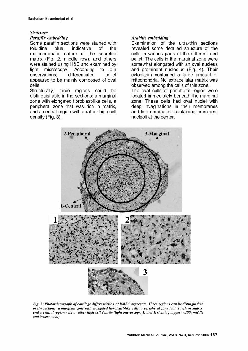
In central zone, the cell density was relatively high and three kinds of cells including oval cells similar to that of peripheral region, fat storing cells were observed (Fig. 6).
Fig. 5: Peripheral zone: nearly all cells were oval in shape. A cell immediately under the elongated cells of marginal zone (A, ×3000). These cells contained plenty of mitochondria (M) and dilated rER (B, ×15000). Deeper cells were highly active with several peripheral secretory vesicles (SV) (C, ×3000). Fibrillar matrix (FM) was observed among these cells (D, × 15000).
Fig. 4: Marginal zone. The cells in this region were somewhat elongated. Superficial cells possess some pinocytotic pits (P) (A, ×3000), but no matrix was observed among the cells (B, ×7000).
A
B
P
168
Structure of Cartilage Produced from hMSCs
Discussion Since the introduction of micromass culture system by Johnston et al. (21), scientists have utilized the system for different purposes. Some researchers used it to study the role of macromolecules such as BMP6 family in promoting chondrogenic differentiation (37-38). Others have cultivated MSCs in micromass culture system to investigate molecular events during in vitro chondrogenesis that resulted in defining the expression pattern of cartilage specific molecule such as collagen II, X, and aggrecan (18, 27-28). Still other scientists have used this culture system to evaluate the chondrogenic potential of the cells isolated by different methods (29-32).
Ichinso et al. (2005) cultured human MSCs in a micromass culture system for 21 days in order to investigate the morphologic changes during in vitro chondrogenesis. They concluded that in vitro chondrogenesis is very similar to that of in vivo (36). In present study, human MSCs were differentiated in micromass culture system in order to examine their structure by either light or transmission electron microscopy. In summary, our investigation showed that the pellet having been cultured in chondrogenic medium for 21 days differentiated into tissue composed of oval cells and a plenty of matrix accumulated among them. Since the cartilage-specific molecules were produced in this tissue as evidenced by RT-PCR and toluidine blue staining, we concluded that the tissue could be cartilage.
Fig. 6: Central zone: Similar to the peripheral zone, the majority of the cells in this region were oval chondroblastic cells (A, ×3000), secreting cartilage matrix (CM) (B, ×3000 and C, ×15000), with well-developed dilated rER (D, ×20000). Some cells loaded with lipid droplet were also observed adjacent to chondroblastic cells (A, ×3000).
D C
A B
M
rE
SV
FM
Yakhteh Medical Journal, Vol 8, No 3, Autumn 2006 169
Baghaban Eslaminejad et al

In general, the cartilage differentiated from MSCs had two characteristic structures. The first tissue type seemed to be rather cellular. The second one comprised of the cells appearing to be in active state, as its structure implied. These characters have been inherited from embryonic cartilage, as they are the characteristic of the cartilage tissue during development. Our results showed some differences among peripheral and central zones in terms of the accumulation of metachromatic matrix. Fewer matrices seemed to be produced in central zone compared to peripheral zone. This issue could be explained by avascular nature of the cartilage. Less matrix was secreted in central region, probably because regional avascularity and insufficient diffusion limit substrate availability. If this speculation is true, the number of initial cells of micromass culture system seems to be not appropriate to produce a mass size capable to be well-supplied by diffusion. This hypothesis was reinforced by observing the dying cells in central region. It was also interesting that no dividing cells were seen among the cells of tissue section. Similar findings have already been reported by Sekiya et al. (2002); they also indicated that DNA radioactivity per cell remained unchanged throughout the micromass culture system (19). They concluded that increase of the pellet size during the micromass culture system was due to matrix production rather than cell proliferation. According to Ichinso et al. (2005), there are three distinct cell layers including superficial fibroblastic, middle apoptotic, and deep chondrocytic-like cells during early days of induction of in vitro chondrogenesis using micromass culture system (36). They showed that by the end of day 21, the apoptotic cell layer disappeared, the superficial fibroblastic layer began disappearing, and the deep chondrocytic-like cells remained and constituted the main mass of the pellet. At this time, these cells had irregular outline and were similar to mature chondrocytes. In present study, after 21 days of chondrogenic induction, the differentiated pellet contained no apoptotic middle layer, but superficial fibroblastic layer remained
and the cells constituting the pellet seemed to be fully active (chondroblastic) in contrast to inactive (chondrocyte) state described by Ichinso et al. Conclusion Taken together, condensation produced in micromass system plus chondrogenic medium with defined materials could start and maintain chondrogenic differentiation, but it was unable to complete the process with producing fully mature tissue. In other words, it seems that the culture condition utilized in this study for chondrogenic differentiation was just able to produce a tissue more resemble to embryonic cartilage rather than fully differentiated mature cartilage found in adult tissues. Acknowledgments This research was supported by the Iranian Small Business Developing Center (SBDC) – LIDCO Grant. __________________________________ References 1. Buckwalter JA. Articular cartilage injuries. Clin Orthop 2002; 402: 21-37 2. Martin JA, Buckwalter JA. Aging, articular cartilage chondrocyte senescence and osteoarthritis. Biogerontology 2002; 3: 357-264 3. Buckwalter JA, mankin HJ. Articular cartilage: degeneration and osteoarthritis, repair, regeneration, and transplantation. Instr Course Lect 1998; 47: 487-504 4. Frenkel SR, Di Cesare PE. Scaffolds for articular cartilage repair. Am Biomed Eng 2004; 32: 26-34 5. Hardingham T, Tew S, Murdoch A. Tissue engineering: chondrocytes and cartilage. Arthritis Res 2002; 4: 563-568 6. Mitchell N, Sheparol N. The resurfacing of adult rabbit articular cartilage by multiple perforations through the subchondral bone. J Bone Joint Surg Am 1976; 58: 230-233 7. Mankin H, Mow V, Buchwalter J, Ionnotti J, Radcliffe A. Form and function of articular cartilage. In: Simon S, ed. Orthopaedic Basic Science. Rosemont IL: American Acaderny of Orthopaedic Surgeons 1994; 443-470 8. Frenkel SR, Clancy RM, Ricci JL, Di Cesare PE, Rediske JJ. Effects of nitric oxide on chondrucyte migration, adhesion, and cytoskeletal assembly. Arthritis Rheum1996; 39: 1905-1912 9. Brittberg MM, Lindahl A, Nilsson A, Ohlsson C, Isaksson O, Peterson L.Treatment of deep cartilage defects in the knee with antologous chondrocyte transplantation. N Engl J Med 1994; 331: 889-895 10. Cancedda R, Dozin B, Giannoi P, Quaro R. Tissue engineering and cell therapy of cartilage and bone. Matrix Biol 2003; 22: 81-91 11. Peterson L, Brittberg M, kiviranta I, Akerlund EL, Lindahl A. Autologous chondrocyte transplantation: biomechanics and long-term durability. Am J Sorts Med 2003; 30: 2-12
170
Structure of Cartilage Produced from hMSCs

12. Peterson L, Minas T, Brittberg M, Nilson A, Sjogren-Janson E, Lindahl A. Two-to 9-year outcome after autologous chondrocyte transplantation of the knee. Clin Orthop 2000; 374: 212-234 13. Richardson JB, Caterson B, Erans EH, Ashton BA, Roberts S. Repair of human articular cartilage after implantation of autologous chondrocyte. J Bone Joint Surg Br 1999; 81: 1064-1068 14. Dozin B, Malpeli M, Camardella L, Cancedd R, Pietrangelo A. Response of young, aged and osteoarthritic human articular chondrocytes to inflammatory cytokines: molecular and cellular aspects. Matrix Biol 2002; 21: 449-459 15. Bruder SP, Jaiswal N, Haynesworth SE. Growth, kinetics, self-renewal, and the osteogenic potential of purified human mesenchymal stem cells during extensive subcultivation and following cryopreservation. J Cell Biochem 1997; 64:278-294 16. Colter DC, Class R, DiGirolamo CM, Prockop DJ. Rapid expansion of recycling stem cells in culture of plastic- adherent cells from human bone marrow. Proc Nat Acad Sci U.S.A. 2000; 97: 3213-3218 17. Kadiyala S, Young RG, Thiede MA, Brnder SP. Culture expanded canine mesenchymal stem cells possess osteochondrogenic potential in vivo and in vitro. Cell Transplant 1997; 6: 125-134 18. Johnstone B, Hering TM, Caplan AI, Goldberg VM, Yoo JU. In vitro chondrogenesis of bone marrow-derived mesenchymal stem cells. Exp Cell Res 1998; 238: 265-272 19. Sekiya I, Vuoristo JT, Larson BL, Prockop DJ. In vitro cartilage formation by human adult stem cells from bone marrow stroma defines the sequence of cellular and molecular events during chondrogenesis. Der Biol 2002; 99: 4397-4402 20. Manning WK, Bonner WMJR. Isolation and culture of chondrocytes from human adult articular cartilage. Arthritis Rheum 1967; 10: 235-239 21. Johnstone B. Mesenchymal stem cells and chondrogenesis. Europ Cell Mat 2002; P: 27. 22. Mackay AM, Beck SC, Murphy JM, Barry FP, Chichester CO, Pittenger MF. Chondrogenic differentiation of cultured human mesenchymal stem cells from marrow. Tissue Eng 1998;4:415-428 23. Carlberg AL, Pucci B, Rallapalli R, Tuan RS, Hall DJ. Efficient chondrogenesis of mesenchymal cells in micromass culture by retroviral gene transfer of BMP-2. Differentiation 2001; 67: 128-138 24. Bosnakovski D, Mizuno M, Kim G, Ishiguro T, Okumura M, Iwanaga T, Kadosawa T, Fujinaga T. Chondrogenic differentiation of bovine bone marrow mesenchymal stem cells in pellet culture system. Exp Hematol 2004; 32: 502-509 25. Bosnakovski D, Mizuno M, Kim G, Takagi S, Okumur M, Fujinag T. Gene expression profile of bovine bone marrow mesenchymal stem cell during spontaneous chondrogenic differentiation in pellet culture system. JPN J Vet Res 2006; 53:127-139 26. Indrawattana N, Chen G, Tadokoro M, Shann
LH, Ohgushi H, Tateishi T, Tanaka J, Bunyaratvej A. Growth factor combination for chondrogenic induction from human mesenchymal stem cell. Biochem Biophys Res Commun 2004; 320: 914-919 27. Yoo UY, Barthel TS, Nishimura K, Solchaga L, Caplan AI, Goldberg VM, Jhonstone B. The chondrogenic potential of human bone-marrow-derived mesenchymal progenitor cells. J Bone Joint Surg 1998; 12: 1745-1757 28. Barry F, Boynton RE, Liu B, Murphy JM. Chondrogenic differentiation of mesenchymal stem cells from bone marrow: Differentiation-dependent gene expression of matrix components. Exp Cell Res 2001; 268: 189-200 29. Peister A, Mellad JA, Larsen LL, Hall BM, Gibson LF, Prockop DJ. Adult stem cells from bone marrow (MSCS) isolated from different strains of inbred mice vary in surface epitopes, rates of proliferation and differentiation potential. Blood 2004; 103: 1662-1668 30. Sun S, Guo Z, Xiao x, Liu B, Liu X, Tang PH, Mao N. Isolation of mouse marrow mesenchymal progenitors by a novel and reliable method. Stem Cells 2003; 21: 527-535 31. Tropel P, Noel D, Platet N, Legrand P, Benabid AL, Berger F. Isolation and characterization of mesenchymal stem cells from adult mouse bone marrow. Exp Cell Res 2004; 295: 395-406 32. Eslaminejad MB, Nikmahzar A, Taghiyar L, Nadri S, Massumi M. Murine mesenchymal stem cells isolated by low density primary culture system. Dev Growth Differ 2006;48: 361-370 33. Denker AE, Haas AR, Nicoll SB, Tuan RS. Chondrogenic differentiation of murine C3H10T1/2 multipotential mesenchymal cells: Stimulation by bone morphogenetic prptein-2 in high-density micromass culture. Differentiation 1999; 64: 67-76 34. Huang JI, Kazmi N, Durbhakula MM, Hering TM, Yoo JU, Johnstone B. Chondrogenic potential of progenitor cells derived from human bone marrow and adipose tissue: a patient-matched comparison. J Orthop Res 2005; 23:1383-1389 35. Lin Y, Luo E, Chen X, Liu L, Qiao J, Yan Z, Li Z, Tang W, Zheng X, Tian W. Molecular and cellular characterization during chondrogenic differentiation of adipose-tissue derived stromal cells in vitro and cartilage formation in vivo. J Cell Mol Med 2005; 9: 929-939 36. Ichinose S, Tagami M, Muneta T, Sekiya I. Morphological examination during in vitro cartilage formation by human mesenchymal stem cells.Cell Tissue Res 2005; 322:217-226 37. Sekiya I, Colter DC, Prockop DJ. BMP-6 enhances chondrogenesis in a subpopulation of human marrow stromal cells. Biochem Biophys Res Commun 2001; 282: 411-418 38. Sekiya I, Larsen BL, Vuoristo JT, Reger RL, Prockop DJ. Comparison of effect of BMP-2, -4, and -6 on in vitro cartilage formation of human adult stem cells from marrow stroma. Cell Tissue Res 2005; 320: 269-276 _________________________________________
Yakhteh Medical Journal, Vol 8, No 3, Autumn 2006 171
Baghaban Eslaminejad et al