The spleen CD4+ T cell response to blood-stage Plasmodium chabaudi malaria develops in two phases...
12
The Spleen CD4 + T Cell Response to Blood-Stage Plasmodium chabaudi Malaria Develops in Two Phases Characterized by Different Properties Sandra Marcia Muxel 1 *, Ana Paula Freitas do Rosa ´ rio 1 , Cla ´ udia Augusta Zago 1 , Sheyla Ine ´ s Castillo- Me ´ ndez 1 , Luiz Roberto Sardinha 1,2 , Se ´ rgio Marcelo Rodriguez-Ma ´ laga 1 , Niels Olsen Saraiva Ca ˆ mara 1 , Jose ´ Maria A ´ lvarez 1 , Maria Regina D’Impe ´ rio Lima 1 1 Departamento de Imunologia, Instituto de Cie ˆncias Biome ´dicas, Universidade de Sa ˜o Paulo, Sa ˜o Paulo, Sa ˜o Paulo, Brazil, 2 Instituto Israelita de Ensino e Pesquisa Albert Einstein, Sa ˜o Paulo, Sa ˜o Paulo, Brazil Abstract The pivotal role of spleen CD4 + T cells in the development of both malaria pathogenesis and protective immunity makes necessary a profound comprehension of the mechanisms involved in their activation and regulation during Plasmodium infection. Herein, we examined in detail the behaviour of non-conventional and conventional splenic CD4 + T cells during P. chabaudi malaria. We took advantage of the fact that a great proportion of CD4 + T cells generated in CD1d -/- mice are I-A b - restricted (conventional cells), while their counterparts in I-A b-/- mice are restricted by CD1d and other class IB major histocompatibility complex (MHC) molecules (non-conventional cells). We found that conventional CD4 + T cells are the main protagonists of the immune response to infection, which develops in two consecutive phases concomitant with acute and chronic parasitaemias. The early phase of the conventional CD4 + T cell response is intense and short lasting, rapidly providing large amounts of proinflammatory cytokines and helping follicular and marginal zone B cells to secrete polyclonal immunoglobulin. Both TNF-a and IFN-c production depend mostly on conventional CD4 + T cells. IFN-c is produced simultaneously by non-conventional and conventional CD4 + T cells. The early phase of the response finishes after a week of infection, with the elimination of a large proportion of CD4 + T cells, which then gives opportunity to the development of acquired immunity. Unexpectedly, the major contribution of CD1d-restricted CD4 + T cells occurs at the beginning of the second phase of the response, but not earlier, helping both IFN-c and parasite-specific antibody production. We concluded that conventional CD4 + T cells have a central role from the onset of P. chabaudi malaria, acting in parallel with non- conventional CD4 + T cells as a link between innate and acquired immunity. This study contributes to the understanding of malaria immunology and opens a perspective for future studies designed to decipher the molecular mechanisms behind immune responses to Plasmodium infection. Citation: Muxel SM, Freitas do Rosa ´rio AP, Zago CA, Castillo-Me ´ndez SI, Sardinha LR, et al. (2011) The Spleen CD4 + T Cell Response to Blood-Stage Plasmodium chabaudi Malaria Develops in Two Phases Characterized by Different Properties. PLoS ONE 6(7): e22434. doi:10.1371/journal.pone.0022434 Editor: Ben L. Kelly, Louisiana State University, United States of America Received September 30, 2010; Accepted June 28, 2011; Published July 21, 2011 Copyright: ß 2011 Muxel et al. This is an open-access article distributed under the terms of the Creative Commons Attribution License, which permits unrestricted use, distribution, and reproduction in any medium, provided the original author and source are credited. Funding: This work was supported by grants from FAPESP (www.fapesp.br), CAPES (www.capes.gov.br), and CNPq (www.cnpq.br). The funders had no role in study design, data collection and analysis, decision to publish, or preparation of the manuscript. Competing Interests: The authors have declared that no competing interests exist. * E-mail: [email protected] Introduction Malaria, the infectious disease caused by Plasmodium parasites, is a major global health problem that is responsible for the death of over a million people every year [1]. Humans with no previous experience of malaria almost invariably develop a febrile illness that may become severe and lead to death. The asexual blood- stage of the parasite is responsible for the clinical symptoms of the disease. Three overlapping syndromes, severe anaemia, cerebral malaria and respiratory distress, account for most of the severe cases and deaths [2]. Because many of the features of severe malaria are similar to those of sepsis [3], over-vigorous responses to parasites have been implicated in the aetiology of these syndromes [4,5]. Thus, although TNF-a and IFN-c appear to be protective against the parasite, very high serum concentrations of proinflammatory cytokines are associated with great morbidity [6,7]. Yet, exposure to one or two malaria infections appears to be sufficient to induce complete protection from severe illness and death [8], while sterile immunity to malaria parasites is probably never achieved. Encouragingly, some aspects in mouse models of malaria appear to mirror the human disease with reasonable accuracy. In acutely infected mice, the type 1 responses from CD4 + and CD8 + T cells are the main participants in the development of both malaria pathogenesis and protective immunity [9,10,11]. While TNF-a and IFN-c are associated with the development of clinical manifestations of the disease [12,13,14], the control of acute infection also depends on proinflammatory cytokines [15,16,17], which together with acute- phase antibodies [18] may promote parasite clearance by macro- phages [19,20]. CD4 + T cells are thought to be essential for complete elimination of the parasite during the late phase of the disease [10,21,22], which may also hold true for humans because these cells are critically required to help B cells produce parasite-specific high- affinity immunoglobulin G (IgG) antibodies. However, it is not yet PLoS ONE | www.plosone.org 1 July 2011 | Volume 6 | Issue 7 | e22434
-
Upload
independent -
Category
Documents
-
view
5 -
download
0
Transcript of The spleen CD4+ T cell response to blood-stage Plasmodium chabaudi malaria develops in two phases...
The Spleen CD4+ T Cell Response to Blood-StagePlasmodium chabaudi Malaria Develops in Two PhasesCharacterized by Different PropertiesSandra Marcia Muxel1*, Ana Paula Freitas do Rosario1, Claudia Augusta Zago1, Sheyla Ines Castillo-
Mendez1, Luiz Roberto Sardinha1,2, Sergio Marcelo Rodriguez-Malaga1, Niels Olsen Saraiva Camara1,
Jose Maria Alvarez1, Maria Regina D’Imperio Lima1
1 Departamento de Imunologia, Instituto de Ciencias Biomedicas, Universidade de Sao Paulo, Sao Paulo, Sao Paulo, Brazil, 2 Instituto Israelita de Ensino e Pesquisa Albert
Einstein, Sao Paulo, Sao Paulo, Brazil
Abstract
The pivotal role of spleen CD4+ T cells in the development of both malaria pathogenesis and protective immunity makesnecessary a profound comprehension of the mechanisms involved in their activation and regulation during Plasmodiuminfection. Herein, we examined in detail the behaviour of non-conventional and conventional splenic CD4+ T cells during P.chabaudi malaria. We took advantage of the fact that a great proportion of CD4+ T cells generated in CD1d-/- mice are I-Ab-restricted (conventional cells), while their counterparts in I-Ab-/- mice are restricted by CD1d and other class IB majorhistocompatibility complex (MHC) molecules (non-conventional cells). We found that conventional CD4+ T cells are the mainprotagonists of the immune response to infection, which develops in two consecutive phases concomitant with acute andchronic parasitaemias. The early phase of the conventional CD4+ T cell response is intense and short lasting, rapidlyproviding large amounts of proinflammatory cytokines and helping follicular and marginal zone B cells to secrete polyclonalimmunoglobulin. Both TNF-a and IFN-c production depend mostly on conventional CD4+ T cells. IFN-c is producedsimultaneously by non-conventional and conventional CD4+ T cells. The early phase of the response finishes after a week ofinfection, with the elimination of a large proportion of CD4+ T cells, which then gives opportunity to the development ofacquired immunity. Unexpectedly, the major contribution of CD1d-restricted CD4+ T cells occurs at the beginning of thesecond phase of the response, but not earlier, helping both IFN-c and parasite-specific antibody production. We concludedthat conventional CD4+ T cells have a central role from the onset of P. chabaudi malaria, acting in parallel with non-conventional CD4+ T cells as a link between innate and acquired immunity. This study contributes to the understanding ofmalaria immunology and opens a perspective for future studies designed to decipher the molecular mechanisms behindimmune responses to Plasmodium infection.
Citation: Muxel SM, Freitas do Rosario AP, Zago CA, Castillo-Mendez SI, Sardinha LR, et al. (2011) The Spleen CD4+ T Cell Response to Blood-Stage Plasmodiumchabaudi Malaria Develops in Two Phases Characterized by Different Properties. PLoS ONE 6(7): e22434. doi:10.1371/journal.pone.0022434
Editor: Ben L. Kelly, Louisiana State University, United States of America
Received September 30, 2010; Accepted June 28, 2011; Published July 21, 2011
Copyright: � 2011 Muxel et al. This is an open-access article distributed under the terms of the Creative Commons Attribution License, which permitsunrestricted use, distribution, and reproduction in any medium, provided the original author and source are credited.
Funding: This work was supported by grants from FAPESP (www.fapesp.br), CAPES (www.capes.gov.br), and CNPq (www.cnpq.br). The funders had no role instudy design, data collection and analysis, decision to publish, or preparation of the manuscript.
Competing Interests: The authors have declared that no competing interests exist.
* E-mail: [email protected]
Introduction
Malaria, the infectious disease caused by Plasmodium parasites, is
a major global health problem that is responsible for the death of
over a million people every year [1]. Humans with no previous
experience of malaria almost invariably develop a febrile illness
that may become severe and lead to death. The asexual blood-
stage of the parasite is responsible for the clinical symptoms of the
disease. Three overlapping syndromes, severe anaemia, cerebral
malaria and respiratory distress, account for most of the severe
cases and deaths [2]. Because many of the features of severe
malaria are similar to those of sepsis [3], over-vigorous responses
to parasites have been implicated in the aetiology of these
syndromes [4,5]. Thus, although TNF-a and IFN-c appear to be
protective against the parasite, very high serum concentrations of
proinflammatory cytokines are associated with great morbidity
[6,7]. Yet, exposure to one or two malaria infections appears to be
sufficient to induce complete protection from severe illness and
death [8], while sterile immunity to malaria parasites is probably
never achieved.
Encouragingly, some aspects in mouse models of malaria appear to
mirror the human disease with reasonable accuracy. In acutely
infected mice, the type 1 responses from CD4+ and CD8+ T cells are
the main participants in the development of both malaria
pathogenesis and protective immunity [9,10,11]. While TNF-a and
IFN-c are associated with the development of clinical manifestations
of the disease [12,13,14], the control of acute infection also depends
on proinflammatory cytokines [15,16,17], which together with acute-
phase antibodies [18] may promote parasite clearance by macro-
phages [19,20]. CD4+ T cells are thought to be essential for complete
elimination of the parasite during the late phase of the disease
[10,21,22], which may also hold true for humans because these cells
are critically required to help B cells produce parasite-specific high-
affinity immunoglobulin G (IgG) antibodies. However, it is not yet
PLoS ONE | www.plosone.org 1 July 2011 | Volume 6 | Issue 7 | e22434

apparent how much of the pathology is dependent on a contribution
from the acquired immune response [4].
The pivotal role of CD4+ T cells in the development of both
malaria pathogenesis and protective immunity makes them putative
targets for new strategies to improve the outcome of the disease. On
top of being important sources of IFN-c and helping B cells to
secrete antibodies, these cells play a key role in the regulation of
immune responses. The acquisition of clinical immunity is likely to
be coordinated by CD4+ T cells, as clinical immunity remains
relatively robust over long periods after being established. The
possibility that CD4+ T cells are implicated in this process is
suggested by the fact that IL-10 or TGF-b deficiency intensifies the
clinical signs in mice suffering from malaria [13,23]. Therefore, a
profound comprehension of the mechanisms involved in CD4+ T
cell activation and regulation during Plasmodium infection may
improve the chances of developing effective vaccines and other
potential immunotherapies to prevent severe malaria syndromes.
CD4+ T cells from the spleen, the main lymphoid organ for
protection against blood-borne infectious diseases, can be divided
into two categories based on how they recognise antigens.
Conventional CD4+ T cells recognise peptides associated with
class II molecules of the major histocompatibility complex (MHC),
whereas non-conventional CD4+ T cells are a heterogeneous
population mostly restricted by class IB MHC molecules [24],
which includes CD1d-restricted natural killer T (NKT) cells that
recognise lipids [25]. It is generally accepted that non-conven-
tional CD4+ T cells are particularly shaped for early immune
responses to infections while conventional CD4+ T cells are
responsible for acquired immunity. Because CD4+ T cells from
these two categories have been implicated in the immune response
to Plasmodium parasites [2], we sought to examine in detail the
behaviour of non-conventional and conventional splenic CD4+ T
cells during blood-stage P. chabaudi AS strain malaria, a suitable
model for the human disease caused by Plasmodium falciparum [26].
The information provided here is fundamental for unravelling the
molecular mechanisms behind the early phase of the CD4+ T cell
response to Plasmodium infection, knowledge that could help
explain why people develop severe malaria and how they get
immune against the symptoms of the disease.
Materials and Methods
Mice, parasite and infectionSix-to-eight-wk-old C57BL/6 (wild type; WT) (originally from
The Jackson Laboratory), CD1d-/- [27] and I-Ab-/- (ABBN12) [28]
male mice were bred under specific pathogen-free conditions at the
Isogenic Mice Facility (Department of Immunology/University of
Sao Paulo, Brazil). CD1d-/- and I-Ab-/- mice were used after nine
backcrosses onto the C57BL/6 background. P. chabaudi AS strain
was maintained as described elsewhere [15]. Mice were inoculated
intraperitoneally (i.p.) with 16106 infected red blood cells (iRBC).
WT and CD1d-/- mice were challenged i.p. with 16108 iRBC on
day 30 postinfection (p.i.). Parasitaemias were monitored by
microscopic examination of Giemsa-stained thin tail blood smears.
Ethics statementAll procedures were in accordance with national regulations of
ethical guidelines for mice experimentation and welfare of the
Conselho Nacional de Saude and Colegio Brasileiro em
Experimentacao Animal (COBEA) - Brazil, and the protocols
were approved by the Health Animal Committee (Comissao de
Etica no Uso de Animais – CEUA – ICB/USP) of the Instituto de
Ciencias Biomedicas of the Universidade de Sao Paulo, Sao Paulo,
Brazil (permit number 0019/2005 and 0036/2007).
Spleen cell suspensionSpleen cells were washed and maintained in cold RPMI 1640,
supplemented with penicillin (100 U/mL), streptomycin (100 mg/
mL), 2-mercaptoethanol (50 mM), L-glutamine (2 mM), sodium
pyruvate (1 mM), and 3% heat-inactivated foetal calf serum. All
supplements were purchased from Invitrogen Life Technologies.
The numbers of cells per spleen were counted using a Neubauer
chamber (Sigma-Aldrich).
Phenotypic analysis of spleen cellsSpleen cells (16106) were stained with allophycocyanin (APC)-,
peridin-clorophyll-protein (PercP)-, fluorescein-isothiocyanate
(FITC)-, phycoerythrin (PE)- or CyChrome (Cy)-labelled mono-
clonal antibodies (mAb) (BD Pharmingen) to CD4 (H129.19),
CD8 (53-6.7), CD45R–B220 (RA3-6B2), CD69 (H1.2F3), CD21
(7G6), CD23 (B3B4) and CD1d (1B1) and with the PE-labelled
CD1d-a-GalCer tetramer from the NIH (National Institutes of
Health - USA) Tetramer Facility. Stained cells were analysed by
flow cytometry using a FACSCalibur device with CellQuest
software (BD Biosciences).
IFN-c intracellular detectionSpleen cells (16106) were cultured with GolgiStop according to
the manufacturer’s instructions in the presence of 5 mg/mL plate-
bound purified anti-CD3 mAb (145-2C11) or medium alone for
10 h at 37uC in a 5% CO2 atmosphere. After washing, cells were
surface stained with FITC- or APC-labelled mAb to CD4 and
CD8. Cells were then fixed with Cytofix/Cytoperm buffer, stained
with PE-labelled mAb to IFN-c (XMG-1.2) diluted in Perm/Wash
buffer, and analysed by flow cytometry. All reagents were
purchased from BD Pharmingen.
CFSE proliferation assayThe proliferative T cell response was measured as previously
described [29]. Briefly, spleen cells (36107) were incubated for
30 min at 37uC with carboxyfluorescein succinimidyl ester (CFSE)
(Molecular Probes) (5 mM) in phosphate-buffered saline (PBS)
supplemented with 0.1% bovine serum albumin (BSA). Cells
(16106) were then cultured in 96-well plates (Costar) with iRBC
(36106) or medium alone for 72 h at 37uC in a 5% CO2
atmosphere. In some experiments, blocking mAb to I-Ab (AF6-
120.1) and IgG2a isotypic control (G155-178) were added to the
cultures (both mAb used at 2.5 mg/mL concentrations). After
incubation, cells were stained with PE or APC-labelled mAb to
CD4 and CD8 and analysed by flow cytometry.
Multicytokine assessmentSeveral cytokines (IFN-c, TNF-a, IL-2, IL-4 and IL-5) were
quantified simultaneously, by the cytometric bead array (CBA; BD
Pharmingen), using supernatants obtained from the same cultures
used in the CSFE proliferation assay. This technique uses flow
cytometry to measure soluble analytes in a particle-based
immunoassay and was conducted according to the manufacturer’s
instructions. The lower limit of detection for all cytokines in this
assay was 20 pg/mL.
Purification of cell subpopulationsSpleen cells (16108) from CD1d-/- mice were incubated with
PE-labelled mAb to CD4 and with anti-PE microbeads diluted in
PBS with 0.5% BSA and 2 mM ethylenediaminetetraacetic acid
(EDTA) (Invitrogen Life Technologies). Cells were then purified
using LS columns (Midi MACS; Miltenyi Biotec). The positive
fraction showed . 85% CD4+ cells. To obtain spleen cell
CD4+ T Cells in Malaria
PLoS ONE | www.plosone.org 2 July 2011 | Volume 6 | Issue 7 | e22434

preparations depleted of dendritic cells (DC), negative selection
with anti-Pan DC microbeads (Miltenyi Biotec) was performed,
and the negative fraction showed , 0.5% DC (CD11c+I-Ab+ cells).
Adoptive cell transfers of conventional CD4+ cellsWT and I-Ab-/- mice received i.p. 46106 purified CFSE-
labelled CD4+ cells obtained from CD1d-/- mice. These mice were
infected i.p. with 16106 iRBC on the day of transfer. Proliferative
CD4+ cell response in vivo was analysed on days 4 and 7 of
infection. Non-infected mice were used as controls.
ELISPOT assay for Ig-producing cellsIg-producing cells were quantified by the ELISPOT assay as
previously described [30]. In brief, 96-well flat-bottom microtest
plates (Costar) coated overnight (4uC) with goat anti-mouse total Ig
(10 mg/mL) were saturated with 1% gelatine (Merck) in PBS for
120 min. Titrated numbers of spleen cells (16106 to 56102 cells/
well) were cultured for 6 h at 37uC in a 5% CO2 atmosphere. The
spots were developed by adding goat anti-mouse IgM, IgG2a or
IgG1 biotinylated antibodies followed by the addition of a
phosphatase alkaline-avidin conjugate (all antibodies and conju-
gates were purchased from Southern Biotechnology Associates). 5-
Bromo-4-chloro-3-indolyl phosphate (BCIP) (Sigma-Aldrich) di-
luted in 2-amino-2-methyl-1-propanol (AMP) (Merck) was used as
the substrate. From the titration plots (number of cells plated vs.
spots) and the total number of splenocytes, the number of total Ig-
producing cells per spleen was calculated.
ELISA test for parasite-specific antibodiesAnti-P. chabaudi antibodies were quantified by ELISA as
previously described [31]. In brief, 96-well flat-bottom microtest
plates (Costar) were coated overnight (4uC) with a total P. chabaudi
extract (8 mg/mL). Plates were saturated with 1% BSA for 1 h.
After washing, 100 mL of mouse serum samples (diluted from 1/2)
was added and left for 90 min at room temperature or overnight
(4uC), followed by goat anti-mouse IgG2a and IgG1 peroxidase-
conjugated antibodies (Southern Biotechnology Associates) for 1 h.
After washing, 100 mL of tetramethylbenzidine (TMB) (Zymed)
was added to each well and 15 min later, the absorbance values
were quantified by a Spectra Max 190 spectrophotometer
(Molecular Devices) with a 650-nm wavelength filter.
Statistical analysisStatistical significance was analysed in Graph Pad Prism 4
software using the Mann-Whitney U and Tukey’s multiple
comparison tests. Differences between groups were considered
significant when p,0.05 (5%).
Results
I-Ab-restricted CD4+ T cells are required for the control ofparasitaemia during the early and late P. chabaudiinfection
To evaluate the protective role of CD1d- and I-Ab-restricted
CD4+ T cells during early and late P. chabaudi malaria, we
determined the parasitaemia curves in WT, CD1d-/- and I-Ab-/-
mice infected with 16106 iRBC. WT and CD1d-/- mice developed
comparable first parasitaemia peaks, but in CD1d-/- mice the
second peak was higher and occurred earlier (on days 13 to 15 p.i.)
when compared to WT mice (on day 17 p.i.) (Figure 1). Despite
these differences, mice from both groups showed efficient control
of parasite growth and survived the infection. Contrarily, I-Ab-/-
mice developed a significantly higher first peak and prominent
recrudescent peaks compared to WT mice. Furthermore, these
mice were unable to eliminate the parasite and around 50% were
dead one month after infection. These results indicate that parasite
control during early and late P. chabaudi malaria is dependent on I-
Ab-restricted CD4+ T cells, whereas the contribution of CD1d-
restricted CD4+ T cells is modest and limited to the second
parasitaemia peak.
The majority of spleen CD4+ T cells activated during thefirst days of P. chabaudi infection are restricted by class IIMHC molecules
As the previous results suggest a protective role for I-Ab-
restricted CD4+ T cells in early P. chabaudi malaria, we set out to
evaluate the kinetics of the CD4+ T cell response to infection in
mice lacking CD1d and I-Ab molecules using as parameters spleen
cellularity and expression of the early activation marker CD69
[32]. The aim of this analysis was to compare non-conventional
and conventional CD4+ T cells, which are thought to be primarily
involved in innate and acquired immunity, respectively. We took
advantage of the fact that a great proportion of CD4+ T cells
generated in CD1d-/- mice are restricted by I-Ab molecules
(conventional CD4+ T cells), whereas their counterparts in I-Ab-/-
mice are mostly restricted by CD1d molecules (non-conventional
CD4+ T cells). According to our data, the lack of CD1d molecules
led to reduced numbers of CD4+ cells per spleen in infected mice
(Figure 2A). Although the CD4+ cell population was notably
smaller in I-Ab-/- mice compared to WT mice, it also expanded
from day 0 to 7 p.i. when maximum cellularity was attained in WT
mice. Regarding the CD69 expression, the mean fluorescence
intensity (MFI) was considerably higher in CD4+ cells in all groups
of infected mice compared to controls (Figure 2B). However, for
CD4+ cells generated in the absence of I-Ab molecules, a two-fold
Figure 1. Parasitaemia curves in P. chabaudi-infected WT, CD1d-/- and I-Ab-/- mice. Mice were infected with 16106 iRBC. Each curvecorresponds to the means 6 SD (n = 5–10). *, p,0.05, CD1d-/- or I-Ab-/- mice compared with WT mice. Data are representative of three experiments.doi:10.1371/journal.pone.0022434.g001
CD4+ T Cells in Malaria
PLoS ONE | www.plosone.org 3 July 2011 | Volume 6 | Issue 7 | e22434
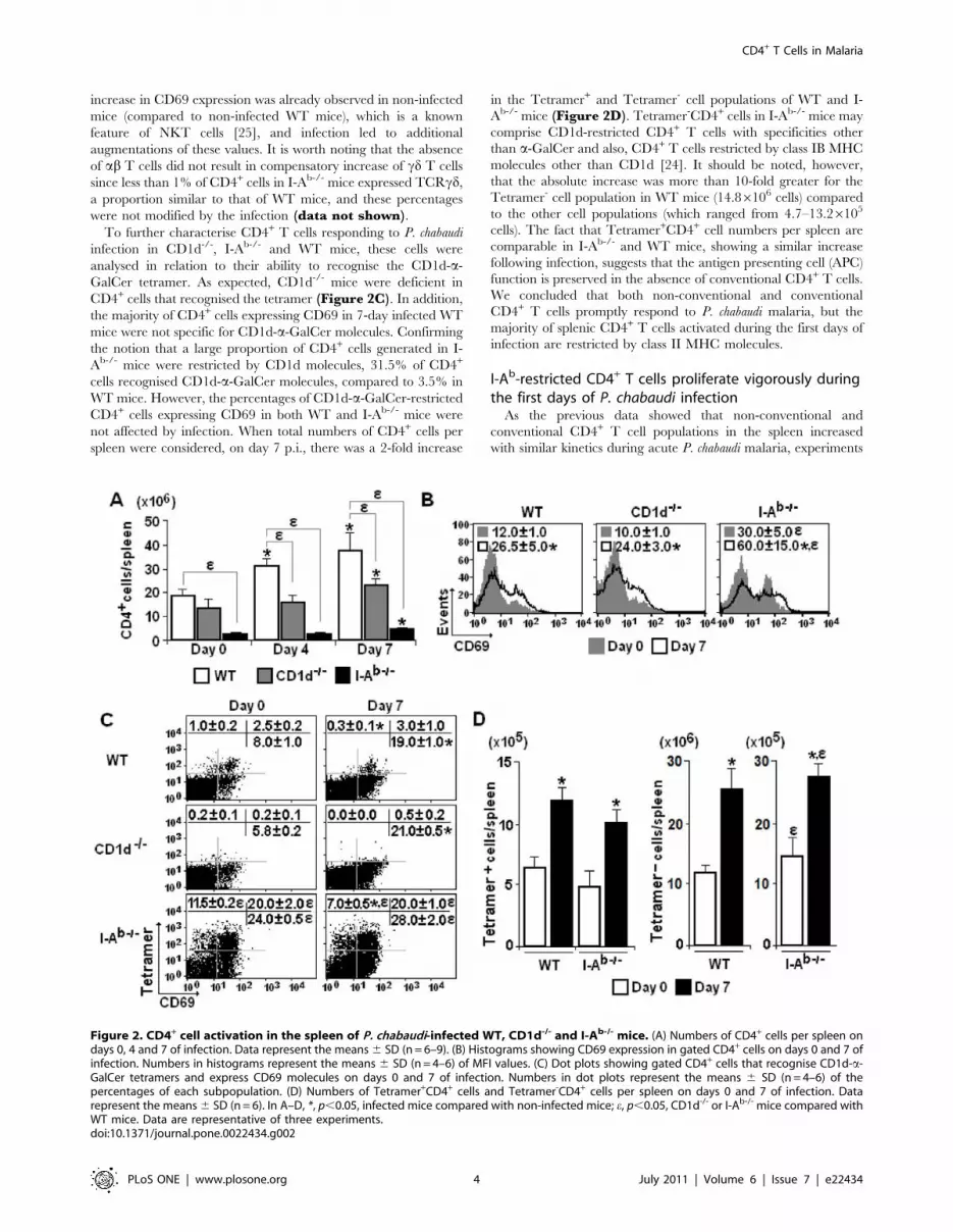
increase in CD69 expression was already observed in non-infected
mice (compared to non-infected WT mice), which is a known
feature of NKT cells [25], and infection led to additional
augmentations of these values. It is worth noting that the absence
of ab T cells did not result in compensatory increase of cd T cells
since less than 1% of CD4+ cells in I-Ab-/- mice expressed TCRcd,
a proportion similar to that of WT mice, and these percentages
were not modified by the infection (data not shown).
To further characterise CD4+ T cells responding to P. chabaudi
infection in CD1d-/-, I-Ab-/- and WT mice, these cells were
analysed in relation to their ability to recognise the CD1d-a-
GalCer tetramer. As expected, CD1d-/- mice were deficient in
CD4+ cells that recognised the tetramer (Figure 2C). In addition,
the majority of CD4+ cells expressing CD69 in 7-day infected WT
mice were not specific for CD1d-a-GalCer molecules. Confirming
the notion that a large proportion of CD4+ cells generated in I-
Ab-/- mice were restricted by CD1d molecules, 31.5% of CD4+
cells recognised CD1d-a-GalCer molecules, compared to 3.5% in
WT mice. However, the percentages of CD1d-a-GalCer-restricted
CD4+ cells expressing CD69 in both WT and I-Ab-/- mice were
not affected by infection. When total numbers of CD4+ cells per
spleen were considered, on day 7 p.i., there was a 2-fold increase
in the Tetramer+ and Tetramer- cell populations of WT and I-
Ab-/- mice (Figure 2D). Tetramer-CD4+ cells in I-Ab-/- mice may
comprise CD1d-restricted CD4+ T cells with specificities other
than a-GalCer and also, CD4+ T cells restricted by class IB MHC
molecules other than CD1d [24]. It should be noted, however,
that the absolute increase was more than 10-fold greater for the
Tetramer- cell population in WT mice (14.86106 cells) compared
to the other cell populations (which ranged from 4.7–13.26105
cells). The fact that Tetramer+CD4+ cell numbers per spleen are
comparable in I-Ab-/- and WT mice, showing a similar increase
following infection, suggests that the antigen presenting cell (APC)
function is preserved in the absence of conventional CD4+ T cells.
We concluded that both non-conventional and conventional
CD4+ T cells promptly respond to P. chabaudi malaria, but the
majority of splenic CD4+ T cells activated during the first days of
infection are restricted by class II MHC molecules.
I-Ab-restricted CD4+ T cells proliferate vigorously duringthe first days of P. chabaudi infection
As the previous data showed that non-conventional and
conventional CD4+ T cell populations in the spleen increased
with similar kinetics during acute P. chabaudi malaria, experiments
Figure 2. CD4+ cell activation in the spleen of P. chabaudi-infected WT, CD1d-/- and I-Ab-/- mice. (A) Numbers of CD4+ cells per spleen ondays 0, 4 and 7 of infection. Data represent the means 6 SD (n = 6–9). (B) Histograms showing CD69 expression in gated CD4+ cells on days 0 and 7 ofinfection. Numbers in histograms represent the means 6 SD (n = 4–6) of MFI values. (C) Dot plots showing gated CD4+ cells that recognise CD1d-a-GalCer tetramers and express CD69 molecules on days 0 and 7 of infection. Numbers in dot plots represent the means 6 SD (n = 4–6) of thepercentages of each subpopulation. (D) Numbers of Tetramer+CD4+ cells and Tetramer-CD4+ cells per spleen on days 0 and 7 of infection. Datarepresent the means 6 SD (n = 6). In A–D, *, p,0.05, infected mice compared with non-infected mice; e, p,0.05, CD1d-/- or I-Ab-/- mice compared withWT mice. Data are representative of three experiments.doi:10.1371/journal.pone.0022434.g002
CD4+ T Cells in Malaria
PLoS ONE | www.plosone.org 4 July 2011 | Volume 6 | Issue 7 | e22434
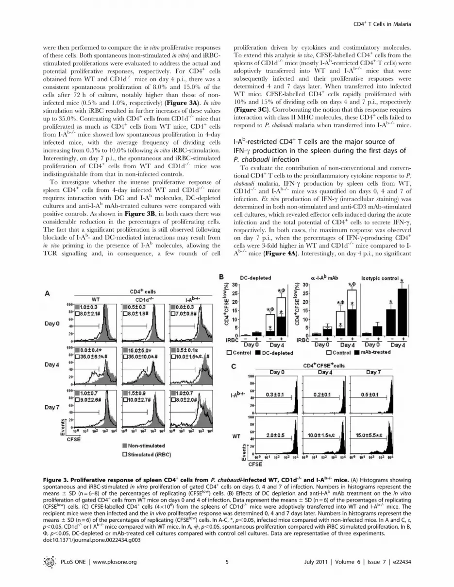
were then performed to compare the in vitro proliferative responses
of these cells. Both spontaneous (non-stimulated in vitro) and iRBC-
stimulated proliferations were evaluated to address the actual and
potential proliferative responses, respectively. For CD4+ cells
obtained from WT and CD1d-/- mice on day 4 p.i., there was a
consistent spontaneous proliferation of 8.0% and 15.0% of the
cells after 72 h of culture, notably higher than those of non-
infected mice (0.5% and 1.0%, respectively) (Figure 3A). In vitro
stimulation with iRBC resulted in further increases of these values
up to 35.0%. Contrasting with CD4+ cells from CD1d-/- mice that
proliferated as much as CD4+ cells from WT mice, CD4+ cells
from I-Ab-/- mice showed low spontaneous proliferation in 4-day
infected mice, with the average frequency of dividing cells
increasing from 0.5% to 10.0% following in vitro iRBC-stimulation.
Interestingly, on day 7 p.i., the spontaneous and iRBC-stimulated
proliferation of CD4+ cells from WT and CD1d-/- mice was
indistinguishable from that in non-infected controls.
To investigate whether the intense proliferative response of
spleen CD4+ cells from 4-day infected WT and CD1d-/- mice
requires interaction with DC and I-Ab molecules, DC-depleted
cultures and anti-I-Ab mAb-treated cultures were compared with
positive controls. As shown in Figure 3B, in both cases there was
considerable reduction in the percentages of proliferating cells.
The fact that a significant proliferation is still observed following
blockade of I-Ab- and DC-mediated interactions may result from
in vivo priming in the presence of I-Ab molecules, allowing the
TCR signalling and, in consequence, a few rounds of cell
proliferation driven by cytokines and costimulatory molecules.
To extend this analysis in vivo, CFSE-labelled CD4+ cells from the
spleens of CD1d-/- mice (mostly I-Ab-restricted CD4+ T cells) were
adoptively transferred into WT and I-Ab-/- mice that were
subsequently infected and their proliferative responses were
determined 4 and 7 days later. When transferred into infected
WT mice, CFSE-labelled CD4+ cells rapidly proliferated with
10% and 15% of dividing cells on days 4 and 7 p.i., respectively
(Figure 3C). Corroborating the notion that this response requires
interaction with class II MHC molecules, these CD4+ cells failed to
respond to P. chabaudi malaria when transferred into I-Ab-/- mice.
I-Ab-restricted CD4+ T cells are the major source ofIFN-c production in the spleen during the first days ofP. chabaudi infection
To evaluate the contribution of non-conventional and conven-
tional CD4+ T cells to the proinflammatory cytokine response to P.
chabaudi malaria, IFN-c production by spleen cells from WT,
CD1d-/- and I-Ab-/- mice was quantified on days 0, 4 and 7 of
infection. Ex vivo production of IFN-c (intracellular staining) was
determined in both non-stimulated and anti-CD3 mAb-stimulated
cell cultures, which revealed effector cells induced during the acute
infection and the total potential of CD4+ cells to secrete IFN-c,
respectively. In both cases, the maximum response was observed
on day 7 p.i., when the percentages of IFN-c-producing CD4+
cells were 3-fold higher in WT and CD1d-/- mice compared to I-
Ab-/- mice (Figure 4A). Interestingly, on day 4 p.i., no significant
Figure 3. Proliferative response of spleen CD4+ cells from P. chabaudi-infected WT, CD1d-/- and I-Ab-/- mice. (A) Histograms showingspontaneous and iRBC-stimulated in vitro proliferation of gated CD4+ cells on days 0, 4 and 7 of infection. Numbers in histograms represent themeans 6 SD (n = 6–8) of the percentages of replicating (CFSElow) cells. (B) Effects of DC depletion and anti-I-Ab mAb treatment on the in vitroproliferation of gated CD4+ cells from WT mice on days 0 and 4 of infection. Data represent the means 6 SD (n = 6) of the percentages of replicating(CFSElow) cells. (C) CFSE-labelled CD4+ cells (46106) from the spleens of CD1d-/- mice were adoptively transferred into WT and I-Ab-/- mice. Therecipient mice were then infected and the in vivo proliferative response was determined 0, 4 and 7 days later. Numbers in histograms represent themeans 6 SD (n = 6) of the percentages of replicating (CFSElow) cells. In A-C, *, p,0.05, infected mice compared with non-infected mice. In A and C, e,p,0.05, CD1d-/- or I-Ab-/- mice compared with WT mice. In A, #, p,0.05, spontaneous proliferation compared with iRBC-stimulated proliferation. In B,F, p,0.05, DC-depleted or mAb-treated cell cultures compared with control cell cultures. Data are representative of three experiments.doi:10.1371/journal.pone.0022434.g003
CD4+ T Cells in Malaria
PLoS ONE | www.plosone.org 5 July 2011 | Volume 6 | Issue 7 | e22434

increase in the percentages of IFN-c-producing CD4+ cells was
observed in I-Ab-/- mice when compared to non-infected mice.
Considering the total cell numbers per spleen on day 7 p.i., WT
and CD1d-/- mice showed, respectively, 30- and 15-fold more
CD4+ cells spontaneously producing IFN-c compared to I-Ab-/-
mice (Figure 4B).
IFN-c and TNF-a were also quantified in vitro in spleen cell
supernatants obtained after 72 h of culture (72-h supernatants).
For WT and CD1d-/- mice, the spontaneous and iRBC-stimulated
production of IFN-c and TNF-a reached the highest levels in the
culture of 4-day infected cells but the values decreased thereafter
(Figure 4C). In contrast, the peak of TNF-a production in cell
cultures from I-Ab-/- mice occurred with spleen cells from day 7 of
infection. Moreover, in I-Ab-/- mice, IFN-c was only detected
following iRBC-stimulation of cells from 4-day infected mice, and
the levels were 2-2.5-fold lower compared to those of correspond-
ing cells from WT and CD1d-/- mice. The IFN-c detected in
supernatants of 4-day infected I-Ab-/- mice is likely to be mainly
produced by CD8+ cells, which were also an important source of
this cytokine as revealed by intracellular staining (Figure S1).Moreover, the fact that CD8+ T cells in I-Ab-/-, CD1d-/- and WT
mice show a vigorous IFN-c response to acute infection
corroborates the assumption that the APC function is preserved
in these mice. IL-2, IL-4, and IL-5 were also quantified in the
same supernatants but the values were below the limit of
trustworthy detection (data not shown). In summary, in early
Figure 4. Production of IFN-c and TNF-a by spleen cells from P. chabaudi-infected WT, CD1d-/- and I-Ab-/- mice. (A) Dot plots showingintracellular IFN-c in gated CD4+ cells on days 0, 4 and 7 of infection. Non-stimulated and anti-CD3-stimulated cell cultures are shown. Numbers in dotplots represent the means 6 SD (n = 4–6) of IFN-c+ cell percentages. (B) Numbers of CD4+IFN-c+ cells per spleen on days 0 and 7 of infection. Datarepresents the means 6 SD (n = 4–6). (C) Spontaneous and iRBC-stimulated IFN-c and TNF-a production in 72-h supernatants from spleens cells ondays 0, 4 and 7 of infection. Data represents the means 6 SD (n = 6–8). In A-C, *, p,0.05, infected mice compared with non-infected mice; e, p,0.05,CD1d-/- or I-Ab-/- mice compared with WT mice. In C, #, p,0.05, spontaneous production compared with iRBC-stimulated production; &, p,0.05,4-day infected mice compared with 7-day infected cells. Data are representative of three experiments.doi:10.1371/journal.pone.0022434.g004
CD4+ T Cells in Malaria
PLoS ONE | www.plosone.org 6 July 2011 | Volume 6 | Issue 7 | e22434

P. chabaudi malaria, IFN-c and TNF-a production by spleen cells is
intense, short-lasting and mostly dependent on conventional CD4+
T cells. These cells are the major source of IFN-c and show similar
kinetics for its production as non-conventional CD4+ T cells.
I-Ab-restricted CD4+ T cells are required for activation ofboth follicular (FO) and marginal zone (MZ) B cells duringearly P. chabaudi infection
The involvement of non-conventional and conventional CD4+
T cells in the early polyclonal B cell response to P. chabaudi malaria
was then investigated by analysing spleen B cells according to
cellularity and CD69 expression on days 0, 4 and 7 of infection. As
we have previously shown that, on days 0 and 7 p.i., 90–95% of
splenic cells expressing high levels of B220 (CD45R) were sIg+ B
cells [33], we decided to analyse the behaviour of this population
during infection. On day 7 p.i., CD1d-/- and I-Ab-/- mice showed
reduced, but statistically significant, augmentation of B cell
numbers per spleen, when compared to WT mice (Figure 5A).Data showing the expansion of the B cell population in 7-day
infected I-Ab-/- mice indicate that at least a proportion of these
cells proliferate without help from conventional CD4+ T cells. This
idea was corroborated by analysis of CD69 expression in B cells
from 7-day infected I-Ab-/- mice, which showed a lower but
statistically significant increase in MFI values compared to B cells
from 7-day infected WT and CD1d-/- mice (Figure 5B).
Theoretically, this B cell response in the absence of help from I-
Ab-restricted CD4+ T cells could either be T-cell independent or
dependent of CD1d-restricted CD4+ T cells. The latter possibility
could be particularly relevant for MZ B cells that express high
levels of CD1d on the surface [34] and are activated during acute
P. chabaudi malaria [33,35].
To verify to what extent non-conventional and conventional
CD4+ T cells are required for the initial steps of FO and MZ B cell
responses to P. chabaudi malaria, spleen B cells from WT, CD1d-/-
and I-Ab-/- mice were evaluated on days 0 and 7 p.i. according to
CD1d, CD21, CD23 and CD69 expression. While infection led to
downregulation of CD21 expression in MZ (CD21highC-
D23lowB220+) B cells and of CD23 expression in FO
(CD21lowCD23highB220+) B cells from WT and CD1d-/- mice,
these phenomena were not observed in B cells from I-Ab-/- mice
(Figure 5C). CD1d expression was also reduced in MZ B cells
from 7-day infected WT mice but not in those from 7-day infected
I-Ab-/- mice (Figure 5D). In addition, on day 7 p.i., the increase
of CD69 expression in both MZ and FO B cells was considerably
higher in WT mice than in I-Ab-/- mice. Based on the B cell
numbers per spleen, we concluded that CD1d-restricted and I-Ab-
restricted CD4+ T cells contribute to the expansion of the spleen B
cell population during early P. chabaudi malaria. However, I-Ab-
restricted CD4+ T cells are essential for the full activation and
phenotypic modifications observed in both MZ and FO B cells.
Figure 5. B cell activation in the spleen of P. chabaudi-infected WT, CD1d-/- and I-Ab-/- mice. (A) Numbers of B220+ cells per spleen on days0, 4 and 7 of infection. Data represent the means 6 SD (n = 6–9). (B) Histograms showing CD69 expression in gated B220+ cells on days 0 and 7 ofinfection. Numbers in histograms represent the means 6 SD (n = 4–6) of MFI values. (C) Dot plots showing MZ (CD21highCD23lowB220+) B cells and FO(CD21lowCD23highB220+) B cells on days 0 and 7 of infection. Numbers in dot plots represent the means 6 SD (n = 4–7) of the percentages of eachsubpopulation. (D) Histograms showing CD1d and CD69 expression in gated MZ and FO B cells on days 0 and 7 of infection. Numbers in histogramsrepresent the means 6 SD (n = 4–7) of MFI values. In A-D, *, p,0.05, infected mice compared with non-infected mice; e, p,0.05, CD1d-/- or I-Ab-/- micecompared with WT mice. Data are representative of three experiments.doi:10.1371/journal.pone.0022434.g005
CD4+ T Cells in Malaria
PLoS ONE | www.plosone.org 7 July 2011 | Volume 6 | Issue 7 | e22434

Production of polyclonal and parasite-specific Ig duringearly and late P. chabaudi infection is mostly dependenton I-Ab-restricted CD4+ T cells
Next, experiments were performed to evaluate the involvement
of non-conventional and conventional CD4+ T cells in polyclonal
and parasite-specific Ig responses to P. chabaudi infection. We
observed that on day 7 p.i., the numbers of total IgM-, IgG2a- and
IgG1-producing cells per spleen increased similarly in WT and
CD1d-/- mice, whereas in I-Ab-/- mice there was only a small
augmentation of IgM-producing cells (Figure 6A). When WT
and CD1d-/- mice were reinfected with 16108 iRBC on day 30
p.i., the parasite-specific IgG2a and IgG1 responses measured 15
days after reinfection (on day 45 p.i.) were higher in mice lacking
CD1d-restricted CD4+ T cells (Figure 6B). Since O.D. values
were still very low on day 30 p.i., making it difficult to compare the
antibody response at earlier times p.i., serum samples were
incubated overnight instead of 90 min to improve detection of
low-affinity Ig. With this approach, WT mice showed significantly
higher levels of parasite-specific IgM (day 30 p.i.), IgG2a (days 7,
15 and 30 p.i.) and IgG1 (day 30 p.i.) compared to CD1d-/- mice
(Figure 6C). Taken together, these results indicate that I-Ab-
restricted CD4+ T cells have a central role in the polyclonal Ig
response induced by the infection, while CD1d-restricted CD4+ T
cells contribute to parasite-specific low-affinity antibody response
but are not required for production of high-affinity antibodies.
The early CD4+ T cell response to P. chabaudi infection isfollowed by the development of acquired immunity
The picture emerging from this study is that soon after parasite
inoculation, a substantial proportion of conventional CD4+ T cells
from the spleen proliferate vigorously and then produce large
amounts of IFN-c and help MZ and FO B cells to produce
polyclonal Ig. To evaluate the spleen CD4+ T cell population
throughout the different phases of P. chabaudi malaria, the cell
numbers per spleen, spontaneous and iRBC-stimulated prolifer-
ation and IFN-c production were analysed during the first month
of infection. As shown in Figure 7A, on day 10 p.i., the numbers
of CD4+ cells per spleen were abruptly reduced and reached
values lower than half of those found in non-infected mice. The
normalisation of this population occurred concomitantly with a
second wave of spontaneous CD4+ cell proliferation that peaked
on day 15 p.i. and decreased thereafter. The spontaneous IFN-cproduction by CD4+ cells and total cells also declined on day 10
p.i., although a small augmentation was observed on day 20 p.i.
concomitant with the second parasitaemia peak. When stimulated
in vitro with iRBC, CD4+ cells showed an impaired ability to
Figure 6. Polyclonal and parasite-specific antibody responses in P. chabaudi-infected WT, CD1d-/- and I-Ab-/- mice. (A) Total numbers ofIgM-, IgG1- and IgG2a-producing cells in the spleen on days 0 and 7 of infection. Each point corresponds to a single mouse. Horizontal lines representthe means of numbers of Ig-producing cells per spleen (n = 6–8). (B) On day 30 p.i., mice were reinfected with 16108 iRBC. Non-reinfected mice wereused as controls. The serum levels of parasite-specific IgG2a and IgG1 antibodies were measured 15 days after re-infection (on day 45 p.i.). To favourhigh-affinity Ig binding, serum samples were incubated with plate-bound parasite antigens during a short period of time (90 min). Each pointcorresponds to a single mouse. Horizontal lines represent the means of O.D. values (n = 5–10). (C) Serum levels of parasite-specific IgM, IgG2a andIgG1 antibodies were measured on days 0, 7, 15 and 30 of infection. To favour low-affinity Ig binding, serum samples were incubated with plate-bound parasite antigens during a long period of time (overnight). Data represents the means 6 SD (n = 4–8). In A, *, p,0.05, infected mice comparedwith non-infected mice. In A and B, e, p,0.05, CD1d-/- or I-Ab-/- mice compared with WT mice. In B, 1, p,0.05, reinfected mice compared with non-reinfected mice. Data in panel B were statistically different from those of non-infected mice, with the exception of IgG1 O.D. values in non-reinfectedmice. Data are representative of three experiments.doi:10.1371/journal.pone.0022434.g006
CD4+ T Cells in Malaria
PLoS ONE | www.plosone.org 8 July 2011 | Volume 6 | Issue 7 | e22434
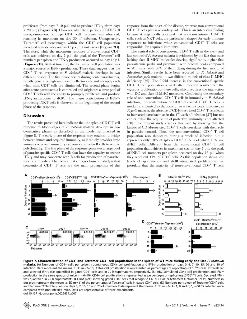
proliferate (from days 7-10 p.i.) and to produce IFN-c (from days
7–20 p.i.) (Figure 7B). However, after these periods of CD4+ cell
unresponsiveness, a huge CD4+ cell response was observed,
reaching its maximum on day 30 of infection. Unexpectedly,
Tetramer+ cell percentages within the CD4+ cell population
increased considerably on day 15 p.i., but not earlier (Figure 7C).Therefore, while the maximum response of conventional CD4+
cells was achieved on day 7 p.i., the peak of the Tetramer+ cell
numbers per spleen and IFN-c production occurred on day 15 p.i.
(Figure 7D). At that time p.i., the Tetramer+ cell population was
a major source of IFN-c production. These data suggest that the
CD4+ T cell response to P. chabaudi malaria develops in two
different phases. The first phase occurs during acute parasitaemia,
rapidly generates high numbers of effector cells and abruptly ends
when most CD4+ cells are eliminated. The second phase begins
after acute parasitaemia is controlled and originates a large pool of
CD4+ T cells with the ability to promptly proliferate and produce
IFN-c in response to iRBC. The major contribution of IFN-c-
producing iNKT cells is observed at the beginning of the second
phase of the response.
Discussion
The results presented here indicate that the splenic CD4+ T cell
response to blood-stages of P. chabaudi malaria develops in two
consecutive phases as described in the model summarised in
Figure 8. The early phase of the response may establish a bridge
between innate and acquired immunity, as it rapidly provides large
amounts of proinflammatory cytokines and helps B cells to secrete
polyclonal Ig. The late phase of the response generates a large pool
of parasite-specific CD4+ T cells that have the capacity to secrete
IFN-c and may cooperate with B cells for production of parasite-
specific antibodies. The picture that emerges from our study is that
conventional CD4+ T cells are the main protagonists of this
response from the onset of the disease, whereas non-conventional
CD4+ T cells play a secondary role. This is an interesting finding
because it is generally accepted that non-conventional CD4+ T
cells, such as NKT cells, are particularly shaped for early immune
responses to infections while conventional CD4+ T cells are
responsible for acquired immunity.
The central role of conventional CD4+ T cells in the early and
late control of P. chabaudi malaria is evidenced by the fact that mice
lacking class II MHC molecules develop significantly higher first
parasitaemia peaks and prominent recrudescent peaks compared
to WT mice, with 50% of them dying during the first month of
infection. Similar results have been reported for P. chabaudi and
Plasmodium yoelii malaria in two different models of class II MHC
deficiency [36]. The 2-fold increase in the conventional splenic
CD4+ T cell population a week after infection results from the
vigorous proliferation of these cells, which requires the interaction
with DC and class II MHC molecules. Confirming the secondary
role of non-conventional CD4+ T cells in immunity to P. chabaudi
infection, the contribution of CD1d-restricted CD4+ T cells is
modest and limited to the second parasitaemia peak. Likewise, in
P. yoelii malaria, the absence of CD1d-restricted CD4+ T cells leads
to increased parasitaemias in the 4th week of infection [37] but not
earlier, while the acquisition of protective immunity is not affected
[38]. The present study clarifies this issue by showing that the
kinetic of CD1d-restricted CD4+ T cells correlates with their role
in parasite control. Thus, the non-conventional CD4+ T cell
population also duplicates during a week of infection but it
represents only 10% of spleen CD4+ T cells of which 40% are
iNKT cells. Different from the conventional CD4+ T cell
population that achieves its maximum size on day 7 p.i., the peak
of iNKT cell numbers per spleen occurred on day 15 p.i. when
they represent 15% of CD4+ cells. As this population shows low
levels of spontaneous and iRBC-stimulated proliferation, we
postulate that the majority of non-conventional CD4+ T cells
Figure 7. Characterization of CD4+ and Tetramer+CD4+ cell populations in the spleen of WT mice during early and late P. chabaudimalaria. (A) Numbers of CD4+ cells per spleen, spontaneous CD4+ cell proliferation and IFN-c production on days 0, 4, 7, 10, 15, 20 and 30 ofinfection. Data represent the means 6 SD (n = 6–10). CD4+ cell proliferation is represented as percentages of replicating (CFSElow) cells. Intracellularand secreted IFN-c was quantified in gated CD4+ cells and in 72-h supernatants, respectively. (B) iRBC-stimulated CD4+ cell proliferation and IFN-cproduction in the same groups of mice (n = 6–10). CD4+ cell proliferation is represented as percentages of replicating (CFSElow) cells. Secreted IFN-cwas quantified in 72-h supernatants. (C) Dot plots showing gated CD4+ cells that recognise CD1d-a-GalCer tetramers (Tetramer+ cells). Numbers indot plots represent the means 6 SD (n = 6) of the percentages of Tetramer+ cells in gated CD4+ cells. (D) Numbers per spleen of Tetramer+CD4+ cellsand Tetramer+CD4+IFN-c cells on days 0, 7, 10, 15 and 20 of infection. Data represent the means 6 SD (n = 6). In A, B and C, *, p,0.05, infected micecompared with non-infected mice. Data are representative of three experiments.doi:10.1371/journal.pone.0022434.g007
CD4+ T Cells in Malaria
PLoS ONE | www.plosone.org 9 July 2011 | Volume 6 | Issue 7 | e22434

only undergo a few rounds of cell division or they migrate from
other tissues.
The main consequence of the early phase of the spleen CD4+ T
cell response to P. chabaudi malaria is the rapid induction of effector
cells that are responsible for the secretion of intense but transient
peaks of TNF-a and IFN-c. These proinflammatory cytokines not
only improve the ability of macrophages and DC to phagocytise
and to present parasite antigens to T cells [19,20,39,40], but also
prime the immune system and thus favour host hyperresponsive-
ness to toll-like receptor (TLR) agonists [14,40,41,42]. Conven-
tional CD4+ T cells are required for the early production of TNF-
a, while CD1d-restricted CD4+ T cells are dispensable for this
response. In addition, conventional CD4+ T cells are the major
source of IFN-c a week after infection, starting its production at
the same time as non-conventional CD4+ T cells. This observation
has a parallel in the pre-erythrocytic stage of P. yoelii infection,
where NK, NKT, cd T and CD4+ T cells from the spleen
simultaneously secrete IFN-c but the latter cell population is the
major source of production [43]. Moreover, similar to our analysis,
in mice infected with P. yoelii sporozoites, IFN-c is produced on
day 5 p.i. but not earlier [43], raising the possibility that the
ligands required for activation of non-conventional CD4+ T cells
are not available during the first days of the disease.
The full activation of MZ and FO B cells at the early P. chabaudi
infection depends on conventional CD4+ T cells, although some
increase in CD69 expression occurs in B cells from mice lacking
class II MHC molecules. Polyclonal Ig secretion is accompanied
by downregulation of CD1d and CD21 in MZ B cells and CD23
in FO B cells. Low levels of CD1d in MZ B cells may result from
disruption of the CD1d recycling machinery, as previously
described for human DCs exposed to high titres of herpes simplex
virus [44]. CD21 is expressed at low levels in various chronic
infectious diseases, such as HIV (human immunodeficiency virus)
infection, being attributed to continuous exposure of B cells to
complement-coupled particles [45]. CD23 binds both CD21 and
IgE, and through these interactions, regulates the synthesis of this
antibody isotype [46], which has been associated with both
protection and aggravation of malaria severity [47,48]. It is
surprising, however, that the entire population of MZ and FO B
cells appears to be engaged in the early response to infection under
the coordination of conventional CD4+ T cells. Although these
cells are also required for induction of rapid secretion of polyclonal
IgM, IgG2a and IgG1, low amounts of polyclonal IgM are
produced during acute infection in mice lacking class II MHC
molecules, indicating that part of this response is T-cell
independent or dependent on non-conventional CD4+ T cells.
However, the polyclonal Ig response is not affected by the absence
of CD1d molecules. The IFN-c-induced switch of polyclonal IgM
to IgG2a [33], which according to our results is also dependent of
conventional CD4+ T cells, may contribute to protection since
antibody from this IgG isotype is the most effective to control
blood-stage parasite growth [31].
With the control of acute parasitaemia, CD4+ T cell numbers
per spleen rapidly decline, and in a few days, reach values that are
lower than those in non-infected controls. This phenomenon is
thought to be primarily due to CD4+ T cell apoptosis [49,50], but
other regulatory mechanisms may also ensure the end of the early
phase of the response since the remaining cells become refractory
to stimulation with iRBC or anti-CD3 mAb [51]. The end of the
early response restricts the undesirable effects of overproduction of
Figure 8. A model for the spleen CD4+ T cell response to blood-stages of P. chabaudi malaria. The spleen CD4+ T cell response to P.chabaudi infection develops in two consecutive phases concomitant with the acute and chronic parasitaemias. The early phase of the responsebegins with intense proliferation of a large proportion of conventional CD4+ T cells, which depends on the interaction with DC and class II MHCmolecules and results in duplication of this population after a week of infection. The non-conventional CD4+ T cell population also duplicates duringthis period, but its low proliferative rate indicates that the majority of these cells undergo only a few rounds of cell division, or alternatively, theymigrate from other tissues. This phase of the response culminates with production of high amounts of proinflammatory cytokines (and also with helpto polyclonal Ig secretion), which is mostly dependent of conventional CD4+ T cells. The abrupt elimination of a great proportion of CD4+ T cells,together with a period of CD4+ T cell unresponsiveness, gives the opportunity to the development of acquired immunity during the late phase of theresponse. At this time, a large pool of CD4+ T cells is generated with the ability to promptly proliferate and produce IFN-c when stimulated with iRBC(and also to cooperate with B cells for production of parasite-specific antibodies). Unexpectedly, the main contribution of non-conventional CD4+ Tcells occurs in the beginning of the second phase of the response. Thus, according to this model, conventional CD4+ T cells are the main protagonistsof this response from the onset of the disease, acting in parallel with non-conventional CD4+ T cells as a bridge between the innate and acquiredimmunity.doi:10.1371/journal.pone.0022434.g008
CD4+ T Cells in Malaria
PLoS ONE | www.plosone.org 10 July 2011 | Volume 6 | Issue 7 | e22434

proinflammatory cytokines, such as the sepsis-like symptoms of
acute malaria, and allows the development of a large population of
parasite-specific CD4+ T cells, a process that occurs in parallel
with a second wave of proliferation. This population increases with
time up to a month of infection, but it progressively declines and
reaches control levels when parasites are completed eliminated
[52]. Despite the persistence of low levels of parasites during the
late phase of the response, IFN-c secretion is only detected at the
second parasitaemia peak, although CD4+ T cells are able to
produce high amounts of IFN-c when stimulated in vitro with
iRBC. Unexpectedly, iNKT cells are a major source of IFN-cproduction at the beginning of the late phase of the response,
when CD1d-restricted CD4+ T cells seem to be required for
helping B cells to secrete low-affinity parasite-specific IgM and IgG
antibodies. This may explain the incomplete control of parasitae-
mia recrudescence in mice lacking CD1d. Our results suggesting
that only the low-affinity antibody response is impaired in the
absence of CD1d-mediated interactions clarify contradictory
studies on the role of CD1d-restricted CD4+ T cells for production
of antibodies against glycosylphosphatidylinositol (GPI)-anchored
parasite molecules [53], the secretion of parasite-specific high-
affinity antibodies and the acquisition of full protective immunity
strictly depending on conventional CD4+ T cells.
The present work adds basic information to the malaria
literature and opens the perspective of future studies to unravel
the molecular mechanisms involved in the early phase of the
spleen CD4+ T cell response to Plasmodium infection. The
engagement of several molecular pathways displaying either
stimulatory or regulatory activities may be required for generating
this response. The possibility that conventional CD4+ T cells
recognise a diverse cohort of parasite components and self-
antigens at different phases of the response is another interesting
topic to be investigated. It is also of outstanding relevance to
consider the influence of pre-erythrocytic stages on the spleen
CD4+ T cell response to infection [43], a limitation of our study
that only addresses the immune response to blood-stage parasites.
We believe that clarifying these issues will help to develop new
strategies to ameliorate the outcome of malaria by minimising the
undesirable effects of the early CD4+ T cell response to Plasmodium
infection.
Supporting Information
Figure S1 IFN-c production by splenic CD8+ cells in P.chabaudi-infected WT, CD1d-/- and I-Ab-/- mice. (A) Dot
plots showing intracellular IFN-c in gated CD8+ cells on days 0, 4
and 7 of infection. Non-stimulated and anti-CD3-stimulated cell
cultures are shown. Numbers in dot plots represent the means 6
SD (n = 4–6) of IFN-c+ cell percentages. (B) Numbers of
CD8+IFN-c+ cells per spleen on days 0, 4 and 7 of infection.
Data represents the means 6 SD (n = 4–6). In A-B, *, p,0.05,
infected mice compared with non-infected mice; e, p,0.05,
CD1d-/- or I-Ab-/- mice compared with WT mice. Data are
representative of three experiments.
(TIF)
Acknowledgments
We thank Meire Ioshie Hiyane and Rogerio Silva Nascimento for technical
assistance, Dr. Albert Bendelac for providing CD1d-/- mice and Dr.
Mitchell Kronenberg and the NIH (National Institutes of Health - USA)
Tetramer Facility for providing the CD1d-a-GalCer tetramer.
Author Contributions
Conceived and designed the experiments: SMM APFR CAZ SIC-M LRS
NOSC JMA MRDL. Performed the experiments: SMM APFR LRS
SMR-M. Analyzed the data: SMM APFR JMA MRDL. Contributed
reagents/materials/analysis tools: NOSC JMA MRDL. Wrote the paper:
SMM APFR JMA MRDL.
References
1. WHO (2007) World Health Organization Malaria, other vectorborne and
parasitic diseases: Regional trend in cases and deaths. Available from: http://www.wpro.who.int/sites/mvp/data/[2009 jun 10].
2. Schofield L, Grau GE (2005) Immunological processes in malaria pathogenesis.Nat Rev Immunol 5: 722–735.
3. Clark IA (1978) Does endotoxin cause both the disease and parasite death inacute malaria and babesiosis? Lancet 2: 75–77.
4. Langhorne J, Ndungu FM, Sponaas AM, Marsh K (2008) Immunity to malaria:more questions than answers. Nat Immunol 9: 725–732.
5. Stevenson MM, Riley EM (2004) Innate immunity to malaria. Nat Rev
Immunol 4: 169–180.
6. Kwiatkowski D (1990) Tumour necrosis factor, fever and fatality in falciparum
malaria. Immunol Lett 25: 213–216.
7. Lyke KE, Burges R, Cissoko Y, Sangare L, Dao M, et al. (2004) Serum levels of
the proinflammatory cytokines interleukin-1 beta (IL-1beta), IL-6, IL-8, IL-10,tumor necrosis factor alpha, and IL-12(p70) in Malian children with severe
Plasmodium falciparum malaria and matched uncomplicated malaria or healthy
controls. Infect Immun 72: 5630–5637.
8. Gupta S, Snow RW, Donnelly CA, Marsh K, Newbold C (1999) Immunity to
non-cerebral severe malaria is acquired after one or two infections. Nat Med 5:340–343.
9. Suss G, Eichmann K, Kury E, Linke A, Langhorne J (1988) Roles of CD4- andCD8-bearing T lymphocytes in the immune response to the erythrocytic stages
of Plasmodium chabaudi. Infect Immun 56: 3081–3088.
10. Podoba JE, Stevenson MM (1991) CD4+ and CD8+ T lymphocytes both
contribute to acquired immunity to blood-stage Plasmodium chabaudi AS.
Infect Immun 59: 51–58.
11. Engwerda C, Belnoue E, Gruner AC, Renia L (2005) Experimental models of
cerebral malaria. Curr Top Microbiol Immunol 297: 103–143.
12. Engwerda CR, Ato M, Cotterell SE, Mynott TL, Tschannerl A, et al. (2002) A
role for tumor necrosis factor-alpha in remodeling the splenic marginal zoneduring Leishmania donovani infection. Am J Pathol 161: 429–437.
13. Li C, Sanni LA, Omer F, Riley E, Langhorne J (2003) Pathology of Plasmodiumchabaudi chabaudi infection and mortality in interleukin-10-deficient mice are
ameliorated by anti-tumor necrosis factor alpha and exacerbated by anti-
transforming growth factor beta antibodies. Infect Immun 71: 4850–4856.
14. Franklin BS, Rodrigues SO, Antonelli LR, Oliveira RV, Goncalves AM, et al.
(2007) MyD88-dependent activation of dendritic cells and CD4(+) Tlymphocytes mediates symptoms, but is not required for the immunological
control of parasites during rodent malaria. Microbes Infect 9: 881–890.
15. Stevenson MM, Tam MF, Belosevic M, van der Meide PH, Podoba JE (1990)
Role of endogenous gamma interferon in host response to infection with blood-stage Plasmodium chabaudi AS. Infect Immun 58: 3225–3232.
16. Su Z, Stevenson MM (2000) Central role of endogenous gamma interferon in
protective immunity against blood-stage Plasmodium chabaudi AS infection.Infect Immun 68: 4399–4406.
17. Taverne J, Tavernier J, Fiers W, Playfair JH (1987) Recombinant tumour necrosis
factor inhibits malaria parasites in vivo but not in vitro. Clin Exp Immunol 67: 1–4.
18. Mota MM, Brown KN, Holder AA, Jarra W (1998) Acute Plasmodiumchabaudi chabaudi malaria infection induces antibodies which bind to the
surfaces of parasitized erythrocytes and promote their phagocytosis bymacrophages in vitro. Infect Immun 66: 4080–4086.
19. Bastos KR, Barboza R, Elias RM, Sardinha LR, Grisotto MG, et al. (2002)
Impaired macrophage responses may contribute to exacerbation of blood-stagePlasmodium chabaudi chabaudi malaria in interleukin-12-deficient mice.
J Interferon Cytokine Res 22: 1191–1199.
20. Sponaas AM, Freitas do Rosario AP, Voisine C, Mastelic B, Thompson J, et al.
(2009) Migrating monocytes recruited to the spleen play an important role incontrol of blood stage malaria. Blood 114: 5522–5531.
21. Meding SJ, Langhorne J (1991) CD4+ T cells and B cells are necessary for the
transfer of protective immunity to Plasmodium chabaudi chabaudi.Eur J Immunol 21: 1433–1438.
22. Cavacini LA, Long CA, Weidanz WP (1986) T-cell immunity in murine malaria:
adoptive transfer of resistance to Plasmodium chabaudi adami in nude mice withsplenic T cells. Infect Immun 52: 637–643.
23. Li C, Corraliza I, Langhorne J (1999) A defect in interleukin-10 leads to
enhanced malarial disease in Plasmodium chabaudi chabaudi infection in mice.Infect Immun 67: 4435–4442.
24. Rodgers JR, Cook RG (2005) MHC class Ib molecules bridge innate and
acquired immunity. Nat Rev Immunol 5: 459–471.
25. Kronenberg M, Gapin L (2002) The unconventional lifestyle of NKT cells. NatRev Immunol 2: 557–568.
CD4+ T Cells in Malaria
PLoS ONE | www.plosone.org 11 July 2011 | Volume 6 | Issue 7 | e22434

26. Cox J, Semoff S, Hommel M (1987) Plasmodium chabaudi: a rodent malaria
model for in-vivo and in-vitro cytoadherence of malaria parasites in the absenceof knobs. Parasite Immunol 9: 543–561.
27. Chen YH, Wang B, Chun T, Zhao L, Cardell S, et al. (1999) Expression of
CD1d2 on thymocytes is not sufficient for the development of NK T cells inCD1d1-deficient mice. J Immunol 162: 4560–4566.
28. Grusby MJ, Johnson RS, Papaioannou VE, Glimcher LH (1991) Depletion ofCD4+ T cells in major histocompatibility complex class II-deficient mice.
Science 253: 1417–1420.
29. Elias RM, Sardinha LR, Bastos KR, Zago CA, da Silva AP, et al. (2005) Role ofCD28 in polyclonal and specific T and B cell responses required for protection
against blood stage malaria. J Immunol 174: 790–799.30. Lima MR, Bandeira A, Falanga P, Freitas AA, Kipnis TL, et al. (1991) Clonal
analysis of B lymphocyte responses to Plasmodium chabaudi infection of normaland immunoprotected mice. Int Immunol 3: 1207–1216.
31. Cavinato RA, Bastos KR, Sardinha LR, Elias RM, Alvarez JM, et al. (2001)
Susceptibility of the different developmental stages of the asexual (schizogonic)erythrocyte cycle of Plasmodium chabaudi chabaudi to hyperimmune serum,
immunoglobulin (Ig)G1, IgG2a and F(ab’)2 fragments. Parasite Immunol 23:587–597.
32. Ziegler SF, Ramsdell F, Alderson MR (1994) The activation antigen CD69.
Stem Cells 12: 456–465.33. Castillo-Mendez SI, Zago CA, Sardinha LR, Freitas do Rosario AP, Alvarez JM,
et al. (2007) Characterization of the spleen B-cell compartment at the early andlate blood-stage Plasmodium chabaudi malaria. Scand J Immunol 66: 309–319.
34. Lopes-Carvalho T, Foote J, Kearney JF (2005) Marginal zone B cells inlymphocyte activation and regulation. Curr Opin Immunol 17: 244–250.
35. Achtman AH, Khan M, MacLennan IC, Langhorne J (2003) Plasmodium
chabaudi chabaudi infection in mice induces strong B cell responses and strikingbut temporary changes in splenic cell distribution. J Immunol 171: 317–324.
36. Cigel F, Batchelder J, Burns JM, Jr., Yanez D, van der Heyde H, et al. (2003)Immunity to blood-stage murine malarial parasites is MHC class II dependent.
Immunol Lett 89: 243–249.
37. Mannoor MK, Weerasinghe A, Halder RC, Reza S, Morshed M, et al. (2001)Resistance to malarial infection is achieved by the cooperation of NK1.1(+) and
NK1.1(-) subsets of intermediate TCR cells which are constituents of innateimmunity. Cell Immunol 211: 96–104.
38. Taniguchi T, Tachikawa S, Kanda Y, Kawamura T, Tomiyama-Miyaji C, et al.(2007) Malaria protection in beta 2-microglobulin-deficient mice lacking major
histocompatibility complex class I antigens: essential role of innate immunity,
including gammadelta T cells. Immunology 122: 514–521.39. Ing R, Stevenson MM (2009) Dendritic cell and NK cell reciprocal cross talk
promotes gamma interferon-dependent immunity to blood-stage Plasmodiumchabaudi AS infection in mice. Infect Immun 77: 770–782.
40. Franklin BS, Parroche P, Ataide MA, Lauw F, Ropert C, et al. (2009) Malaria
primes the innate immune response due to interferon-gamma induced
enhancement of toll-like receptor expression and function. Proc Natl Acad
Sci U S A 106: 5789–5794.
41. Ropert C, Franklin BS, Gazzinelli RT (2008) Role of TLRs/MyD88 in host
resistance and pathogenesis during protozoan infection: lessons from malaria.
Semin Immunopathol 30: 41–51.
42. Seixas E, Moura Nunes JF, Matos I, Coutinho A (2009) The interaction between
DC and Plasmodium berghei/chabaudi-infected erythrocytes in mice involves
direct cell-to-cell contact, internalization and TLR. Eur J Immunol 39:
1850–1863.
43. Soulard V, Roland J, Gorgette O, Barbier E, Cazenave PA, et al. (2009) An
early burst of IFN-gamma induced by the pre-erythrocytic stage favours
Plasmodium yoelii parasitaemia in B6 mice. Malar J 8: 128.
44. Donovan MJ, Jayakumar A, McDowell MA (2007) Inhibition of groups 1 and 2
CD1 molecules on human dendritic cells by Leishmania species. Parasite
Immunol 29: 515–524.
45. Moir S, Malaspina A, Li Y, Chun TW, Lowe T, et al. (2000) B cells of HIV-1-
infected patients bind virions through CD21-complement interactions and
transmit infectious virus to activated T cells. J Exp Med 192: 637–646.
46. Gould HJ, Sutton BJ (2008) IgE in allergy and asthma today. Nat Rev Immunol
8: 205–217.
47. Duarte J, Deshpande P, Guiyedi V, Mecheri S, Fesel C, et al. (2007) Total and
functional parasite specific IgE responses in Plasmodium falciparum-infected
patients exhibiting different clinical status. Malar J 6: 1.
48. Pleass RJ (2009) Fc-receptors and immunity to malaria: from models to vaccines.
Parasite Immunol 31: 529–538.
49. Helmby H, Jonsson G, Troye-Blomberg M (2000) Cellular changes and
apoptosis in the spleens and peripheral blood of mice infected with blood-stage
Plasmodium chabaudi chabaudi AS. Infect Immun 68: 1485–1490.
50. Sanchez-Torres L, Rodriguez-Ropon A, Aguilar-Medina M, Favila-Castillo L
(2001) Mouse splenic CD4+ and CD8+ T cells undergo extensive apoptosis
during a Plasmodium chabaudi chabaudi AS infection. Parasite Immunol 23:
617–626.
51. Muxel SM, Freitas do Rosario AP, Sardinha LR, Castillo-Mendez SI, Zago CA,
et al. (2010) Comparative Analysis of Activation Phenotype, Proliferation, and
IFN-gamma Production by Spleen NK1.1(+) and NK1.1(-) T Cells During
Plasmodium chabaudi AS Malaria. J Interferon Cytokine Res.
52. Freitas do Rosario AP, Muxel SM, Rodriguez-Malaga SM, Sardinha LR,
Zago CA, et al. (2008) Gradual decline in malaria-specific memory T cell
responses leads to failure to maintain long-term protective immunity to
Plasmodium chabaudi AS despite persistence of B cell memory and circulating
antibody. J Immunol 181: 8344–8355.
53. Schmieg J, Gonzalez-Aseguinolaza G, Tsuji M (2003) The role of natural killer
T cells and other T cell subsets against infection by the pre-erythrocytic stages of
malaria parasites. Microbes and Infection 5: 499–506.
CD4+ T Cells in Malaria
PLoS ONE | www.plosone.org 12 July 2011 | Volume 6 | Issue 7 | e22434