The selection of tumor variants with altered expression of classical and nonclassical MHC class I...
7
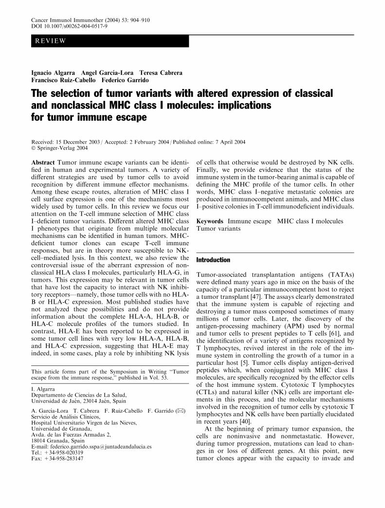
REVIEW Ignacio Algarra Angel Garcı´a-Lora Teresa Cabrera Francisco Ruiz-Cabello Federico Garrido The selection of tumor variants with altered expression of classical and nonclassical MHC class I molecules: implications for tumor immune escape Received: 15 December 2003 / Accepted: 2 February 2004 / Published online: 7 April 2004 Ó Springer-Verlag 2004 Abstract Tumor immune escape variants can be identi- fied in human and experimental tumors. A variety of different strategies are used by tumor cells to avoid recognition by different immune effector mechanisms. Among these escape routes, alteration of MHC class I cell surface expression is one of the mechanisms most widely used by tumor cells. In this review we focus our attention on the T-cell immune selection of MHC class I–deficient tumor variants. Different altered MHC class I phenotypes that originate from multiple molecular mechanisms can be identified in human tumors. MHC- deficient tumor clones can escape T-cell immune responses, but are in theory more susceptible to NK- cell–mediated lysis. In this context, we also review the controversial issue of the aberrant expression of non- classical HLA class I molecules, particularly HLA-G, in tumors. This expression may be relevant in tumor cells that have lost the capacity to interact with NK inhibi- tory receptors—namely, those tumor cells with no HLA- B or HLA-C expression. Most published studies have not analyzed these possibilities and do not provide information about the complete HLA-A, HLA-B, or HLA-C molecule profiles of the tumors studied. In contrast, HLA-E has been reported to be expressed in some tumor cell lines with very low HLA-A, HLA-B, and HLA-C expression, suggesting that HLA-E may indeed, in some cases, play a role by inhibiting NK lysis of cells that otherwise would be destroyed by NK cells. Finally, we provide evidence that the status of the immune system in the tumor-bearing animal is capable of defining the MHC profile of the tumor cells. In other words, MHC class I–negative metastatic colonies are produced in immunocompetent animals, and MHC class I–positive colonies in T-cell immunodeficient individuals. Keywords Immune escape MHC class I molecules Tumor variants Introduction Tumor-associated transplantation antigens (TATAs) were defined many years ago in mice on the basis of the capacity of a particular immunocompetent host to reject a tumor transplant [47]. The assays clearly demonstrated that the immune system is capable of rejecting and destroying a tumor mass composed sometimes of many millions of tumor cells. Later, the discovery of the antigen-processing machinery (APM) used by normal and tumor cells to present peptides to T cells [61], and the identification of a variety of antigens recognized by T lymphocytes, revived interest in the role of the im- mune system in controlling the growth of a tumor in a particular host [5]. Tumor cells display antigen-derived peptides which, when conjugated with MHC class I molecules, are specifically recognized by the effector cells of the host immune system. Cytotoxic T lymphocytes (CTLs) and natural killer (NK) cells are important ele- ments in this process, and the molecular mechanisms involved in the recognition of tumor cells by cytotoxic T lymphocytes and NK cells have been partially elucidated in recent years [40]. At the beginning of primary tumor expansion, the cells are noninvasive and nonmetastatic. However, during tumor progression, mutations can lead to chan- ges in or loss of different genes. At this point, new tumor clones appear with the capacity to invade and This article forms part of the Symposium in Writing ‘‘Tumor escape from the immune response,’’ published in Vol. 53. I. Algarra Departamento de Ciencias de La Salud, Universidad de Jae´n, 23014 Jae´n, Spain A. Garcı´a-Lora T. Cabrera F. Ruiz-Cabello F. Garrido (&) Servicio de Ana´lisis Clı´nicos, Hospital Universitario Virgen de las Nieves, Universidad de Granada, Avda. de las Fuerzas Armadas 2, 18014 Granada, Spain E-mail: [email protected] Tel.: +34-958-020319 Fax: +34-958-283147 Cancer Immunol Immunother (2004) 53: 904–910 DOI 10.1007/s00262-004-0517-9
-
Upload
independent -
Category
Documents
-
view
0 -
download
0
Transcript of The selection of tumor variants with altered expression of classical and nonclassical MHC class I...
REVIEW
Ignacio Algarra Æ Angel Garcıa-Lora Æ Teresa Cabrera
Francisco Ruiz-Cabello Æ Federico Garrido
The selection of tumor variants with altered expression of classicaland nonclassical MHC class I molecules: implicationsfor tumor immune escape
Received: 15 December 2003 / Accepted: 2 February 2004 / Published online: 7 April 2004� Springer-Verlag 2004
Abstract Tumor immune escape variants can be identi-fied in human and experimental tumors. A variety ofdifferent strategies are used by tumor cells to avoidrecognition by different immune effector mechanisms.Among these escape routes, alteration of MHC class Icell surface expression is one of the mechanisms mostwidely used by tumor cells. In this review we focus ourattention on the T-cell immune selection of MHC classI–deficient tumor variants. Different altered MHC classI phenotypes that originate from multiple molecularmechanisms can be identified in human tumors. MHC-deficient tumor clones can escape T-cell immuneresponses, but are in theory more susceptible to NK-cell–mediated lysis. In this context, we also review thecontroversial issue of the aberrant expression of non-classical HLA class I molecules, particularly HLA-G, intumors. This expression may be relevant in tumor cellsthat have lost the capacity to interact with NK inhibi-tory receptors—namely, those tumor cells with no HLA-B or HLA-C expression. Most published studies havenot analyzed these possibilities and do not provideinformation about the complete HLA-A, HLA-B, orHLA-C molecule profiles of the tumors studied. Incontrast, HLA-E has been reported to be expressed insome tumor cell lines with very low HLA-A, HLA-B,and HLA-C expression, suggesting that HLA-E mayindeed, in some cases, play a role by inhibiting NK lysis
of cells that otherwise would be destroyed by NK cells.Finally, we provide evidence that the status of theimmune system in the tumor-bearing animal is capable ofdefining the MHC profile of the tumor cells. In otherwords, MHC class I–negative metastatic colonies areproduced in immunocompetent animals, and MHC classI–positive colonies in T-cell immunodeficient individuals.
Keywords Immune escape Æ MHC class I molecules ÆTumor variants
Introduction
Tumor-associated transplantation antigens (TATAs)were defined many years ago in mice on the basis of thecapacity of a particular immunocompetent host to rejecta tumor transplant [47]. The assays clearly demonstratedthat the immune system is capable of rejecting anddestroying a tumor mass composed sometimes of manymillions of tumor cells. Later, the discovery of theantigen-processing machinery (APM) used by normaland tumor cells to present peptides to T cells [61], andthe identification of a variety of antigens recognized byT lymphocytes, revived interest in the role of the im-mune system in controlling the growth of a tumor in aparticular host [5]. Tumor cells display antigen-derivedpeptides which, when conjugated with MHC class Imolecules, are specifically recognized by the effector cellsof the host immune system. Cytotoxic T lymphocytes(CTLs) and natural killer (NK) cells are important ele-ments in this process, and the molecular mechanismsinvolved in the recognition of tumor cells by cytotoxic Tlymphocytes and NK cells have been partially elucidatedin recent years [40].
At the beginning of primary tumor expansion, thecells are noninvasive and nonmetastatic. However,during tumor progression, mutations can lead to chan-ges in or loss of different genes. At this point, newtumor clones appear with the capacity to invade and
This article forms part of the Symposium in Writing ‘‘Tumorescape from the immune response,’’ published in Vol. 53.
I. AlgarraDepartamento de Ciencias de La Salud,Universidad de Jaen, 23014 Jaen, Spain
A. Garcıa-Lora Æ T. Cabrera Æ F. Ruiz-Cabello Æ F. Garrido (&)Servicio de Analisis Clınicos,Hospital Universitario Virgen de las Nieves,Universidad de Granada,Avda. de las Fuerzas Armadas 2,18014 Granada, SpainE-mail: [email protected].: +34-958-020319Fax: +34-958-283147
Cancer Immunol Immunother (2004) 53: 904–910DOI 10.1007/s00262-004-0517-9
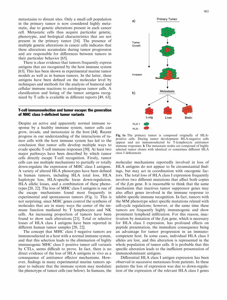
metastasize to distant sites. Only a small cell populationin the primary tumor is now considered highly meta-static, due to genetic alterations present in each cancercell. Metastatic cells thus acquire particular genetic,phenotypic, and biological characteristics that are notpresent in the primary tumor [16]. The presence ofmultiple genetic alterations in cancer cells indicates thatthese alterations accumulate during tumor progressionand are responsible for differences between tumors intheir particular behavior [65].
There is clear evidence that tumors frequently expressantigens that are recognized by the host immune system[63]. This has been shown in experimental murine tumormodels as well as in human tumors. In the latter, theseantigens have been defined on the molecular level bytechniques and methods for the analysis of humoral andcellular immune reactions to autologous tumor cells. Aclassification and listing of the tumor antigens recog-nized by T cells is available in different reports [49, 63].
T-cell immunoselection and tumor escape: the generationof MHC class I–deficient tumor variants
Despite an active and apparently normal immune re-sponse by a healthy immune system, tumor cells cangrow, invade, and metastasize in the host [44]. Recentprogress in our understanding of the interactions of tu-mor cells with the host immune system has led to theconclusion that tumor cells develop multiple ways toevade specific T-cell immune responses [50]. At least twomajor pathways have been described by which tumorcells directly escape T-cell recognition. Firstly, tumorcells can use multiple mechanisms to partially or totallydown-regulate the expression of MHC class I antigens.A variety of altered HLA phenotypes have been definedin human tumors, including HLA total loss, HLAhaplotype loss, HLA-specific locus down-regulation,HLA allelic losses, and a combination of these pheno-types [20, 22]. The loss of MHC class I antigens is one ofthe escape mechanisms found most frequently inexperimental and spontaneous tumors (Fig. 1). This isnot surprising, since MHC genes control the synthesis ofmolecules that are in many ways the center of the im-mune function mediated by T lymphocytes and NKcells. An increasing proportion of tumors have beenfound to show such alterations [21]. Total or selectivelosses of HLA class I antigens have been reported indifferent human tumor samples [20, 22].
The concept that MHC class I–negative tumors areimmunoselected in a host with a normal immune system,and that this selection leads to the elimination of highlyimmunogenic MHC class I–positive tumor cell variantsby CTLs, seems difficult to prove. In fact, there is nodirect evidence of the loss of HLA antigens in vivo as aconsequence of antitumor effector mechanisms. How-ever, findings in many experimental murine tumors ap-pear to indicate that the immune system may modulatethe phenotype of tumor cells (see below). In humans, the
molecular mechanisms reportedly involved in loss ofHLA antigens do not appear to be circumstantial find-ings, but may act in coordination with oncogenic fac-tors. The total loss of HLA class I expression frequentlyinvolves two different mutations that affect both copiesof the b2m gene. It is reasonable to think that the samemechanism that inactives tumor suppressor genes mayalso affect genes involved in the immune response toinhibit specific immune recognition. In fact, tumors withthe MM phenotype select specific mutations related withcell-cycle regulations; however, at the same time thesetumors are frequently highly immunogenic and showprominent lymphoid infiltration. For this reason, inac-tivation by mutation of the b2m gene, which is necessaryfor HLA class I expression, has profound effects onpeptide presentation, the immediate consequence beingan advantage for tumor progression in an immuno-competent host. In some cases, individual HLA class Ialleles are lost, and this alteration is represented in thewhole population of tumor cells. It is probable that thisspecific alteration leads to the inefficient presentation ofimmunodominant antigens.
Differential HLA class I antigen expression has beenobserved in successive metastases from patients. In thesepatients the loss of expression was due to down-regula-tion of the expression of the relevant HLA class I genes
Fig. 1a The primary tumor is composed originally of HLA-positive cells. During tumor development HLA-negative cellsappear and are immunoselected by T-lymphocyte antitumorimmune responses. b The metastatic nodes are composed of highlyselected tumor clones with identical or sometimes different HLAclass I deficiencies
905
[34]. In other cases, tumor cells can also be immunos-elected by generating tumor-specific antigen loss vari-ants that are not presented to CD8+ cytotoxic T cells[29]. This has been shown in two metastatic cell linesfrom a patient with melanoma, in whom a significantdifference in Melan-A/MART-1 expression was ob-served after immunotherapy [38]. These alterations maylead to the preferential immunoselection of melanomaantigen-negative variants during disease progression,providing a mechanism of escape from immune inter-vention [34].
The most frequent HLA class I alteration involves theloss of an HLA haplotype and at the same time the lossof the activatory NK receptor MICA and MICB alleles.These alterations may also favor the growth of tumorcells by simultaneous escape from T- and NK-cell rec-ognition. Finally, down-regulation of HLA class Iantigens may also be the result of a defect in the APM(TAP and LMP), which is frequently reversible by IFN-ctreatment. These APM genes are regulated by thiscytokine, and the alterations may thus be the conse-quence of inhibition of proinflammatory cytokines thatare mediated by oncogenic factors [44].
The role of nonclassical HLA class I molecules HLA-Gand HLA-E in tumor escape
There are reports from different laboratories that HLA-G is expressed aberrantly in some human tumors tissueand cell lines [8, 45]. These findings may indicate thatHLA-G expression is another mechanism of tumor es-cape from NK-cell attack. Such a mechanism may par-allel the manner in which HLA-G protects against NKrejection of fetal tissues in the placenta [33]. However,this is still a controversial issue, since other laboratories(including ours) have not found HLA-G expression atthe tumor cell surface with flow cytometry techniques[48]. However, we have found in many normal and tu-moral tissues that HLA-G transcription was not fol-lowed by cell surface expression. These findings indicatestrong posttranscriptional control of the expression ofthis gene in its different isoforms (from HLA-G1 toHLA-G5). It is also important to take into account thatif a tumor cell is already expressing classical HLA-B orHLA-C molecules, NK cells are already effectivelyinhibited because of the strong inhibitory capacity ofthese two HLA locus products through their interactionwith NK inhibitory receptors [40]. In these tumors,aberrant HLA-G expression is not required for tumorescape. Aberrant expression may play a role in tumorescape in some tumors, as reported in renal cell carci-nomas [8], but it is far from clear whether this is so inmany other tumor types such as melanomas, whereHLA-G is not expressed at the cell surface [14, 43, 48].
Similarly, other nonclassical HLA class I moleculessuch as HLA-E might also contribute to NK tumor cellescape. It is well documented that HLA-E is widelytranscribed in most tissues, and that this molecule is
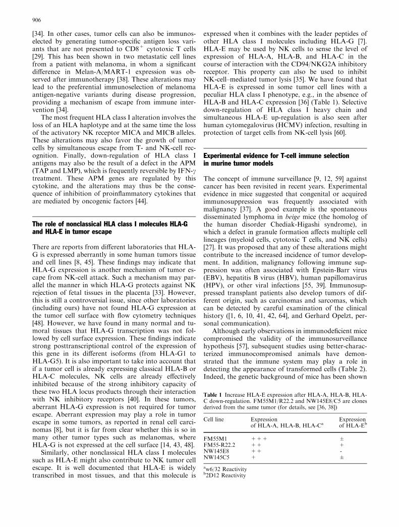
expressed when it combines with the leader peptides ofother HLA class I molecules including HLA-G [7].HLA-E may be used by NK cells to sense the level ofexpression of HLA-A, HLA-B, and HLA-C in thecourse of interaction with the CD94/NKG2A inhibitoryreceptor. This property can also be used to inhibitNK-cell–mediated tumor lysis [35]. We have found thatHLA-E is expressed in some tumor cell lines with apeculiar HLA class I phenotype, e.g., in the absence ofHLA-B and HLA-C expression [36] (Table 1). Selectivedown-regulation of HLA class I heavy chain andsimultaneous HLA-E up-regulation is also seen afterhuman cytomegalovirus (HCMV) infection, resulting inprotection of target cells from NK-cell lysis [60].
Experimental evidence for T-cell immune selectionin murine tumor models
The concept of immune surveillance [9, 12, 59] againstcancer has been revisited in recent years. Experimentalevidence in mice suggested that congenital or acquiredimmunosuppression was frequently associated withmalignancy [37]. A good example is the spontaneousdisseminated lymphoma in beige mice (the homolog ofthe human disorder Chediak-Higashi syndrome), inwhich a defect in granule formation affects multiple celllineages (myeloid cells, cytotoxic T cells, and NK cells)[27]. It was proposed that any of these alterations mightcontribute to the increased incidence of tumor develop-ment. In addition, malignancy following immune sup-pression was often associated with Epstein-Barr virus(EBV), hepatitis B virus (HBV), human papillomavirus(HPV), or other viral infections [55, 39]. Immunosup-pressed transplant patients also develop tumors of dif-ferent origin, such as carcinomas and sarcomas, whichcan be detected by careful examination of the clinicalhistory ([1, 6, 10, 41, 42, 64], and Gerhard Opelzt, per-sonal communication).
Although early observations in immunodeficient micecompromised the validity of the immunosurveillancehypothesis [57], subsequent studies using better-charac-terized immunocompromised animals have demon-strated that the immune system may play a role indetecting the appearance of transformed cells (Table 2).Indeed, the genetic background of mice has been shown
Table 1 Increase HLA-E expression after HLA-A, HLA-B, HLA-C down-regulation. FM55M1/R22.2 and NW145E8/C5 are clonesderived from the same tumor (for details, see [36, 38])
Cell line Expressionof HLA-A, HLA-B, HLA-Ca
Expressionof HLA-Eb
FM55M1 +++ ±FM55-R22.2 ++ +NW145E8 ++ -NW145C5 + ±
aw6/32 Reactivityb2D12 Reactivity
906
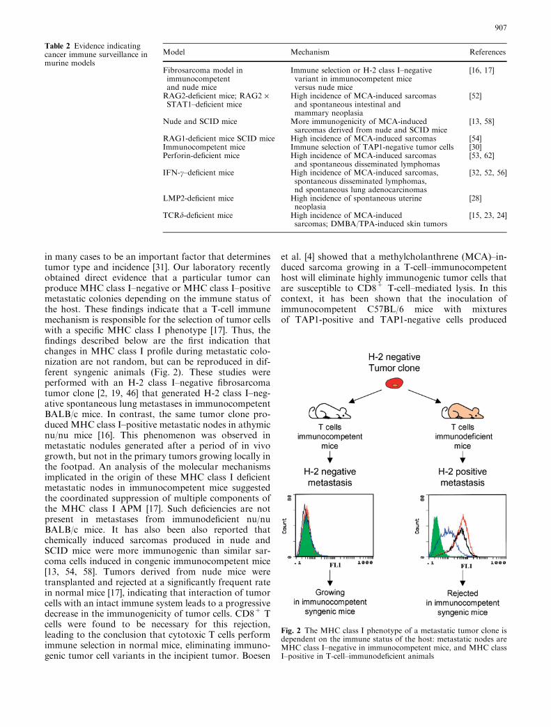
in many cases to be an important factor that determinestumor type and incidence [31]. Our laboratory recentlyobtained direct evidence that a particular tumor canproduce MHC class I–negative or MHC class I–positivemetastatic colonies depending on the immune status ofthe host. These findings indicate that a T-cell immunemechanism is responsible for the selection of tumor cellswith a specific MHC class I phenotype [17]. Thus, thefindings described below are the first indication thatchanges in MHC class I profile during metastatic colo-nization are not random, but can be reproduced in dif-ferent syngenic animals (Fig. 2). These studies wereperformed with an H-2 class I–negative fibrosarcomatumor clone [2, 19, 46] that generated H-2 class I–neg-ative spontaneous lung metastases in immunocompetentBALB/c mice. In contrast, the same tumor clone pro-duced MHC class I–positive metastatic nodes in athymicnu/nu mice [16]. This phenomenon was observed inmetastatic nodules generated after a period of in vivogrowth, but not in the primary tumors growing locally inthe footpad. An analysis of the molecular mechanismsimplicated in the origin of these MHC class I deficientmetastatic nodes in immunocompetent mice suggestedthe coordinated suppression of multiple components ofthe MHC class I APM [17]. Such deficiencies are notpresent in metastases from immunodeficient nu/nuBALB/c mice. It has also been also reported thatchemically induced sarcomas produced in nude andSCID mice were more immunogenic than similar sar-coma cells induced in congenic immunocompetent mice[13, 54, 58]. Tumors derived from nude mice weretransplanted and rejected at a significantly frequent ratein normal mice [17], indicating that interaction of tumorcells with an intact immune system leads to a progressivedecrease in the immunogenicity of tumor cells. CD8+ Tcells were found to be necessary for this rejection,leading to the conclusion that cytotoxic T cells performimmune selection in normal mice, eliminating immuno-genic tumor cell variants in the incipient tumor. Boesen
et al. [4] showed that a methylcholanthrene (MCA)–in-duced sarcoma growing in a T-cell–immunocompetenthost will eliminate highly immunogenic tumor cells thatare susceptible to CD8+ T-cell–mediated lysis. In thiscontext, it has been shown that the inoculation ofimmunocompetent C57BL/6 mice with mixturesof TAP1-positive and TAP1-negative cells produced
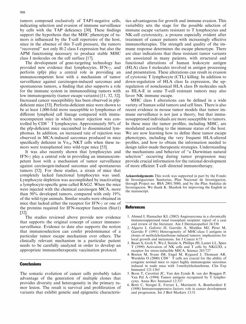
Table 2 Evidence indicatingcancer immune surveillance inmurine models
Model Mechanism References
Fibrosarcoma model inimmunocompetentand nude mice
Immune selection or H-2 class I–negativevariant in immunocompetent miceversus nude mice
[16, 17]
RAG2-deficient mice; RAG2 ·STAT1–deficient mice
High incidence of MCA-induced sarcomasand spontaneous intestinal andmammary neoplasia
[52]
Nude and SCID mice More immunogenicity of MCA-inducedsarcomas derived from nude and SCID mice
[13, 58]
RAG1-deficient mice SCID mice High incidence of MCA-induced sarcomas [54]Immunocompetent mice Immune selection of TAP1-negative tumor cells [30]Perforin-deficient mice High incidence of MCA-induced sarcomas
and spontaneous disseminated lymphomas[53, 62]
IFN-c–deficient mice High incidence of MCA-induced sarcomas,spontaneous disseminated lymphomas,nd spontaneous lung adenocarcinomas
[32, 52, 56]
LMP2-deficient mice High incidence of spontaneous uterineneoplasia
[28]
TCRd-deficient mice High incidence of MCA-inducedsarcomas; DMBA/TPA-induced skin tumors
[15, 23, 24]
Fig. 2 The MHC class I phenotype of a metastatic tumor clone isdependent on the immune status of the host: metastatic nodes areMHC class I–negative in immunocompetent mice, and MHC classI–positive in T-cell–immunodeficient animals
907
tumors composed exclusively of TAP1-negative cells,indicating selection and evasion of immune surveillanceby cells with the TAP deficiency [30]. These findingssupport the hypothesis that the MHC phenotype of tu-mors is influenced by the T-cell repertoire of the host,since in the absence of this T-cell pressure, the tumors‘‘recovered’’ not only H-2 class I expression but also theAPM functioning necessary to produce stable MHCclass I molecules on the cell surface [17].
The development of gene-targeting technology hasprovided new evidence that lymphocytes, IFN-c, andperforin (pfp) play a central role in providing animmunocompetent host with a mechanism of tumorsurveillance against carcinogen-induced sarcomas andspontaneous tumors, a finding that also supports a rolefor the immune system in immunoediting tumors withlow immunogenicity (tumor escape variants) [11, 32, 52].Increased cancer susceptibility has been observed in pfp-deficient mice [53]. Perforin-deficient mice were shown tobe at least 1,000-fold more susceptible to lymphomas ofdifferent lymphoid cell lineage compared with immu-nocompetent mice in which tumor rejection was con-trolled by CD8+ T lymphocytes. Approximately 50% ofthe pfp-deficient mice succumbed to disseminated lym-phomas. In addition, an increased rate of rejection wasobserved in MCA-induced sarcomas produced in micespecifically deficient in Va14 NKT cells when these tu-mors were transplanted into wild-type mice [53].
It was also recently shown that lymphocytes andIFN-c play a central role in providing an immunocom-petent host with a mechanism of tumor surveillanceagainst carcinogen-induced sarcomas and spontaneoustumors [52]. For these studies, a strain of mice thatcompletely lacked functional lymphocytes was used.Lymphocyte depletion was accomplished by inactivatinga lymphocyte-specific gene called RAG2. When the micewere injected with the chemical carcinogen MCA, morethan 50% developed tumors, compared with only 19%of the wild-type animals. Similar results were obtained inmice that lacked either the receptor for IFN-c or one ofthe proteins required for IFN-receptor function (Stat1)[32].
The studies reviewed above provide new evidencethat supports the original concept of cancer immuno-surveillance. Evidence to date also supports the notionthat immunoselection can confer predominance of aparticular tumor escape mechanism over others. Theclinically relevant mechanism in a particular patientneeds to be carefully analyzed in order to develop anappropriate immunotherapeutic vaccination protocol.
Conclusions
The somatic evolution of cancer cells probably takesadvantage of the generation of multiple clones thatprovides diversity and heterogeneity in the primary tu-mor lesion. The result is survival and proliferation ofvariants that exhibit genetic and epigenetic characteris-
tics advantageous for growth and immune evasion. Thisvariability sets the stage for the possible selection ofimmune escape variants resistant to T lymphocytes andNK-cell cytotoxicity, a process especially evident aftertreatment of cancer patients with increasingly effectiveimmunotherapies. The strength and quality of the im-mune response determines the escape phenotype. Thereare clear indications that these resistant tumor variantsare associated in many patients, with structural andfunctional alterations of human leukocyte antigen(HLA) class I molecules involved in antigen processingand presentation. These alterations can result in evasionof cytotoxic T lymphocyte (CTL) killing. In addition todown-regulation of HLA class Ia expression, the up-regulation of nonclassical HLA class Ib molecules suchas HLA-E in some T-cell–resistant tumors may alsofavor NK immune escape.
MHC class I alterations can be defined in a widevariety of human solid tumors and cell lines. There is alsorecent evidence in mouse tumor models that tumor im-mune surveillance is not just a theory, but that immu-nosuppressed individuals are more susceptible to tumors.In these mice the tumor profiles, including MHC, aremodulated according to the immune status of the host.We are now learning how to define these tumor escapephenotypes, including the very frequent HLA-alteredprofiles, and how to obtain the information needed todesign tailor-made therapeutic strategies. Understandingthe mechanisms and behavior of ‘‘a Darwinian naturalselection’’ occurring during tumor progression mayprovide crucial information for the rational developmentof more efficient T-cell–directed immunotherapy.
Acknowledgements This work was supported in part by the Fondode Investigaciones Sanitarias, Plan Nacional de Investigacion,through Project no. BSA 2001/3080, and by the Plan Andaluz deInvestigacion. We thank K. Shashok for improving the English inthe manuscript.
References
1. Ahmed I, Hamacher KL (2002) Angiosarcoma in a chronicallyimmunosuppressed renal transplant recipient: report of a caseand review of the literature. Am J Dermatopathol 24:330
2. Algarra I, Gaforio JJ, Garrido A, Mialdea MJ, Perez M,Garrido F (1991) Heterogeneity of MHC-class I antigens inclones of methylcholanthrene induced tumors: implications forlocal growth and metastasis. Int J Cancer 6:73
3. Bauer S, Groh V, Wu J, Steinle A, Phillips JH, Lanier LL, SpiesT (1999) Activation of NK cells and T cells by NKG2D, areceptor for stress-inducible MICA. Science 285:727
4. Boesen M, Svane IM, Engel M, Rygaard J, Thomsen AR,Werdelin O (2000) CD8+ T cells are crucial for the ability ofcongenic normal mice to reject highly immunogenic sarcomasinduced in nude mice with 3-methylcholanthrene. Clin ExpImmunol 121:1365
5. Boon T, Cerottini JC, Van den Eynde B, van der Bruggen P,Van Pel A (1994) Tumor antigens recognized by T lympho-cytes. Annu Rev Immunol 12:337
6. Botti C, Seregni E, Ferrari L, Martinetti A, Bombardieri E(1998) Immunosuppressive factors: role in cancer developmentand progression. Int J Biol Markers 13:51
908
7. Braud V, Jones EY, McMichael A (1997) The human majorhistocompatibility complex class Ib molecule HLA-E bindssignal sequence-derived peptides with primary anchor residuesat positions 2 and 9. Eur J Immunol 27:1164
8. Bukur J, Seliger B (2003) The role of HLA G for protection ofhuman renal cell-carcinoma cells from immune-mediated lysis:implications for immunotherapies. Sem Cancer Biol 13:353
9. Burnet FM (1970) The concept of immunological surveillance.Prog Exp Tumor Res 13:1
10. Cadranel J, Naccache J, Wislez M, Mayaud C (1999) Pulmo-nary malignancies in the immunocompromised patient. Respi-ration 66:289
11. Dunn GP, Bruce AT, Ikeda H, Old LJ, Schreiber RD (2002)Cancer immunoediting: from immunosurveillance to tumorescape. Nat Immunol 3:991
12. Ehrlich P (1909) Ueber den jetzigen Stand der Karzinomf-orschung (About the current state of the art of cancer re-search). NedTijdschr Geneesk 5:273
13. Engel AM, Svane IM, Rigaard J, Werdelin O (1997) MCAsarcomas induced in scid mice are more immunogenic thanMCA sarcomas induced in congenic, immunocompetent mice.Scand J Immunol 45:463
14. Frumento G, Franchello S, Palmisano GL, Nicotra MR,Giacomini P, Loke YW, Geraghty DE, Maio M, Manzo C,Natali PG, Ferrara GB (2000) Melanomas and melanoma celllines do not express HLA-G, and the expression cannot beinduced by gamma-IFN treatment. Tissue Antigens 56:30
15. Gao Y, Yang W, Pan M, Scully E, Girardi M, Augenlicht LH,Craft J, Yin Z (2003) Gamma delta T cells provide an earlysource of interferon gamma in tumor immunity. J Exp Med198:433
16. Garcia-Lora A, Algarra I, Gaforio JJ, Ruiz-Cabello F, GarridoF (2001) Immunoselection by T lymphocytes generates re-peated MHC class I deficient metastatic tumor variants. IntJ Cancer 91:109
17. Garcia-Lora A, Martinez M, Algarra I, Gaforio JJ, GarridoF (2003) MHC class I-deficient metastatic tumor variantsimmunoselected by T lymphocytes originate from the coor-dinated downregulation of APM components. Int J Cancer106:521
18. Garcia-Lora A, Algarra I, Garrido F (2003) MHC class Iantigens, immune surveillance and tumor immune escape.J Cell Physiol 195:346
19. Garrido A, Perez M, Delgado C, Garrido ML, Rojano J,Algarra I, Garrido F (1986) Influence of class I H-2 geneexpression on local tumor growth. Exp Clin Immunogenet13:98
20. Garrido F, Algarra I (2001) MHC antigens and tumor escapefrom immune surveillance. Adv Cancer Res 83:117
21. Garrido F, Cabrera T, Concha A, Glew S, Ruiz-Cabello F,Stern PL (1993) Natural history of HLA expression duringtumour development. Immunol Today 14:491
22. Garrido F, Ruiz-Cabello F, Cabrera T, Perez-Villar JJ, Lopez-Botet M, Duggan-Keen M, Stern P (1997) Implications forimmune surveillance of altered HLA class I phenotypes inhuman tumors. Immunol Today 18:89
23. Girardi M, Oppenheim DE, Steele CR, Lewis JM, Glusac E,Filler R, Hobby P, Sutton B, Tigelaar RE, Hayday AC (2001)Regulation of cutaneous malignancy by gammadelta T cells.Science 294:605
24. Girardi M, Glusac E, Filler RB, Roberts SJ, Propperova I,Lewis J, Tigelaar RE, Hayday AC (2003) The distinct contri-butions of murine T cell receptor (TCR)gammadelta+ andTCRalphabeta+ T cells to different stages of chemically in-duced skin cancer. J Exp Med 198:747
25. Groh V, Steinle A, Bauer S, Spies T (1998) Recognition ofstress-induced MHC molecules by intestinal epithelial gam-madelta T cells. Science 279:1737
26. Groh V, Rhinehart R, Randolph-Habecker J, Topp MS,Riddell SR, Spies T (2001) Costimulation of CD8alphabeta Tcells by NKG2D via engagement by MIC induced on virus-infected cells. Nat Immunol 2:255
27. Haliotis T, Ball JK, Dexter D, Roder JC (1985) Spontaneousand induced primary oncogenesis in natural killer (NK)-celldeficient beige mutant mice. Int J Cancer 35:505
28. Hayashi T, Faustman DL(2002) Development of spontaneousuterine tumors in low molecular mass polypeptide-2 knockoutmice. Cancer Res 62:24
29. Jager E, Ringhoffer M, Karbach J, Arand M, Oesch F, Knuth(1996) A Inverse relationship of melanocyte differentiationantigen expression in melanoma tissues and CD8+ cytotoxic-T-cell responses: evidence for immunoselection of antigen-lossvariants in vivo. Int J Cancer 66:470
30. Johnsen A, Templeton DJ, Sy MS, Harding CV (1999) Defi-ciency of transporter for antigen presentation (TAP) in tumorcells allows evasion of immune surveillance and increasestumorigenesis. J Immunol 163:4224
31. Kagi DB, Ledermann B, Burki K, Seiler P, Odermatt B, OlsenKJ, Podack ER, Zinkernagel RM, Hengartner H (1994)Cytotoxicity mediated by T cells and natural killer cells isgreatly impaired in perforin-deficient mice. Nature 369:31
32. Kaplan DH, Shankaran V, Dighe AS, Stockert E, Aguet M,Old LJ, Schreiber RD (1998) Demonstration of an interferongamma dependent tumor surveillance system in immunocom-petent mice. Proc Natl Acad Sci U S A 95:7556
33. Le Bouteiller P (1997) HLA-G: on the track of immunologicalfunctions. Eur J Immunogenet 24:397
34. Lehmann F, Marchand M, Hainaut P, Pouillart P, Sastre X,Ikeda H, Boon T, Coulie PG (1995) Differences in the antigensrecognized by cytolytic T cells on two successive metastases of amelanoma patient are consistent with immune selection. EurJ Immunol 25:340
35. Llano M, Lee N, Navarro F, Garcia P, Albar JP, Geraghty DE,Lopez-Botet M (1998) HLA-E-bound peptides influence rec-ognition by inhibitory and triggering CD94/NKG2 receptors:preferential response to an HLA-G-derived nonamer. EurJ Immunol 28:2854
36. Marin R, Ruiz-Cabello F, Pedrinaci S, Mendez R, Jimenez P,Geraghty DE, Garrido F (2003) Analysis of HLA-E expressionin human tumors. Immunogenetics 54:767
37. McClain KL (1997) Immunodeficiency states and relatedmalignancies. Cancer Treat Rev 92:39
38. Mendez R, Serrano A, Jager E, Maleno I, Ruiz Cabello F,Knuth A, Garrido F (2001) Analysis of HLA class I expressionin different metastases from two melanoma patients undergoingpeptide immunotherapy. Tissue Antigens 57:508
39. Meyer T, Arndt R, Nindl I, Ulrich C, Christophers E,Stockfleth E (2003) Association of human papillomavirusinfections with cutaneous tumors in immunosuppressedpatients. Transpl Int 16:146
40. Moretta A, Bottino C, Vitale M, Pende D, Biassoni R, MingariMC, Moretta L (1996) Receptors for HLA class-I molecules inhuman natural killer cells. Annu Rev Immunol 14:619
41. Nemes B, Zalatnai A, Podder H, Jaray J, Sotonyi P Jr, Schaff Z,Foldes K, Perner F (2000) Papillary microcarcinoma of thethyroid gland in renal transplant patients. Pathol Oncol Res 6:72
42. Otley CC, Pittelkow MR (2000) Skin cancer in liver transplantrecipients. Liver Transpl 6:253
43. Pangault C, Amiot L, Caulet-Maugendre S, Brasseur F, BurtinF, Guilloux V, Drenou B, Fauchet R, Onno M (1999) HLA-Gprotein expression is not induced during malignant transfor-mation. Tissue Antigens. 53:335
44. Pardoll DM (2003) Does the immune system see tumors asforeign or self? Annual Rev Immunol 21:807
45. Paul P, Rouas-Freiss N, Khalil-Daher I, Moreau P, Riteau B,Le Gal FA, Avril MF, Dausset J, Guillet JG, Carosella ED(1998) HLA-G expression in melanoma: a way for tumor cellsto escape from immunosurveillance. Proc Natl Acad Sci U S A95:4510
46. Perez M, Algarra I, Ljunggren HG, Caballero A, Mialdea MJ,Gaforio JJ, Garrido F (1990) A weakly tumorigenic phenotypewith high MHC class I expression is associated with high meta-static potential after surgical removal of the primary murinefibrosarcoma. Int J Cancer 46:258
909
47. Prehn RT, Main JM (1957) Immunity to methylcholanthreneinduced sarcomas. J Natl Cancer Inst 18:769
48. Real LM, Cabrera T, Collado A, Jimenez P, Garcia A, Ruiz-Cabello F, Garrido F (1999) Expression of HLA G in humantumors is not a frequent event. Int J Cancer 81:512
49. Renkvist N, Castelli C, Robbins PF, Parmiani G (2001) Alisting of human tumor antigens recognized by T cells. CancerImmunol Immunother 50:3
50. Restifo NP, Antony PA, Finkelstein SE, Leitner WW, SurmanD, Theoret MR, Touloukian CE (2002) Assumptions of thetumor ‘escape’ hypothesis. Sem Cancer Biol 12:81
51. Seliger B, Cabrera T, Garrido F, Ferrone S (2002) HLA class Iantigen abnormalities and immune escape by malignant cells.Sem Cancer Biol 12:3
52. Shankaran V, Ikeda H, Bruce AT, White JM, Swanson PE, OldLJ, Scheriber RD (2001) IFNc and lymphocytes prevent pri-mary tumour development and shape tumour immunogenicity.Nature 26:1107
53. Smyth MJ, Thia KY, Street SE, MacGregor D, Godfrey DI,Trapani JA (2000) Perforin-mediated cytotoxicity is critical forsurveillance of spontaneous lymphoma. J Exp Med 192:755
54. Smyth MJ, Crowe NY, Godfrey DI (2001) NK cells and NKTcells collaborate in host protection from methylcholanthrene-induced fibrosarcoma. Int Immunol 13:459
55. Stern PL (1996) Immunity to human papillomavirus associatedcervical neoplasia. Adv Cancer Res 69:175
56. Street SE, Cretney E, Smyth MJ (2001) Perforin and interferon-gamma activities independently control tumor initiation,growth, and metastasis. Blood 97:192
57. Stutman O (1979) Chemical carcinogenesis in nude mice:comparison between nude mice from homozygous matings and
heterozygous matings and effect of age and carcinogen dose.J Natl Cancer Inst 62:353
58. Svane IM, Engel AM, Nielsen MB, Ljunggren HG, Rygaard J,Werdelin O (1996) Chemically induced sarcomas from nudemice are more immunogenic than similar sarcomas from con-genic normal mice. Eur J Immunol 26:1844
59. Thomas L (1959) Discussion of cellular and humoral aspects ofthe hypersensitivity states. In: Lawrence HS (ed) Cellular andhumoral aspects of the hypersensitive states. Hoeber-Harper,New York, p 529
60. Tomasec P, Braud V, Rickards C, Powell M, McSharry B,Gadola S, Cerundolo V, Borysiewicz L, McMichael A, Wil-kinson G (2000) Surface expression of HLA-E, an inhibitor ofnatural killer cells, enhanced by human cytomegalovirusgpUL40. Science 287:1031
61. Townsend AR, Rothbard J, Gotch FM, Bahadur G, Wraith D,McMichael AJ (1986) The epitopes of influenza nucleoproteinrecognized by cytotoxic T lymphocytes can be defined withshort synthetic peptides. Cell 28:959
62. van den Broek MF, Kagi D, Zinkernagel RM, Hengartner H(1995) Perforin dependence of natural killer cell-mediated tu-mor control in vivo. Eur J Immunol 25:3514
63. Van den Eynde BJ, Van der Bruggen P (1997) T cell definedtumor antigens. Curr Opin Immunol 9:684
64. Winter P, Schoeneich G, Miersch WD, Klehr HU (1997)Tumour induction as a consequence of immunosuppressionafter renal transplantation. Int Urol Nephrol 29:701
65. Yokota J (2000) Tumour progression and metastasis. Carci-nogenesis 21:497
910