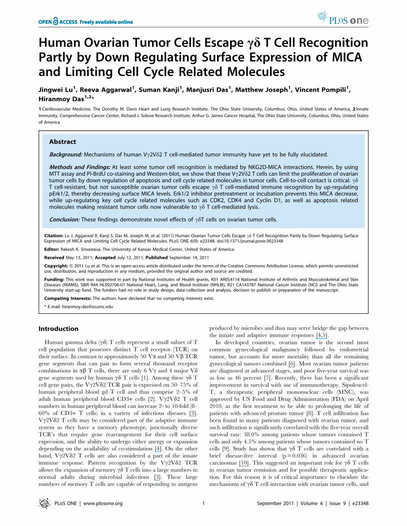
Human Ovarian Tumor Cells Escape gammadelta T Cell Recognition Partly by Down Regulating Surface...
12
Human Ovarian Tumor Cells Escape cd T Cell Recognition Partly by Down Regulating Surface Expression of MICA and Limiting Cell Cycle Related Molecules Jingwei Lu 1 , Reeva Aggarwal 1 , Suman Kanji 1 , Manjusri Das 1 , Matthew Joseph 1 , Vincent Pompili 1 , Hiranmoy Das 1,2 * 1 Cardiovascular Medicine, The Dorothy M. Davis Heart and Lung Research Institute, The Ohio State University, Columbus, Ohio, United States of America, 2 Innate Immunity, Comprehensive Cancer Center, Richard J. Solove Research Institute, Arthur G. James Cancer Hospital, The Ohio State University, Columbus, Ohio, United States of America Abstract Background: Mechanisms of human Vc2Vd2 T cell-mediated tumor immunity have yet to be fully elucidated. Methods and Findings: At least some tumor cell recognition is mediated by NKG2D-MICA interactions. Herein, by using MTT assay and PI-BrdU co-staining and Western-blot, we show that these Vc2Vd2 T cells can limit the proliferation of ovarian tumor cells by down regulation of apoptosis and cell cycle related molecules in tumor cells. Cell-to-cell contact is critical. cd T cell-resistant, but not susceptible ovarian tumor cells escape cd T cell-mediated immune recognition by up-regulating pErk1/2, thereby decreasing surface MICA levels. Erk1/2 inhibitor pretreatment or incubation prevents this MICA decrease, while up-regulating key cell cycle related molecules such as CDK2, CDK4 and Cyclin D1, as well as apoptosis related molecules making resistant tumor cells now vulnerable to cd T cell-mediated lysis. Conclusion: These findings demonstrate novel effects of cdT cells on ovarian tumor cells. Citation: Lu J, Aggarwal R, Kanji S, Das M, Joseph M, et al. (2011) Human Ovarian Tumor Cells Escape cd T Cell Recognition Partly by Down Regulating Surface Expression of MICA and Limiting Cell Cycle Related Molecules. PLoS ONE 6(9): e23348. doi:10.1371/journal.pone.0023348 Editor: Rakesh K. Srivastava, The University of Kansas Medical Center, United States of America Received May 13, 2011; Accepted July 12, 2011; Published September 14, 2011 Copyright: ß 2011 Lu et al. This is an open-access article distributed under the terms of the Creative Commons Attribution License, which permits unrestricted use, distribution, and reproduction in any medium, provided the original author and source are credited. Funding: This work was supported in part by National Institutes of Health grants, K01 AR054114 National Institute of Arthritis and Musculoskeletal and Skin Diseases (NIAMS), SBIR R44 HL092706-01 National Heart, Lung, and Blood Institute (NHLBI), R21 CA143787 National Cancer Institute (NCI) and The Ohio State University start-up fund. The funders had no role in study design, data collection and analysis, decision to publish or preparation of the manuscript. Competing Interests: The authors have declared that no competing interests exist. * E-mail: [email protected] Introduction Human gamma delta (cd) T cells represent a small subset of T cell population that possesses distinct T cell receptor (TCR) on their surface. In contrast to approximately 50 Va and 50 Vb TCR gene segments that can pair to form several thousand receptor combinations in ab T cells, there are only 6 Vc and 4 major Vd gene segments used by human cd T cells [1]. Among these cd T cell gene pairs, the Vc2Vd2 TCR pair is expressed on 50–75% of human peripheral blood gd T cell and thus comprise 2–5% of adult human peripheral blood CD3+ cells [2]. Vc2Vd2 T cell numbers in human peripheral blood can increase 2- to 10-fold (8– 60% of CD3+ T cells) in a variety of infectious diseases [3]. Vc2Vd2 T cells may be considered part of the adaptive immune system as they have a memory phenotype, junctionally diverse TCR’s that require gene rearrangement for their cell surface expression, and the ability to undergo either anergy or expansion depending on the availability of co-stimulation [4]. On the other hand, Vc2Vd2 T cells are also considered a part of the innate immune response. Pattern recognition by the Vc2Vd2 TCR allows the expansion of memory cd T cells into a large numbers in normal adults during microbial infections [3]. These large numbers of memory T cells are capable of responding to antigens produced by microbes and thus may serve bridge the gap between the innate and adaptive immune responses [4,5]. In developed countries, ovarian tumor is the second most common gynecological malignancy followed by endometrial tumor, but accounts for more mortality than all the remaining gynecological tumors combined [6]. Most ovarian tumor patients are diagnosed at advanced stages, and poor five-year survival was as low as 46 percent [7]. Recently, there has been a significant improvement in survival with use of immunotherapy. Sipuleucel- T, a therapeutic peripheral mononuclear cells (MNC), was approved by US Food and Drug Administration (FDA) on April 2010, as the first treatment to be able to prolonging the life of patients with advanced prostate tumor [8]. T cell infiltration has been found in many patients diagnosed with ovarian tumor, and such infiltration is significantly correlated with the five-year overall survival rate: 38.0% among patients whose tumors contained T cells and only 4.5% among patients whose tumors contained no T cells [9]. Study has shown that cd T cells are correlated with a brief disease-free interval (p = 0.036) in advanced ovarian carcinomas [10]. This suggested an important role for cd T cells in ovarian tumor remission and for possible therapeutic applica- tion. For this reason it is of critical importance to elucidate the mechanisms of cd T cell interaction with ovarian tumor cells, and PLoS ONE | www.plosone.org 1 September 2011 | Volume 6 | Issue 9 | e23348
-
Upload
independent -
Category
Documents
-
view
1 -
download
0
Transcript of Human Ovarian Tumor Cells Escape gammadelta T Cell Recognition Partly by Down Regulating Surface...
Human Ovarian Tumor Cells Escape cd T Cell RecognitionPartly by Down Regulating Surface Expression of MICAand Limiting Cell Cycle Related MoleculesJingwei Lu1, Reeva Aggarwal1, Suman Kanji1, Manjusri Das1, Matthew Joseph1, Vincent Pompili1,
Hiranmoy Das1,2*
1 Cardiovascular Medicine, The Dorothy M. Davis Heart and Lung Research Institute, The Ohio State University, Columbus, Ohio, United States of America, 2 Innate
Immunity, Comprehensive Cancer Center, Richard J. Solove Research Institute, Arthur G. James Cancer Hospital, The Ohio State University, Columbus, Ohio, United States
of America
Abstract
Background: Mechanisms of human Vc2Vd2 T cell-mediated tumor immunity have yet to be fully elucidated.
Methods and Findings: At least some tumor cell recognition is mediated by NKG2D-MICA interactions. Herein, by usingMTT assay and PI-BrdU co-staining and Western-blot, we show that these Vc2Vd2 T cells can limit the proliferation of ovariantumor cells by down regulation of apoptosis and cell cycle related molecules in tumor cells. Cell-to-cell contact is critical. cdT cell-resistant, but not susceptible ovarian tumor cells escape cd T cell-mediated immune recognition by up-regulatingpErk1/2, thereby decreasing surface MICA levels. Erk1/2 inhibitor pretreatment or incubation prevents this MICA decrease,while up-regulating key cell cycle related molecules such as CDK2, CDK4 and Cyclin D1, as well as apoptosis relatedmolecules making resistant tumor cells now vulnerable to cd T cell-mediated lysis.
Conclusion: These findings demonstrate novel effects of cdT cells on ovarian tumor cells.
Citation: Lu J, Aggarwal R, Kanji S, Das M, Joseph M, et al. (2011) Human Ovarian Tumor Cells Escape cd T Cell Recognition Partly by Down Regulating SurfaceExpression of MICA and Limiting Cell Cycle Related Molecules. PLoS ONE 6(9): e23348. doi:10.1371/journal.pone.0023348
Editor: Rakesh K. Srivastava, The University of Kansas Medical Center, United States of America
Received May 13, 2011; Accepted July 12, 2011; Published September 14, 2011
Copyright: � 2011 Lu et al. This is an open-access article distributed under the terms of the Creative Commons Attribution License, which permits unrestricteduse, distribution, and reproduction in any medium, provided the original author and source are credited.
Funding: This work was supported in part by National Institutes of Health grants, K01 AR054114 National Institute of Arthritis and Musculoskeletal and SkinDiseases (NIAMS), SBIR R44 HL092706-01 National Heart, Lung, and Blood Institute (NHLBI), R21 CA143787 National Cancer Institute (NCI) and The Ohio StateUniversity start-up fund. The funders had no role in study design, data collection and analysis, decision to publish or preparation of the manuscript.
Competing Interests: The authors have declared that no competing interests exist.
* E-mail: [email protected]
Introduction
Human gamma delta (cd) T cells represent a small subset of T
cell population that possesses distinct T cell receptor (TCR) on
their surface. In contrast to approximately 50 Va and 50 Vb TCR
gene segments that can pair to form several thousand receptor
combinations in ab T cells, there are only 6 Vc and 4 major Vdgene segments used by human cd T cells [1]. Among these cd T
cell gene pairs, the Vc2Vd2 TCR pair is expressed on 50–75% of
human peripheral blood gd T cell and thus comprise 2–5% of
adult human peripheral blood CD3+ cells [2]. Vc2Vd2 T cell
numbers in human peripheral blood can increase 2- to 10-fold (8–
60% of CD3+ T cells) in a variety of infectious diseases [3].
Vc2Vd2 T cells may be considered part of the adaptive immune
system as they have a memory phenotype, junctionally diverse
TCR’s that require gene rearrangement for their cell surface
expression, and the ability to undergo either anergy or expansion
depending on the availability of co-stimulation [4]. On the other
hand, Vc2Vd2 T cells are also considered a part of the innate
immune response. Pattern recognition by the Vc2Vd2 TCR
allows the expansion of memory cd T cells into a large numbers in
normal adults during microbial infections [3]. These large
numbers of memory T cells are capable of responding to antigens
produced by microbes and thus may serve bridge the gap between
the innate and adaptive immune responses [4,5].
In developed countries, ovarian tumor is the second most
common gynecological malignancy followed by endometrial
tumor, but accounts for more mortality than all the remaining
gynecological tumors combined [6]. Most ovarian tumor patients
are diagnosed at advanced stages, and poor five-year survival was
as low as 46 percent [7]. Recently, there has been a significant
improvement in survival with use of immunotherapy. Sipuleucel-
T, a therapeutic peripheral mononuclear cells (MNC), was
approved by US Food and Drug Administration (FDA) on April
2010, as the first treatment to be able to prolonging the life of
patients with advanced prostate tumor [8]. T cell infiltration has
been found in many patients diagnosed with ovarian tumor, and
such infiltration is significantly correlated with the five-year overall
survival rate: 38.0% among patients whose tumors contained T
cells and only 4.5% among patients whose tumors contained no T
cells [9]. Study has shown that cd T cells are correlated with a
brief disease-free interval (p = 0.036) in advanced ovarian
carcinomas [10]. This suggested an important role for cd T cells
in ovarian tumor remission and for possible therapeutic applica-
tion. For this reason it is of critical importance to elucidate the
mechanisms of cd T cell interaction with ovarian tumor cells, and
PLoS ONE | www.plosone.org 1 September 2011 | Volume 6 | Issue 9 | e23348
to understand the evasion mechanisms by which tumors escape
from immunosurveillance by cytotoxic T cells.
MHC class I chain related molecules A and B (MICA and
MICB, danger signals), which are widely expressed in epithelial
tumor cells, and virally or bacterially infected cells, can be
recognized by cd T cells and NK cells via NKG2D; a signaling
pathway is responsible for enhanced cytotoxicity against infected
cells or tumors [11,12,13]. MICA is preserved in most mammals
except for rodents and is present at high levels in gastrointestinal
epithelium, and on tumor cells of epithelial origin [14]. Other
ligands for NKG2D such as UL16 binding protein 1,2,3,4
(ULBP1, 2, 3, 4) RAET1G (ULBP5) and RAET1L (ULBP6) have
also been reported as targets of the host immune response [15].
The expression and modulation of MICA on tumor cells have
effects on cell survival [16,17,18]. Largely unknown, however is
the mechanism by which cd T cells affect tumor cells other than
cytotoxicity and Th-1 type cytokine. In the present study, we
assessed the effect of cd T cells on proliferation of tumor cells using
two ovarian tumor cell lines, A2780 and OV4, resistant and
susceptible to cytotoxic lysis, respectively. We further evaluated the
mechanisms by which tumor cells escape immune recognition by
cd T cells, focusing on surface molecules and cell cycle related
molecules. Herein we show that the tumor cell survival from the
recognition of cd T cells is correlated with the down regulation of
cell surface MICA levels and reduced expression of cell cycle
related molecules, as well as up-regulation of Erk1/2 signaling.
Erk1/2 inhibitor was able to bring back surface expression of
MICA, induced higher levels of cell cycle related molecules and
resulted in effective cytotoxic lysis by cd T cells. These new
findings help define novel mechanisms for tumor evasion against
innate host immune response.
Results
In Vitro cd T Cell Expansion and CharacterizationAs previously reported [19,20] human cd T cells were expanded
from freshly isolated PBMCs. One million of PBMC was seeded in
a well of a 24-well plate and 10 mm of risedronate was added to the
culture in a complete RPMI-1640 media. Recombinant IL-2
(0.5 nM) was supplemented to the culture on day 3 and 7 during
the culture. Cells were subjected to split upon overgrowth. At day
14 flowcytometric analysis was performed to assess cell expansion
and to determine their phenotype. Flowcytometric analysis
revealed that almost all expanded cells were CD3 positive. Among
expanded cells almost 90% cells were Vd2 positive cd T cells (Fig.
S1). There were almost no Vd1 positive cd T cells. Similar cells
were used for the subsequent experiments.
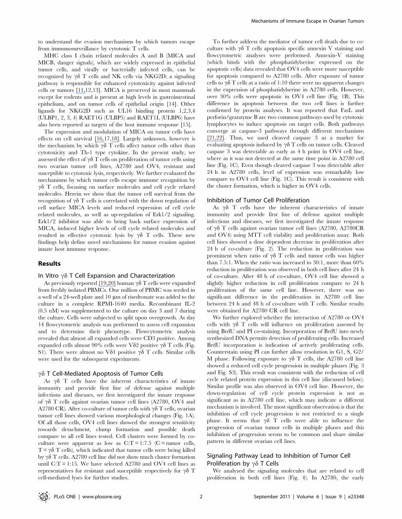
cd T Cell-Mediated Apoptosis of Tumor CellsAs cd T cells have the inherent characteristics of innate
immunity and provide first line of defense against multiple
infections and diseases, we first investigated the innate response
of cd T cells against ovarian tumor cell lines (A2780, OV4 and
A2780 CR). After co-culture of tumor cells with cd T cells, ovarian
tumor cell lines showed various morphological changes (Fig. 1A).
Of all those cells, OV4 cell lines showed the strongest sensitivity
towards detachment, clump formation and possible death
compare to all cell lines tested. Cell clusters were formed by co-
culture were apparent as low as C:T = 1:7.5 (C = tumor cells,
T = cd T cells), which indicated that tumor cells were being killed
by cd T cells. A2780 cell line did not show much cluster formation
until C:T = 1:15. We have selected A2780 and OV4 cell lines as
representatives for resistant and susceptible respectively for cd T
cell-mediated lyses for further studies.
To further address the mediator of tumor cell death due to co-
culture with cd T cells apoptosis specific annexin V staining and
flowcytometric analyses were performed. Annexin-V staining
(which binds with the phosphatidylserine expressed on the
apoptotic cells) data revealed that OV4 cells were more susceptible
for apoptosis compared to A2780 cells. After exposure of tumor
cells to cd T cells at a ratio of 1:10 there were no apparent changes
in the expression of phosphatidylserine in A2780 cells. However,
over 30% cells were apoptotic in OV4 cell line (Fig. 1B). This
difference in apoptosis between the two cell lines is further
confirmed by protein analyses. It was reported that FasL and
perforin/granzyme B are two common pathways used by cytotoxic
lymphocytes to induce apoptosis on target cells. Both pathways
converge at caspase-3 pathways through different mechanisms
[21,22]. Thus, we used cleaved caspase 3 as a marker for
evaluating apoptosis induced by cd T cells on tumor cells. Cleaved
caspase 3 was detectable as early as 4 h point in OV4 cell line,
where as it was not detected at the same time point in A2780 cell
line (Fig. 1C). Even though cleaved caspase 3 was detectable after
24 h in A2780 cells, level of expression was remarkably low
compare to OV4 cell line (Fig. 1C). This result is consistent with
the cluster formation, which is higher in OV4 cells.
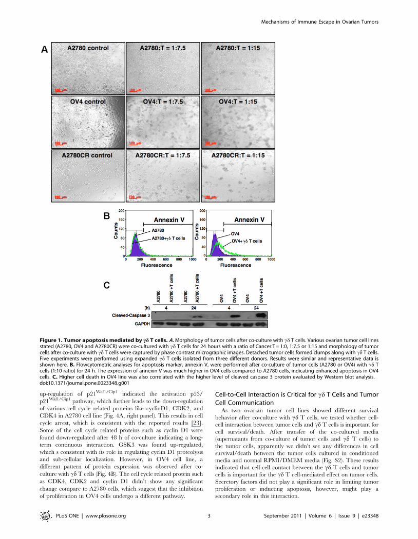
Inhibition of Tumor Cell ProliferationAs cd T cells have the inherent characteristics of innate
immunity and provide first line of defense against multiple
infections and diseases, we first investigated the innate response
of cd T cells against ovarian tumor cell lines (A2780, A2780CR
and OV4) using MTT cell viability and proliferation assay. Both
cell lines showed a dose dependent decrease in proliferation after
24 h of co-culture (Fig. 2). The reduction in proliferation was
prominent when ratio of cd T cells and tumor cells was higher
than 7.5:1. When the ratio was increased to 30:1, more than 60%
reduction in proliferation was observed in both cell lines after 24 h
of co-culture. After 48 h of co-culture, OV4 cell line showed a
slightly higher reduction in cell proliferation compare to 24 h
proliferation of the same cell line. However, there was no
significant difference in the proliferation in A2780 cell line
between 24 h and 48 h of co-culture with T cells. Similar results
were obtained for A2780 CR cell line.
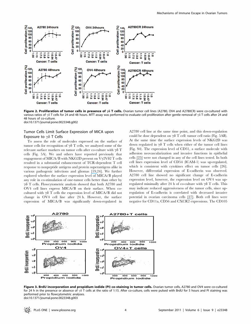
We further explored whether the interaction of A2780 or OV4
cells with cd T cells will influence on proliferation assessed by
using BrdU and PI co-staining. Incorporation of BrdU into newly
synthesized DNA permits detection of proliferating cells. Increased
BrdU incorporation is indication of actively proliferating cells.
Counterstain using PI can further allow resolution in G1, S, G2/
M phase. Following exposure to cd T cells, the A2780 cell line
showed a reduced cell cycle progression in multiple phases (Fig. 3
and Fig. S3). This result was consistent with the reduction of cell
cycle related protein expression in this cell line (discussed below).
Similar profile was also observed in OV4 cell line. However, the
down-regulation of cell cycle protein expression is not as
significant as in A2780 cell line, which may indicate a different
mechanism is involved. The most significant observation is that the
inhibition of cell cycle progression is not restricted to a single
phase. It seems that cd T cells were able to influence the
progression of ovarian tumor cells in multiple phases and this
inhibition of progression seems to be common and share similar
pattern in different ovarian cell lines.
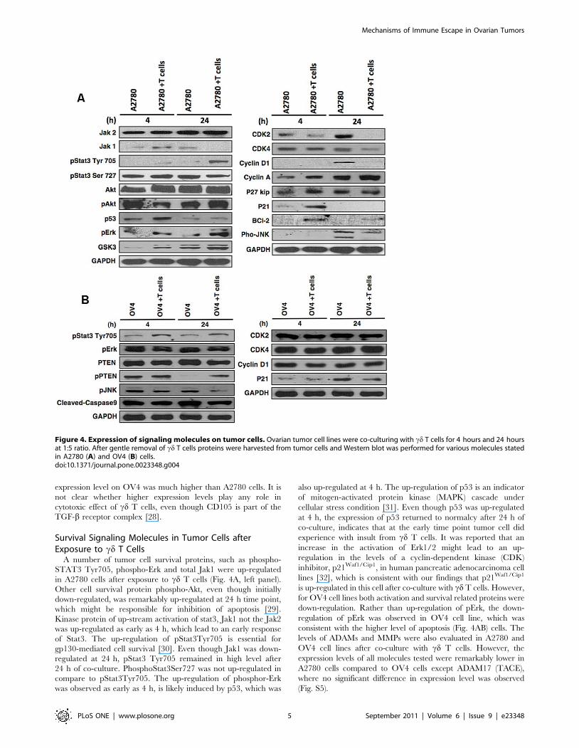
Signaling Pathway Lead to Inhibition of Tumor CellProliferation by cd T Cells
We analyzed the signaling molecules that are related to cell
proliferation in both cell lines (Fig. 4). In A2780, the early
Mechanisms of Immune Escape in Ovarian Tumors
PLoS ONE | www.plosone.org 2 September 2011 | Volume 6 | Issue 9 | e23348
up-regulation of p21Waf1/Cip1 indicated the activation p53/
p21Waf1/Cip1 pathway, which further leads to the down-regulation
of various cell cycle related proteins like cyclinD1, CDK2, and
CDK4 in A2780 cell line (Fig. 4A, right panel). This results in cell
cycle arrest, which is consistent with the reported results [23].
Some of the cell cycle related proteins such as cyclin D1 were
found down-regulated after 48 h of co-culture indicating a long-
term continuous interaction. GSK3 was found up-regulated,
which s consistent with its role in regulating cyclin D1 proteolysis
and sub-cellular localization. However, in OV4 cell line, a
different pattern of protein expression was observed after co-
culture with cd T cells (Fig. 4B). The cell cycle related protein such
as CDK4, CDK2 and cyclin D1 didn’t show any significant
change compare to A2780 cells, which suggest that the inhibition
of proliferation in OV4 cells undergo a different pathway.
Cell-to-Cell Interaction is Critical for cd T Cells and TumorCell Communication
As two ovarian tumor cell lines showed different survival
behavior after co-culture with cd T cells, we tested whether cell-
cell interaction between tumor cells and cd T cells is important for
cell survival/death. After transfer of the co-cultured media
(supernatants from co-culture of tumor cells and cd T cells) to
the tumor cells, apparently we didn’t see any differences in cell
survival/death between the tumor cells cultured in conditioned
media and normal RPMI/DMEM media (Fig. S2). These results
indicated that cell-cell contact between the cd T cells and tumor
cells is important for the cd T cell-mediated effect on tumor cells.
Secretory factors did not play a significant role in limiting tumor
proliferation or inducting apoptosis, however, might play a
secondary role in this interaction.
Figure 1. Tumor apoptosis mediated by cd T cells. A. Morphology of tumor cells after co-culture with cd T cells. Various ovarian tumor cell linesstated (A2780, OV4 and A2780CR) were co-cultured with cd T cells for 24 hours with a ratio of Cancer:T = 1:0, 1:7.5 or 1:15 and morphology of tumorcells after co-culture with cd T cells were captured by phase contrast micrographic images. Detached tumor cells formed clumps along with cd T cells.Five experiments were performed using expanded cd T cells isolated from three different donors. Results were similar and representative data isshown here. B. Flowcytometric analyses for apoptosis marker, annexin V, were performed after co-culture of tumor cells (A2780 or OV4) with cd Tcells (1:10 ratio) for 24 h. The expression of annexin V was much higher in OV4 cells compared to A2780 cells, indicating enhanced apoptosis in OV4cells. C. Higher cell death in OV4 line was also correlated with the higher level of cleaved caspase 3 protein evaluated by Western blot analysis.doi:10.1371/journal.pone.0023348.g001
Mechanisms of Immune Escape in Ovarian Tumors
PLoS ONE | www.plosone.org 3 September 2011 | Volume 6 | Issue 9 | e23348
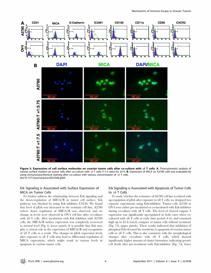
Tumor Cells Limit Surface Expression of MICA uponExposure to cd T Cells
To assess the role of molecules expressed on the surface of
tumor cells for recognition of cd T cells, we analyzed some of the
relevant surface markers on tumor cells after co-culture with cd T
cells (Fig. 5A). We and others have reported previously that
engagement of MICA/B with NKG2D present on Vc2Vd2 T cells
resulted in a substantial enhancement of TCR-dependent T cell
response to nonpeptide antigens and protein superantigens alike in
various pathogenic infections and gliomas [19,24]. We further
explored whether the surface expression level of MICA/B played
any role in co-stimulation of one-tumor cells better than other by
cd T cells. Flowcytometric analysis showed that both A2780 and
OV4 cell lines express MICA/B on their surface. When co-
cultured with cd T cells the expression level of MICA/B did not
change in OV4 cell line after 24 h. However, the surface
expression of MICA/B was significantly down-regulated in
A2780 cell line at the same time point, and this down-regulation
could be dose dependent on cd T cell: tumor cell ratio (Fig. 5AB).
At the same time the surface expression levels of NKG2D was
down regulated in cd T cells when either of the tumor cell lines
(Fig. S4). The expression level of CD31, a surface molecule with
adhesion neovascularization and invasive functions in epithelial
cells [25] were not changed in any of the cell lines tested. In both
cell lines expression level of CD54 (ICAM-1) was up-regulated,
which is consistent with cytokines effect on tumor cells [26].
However, differential expression of E-cadherin was observed.
A2780 cell line showed no significant change of E-cadherin
expression level, however, the expression level on OV4 was up-
regulated minimally after 24 h of co-culture with cd T cells. This
may indicate reduced aggressiveness of the tumor cells, since up-
regulation of E-cadherin is correlated with decreased invasive
potential in ovarian carcinoma cells [27]. Both cell lines were
negative for CD11a, CD56 and CXCR2 expressions. The CD105
Figure 2. Proliferation of tumor cells in presence of cd T cells. Ovarian tumor cell lines (A2780, OV4 and A2780CR) were co-cultured withvarious ratios of cd T cells for 24 and 48 hours. MTT assay was performed to evaluate cell proliferation after gentle removal of cd T cells after 24 and48 hours of co-culture.doi:10.1371/journal.pone.0023348.g002
Figure 3. BrdU incorporation and propidium iodide (PI) co-staining in tumor cells. Ovarian tumor cells, A2780 and OV4 were co-culturedfor 24 h in the presence or absence of cd T cells at the ratio of 1:15. After co-culture, cells were pulsed with BrdU for 5 hours and PI staining wasperformed prior to flowcytometric analyses.doi:10.1371/journal.pone.0023348.g003
Mechanisms of Immune Escape in Ovarian Tumors
PLoS ONE | www.plosone.org 4 September 2011 | Volume 6 | Issue 9 | e23348
expression level on OV4 was much higher than A2780 cells. It is
not clear whether higher expression levels play any role in
cytotoxic effect of cd T cells, even though CD105 is part of the
TGF-b receptor complex [28].
Survival Signaling Molecules in Tumor Cells afterExposure to cd T Cells
A number of tumor cell survival proteins, such as phospho-
STAT3 Tyr705, phospho-Erk and total Jak1 were up-regulated
in A2780 cells after exposure to cd T cells (Fig. 4A, left panel).
Other cell survival protein phospho-Akt, even though initially
down-regulated, was remarkably up-regulated at 24 h time point,
which might be responsible for inhibition of apoptosis [29].
Kinase protein of up-stream activation of stat3, Jak1 not the Jak2
was up-regulated as early as 4 h, which lead to an early response
of Stat3. The up-regulation of pStat3Tyr705 is essential for
gp130-mediated cell survival [30]. Even though Jak1 was down-
regulated at 24 h, pStat3 Tyr705 remained in high level after
24 h of co-culture. PhosphoStat3Ser727 was not up-regulated in
compare to pStat3Tyr705. The up-regulation of phosphor-Erk
was observed as early as 4 h, is likely induced by p53, which was
also up-regulated at 4 h. The up-regulation of p53 is an indicator
of mitogen-activated protein kinase (MAPK) cascade under
cellular stress condition [31]. Even though p53 was up-regulated
at 4 h, the expression of p53 returned to normalcy after 24 h of
co-culture, indicates that at the early time point tumor cell did
experience with insult from cd T cells. It was reported that an
increase in the activation of Erk1/2 might lead to an up-
regulation in the levels of a cyclin-dependent kinase (CDK)
inhibitor, p21Waf1/Cip1, in human pancreatic adenocarcinoma cell
lines [32], which is consistent with our findings that p21Waf1/Cip1
is up-regulated in this cell after co-culture with cd T cells. However,
for OV4 cell lines both activation and survival related proteins were
down-regulation. Rather than up-regulation of pErk, the down-
regulation of pErk was observed in OV4 cell line, which was
consistent with the higher level of apoptosis (Fig. 4AB) cells. The
levels of ADAMs and MMPs were also evaluated in A2780 and
OV4 cell lines after co-culture with cd T cells. However, the
expression levels of all molecules tested were remarkably lower in
A2780 cells compared to OV4 cells except ADAM17 (TACE),
where no significant difference in expression level was observed
(Fig. S5).
Figure 4. Expression of signaling molecules on tumor cells. Ovarian tumor cell lines were co-culturing with cd T cells for 4 hours and 24 hoursat 1:5 ratio. After gentle removal of cd T cells proteins were harvested from tumor cells and Western blot was performed for various molecules statedin A2780 (A) and OV4 (B) cells.doi:10.1371/journal.pone.0023348.g004
Mechanisms of Immune Escape in Ovarian Tumors
PLoS ONE | www.plosone.org 5 September 2011 | Volume 6 | Issue 9 | e23348
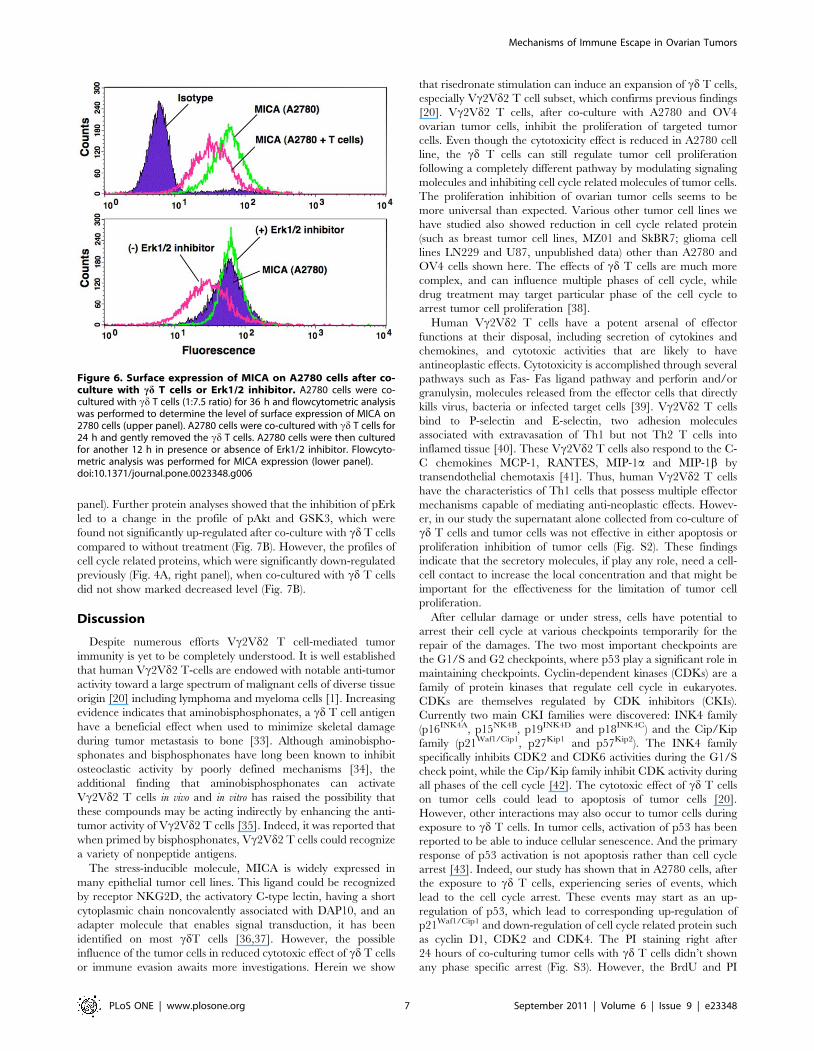
Erk Signaling is Associated with Surface Expression ofMICA on Tumor Cells
To further address the relationship between Erk signaling and
the down-regulation of MICA/B in tumor cell surface, Erk
pathway was blocked by using Erk inhibitor U0126. We found
that level of pErk was increased in the resistant cell line, A2780
(where down regulation of MICA/B was observed) and no
change in levels were observed in OV4 cell line after co-culture
with cd T cells. After incubation with Erk inhibitor with A2780
cells, the MICA/B surface expression was completely recovered
to normal level (Fig. 6, lower panel). It is possible that Erk may
play a critical role in the expression of MICA/B and recognition
of cd T cells as a result. The changes in pErk expression levels
after exposure to cd T cells may lead to differential regulation of
MICA expressions, which might result in various levels in
apoptosis in various tumor cells.
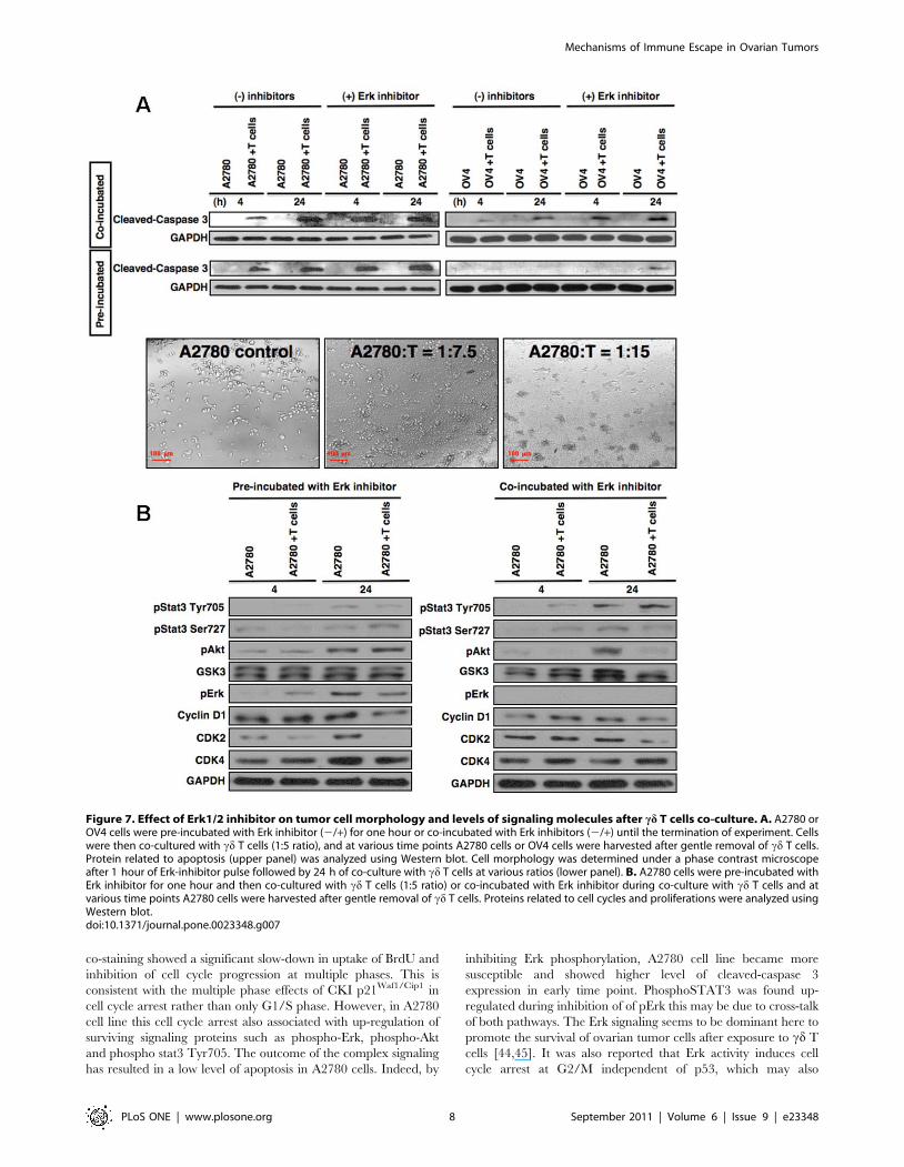
Erk Signaling is Associated with Apoptosis of Tumor Cellsto cd T Cells
To study whether the resistance of A2780 cell line is related with
up-regulation of pErk after exposure to cd T cells, we designed two
separate experiments using Erk-inhibitor. Tumor cells A2780 or
OV4 were either pre-incubated or co-incubated with Erk-inhibitor
during co-culture with cd T cells. The level of cleaved caspase 3
expression was significantly up-regulated in both cases when co-
cultured with cd T cells at early time period (4 h), and remained
high up to 24 h tested, compare to tumor cells without treatment
(Fig. 7A, upper panels). These results indicated that inhibition of
phosphor-Erk elevated the sensitivity to apoptosis of ovarian tumor
cells to cd T cells. This is also consistent with the morphological
changes after co-culture with cd T cells, which showed
significantly higher amount of cluster formation, indicating greater
cell death after pre-treatment with Erk-inhibitor (Fig. 7A, lower
Figure 5. Expression of cell surface molecules on ovarian tumor cells after co-culture with cd T cells. A. Flowcytometric analysis ofvarious surface markers on tumor cells after co-culture with cd T cells (1:7.5 ratio) for 24 h. B. Expression of MICA on A2780 cells was evaluated byusing immunocytochemical staining after co-culture with various concentration of cd T cells.doi:10.1371/journal.pone.0023348.g005
Mechanisms of Immune Escape in Ovarian Tumors
PLoS ONE | www.plosone.org 6 September 2011 | Volume 6 | Issue 9 | e23348
panel). Further protein analyses showed that the inhibition of pErk
led to a change in the profile of pAkt and GSK3, which were
found not significantly up-regulated after co-culture with cd T cells
compared to without treatment (Fig. 7B). However, the profiles of
cell cycle related proteins, which were significantly down-regulated
previously (Fig. 4A, right panel), when co-cultured with cd T cells
did not show marked decreased level (Fig. 7B).
Discussion
Despite numerous efforts Vc2Vd2 T cell-mediated tumor
immunity is yet to be completely understood. It is well established
that human Vc2Vd2 T-cells are endowed with notable anti-tumor
activity toward a large spectrum of malignant cells of diverse tissue
origin [20] including lymphoma and myeloma cells [1]. Increasing
evidence indicates that aminobisphosphonates, a cd T cell antigen
have a beneficial effect when used to minimize skeletal damage
during tumor metastasis to bone [33]. Although aminobispho-
sphonates and bisphosphonates have long been known to inhibit
osteoclastic activity by poorly defined mechanisms [34], the
additional finding that aminobisphosphonates can activate
Vc2Vd2 T cells in vivo and in vitro has raised the possibility that
these compounds may be acting indirectly by enhancing the anti-
tumor activity of Vc2Vd2 T cells [35]. Indeed, it was reported that
when primed by bisphosphonates, Vc2Vd2 T cells could recognize
a variety of nonpeptide antigens.
The stress-inducible molecule, MICA is widely expressed in
many epithelial tumor cell lines. This ligand could be recognized
by receptor NKG2D, the activatory C-type lectin, having a short
cytoplasmic chain noncovalently associated with DAP10, and an
adapter molecule that enables signal transduction, it has been
identified on most cdT cells [36,37]. However, the possible
influence of the tumor cells in reduced cytotoxic effect of cd T cells
or immune evasion awaits more investigations. Herein we show
that risedronate stimulation can induce an expansion of cd T cells,
especially Vc2Vd2 T cell subset, which confirms previous findings
[20]. Vc2Vd2 T cells, after co-culture with A2780 and OV4
ovarian tumor cells, inhibit the proliferation of targeted tumor
cells. Even though the cytotoxicity effect is reduced in A2780 cell
line, the cd T cells can still regulate tumor cell proliferation
following a completely different pathway by modulating signaling
molecules and inhibiting cell cycle related molecules of tumor cells.
The proliferation inhibition of ovarian tumor cells seems to be
more universal than expected. Various other tumor cell lines we
have studied also showed reduction in cell cycle related protein
(such as breast tumor cell lines, MZ01 and SkBR7; glioma cell
lines LN229 and U87, unpublished data) other than A2780 and
OV4 cells shown here. The effects of cd T cells are much more
complex, and can influence multiple phases of cell cycle, while
drug treatment may target particular phase of the cell cycle to
arrest tumor cell proliferation [38].
Human Vc2Vd2 T cells have a potent arsenal of effector
functions at their disposal, including secretion of cytokines and
chemokines, and cytotoxic activities that are likely to have
antineoplastic effects. Cytotoxicity is accomplished through several
pathways such as Fas- Fas ligand pathway and perforin and/or
granulysin, molecules released from the effector cells that directly
kills virus, bacteria or infected target cells [39]. Vc2Vd2 T cells
bind to P-selectin and E-selectin, two adhesion molecules
associated with extravasation of Th1 but not Th2 T cells into
inflamed tissue [40]. These Vc2Vd2 T cells also respond to the C-
C chemokines MCP-1, RANTES, MIP-1a and MIP-1b by
transendothelial chemotaxis [41]. Thus, human Vc2Vd2 T cells
have the characteristics of Th1 cells that possess multiple effector
mechanisms capable of mediating anti-neoplastic effects. Howev-
er, in our study the supernatant alone collected from co-culture of
cd T cells and tumor cells was not effective in either apoptosis or
proliferation inhibition of tumor cells (Fig. S2). These findings
indicate that the secretory molecules, if play any role, need a cell-
cell contact to increase the local concentration and that might be
important for the effectiveness for the limitation of tumor cell
proliferation.
After cellular damage or under stress, cells have potential to
arrest their cell cycle at various checkpoints temporarily for the
repair of the damages. The two most important checkpoints are
the G1/S and G2 checkpoints, where p53 play a significant role in
maintaining checkpoints. Cyclin-dependent kinases (CDKs) are a
family of protein kinases that regulate cell cycle in eukaryotes.
CDKs are themselves regulated by CDK inhibitors (CKIs).
Currently two main CKI families were discovered: INK4 family
(p16INK4A, p15NK4B, p19INK4D and p18INK4C) and the Cip/Kip
family (p21Waf1/Cip1, p27Kip1 and p57Kip2). The INK4 family
specifically inhibits CDK2 and CDK6 activities during the G1/S
check point, while the Cip/Kip family inhibit CDK activity during
all phases of the cell cycle [42]. The cytotoxic effect of cd T cells
on tumor cells could lead to apoptosis of tumor cells [20].
However, other interactions may also occur to tumor cells during
exposure to cd T cells. In tumor cells, activation of p53 has been
reported to be able to induce cellular senescence. And the primary
response of p53 activation is not apoptosis rather than cell cycle
arrest [43]. Indeed, our study has shown that in A2780 cells, after
the exposure to cd T cells, experiencing series of events, which
lead to the cell cycle arrest. These events may start as an up-
regulation of p53, which lead to corresponding up-regulation of
p21Waf1/Cip1 and down-regulation of cell cycle related protein such
as cyclin D1, CDK2 and CDK4. The PI staining right after
24 hours of co-culturing tumor cells with cd T cells didn’t shown
any phase specific arrest (Fig. S3). However, the BrdU and PI
Figure 6. Surface expression of MICA on A2780 cells after co-culture with cd T cells or Erk1/2 inhibitor. A2780 cells were co-cultured with cd T cells (1:7.5 ratio) for 36 h and flowcytometric analysiswas performed to determine the level of surface expression of MICA on2780 cells (upper panel). A2780 cells were co-cultured with cd T cells for24 h and gently removed the cd T cells. A2780 cells were then culturedfor another 12 h in presence or absence of Erk1/2 inhibitor. Flowcyto-metric analysis was performed for MICA expression (lower panel).doi:10.1371/journal.pone.0023348.g006
Mechanisms of Immune Escape in Ovarian Tumors
PLoS ONE | www.plosone.org 7 September 2011 | Volume 6 | Issue 9 | e23348
co-staining showed a significant slow-down in uptake of BrdU and
inhibition of cell cycle progression at multiple phases. This is
consistent with the multiple phase effects of CKI p21Waf1/Cip1 in
cell cycle arrest rather than only G1/S phase. However, in A2780
cell line this cell cycle arrest also associated with up-regulation of
surviving signaling proteins such as phospho-Erk, phospho-Akt
and phospho stat3 Tyr705. The outcome of the complex signaling
has resulted in a low level of apoptosis in A2780 cells. Indeed, by
inhibiting Erk phosphorylation, A2780 cell line became more
susceptible and showed higher level of cleaved-caspase 3
expression in early time point. PhosphoSTAT3 was found up-
regulated during inhibition of of pErk this may be due to cross-talk
of both pathways. The Erk signaling seems to be dominant here to
promote the survival of ovarian tumor cells after exposure to cd T
cells [44,45]. It was also reported that Erk activity induces cell
cycle arrest at G2/M independent of p53, which may also
Figure 7. Effect of Erk1/2 inhibitor on tumor cell morphology and levels of signaling molecules after cd T cells co-culture. A. A2780 orOV4 cells were pre-incubated with Erk inhibitor (2/+) for one hour or co-incubated with Erk inhibitors (2/+) until the termination of experiment. Cellswere then co-cultured with cd T cells (1:5 ratio), and at various time points A2780 cells or OV4 cells were harvested after gentle removal of cd T cells.Protein related to apoptosis (upper panel) was analyzed using Western blot. Cell morphology was determined under a phase contrast microscopeafter 1 hour of Erk-inhibitor pulse followed by 24 h of co-culture with cd T cells at various ratios (lower panel). B. A2780 cells were pre-incubated withErk inhibitor for one hour and then co-cultured with cd T cells (1:5 ratio) or co-incubated with Erk inhibitor during co-culture with cd T cells and atvarious time points A2780 cells were harvested after gentle removal of cd T cells. Proteins related to cell cycles and proliferations were analyzed usingWestern blot.doi:10.1371/journal.pone.0023348.g007
Mechanisms of Immune Escape in Ovarian Tumors
PLoS ONE | www.plosone.org 8 September 2011 | Volume 6 | Issue 9 | e23348
contribute to the cell cycle arrest at multiple phases [46]. However,
in OV4 cell line, even though the signaling profile is different, cell
cycle arrest is also evident, which indicates that the inhibition of
cell proliferation is common among different cell lines and
different profile protein expression.
In summary, herein we report the mechanisms by which ovarian
tumor cells escape immune recognition upon exposure to cd T cells.
Tumor cells when recognized by cd T cells, cytotoxic pathway
predominates and apoptotic signal is more effective as a result tumor
cell become susceptible for lysis. However, some tumor cells can
modulate their surface molecules as well as signaling molecules to
hide recognition from immune cells and the pathway is dominant in
the resistant ovarian tumor cells, A2780. Even though resistant
tumor cells can escape immune recognition from cd T cells by down
regulating surface expression of MICA, cd T cells can still be able to
slow down the progression of the tumor cell proliferation by
inhibiting cell cycle related molecules CDK2, CDK4 and Cyclin
D1. Our findings clearly demonstrate that the effects of cd T cells on
ovarian tumor cells are more complicated than previous thought. By
modulating certain signaling molecules, in this case, inhibiting Erk
pathway, resistant ovarian tumor cells could be turned into
susceptible towards cd T cells-mediated immune recognition and
resulted in lysis. As cd T cells could be expanded in a large number
using aminobisphosphonate in vitro, the possibility of expanding cells
from peripheral blood and combining with molecules, which have
potential for modulating particular signal might result in an effective
immune therapy.
Methods
Derivation of cd T CellsHuman peripheral blood was collected (30 ml) from adult
healthy donors after obtaining the IRB approval from the Ohio
State University Medical Center and obtaining written consents
from donors. The ethic committee has also approved the
procedure and records are saved in the laboratory logbook.
Freshly collected blood was processed to isolate peripheral blood
mononuclear cells (PBMC) following the similar protocol pub-
lished earlier [19,20,47]. In brief, the peripheral blood was diluted
twice with phosphate buffer saline (PBS, pH 7.4) and carefully
layered over 10 ml of Ficoll-Paque Plus solution (GE Healthcare,
Uppsala, Sweden). After 30 min of centrifugation in a swinging
bucket rotor at 1400 rpm at room temp (24uC), the upper layer
was aspirated out and the mononuclear cell layer (buffy coat) was
collected. Buffy coat was washed three times with PBS to remove
platelets. One million of PBMC in each well was stimulated with
10 mM risedronate in a 24-well plate using 1 ml RPMI 1640
supplemented with 10% fetal bovine serum (FBS, HyClone Lab
Inc, Logan, UT), 2 mM glutamine, 1 nM b-mercapto ethanol,
1 nM HEPES and 100 IU of penicillin and streptomycin at 37uCincubator. Recombinant IL-2, 0.5 nM (PeproTech Inc. Rocky
Hill, NJ) was added to the culture on days 3 and 7. Cells were split
after day 10 using the complete RPMI 1640 media supplemented
with 0.5 nM rIL-2. Flowcytometric (FACS) analysis was per-
formed (using a FACS Calibur machine, BD Biosciences, CA) at
day 14, to evaluate phenotype of the expanded cell. FACS analysis
data revealed that 99.8% of the expanded cells were CD3+, and
89.5% of them were Vd2+. Cells were used between 15–19 days of
initial culture for further experiments discussed below.
Morphology of Ovarian Tumor Cells after Co-Culture withcd T Cells
Ten thousand ovarian tumor cells (A2780, OV4 and A2780CR;
purchased from ATCC, VA, and used within six months after
receipt) were cultured in a well of a 96-well culture plate in
DMEM media supplemented with 10% FBS. The cd T cells (T)
were added to the well and co-cultured with ovarian cancer (C)
cells with a ratio of C:T = 1:1 or 1:7.5 or 1:15 or 1:30 for 24 h or
48 h at 37uC incubator with 5% CO2. Phase contrast micrographs
were taken at 24 h and 48 h time points. The images were
captured under an epifluorescence microscope (Axioplan2; Carl
Zeiss) using Zeiss Axiovision imaging software. Separate sets of
experiments were performed pre-treating A2780 cell line for one
hour with Erk inhibitor U0126 at 10 mM final concentration or
co-culturing with same concentration of U0126 for 24 h. Cells
were then exposed to same ratio of cd T cells following methods
discussed above.
Cell Viability and Proliferation AssaysTumor cell proliferation was performed using MTT assays (3–
4,5-Dimethylthiazol-2-yl-2,5-diphenyltetrazolium bromide), a yel-
low tetrazole, is reduced to purple formazan in living cells. The
principal of the method uses the conversion of MTT to formazan
via mitochondrial oxidation and colorimetric assay was used to
detect the formazan as previously described with simple
modifications [48]. Briefly, ten thousand of ovarian tumor cells
(either OV4 or A2780WT or A2780CR) were co-cultured with cdT cells at a ratio of 1:30 or 1:15 or 1:7.5 or 1:3.75 or 1:1.9 or
1:0.94 or 1:0.47 or 1:0, in a well of a 96-well plate and MTT assay
was performed at 24 h or 48 h time points. Before performing
MTT assays, cd T cells were gently washed out once with 150 ml
PBS to minimize the interference with tumor cells read-out. Most
of the cd T cells were washed away and very few tumor cells were
drifted away during the process of washing observed under
microscope. After washing out the cd T cells, 100 ml of DMEM
medium/well with a concentration of 500 mg/ml MTT reagent
(Sigma) was added to the plate. Plates were incubated at 37uC in
the incubator for 3 h and 100 ml of diluting solution (80%
isopropyl alcohol, 10% HCl and 10% TritonX 100) was added to
each well and mixed well by using auto mix shaker. The developed
color value was recorded at 570 nM spectra and the reference
spectrum was kept at 690 nM. MTT results were calculated using
the equation:
Result %~½signal (cancerzT)� signal(T)�=
½singal (cancer)� signal(DMEM)�
Analysis of Apoptosis in Tumor CellsTo evaluate apoptosis tumor cells (150 K) were co-cultured with
cd T cells at 1:10 ratio for 24 hours. All cells were harvested
without any wash using non-enzymatic cell dissociation buffer.
Cells were then incubated on ice with FITC-conjugated annexin V
Ab for 30 min and washed with 16 PBS, and processed as
described above. Flowcytometric analysis was performed to
evaluate annexin V-positive cells. Cells negative for annexin V
staining were considered as live cells, and annexin V-positive cells
were considered as apoptotic cells.
Effect of Conditional Media on Tumor Cell ProliferationTo verify whether cell-cell contact is necessary for cd T cells to
interact with tumor cells, ovarian tumor cells (A2780 or OV4,
10 K cells/well) were co-cultured with cd T cells at a ratio of 1:0,
1:15 and 1:30 in 96-well plates (in 200 ml volume). The cd T cells
were also cultured without tumor cells with a concentration of
150 K cells/well and 300 K cells/well in DMEM medium with
Mechanisms of Immune Escape in Ovarian Tumors
PLoS ONE | www.plosone.org 9 September 2011 | Volume 6 | Issue 9 | e23348
10% FBS for 24 h. A second set of similar tumor cells were grown
at the same time without cd T cells using similar plates. Tumor
cells were cultured at the concentration of 60 K cells/well. After
24 h, 100 ml supernatant from first set was taken out and replaced
with the medium of second set of tumor cells accordingly. MTT
assay was performed after 24 h further incubation following the
same procedure as described above.
Flowcytometric AnalysisTo assess the effect of cdT cells on surface molecules of tumor
cells, cd T cells were co-cultured with 300 K ovarian tumor cells
per well either A2780 or OV4 at 7.5:1 ratio in 6-well plate using
RPMI medium (for A2780) or DMEM medium (for OV4) with
10% FBS. Cells were harvest using non-enzymatic cell dissoci-
ation solution (Sigma) after 24 h of co-culture and washed with
PBS. Cells were then incubated with primary and secondary
antibodies (if applicable) for 45 min each. Surface markers such
as CD31, MICA, CD56, CD105, CD11a, CXCR2, ICAM-1
(PE-conjugated, Pharmingen), E-cadherin (Invitrogen, secondary
antibody PE-conjugated) were tested using flowcytometry as
described above.
To assess the effect of tumor cells towards the surface expression
level of NKG2D on cd T cells, similar experiments were
performed as mentioned above, collected cd T cells after
incubation with tumor cells and flowcytometric analysis was
performed using NKG2D Ab (PE-conjugated, Pharmingen).
Immunocytochemical Study of MICA Expression onTumor Cells
A2780 tumor cells were co-cultured with cd T cells with various
ratios such as 1:0, 1:3.75, and 1:7.5 for 24 h. Cells were then fixed
with 4% para formaldehyde at room temperature for 10 min,
stained with anti-MICA Abs (kind gift from Dr. Spies, Seattle,
WA) and DAPI. The staining was observed under an epifluores-
cence microscope (Axioplan2; Carl Zeiss) and images were
captured with Zeiss Axiovision imaging software.
BrdU Incorporation and Propidium Iodide Co-StainingTo evaluate the effect of cdT cells to arrest cell cycle of tumor
cells, cd T cells were co-cultured with three hundred thousand
ovarian tumor cells A2780 or OV4 in a well at 15:1 ratio in a 6-
well plate. After 24 h, cd T cells were removed by three times
gentle washing with 16PBS. Respective culture media such as
DMEM (for OV4) or RPMI (for A2780) with 10% FBS and
10 mM BrdU were added to each plate. Cells were incubated at
37uC for another 5 h in an incubator. The cells were isolated by
using non-enzymatic cell dissociation buffer (Sigma) and collected
in 15 ml centrifuge tube, centrifuged at 3006g at 4uC. Cells were
then washed with PBS once and fixed with 75% ethanol for over
night. After centrifugation, pellet was resuspended in 2 M HCl
solution and incubated for 20 min. Cells were washed with PBS
and then resuspended with 0.1 M Sodium borate solution, and
again incubated for 2 min to neutralize the residual acid. After
washing with PBS, cells were incubated with primary antibody
(anti-BrdU monoclonal antibody, Invitrogen) in 16PBS contain-
ing 0.5% Tween-20 and 0.5% BSA for 45 min. Unbound primary
antibody was removed using washing buffer (16PBS containing
0.5% BSA). PE-conjugated secondary antibody (Pharmingen) was
added and incubated with cells for another 45 min. Cells were
then washed and incubated in PBS buffer containing RNase
OneTM Ribonuclease (10 U/ml, Promega) and propidium iodide
(PI, 3 mM). Cells were then analyzed by using a flowcytometer
(FACS Calibur, BD).
Propidium Iodide StainingThe cd T cells were co-cultured with three hundred thousand
ovarian tumor cells, A2780 WT or OV4 in a well of a 6-well plate
with a 10:1 ratio. After 24 hours, cd T cells were gently removed by
washing with 16PBS three times. Cells were dissociated and
centrifuged at 3006 g. Pellet was collected and washed with PBS
once. Cells were then fixed and permeabilized with 80% ethanol
over night. Fixed cells were centrifuged with and pellet was
incubated in PBS for rehydration for 10 mins. Then cells were
washed once with ice cold PBS. PI staining solution (Sigma, PI;
1:1000 and RNase One; 1:1000 in 16PBS) was added to the cells
and incubated for 20 mins at 37uC before analysis. PI was
purchased from Sigma and BrdU antibody was from Pharmingen.
Flowcytometry was performed using BD FACS CaliburTM machine
and data analysis was performed by using Cell Quest software.
Protein AnalysesTotal protein analysis was performed using standard Western
blot technology. Half a million of tumor cells (A2780 or OV4) were
pre-seeded in 3 cm Petri dish for 10 h before adding cd T cells.
Control plates were also plated at the same time to maintain equal
cell numbers, we did not observe any significant change on cell
numbers after 10 h of pre-seeding. Fresh medium (RPMI for A2780
and DMEM for OV4) containing 10% FBS were replaced to the
culture plates. Two and half million cd T cells were added to the
tumor cells to make a ratio of C:T = 1:5 (where ever applicable).
Protein was isolated after 4 and 24 h of addition of cd T cells. Before
isolation of total protein, cd T cells were gently removed by washing
with 16PBS. Various antibodies have been used for Western blot,
such as total Jak1, pan Akt, pStat3 (Ser 705), pErk, CDK2,
GAPDH, b-Actin (all from Cell Signaling Technology), p53, GSK3,
CDK4, cyclin D1, cyclin A, p27, kip, p21Waf1/Cip1, cleaved-
Caspase3, ADAM10, ADAM17, MMP9, MMP14 (all from Santa
Cruz Biotech) to detect their level of expression on tumor cells at
various time points of co-culture with cd T cells.
To determine the role of Erk signaling in modulation of ovarian
tumor cells (A2780 and OV4) and cd T cells interaction, A2780
cells or OV4 cells (500 K cells plated a day before) were pre-
treated with Erk inhibitor (U0126, 10 mM final concentration, Cell
Signaling) for one hour and then co-cultured with cd T cells (1:5
ratio) for 4 h and 24 h. Western blot was performed from isolated
proteins from tumor cells by removing cd T cells following the
same procedure as mentioned above. A separate set of
experiments were performed by incubating A2780 or OV4 cells
and cd T cells (1:5 ratio) together with Erk inhibitor (10 mM,
U0126) for 4 h and 24 h, and then processed for Western blot
analyses following above-mentioned protocol.
Effect of Erk-Inhibitor in Recovery of Surface Expressionof MICA
Ovarian tumor cells, A2780 (300 K) were co-cultured with cd T
cells with a ratios of 1:7.5 or 1:0 in a 6-well plate with RPMI
medium containing 10% FBS. After 24 h of incubation, cd T cells
were gently removed by washing with PBS. FBS free RPMI
medium was added to the plate with/without Erk inhibitor U0126
(10 mM, final concentration). Cells were cultured for another 12 h.
Then cells were harvested and processed for flowcytometric
analysis for surface expression of MICA.
Statistical AnalysisValues were expressed as mean6SEM and statistical analysis
was performed by ANOVA. Students t- test was also performed
and the results were considered significant when values of p,0.05.
Mechanisms of Immune Escape in Ovarian Tumors
PLoS ONE | www.plosone.org 10 September 2011 | Volume 6 | Issue 9 | e23348
Supporting Information
Figure S1 Flowcytometric analyses of expanded cd Tcells. Total human PBMC was stimulated with risedronate, an
aminobisphosphonate in T-cell media supplemented with rIL-2 at
day 3 and 7 and flowcytometric analysis was performed at day 17
for T cell subtypes after expansion.
(TIFF)
Figure S2 Cell-to-cell contact is necessary not the cellsecretory molecules to inhibit tumor cell proliferation.A2780 or OV4 cells were co-cultured with cd T cells and culture
supernatants were added to the respective tumor cells to evaluate
effects on proliferation of tumor cells using MTT assays. Tumor
cell culture media or cd T cell culture media were used as controls.
(TIFF)
Figure S3 Cell cycle analysis of tumor cells usingpropidium iodide staining. Tumor cells were co-cultured in
presence or absence of cd T cells at a ratio of 1:10 for 24 hours.
Propidium iodide staining was done after gentle removal of cd T
cells and flowcytometic analysis was performed for evaluation of
cell cycle status.
(TIFF)
Figure S4 Expression of NKG2D on cd T cells after co-culture with tumor cells. Filled histogram indicates surface
expression level of NKG2D on cd T cells without co-culture with
any tumor cells. Green line indicates surface expression level of
NKG2D on cd T cells after co-cultured with tumor cell line A2780
at a ratio of 7.5:1 for 24 hours. Magenta line indicate surface
expression level of NKG2D on cd T cells after co-cultured with
tumor cell line OV4 at same ratio of cells and same time point.
(TIFF)
Figure S5 The levels of ADAMs and MMPs in A2780 andOV4 cell lines after co-culture with cdT cells. Ovarian
tumor cell lines, A2780 or OV4 were co-cultured with cdT cells
for 4 h and 24 h at 1:5 ratio. After gentle removal of cd T cells
total proteins were harvested from tumor cells and Western blot
was performed for levels of ADAMs and MMPs.
(TIFF)
Acknowledgments
Authors are thankful to Drs. Jack F. Bukowski (BWH, Harvard Medical
School) and Martin Lubow (The Ohio State University Medical Center)
for their critical reading and suggestions for the manuscript.
Author Contributions
Conceived and designed the experiments: JL HD. Performed the
experiments: JL RA SK MD MJ HD. Analyzed the data: JL RA SK
MD MJ VP HD. Wrote the paper: JL HD.
References
1. Porcelli S, Brenner MB, Band H (1991) Biology of the human gamma delta T-
cell receptor. Immunol Rev 120: 137–183.
2. Morita CT, Parker CM, Brenner MB, Band H (1994) TCR usage and
functional capabilities of human gamma delta T cells at birth. J Immunol 153:
3979–3988.
3. Hara T, Mizuno Y, Takaki K, Takada H, Akeda H, et al. (1992) Predominant
activation and expansion of V gamma 9-bearing gamma delta T cells in vivo as
well as in vitro in Salmonella infection. J Clin Invest 90: 204–210.
4. Bukowski JF, Morita CT, Brenner MB (1999) Human gamma delta T cells
recognize alkylamines derived from microbes, edible plants, and tea:
implications for innate immunity. Immunity 11: 57–65.
5. Morita CT, Jin CG, Sarikonda G, Wang H (2007) Nonpeptide antigens,
presentation mechanisms, and immunological memory of human V gamma 2 V
delta 2 T cells: discriminating friend from foe through the recognition of prenyl
pyrophosphate antigens. Immunological Reviews 215: 59–76.
6. Kyriazi S, Kaye SB, deSouza NM (2010) Imaging ovarian cancer and peritoneal
metastases–current and emerging techniques. Nat Rev Clin Oncol 7: 381–393.
7. Jemal A, Siegel R, Ward E, Hao Y, Xu J, et al. (2009) Cancer statistics, 2009.
CA Cancer J Clin 59: 225–249.
8. Kantoff PW, Higano CS, Shore ND, Berger ER, Small EJ, et al. (2010)
Sipuleucel-T immunotherapy for castration-resistant prostate cancer.
N Engl J Med 363: 411–422.
9. Zhang L, Conejo-Garcia JR, Katsaros D, Gimotty PA, Massobrio M, et al.
(2003) Intratumoral T cells, recurrence, and survival in epithelial ovarian cancer.
N Engl J Med 348: 203–213.
10. Raspollini MR, Castiglione F, Rossi Degl’innocenti D, Amunni G, Villanucci A,
et al. (2005) Tumour-infiltrating gamma/delta T-lymphocytes are correlated
with a brief disease-free interval in advanced ovarian serous carcinoma. Ann
Oncol 16: 590–596.
11. Wu J, Groh V, Spies T (2002) T cell antigen receptor engagement and specificity
in the recognition of stress-inducible MHC class I-related chains by human
epithelial gamma delta T cells. J Immunol 169: 1236–1240.
12. Oppenheim DE, Roberts SJ, Clarke SL, Filler R, Lewis JM, et al. (2005)
Sustained localized expression of ligand for the activating NKG2D receptor
impairs natural cytotoxicity in vivo and reduces tumor immunosurveillance. Nat
Immunol 6: 928–937.
13. Eleme K, Taner SB, Onfelt B, Collinson LM, McCann FE, et al. (2004) Cell
surface organization of stress-inducible proteins ULBP and MICA that stimulate
human NK cells and T cells via NKG2D. Journal of Experimental Medicine
199: 1005–1010.
14. Groh V, Rhinehart R, Secrist H, Bauer S, Grabstein KH, et al. (1999) Broad
tumor-associated expression and recognition by tumor-derived gamma delta T
cells of MICA and MICB. Proc Natl Acad Sci U S A 96: 6879–6884.
15. Champsaur M, Lanier LL (2010) Effect of NKG2D ligand expression on host
immune responses. Immunol Rev 235: 267–285.
16. Groh V, Wu J, Yee C, Spies T (2002) Tumour-derived soluble MIC ligands
impair expression of NKG2D and T-cell activation. Nature 419: 734–738.
17. Kaiser BK, Yim D, Chow IT, Gonzalez S, Dai Z, et al. (2007) Disulphide-
isomerase-enabled shedding of tumour-associated NKG2D ligands. Nature 447:482–486.
18. Salih HR, Rammensee HG, Steinle A (2002) Cutting edge: down-regulation ofMICA on human tumors by proteolytic shedding. J Immunol 169: 4098–4102.
19. Das H, Groh V, Kuijl C, Sugita M, Morita CT, et al. (2001) MICA engagement
by human V gamma 2 V delta 2 T cells enhances their antigen-dependenteffector function. Immunity 15: 83–93.
20. Das H, Wang L, Kamath A, Bukowski JF (2001) Vgamma2Vdelta2 T-cellreceptor-mediated recognition of aminobisphosphonates. Blood 98: 1616–1618.
21. Goping IS, Barry M, Liston P, Sawchuk T, Constantinescu G, et al. (2003)Granzyme B-induced apoptosis requires both direct caspase activation and relief
of caspase inhibition. Immunity 18: 355–365.
22. Vermijlen D, Froelich CJ, Luo DZ, Suarez-Huerta N, Robaye B, et al. (2001)
Perforin and granzyme B induce apoptosis in FasL-resistant colon carcinoma
cells. Cancer Immunology Immunotherapy 50: 212–217.
23. Yazlovitskaya EM, DeHaan RD, Persons DL (2001) Prolonged wild-type p53
protein accumulation and cisplatin resistance. Biochemical and BiophysicalResearch Communications 283: 732–737.
24. Friese MA, Platten M, Lutz SZ, Naumann U, Aulwurm S, et al. (2003) MICA/NKG2D-mediated immunogene therapy of experimental gliomas. Cancer
Research 63: 8996–9006.
25. Sapino A, Bongiovanni M, Cassoni P, Righi L, Arisio R, et al. (2001) Expression
of CD31 by cells of extensive ductal in situ and invasive carcinomas of the breast.
Journal of Pathology 194: 254–261.
26. Hutchins D, Steel CM (1994) Regulation of Icam-1 (Cd54) Expression in
Human Breast-Cancer Cell-Lines by Interleukin-6 and Fibroblast-DerivedFactors. International Journal of Cancer 58: 80–84.
27. Kajiyama H, Kikkawa F, Khin E, Shibata K, Ino K, et al. (2003) Dipeptidylpeptidase IV overexpression induces up-regulation of E-cadherin and tissue
inhibitors of matrix metalloproteinases, resulting in decreased invasive potential
in ovarian carcinoma cells. Cancer Research 63: 2278–2283.
28. Xu G, Chakraborty C, Lala PK (2001) Expression of TGF-beta signaling genes
in the normal, premalignant, and malignant human trophoblast: loss of smad3 inchoriocarcinoma cells. Biochem Biophys Res Commun 287: 47–55.
29. Bellacosa A, Kumar CC, Di Cristofano A, Testa JR (2005) Activation of AKTkinases in cancer: Implications for therapeutic targeting. Advances in Cancer
Research Vol 94 94: 29– +.
30. Syed V, Ulinski G, Mok SC, Ho SM (2002) Reproductive hormone-induced,
STAT3-mediated interleukin 6 action in normal and malignant human ovarian
surface epithelial cells. Journal of the National Cancer Institute 94: 617–629.
31. Fang L, Li GN, Liu GZ, Lee SW, Aaronson SA (2001) p53 induction of heparin-
binding EGF-like growth factor counteracts p53 growth suppression throughactivation of MAPK and PI3K/Akt signaling cascades. Embo Journal 20:
1931–1939.
32. DeHaan RD, Yazlovitskaya EM, Persons DL (2001) Regulation of p53 target
gene expression by cisplatin-induced extracellular signal-regulated kinase.Cancer Chemotherapy and Pharmacology 48: 383–388.
Mechanisms of Immune Escape in Ovarian Tumors
PLoS ONE | www.plosone.org 11 September 2011 | Volume 6 | Issue 9 | e23348
33. Hortobagyi GN, Theriault RL, Porter L, Blayney D, Lipton A, et al. (1996)
Efficacy of pamidronate in reducing skeletal complications in patients withbreast cancer and lytic bone metastases. Protocol 19 Aredia Breast Cancer Study
Group. N Engl J Med 335: 1785–1791.
34. Russell RG, Rogers MJ (1999) Bisphosphonates: from the laboratory to the clinicand back again. Bone 25: 97–106.
35. Kunzmann V, Bauer E, Feurle J, Weissinger F, Tony HP, et al. (2000)Stimulation of gammadelta T cells by aminobisphosphonates and induction of
antiplasma cell activity in multiple myeloma. Blood 96: 384–392.
36. Wu J, Song Y, Bakker AB, Bauer S, Spies T, et al. (1999) An activatingimmunoreceptor complex formed by NKG2D and DAP10. Science 285:
730–732.37. Bauer S, Groh V, Wu J, Steinle A, Phillips JH, et al. (1999) Activation of NK
cells and T cells by NKG2D, a receptor for stress-inducible MICA. Science 285:727–729.
38. Denkert C, Furstenberg A, Daniel PT, Koch I, Kobel M, et al. (2003) Induction
of G0/G1 cell cycle arrest in ovarian carcinoma cells by the anti-inflammatorydrug NS-398, but not by COX-2-specific RNA interference. Oncogene 22:
8653–8661.39. Spada FM, Grant EP, Peters PJ, Sugita M, Melian A, et al. (2000) Self-
recognition of CD1 by gamma/delta T cells: implications for innate immunity.
J Exp Med 191: 937–948.40. Austrup F, Vestweber D, Borges E, Lohning M, Brauer R, et al. (1997) P- and E-
selectin mediate recruitment of T-helper-1 but not T-helper-2 cells intoinflammed tissues. Nature 385: 81–83.
41. Roth SJ, Diacovo TG, Brenner MB, Rosat JP, Buccola J, et al. (1998)
Transendothelial chemotaxis of human alpha/beta and gamma/delta Tlymphocytes to chemokines. Eur J Immunol 28: 104–113.
42. Pietenpol JA, Stewart ZA (2002) Cell cycle checkpoint signaling: cell cycle arrest
versus apoptosis. Toxicology 181–182: 475–481.43. Xue W, Zender L, Miething C, Dickins RA, Hernando E, et al. (2007)
Senescence and tumour clearance is triggered by p53 restoration in murine livercarcinomas. Nature 445: 656–660.
44. Chatterjee M, Jain S, Stuhmer T, Andrulis M, Ungethum U, et al. (2007)
STAT3 and MAPK signaling maintain overexpression of heat shock proteins90alpha and beta in multiple myeloma cells, which critically contribute to
tumor-cell survival. Blood 109: 720–728.45. Sengupta TK, Talbot ES, Scherle PA, Ivashkiv LB (1998) Rapid inhibition of
interleukin-6 signaling and Stat3 activation mediated by mitogen-activatedprotein kinases. Proc Natl Acad Sci U S A 95: 11107–11112.
46. Tang D, Wu D, Hirao A, Lahti JM, Liu L, et al. (2002) ERK activation mediates
cell cycle arrest and apoptosis after DNA damage independently of p53. J BiolChem 277: 12710–12717.
47. Das H, Sugita M, Brenner MB (2004) Mechanisms of Vdelta1 gammadelta Tcell activation by microbial components. J Immunol 172: 6578–6586.
48. Wisel S, Khan M, Kuppusamy ML, Mohan IK, Chacko SM, et al. (2009)
Pharmacological preconditioning of mesenchymal stem cells with trimetazidine(1-[2,3,4-trimethoxybenzyl]piperazine) protects hypoxic cells against oxidative
stress and enhances recovery of myocardial function in infarcted heart throughBcl-2 expression. J Pharmacol Exp Ther 329: 543–550.
Mechanisms of Immune Escape in Ovarian Tumors
PLoS ONE | www.plosone.org 12 September 2011 | Volume 6 | Issue 9 | e23348