The scleral ossicles of sceloporine iguanids: A reexamination with comments on their phylogenetic...
10
Herpetologica, 38(2), 1982,302311 @ 1982 by The Herpetologists' League, Inc. THE SCLERAL OSSICLES OF SCELOPORINE IGUANIDS: A REEXAMINATION WITH COMMENTS ON THEIR PHYLOGENETIC SIGNIFICANCE ABSTRACT: A reexamination of the scleral ossicles of sceloporine iguanids was undertaken after conflicting data sets were discovered in the literature. The scleral ossicles of certain other iguanids were also studied for conlparative purposes. Sceloporines exhibit much greater variation in ossicle configuration than has previously been reported. This variation is often too great within genera and species to provide useful characters for inferring intergeneric relationships, although a few derived characters do suggest monophyletic groupings of genera. A large number of iguanids exhibit an ossicle arrangement in which there are 14 ossicles of approximately equal size in each eye; three positive and three negative ossicles occupy the same positions in the scleral rings of all these iguanids. This condition is thought to be either the direct or indirect precursor to all patterns of ossicle configuration seen in sceloporines. These patterns seem to have been derived from their presumed precursor by both reductions and losses of ossicles in the dorsal portion of the ring. Key words: Eye; Homology; Iguanidae; Phylogeny; Sceloporines; Scleral ossicles THE outermost sheath of the vertebrate their neighbors on either side; negative eye is the scleroid coat or sclera, which ossicles are overlapped by their neigh- is composed primarily of fibrous connec- bors on either side; and halfway (or im- tive tissue and functions to preserve the bricating) ossicles overlap ossicles on shape of the eyeball. In many verte- one side but are overlapped by ossicles brates, this function is enhanced by a ring on the other side. The positive ossicle in of small bones lying within the scleroid the temporoventral corner of the eye is coat, the scleral ossicles (Romer, 1970). designated as number 1, and consecutive Among living reptiles, these ossicles are ossicles are numbered up the temporal known to occur in turtles, most lizards, side of the eye and down the nasal side. some amphisbaenians, and Sphenodon, Underwood (1970) modified this conven- but they are absent in snakes and croco- tion slightly so that only the ossicles im- dilians. Scleral ossicles are also known mediately adjacent to the ossicle in ques- from various extinct reptiles, including tion are taken into consideration when certain crocodilians (Romer, 1956; Un- scoring an ossicle as positive, negative, derwood, 1970). The scleral ossicles of or imbricating. In addition, he noted that lizards are thin, sigmoidally flexed plates certain lizards have ossicles with that overlap one another in a fashion re- S-shaped margins and mutual overlap be- sembling the leaves of a camera's iris dia- tween two adjacent ossicles. In such phragm. The number of ossicles in liz- cases, the overlap nearer the corneal end ards varies from 6-40 in a single eye, with is scored. 10-17 being most common (Underwood, Using Underwood's (1970) modified 1970). system of scoring, Presch (1970) de- Gugg (1939) proposed a standardized scribed the scleral ossicles of the scelop- method for counting and numbering the orine lizards (family Iguanidae; see Eth- individual ossicles. He defined three eridge, 1964; Presch, 1969). He divided types of ossicles on the basis of their pat- the sceloporines into two groups based tern of overlap with adjacent ossicles on the configuration of their scleral ossi- (Fig. 1 below). When viewed from the cles. Members of Group 1 (Sceloporus, corneal aspect, positive ossicles overlap Sator, Urosaurus, Uta, and Petrosaurus)
Transcript of The scleral ossicles of sceloporine iguanids: A reexamination with comments on their phylogenetic...
Herpetologica, 38(2), 1982,302311 @ 1982 by The Herpetologists' League, Inc.
THE SCLERAL OSSICLES OF SCELOPORINE IGUANIDS: A REEXAMINATION WITH COMMENTS ON THEIR
PHYLOGENETIC SIGNIFICANCE
ABSTRACT: A reexamination of the scleral ossicles of sceloporine iguanids was undertaken after conflicting data sets were discovered in the literature. The scleral ossicles of certain other iguanids were also studied for conlparative purposes. Sceloporines exhibit much greater variation in ossicle configuration than has previously been reported. This variation is often too great within genera and species to provide useful characters for inferring intergeneric relationships, although a few derived characters do suggest monophyletic groupings of genera. A large number of iguanids exhibit an ossicle arrangement in which there are 14 ossicles of approximately equal size in each eye; three positive and three negative ossicles occupy the same positions in the scleral rings of all these iguanids. This condition is thought to be either the direct or indirect precursor to all patterns of ossicle configuration seen in sceloporines. These patterns seem to have been derived from their presumed precursor by both reductions and losses of ossicles in the dorsal portion of the ring.
Key words: Eye; Homology; Iguanidae; Phylogeny; Sceloporines; Scleral ossicles
THE outermost sheath of the vertebrate their neighbors on either side; negative eye is the scleroid coat or sclera, which ossicles are overlapped by their neigh- is composed primarily of fibrous connec- bors on either side; and halfway (or im- tive tissue and functions to preserve the bricating) ossicles overlap ossicles on shape of the eyeball. In many verte- one side but are overlapped by ossicles brates, this function is enhanced by a ring on the other side. The positive ossicle in of small bones lying within the scleroid the temporoventral corner of the eye is coat, the scleral ossicles (Romer, 1970). designated as number 1, and consecutive Among living reptiles, these ossicles are ossicles are numbered up the temporal known to occur in turtles, most lizards, side of the eye and down the nasal side. some amphisbaenians, and Sphenodon, Underwood (1970) modified this conven- but they are absent in snakes and croco- tion slightly so that only the ossicles im- dilians. Scleral ossicles are also known mediately adjacent to the ossicle in ques- from various extinct reptiles, including tion are taken into consideration when certain crocodilians (Romer, 1956; Un- scoring an ossicle as positive, negative, derwood, 1970). The scleral ossicles of or imbricating. In addition, he noted that lizards are thin, sigmoidally flexed plates cer ta in l izards have ossicles wi th that overlap one another in a fashion re- S-shaped margins and mutual overlap be- sembling the leaves of a camera's iris dia- tween two adjacent ossicles. I n such phragm. The number of ossicles in liz- cases, the overlap nearer the corneal end ards varies from 6-40 in a single eye, with is scored. 10-17 being most common (Underwood, Using Underwood's (1970) modified 1970). system of scoring, Presch (1970) de-
Gugg (1939) proposed a standardized scribed the scleral ossicles of the scelop- method for counting and numbering the orine lizards (family Iguanidae; see Eth- individual ossicles. He defined three eridge, 1964; Presch, 1969). He divided types of ossicles on the basis of their pat- the sceloporines into two groups based tern of overlap with adjacent ossicles on the configuration of their scleral ossi- (Fig. 1 below). When viewed from the cles. Members of Group 1 (Sceloporus, corneal aspect, positive ossicles overlap Sator, Urosaurus, Uta, and Petrosaurus)
lune 19821 HERPETO
have a total of 14 ossicles in each eye. Numbers 1, 6, and 8 are positive; num- bers 4, 7, and 10 are negative; and the remainder are imbricating (as in Fig. 1A below).
Members of Presch's Group 2 are Phry- nosoma, Uma, Callisaurus, and Hol- brookia (including Cophosaurus texan- us) . They were reported to have 12 scleral ossicles in each eye. Presch divid- ed this group into two subgroups. Mem- bers of Group 2B (Phrynosoma) were stated to differ from those of Group 2A (Uma, CaElisaurus, and Holbrookia) in that the eighth ossicle of the former is overlapped by the ninth but fails to reach the edge of the seventh. In Group 2A, the eighth ossicle was reported to extend be- yond the edges of both immediately ad- jacent ossicles and to be negative. I n Group 2A, ossicles 1, 5, and 7 were said to be positive; 4, 6, and 8 were said to be negative.
While practicing the technique of ex- posing the scleral ossicles, I noticed an ossicle arrangement in Uma different from that reported by Presch (1970). The number and configuration of scleral os- sicles reported for various sceloporines by Presch (1970) are similarly incon- gruent with reports on the same taxa by Underwood (1970). Prompted by these apparent inconsistencies, I have under- taken a reexamination of the scleral os- sicles in sceloporine lizards. This reex- aminat ion has also resu l t ed i n a reassessment of the significance of the different configurations of these ossicles in attempts to reconstruct sceloporine phylogeny.
I en~ployed two methods for obtaining intact scleral rings that had been partially freed of associated tissues, Some scleral rings were obtained from skeletons cleaned by dermestid beetles; most, however, were removed from wet speci- mens. Longitudinal incisions were made anteriorly and posteriorly from the nasal
and temporal corners of the eye, respec- tively. The skin was then peeled back and the eyeball removed. The orbital hemisphere of the eyeball was cut away, and much of the remaining tissue was re- moved with fine forceps. By leaving some of the tissue on the orbital surface of the scleral ring intact, the ring was less likely to disarticulate upon further pro- cessing.
After the ossicles were partially freed of associated tissues by either of the methods described above, they were im- mersed in 5% sodium hypochlorite (bleach) for 20-30 s to remove additional tissue. The action of the sodium hypo- chlorite was arrested by immersion in 95% ethanol.
The techniques described above have certain drawbacks. Identification of ossi- cle number 1 depends in part upon its location in the intact eye. The techniques used in this study preclude determina- tion of the orientation of individual ossi- cles as they would appear in the intact eye. Only the spatial relationships of the ossicles to one another were studied. The configuration of the ossicles, however, is conservative enough that ossicle number 1 can be determined by the pattern of overlap alone. Whether or not the entire ring has been rotated was not deter-mined. The number and overlap pattern of the ossicles are described using Gugg's (1939) conventions as modified by Underwood (1970).
Representatives of all genera of scelop- orine lizards and certain members of oth- e r iguanid groups (Etheridge, repro- duced in Paul1 e t al., 1976) were examined using the above techniques. Because members of the sand lizard group (Smith, 1946) exhibited a high de- gree of variation in ossicle pattern, all species in this group of related genera (Etheridge, 1964) were examined. The genus Sator is here regarded as a mem- ber of Sceloporus, following Wyles and Gormzn (1978). Generic nomenclature for sand lizards follows Clarke (1965) for
304 HERPETOLOGICA IVol. 38. No. 2
ease of discussion, though his classifica- tion is regarded by some as oversplit (Ad- est, 1978; Wyles and Gorman, 1978).
In order to be able to infer genealogical relationships based upon the distribu- tions of particular features of the scleral ossicles among the sceloporine taxa, something must be known about the
L 7
probable pathways of evolutionary change in various characteristics of these ossi- cles. In order to make such evolutionary inferences, I have relied solely on the method of outgroup comparison, in which the condition of a variable char- acter in the sceloporines that is found in the majority of closely related taxa is in- ferred as being primitive for the scelop- orines. Onlv shared, derived characters (synapornorphies) are taken to be indic- ative of close genealogical relationship (Hennig, 1966).
Although the numbering system used here provides an unambiguous method for describing the number and pattern of ossicles in a scleral ring, ossicles of the same nuinber in rings that differ in total -ossicle number are not necessarily ho- mologous. A second numbering system is used to indicate homologies between os- sicles in different rings: individual ossi- cles are numbered to-indicate their pre- sumed homologies with ossicles in rings containing 14 ossicles numbered accord- ing to the first system. Homology be- tween ossicles in different rings is in- ferred by assuming (1) a consistent pattern of overlap in all rings, and (2) that ossicles in a state of reduction are more likely to be lost than those that are not reduced.
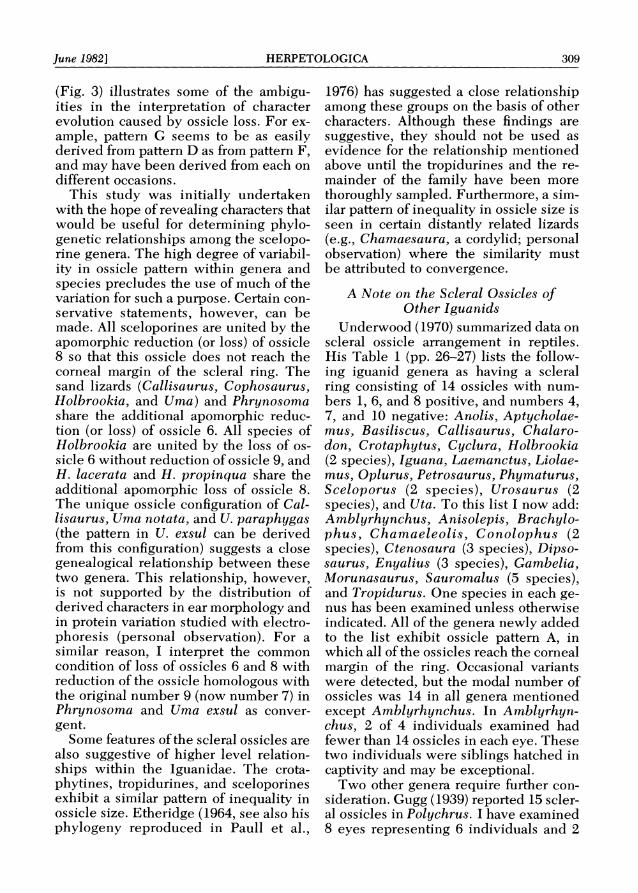
This study revealed nine patterns in the scleral rings of sceloporines and other iguanid lizards (see also Underwood, 1970). These patterns are as follows:
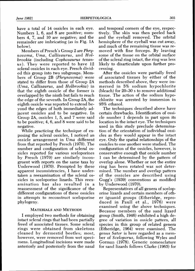
Pattern A.-14 ossicles: 1 , 6 , 8 positive; 4, 7, 10 negative; all ossicles contribute to the corneal margin of the ring (Fig. 1A).
Pattern B.-14 ossicles: 1 , 6 , 8 positive; 4 ,7 ,10 negative; number 8 reduced so as not to reach the corneal margin (Fig. 1B).
Pattern C.-14 ossicles: 1, 6, 8 posi- tive; 4, 7, 10 negative; ossicles 6 and 8 reduced so as not to reach the corneal margin (Fig. 1C).
Pattern 0.-14 ossiclss: 1, 6, 8 posi- tive; 4, 7, 10 negative; numbers 6, 8, and 9 reduced so as to be excluded from the corneal margin. Number 8 is usually the smallest, followed by 6, then 9 (Fig. ID).
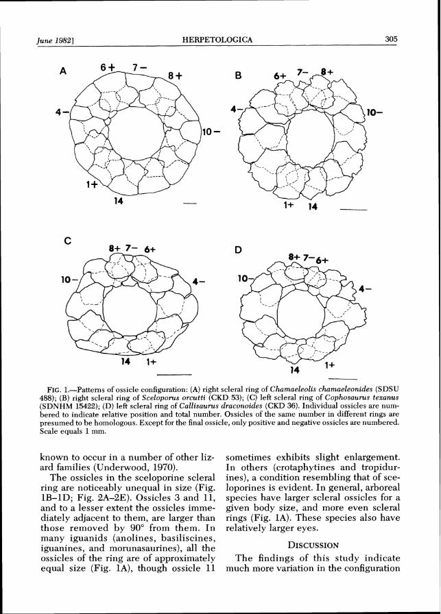
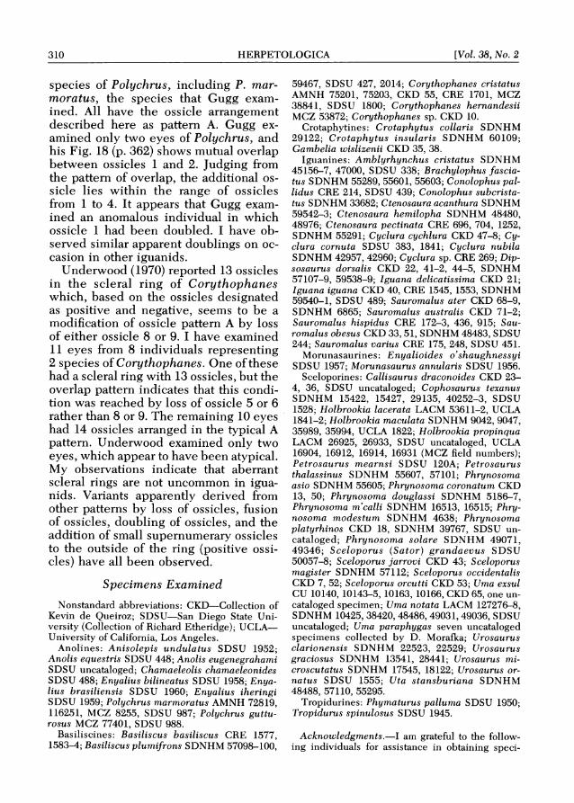
Pattern E.-13 ossicles: 1 , 6 , 8 positive; 4 , 7 , 9 negative; 6 and 8 reduced so as not to reach the corneal margin. Number 6 is smaller than 8 (Fig. 2A).
Pattern F.-13 ossicles: 1 , 5 , 7 positive; 4, 6, 9 negative; 7 reduced so as to be excluded from the corneal margin (Fig. 2B).
Pattern G.-13 ossicles: 1, 5 , 7 posi- tive; 4, 6, 9 negative; neither 7 nor 8 reaches the corneal margin. Number 7 is smaller than number 8 (Fig. 2C).
Pattern H.-12 ossicles: 1, 5, 7 posi- tive; 4 , 6 , 8 negative; all reach the corneal margin (Fig. 2D).
Pattern 1.-12 ossicles: 1, 5 , 7 positive; 4, 6, 8 negative; 7 reduced and excluded from the corneal margin (Fig. 2E).
The distribution of these ossicle pat- terns in various sceloporine taxa is pre- sented in Table 1.
Using the modal number of eyes for each taxon, the sceloporine taxa in Table 1can be characterized by ossicle pattern as follows: pattern B: Petrosaurus, Sce- loporus, Urosaurus, and Uta; pattern C: Cophosaurus; pattern D: Callisaurus, Uma notata (including races formerly recognized as U. inornata, U, notata, and U . scoparia now included within a single species [Adest, 1977; Zalusky e t al., 19801), and U. paraphygas; pattern F: Holbrookia maculata; pattern H: Hol- brookia lacerata and H. propinqua; pat- tern I: Phrynosoma and Uma exsul. None of these taxa exhibits a statistical mode for ossicle pattern A, E, or G, although pattern A is the most common pattern in iguanids other than sceloporines and is
305 June 19821 HERPETOLOGICA
FIG.1.-Patterns of ossicle configuration: (A) right scleral ring of Chatnaeleolis chamaeleonides (SDSU 488); ( B ) right scleral ring of Sceloporus orcutti (CKD 53); ( C ) left scleral ring of Cophosaurus texanus (SDNHM 15422); (D) left scleral ring of Callisaurus draconoides (CKD 36). Individual ossicles are num- bered to indicate relative position and total number. Ossicles of the same number in different rings are presumed to be homologous. Except for the final ossicle, only positive and negative ossicles are numbered. Scale equals 1mm.
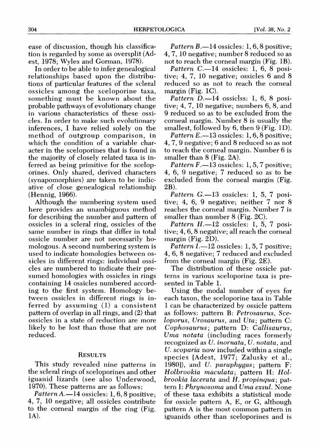
known to occur in a number of other liz- sometimes exhibits slight enlargement. ard families (Underwood, 1970). In others (crotaphytines and tropidur-
The ossicles in the sceloporine scleral ines), a condition resembling that of sce- ring are noticeably unequal in size (Fig. loporines is evident. In general, arboreal 1B-1D; Fig. 2A-2E). Ossicles 3 and 11, species have larger scleral ossicles for a and to a lesser extent the ossicles imme- given body size, and more even scleral diately adjacent to them, are larger than rings (Fig. 1A). These species also have those removed by 90" from them. I n relatively larger eyes. many iguanids (anolines, basiliscines, iguanines, and morunasaurines), all the DISCUSSION ossicles of the ring are of approximately The findings of this study indicate equal size (Fig. IA), though ossicle 11 much more variation in the configuration
306 HERPETOLOGICA [Vol. 38, No. 2
FIG, 2.-Patterns of ossicle configuration: (A) left scleral ring of Callisaurus draconoides (CKD 24); (B) left scleral ring of Holbrookia maculata (SDNHM 35994); (C) right scleral ring of Uma exsul (CU 10163); (D) right scleral ring of Holbrookia lacerata (UCLA 1842); (E) left scleral ring of Phrynosoma coronatum (CKD 13). Individual ossicles are numbered to indicate relative position and total number as in Fig. 1 (nonparenthetical), and to indicate presumed homologies with ossicles in rings with 14 ossicles where these numbers differ from the former (parenthetical). Except for the final ossicle, only positive and negative ossicles are numbered. Scale equals 1mm.
307 June 19821 HERPETOLOGICA
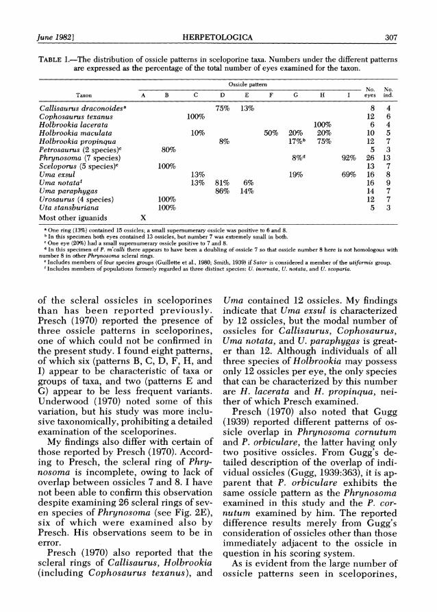
TABLE1.-The distribution of ossicle patterns in sceloporine taxa. Numbers under the different patterns are expressed as the percentage of the total number of eyes examined for the taxon.
Ossicle pattern No. No.
C D E F G H I eyes ind.Taxon A B
Callisaurus draconoidesa Cophosaurus texanus Holbrookia lacerata Holbrookia maculata Holbrookia propinqua Petrosaurus (2 specie^)^ Phrynosoma (7 species) Sceloporus (5 species)' Uma exsul Uma notataf Uma paraphygas Urosaurus (4 species) Uta stansburiana Most other iguanids X
" O n e ring (13%) contained 15 ossicles; a small supernumerary ossicle was positive to 6 and 8. In this specimen both eyes contained 13 ossicles, but number 7 was extremely small in both. One eye (20%)had a small supernumerary ossicle positive to 7 and 8.
* I n this specimen of P. m'calli there appears to have been a doubling of ossicle 7 so that ossicle number 8 here is not homologous with number 8 in other Phrynosoma scleral rings.
"Includes members of four species groups (Guillette et al., 1980; Smith, 1939) if Sator is considered a member of the uttjormis group. 'Includes members of populations formerly regarded as three distinct species: U. inornata, U. notata, and U. scoparia.
of the scleral ossicles in sceloporines than has been reported previously. Presch (1970) reported the presence of three ossicle patterns in sceloporines, one of which could not be confirmed in the present study. I found eight patterns, of which six (patterns B, C, D, F, H, and I) appear to be characteristic of taxa or groups of taxa, and two (patterns E and G) appear to be less frequent variants. Underwood (1970) noted some of this variation, but his study was more inclu- sive taxonomically, prohibiting a detailed examination of the sceloporines.
My findings also differ with certain of those reported by Presch (1970). Accord- ing to Presch, the scleral ring of Phry-nosoma is incomplete, owing to lack of overlap between ossicles 7 and 8. I have not been able to confirm this observation despite examining 26 scleral rings of sev- en species of Phrynosoma (see Fig. 2E), six of which were examined also by Presch. His observations seem to be in error.
Presch (1970) also reported that the scleral rings of Callisaurus, Holbrookia (including Cophosaurus texanus), and
Uma contained 12 ossicles. My findings indicate that Uma exsul is characterized by 12 ossicles, but the modal number of ossicles for Callisaurus, Cophosaurus, Uma notata, and U. paraphygas is great- er than 12. Although individuals of all three species of Holbrookia may possess only 12 ossicles per eye, the only species that can be characterized by this number are H. lacerata and H. propinqua, nei-ther of which Presch examined.
Presch (1970) also noted that Gugg (1939) reported different patterns of os- sicle overlap in Phrynosoma cornutum and P. orbiculare, the latter having only two positive ossicles. From Gugg's de- tailed description of the overlap of indi- vidual ossicles (Gugg, 1939:363), it is ap- parent that P. orbiculare exhibits the same ossicle pattern as the Phrynosoma examined in this study and the P. cor- nutum examined by him. The reported difference results merely from Gugg's consideration of ossicles other than those immediately adjacent to the ossicle in question in his scoring system.
As is evident from the large number of ossicle patterns seen in sceloporines,

308 HERPETOLOGICA [Vol. 38, No. 2
loss o f 6
- 1 l o s s o f 8
reduct ion of 9 ~ g / / l s a u r u ~
further r e d u c t i o n / ; y . t a \ Uma peraphygas
C
, 44 Phrynosoma
Uma exsul '1 G reduction
I/ reduction o f 9
l o s s Of 8
fB e n l a r g e m e n t o f 9 Petrosaurus
Sceloporus ",F ,H
Holbrookia Holbrookia l ace ra te U t a
macu la ta Holbrookla proplnqua
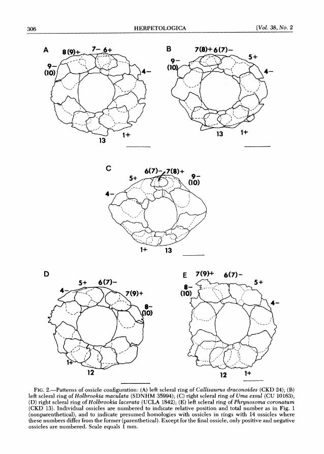
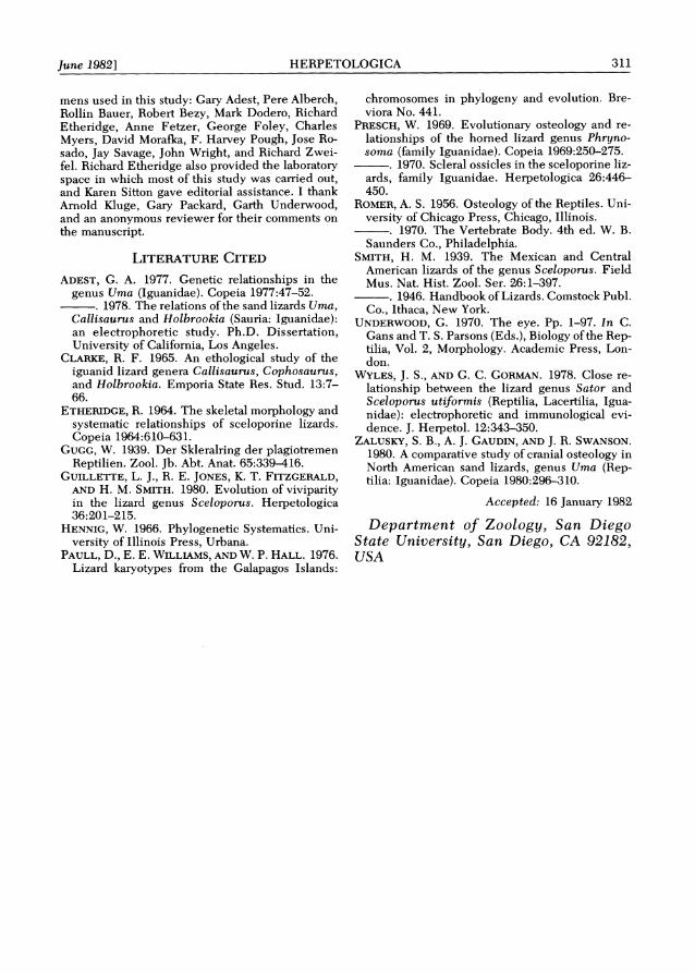
FIG. 3.-Hypothetical evolutionary scheme for patterns of scleral ossicle configuration in sceloporines (see Figs. 1and 2). Sceloporine taxa are listed under the ossicle pattern for which they exhibit a statistical mode. A double line signifies that the evolutionary step is supported by variation within taxa (Table 1). This scheme of character evolution is not to be interpreted as a phylogeny for the taxa, because conver- gence in ossicle pattern probably occurs in certain cases. Here each ossicle number refers to the number of the homologous ossicle in the 14-ossicle ring.
evolution has been relatively rapid in this character complex when compared with the rest of the family Iguanidae, nearly all of which possess pattern A. Be-cause ossicle pattern A is widespread in the Iguanidae and is known to occur in other lizard families, I regard this pattern as primitive for the Iguanidae and the precursor to those arrangements occur- ring in sceloporines. Furthermore, all patterns seen in sceloporines are deriv- able from pattern A by simple reduction or loss of ossicles in the dorsal portion of the scleral ring.
Figure 3 gives a hypothetical character phylogeny for the different ossicle pat- terns found in sceloporine iguanids. The evolutionary steps in this figure are in- tended to be the simplest changes re-
quired to derive one ossicle pattern from another. Variation in ossicle pattern with- in taxa (Table 1)supports some of these steps (D-E, C-F, F-G, G-H, F-H, D-G, G-I, and C-D), but not others.
Inequality in ossicle size within a scleral ring may be a necessary precursor to the loss of ossicles 6 and 8 in scelopo- rines. Scleral rings with relatively small ossicles in their dorsal series exhibit greater overlap between ossicles that are not immediate neighbors than do more even rings: compare ossicles 5, 7, and 9 in Fig. 1B-1D with the same ossicles in Fig. 1A. Thus, when ossicle 6 or 8 is lost, the continuity of the ring is preserved, and ossicle 5 or 9, respectively, changes from imbricating to positive.
The hypothetical character phylogeny
309 lune 19821 HERPETOLOGICA
(Fig. 3) illustrates some of the ambigu- ities in the inter~retation of character evolution caused by ossicle loss. For ex- ample, pattern G seems to be as easily derived from pattern D as from pattern F, and mav have been derived from each on different occasions.
This study was initially undertaken with the hope of revealing characters that would be useful for detennining phylo- genetic relationships among the scelopo- rine genera. The high degree of variabil- ity in ossicle pattern within genera and species precludes the use of much of the variation for such a purpose. Certain con- servative statements, however, can be made. All sceloporines are united by the apomorphic reduction (or loss) of ossicle 8 so that this ossicle does not reach the corneal margin of the scleral ring. The sand lizards (Callisaurus, Cophosaurus, Holbrookia, and U m a ) and Phrynosoma share the additional apomorphic reduc- tion (or loss) of ossicle 6. All species of Holbrookia are united by the loss of os- sicle 6 without reduction of ossicle 9, and H. laceruta and H. propinqua share the additional apomorphic loss of ossicle 8. The unique ossicle configuration of Cal-lisaurus, Uma notata, and U. paraphygas (the pattern in U . exsul can be derived from this configuration) suggests a close genealogical relationship between these two genera. This relationship, however, is not supported by the distribution of derived characters in ear morphology and in protein variation studied with electro- phoresis (personal observation). For a similar reason, I interpret the common condition of loss of ossicles 6 and 8 with reduction of the ossicle homologous with the original number 9 (now number 7) in Phrynosoma and Uma exsul as conver- gent.
Some features of the scleral ossicles are also suggestive of higher level relation- ships within the Iguanidae. The crota- phytines, tropidurines, and sceloporines exhibit a similar pattern of inequality in ossicle size. Etheridge (1964, see also his phylogeny reproduced in Paul1 e t al.,
1976) has suggested a close relationship among these groups on the basis of other characters. Although these findings are suggestive, they should not be used as evidence for the relationship mentioned above until the tropidurines and the re- mainder of the family have been more thoroughly sampled. Furthermore, a sim- ilar pattern of inequality in ossicle size is seen in certain distantly related lizards (e.g., Chamaesaura, a cordylid; personal observation) where the similarity must be attributed to convergence.
A Note on the Scleral Ossicles of Other Iguanids
Underwood (1970) summarized data on scleral ossicle arrangement in reptiles. His Table 1 (pp. 26-27) lists the follow- ing iguanid genera as having a scleral ring consisting of 14 ossicles with num- bers 1, 6, and 8 positive, and numbers 4, 7, and 10 negative: Anolis, Aptycholae- mus , Basiliscus, Callisaurus, Chalaro- don, Crotaphytus, Cyclura, Holbrookia (2 species), Iguana, Laemanctus, Liolae- mus , Oplurus, Petrosaurus, Phymaturus, Sceloporus ( 2 species), Urosaurus (2 species), and Uta. To this list I now add: Ambl yrhynchus, Anisolepis, Brachylo- p h u s , C h a m a e l e o l i s , C o n o l o p h u s (2 species), Ctenosaura (3 species), Dipso-saurus, Enyalius (3 species), Gambelia, Morunasaurus, Sauromalus (5 species), and Tropidurus. One species in each ge- nus has been examined unless otherwise indicated. All of the genera newly added to the list exhibit ossicle pattern A, in which all of the ossicles reach the corneal margin of the ring. Occasional variants were detected, but the modal number of ossicles was 14 in all genera mentioned except Amblyrhynchus. In Amblyrhyn-chus, 2 of 4 individuals examined had fewer than 14 ossicles in each eye. These two individuals were siblings hatched in captivity and may be exceptional.
Two other genera require further con- sideration. Gugg (1939) reported 15scler-a1 ossicles in Polychrus. I have examined 8 eyes representing 6 individuals and 2
310 HERPETOILOGICA [Val. 38, No. 2
species of Polychrus, including P. mar- moratus, the species that Gugg exam-ined. All have the ossicle arrangement described here as pattern A. Gugg ex- amined only two eyes of Polychrus, and his Fig. 18 (p. 362) shows mutual overlap between ossicles 1 and 2. Judging from the pattern of overlap, the additional os- sicle lies within the range of ossicles from 1 to 4. It appears that Gugg exam- ined an anomalous individual in which ossicle 1 had been doubled. I have ob- served similar apparent doublings on oc- casion in other iguanids.
Underwood (1970) reported 13 ossicles in the scleral ring of Corythophanes which, based on the ossicles designated as positive and negative, seems to be a modification of ossicle pattern A by loss of either ossicle 8 or 9. I have examined 11 eyes from 8 individuals representing 2 species of Corythophanes. One of these had a scleral ring with 13 ossicles, but the overlap pattern indicates that this condi- tion was reached by loss of ossicle 5 or 6 rather than 8 or 9. The remaining 10 eyes had 14 ossicles arranged in the typical A pattern. Underwood examined only two eyes, which appear to have been atypical. My observations indicate that aberrant scleral rings are not uncommon in igua- nids. Variants apparently derived from other patterns by loss of ossicles, fusion of ossicles, doubling of ossicles, and the addition of small supernumerary ossicles to the outside of the ring (positive ossi- cles) have all been observed.
Specimens Examined
Nonstandard abbreviations: CKD-Collection o f Kevin de Queiroz; SDSU-San Diego State Uni- versity (Collection o f Richard Etheridge); UCLA- University o f California, Los Angeles.
Anolines: Anisolepis undulatus SDSU 1952; Anolis equestris SDSU 448; Anolis eugenegrahami SDSU uncataloged; Chamaeleolis chamaeleonides SDSU 488; Enyalius bilineatus SDSU 1958; Enya-lius brasiliensis SDSU 1960; Enyalius iheringi SDSU 1959; Polychrus marmoratus AMNH 72819, 116251, MCZ 8255, SDSU 987; Polychrus guttu- rosus MCZ 77401, SDSU 988.
Basiliscines: Basiliscus basiliscus C R E 1577, 1583-4;Basiliscus plumifrons SDNHM 57098-100,
59467, SDSU 427, 2014; Corythophanes cristatus AMNH 75201, 75203, C K D 55, CRE 1701, MCZ 38841, SDSU 1800; Corythophanes hernandesii MCZ 53872; Corythophanes sp. C K D 10.
Crotaphytines: Crotaphytus collaris SDNHM 29122; Cro taphy tus insularis S D N H M 60109; Gambelia wislizenii C K D 35, 38.
Iguanines: Amblyrhynchus cristatus SDNHM 45156-7, 47000, SDSU 338; Brachylophus fascia- tus SDNHM 55289,55601,55603;Conolophus pal- lidus CRE 214, SDSU 439; Conolophus subcrista- tus SDNHM 33682; Ctenosaura acanthura SDNHM 59542-3; Ctenosaura hemilopha SDNHM 48480, 48976; Ctenosaura pectinata CRE 696, 704, 1252, SDNHM 55291; Cyclura cychlura C K D 47-8; Cy-clura cornuta SDSU 383, 1841; Cyclura nubila SDNHM 42957,42960; Cyclura sp. CRE 269; Dip-sosaurus dorsalis C K D 22, 41-2, 44-5, SDNHM 57107-9, 59538-9; Iguana delicatissima C K D 21; Iguana iguana C K D 40, CRE 1545, 1553, SDNHM 59540-1, SDSU 489; Sauromalus ater CKD 68-9, SDNHM 6865; Sauromalus australis C K D 71-2; Sauromalus hispidus CRE 172-3, 436, 915; Sau-romalus obesus C K D 33,51, SDNHM 48483, SDSU 244; Sauromalus varius CRE 175, 248, SDSU 451.
Morunasaurines: Enyalioides o'shaughnessyi SDSU 1957; Morunasaurus annularis SDSU 1956.
Sceloporines: Callisaurus draconoides C K D 23-4, 36, SDSU uncataloged; Cophosaurus texanus SDNHM 15422, 15427, 29135, 40252-3, SDSU 1528; Holbrookia Eacerata LACM 53611-2, U C L A 1841-2; Holbrookia maculata SDNHM 9042,9047, 35989, 35994, U C L A 1822; Holbrookia propinqua LACM 26925, 26933, SDSU uncataloged, U C L A 16904, 16912, 16914, 16931 ( M C Z field numbers); Petrosaurus mearnsi SDSU 120A; Petrosaurus thalassinus SDNHM 55607, 57101; Phrynosoma asio SDNHM 55605; Phrynosoma coronatum CKD 13, 50; Phrynosoma douglassi SDNHM 5186-7, Phrynosoma m'calli SDNHM 16513, 16515; Phry-nosoma modes tum SDNHM 4638; Phrynosoma platyrhinos C K D 18, SDNHM 39767, SDSU un-cataloged; Phrynosoma solare S D N H M 49071, 49346; Sceloporus ( S a t o r ) grandaecus SDSU 50057-8; Sceloporus jarroui C K D 43; Sceloporus magister SDNHM 57112; Sceloporus occidentalis C K D 7, 52; Sceloporus orcutti C K D 53; Uma exsul CU 10140,10143-5,10163,10166, C K D 65, one un- cataloged specimen; Uma notata LACM 127276-8, SDNHM 10425,38420,48486,49031,49036,SDSU uncataloged; Uma paraphygas seven uncataloged specimens collected b y D . Morafka; Urosaurus clarionensis S D N H M 22523, 22529; Urosaurus graciosus SDNHM 13541, 28441; Urosaurus mi- croscutatus SDNHM 17545, 18122; Urosaurus or- natus SDSU 1555; Uta stansburiana S D N H M 48488, 57110, 55295.
Tropidurines: Phymaturus palluma SDSU 1950; Tropidurus spinulosus SDSU 1945.
Acknowledgments.-I am grateful to the follow- ing individuals for assistance i n obtaining speci-
311 June 19821 HERPETOLOGICA
mens used in this study: Gary Adest, Pere Alberch, Rollin Bauer, Robert Bezy, Mark Dodero, Richard Etheridge, Anne Fetzer, George Foley, Charles Myers, David Morafka, F. Harvey Pough, Jose Ro- sado, Jay Savage, John Wright, and Richard Zwei- fel. Richard Etheridge also provided the laboratory space in which most of this study was carried out, and Karen Sitton gave editorial assistance. 1 thank Arnold Kluge, Gary Packard, Garth Underwood, and an anonymous reviewer for their comments on the manuscript.
ADEST, G. A. 1977. Genetic relationships in the genus Uma (Iguanidae). Copeia 1977:47-52.
-. 1978. The relations of the sand lizards Uma, Callisaurus and Holbrookia (Sauria: Iguanidae): an electrophoretic study. Ph.D. Dissertation, University of California, Los Angeles.
CLARKE, R. F. 1965. An ethological study of the iguanid lizard genera Callisaurus, Cophosaurus, and Holbrookia. Emporia State Res. Stud. 13:7- 66.
ETHERIDGE,R. 1964. The skeletal morphology and systematic relationships of sceloporine lizards. Copeia 1964:610-631.
GUGG, W. 1939. Der Skleralring der plagiotremen Reptilien. Zool. Jb. Abt. Anat. 65:339-416.
GUILLETTE, L. J., R. E. JONES, K. T. FITZGERALD, AND H. M. SMITH. 1980. Evolution of viviparity in the lizard genus Sceloporus. Herpetologica 36:201-215.
HENNIG,W. 1966. Phylogenetic Systematics. Uni- versity of Illinois Press, Urbana.
PAULL, D., E. E. WILLIAMS, AND W. P. HALL. 1976. Lizard karyotypes from the Galapagos Islands:
chromosomes in phylogeny and evolution. Bre- viora No. 441.
PRESCH, W. 1969. Evolutionary osteology and re- lationships of the horned lizard genus Phryno-soma (family Iguanidae). Copeia 1969:250-275.
. 1970. Scleral ossicles in the scelo~orine liz- ards, family Iguanidae. Herpetologica 26:446- 451).
ROMER,A. S. 1956. Osteology of the Reptiles. Uni- versity of Chicago Press, Chicago, Illinois.
-. 1970. The Vertebrate Body. 4th ed. W. B. Saunders Co., Philadelphia.
SMITH, H. M. 1939. The Mexican and Central American lizards of the genus Sceloporus. Field Mus. Nat. Hist. Zool. Ser. 26:l-397. -1946. Handbook of Lizards. Comstock Publ.
Co., Ithaca, New York. UNDERWOOD,G. 1970. The eye. Pp. 1-97. In C.
Gans and T. S. Parsons (Eds.), Biology ofthe Rep- tilia, Vol. 2, Morphology. Academic Press, Lon- don.
WYLES, J. S., AND G. C. CORMAN. 1978. Close re- lationship between the lizard genus Sator and Sceloporus utijormis (Reptilia, Lacertilia, Igua- nidae): electrophoretic and immunological evi- dence. J. Herpetol. 12:343-350.
ZALUSKY,S. B., A. J. GAUDIN,AND J. R. SWANSON. 1980. A comparative study of cranial osteology in North American sand lizards, genus Uma (Rep-tilia: Iguanidae). Copeia 1980:296-310.
Accepted: 16 January 1982
Department of Zoology, Sun Diego State University, Sun Diego, C A 92182, U S A