The role(s) of calcium ions in phytochrome action
21
Phorochrrni.stry mid Photobiology Vol. 54, No. 6, pp. 1135-1155, 1991 Printed in Great Britain. All rights reserved 003 1 -XhS5/9 I $03.00+0.00 Copyright 0 1W1 Pcrgamon Press plc YEARLY REVIEW THE ROLE(S) OF CALCIUM IONS IN PHYTOCHROME ACTION Introduction Phytochrome, the red (R)*- and far-red light (FR)- absorbing morphogenetic photoreceptor, which occurs throughout the Plant Kingdom, was disco- vered by scientists at the US Department of Agricul- ture, Beltsville, MD. This year is the 40th anniver- sary of the prediction of the R/FR reversible pigment in plants and more than 30 years since its first spectroscopic detection. During the last four decades extensive progress has been made in under- standing the molecular structure and function of phytochrome (for review see Furuya, 1987). Phy- tochrome genes have now been cloned for a few plant species (Quail et al., 1987; Furuya, 1989; Shar- rock and Quail, 1989). The photoregulation of genes, including the phytochrome gene itself, has been extensively studied (Quail et a/., 1987; Nagy et a/., 1988; Furuya, 1989; Tomizawa et a[., 1990). However, the molecular mechanism of phyto- chrome action is still obscure. Multiple response types could be an indication of different modes of action of the photoreceptor (Jordan et al., 1986; Kronenberg and Kendrick. 1986; Schafer et al., 1986; Furuya, 1989). One attractive hypothesis is that calcium ions (Ca2+) participate as a second messenger (Roux, 1984). During the last decade many papers have been published about the involve- ment of Ca” in the regulation of different phyto- chrome-regulated processes. Some o’f them were reviewed by Roux et 121. in 1986. Since that time *Abbreviations: AC, adenylate cyclase; Ca:!+, calcium ion; [Ca”]‘, cytoplasmic calcium ion; CaBP, calcium-hind- ing physode; CPZ, chlorpromazine; DAG, 1,2-diacygly- cerol; DB-CAMP, dibuthyryl CAMP: DHP, dihydroxy- pyridine; EDTA, ethylenediaminetelraacetic acid; EGTA, ethyleneglycol-his-(P-aminoethyl ether)-N,N, N’,N-tetraacetic acid; FR, far-red light; GA, gihherel- lin; H,, 1-(5-isoquinoIine sulfonyl)-2-mt.thylpiperazinc; I,,,, calcium channel currcnt; IP,, inositol 1-phosphate; IP,, inositol 1,4-bisphosphate; IP,, inositol 1,4,5-tris- phosphate; OAG, 1-oleoyl-2-acetyl-sn-glycerol; PDE, phosphodiesterase; PEG, polyethylene glycol; Pfr, FR- absorbing form of phytochrome; PI, phasphatidylinosi- tol; PIP2, phosphatidylinositol 4,s-hisphosphate; PKA, protein kinase A; PKC, protein kinase C; PLA,, phos- pholipase A:; PLC, phospholipase C; PMA, phorbol 12-myristate 13-acetate; Pr, R-absorbing form of phyto- chrome; R. red light; TFP, triHuoperazine; TMB-8,3,4, 5-trimethoxy-benzoic acid 8-(diethylamino)-octyl ester; W,, N-(6-amonohexyl)-l-naphthalenesulfonamide; W,, N-(h-aminohexyl)-5-chloro-1-naphthalenesulfonamide. new and sometimes contradictory data have been published and this review is an update of knowledge concerning participation of Ca’+ in phytochrome action. The basic evidence for participation of Ca2 ’ in phytochrome action in different plant systems is reviewed and possible mechanisms of action of Ca’ ’ in plant photomorphogenesis are discussed. Participation of’ Ca’ in Phytochrome Action in Different Plant Systems Biopotentials The plasma membrane of some plants is electri- cally excitable. In a few lower-plant species light can trigger an “action potential” which appears to be an all-or-none response when the stimulus exceeds a certain threshold value (Trqbacz, 1989 and references therein). In isolated internodes of the green algae Nitel- lopsis sp., Nitella opaca AG. and Nitel1nflexili.v (L.) AG., R (fluence 8.4 W m-*) caused depolarization of their plasma membrane, which had a mean lag- time of 1.7 * 0.3 s and reached a steady state within 1-2 min upon continuous irradiation (Weisenseel and Ruppert, 1077). Repolarization of the mem- brane occurred a few seconds after the light was turned off and was accelerated by FR applied immediately after the R pulse. The magnitude of R-triggered membrane depolarization in the algal cells was dependent upon the [Ca”] of the external medium. In Ca2+ -free medium the depolarization was only about 1 mV, whereas in the presence of 5 mM Ca2’ a mean depolarization of approximately 33 mV was recorded (Weisenseel and Ruppert, 1977). Red and FR change the mobility of the uni- cellular green alga Mesotaenium caldariorum in an electric field. The Zeta-potential. indicating the sur- face charge of the cells, was calculated from the velocity of the movement (Stenz and Weisenseel, 1986). After FR irradiation the electrophoretic mobility of the cells was faster and the Zeta-poten- tial more negative than after R irradiation. There was no change in Zeta-potential after R and FR treatment in the presence of 1 mM ethyleneglycol- bis-(P-aminoethyl ether)-N,N,N’,N’-tetraacetic acid (EGTA). The R effect on the Zeta-potential was also reduced by 10 p.M verapamil to the level of the FR control. From these data, Stenz and Weisenseel (1986) argued that phytochrome increases Ca2 ’ influx into the algal cells. In this way phytochrome 1135
-
Upload
independent -
Category
Documents
-
view
0 -
download
0
Transcript of The role(s) of calcium ions in phytochrome action

Phorochrrni.stry mid Photobiology Vol. 54, No. 6 , pp. 1135-1155, 1991 Printed in Great Britain. All rights reserved
003 1 -XhS5/9 I $03.00+0.00 Copyright 0 1W1 Pcrgamon Press plc
YEARLY REVIEW
THE ROLE(S) OF CALCIUM IONS IN PHYTOCHROME ACTION
Introduction
Phytochrome, the red (R)*- and far-red light (FR)- absorbing morphogenetic photoreceptor, which occurs throughout the Plant Kingdom, was disco- vered by scientists at the US Department of Agricul- ture, Beltsville, MD. This year is the 40th anniver- sary of the prediction of the R/FR reversible pigment in plants and more than 30 years since its first spectroscopic detection. During the last four decades extensive progress has been made in under- standing the molecular structure and function of phytochrome (for review see Furuya, 1987). Phy- tochrome genes have now been cloned for a few plant species (Quail et al . , 1987; Furuya, 1989; Shar- rock and Quail, 1989). The photoregulation of genes, including the phytochrome gene itself, has been extensively studied (Quail et a / . , 1987; Nagy et a / . , 1988; Furuya, 1989; Tomizawa et a[., 1990). However, the molecular mechanism of phyto- chrome action is still obscure. Multiple response types could be an indication of different modes of action of the photoreceptor (Jordan et al., 1986; Kronenberg and Kendrick. 1986; Schafer et al., 1986; Furuya, 1989). One attractive hypothesis is that calcium ions (Ca2+) participate as a second messenger (Roux, 1984). During the last decade many papers have been published about the involve- ment of Ca” in the regulation of different phyto- chrome-regulated processes. Some o’f them were reviewed by Roux et 121. in 1986. Since that time
*Abbreviations: A C , adenylate cyclase; Ca:!+, calcium ion; [Ca”]‘, cytoplasmic calcium ion; CaBP, calcium-hind- ing physode; CPZ, chlorpromazine; D A G , 1,2-diacygly- cerol; DB-CAMP, dibuthyryl CAMP: D H P , dihydroxy- pyridine; EDTA, ethylenediaminetelraacetic acid; EGTA, ethyleneglycol-his-(P-aminoethyl ether)-N,N, N’,N-tetraacetic acid; FR, far-red light; G A , gihherel- lin; H,, 1-(5-isoquinoIine sulfonyl)-2-mt.thylpiperazinc; I , , , , calcium channel currcnt; I P , , inositol 1-phosphate; IP,, inositol 1,4-bisphosphate; IP,, inositol 1,4,5-tris- phosphate; O A G , 1-oleoyl-2-acetyl-sn-glycerol; PDE, phosphodiesterase; PEG, polyethylene glycol; Pfr, FR- absorbing form of phytochrome; PI, phasphatidylinosi- tol; PIP2, phosphatidylinositol 4,s-hisphosphate; PKA, protein kinase A; PKC, protein kinase C; PLA,, phos- pholipase A:; PLC, phospholipase C ; PMA, phorbol 12-myristate 13-acetate; Pr, R-absorbing form of phyto- chrome; R. red light; TFP, triHuoperazine; TMB-8,3,4, 5-trimethoxy-benzoic acid 8-(diethylamino)-octyl ester; W,, N-(6-amonohexyl)-l-naphthalenesulfonamide; W,, N-(h-aminohexyl)-5-chloro-1-naphthalenesulfonamide.
new and sometimes contradictory data have been published and this review i s an update of knowledge concerning participation of Ca’+ in phytochrome action. The basic evidence for participation of Ca2 ’ in phytochrome action in different plant systems is reviewed and possible mechanisms of action of Ca’ ’ in plant photomorphogenesis are discussed.
Participation of’ Ca’ in Phytochrome Action in Different Plant Systems
Biopotentials
The plasma membrane of some plants is electri- cally excitable. In a few lower-plant species light can trigger an “action potential” which appears to be an all-or-none response when the stimulus exceeds a certain threshold value (Trqbacz, 1989 and references therein).
In isolated internodes of the green algae Nitel- lopsis sp., Nitella opaca AG. and Nitel1nflexili.v (L.) AG. , R (fluence 8.4 W m-*) caused depolarization of their plasma membrane, which had a mean lag- time of 1.7 * 0.3 s and reached a steady state within 1-2 min upon continuous irradiation (Weisenseel and Ruppert, 1077). Repolarization of the mem- brane occurred a few seconds after the light was turned off and was accelerated by FR applied immediately after the R pulse. The magnitude of R-triggered membrane depolarization in the algal cells was dependent upon the [Ca”] of the external medium. In Ca2+ -free medium the depolarization was only about 1 mV, whereas in the presence of 5 mM Ca2’ a mean depolarization of approximately 33 mV was recorded (Weisenseel and Ruppert, 1977). Red and FR change the mobility of the uni- cellular green alga Mesotaenium caldariorum in an electric field. The Zeta-potential. indicating the sur- face charge of the cells, was calculated from the velocity of the movement (Stenz and Weisenseel, 1986). After FR irradiation the electrophoretic mobility of the cells was faster and the Zeta-poten- tial more negative than after R irradiation. There was no change in Zeta-potential after R and FR treatment in the presence of 1 mM ethyleneglycol- bis-(P-aminoethyl ether)-N,N,N’,N’-tetraacetic acid (EGTA). The R effect on the Zeta-potential was also reduced by 10 p.M verapamil to the level of the FR control. From these data, Stenz and Weisenseel (1986) argued that phytochrome increases Ca2 ’ influx into the algal cells. In this way phytochrome
1135

1136 ANDRZEJ TRETKN et al.
may stimulate depolarization of the plasma mem- brane andlor secretion of less negatively or posi- tively charged material to the surface of the cells.
Protoplast swelling
In 1983, Blakeley et al. published the first paper on phytochrome-controlled swelling of protoplasts, obtained from etiolated cereal leaves. Protoplasts, isolated from etiolated primary wheat (Tritium aes- tivum L.) leaves (Blakeley et ul., 1983; Bossen et al., 1988), oat (Avena sativa L.) (Kim et al., 1986; Chung et al., 1988), maize (Zea mays L) (Zhou et al., 1990) and barley (Hordeurn vufgare L.) (U et al., 1991), maintained at constant osmotic potential swelled upon R irradiation. The effect of R on protoplast swelling was photoreversible by sub- sequent FR, indicating the involvement of phyto- chrome (Blakeley et af., 1983; Kim et al., 1986; Bossen et al., 1988; Zhou et al. , 1990; U et al., 1991). Besides this effect via phytochrome, proto- plasts also swelled in darkness after treatment with plant growth subtances: gibberellic acid (Blakeley et al., 1983; Chung et al., 1988; Bossen et al., 1991; U et al., 1991), indole-3-acetic acid, cr-naphtha- leneacetic acid, banzylaminopurine, abscisic acid (Bossen et al., 1991), acetylcholine (Tretyn et al., 1990a,d; Bossen et al. , 1991) and CAMP (Kim et al., 1986; Chung et al . , 1988; Bossen et al., 1990; U et al., 1991). At 15°C protoplasts started to swell almost immediately after a R pulse and reached their maximum volume after 10 min in darkness at 15°C (Bossen et al., 1988). Maize mesophyll proto- plasts began to swell 5 min after R irradiation and increased by about 30% in comparison to the green safe light control (Zhou et al., 1990). However, at 4°C the process only reached completion after 12 h incubation (Chung et al . , 1988).
In some cases, R-induced protoplast swelling appeared to be stimulated in the presence of K+ (Blakeley et al., 1983) or in an isosmotic modified Murashige-Skoog’s nutrient solution (Kim et al., 1986; Chung et al., 1988). Bossen et al. (1988) found that phytochrome-controlled swelling of etiolated wheat protoplasts had an absolute prerequisite for Ca2+ in the surrounding medium and that the pres- ence or absence of K+ had no influence on the response. Neither Mg2+ nor Ba2+ replaced this requirement for Ca2+. Furthermore, protoplast swelling was stimulated in darkness in the presence of Ca2+ and the calcium ionophore A 23187 (calimycin) (Bossen et al., 1988; U et al., 1991). However, phytochrom-stimulated, Ca2+-dependent swelling of protoplasts failed in a medium containing the chelator EGTA (Bossen et al., 1988; U et al., 1991) or calcium-channel blockers verapamil and La3+ (Bossen et al., 1988). These results strongly suggest that R causes the opening of calcium chan- nels in the plasma membrane. This suggestion has been recently confirmed by Tretyn et al. (1990c),
who found that nifedipine, a very specific calcium- channel blocker, completely prevents the R- induced, Ca2+-dependent swelling of wheat proto- plasts. However, nifedipine is only active when it is added to the medium before a R pulse (Tretyn et al., 1990~). Moreover, as with A 23187, protoplasts swell in darkness in a medium containing Ca2+ and the agonist of the “L-type” animal calcium channels, Bay K-8644 (Tretyn et al . , 1990~). Bossen et al. (1988) and subsequently Tretyn et al. (1990~) pro- posed that a R-induced swelling of protoplasts is stimulated by an increase in cytoplasmic [Ca2+] ([Caz+],). Recently, U et al. (1991) have shown that R increases 45Ca2+ uptake by etiolated barley mesophyll protoplasts and FR overcomes the effect of R, suppressing uptake of this ion. A R pulse increases and FR pulse decreases [Ca2+Ic in etio- lated oat protoplasts (Chae et al. , 1990). The phyto- chrome-controlled transduction chain leading to protoplast swelling has been studied by Bossen et al. (1988, 1990, 1991) and U et al. (1991). A GTP- binding protein (G-protein), phosphatydylinosit- ides, adenosine 3’:5’-cyclic monophosphate (CAMP) and the calcium-binding protein, calrnodulin, could all take part in phytochrome-induced protoplasts swelling. The non-hydrolyzable GTP analogue, gua- nosine-S’-O-(3-thiotriphosphate) (GTP-y-S) added to electroporated protoplasts stimulated their swell- ing in darkness or after a control FR irradiation. After a recovery period this compound was only active when Ca2+ was added to the external medium. Introduction of GDP-P-S by electropor- ation into protoplasts before a R pulse inhibited the phytochrome-stimulated swelling response (Bossen et al., 1990).
The phospholipase C (PLC) inhibitor, neomycin (10 p M ) , as well as lithium chloride (10 pM), an inhibitor of inositol-1-phosphatase, prevented R and GTP-y-S-induced swelling of wheat protoplasts, while having no effect on the FR-irradiated control. The precursor of the phosphoinositide pathway, myo-inositol, had no effect on protoplast volume after R or FR treatment. However, this compound at 1 mM nullifed the inhibitory effect of Li+ on the R-induced swelling response (Bossen et al., 1990). The participation of calmodulin in protoplast swell- ing is unclear. A very specific antagonist of this regulatory protein, N-(6-aminohexyl)-5-chloro-l- naphthalenesulfonamide (W,), stimulated an in- crease in protoplast volume both in darkness or after control FR irradiation and failed to prevent the R-induced response. A non-active analogue of this compound, N-(6-aminohexyl)-l-naphthalene- sulfonamide (W,), used at the same concentration as W7 (10 p.M) failed to affect R or FR irradiated protoplasts (Bossen et al., 1990). These observations were explained on the basis of an indirect effect on the [Ca2+Ic via a calmodulin-activated plasma membrane ATPase being inactivated (Bossen et al., 1990). Effectiveness of the calmodulin inhibitor

Yearly Review 1137
(chlorpromazine [CPZ]) in blocking the R-induced, Ca*+-dependent swelling response could be due to a non-specificity mechanism (Tretyn et a/. , 1940a).
The effect of CAMP and the participation of CAMP-dependent protein kinase C (PKC) in the regulation of phytochrome-controlled protoplast swelling have been described (Kim et a / . , 1986; Chung et a/., 1988; Bossen et a/., 1990; U et al . , 1991). The membrane-permeable analogue of CAMP, dibuthyryl-CAMP (DB-CAMP) (Kim et a/. , 1986; Chung et a / . . 1988; Bossen et a / . , 1990), pro- stacycline, an activator of adenylate cyclase (AC) (Kim et al . , 1986), and phorbol 12-myristate 13- acetate (PMA), an activator of PKC, stimulate swelling of oat or wheat mesophyll protoplasts in darkness or after control FR irradiation. However, R or PMA-induced increase in protciplast volume was completely prevented by the PKC inhibitor, 1- (5-isoquinoline sulfonyl)-2-methylpiperazine) (H,) (Bossen et a/. , 1990). The stimulation of protoplast swelling by DB-CAMP in darkness was not inhibited by neomycin and Lit (Bossen et al., 1990).
Recently, Zhou et al. (1990) have studied the biological significance of phytochrome-mediated swelling of etiolated leaf protoplasts. They com- pared the effect of phytochrome on the swelling of protoplasts isolated from Pisum sati iwn L. (pea) leaves, maize coleoptiles and both etiolated and R pre-irradiated maize leaves. Red light failed to induce swelling of pea leaf and maize coleoptile protoplasts. Furthermore, R failed to stimulate swelling of protoplasts isolated from R-pretreated unrolled maize leaves. However, R stimulated swelling of protoplasts isolated from etiolated maize leaves (Zhou et al., 1990). These authors suggest that phytochrome-stimulated swelling of protoplasts is correlated with the leaf unrolling response and not leaf expansion. Both swelling of etiolated wheat protoplasts and unrolling of the (etiolated leaf material from which protoplasts were isolated are under phytochrome control and only occur if Ca2+ is present in the medium (Tretyn and Kendrick, 1990; for details see below).
Mougeotia chloroplast reorientation
Considerable indirect evidence has accumulated on the role of Caz+ in the sensory transduction pathway from the photoreceptors (phytochrome and blue-light photoreceptor) (Haupt, 1987; Schon- bohm, 1987; Walczak et a / . , 1984) to the terminal response (actin-based chloroplast reorientation) in the green alga Mougeotia (Wagner and Klein, 1981; Grolig and Wagner, 1988; Wagner and Grolig, 1991). Furthermore, calmodulin or a closely related homologue has been isolated from Mougeotia and its involvement in the signalling process has been proposed (Wagner et a/. , 1984). However, pharma- cological studies in different laboratories using cal- modulin inhibitors (trifluoperazine [TFP], W7 and
others) have led to conflicting interpretations with respect to the requirement of calmodulin in this process (Serlin and Roux, 1984; Wagner e t a / . , 1984; Schonbohm etnl., 1990b). Schonbohm e ta / . (IY9Ob) observed that TFP at 10 pM, applied over extended time periods, had no effect on the final chloroplast response, while higher TFP concentrations merely resulted in considerable side effects. Hence, they concluded no specific TFP effect and therefore no involvement of calmodulin in the chloroplast move- ment of Mougeotia. However, Wagner et d . (1984) showed that the application of 10 pM TFP, 15 min before the induction of movement in Mougeotiu, noticeably delayed the beginning of reorientation without effect on the percentage of finally responding cells. This result indicated that TFP (possibly via calmodulin) influences the kinetics of chloroplast movement in Mougeotia. The difference noted by Schonbohm’s group is apparently a consequence of the different kinetic parameter they utilized.
Two hypotheses have been put forward on the role of phytochrome in the chloroplast reorientation of Mougeotia: (1) The phytochrome gradient is translated directly into a calcium gradient, to steer chloroplast positioning (Haupt and Weisenseel, 1976; Serlin and Roux, 1984). The vectorial mech- anism could be envisaged as a FR-absorbing form of phytochrome (Pfr)-mediated opening of calcium channels in the plasma membrane (vectorial calcium effect). The suggestion made earlier, that phyto- chrome itself is the plasma membrane calcium chan- nel (Hendricks and Borthwick, 1967) appears unlikely in light of recent evidence indicating that phytochrome in Mesotaenium and Mougeotiu is at best only a peripheral membrane protein (Kidd and Lagarias, 1990; Hanstein et a/. , 1991). (2) Calcium is uniformly distributed in the cytoplasm and its sole function is to establish cytoskeletal competence for the performance of chloroplast movement (scalar calcium effect). The vectorial mechanism can be envisaged as a Pfr-mediated regulation of anchorage sites to actin on the Mougeotia plasma membrane (vectorial anchorage effect) (Wagner and Klein, 1981; Grolig and Wagner, 1988).
The first hypothesis originally appeared to be supported by results obtained by two different approaches: observation of WFR reversible J-iCa2+ influx into the cell from the external medium by micro-autoradiography (Dreyer and Weisenseel, 1979); the action of phytochrome could be mim- icked by local application of calcium ionophore A 23187 (Serlin and Roux, 1984). Recently, this hypothesis has been challenged by the following results: application of various calcium-entry block- ers (Schonbohm et a / . , 1990a) or incubating the cells in calcium-depleted media (Schonbohm et a/. , 1990a; Russ et a/ . , 1991) failed to influence the final chloroplast position with respect to light. Further- more, patch-clamp data on Mougeotiu protoplasts

1138 ANDRZEJ TRETYN et al.
(cell-attached configuration), do not support phyto- chrome-regulation of ion-channel activity compa- tible with the rapid physiological response (Lew et al., 1990a,b). Only after a considerable delay of 2-5 min can a R-activated potassium channel be observed. This response is too slow to establish an ion gradient affecting chloroplast reorientation, because by the time it occurs the chloroplast has already started to reorient (Haupt and Trump, 1975; Walczak et a/ . , 1984). Finally the calcium antagon- ist, 3,4,5-trimethoxy-benzoic acid 8-(diethylamino)- octyl ester (TMB-8), fails to prevent chloroplast movement (Lew etul. , 1990b). Evidence is therefore mounting against the involvement of phytochrome- controlled calcium channels in the reorientation of the Mougeotia chloroplast. Evidence is growing in favour of the second hypothesis. Calcium-binding vesicles, evidently identical to physodes (Tretyn et ul., 1991a), have been discovered within the Mouge- otia cytoplasm, but most abundantly in the area where the chloroplast is in contact with the cortical cytoplasm (Wagner and Rossbacher, 1980; Grolig and Wagner, 1987). Recent investigations demon- strate the capability of these globules to exchange calcium for osmium, potassium or neutral red, much like an ion-exchanger (Russ et al.. 1988; Grolig and Wagner, 1989; Tretyn et al., 1991a). Since the Mougeotia physodes, like those in brown algae (Ragan, 1976) lack a membrane, they may have this function in vivo (Tretyn et al., 1991a).
Measurements of [Ca2+], in M . scalaris with the fluorescent calcium indicator-dye indo-1 showed that pulsed irradiation with 365 nm light caused a rapid increase in [Ca2+Ic (within less than 1 s) independent of the external [Ca2+] (Russ et al., 1991). These observations suggest that calcium is released from intracellular stores, such as the cal- cium-binding physodes (Wagner et al . , 1992). Light of 365 nm was at least twice as effective in raising [Ca2+], as 450 nm light, whereas the effect of R was undetectable. The experimental procedure used included pulses of 365 nm light to excite indo-1 to enable measurement. Hence, Russ et al. (1991) argued that any small R-induced [Ca2+] changes would have been masked by the strong blue-light effect on calcium release during indo-1 excitation. This is also supported by the finding that fluor- escence of chlorotetracycline-strained calcium-bind- ing physodes (CaBP) decreased in a FR-reversible manner after irradiation with R (Wagner et al., 1987). Therefore the slow R-induced activation of a calcium-dependent potassium channel (Lew et al., 1990a,b) could be a consequence of a minor R- mediated calcium release from the CaBP, which leaves the microtubules relatively unaffected (see below; Grolig, 1992).
How is the observed light-induced rise in [Ca2+Ic related to the chloroplast movement? In addition to the phenomenon of chloroplast reorientation with respect to the direction of light, the velocity of the
movement has also been reported to depend on the light conditions (Haupt, 1982). In accordance with Schonbohm (1987), Russ e ta / . (1991) observed that blue light-mediated movement is faster than that induced by R, and UV-A-mediated movement is the fastest of all. This behaviour matches perfectly with the progressive rise of [Ca2+], observed at shorter wavelengths. This conclusion is further strengthened by the observation that both the cal- cium-channel agonist Bay K-8644 and the calcium ionophore A 23187 increased the velocity of low irradiance chloroplast movement under low irradiance conditions (Russ et a/ . , 1991).
The prominent rise of [Ca2+Ic after treatment with 365 or 450 nm light is reflected by changes in the mechanical properties of the cytoplasm (Weisenseel, 1968; Schonbohm and Hellwig, 1979). Schonbohm (1987) found an immediate decrease of chloroplast anchorage in high irradiance blue light, in contrast to a slow increase of chloroplast anchor- age in R. Similarly, Weisenseel (1968) has shown that Mougeotia cells kept at low temperature retain the capacity to carry out reorientational movement in blue light, but not in R. Finally, Kraml et al. (1988) reported a considerable increase of the vel- ocity of phytochrome-mediated chloroplast move- ment, if cells of the closely related alga Mesotaenium were irradiated with blue light in addition to R. All these light-mediated changes in cytoplasmic vis- cosity could be taken as an indication for the exis- tence of a cytoskeletal network which resists chloro- plast repositioning under R , low [Ca2+Ic conditions, but “loosens up” under blue light, high [Ca2+], conditions.
Microtubules in various systems are known to depolymerize at high [Ca’’], levels (Zhang et al., 1990). In Mougeotia interphase cells, the microtu- bules are located just beneath and in close contact to the plasma membrane (Foos, 1970; Galway and Hardham, 1986). It is not known to what extent this feature interacts with the observed [Ca2+], changes in vivo, but in “ghosts” of Mougeotia protoplasts membrane-bound microtubules are remarkably stable and depolymerize at high [Ca”] only after pretreatment with Triton X-100 (Kakimoto and Shi- baoka, 1986). The microtubule-depolymerizing agents colchicine, nocodazole and vinblastine all accelerate R-mediated chloroplast movement by a factor of 2, while taxol, a microtubule-polymerizing agent, retards movement (Serlin and Ferrell, 1989).
The data on the involvement of calmodulin dis- cussed above led Russ et a/. (1991) to suggest that a physically- or chemically-induced release of Ca2+ from the calcium-binding “vesicles”/physodes, may cause a local calmodulin-mediated (Wagner et al., 1984; Gratzer and Baines, 1987) microtubule depolymerization and this in turn could lead to a decrease in viscosity in the cortical region of Mouge- otia cells (Grolig, 1992; Wagner et al., 1992). One ecological function for the faster chloroplast reori-

Yearly Review 1139
entation under UV-A and blue light could be the necessity to prevent damage of the photosynthesis apparatus as a result of sudden intense irradiation (Nultsch et al . , 1981).
Summarizing, the hypothesis that the role of cal- cium in the light-induced chloroplast reorientation (Wagner and Klein, 1981; Grolig and Wagner, 1988), typically occurring in Mougeotia and Mesota- enium, is not part of the signalling process has been strengthened by recent research. Proba.bly, the main function of calcium is to establish cytoskeletal com- petence in the amazingly thin cell cortex of 0 .24 .3 pni enabling rapid response when a signal is dispatched by an incipient beam of light (Wagner et al . , 1992). Except for calmodulin, the cytoskeletal components and regulatory factors that are involved in establishing competence remain unknown. How- ever, microtubules are good candidates to play a role since their polymerizatioddepolymerization behaviour to modifications in [Ca2+Ic is compatible with the observed changes in cytoplasmic viscosity accompanying chloroplast reorientation.
Germination of fern spores and Spirodela turions
Germination of many unicellular non-green (Adiantum capillus-veneris L., Dryopteris paleacea SW.) and green (Onclea sensibilis L.) fern spores and multicellular Spirodela polyrhyza L. Schleiden turions is stimulated by light.
Low fluence R stimulates germinatnon of Onclea sensibilis spores and FR applied immediately after R reverses the effect (Wayne and Hepler, 1984). Red-light stimulation of fern-spore germination has only been observed in the presence of Ca2+: 3 p M of this cation stimulates 50% germination of R- irradiated spores. Phytochrome-stimulated germi- nation of Onocka species occurs even if Ca2+ is added to the medium 8 h after R irradiation. A longer interval betwen the R pulse and application of Ca2+ results in a rapid decline in germination. The calcium ionophore, A 23187, also stimulates spore germination of Onoclea in darkness in the presence of Ca2+. The calcium-channel blockers La'+ and Co2+, and calmodulin inhibitors TFP and CPZ all inhibit the R-induced, Ca2+-dependent spore germination of Onoclea (Wayne and Hepler, 1984).
By means of atomic-absorption spectroscopy, Wayne and Hepler (1985) have shown that about 5 min after the onset of R irradiation there is an increase in [Ca2+], in hydrated Onoclea spores. They also showed that La3+ (300 p M ) added to the medium before a R pulse completely prevents Ca2' uptake.
Iino et al. (1989) studied the effect of R on the cell cycle progression and DNA content of germi- nated non-green Adiantum capillus-veneris spores. The first step in germination of Go-stopped (Go = part of the interphase of the cell cycle) spores
is stimulation of DNA synthesis during the S phase of the cell cycle and subsequent cell division. Spores remain quiescent when hydrated in darkness in a solution containing high (1 mhl) or low (32 nM) [Ca2+]. Moreover, a gradual decrease in DNA con- tent was observed, especially in spores incubated in a low [Ca2+] medium. A R pulse stimulated DNA increase in spores incubated in a high [Ca'+] medium. Spores were most sensitive to a R pulse (10 min, fluence 3 x 10' J m-') after 2 days of dark hydration in a medium containing 1 mM CaCI2. An increase in DNA ranging from 60 to 100% was observed 5@70 h after the R pulse. The highest increase in DNA content was found when Ca' was added to the medium immediately after the R pulse. However, R resulted in an increase in the DNA content even if the 1 mM Ca'' was delayed by 15 h. Longer delays result in the R-inductive effect being progressively lost (Iino et al . , 1989). A FR pulse (10 min, fluence 10' J m-') after R almost com- pletely prevented the DNA increase, even if it was applied 10 h after the R pulse. After this time FR was less effective (Iino et al., 1989).
The effect of phytochrome and Ca2+ on germi- nation of Dryopteris paleacea spores has been exten- sively studied by Scheuerlein and colleagues (Durr and Scheuerlein, 1990; Scheuerlein et al., 1988, 1989, 1991). In non-green Dryopteris spores the first visible evidence of germination is the appearance of chlorophyll synthesis. Using an epifluorescence microscope or a spectroscopic determination of extracted chlorophyll it is possible to determine the first step of spore germination 2 days after light treatment (Scheuerlein et al., 1988). Both in vivo and in vitro determined chlorophyll synthesis occurs only if Ca2+ is present in the surrounding medium (Scheuerlein et al., 1988). The half-maximal response was observed in a medium containing 3 p M free Ca2+ and the optimum was reached around 100 p M . Red light-stimulated germination of Dryopteris occurred if Ca2' was added during the first 30 h after the light treatment. After longer periods Ca2+ was less effective in stimulating germi- nation and 48 h after a R pulse no significant response was observed, compared to the Ca2+-free control (Durr and Scheuerlein, 1990). During this period (from 30 to 50 h after R treatment) CaZ+ must be present in the medium for at least 9 h for maximal response (Durr and Scheuerlein, 1990). A second R pulse applied 24 or 42 h after the first pulse increased the response to Ca2-', as well as shifting sensitivity to this cation during prolonged dark intervals (Durr and Scheuerlein, 1990). Poly- ethylene glycol (PEG), which is thought to inhibit the action of Pfr (Psaras and Haupt, 1989), also delayed the Ca2+ requirement by approx. 30 h. Red light-stimulated germination of Dryopteris spores can be completely reversed by a subsequent FR treatment given up to 10 h after a R pulse. Spores irradiated with R reached 50% escape from reversi-

1140 ANDRZEJ TRETYN ef a1
bility after 22 h incubation (Diirr and Scheuerlein, 1990). Red light-induced Ca2+-dependent Dryop- teris germination was significantly reduced by addition of 10 p M or higher La3+ to the medium; germination was completely blocked at 10 mM La3+ (Scheuerlein et al., 1989).
Dormant turions of Spirodela polyrhyza after a cold after-ripening period can be stimulated to ger- minate by activation of phytochrome (Appenroth et al., 1989). Red light, followed by a few days of darkness, results in germination of Spirodela turions, whereas FR has no effect (Appenroth and Augsten, 1990; Appenroth et al., 1990). Far-red light fully reverses the R effect if given during the first day after R irradiation, after which its effective- ness gradually decreases. Red light-induced turion germination occurs only when Ca2+ is present in the medium. The half-maximal response for Ca2+ was observed at 16 * 2 p M (Appenroth et al., 1990). The presence of Ca2+ before or during the first 12 h after R irradiation had no effect on turion germination. The highest sensitivity of Spirodela to Ca2+ was observed on the second day after R, after which there was a sharp decrease in response (Appenroth and Augsten, 1990).
Treatment of etiolated Spirodela with the calcium ionophore A 23187 (1 p M ) for 24 h stimulated ger- mination of about 25% of turions in the presence of 1 mM Ca(N03)2. Both shorter or longer treat- ment of turions incubated in a medium containing Ca2+ and A 23187 had no effect on their germi- nation (Appenroth et al., 1990). Inhibition of Ca2+ uptake produced by calcium-influx blockers: La3+, Co2+ and Mn2+ influence turion germination to different degrees. Whereas La3+ and Co2+ nullify the R-inductive effect, Mn2+ was much less effec- tive. Lanthanium ions were virtually ineffective if added to the medium containing Ca2+ during the first 24 h after a R pulse. Verapamil, another cal- cium-channel blocker, at concentrations higher than 100 p M decreases the R-induced, Ca2+-dependent response (Appenroth et al., 1990). However, calmo- dulin inhibitors TFP and CPZ were more inhibitory at concentrations one order of magnitude lower than verapamil (Appenroth et al., 1990).
Seed germination
Phytochrome controls germination of seeds of many plant species. Light can promote or inhibit germination and such seeds are referred to as posi- tively and negatively photosensitive seeds, respec- tively (Cone and Kendrick, 1986).
Presence of Ca2+ (1 mM) or EGTA (1 mM) in the incubation medium had no effect on germination of negatively photosensitive seeds of Phacelia tana- cetifolia Benth. cv. Blue Clai in darkness. However, in the presence of A 23187 (10 pM) and EGTA a 50% reduction of germination was observed, whereas a calmodulin antagonist, calmidazolium
(10 p M ) nearly completely stopped this process (Cocucci and Negrini, 1991), indicating that acti- vation of calmodulin is a requirement for germi- nation. In addition, Ca2+ or EGTA in the presence or absence of A 23187 failed to affect photo- inhibition of germination of P. tanacetifolia (Cocucci and Negrini, 1991).
In the case of many species gibberellins (GA) can overcome the requirement of seeds for light (Hilhorst, 1990, and reference therein). For exam- ple, germination of Sisymbrium offkinale (L.) Scop. is stimulated by R or GA in darkness. It was con- cluded that Pfr increases the rate of GA synthesis and/or the sensitivity of seeds to GA (Hilhorst, 1990). Similar interaction between Pfr and GA has been also observed during germination of the fern spores Lygodium japonicum (Kagawa and Sugai, 1991). In both cases the authors concluded that Ca2+ may be a second messenger in phytochrome- controlled germination. Participation of Ca2+ in GA-stimulated synthesis and secretion of some hydrolases (occur during the early stages of germination) has been demonstrated with aleurone cells of barley seeds (caryopses) (Bush and Jones, 1988 and references therein).
The oat seedling as a model system
Aerial tissues of etiolated oat seedlings are one of the richest sources of phytochrome, from which it has been isolated and biochemically characterized (Vierstra and Quail, 1986). The structure and regu- lation of phytochrome genes in this species are well documented (Quail et al., 1987), but its photo- morphogenesis, although often studied, is less well understood.
Phytochrome controls membrane permeability to Ca2+ and its subcellular distribution in etiolated oat cells. Hale and Roux (1980), using the chromo- metallic dye murexide, found that R induces an efflux of Ca2' from etiolated oat protoplasts to the surrounding medium. Far-red light reversed the R effect resulting in a decrease in external [Ca2+]. Furthermore, R and FR had no effect on membrane permeability to Ca2+ in protoplasts isolated from green (light-grown) oat-leaf tissues.
Tretyn (1987) studied the effect of phytochrome on 4sCa2i accumulation by etiolated oat coleoptile sections and found that R irradiation stimulates its uptake. Far-red light not only nullified the effect of R, but decreased the rate of 4sCa2+ accumulation compared to the dark control. Red-light stimulation of Ca2+ uptake by sections of etiolated oat coleop- tiles was decreased by about 43 and 52% by the calcium channel blockers verapamil (100 pM) and La3+ (100 p M ) , respectively (Tretyn, 1987).
The effect of phytochrome on free [Ca2*Ic in oat cells was studied by Chae and co-workers (1990) using the acetoxymethyl esterified form of the Ca2' fluorescent-indicator dye, quin-2 (quin-2lAM). In
Yearly Review 1141
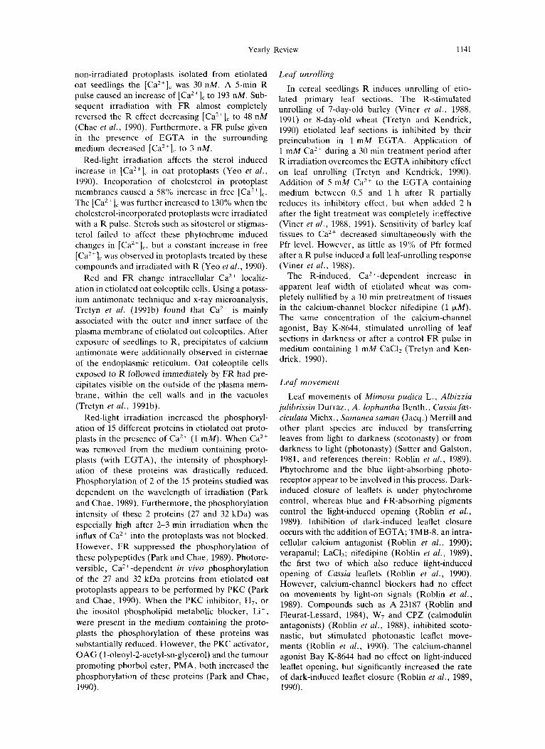
non-irradiated protoplasts isolated from etiolated oat seedlings the [Ca*+], was 30 nM. A 5-min R pulse caused an increase of [Ca2+], to 193 nM. Sub- sequent irradiation with FR almost completely reversed the R effect decreasing [Ca2i]c to 48 nM (Chae et af., 1990). Furthermore, a FR pulse given in the presence of EGTA in the surrounding medium decreased [Ca2'Ic to 3 nM.
Red-light irradiation affects the sterol induced increase in [Ca2+], in oat protoplasts (Ye0 et al., 1990). Incoporation of cholesterol in protoplast membranes caused a S8% increase in free [Ca2+Ic. The [Ca2i]c was further increased to 130%) when the cholesterol-incorporated protoplasts were irradiated with a R pulse. Sterols such as sitosteral or stigmas- terol failed to affect these phytochrome induced changes in [Ca*+],., but a constant increase in free [Ca2+lc was observed in protoplasts treated by these compounds and irradiated with R (Yeo e ta / . , 1990).
Red and FR change intracellular Ca2+ localiz- ation in etiolated oat coleoptile cells. Using a potass- ium antimonate technique and x-ray microanalysis, Tretyn et al. (1991b) found that Ca"+ is mainly associated with the outer and inner surface of the plasma membrane of etiolated oat coleoptiles. After exposure of seedlings to R, precipitates of calcium antimonate were additionally observed in cisternae of the endoplasmic reticulum. Oat coleoptile cells exposed to R followed immediately by FR had pre- cipitates visible on the outside of the plasma mem- brane, within the cell walls and in the vacuoles (Tretyn et al., 1991b).
Red-light irradiation increased the phosphoryl- ation of 15 different proteins in etiolated oat proto- plasts in the presence of Ca2' (1 mM). When Ca2+ was removed from the medium containing proto- plasts (with EGTA), the intensity of phosphoryl- ation of these proteins was drastically reduced. Phosphorylation of 2 of the 15 proteins studied was dependent on the wavelength of irradiation (Park and Chae, 1989). Furthermore, the phosphorylation intensity of these 2 proteins (27 and 32 kDa) was especially high after 2-3 min irradiation when the influx of Ca2+ into the protoplasts was not blocked. However, FR suppressed the phosphorylation of these polypeptides (Park and Chae, 1989). Photore- versible, Ca2+-dependent in vivo phosphorylation of the 27 and 32 kDa proteins from etiolated oat protoplasts appears to be performed by PKC (Park and Chae, 1990). When the PKC inhibitor, H,, or the inositol phospholipid metabolic blocker, Li+ , were present in the medium containing the proto- plasts the phosphorylation of these proteins was substantially reduced. However, the PKC activator, OAG (1-oleoyl-2-acetyl-sn-glycerol) and the tumour promoting phorbol ester, PMA, both increased the phosphorylation of these proteins (Pa:rk and Chae, 1990).
Leaf unrolling
In cereal seedlings R induces unrolling of etio- lated primary leaf sections. The R-stimulated unrolling of 7-day-old barley (Viner et al . , 1988, 1991) or 8-day-old wheat (Tretyn and Kendrick, 1990) etiolated leaf sections is inhibited by their preincubation in 1 mM EGTA. Application of 1 mM Ca2+ during a 30 min treatment period after R irradiation overcomes the EGTA inhibitory effect on leaf unrolling (Tretyn and Kendrick, 1990). Addition of 5 mM Ca2' to the EGTA containing medium between 0.5 and 1 h after R partially reduces its inhibitory effect, but when added 2 h after the light treatment was completely ineffective (Viner et al. , 1988, 1991). Sensitivity of barley leaf tissues to Ca2+ decreased simultaneously with the Pfr level. However, as little as 19% of Pfr formed after a R pulse induced a full leaf-u,nrolling response (Viner et a/., 1988).
The R-induced, Ca2+-dependent increase in apparent leaf width of etiolated wheat was com- pletely nullified by a 10 min pretreatment of tissues in the calcium-channel blocker nifedipine (1 F M ) . The same concentration of the calcium-channel agonist, Bay K-8644, stimulated unrolling of leaf sections in darkness or after a control FR pulse in medium containing 1 mM CaCI, (Tretyn and Ken- drick, 1990).
Leaf movement
Leaf movements of Mimosa pudica L., Albizzia julibrissin Durraz., A. lophantha Benth., Cassia fas- ciculata Michx., Samanea saman (Jacq.) Merrill and other plant species are induced by transferring leaves from light to darkness (scotonasty) or from darkness to light (photonasty) (Satter and Galston, 1981, and references therein; Roblin et a/ . , 1989). Phytochrome and the blue light-absorbing photo- receptor appear to be involved in this process. Dark- induced closure of leaflets is under phytochrome control, whereas blue and FR-absorbing pigments control the light-induced opening (Roblin et al., 1989). Inhibition of dark-induced leaflet closure occurs with the addition of EGTA; TMB-8, an intra- cellular calcium antagonist (Roblin et a/ . , 1990); verapamil; LaCI,; nifedipine (Roblin et al., 1989), the first two of which also reduce light-induced opening of Cassia leaflets (Roblin et a/., 1990). However, calcium-channel blockers had no effect on movements by light-on signals (Roblin et a / . , 1989). Compounds such as A 23187 (Roblin and Fleurat-Lessard, 1984), W7 and CPZ (calmodulin antagonists) (Roblin et al., 1988), inhibited scoto- nastic, but stimulated photonastic leaflet move- ments (Roblin et a/., 1990). The calcium-channel agonist Bay K-8644 had no effect on light-induced leaflet opening, but significantly increased the rate of dark-induced leaflet closure (Roblin et al., 1989, 1990).

1142 ANDRZEJ TRETYN el al.
Phytochrome controls nyctinastic movement of A . lophuntha leaflets (Moysset and Simon, 1989). Depending on the external concentration applied A 23187 or Ca2+, can induce an increase in nyctinas- tic closure of leaflets. In adequate concentrations they counteract FR-induced leaflet closure (Moysset and Simon, 1989). The intracellular calcium antag- onist, TMB-8, inhibits the effect of R, but had no effect on the FR-induced response (Moysset and Simon, 1989).
Flower induction
Halaban and Hillman (1970) found that flowering of Lemna perpusilla Torr. grown under an inductive photoperiod on a full nutrient solution with sucrose could be inhibited by transfer of plants to water at particular times during the dark period. The highest degree of inhibition of the process was observed about 1-2 h after the time of maximal sensitivity of Lemna to light treatments that influence flowering. Addition of either Ca(N03)2 or K2HP04 partially prevents the effect, whereas NH4N03 or MgS04 increases the degree of inhibition (Halaban and Hill- man, 1970).
Recently, Friedman et al. (1989) have presented data concerning participation of Ca2+ in the phyto- chrome-controlled flower induction of Pharbitis nil CHOIS. This short-day plant can be induced to flower by a single photoinductive dark period. Induction of flowering occurs after pretreatment with 20 mM EGTA given 8 h before the dark per- iod. Application of 30 mM CaC12, 30 rnin after EGTA application completely reverses the inhibi- tory action of the chelator. Furthermore, plants sprayed with A 23187 at different times from the beginning until 4 h of the subinductive dark period (11 h 45 min) produced more flowers per plant than non-treated controls. However, treatment with LaC13 (5 mM), W7 (150 pM) and CPZ (200 pM) all reduced the flowering response (Friedman et al., 1989). Tretyn et al. (1990b) failed to confirm these results using EGTA, calcium-channel blockers (La3, nifedipine and verapamil) and the calcium-channel agonist (Bay K-8644). The calmodulin inhibitor CPZ and the inositol phospholipid metabolic inhibi- tor Li+ also had no effect.
Interaction Between Phytochrome and CaZ+
In order to characterize the relationship between phytochrome and Ca2+, the Ca2+-sensitive phase after R irradiation has been studied for several responses. Results of these experiments are summa- rized in Table 1.
Bossen et al. (1988) showed that to obtain a full swelling response of etiolated wheat protoplasts, Ca2+ had to be supplied to the medium within the first 10 rnin after a 1 rnin saturating R pulse. In the case of etiolated barley leaf sections, maximum unrolling occurred if Ca2+ was supplied to the bath-
Table 1. Investigation of the Ca’+-dependent phase for differ- ent phytochrome responses. Different plant systems were irradiated with a red-light pulse in Caz+-free medium and then CaZ+ was supplied at different times after the irradiation. The escape time is the time after which responsiveness to Ca”
addition is lost
Escape Reference Response time
Wheat protoplast < 10 rnin Bossen et al. (1988) swelling
Barley leaf unrolling 1-2 h Viner et al. (1988)
Spirodela turion > 12 h Appenroth and germination Augsten (1990)
Fern more germination
Onclea sensibilis < 8 h Wayne and Hepler ( 1984)
Dryopterk = 15 h Durr and
Adiantum 15-20 h Iino et al. (1989) puleacea Scheuerlein (1990)
cupillus-veneris
ing solution up to 2 h after a R inductive pulse (Viner et al., 1988). Experiments which were per- formed with Spirodela turions showed that Ca2+ only stimulates germination when applied during the Pfr-requiring phase (Appenroth and Augsten, 1990). The most Ca2+-sensitive period was found to be between 24 and 48 h after a R pulse. Indepen- dent of the delay between a R pulse and a FR pulse to remove Pfr, only a negligible response to Ca2+ addition was observed (Appenroth and Augsten, 1990). Interaction between phytochrome and Ca2+ observed during swelling of wheat protoplasts (Bossen et al., 1988), unrolling of barley leaf sec- tions (Viner et al., 1988), and germination of Spirod- ela turions (Appenroth and Augsten, 1990) differed from that recorded for R-induced fern spore germi- nation (Wayne and Hepler, 1984; Iino et al. , 1989; Scheuerlein et al., 1989). Wayne and Hepler (1984) studied the relationship between phytochrome photoconversion and Ca2+ transport. Red light- stimulated germination of Onoclea occurs even if CaZ+ was added to the medium up to 8 h after irradiation. In the absence of Ca2+, FR partially reversed the inductive effect of R, but only when given up to 5 min after R. The ability of FR to reverse the inductive R effect was not observed when Ca2+ was present at the time of R irradiation (Wayne and Hepler, 1984). Phytochrome photo- transformation clearly does not require Caz+. The first cell division of Adiantum spores is induced by R even in the absence of Ca2+. A R-induced DNA increase in the spores was unaffected even if a satu- rating concentration of Ca2’ was added 15 h after R (Iino et al., 1989). Much longer retention of the capacity to respond to Ca2+ after phytochrome photoconversion was observed by Scheuerlein et al. (1989) for R-induced germination of fern spores

Yearly Review 1143
Table 2. The effect of red-light via phylochrome on Ca2+ influxiefflux intoifrom different plant cells or on the intracellular free [Ca*+] ([Ca"]") measured by different analytical methods
Method of measurement Plant material Effect Reference
Murexide- Mougeotia protoplasts Efflux Roux (1984) spectrophotometry Vallisneria protoplasts Efflux Takagi and Nagai (1988)
Oat protoplasts Efflux Hale and Roux (1980) Wheat protoplasts Efflux Bossen (1990)
Autoradiography Mougeotia cells Influx Dreyer and Weisenseel (1979)
Atomic absorption spectroscopy Onoclea spores Influx Wayne and Hepler (1985)
45Ca2+ uptake studies Maize protoplasts Influx Das and Sopory (1985) Oat coleoptile sections Influx Tretyn (1987) Mesotaenium protoplasts Influx Berkelman and Lagarias (1990) Barley protoplasts Influx U ei al. (1991)
Quin-2-fluorimetry Oat protoplasts Increase [Caz+Ic Chae et al. (1990)
Fura-2-fluorimetry Dryopteris spores Decrease [CaZ+], Scheuerlein et al. (1991)
of Dryopteris paleacea. In this case there was no germination induction when Ca2+ was added to the medium as late as 40 h after R. Moreover, Ca2+ addition was fully effective 24 h after R treatment, when the spores had escaped from FR reversibility (Scheuerlein et ul., 1989).
Phytochrome control of Ca2+ fluxes
Phytochrome controls membrane permeability to different ions, including Ca2+ (Kendrick and Bossen, 1987). Results concerning the influence of R on Ca2+ fluxes are contradictory (Table 2). Dreyer and Weisenseel (1979) showed by means of autoradiography that R stimulates 45C'a2+ accumu- lation into the cells of the green alga Mougeotia. A few years later Wayne and Hepler (1985) using atomic absorption spectroscopy discovered that R stimulates an increase in the total [Ca2+], of Ono- clea spores. Both authors showed that subsequent exposure to FR nullifies R-induced Ca2+ accumu- lation by both Mougeotia and Onoclea cells. Stimu- lation of 45Ca2+ uptake by R has been confirmed by a number of authors for etiolated maize (Das and Sopory, 1985), barley (U et al., 1991) protoplasts, Mesotuenium protoplasts (Berkelman and Lagarias, 1990), and etiolated oat coleoptile section cells (Tretyn, 1987). In all these plant systems FR, applied after a R pulse, significantly reduces the R stimulation of 45Ca2+ uptake.
Results which contradict the above cases have been obtained on the basis of studies using the Ca2+-sensitive dye, murexide. Hale and Roux (1980), Roux (1984), Takagi and Nagai (1988) and Bossen (1990) have all shown that R stimulates an efflux and FR results in influx of Ca2+ into the cells of etiolated protoplasts of oat, Mougeotia sp., Vallisneria gigantea Graebner and wheat (Table 2).
There are a limited number of studies on phyto- PAP 54:6-R
chrome control of [Ca2+],. Using an acetoxymethyl ester of the Ca2+ fluorescent indicator dye, quin-2 (quin-2iAM) Chae et al. (1990) have found that 5 min R irradiation of etiolated oat protoplasts results in an approximately 5-fold increase in [Ca2+],, while a subsequent irradiation with 5 min FR leads to a significant reduction in [Ca2+lc. How- ever, Scheuerlein et al. (1991) utilizing a different fluorescent calcium indicator dye, fura-2, discovered that in germinated spores of Dryopteris, R leads to a decrease in [Ca2+], in comparison to non-irradiated cells. Since these measurements were performed 20 h after R treatment it is difficult to say if observed changes in [Ca2+], in individual Dryopteris spores represent primary or secondary effects.
The question as to whether R and FR directly influence intracellular free Ca2+ is still unanswered. However, it is clear that phytochrome can change membrane permeability to Ca2+ ions. In our opin- ion, results of experiments performed with murex- ide are rather difficult to interpret. Murexide does not penetrate plasma membrane and the results only show net changes of Ca2+ in the surrounding medium. From studies with Ca2+ fluorescent-indi- cator dyes it is known that the presence of chelating agents of this ion in the medium decrease [Ca,,], of cells maintained in darkness (Scheuerlein et al., 1991) or irradiated with R (Chae et al., 1990; Scheuerlein et al., 1991). Presence of membrane impermeable chelators of murexide in the surround- ing medium can lead to a non-physiological efflux of the Ca2+ from the cytoplasm to the external medium. Red light-stimulated increase of mem- brane permeability to Ca2+ may increase its leakage from the cytoplasm to the medium.
In etiolated plant cells [Ca2+], is in the same concentration range as in non-stimulated animal cells (Chae et al., 1990; Scheuerlein et al., 1991)

1144 ANDRZEJ TRETYN et al.
and increases significantly after R irradiation (Chae et al., 1990). In animal cells different stimuli change [Ca2+], via an increase or decrease of membrane permeability for extracellular Ca2+. Membrane fluxes of this ion are regulated by two well defined elements: calcium channels (Tsien and Tsien, 1990) and calcium pumps (Schatzman, 1989). There is some evidence pointing to the existence of similar elements in the membranes of plant cells. The pres- ence and molecular properties of different plant ion channels including those for Ca2+ have been reviewed recently by Sanders and Slyman (1989) and Tester (1990). Influx of Ca2+ into many plant cells is modified by different compounds which inhibit or stimulate, in a manner consistent with the operation of particular types of calcium channels similar to those in animals (Tester, 1990). Some of these agents can influence Ca2+-dependent photo- morphogenetic responses. Wayne and Hepler (1985) showed that La3+ inhibits the R-induced increase in intracellular calcium in the spores of Onoclea sensibilis. Stenz and Weisenseel (1986) found that the R-induced reduction of the Zeta- potential of the green alga Mesotaenium disap- peared in media with a low [Ca2+] or after adding the calcium-channel blocker, verapamil. Both La3+ and verapamil decrease the rate of 4sCa2+ uptake by cells of oat etiolated coleoptile sections (Tretyn, 1987). Takagi and Nagai (1988) have reported that three different calcium-channel blockers, La3+, nifedipine, and verapamil, result in the cessation of phytochrome-controlled cytoplasmic streaming in Vallisneriu gigantea Graebner protoplasts. Calcium- dependent streaming is induced only when phyto- chrome exists in the Pfr form (Takagi et al., 1990). All three of the above-mentioned calcium-channel blockers inhibited phytochrome-mediated move- ments of Cassia fasciculata leaflets (Roblin et al. , 1989, 1990). Lanthanium ions inhibit the phyto- chrome-controlled germination of Onoclea sensibilis (Wayne and Hepler, 1984), Dryopteris paleacea fern spores (Scheuerlein et al . , 1989) and Spirodela polyrha turions (Appenroth et al., 1990).
Both Ca2+-dependent, phytochrome-stimulated swelling of etiolated wheat protoplasts (Bossen et al., 1988; Tretyn et al., 1990c) and unrolling of etiolated wheat leaves (Tretyn and Kendrick, 1990) is nullified by verapamil, La3+, and nifedipine, a DHP derivative. Nifedipine was only active when it was added to the medium containing protoplasts before a R pulse (Tretyn et a/., 1990~). In contrast, another DHP derivative, Bay K-8644, well known as an antagonist of “L-type” calcium channels (Tsien and Tsien, 1990), stimulates wheat protoplast swell- ing (Tretyn et a/ . , 1990c) and leaf unrolling (Tretyn and Kendrick, 1990) after control FR irradiation. Furthermore, Bay K-8644 promotes phytochrome- mediated Cassia fasciulata leaf movement (Roblin et al., 1989, 1990) and increases the velocity of the R-mediated Mougeotia scalaris Hassel chloroplast
rotation (Russ et al., 1991). Most authors agree that phytochrome regulates transmembrane Ca2 + fluxes (Hepler and Wayne, 1985; Moysset and Simon, 1989; Roux et al., 1986; Stenz and Weisenseel, 1986; Takagi and Nagai, 1988; Takagi et al., 1990; U et al., 1991; Wayne and Hepler, 1984, 1985; Weisen- see1 and Ruppert, 1977) via regulation of calcium- channel activity (Bosen, 1990; Bossen et al . , 1988; Roblin et al., 1989; Tretyn, 1987; Tretyn and Ken- drick, 1990; Tretyn et al., 1990a). Tretyn ef al. (1990~) have proposed that phytochrome controls the activity of DHP-sensitive “L-type” calcium channels.
Unlike calcium channels there is direct evidence concerning the effect of phytochrome on calcium pumps (Ca2+-translocating ATPase). Calcium pumps have been identified and biochemically characterized from tissues of many plant species (Rasi-Caldogno et a/., 1989; see Briskin, 1990, for review). Dieter and Marme (1981, 1983) demon- strated that activity of the microsomal Ca2+ pump is stimulated by calmodulin. However, when vesicles were prepared from FR-irradiated corn seedlings the pump was no longer under the control of calmo- dulin (Dieter and Marme, 1981, 1983).
In some plant systems FR irradiation decreases the rate of 4sCa2+ uptake (Tretyn, 1987; U et a/., 1991) and [Ca2+Ic (Chae et a/., 1990) compared to dark controls. However, the mechanism of action of the physiologically inactive R-absorbing form of phytochrome (Pr) on these processes remains un- clear.
Sources of Ca2+
There are two different Ca2+ sources available for utilization by light-stimulated cells: from the extracellular sources and intracellular stores. The concentrations in soil solutions or in “fresh” water are not very high, values of 1 pA4 Ca2+ or less. These ranges of [Ca2+] are sufficient, but not opti- mal for phytochrome-stimulation of germination of fern spores: Onoclea (Wayne and Hepler, 1984), Dryopteris (Scheuerlein et a/ . , 1989), Adiantum (Iino et a/., 1989) or Spirodela turions (Appenroth and Augsten, 1990). During plant development Ca2+ may accumulate inside particular cells or tis- sues. For example, in Onoclea spores, large amounts of Ca2’ have been found within the cell wall (Wayne and Hepler, 1985). Only after depletion of the cell wall-localized Ca2+ by EGTA is it possible to demonstrate that phytochrome-con- trolled fern-spore germination is a Ca2+-dependent process (Wayne and Hepler, 1984; Scheuerlein et al., 1989). Moreover, adequate amounts of the cal- cium chelator can inhibit other R-stimulated pro- cesses: leaf unrolling (Viner et d., 1988; Tretyn and Kendrick, 1990), leaf movement (Roblin e ta / . , 1990) and photoperiodic flower induction (Friedman et al., 1989; Tretyn et al., 1990b). On

Yearly Review 1145
these grounds we believe that in many plant systems phytochrome controls movement of Ca2+ from the apoplast to the symplast. Experiments on protoplast swelling show that even after removing the cell wall they must be very carefully washed .with EGTA solution before being used in an experiment (Rossen et al., 1988; Tretyn et al., 1990b). Cytochemical studies performed on etiolated oat cokoptiles have shown that most of the antimonate precipitable Ca2+ is present both in the proximity o f the extra- cellular and intracellular side of the plasma mem- brane (Tretyn et al., 1991b). In animal cells special protein, so-called “annexin”, can take part in bind- ing of Ca2’ to the phospholiped components of the plasma membrane cytoplasmic surface. Recently, Clark et al. (1991) have found annexin-like protein in pea plumules which has properties similar to that found in animal cells. It appears that plasma membrane-bound calcium moves into the cytosol of the oat coleoptile cells after R irradiation (Tretyn etal . , 1991b). After washing the cells with ethylene- diaminetetraacetic acid (EDTA) or EGTA it is possible to remove Ca2+ bounded to the cell wall and extracellular side of the plasma membrane (Wayne and Hepler, 1985).
Some Ca2+-dependent, light-regulated processes, such as Mougeotia chloroplast re-orientation, have a very low [Ca2+] requirement in the surrounding medium. However, special osmiophilic globules are observed inside the cells of this green alga (Grolig and Wagner, 1989). These CaBP are devoid of any membrane-like structures and accumulate large amounts of calcium (Tretyn et al . , 1991). A similar function of Mougeotia CaBP is shown by the “tan- nin” vacuoles observed in cortical parenchyma cells of motor organs localized at the leaflet base of Mimosa (Toryiama and Jaffe, 1972). The way in which light initiates release of Ca2’~ from such internal stores is difficult to envisage. Roblin et al. (1989) have proposed that R stimulates Ca2+ influx from the apoplast into the cells, while blue light can release the Ca2+ from its intracellular stores.
The Mechanism of Action of Ca2+ in Photomorphogenesis
Light is one of the most important environmental stimuli for bacteria, fungi, animals and plants. Thus nearly all these organisms, during their evolution, have developed mechanisms by which they are able to detect both quantitative and qualitative aspects of the light environment, indicating the direction of irradiation. Moreover, some bacteria and both lower and higher plants utilize light as an energy source in photosynthesis.
The best characterized photoreceptor in animals is rhodopsin (Fung, 1985; Rayer et al. , 1990); it has also been found in some bacteria (bac- teriorhodopsin) and the green alga Chlamydomonas (for review see Wagner and Marwan, 1991). Rho-
dopsin is a membrane component of rods and cones of the eye retina in vertebrates and in visual cells of invertebrates (Rayer et al., 1990). In the case of Halobacterium and Chlamydomonas this pigment is also an integrated membrane component (Wagner and Marwan, 1991). Rhodopsin consists of a poly- peptide (opsin) to which a carotenoid-derivative chromophore (retinal) is covalently linked (Rayer et al., 1990). The primary structure of opsin in different species has many characteristics in com- mon with all rhodopsins and other membrane recep- tors. The proteins possess seven transmembrane segments interconnected by loops on both side of the membrane. First and second cytoplasmic loops are the target for a G-protein in animals (Fung, 1985) called trandsducin (Gt). Moreover, in the C- terminal part of the polypeptide of both inver- tebrates and vertebrates rhodopsin has serine and threonine residues which undergo light-induced phosphorylation (Rayer et al. , 1990). After absorp- tion of one photon by rhodopsin, isomerization of the retinal chromophore from 11-cis to the all-trans configuration is observed (Rayer et al., 1990). In the case of Halobacterium, light stimulates isomeri- zation of all-trans retinal to the 13-cis form of the chromophore (Wagner and Marwan, 1991). Re-iso- merization of the chromophore group of rhodopsin is accompanied by conformational changes of the protein (Fung, 1985). In invertebrates the light acti- vated form of rhodopsin (meta-rhodopsin) is ther- mally stable at physiological temperatures and is re- isomerized by light to the physiologically inactive form (Rayer et al., 1990).
Some biochemical and physicochemical proper- ties of rhodopsin are very similar to those of phyto- chrome. Both pigments consists of protein and chromophore components. Absorption of light by the chromophores of rhodopsin and phytochrome leads to their isomerization (in the case of phyto- chrome it is 15-cisitrans isomerization, Rudiger, 1987). Similar to the retinal rhodopsin chromophore in invertebrates, cisltrans and translcis isomerization of the phytochrome chromophore by light are accompanied by conformational changes of the pro- tein resulting in the physiologically active Pfr form being converted to the inactive (Pr) form, and vice versa. It has been shown by means of monoclonal antibodies that conformational differences between Pr and Pfr extend over the whole length of the molecule (Schneider-Poetsch et al. , 1989). How- ever, only certain regions of the phytochrome poly- peptide chain undergo photoconversion-induced conformational changes and these are probably involved in phytochrome’s mode of action: segments corresponding to 4-10 kDa, 74, 83-84 and 100 kDa from the N-terminus (Quail et al.. 1987). Recently, Schneider-Poetsch and coworkers (Schneider- Poetsch and Braun, 1991; Schneider-Poetsch et al., 1991) analysed published phytochrome sequence data and made a comparison with bacterial sensor

1146 ANDRZEJ TRETYN et al.
proteins responsive to environmental stimuli. They found 23-2670 homology for a 240 amino acid seg- ment near to the C-terminus. Both in phytochrome and bacterial sensor proteins this domain undergoes conformational changes which are caused by stimu- lus of the N-terminal region. By analogy to bacterial sensor protein these workers postulate that phyto- chrome, after light-stimulated conformational change of the C-terminus, transduces this signal by interactions with additional (not identified) pro- teins.
Phytochrome and rhodopsin have one important difference. Whereas rhodopsin is a typical integral membrane protein, there is no such evidence for this in the case of phytochrome in plants. Immunocytochemical studies have shown that phytochrome is a cytoplasmic protein. Moreover, analysis of phytochrome polypeptides shows no extended hydrophobic sequences typical of mem- brane proteins (for review see Jordan et al., 1986; Vierstra and Quail, 1986). This difference between rhodopsin and phytochrome probably reflects the former's single mode of action within the mem- brane, while phytochrome might interact with sev- eral different components within the cell. In fact, in animal (Rayer et al., 1990) and Chlamydomonas cells (Harz and Hegemann, 1991) rhodopsin regu- lates membrane conductance. In contrast to phyto- chrome, in animals rhodopsin does not take part in the regulation of photomorphogenesis. Similar to phytochrome, bacterial sensor proteins also lack hydrophobic transmembrane regions in the N-ter- minal region of the polypeptide chain (Schneider- Poetsch et al., 1991).
Signal Transduction Chains
Light absorption by phytochrome is transduced by an unknown cascade which leads to changes in membrane conductance, growth and development. Photoconversion of phytochrome and ultimate photomorphogenetic responses appear to be con- nected by internal messenger(s). Calcium ions have been proposed as playing this role (Roux et al., 1986) as discussed above. This section reviews data concerning participation of second messengers in phytochrome action.
There are only a limited number of studies about the signal transduction chain involved in phyto- chrome action. At the present state of knowledge it seems that the process of light transduction which occurs in Halobacterium, rod cells of vertebrates and visual cells of invertebrates and in the green alga Chlamydomonas, is similar to that observed in many plants (Table 3). The transduction of light stimuli which takes place in invertebrates especially is very similar to that observed during phytochrome action. In invertebrates this involves light activation of a G,, the target of which is PLC, a rhodopsin kinase and finally Ca2+ as the second messenger
(Fung, 1985; Rayer et al . , 1990). All these elements have also been recognized in plant cells and they probably function in the same way as in animals.
GTP-binding proteins
The G-proteins in animals are a heterotrimer con- sisting of three distinct polypeptides c1 (==40 kDa), P (=36 kDa) and y (8 kDa). The c1 subunit binds GTP and is the activator of target proteins, whereas P and y subunits form a complex which modulates the activity of the c1 subunit (Stryer and Bourne, 1986; Gilman, 1987). Furthermore, the a subunit of a G, can be specifically activated through ADP- ribosylation by bacterial toxins (Stryer and Bourne, 1986; Gilman, 1987).
The presence of a protein which binds ["SIGTP- y-S has been found in many plant systems (Hasunuma et al., 1987; Blum et al., 1988; Drcbbak et a l . , 1988; Korolkov et al., 1990; Romero et al., 1991b). These proteins were ADP-ribosylated in the presence of cholera toxin (Romero et al., 1991a) and they possess GTPase activity (Korolkov et al., 1990) and were recognized by antibodies raised against a highly conserved peptide of most G, a- subunits (Blum et al., 1988; Korolkov et al., 1990; Romero et al., 1991b). Furthermore, [35]GTP-y-S- binding proteins have been isolated from membrane fractions (Hasunuma et al., 1987; Blum et al., 1988; Drcbbak et al. , 1988; Korolkov et al., 1990). Despite the fact that G-proteins isolated from plants may differ in molecular mass they have similar biochemi- cal and molecular properties to G, isolated from animal cells (Stryer and Bourne, 1986; Gilman, 1987).
There is limited evidence that G-protein acti- vation is involved in phytochrome action. Bossen and co-workers (1990) have shown that the R- induced, Ca*+-dependent, swelling of etiolated wheat mesophyll protoplasts is nullified by an inhibi- tor of G-proteins, GDP-P-S. However, an activator of G-proteins, GTP-y-S, induced swelling of the protoplasts to the same extent as after R, both in darkness and after a control FR irradiation as long as essential Ca2+ was present (Bossen et a[., 1990).
Hasunuma et al. (1987) were the first to report binding of [35S]GTP-y-S to proteins in extracts of Lemna paucicostata 441 containing membrane com- ponents. They found that R and FR inhibited GTP- binding to these proteins and proposed that phyto- chrome (Pfr) may function in the same way as the photoexcited state of rhodopsin in animals. Sub- sequently, Korolkov et al. (1990) published data confirming the presence of a G-protein in plants: the eyespot of Chlamydomonas reinhardtii. This protein can take part in light induced and rhodopsin-depen- dent locomotion of the alga (Korolkov et al., 1990).
Extensive studies of the possible involvement of G-proteins in the phytochrome transduction chain have been performed by Romero and colleagues

Yearly Review 1147
Table 3. Comparison of bacterial, animal and plant photoreceptors. (Data from: Fung, 1985; Rayer et al., 1990; Brederoo et al., 1991; Wagner and Marwan, 1991) .
Photoreceptor Transduction chain
Taxonomic group Type Location
Bacteria Halohacterium Rhodopsin Membrane
Animal5 Invertebrate Rhodopsin Membrane Vertcbrate Rhodopsin Mcmbrane
Plants Chlamvdomonas Rhodomin Membrane Other plants Phytochkme Cytosol
Ca2+ G IP3 Location
- + ND* Within the membrane
+ + + Within the membrane + + + In the cytosol
+ + + In the cytosol + + + In the cytosol
*ND = not determined; G = GTP-binding protein; IP2 = inositol 1,4,5-trisphosphate
(Romero et al., 1991a,b). They found that compared to FR or the dark control, R increases binding of ["SIGTP-y-S to a protein fraction isolated from pre-irradiated etiolated oat seedlings. Moreover, in vitro binding of GTP-y-S to G-protein(s) from the oat extract was inactivated by the Pr form of exogen- ously added purified phytochrome. However, the binding capacity remained fully active when the Pfr form was added to the medium (Romero et al. , 1991b). Romero et al. (1991a) have also studied the effect of cholera toxin on the chlorophyll aib-bind- ing protein Cab and phytochrome Phy genes. Expression of both these genes is phytochrome- regulated, the former positively and the latter nega- tively. After 2-h incubation of etiolated oat seed- lings in darkness in different doses of cholera toxin, Romero et al. (1991a) found that the toxin regulates the expression of Cab and Phy genes: Cab up-, and Pliy down-regulated.
From studies with animal cells it is well known that activation of G, leads to an exchange of GDP for GTP bound to their a-subunit. The GTP-bound to the ci subunit affects the activity of other effector molecules, that generate intracellular signals. Aden- ylate cyclase, cGMP phosphodiesterase (cGMP PDE), possibly PLC and phospholipase A2 (PLA2) and more than one type of ionic channel have been shown to be direct targets for G proteins (Brown and Brinbaumer, 1990). A single G protein may have several different membrane targets and can be linked to about 70 different receptors, at least 8 different K + , 1 Na+ and 2 different CaZ+ channels (Brown and Brinbaumer, 1990). In different animal cells they directly regulate voltage and DHP-sensi- tive Ca2+ channels (Brown and Brinbaumer, 1990; Schultz et al., 1990; Trautwein and Hescheler, 1990). The presence of similar channels in etiolated wheat mesophyll protoplasts has been postulated by Tretyn et al. (1990~). Furthermore, Bossen et al. (1990) have obtained results consistent with the existence of a G-protein in the protoplasts. We believe that, at least in etiolated wheat protoplasts,
membrane Ca2+ transport can be controlled by a G,-type system which may be directly bound to a DHP-sensitive L-type channel.
The phosphoinositide cascade
During hydrolysis of phosphatidylinositol 4,s- bisphosphate (PIP2) by membrane-bound PLC, two intracellular signal molecules, viz. 1,2-diacylglycerol (DAG) and inositol 1,4,5-trisphosphate (IP3) are generated, which activate PKC, whereas IP3 mobil- izes Ca2+ from intracellular stores (Berridge, 1987). The activity of a plasma membrane located PLC can be modulated via a PKC-controlled phosphoryl- ation, whereas IP3 receptors are associated with the endoplasmic reticulum by phosphorylation by a CAMP-dependent protein kinase A (PKA) (Bansal and Majerus, 1990). The breakdown of PIP2 to DAG and IP, appears to be regulated by a G- protein (Stryer and Bourne, 1986; Berridge, 1987).
Phosphoinositides have been isolated from differ- ent plant systems (for review see Boss, 1989; Hart- mann and Pfaffmann, 1990; Bonner et al., 1991). Furthermore, PLC has been isolated from wheat (Melin et al., 1987) and Pharbitis nil (Bonner et al., 1991) membranes. In both cases the enzyme was activated by a low [Ca2+], wheres G, activators failed to affect PLC activity (Melin ef al., 1987; Bonner et al., 1991). In Samanea leaflets, rapid light-induced changes have been shown in the con- centration of inositol 1-phosphate (IP,) and phos- pholipids (Morse et al . , 1987, 1990). The very con- venient system, moss protonemata, has been used for a study on phytochrome-controlled signal trans- duction in lower plants (for review see Hartmann and Pfaffmann, 1990). Phototropic response of Cer- atodon purpureus (Hedw.) Brid. protonemata is regulated via phytochrome. A 5 min pulse of R increased the activity of PLC in dark-grown cells, whereas FR after R reversed the effect to the level of the dark control. Furthermore, R increased both the rate of PIP2 degradation and the levels of inosi-

1148 ANDRZEJ TRETYN et al.
to1 1,4-bisphosphate (IP,) and IPS. Lithium ions (inhibitor of inositol-1-phosphatase) decreased the level of phosphatidylinositol (PI), and increased the endogenous levels of IP1, IP2 and IPS in R-preir- radiated protonemal tissues (Hartmann and Pfaffmann, 1989). Recently, Bossen et al. (1990) found that Li+ and neomycin, an inhibitor of PLC, inhibit both R- and GTP-y-S induced swelling of protoplasts while having no effect on the FR- irradiated phytochrome-controlled phototropic response of Ceratodon purpureus (Hartmann and Pfaffman, 1989). In the latter case, Li+ M ) was only active if it was present during or after R irradiation (Hartmann and Pfaffmann, 1989). Lith- ium ions also inhibited another phytochrome-regu- lated process: R-induced chloroplast reorientation in Mougeotia scalaris (Wagner et al., 1989).
Phytochrome and proteins phosphorylation
Since the discovery of CAMP-dependent PKA in 1968, reversible phosphorylation of proteins has been shown to be the major mechanism for the regulation of protein function in animal cells (for review see Kikkawa and Nishizuka, 1986). As in animals, reversible protein phosphorylation also plays a regulatory role in the transduction of many different extracellular signals in plants (for review see Ranjeva and Boudet, 1987; Poovaiah et al. , 1987; Blowers and Trewavas, 1989), including light (for review see Singh and Song, 1990).
A calcium-dependent protein kinase has been found in soluble as well as membrane fractions of many plant systems (Anderson, 1989; Blowers and Trewavas, 1989). Datta et al. (1985) have shown the R, Ca2 +-dependent phosphorylation of three different proteins: 47, 64 and 77 kDa. Far-red irradiation, calcium chelators and calmodulin inhibi- tors nullify these phosphorylations in isolated pea nuclei. From the same material a polyamine-stimu- lated protein kinase was found and purified to 90% homogeneity (Li and Roux, 1991). To date nothing is known about phytochrome regulation of this enzyme. Phytochrome-controlled dephosphoryl- ation of two proteins (29 and 30 kDa) and phos- phorylation of another (30 kDa) in etiolated oat coleoptile has been described (Otto and Schafer, 1988), as well as phosphorylation of two proteins (27 and 32 kDa) in etiolated oat protoplasts (Park and Chae, 1989). Phosphorylation of the 27 and 32 kDa proteins from oat protoplasts was sub- stantially reduced both by the PKC inhibitor (H7) and Li+ , whereas the activators of PKC, OAG and PMA, both enhanced phosphorylation of these pro- teins. From their in vivo studies Park and Chae (1990) proposed that phytochrome action in oat protoplasts is associated with a PKC-dependent pro- tein phosphorylation. Participation of PKC in regu- lation phytochrome-controlled processes has also been postulated by Bossen et al. (1990) and Haas
et al. (1991). Bossen et al. (1990) have shown that H, inhibits the R-induced swelling of etiolated wheat protoplasts. However, PMA induced swelling of the protoplasts after control FR irradiation (Bossen et al., 1990). Staurosporine, an inhibitor of PKC sig- nificantly decreases phytochrome-mediated, Ca2+- dependent germination of Dryopteris filix-mas L. spores (Haas et al., 1991). The presence of a protein kinase activity associated with the phytochrome molecule itself has been a matter of much dis- cussion. Wong et al. (1986, 1989) have shown that purified phytochrome preparations possess protein kinase activity. However, Kim et al. (1989) and Grimm et al. (1989) have shown that the phyto- chrome-associated protein kinase activity is due to a protein which co-purifies with phytochrome. Despite this discrepancy, the fact that phytochrome co-purifies with a protein kinase may suggest that these proteins have affinity with each other. The phytochrome molecule itself could be a substrate for protein kinases. Wong et al. (1986) found that mammalian PKA phosphorylates both Pr and Pfr, whereas PKC intensively phosphorylates the Pr form. Wong et al. (1990) and McMichael and Lagar- ias (1990) have demonstrated that an in vitro poly- cation-stimulated protein kinase phosphorylates the serine rich N-terminus of purified oat phytochrome.
Using isolated nuclei from etiolated oat seedlings Romero et al. (1991a) have studied the mechanism of phytochrome-regulated protein phosphorylation. By means of Western blot analysis and antibodies against animal G-protein at least two G-proteins (24 and 75 kDa) were discovered in the etiolated oat seeding nuclei. Cholera toxin, which stimulates ADP-ribosylation of a G-protein, enhances phos- phorylation of two proteins (10 and 54 kDa) and inhibits phosphorylation of two others (60 and 75 kDa). The phosphorylation of 60 and 75 kDa proteins was also stimulated by R. Addition of GDP-y-S to the nuclei preparations stimulated phosphorylation of a 60-kDa protein, however, the phosphorylation of three other proteins were inhibited. The increase in GDP-y-S-induced phos- phorylation of the 60-kDa protein was reduced by FR irradiation of oat nuclei (Romero et al. , 1991a). Participation of plant nuclear protein phosphoryl- ation in light-regulated gene expression has been postulated (Datta and Cashmore, 1989). At least in the oat system this process could be regulated by a G-protein signal transduction cascade (Romero et al., 1991b).
Calrnodulin-calcium binding protein
The universal role of calmodulin, in Ca2+-depen- dent, phytochrome-controlled processes was pointed out by Roux et al. (1986). Since that time, many new results concerning participation of calmo- dulin in phytochrome action have been published.
Calmodulin has been found in all plants investi-
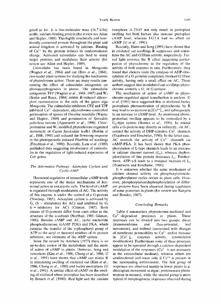
Yearly Review 1149
gated so far. It is low-molecular mass (16.7 kDa) acidic, calcium-binding protein (for review see Allan and Hepler, 1989). This highly structurally and func- tionally conserved protein throughout the plant and animal kingdom is activated by calcium. Binding of Ca2+ by the protein induces its conformational change. Activated calmodulin can bind to many target proteins and modulates their activity (for review see Allan and Hepler, 1989).
Calmodulin has been found in Mougeotia (Wagner et al., 1984) and oat (Biro t't al., 1984), two model plant systems for studying the mechanism of phytochrome action. There are many results con- cerning the effect of calmodulin antagonists on photomorphogenesis in plants. The calmodulin antagonists TFP (Wagner et a/. , 1984, 1987) and W7 (Serlin and Roux, 1984) inhibit R-induced chloro- plast reorientation in the cells of the green alga Mougeotia. The calmodulin inhibitors CPZ and TFP inhibited Ca2+-dependent, phytochronie-controlled germination of spores of Onoclea senszbilis (Wayne and Hepler, 1984) and germination of Spirodela polyrhiza turions (Appenroth et al., 1990). Chlor- promazine and W7 modified phytochrome-regulated movement of Cassia fasciculata leaflet (Roblin et al., 1988, 1990) and reduced the flowering response in the photoperiodic induced Pharbitis nil seedlings (Friedman et al., 1989). Recently, Lam et al. (1989) published data suggesting involvement of calmodu- lin in the regulation of light-induced expression of Cab genes.
The Alternative Pathway: Adenylate Cyclase and Cyclic-AMP
Hormonal regulation of intracellular cAMP levels represents one of the major mechanisms of hor- monal action in eukaryotic cells. The level of cAMP is regulated through modulation of AC. The activity of this enzyme is under the control of a G-protein (Northup, 1985). Adenylate cyclase is activated by G, (S = stimulatory for AC) and inhibited by G, (i = inhibitory for AC) (Gilman, 1987). Both classes of G-proteins differ from each other in the structure of the a-subunit (Northup, 1985; Gilman, 1986). Besides cAMP and AC, cyclic nucleotide phosphodiesterase (CAMP PDE) and PKA, which catalyze the transfer of the y-phosphoryl group of ATP to the seryl or threonyl residues of its protein substrate, are elements of the AMP !system.
Since the review by Amrhein (1977) there is no up-to-date review of the metabolism and the mode of action of cAMP in plants. However, Song and coworkers (Kim et al., 1986; Chung et a/., 1988; U et al., 1991) have shown that cAMP can mimic R in stimulating swelling of etiolated oat (Kim et a/., 1986; Chung et al., 1988) and barley protoplasts (U et al . , 1991). A similar effect of cAMP on the swell- ing of etiolated wheat protoplast has been described by Bossen et al. (1990). Red light and the calcium
ionophore A 23187 not only result in protoplast swelling but both factors also increase protoplast cAMP level, whereas EGTA had no effect on cAMP (U et al., 1991).
Recently, Hahn and Song (1991) have shown that in etiolated oat seedlings R suppresses and stimu- lates the AC and GTPase activity, respectively. Far- red light reverses the R effect suggesting partici- pation of phytochrome in the regulation of the activity of both enzymes in oats. Furthermore, they found that cholera toxin (by catalysis of ADP-ribo- sylation of a G-protein) completely blocked GTPase activity, having only a small effect on AC. These authors suggest that in etiolated oat seedlings phyto- chrome controls a G, of G-protein.
The mechanism of action of cAMP in phyto- chrome-regulated signal transduction is unclear. U et a/. (1991) have suggested that in etiolated barley protoplasts photoactivation of phytochrome by R may lead to an increase in [Ca'+],, and subsequently to an increase in cAMP level. As mentioned above, protoplast swelling appears to be controlled by a G,-type system (Bossen et al., 1990). In animal cells a G-protein may directly or indirectly, via AC, control the activity of DHP-sensitive Ca2 channels (Trautwein and Hescheler, 1990). In the latter case, AC controls the activity of Ca2' channels via CAMP-PKA. It has been shown that PKA phos- phorylation of L-type channels leads to an increase in calcium channel current (Ica), whereas dephos- phorylation of this protein decreases I,.:,. Further- more, ATP-y-S leads to a maximal increase of I,, (Trautwein and Hescheler, 1990).
It is unknown whether the same mechanism of calcium channel activity via phosphorylationide- phosphorylation cycles occurs in plant cells. How- ever, phosphorylation/dephosphorylation of differ- ent proteins have been observed during regulation of some processes in plants (for review see Ranjeva and Boudet, 1987).
Concluding Remarks
Table 4 summarizes phytochrome-mediated and Ca2+-dependent processes in plants. These responses can be divided into two groups: direct (transmembrane and Zeta-potential, leaf movement), and indirect (associated with changes of membrane permeability to Ca2 ' and/or increase in [Ca2+Ic, enzymes activity, cytoskeleton modification). Furthermore some of these processes appear to be operated through a calcium-dependent modulation of the responses (Ca' ' is not necessary in the extracellular medium), whereas others are unidirectional and occur only if Ca2+ is present in the surrounding environment. The first group of responses are observed mainly in lower plants (e.g. chloroplast movement in algae; protonemata photo- tropism in mosses), while the second group is more typical of morphogenetic responses observed during

1150 ANDRZEJ TRETYN et al.
Table 4. The participation of Ca2+ in different phytochrome-controlled phenomena
Phytochrome-regulated Level of control Plant species process Effect of Ca2+ References
Membrane properties and permeability
Membrane permeability, osmotic pressure, and cytoskeleton activity
Enzyme and metabolic activity
Gene expression, DNA and protein synthesis
Nitella sp. Mesotaenium cadariorum
Phaseolus aureus
Triticum aestivum Cassia fasciculata Albizzia lophantha Hordeum vulgare Triticum aestivum Mougeotia scalaris
Avena sativa Lygodium japonicum
Zea mays Spinacia oleracea
Onoclea sensibilis Dryopteris paleacea Adiantum capillus-
veneris Spirodela polyrhiza
Pharbitis nil
Membrane potential
Zeta-potential
Root-tip adhesion
Protoplast swelling Leaf movement
Leaf unrolling
Chloroplast movement
Acetylcholinesterase Gibberellin synthesis NAD kinase Mitochondria1 ATPase Peroxidase secretion
Spore germination
DNA synthesis
Turion germination
Flower induction
Depolarization
Decreasing
Stimulation
Stimulation Modulation
Stimulation
Stimulation
Inhibition Stimulation Activation Inhibition Stimulation
Stimulation
Stimulation
Stimulation
Participation
Weisenseel and RUDDert ' I
(1977) Stanz and Weisenseel (1986) Tanada (1968)
Bossen et al. (1988) Roblin er al. (1990) Moysset and Simon (1989) Viner et al. (1988) Tretyn and Kendrick (1990) Grolig and Wagner (1988)
Kim et al. (1990) Kagawa and Sugai (1991) Dieter and Marmt (1980) Serlin et al. (1984) Sticher et al. (1991)
Wayne and Hepler (1984) Diirr and Scheuerlein (1990)
Iino et al. (1989)
Appenroth and Augsten (1990) Friedman el al. (1989)
cell division (e.g. fern spores) and differentiation of higher plants (e.g. leaf unrolling). In some cases, such as in phytochrome-controlled flower induction or regulation of seed germination, participation of Ca2+ in regulation of these processes seems to be very complicated. Collectively published data strongly suggests that there is not simply one type of Ca2+ action in photomorphogenesis. The mode of action of Ca2+ in phytochrome-controlled processes appears to be determined by state of differentiation (cell competence). However, even in one cell alter- native phytochrome-regulated pathways (calcium- dependent and independent) are likely to exist.
Acknowledgements-The authors are indebted to col- leagues who made unpublished results available while this review was being prepared. A. T . was supported by The Alexander-von-Humboldt Foundation, Bonn, Fed. Rep. Germany.
ANDRZEJ TRETYN*
Institute of Biology N . Copernicus University Gagarina 9 PL-87-100 Toruri Poland
*To whom correspondence should be addressed.
RICHARD E. KENDRICK
Laboratory of Plant Physiology Research Wageningen Agricultural University Generaal Foulkesweg 72 NL-6703 BW Wageningen The Netherlands
GOITFRIED WAGNER
Membran- und Bewegungsphysiologie, Botanik I , Justus-Liebig- Universitat Senckenbergstr. 17-25 0-6300 Giessen Fed. Rep. Germany
REFERENCES
Anderson, J. M. (1989) Membrane-derived fatty acids as precursors to second messengers. In Second Messengers in Plant Growth and Development (Edited by W. F. Boss and D. J. Morrt), pp. 289-303. Alan R. Liss, New York.
Allan, E. and P. K. Hepler (1989) Calmodulin and cal- cium-binding proteins. In T h e Biochemistry of Plants (Edited by A. Marcus), Vol. 15, pp. 455-484. Aca- demic Press, San Diego.
Amrhein, N. (1977) The current status of cyclic AMP in higher plants. Ann. Rev. Plant Physiol. 28, 123-132.
Appenroth, K.-J. and H. Augsten (1990) Photophysiology of turion germination in Spirodela polyrhiza (L.) Schleiden-V. Demonstration of a calcium-requiring phase during phytochrome-mediated germination. Pho- tochem. Photobiol. 52, 61-65.
Appenroth, K.-J., R. Klinger, R. Wetzker and H. Augsten (1990) Photophysiology of turion germination in

Yearly Review 1151
Spirodela polyrhiza (L.) Schleiden-VI. Importance of calcium and calmodulin, Biochem. Physiol. Pfanzen.
Appenroth, K.-J., J . Opfermann, W. Hertel and H. Augsten (1989) Photophysiology of turion germination in Spriodela polyrhiza (L.) Schleiden. 11. Influence of after-ripening on germination kinetics. J . Plant Physiol.
Bansal, V. S. and P. W. Majerus (1990) Phosphatidylinosi- tol-derived precursors and signals. Ann. Rev. Cell. Biol.
Berkelman, T. and J . C. Lagarias (1990) Calcium transport in the unicellular green alga Mesotaenium caldariorum: a model system for phytochrome-mediated responses. In Calcium in Plant Growth and Development (Edited by R. T. Leonard and P. K. Hepler), pp. 151-160. Am. SOC. Plant Physiol., Bethesda, MD.
Berridge, M. J. (1987) Inositol triphosphate and diacylgly- cerol: Two integrating second messengers. Ann. Rev. Biochem. 56, 159-193.
Biro, R. L., S . Davy, B. S. Serlin, M. E. Terry, N. Datta, S . K. Sopory and S. J . Roux (1984) Characterization of oat calmodulin and radioimmunoassay of its subcellular distribution. Plant Physiol. 75, 382-386.
Blakeley, S. D., B. Thomas, J. L. Hall and D. Vince- Prue (1983) Regulation of swelling of etiolated-wheat- leaf protoplasts by phytochrome and gibberellic acid. Planta 158, 416-421.
Blowers, D. P. and A . J . Trewavas (1989) Second messen- gers: Their existence and relationship to protein kinases. In Second Messengers in Plant Growth and Development (Edited by W. F. Boss and D. J. MorrC), pp. 1-28. Alan R. Liss, New York.
Blum, W., K.-D. Hinsch, G. Schultz and E. W. Weiler (1988) Identification of GTP-binding proteins in the plasma membrane of higher plants. Biochem. Biophys. Res. Commun. 156, 954-959.
Bonner, P. L. R., S. L. Prior, A . M. Hetherington and P. J. Lumsden (1991) Phosphatidylinositol 4,5- bisphosphate specific phospholipase C in Pharbitis nil membranes. Biologia Plantarum. In press.
Boss, W. F. (1989) Phosphoinositide metabolism: Its relation to signal transduction in plants. In Second Mes- sengers in Plant Growth and Development (Edited by W. F. Boss and D. J. Morrt), pp. 29-56. Alan R. Liss, New York.
Bossen, M. E . (1990) Plant protoplasts as a model system to study phytochrome-regulated changes in the plasma membrane. Ph. D. Thesis, Wageningen Agricultural University, The Netherlands, pp. 98.
Bossen, M. E., H. H. A. Dassen, R. E. Kendrick and W. J. Vredenberg (1988) The role of calcium ions in phytochrome-controlled swelling of etiolated wheat (Triticum aestivum L.) protoplasts. Planta 174, 94-100.
Bossen, M. E., A. Tretyn, R. E. Kendrick and W. J. Vredenberg (1991) Comparison between swelling of etiolated wheat (Triticum aestivum L.) protoplasts induced by phytochrome and a-naphthaleneacetic acid, benzylaminopurine, gibberellic acid, abscisic acid and acetylcholine. J. Plant Physiol. 137, 704-710.
Bossen, M. G., R. E. Kendrick and W. J. Vredenberg (1990) The involvement of a G-protein in phytochrome- regulated, Ca2'-dependent swelling of etiolated wheat protoplasts. Physiol. Plant. 80, 55-62.
Brederoo, J . , P. Wildt, C. Popp-Snijders, R. F. Irvine, A . Musgrave and H. Ende (1991) Phosphoinositol lipids in Chlamydomonas euganietos gametes. Planta 184, 175-181.
Briskin, D. P. (1990) CaZ+-Translocating, ATPase of the plant plasma membrane. Plant Physiol. 94, 397-400.
Brown, A. M. and L. Brinbaumer (1990) Ionic channels and their regulation by G protein subunits. Annu. Rev. Physiol. 52, 197-213.
186, 209-219.
135, 274-279.
6 , 41-67.
Bush, D. S. and R. L. Jones (1988) Cytoplasmic calcium and a-amylase secretion from barley aleurone proto- plasts. Eur. J. Cell Biol. 46, 446-469.
Chae, Q. , H . J. Park and S. D. Hong (1990) Loading of quin2 into the oat protoplast and measurement of cytosolic calcium ion concentration changes by phyto- chrome action. Biochem. Biophys. Acta 1051, 115-122.
Chung, C. H. , S.-U. Lim, P . 4 . Song, J . D . Berlin and J . R. Goodin (1988) Swelling of etiolated oat protoplasts induced by phytochrome, CAMP and gibberellic acid: a kinetic study. Plant Cell Physiol. 29, 75-78.
Clark, G., M. Dauwalder and S. J. Roux (1991) Purifi- cation, biochemical characterization and localization of a calcium-phospholipid binding protein in peas. Plant Physiol. 96 (Suppl.), 67.
Cocucci, M. and N. Negrini (1991) Calcium-calmodulin in germination of Phacelia tanacetifolia seeds: effects of light, temperature, fusicoccin and calmodulin antagon- ists. Physiol. Plant. 82, 143-149.
Cone, J. W. and R . E. Kendrick (1986) Photocontrol of seed germination. In Photomorphogenesis in Plants (Edited by R. E. Kendrick and G . H. M. Kronenberg), pp. 443-465. Martinus Nijhoff, Dordrecht.
Das, R. and S. K. Sopory (1985) Evidence of regulation of calcium uptake by phytochrome in maize protoplasts. Biochem. Biophys. Res. Commun. 128, 1455-1460.
Datta, N. and A. R. Cashmore (1989) Binding of a pea nuclear protein to promotors of certain photoregulated genes is mediated by phosphorylation. The Plant Cell 1,
Datta, N., Y.-R. Chen and S. J. Roux (1985) Phytochromc and calcium stimulation of protein phosphorylation in isolated pea nuclei. Biochem. Biophys. Res. Commun.
Dieter, P. and D. Marmt (1980) Partial purification of plant NAD kinase by calmodulin-Sepharose affinity chromatography. Cell Calcium 1, 279-286.
Dieter, P. and D. Marme (1981) Far red light irradiation of intact corn seedlings affects mitochondria1 and cal- modulin-dependent microsomal CaZ+ transport. Biochem. Biophys. Res. Commun. 101, 749-755.
Dieter, P. and D. Marme (1983) The effect of calmodulin and far red light on the kinetic properties of the mito- chondrial and microsomal calcium-ion transport system from corn. Planta 159, 277-281.
Dreyer, E. M. and M. H. Weisenseel (1979) Phytochrome- mediated uptake of calcium in Mougeotia cells. Planta
Drobak, B. K., E. F. Allan, J. G. Comerfold, K. Roberts and A. P. Dawson (1988) Presence of guanine nucleo- tide-binding proteins in a plant hypocotyl microsomal fraction. Biochem. Biohys. Res. Commun. 150,899-903.
Diirr, S. and R. Scheuerlein (1990) Characterization of a calcium-requiring phase during phytochrome-mediated fern-spore germination of Dryopteris paleacea SW. Pho- tochem. Photobiol. 52, 73-82.
Foos, K. (1970) Mikrotubuli bei Mougeotia spec. Z. Pfanzenphysiol. 62, 201-203.
Friedman, H . , E. E. Goldschmidt and A. H. Halevy (1989) Involvement of calcium in the photoperiodic flower induction process in Pharbitis nil. Plant Physiol. 89, 53G-534.
Fung, B. (1985) The light-activated cyclic GMP phosphod- iesterase system in retinal rods. In Molecular Mechan- i s m of Transmembrane Signalling (Edited by P. Cohen and M. D. Housley), pp. 183-214. Elsevier, Amster- dam.
Furuya, M. (1987) The history of phytochrome. In Phy- tochrome and Photoregulation in Plants (Edited by M. Furuya), pp. 3-8. Academic Press, Tokyo.
Furuya, M. (1989) Molecular properties and biogenesis of phytochrome I and 11. Adv. Biophys. 25, 133-167.
Galway, M. E. and A. R. Hardham (1986) Microtubule
1069- 1077.
128, 1403-1408.
146, 31-39.

1152 ANDRZEJ TRETYN et al.
reorganization, cell wall synthesis and establishment of the axis of elongation in regenerating protoplasts of the alga Mougeotia. Protoplasma 135, 130-143.
Gilman, A. G. (1987) G protein: transducer of receptor- generated signals. Ann. Rev. Biochem. 56, 615-649.
Gratzer, W. B. and A. J. Baines, (1987) Calmodulin and cytoskeletal function. In Calmodulin (Edited by P. Cohen and C. B. Klee), pp. 329-340. Elsevier, Amster- dam.
Grimm, R., D. Gast and W. Riidiger (1989) Characteriz- ation of a protein-kinase activity associated with phyto- chrome from etiolated oat (Avena sativa L.) seedlings. Planta 178, 199-206.
Grolig, F. (1992) The cytoskeleton of the Zygnemataceae. In The Cytoskeleton ofthe Algae (Edited by D. Menzel). CRC Press, Boca Raton. In press.
Grolig, F. and G . Wagner (1987) Vital staining permits isolation of calcium vesicles from the green alga Mouge- otia. Planta 171, 433-437.
Grolig, F. and G. Wagner (1988) Light-dependent chloro- plast reorientation in Mougeotia and Mesotaenium: Biased by pigment-regulated plasmalemma anchorage sites to actin filaments? Botanica Acta 101, 2-6.
Grolig, F. and G. Wagner (1989) Characterization of the isolated calcium-binding vesicles from the green alga Mougeotia scalaris, and their relevance to chloroplast movement. Planta 177, 169-177.
Haas, C. J., R. Scheuerlein and S. J. Roux (1991) Phyto- chrome-mediated germination and early development in spores of Dryopteris jZx-mas L. : Phase-specific and non-phase-specific inhibition by staurosporine. J . Plant Physiol. 138, 350-357.
Hahn, T.-R. and P.-S. Song (1991) Red light modulation of the activity of adenyl cyclase and GTPase in Avena sativa. Photochem. Photobiol. 53, Suppl., 12s.
Halaban, R. and W. S. Hillman (1970) Response of Lemna perpusilla to periodic transfer to distilled water. Plant Physiol. 46, 641-644.
Hale, C. C. 11. and S. J. Roux (1980) Photoreversible calcium fluxes induced by phytochrome in oat coleoptile cells. Plant Physiol. 65, 658-662.
Hanstein, C., U. Lange, H. A . W. Schneider-Poetsch, F. Grolig and G. Wagner (1991) Immunolocalization of phytochrome and immunodetection of actin in Mougeo- tia. In Phytochrome Properties and Biological Action (Edited by B. Thomas and C. B. Johnson) pp. 249-258. Springer, Heidelberg.
Hartmann, E. and H. Pfaffmann (1990) Phosphatidylinosi- to1 and phytochrome-mediated phototropism of moss protonema tip cells. In Inositol Metabolism in Plants (Edited by D. J. MorrC, W. F. Boss and F. A. Loewus), pp. 259-275. Wiley-Liss, New York.
Harz, H . and P. Hegemann (1991) Rhodopsin-regulated calcium currents in Chlamydomonas. Nature 351, 489-491.
Hasunuma, K., K. Furukawa, K. Funadera, M. Kubata and M. Watanabe (1987) Partial characterization and light-induced regulation of GTP-binding protein in Lemna paucicostata. Photochem. Photobiol. 46,
Haupt, W. (1982) Light-mediated movements of chloro- plasts. Ann. Rev. Plant Physiol. 33, 205-233.
Haupt, W. (1987) Phytochrome control of intracellular movement. In Phytochrorne and Photoregulation in Plants (Edited by M. Furuya) pp. 225-237. Academic Press, Tokyo.
Haupt, W . and K. Trump (1975) Lichtorientierte Chloro- plastenbewegung bei Mougeotia: Die Grope des Phy- tochromgradienten steuert die Bewegungsgeschwindig- keit. Biochem. fhysio. Pjlanzen 168, 131-140.
Haupt, W. and M. H. Weisenseel (1976) Physiological evidence and some thoughts on localized responses, intracellular localization and action of phytochrome. In
531-535.
Light and Plant Development (Edited by H. Smith), pp. 63-74. Butterworth, London.
Hendricks, S. B. and H. A. Borthwick (1967) The function of phytochrome in regulation of plant growth. Proc. Natl. Acad. Sci. 58, 2125-2130.
Hepler, P. K. and R. 0. Wayne (1985) Calcium and plant development. Ann. Rev. Plant Physiol. 36, 397-439.
Hilhorst, H. W. M. (1990) Seed dormancy and germi- nation: Light and nitrate. Ph.D. Thesis, Wageningen Agricultural University, The Netherlands, p. 109.
Iino, M., M. Endo and M. Wada (1989) The occurrence of a Ca2+-dependent period in the red light-induced late G1 phase of germinating Adiantum spores. Plant Physiol. 91, 61&616.
Jordan, B. R., M. D. Partis and B. Thomas (1986) The biology and molecular biology of phytochrome. Oxford Surveys of Plant Molecular and Cell Biology, 3,315-362.
Kagawa, T. and M. Sugai (1991) Involvement of gibberel- lic acid in phytochrome-mediated spore germination of the fern Lygodium japonicum. J. Plant Physiol. 138,
Kakimoto, T. and T. H. Shibaoka (1986) Calcium-sensi- tivity of cortical microtubules in the green alga Mougeo- tia. Plant Cell Physiol. 27, 91-101.
Kendrick, R. E. and M. E. Bossen (1987) Photocontrol of ion fluxes and membrane properties in plants. In Phytochrome and Photoregulation in Plants (Edited by M. Furuya), pp. 215-224. Academic Press, Tokyo.
Kidd, D. G. and J. C. Lagarias (1990) Phytochrome from the green alga Mesotaenium caldariorum. Purification and preliminary characterization. J . Biol. Chem. 265,
Kikkawa, U. and Y. Nishizuka (1986) The role of protein kinase C in transmembrane signalling. Annu. Rev. Cell Biol. 2, 149-178.
Kim, I . -S. , U. Bai and P . 4 . Song (1989) A purified 124- kDa oat phytochrome does not possess protein kinase activity. Photochem. Photobiol. 49, 319-323.
Kim, H. Y., T. I. Kim, H . K. Kim and 0. Chae (1990) The effect of phytochrome action on the activity of cytosolic cholinesterase in oat cells. Biochem. Biophys. Res. Commun. 169, 159-164.
Kim, Y.-S., D.-K. Moon, J. R. Goodin and P . 4 . Song (1986) Swelling of etiolated oat protoplasts induced by CAMP and phytochrome. Plant Cell Physiol. 21, 193-197.
Korolkov, S. N., M. N. Garnovskaya, A. S. Basov, A. S. Chunaev and I. L. Dumler (1990) The detection and characterization of G-proteins in the eyespot of Chlamy- domonas reinhardtii. FEBS Lett. 270, 132-134.
Kraml, M . , G. Biittner, W. Haupt and H. Herrmann (1988) Chloroplast orientation in Mesotaenium: the phytochrome effect is strongly potentiated by interaction with blue light. Protoplasma 1, 172-179.
Kronenberg, G . H. M. and R. E. Kendrick (1986) The physiology of action. In Photomorphogenesis in Plants (Edited by R. E. Kendrick and G. H. M. Kronenberg), pp. 99-114. Martinus Nijhoff, Dordrecht.
Lam, E., M. Benedyk and N.-H. Chua (1989) Characteri- zation of phytochrome-regulated gene expression in a photoautotrophic cell suspension: possible role for cal- modulin. Mol. Cell. Biol. 9, 4819-4823.
Lew, R. R., B. S. Serlin, L. Schauf and M. E. Stockton (1990a) Red light regulates calcium-activated potassium channels in Mougeotia plasma membrane. Plant Physiol.
Lew, R. R., B. S. Serlin, C. L. Schauf and M. E. Stockton (1990b) Calcium activation of Mougeotia potassium channels. Plant Physiol. 92, 831-836.
Li, H. and S. J. Roux (1991) Purification and partial characterization of a polyamine-stimulated protein kin- ase from pea nuclei. Plant Physiol. 96 (Suppl.), 66.
McMichael, R. W. Jr. and J. C. Lagarias (1990) Phospho-
299-303.
7029-7035.
92, 822-830.
Yearly Review 1153
peptide mapping of Avena phytochrome phosphorylated by protein kinases in vitro. Biochem. 29, 3872-3878.
Melin, P. M., M. Sommarin, A. S. Sandelius and B. Jergil (1987) Identification of Ca2+ stimulated polyphospho- inositide phospholipase C in isolated plant plasma mem- branes. FEBS Lett. 223, 87-91.
Morse, M., R. Crain, G. Cot6 and R. Satter (1990) Light- stimulated inositol phospholipid turnover in Samanea saman pulvini. Increased levels of diacylglycerol. Plant Physiol. 89, 724-727.
Morse, M., R . Crain and R. Satter (1987) Light-stimulated inositol phospholipid turnover in Samanea saman pul- vini. Proc. Natl. Acad. Aci. USA 84, 7075-7078.
Moysset, L. and E. Simon (1989) Role of calcium in phytochrome-controlled nyctinastic movements of Albizzia lophantha leaflets. Plant Physiol. 90, 1108-1114.
Nagy, F., S. A. Kay and N.-H. Chua (1988) Gene regu- lation by phytochrome. Trends Genet. 4, 37-42.
Northup, J . K. (1985) Overview of the guanine nucleotide regulatory protein systems, N, and N,, which regulate adenylate cyclase activity in plasma membranes. In Mol- ecular Mechanisms of Transmembrane Signalling (Edited by P. Cohen and M. D. Houslay), pp. 91-116. Elsevier, Amsterdam.
Nultsch, W., J. Pfau and U. Ruffer (1981) Do correlations exist between chromatophore arrangement and photo- synthetic activity in seaweeds? Mar. B id . 62, 111-117.
Otto, V. and E. Schafer (1988) Rapid phytochrome-con- trolled protein phosphorylation and dephosphorylation in Avena sativa L. Plant Cell Physiol. 29, 1115-1121.
Park, M.-H. and Q. Chae (1989) Intracellular protein phosphorylation in oat (Avena sativa L.) protoplasts by phytochrome action. (1) Measurement of action spectra for the protein phosphorylation. Biochem. Biophys. Res. Comun. 162, 9-14.
Park, M.-H. and Q. Chae (1990) Intracellular protein phosphorylation in oat (Avena sativa L.) protoplast by phytochrome action: Involvement of protein kinase C. Biochem. Biophys. Res. Commun. 169, 1185-1190.
Poovaiah, B. W., J . J . McFadden and A . S. N. Reddy (1987) The role of calcium ions in gravity signal percep- tion and transduction. Physiol. Plant. 71, 401-407.
Psaras, G. K. and W. Haupt (1989) Light induced fern- spore germination under reduced water potential. Bo- tanica Acta 102, 222-228.
Quail, P. H., C. Gatz, H. P. Hershey, A. M. Jones, J. L. Lissemore, B. M. Parks, R. A. Sbarrock, R. F. Barker, K. Idler, M. G. Murray, M. Koornneef and R. E. Kendrick (1987) Molecular biology of phytochrome. In Phytochrome and Photoregulation in Plants (Edited by M. Furuya), pp. 23-37. Academic Press, Tokyo.
Ragan. M. A. (1976) Physodes and the phenolic com- pounds of brown algae. Composition and significance of physodes in vivo. Botanica Marina 14, 145-154.
Ranjeva, R. and A. M. Boudet (1987) Phosphorylation of protein in plants. Regulatory effects and potential involvement in stimulus response coupling. Ann. Rev. Plant Physiol. 38, 73-93.
Rasi-Caldogno, F. , M. C. Pugliarello, C. Olivari and M. I. De Michelis (1989) The calcium pump of the plasma membrane of higher plants. In Plant Membrane Trans- port (Edited by J. Dainty, M. I. De Michelis, E. MarrC and R . Rasi-Caldogno), pp. 225-230. Elsevier, Amster- dam.
Rayer, B., M. Naynert and H. Stieve (1990) Photo- transduction: different mechanisms in vertebrates and invertebrates. J . Photochem. Photobiol. B: Biol. 7,
Roblin, G . and P. Fleurat-Lessard (1984) A possible mode of calcium involvement in dark- and light-induced leaflet movements in Cassia fasciculata Michx. Plant Cell Phy-
107-148.
siol. 25, 1495-1499.
Roblin, G., P. Fleurat-Lessard and J. Bonmort (1988) Effets d’antagonistes de la calmoduline sur les mouvments foliaires induits une pkriode d’obscuritk o u d’kclairement chez Cassia fasciculata Michx. C. R. Acad. Sci. Paris. 306, 179-184.
Roblin, G., P. Fleurat-Lessard and J . Bonmort (1989) Effects of compounds affecting calcium channels on phytochrome- and blue pigment-mediated pulvinar movements of Cassia fasciculata. Plant Physiol. 90, 697-701.
Roblin, G . , P. Fleurat-Lessard, A. Everat-Bourbouloux. J. Bonmort and C. Moyen (1990) Phytochrome- and blue pigment-mediated leaf movements: experimental approach in the signal transduction. Photochem. Phoro- biol. 52, 197-202.
Romero, L. C., B. Biswal and P . 4 . Song (1991a) Protein phosphorylation in isolated nuclei from etiolated Avena seedlings. Effects of redifar-red light and cholera toxin. FEBS Lett. 282, 347-350.
Romero, L. C., D. Sommer, C. Gotor and P.-S. Song (1991b) G-protein in etiolated Avena seedlings. Possible phytochrome regulation. FEBS Lett. 282, 341-346.
Roux, S. J. (1984) Caz+ and phytochrome action in plants. Bioscience 34, 25-29.
Roux, S. J., R . C. Wayne and N. Datta (1986) Role of calcium ions in phytochrome responses: an update. Physiol. Plantarum 66, 344-348.
Rudiger, W. (1987) Biochemistry of the phytochromc chromophore. In Phytochrome and Photoregulation in Plants (Edited by M. Furuya), pp. 127-137. Academic Press, Tokyo.
Russ, U., F. Grolig and G . Wagner (1988) Differentially adsorbed vital dyes inhibit chloroplast movement in Mougeotia scalaris Protoplasma (Suppl. l ) , 180-184.
Russ, U., F. Grolig and G. Wagner (1991) Changes of cytoplasmic free calcium in the green alga Mougrofiu scalaris: monitored with indo-1 and effect on the velocity of the chloroplast movements. Planta 184, 105-1 12.
Sanders, D. and C. L. Slayman (1989) Transport at the plasma membrane of plant cells: a review. In Plant Membrane Transport (Edited by J. Dainty, M. I . De Michelis, E. MarrC and R. Rasi-Caldogno) pp. Si-11. Elsevier, Amsterdam.
Satter, R. L. and A. W. Galston (1981) Mechanisms of control of leaf movements. Ann. Rev. Plant Physiol. 32, 83-100.
Schafer, E. , K. Apel, A. Batschauer and E. Mosinger (1986) Phytochrome: The molecular biology of action. In Photomorphogenesis in Plants (Edited by R. E. Ken- drick and G . H. M. Kronenberg), pp. 83-98. Martinus Nijhoff, Dordrecht.
Schatzman, H. J. (1989) The calcium pump of the surface membrane and of the sarcoplasmic reticulum. Annu. Rev. Physiol. 51, 473-485.
Scheuerlein, R., K. Schmidt, M. Poenie and S. J . Roux (1991) Determination of cytoplasmic calcium concen- tration in Dryopreris spores: A developmentally non- disruptive technique for loading of the calcium indicator fura-2. Planta 184, 166-174.
Scheuerlein, R., R. Wayne and S. J . Roux (1988) Early quantitative method for measuring germination in non- green spores of Dryopteris paleacea: An epifluoresc- ence-microscope technique. Physiol. Plant. 73,505-5 11.
Scheuerlein, R., R. Wayne and S. J . Roux (1989) Calcium requirement of phytochrome-mediated fern-spore ger- mination: No direct phytochrome-calcium interaction in the phytochrome-initiated transduction chain. Planta
Schneider-Poetsch, H. A. W. and B . Braun (1991) Pro- posal on the nature of phytochrome action based on the C-terminal sequences of phytochrome. J . Plant Physiol. 137, 576580.
Schneider-Poetsch, H. A. W., B. Braun, S. Marx and A.
178, 25-30.

1154 ANDRZEJ TRETYN et al.
Schaumburg (1991) Phytochromes and bacterial proteins are related by structural and functional homologies. Hypothesis on phytochrome-mediated signal-transduc- tion. FEBS Lett. 281, 245-249.
Schneider-Poetsch, H. A. W., B. Braun and W. Rudiger (1989) Phytochrome-all regions marked by a set of monoclonal antibodies reflect conformational changes. Planta 177, 511-514.
Schonbohm, E. (1987) Movement of Mougeotia chloro- plasts under continuous weak and strong light. Acta Physiol. Plant. 9, 109-135.
Schonbohm, E. and H. Hellwig (1979) Zum Photorezep- tor-Problem der Schwachlichtbewegung des Mougeotia Chloroplasten im Blau, bzw. Hellrot bei niederen Tem- peraturen. Ber. Deutsch. Bot. Ges. 92, 749-762.
Schonbohm, E., J. Meyer-Wegener and E. Schonbohm (1990a) No evidence for influx as an essential link in the signal transduction chains of either light-oriented chloroplast movements or Pfr-mediated chloroplast anchorage in Mougeotia. J. Photochem. Photobiol. 5,
Schonbohm, E., E. Schonbohm and J . Meyer-Wegener (1990b) On the signal-transduction chains of two Pfr- mediated short-term processes: increase of anchorage and movement of Mougeotia chloroplasts. Photochem. Photobiol. 52, 203-209.
Schultz, G., W. Rosenthal, J. Hescheler and W. Trautwein (1990) Role of G proteins in calcium channel modu- lation. Annu. Rev. Physiol. 52, 275-292.
Serlin, B. S. and S. Ferrel (1989) The involvement of microtubules in chloroplast rotation in the alga Mougeo- ria. Plant Sci. 60, 1-8.
Serlin, B. S. and S. J. Roux (1984) Modulation of chloro- plast movement in the green alga Mougeotia by the CaZ+ ionophore A 23187 and by calmodulin antagonists. Proc. Natl. Acad. Sci. USA 81, 6368-6372.
Serlin, B. S. , S. K. Sopory and S. J . Roux (1984) Modu- lation of oat mitochondria1 ATPase activity by Caz+ phytochrome. Plant Physiol. 74, 827-833.
Sharrock, R. A. and P. H. Quail (1989) Novel phyto- chrome sequences in Arabidopsis thaliana: Structure, evolution, and differential expression of a plant regula- tory photoreceptor family. Genes Develop. 3,
Singh, B. R. and P.-S. Song (1990) Phytochrome and protein phosphorylation. Photochem. Photobiol. 52, 249-254.
Stenz, H.-G. and M. H. Weisenseel (1986) Phytochrome mediates a reduction of the surface charge of Mesotaen- ium cell. J . Plant Physiol. 122, 159-168.
Sticher, L., C. Penel and H . Greppin (1981) Calcium requirement for the secretion of peroxidases by plant cell suspensions. J. Cell. Sci. 48, 345-353.
Stryer, L. and H. R. Bourne (1986) G proteins: a family of signal transducers. Annu. Rev. Cell Biol. 2, 391-419.
Takagi, S. and R. Nagai (1988) Light-affected Caz+ Huxes in protoplasts from Vallisneria mesophyll cells. Plant Physiol. 88, 228-232.
Takagi, S., K. T. Yamamoto, M. Furuya and R. Nagai (1990) Cooperative regulation of cytoplasmic streaming and Ca2* fluxes by Pfr and photosynthesis in Vallisneria mesophyll cells. Plant Physiol. 94, 1702-1708.
Tanada, T. (1968) Subtances essential for a red, far red light reversible attachment of mung bean root tips to glass. Plant Physiol. 43, 207s2071.
Tester, M. (1990) Plant ion channels: whole-cell and sin- gle-channel studies. New Phytol. 114, 305-340.
Thompson, W. F. and M. J . White (1991) Physiological and molecular studies of light-regulated nuclear genes in higher plants. Annu. Rev. Plant Physiol. 42,423-466.
Tomizawa, K.-I . , A. Nagatani and M. Furuya (1990) Phy- tochrome genes: studies using the tools of molecular biology and photomorphogenetic mutants. Photochem.
331-341.
1745-1757.
Photobiol. 52, 265-275. Toriyama, H. and M. J. Jaffe (1972) Migration of calcium
and its role in the regulation of seismonasty in the motor cell of Mimosa pudica L. Plant Physiol. 49, 72-81.
Trautwein, W. and J. Hescheler (1990) Regulation of cardiac L-type calcium current by phosphorylation and G proteins. Annu. Rev. Physiol. 52, 257-274.
Trebacz, K. (1989) Light-triggered action potentials in plants. Acta SOC. Botanic. Polon. 58, 141-156.
Tretyn, A. (1987) Influence of red light and acetylcholine on 45CaZ+ uptake by oat coleoptile cells. Cell Bid. Int. Rep. 11, 887-896.
Tretyn, A., M. E. Bossen and R. E. Kendrick (1990a) The influence of acetylcholine on the swelling of wheat (Triticurn aestivum L.) protoplasts. J . Plant Physiol. 136,
Tretyn, A., M. Cymerski, J. Czaplewska, H. Lukasiewicz, A. Pawlak and J . Kopcewicz (1990b) Calcium and photoperiodic flower induction in Pharbifis nil. Physiol. Plant. 80, 388-392.
Tretyn, A., F. Grolig, G. Magdowski and G. Wagner (1991a) Electronmicroscopic characterization of the cal- cium-binding, physodes in the green alga Mougeotia scalaris. Histochemistry. Submitted.
Tretyn, A. and R. E . Kendrick (1990) Induction of leaf unrolling by phytochrome and acetylcholine in etiolated wheat seedlings. Photochem. Photobiol. 52, 123-129.
Tretyn, A., R. E . Kendrick and M. E. Bossen (1990~) The effect of a calcium-channel antagonist nifedipine and agonist Bay K-8644 on the phytochrome-controlled swelling of etiolated wheat protoplasts. Physiol. Plant. 78, 230-335.
Tretyn, A., R. E. Kendrick, M. E. Bossen and W. J. Vredenberg (1990d) Influence of acetylcholine agonists and antagonists on the swelling of etiolated wheat (Triticum aestivum L.) mesophyll protoplasts. PIanta 182, 4 7 s 479.
Tretyn, A., R. E. Kendrick and J . Kopcewicz (1991b) Cytochemical studies on phytochrome-mediated changes of Caz+ localization in etiolated oat coleoptile cells. J. Exp. Bor. In press.
Tsien, R. W. and R. Y . Tsien (1990) Calcium channels, stores, and oscillations. Annu. Rev. Cell Biol. 6,
U, Z-K., S.-J. Song, D.-K. Moon and P.-S. Song (1991) Involvement of phytochrome in Caz+ ions in the swelling of etiolated barley protoplasts. Plant Cell Physiol. Sub- mitted.
Vierstra, R. D. and P. H. Quail (1986) The protein. In Photomorphogenesis in Plants (Edited by R. E. Ken- drick and G. H . M. Kronenberg), pp. 35-60. Martinus Nijhoff, Dordrecht.
Viner, N., G. Whitelam and H. Smith (1988) Calcium and phytochrome control of leaf unrolling in dark-grown barley seedlings. Planta 175, 209-213.
Viner, N., G. C. Whitelam and H. Smith (1991) Phyto- chrome/response coupling: a phytochrome deactivation process in the low Huence responses of etiolated barley leaf sections. Plant Cell Physiol. Submitted.
Wagner, G., P. G. Eilfield, U. Russ and F. Grolig (1989) Binding of heterologous cytosolic phytochrome to the calcium-binding vesicles in vitro, and inhibitory effect of lithium on chloroplast movement in Mougeotia scalaris. Plant Physiol. Biochern. 27, 817-821.
Wagner, G. and F. Grolig (1991) Algal chloroplast move- ments. In Algal Cell Motility (Edited by M. Melkonian). Chapman and Hall, New York. In press.
Wagner, G., F. Grolig and D. Altmuller (1987) Transduc- tion chain of low irradiance response of chloroplast reorientation in Mougeotia in blue or red light. Photobi- ochem. Photobiophys. (Suppl.), 183-189.
Wagner, G. and K. Klein (1978) Differential effect of calcium on chloroplast movement in Mougeotia. Photo-
24-29.
7 15-760.

Yearly Review 1155
chem. Photobiol. 27, 137-140. Wagner, G. and K. Klein (1981) Mechanism of chloroplast
movement in Mougeotia. Protoplasma 109, 169-185. Wagner, G. and W. Marwan (1991) Movement. Loco-
motion. Puog. Bot. 53. In press. Wagner, G. and R. Rossbacher (1980) X-ray microanalysis
and chlorotetracycline staining of calcium vesicles in the green alga Mougeotia. Planta 149, 298-305.
Wagner, G . , U. Russ and H. Quader (1992) Calcium, a regulator of cytoskeletal activity and cellular com- petence. In The Cytoskeleton of the Algae (Edited by D. Menzel). CRC Press, Boca Raton, FL. In press.
Wagner, G . , P. Valentin, P. Dieter and D. MarmC (1984) Identification of calrnodulin in the green alga Mougeotia and its possible function in chloroplasts reorientation movement. Planta 162, 62-67.
Walczak, T., H. GabryS and W. Haupt (1984) Flavin- mediated weak-light chloroplast movement in Mougeo- tia. In Blue Light Effects in Biological Systems (Edited by H. Senger) pp. 454-459. Springer, Heidelberg.
Wayne, R. 0. and P. K. Hepler (1984) The role of calcium ions in phytochrome-mediated germination of spores of Onoclea sensibilis L. Planta 160, 12-20.
Wayne, R. 0. and P. K. Hepler (1985) Red light stimu- lates an increase in intracellular calcium in the spores of Onoclea sensibilis. Plant Physiol. 11, 8-11.
Weisenseel, M. (1968) Vergleichende Untersuchungen
zum Einflup der Temperatur auf die lichtinduzierte Chloroplastenverlagerung . Z. Pflanzenphysiol. 59, 56-69.
Weisenseel, M. H. and H. K. Ruppert (1077) Phyto- chrome and calcium ions are involved in light-induced membrane depolarisation in Nitellu. Plantu 137,
Wong, Y. S., H. C. Cheng, D. A. Walsh and J . C. Lagarias (1986) Phosphorylation of A vena phytochrome in vitro as a probe of light induced conformational changes. J . Biol. Chem. 261, 12089-12097.
Wong, Y. S., R. W. McMichael, Jr . and J. C. Lagarias (1989) Properties of a polycation-stimulated protein kin- ase associated with purified Avena phytochromc. Plant Physiol. 91, 709-718.
Yeo, J . W., H. Y. Kim, D-Ch Yang and Q. Chae (1990) The effect of cholesterol and phytosterols on the calcium ion influx in oat protoplast. Korean Biochem. J . 23, 110-1 16.
Zhang, D. H., D. A. Callaham and P. K. Hepler (1990) Regulation of anaphase chromosome motion in Trades- cantia stamen hair cells by calcium and related signaling agents. J . Cell. Biol. 111, 171-182.
Zhou, S., A. M. Jones and T. K. Scott (1990) Phyto- chrome-mediated swelling of etiolated leaf protoplasts and its possible biological significance. Plant Cell Rep.
225-229.
9, 435-438.