The role of the chromatin organization in DNA double strand ...
162
HAL Id: tel-03325159 https://tel.archives-ouvertes.fr/tel-03325159 Submitted on 24 Aug 2021 HAL is a multi-disciplinary open access archive for the deposit and dissemination of sci- entific research documents, whether they are pub- lished or not. The documents may come from teaching and research institutions in France or abroad, or from public or private research centers. L’archive ouverte pluridisciplinaire HAL, est destinée au dépôt et à la diffusion de documents scientifiques de niveau recherche, publiés ou non, émanant des établissements d’enseignement et de recherche français ou étrangers, des laboratoires publics ou privés. The role of the chromatin organization in DNA double strand break repair in mouse embryonic stem cells Liubov Chechik To cite this version: Liubov Chechik. The role of the chromatin organization in DNA double strand break repair in mouse embryonic stem cells. Agricultural sciences. Université de Strasbourg, 2020. English. NNT : 2020STRAJ033. tel-03325159
-
Upload
khangminh22 -
Category
Documents
-
view
2 -
download
0
Transcript of The role of the chromatin organization in DNA double strand ...
HAL Id: tel-03325159https://tel.archives-ouvertes.fr/tel-03325159
Submitted on 24 Aug 2021
HAL is a multi-disciplinary open accessarchive for the deposit and dissemination of sci-entific research documents, whether they are pub-lished or not. The documents may come fromteaching and research institutions in France orabroad, or from public or private research centers.
L’archive ouverte pluridisciplinaire HAL, estdestinée au dépôt et à la diffusion de documentsscientifiques de niveau recherche, publiés ou non,émanant des établissements d’enseignement et derecherche français ou étrangers, des laboratoirespublics ou privés.
The role of the chromatin organization in DNA doublestrand break repair in mouse embryonic stem cells
Liubov Chechik
To cite this version:Liubov Chechik. The role of the chromatin organization in DNA double strand break repair inmouse embryonic stem cells. Agricultural sciences. Université de Strasbourg, 2020. English. �NNT :2020STRAJ033�. �tel-03325159�
0
UNIVERSITÉ DE STRASBOURG
ÉCOLE DOCTORALE DES SIENCES DE LA VIE ET DE LA SANTÉ DE
STRASBOURG
IGBMC – CNRS UMR 7104 – Inserm U 964]
THÈSE présentée par :
Liubov CHECHIK
soutenue le : 11 décembre 2020
pour obtenir le grade de : Docteur de l’université de Strasbourg
Discipline/ Spécialité : Aspects moléculaires et cellulaires de la biologie
The role of the chromatin organization in DNA double strand break repair in mouse
embryonic stem cells
THÈSE dirigée par :
Dr. SOUTOGLOU Evi DR, IGBMC, Université de Strasbourg
RAPPORTEURS :
Prof. LOPES Massimo Professor, UZH, Zurich, Switzerland
Prof. HAJKOVA Petra Professor, Imperial College London, UK
AUTRES MEMBRES DU JURY :
Dr. SCHREIBER Valerie DR, IGBMC, Université de Strasbourg

1
Table of Contents Acknowledgements ......................................................................................................................................................... 3
List of figures ................................................................................................................................................................... 6
List of abbreviations ........................................................................................................................................................ 8
Thesis summary ............................................................................................................................................................ 13
Thesis summary in French ............................................................................................................................................. 18
Introduction .................................................................................................................................................................. 24
DNA damage and repair ............................................................................................................................................ 24
DNA damage ......................................................................................................................................................... 24
DNA repair ............................................................................................................................................................. 26
DSB recognition and DNA damage response ............................................................................................................ 30
DSB sensing and DDR activation ........................................................................................................................... 30
gH2AX and MDC1 foci formation .......................................................................................................................... 31
Role of parylation in DDR ...................................................................................................................................... 33
Signal transduction and amplification. DDR kinases ............................................................................................. 33
Role of ubiquitylation and SUMOylation in DSB repair ........................................................................................ 36
DDR activation consequences ............................................................................................................................... 39
Double-strand break repair pathways ...................................................................................................................... 40
Non-homologous end joining ................................................................................................................................ 40
Homologous recombination ................................................................................................................................. 42
Microhomology-mediated end joining ................................................................................................................. 45
Single-strand annealing ......................................................................................................................................... 46
Double-strand break repair pathway choice ........................................................................................................ 47
Chromatin ................................................................................................................................................................. 49
Euchromatin and heterochromatin ...................................................................................................................... 52
DNA repair in the chromatin context .................................................................................................................... 54
Bivalency ............................................................................................................................................................... 57
Embryonic stem cells ................................................................................................................................................ 58
Chromatin in embryonic stem cells ...................................................................................................................... 59
DNA repair in embryonic stem cells ...................................................................................................................... 60
Genome editing ......................................................................................................................................................... 62
Genome editing fidelity studies ................................................................................................................................ 64
Goals .............................................................................................................................................................................. 72
2
Results ........................................................................................................................................................................... 73
Establishing an experimental system ........................................................................................................................ 73
Target choice and guide RNA design......................................................................................................................... 77
TIDE analysis. Indel pattern is sequence-specific and is not influenced by a cell type or a chromatin context ....... 78
3T3 cells have a higher rate of error-prone repair in housekeeping genes but not in pluripotency regulators ...... 83
HR-TIDE: improving methodological procedures in order to detect HR ................................................................... 87
Overall cutting efficiency is slightly higher in 3T3 than in ES cells ............................................................................ 88
HR rates are higher in ES than in 3T3 cells ................................................................................................................ 90
Bivalent and inactive genes....................................................................................................................................... 93
Exceptions ................................................................................................................................................................. 95
LADs ........................................................................................................................................................................... 95
The cell cycle is partially responsible for the low HR rate in 3T3. Cell cycle block causes an increase in HR
utilisation but cannot fully explain differences between cell types ......................................................................... 96
Discussion ...................................................................................................................................................................... 99
Sequence predominantly defines the DSB repair outcome. Chromatin and cell type might have no more than
moderate influence on the DSB repair outcome .................................................................................................... 100
ES cells use HR more actively than differentiated cells .......................................................................................... 101
HR is enhanced in transcriptionally active genes. It is more prominent in ES than in differentiated cells ............ 101
HR levels are low in bivalent domains and facultative heterochromatin ............................................................... 102
Influence of the cell cycle on the enhanced HR utilization in ES cells .................................................................... 102
Perspectives ................................................................................................................................................................ 104
Materials and methods ............................................................................................................................................... 107
Cell culture .............................................................................................................................................................. 107
Cell cycle analysis .................................................................................................................................................... 110
Western blotting ..................................................................................................................................................... 110
Chromatin Immunoprecipitation (ChIP) ................................................................................................................. 111
TIDE ......................................................................................................................................................................... 113
Bibliography ................................................................................................................................................................ 117

3
Acknowledgements
I dedicate my work and this thesis to two people whom I would never
be able to thank in person, as they have already left this world. One of
them is Professor Andrey Perevozchikov, my first ever group leader. He
accepted me to his team for an internship when I was a high school
student, naïve and ignorant. He opened a wide world of biological
research before me, like a wonderland. I will never forget his kindness,
his sense of humour, his introductions into molecular biology, and first
scientific papers that I read upon his recommendation.
Another is my granny. I always knew I had her love and her support,
whatever happens. She always believed in me. I know she would have
been happy and proud for me if she had known. She will not see it, she
will not read it, but I want to leave it here for her, to let her know and
thank her. Granny, your trust in me helps me even after you are gone.
I would like to thank all people who helped during my time here. I would have never managed without
them.
First of all, I would like to express my sincere gratitude to my supervisor, Dr. Evi Soutoglou for everything
that happened in this four years. Thank you for accepting me to your lab and letting me work on my project.
Thank you for putting your trust in me. Thank you for being so kind and gentle all this years. Thank you for
being always ready to help with solving problems with my project or give more ideas for it. Thank you for
being willing to listen to my ideas and letting me try them, and to explain yours when I could not understand
them. Thank you for making me believe I can think, and for teaching to do it. Thank you for being so positive
and motivating. Thank you for your support and your patience. Thank you for being a model for me,
sometimes not only in science but also in life. I do not know how to express what I think and what I feel,
because nothing I can say would be enough, and words fall short. I can only hope you will understand what
is behind them.

4
I would like to thank Dr. Massimo Lopes, Dr. Petra Hajkova, Dr. Valerie Schreiber and Dr. Anna Poetsch for
their kind agreement to become jury members for my defense. I am also very grateful to Dr. Anna Poetsch
for her interest to my project, for taking time for discussion and the feedback I have got from her.
I would like to express my deep gratitude to my midthesis committee members: Dr. Thomas Sexton, Dr.
Massimo Lopes and Dr. Maria-Elena Torres Padilla for their help and feedback for my project. I thank Dr.
Maria-Elena Torres-Padilla for her kindness and support, for believing in me and for encouraging me. I do
not know if I would be where I am without it.
I want to thanks all lab members, past and present, for this great four years, for support and help in science
and in life. Audrey, Duygu, Indrajeet, Sylvain, Ophelie, Ioanna, Michalis, Alex, Ujjwal, Alkmini, Katerina,
Lucile, Celine, Kostas, thank you!
Thank you, Audrey, for the time we were working together – it was a wonderful time. Thank you for always,
from the very first day, taking care of me, thank you for helping me to solve my household issues and
problems with documents all the times. I would have never managed it all on my own. Thank you for being
so uncommonly nice. Thank you, Duygu, for the joy of working together with you. Thank you for being kind
and patient. And thank you for helping me with translating all forms and documents from and for university
for me. I do not know what I would do without you. Thank you, Indrajeet, for the fun time. Thank you for
being there for me, for your support and for your advice. Thank you for being almost like a brother. Thank
you, Sylvain, for being so positive, thank you for the fun. Thank you for your advice and for being ready to
help me with troubleshooting and with translations to French. Thank you, Ophelie, for being such a great
neighbour in the lab. Thank you for your help with my project. Thank you for taking over it: I am happy I
leave it in such reliable hands. Thank you, Ioanna, for being patient with my attempts to read in Greek.
Thank you, Alex and Michalis, for the time you were working with me as trainees. It was a great experience.
Thank you, Ujjwal, for being such a miscellaneous interlocutor and for your patience while teaching me how
to do ChIP. Thank you, Alkmini, for being sincere, and fun as well, and for teaching me Greek tong twisters.
Thank you for teaching me at the time when I just came to the lab and had to learn everything. Thank you,
Lucile, for the pleasure of working with you and getting to know your absolutely amazing personality. Thank
you, Celine, for your honesty and liveliness. Thank you, Katerina, for teaching me so many protocols at the
beginning. Thank you, Kostas, for being a nice colleague, even for a short period of time.

5
I would like to thank Le Programme Investissements d’Avenir and Association pour la Recherche sur le
Cancer for funding my work.
I want to thank my friends here for making my life here bright and colourful, and for so many wonderful
moments. Mumin, thank you for always being there for me, for being the friend to whom I can come with
any trouble and with any reason for happiness however stupid they are. Thank you, Olga, Rocio and Sushil,
for your support, for being there to listen to me, to cheer me up or to have fun. Monica, thank you for
always reminding me of bright sides of life, for being a local little sun that brings light and warmth into lives
of everybody around, and mine as well. Thank you, Salvatore, for fun we had and for support you gave me,
and for keeping my wheels rolling even when I could not do so on my own. Iskander, thank you for teaching
me how to play football and for your help with troubleshooting my experiments, thank you for the fun
times and for supporting me when it was not so much fun. Alexey and Sergey, thank you for the possibility
to discuss everything from art to literature and from music to philosophy. Annabela, Giovanni, Margarita,
Nemanja, Marta, Xieyang, Dimitra, Paul, thank you for the happiness of being friends with you.
I would like to thank people without who my work would much more difficult, if not impossible.
I thank Betty, Amelie, Marion, Patricia and the whole cell culture facility. I thank Claudine and Murielle for
their help with flow cytometry. I want to thank Armelle, France, Francine and Annick for their help with all
kind of documents and administrative issues. I would like to express my gratitude to all IGBMC personnel,
whose work is often invisible to us but so important for us.
I want to thank Ines and Cedric for always keeping my spirits up.
I thank my friends who are far from me but whose support helped me to get through all troubles that appear
on the way: Michael, Peter, Denis, Michael, Ekaterina, Michael, Gustavo, Monika and Yaarub.
I thank Andrew, who was always there, the closest one, even being so far physically.
And of course, thank you, mom! None of it would have been possible without you always being on my side,
being ready to listen, and comfort, and support, and encourage me. I am what I am, and I am where I am
thanks to you.

6
List of figures
1. Types of DNA lesions, their sources and repair pathways
2. Schematic representation of early steps of DDR activation
3. Roles of PI3KKs in the maintenance of genome stability
4. Consequences of DNA damage-induced Chk1 and Chk2 mediated cell cycle checkpoint activation
5. Schematic representation of cNHEJ pathway
6. Schematic representation of HR pathway
7. Schematic representation of MMEJ pathway
8. Schematic representation of SSA pathway
9. DSB repair pathway choice
10. Chromatin environment influence over DNA repair pathway choice
11. Schematic representation of CRISPR-Cas0 system mechanism of action
12. A scheme of the construct used to create stable cell lines
13. FACS results for analysed clones
14. gH2AX levels are increased after transfecting with a guide RNA for major satellites
15. ChIP-qPCR results for selected targets
16. A scheme of TIDE experiment
17. Reproducibility of indel patterns
18. TIDE results: indel pattern
19. Indel size assessment
20. Indel size assessment. Larger insertions are more prominent in ES than in 3T3 cells
21. TIDE results comparison between cell types
22. Quantification of a cell cycle distribution with or without transfection
23. RT-qPCR in ES cells
24. Scheme of HR-TIDE approach
25. A representative graph returned by TIDE software after co-transfecting guide RNA for Oct4 and a
corresponding HR template
26. Comparison of cutting efficiency
27. Comparison between HR efficiencies in 3T3 and ES cells
7
28. Comparison of HR use between cell types on exampl of single guide RNAs
29. Comparison of arrays of guide RNAs targeting housekeeping genes or pluripotency regulators
between cell lines
30. Comparison betwee different target groups in the same cell type
31. Comparison of bivalent and heterochromatic targets between two cell lines on single guide RNAs
and on combined arrays
32. Comparison of targets located in LADs targets between two cell types on single guide RNAs
33. HR proportion grows considerably after cell cycle block with RO inhibitor
34. Cell cycle distribution difference between cells cultured in the presence of serum or in serum-free
2i medium
35. Comparison of HR proportion in two culture mediums shows that it is not consistent between
different targets and no conclusion can be drawn
8
List of abbreviations
2i cocktail of two inhibitors: for MEK1 and GSK3b
53BP1 TP53-binding protein 1
ABRA1 BRCA1-A complex subunit Abraxas 1
Alt-EJ alternative end-joining
AP apurinic-apirimidinic
APE1 Apurinic-apyrimidic endonuclease 1
APTX Aprataxin
ATR Ataxia telangiectasia and Rad3-related protein
ATRIP ATR-interacting protein
BARD1 BRCA1-associated RING domain protein 1
BER base excision repair
BIR break-induced replication
BLM Bloom syndrome helicase
Bmi1 Polycomb complex protein BMI-1 (B cell-specific Moloney murine leukemia virus integration site 1)
BRCA1, 2 Breast cancer type 1, 2 susceptibility protein
BRCC36 BRCA1/BRCA2-containing complex subunit 36
Cas9 CRISPR-associated protein 9
Cdc25A M-phase inducer phosphatase 1
Cdks cyclin-dependent kinases
CENPA Histone H3-like centromeric protein A
CETN2 Centrin 2
CHFR checkpoint protein with FHA and RING domain
Chk1,2 Checkpoint kinase 1,2
cNHEJ classical non-homologous end-joining
CRISPR clustered regularly interspaced short palindromic repeats
crRNA CRISPR RNA
9
CSA-B Cockayne Syndrome protein A-B
CTCF CCCTC-binding factor
CtIP CtBP-interacting protein
DD destabilisation domain
DDR DNA damage response
DNA deoxyribonucleic acid
DNA2 DNA replication ATP-dependent helicase/nuclease DNA2
DNA-PKcs DNA-dependent protein kinase catalytic subunit
DSB double strand break
dsDNA double-stranded DNA
DYNLL1 Dynein light chain 1
ERCC1 DNA excision repair protein ERCC1
ERK Extracellular signal-regulated kinase
ES cells embryonic stem cells
ETAA1 Ewing's tumor-associated antigen 1
EXO1 Exonuclease 1
EZH2 Enhancer of zeste homolog 2
FA Fancony anemia
FAAP24 FA associated protein of 24 kDA
FANCA-M Fanconi anemia group A-M protein FEN1 Flap endonuclease 1
FGF Fibroblast growth factor GG-NER global genome NER
GSK3b Glycogen synthase kinase-3 beta
HERC2 HECT domain and RCC1-like domain-containing protein 2
HMGB1 High mobility group box 1 protein
HMT Histone methyltransferases
HP1 Heterochromatin protein 1
10
HR homologous recombination
ICL interstrand crosslink
Indels insertions and deletions
IR ionizing radiation
Kap1 KRAB-associated protein 1
Ku 70/80 Ku autoantigen protein p70/80 homolog
LADs lamina-associated domains
LEDGF Lens epithelium-derived growth factor
LIF Leukemia inhibitory factor 1
Lig1-4 DNA ligases 1-4
LTGC long-tract gene conversion
MDC1 Mediator of DNA damage checkpoint protein 1
Mec1 Mitosis entry checkpoint 1
MEK1 MAPK/ERK kinase 1
MFH Forkhead box protein C2
MLL2 Myeloid/lymphoid or mixed-lineage leukemia protein 2
MMEJ microhomology-mediated end joining
MMR mismatch repair
Mre11 Meiotic recombination 11 homolog 1
MRN Mre11-Rad50-Nbs1 complex
MSH MutS homolog
Mst 1,2 Mammalian STE20-like protein kinase 1,2
Myc Myc proto-oncogene protein
Nbs1 Nijmegen breakage syndrome protein 1
NER nucleotide excision repair
NHEJ Non-homologous end joining
Oct4 Octamer-binding protein 4
PALB2 Partner and localizer of BRCA2
PARP Poly (ADP-ribose) polymerase
11
PCNA Proliferating cell nuclear antigen
PIKK phosphoinositide 3-kinase (PI3K)-related kinase
PNKP polynucleotide kinase 3’-phospate
PolII RNA polymerase II
Pola-s DNA polymerase a-s
Pou5f1 POU domain, class 5, transcription factor 1
PRC1, 2 Polycomb repressive complex 1, 2
PTIP PAX transactivation activation domain-interacting protein
Rad51, 52, 54 DNA repair protein RAD51, 52, 54 homolog
RAP80 Receptor-associated protein 80
REV1, 7 Rev1, 7-like terminal deoxycytidyl transferase
RFC Replication factor C
RIF1 Rap1-interacting factor 1 homolog
RNAPII RNA polymerase II
Rnf2, 8 , 168 RING finger protein 2, 8, 168
ROS reactive oxigene species
RPA Replication protein A
Sall4 Sal-like protein 4
SHLD1-3 Shieldin complex subunit 1-3
SMC1 Structural maintenance of chromosomes protein 1
SSA single-strand annealing
SSB single-strand break
SSBR single strand break repair
SDSA Synthesis-dependent strand annealing pathway
Sox2 SRY-box 2
ssDNA single-strand DNA
STAT3 Signal transducer and activator of transcription 3
SUMO Small Ubiquitin-like Modifier
12
TAD topologically associated domain
TALENs Transcription activator-like effector nucleases
TC-NER transcription-coupled NER
TDP1 tyrosyl-DNA phosphodiestherase 1
TFIIH Trancription factor II H
TIDE tracking indels by decomposition
Tip60 60 kDa Tat-interactive protein
TLS translesion synthesis
TOP1 DNA topoisomerase 1
TopBP1 DNA topoisomerase 2-binding protein 1
tracrRNA trans-activating RNA
Ubc13 ubiquitin-conjugating enzyme 13
UBQLN4 Ubiquilin-4
USP3, 11, 16 ubiquitin-specific proteases 3, 11, 16
UV ultraviolet
XLF XRCC4-like factor
XPA-G Xeroderma Pigmentosum, complementation group A-G
XPF Excision repair cross-complementing rodent repair deficiency, complementation group 4
XRCC1-4 X-ray repair cross-complementing protein 1-4
ZNFs zinc-finger nucleases
13
Thesis summary
Thousands of acts of DNA damage happen in multicellular organisms every day. This makes the process of
DNA repair, particularly of double-strand breaks, extremely important to study. Evidence grows to
support the hypothesis that chromatin organization plays a notable role in repair pathway choice. It has
been shown by multiple groups that the local chromatin organization around the site of a double-strand
break (DSB) can influence the repair outcome. For example, transcriptionally active chromatin marks such
as trimethylation of the lysine 36 of histone H3 (H3K36me3) and acetylation of a lysine 16 of the histone
H4 (H4K16ac) has been observed to promote homologous recombination (HR). Moreover, it has recently
been demonstrated that in case of DSBs induced in transcrtiptionally active regions in G1, where HR is not
possible due to the absence of a sister chromatid that should serve as a template, repair can even be
postponed till G2 phase where HR is enabled. On the other hand, it has been shown that the presence of
repressive chromatin marks, such as mono- and dimethylation of the lysine 20 of histone H4 (H4K20me1
and H4K20me2) favours the choice of non-homologous end joining (NHEJ) as the main repair pathway.
Results obtained in our laboratory a few years ago showed that in lamina-associated domains (LADs) that
represent highly repressed chromatin type HR is absolutely prohibited and cells even use potentially
deleterious microhomology-mediated end-joining (MMEJ) instead. It has also been observed in the lab
that in pericentric heterochromatin, another case when HR can potentially be deleterious, there are
certain precautions cells take to prevent undesirable consequences of its use by translocating breaks to
the periphery of a chromocenter for repair. DSB repair in heterochromatin, especially constitutive, is
much better studied than in euchromatin or facultative heterochromatin. It has been shown that
euchromatin is more susceptible to DNA damage and generally promotes HR but overall it requires more
detailed studies.
One of the chromatin types, relatively abundant in stem cells, that has yet never been studied in the field
of DNA repair is so-called bivalent chromatin. Bivalent domains is a name for chromatin stretches marked
with both permissive and repressive histone modifications. At first, the combination of H3K4me3 and
H3K27me3 was observed, which is now also named ‘classic bivalent domains’; later on H3K4me3 and
H3K9me3 or H3K36me2/3 and H3K9me3 containing bivalent domains were found in human mesenchymal
stem cells and preadipocytes and HEK293 cells respectively. For some years after their discovery, they
14
were considered a cell culture artifact by many scientists. However, within several last years, they were
observed in vivo by several groups, both during normal development and in cancer. All in all, we consider
it an interesting phenomenon, worth thorough investigation.
In our laboratory we are working on various aspects of DSB repair regulation by the chromatin context,
using various chromatin types, experimental approaches, and model organisms. In my project, I used
mouse ESCs as a model to study stem cell-specific features of chromatin influence over the DNA repair
process and study the specific features of DNA repair in bivalent chromatin.
The main questions I wanted to answer are the following:
Do bivalently marked regions represent a distinct chromatin type in terms of DNA repair?
Does chromatin structure affect repair of the same loci in the same cell line during differentiation?
Are there ES-specific features of DNA repair in the chromatin context?
The goal of my project was to study the kinetics and mechanisms of DSB repair and its relation to the
chromatin context that the breaks are induced. To this end, I was using CRISPR-Cas9 to induce double-
strand breaks (DSBs) in various chromatin contexts in ESCs or differentiated cells. I have generated stable
mES cell lines expressing wtCas9 fused to GFP and to a destabilisation domain (DD) that leads to constant
degradation of the wtCas9. In the presence of the chemical molecule called Shield1, Cas9 is stabilized. I
have observed that 8-10h after shield addition, Cas9GFP reaches its max levels and 10h after shield
withdrawal it drops almost to the levels observed in the absence of Shield1.
To induce DSBs at the different chromatin contexts, I use plasmids or in vitro transcribed guide RNAs that
target genomic locations decorated by different chromatin modifications. As one of the chromatin types
of our interest is bivalent domains that have not been studied before in terms of DNA repair and are often
observed at developmental regulators and thought to enable fast differentiation, we chose to induce
breaks at several bivalently marked genes. These genes (Pax6, Zic1, and Ngn2) are reported to be
expressed at very low levels in ESCs and in high levels in some differentiated cell types and regulate
neuronal development. These loci were compared with genes that are considered to be markers of
15
pluripotency (Nanog, Pou5f1, Tfcp2l1, Zfp42) that are expressed highly in mESCs and are shut down
during differentiation and with housekeeping genes (Actb, Gapdh) that are expressed all the time, and
genes that are reported to be totally repressed by H3K27me3 in ES cells (Hoxb1, Tdrd1, Mc4r). We also
sought to include some genes that belong to LADs in ES cells (Sox6, Ptn, Nrp1) into the comparison.
To this end, we designed two guide RNAs per gene to introduce DSBs into their promoters. We used the
Cas9-expressing NIH 3T3 cell line previously established in the lab to get a comparison with a
differentiated cell type.
To measure NHEJ efficiently as well as repair fidelity of the breaks induced in the above-described
chromatin contexts in mESCs and NIH3T3 cells we have employed a method named TIDE (Tracking of
Indels by Decomposition). TIDE is a sequencing-based method allowing us to quantify a percentage of
incorrectly repaired sequences in the population, as well as a percentage of particular insertions or
deletions. Our results from TIDE showed that ESCs are using non-homologous end joining (NHEJ) or other
error-prone pathways more than expected from the literature. Interestingly, we also observed that the
relative frequency of erroneous repair varies depending on a type of gene where the break was
introduced. ESCs are using error-prone repair less than 3T3 cells for housekeeping genes and
developmental regulators but, surprisingly, not for pluripotency markers, suggesting that both the
chromatin structure as well as the levels of transcription influence TIDE efficiency. The possibility that
disrupting a promoter of a pluripotency regulator we interfere with the cell cycle and increase the
proportion of cell in G1 leading to increase of NHEJ use was discharged as we could show that there are
no changes at the ESCs cell cycle profiles in all conditions.
We also noticed although the overall TIDE efficiency depends on the cell type, the pattern of insertions
and deletions (indels) is quite similar between ESCs and 3T3 cells and depends on guide RNAs per se. As
deletions less than 10nt are considered products of NHEJ and more than 10nt alternative end-joining
(AltEJ), our results suggest that the NHEJ/AltEJ balance does not largely depend on chromatin structure or
cell type.
Unfortunately, the TIDE method did not allow us to distinguish between the DNA that was not cut or
repaired using HR. To measure HR efficiency, we needed to modify the method, taking advantage of the
TIDE sensitivity of measuring small insertion efficiency after a break induction. To this end, for each guide
16
RNA, we designed a specific HR template that consists of 1000bp homology arms (500bp at each side of
the break) and a 9 bp unique DNA that is inserted by HR at the break site. ESCs and 3T3 cells that stably
express Cas9 were co-transfected with a guide RNA together with a template, and the locus was amplified
by PCR and subjected to TIDE. The frequency of 9 bp insertion at each genomic location indicated the HR
frequency. Using this modified TIDE method (which we called HR-TIDE) we were able to get a comparison
of HR efficiency between different chromatin types, as well as between 3T3 and ES cells. We could see
that HR frequency in 3T3 cells was generally low, and never exceeded 20%. ES cells demonstrated greater
variability, from as low as 3% in some inactive up to 40% in active genes. In general, using HR-TIDE we
could not confirm previous reports that 80% of DSBs are repaired using HR, although it was giving a
greater contribution than in differentiated cells, which goes in line with current ideas in the field.
Comparing HR efficiency between different chromatin contexts we could confirm the observation that
transcriptionally active chromatin is rather promotive for it, whereas facultative heterochromatin
represents a repressive environment. We also observed that bivalent domains show intermediate levels
of HR, supporting the idea that they represent an intermediate state between active and repressed
chromatin. As for genes located in LADs, we saw some variability, with some genes totally repressed and
some demonstrating higher HR proficiency, which might be explained by their location inside or at a
border of domains.
Seeing differences in HR frequency we bared in mind that the cell cycle differs between ES and 3T3 cells,
the former having a much shorter G1 and thus a larger percentage of cells capable of this way of repair.
To address this question, we first decided to block 3T3 cells in the G2 phase of the cell cycle using RO
inhibitor. In accordance with previous reports, we could see a considerable increase in the percentage of
HR. However, we required complementary proof that extending the G1 phase of ES cells would lead to an
HR efficiency drop. In order to model such a situation without driving cells into a commitment for
differentiation, we decided to use 2i medium. 2i medium for culturing ES cells is serum-free and includes a
cocktail of two inhibitors (MEK inhibitor and GSK-3 inhibitor, that block MEK/ERK and Wnt/b-Catenin
signalling pathways respectively). However, we could not observe a significant drop in HR efficiency in
active genes, which indicates that cell cycle differences between cell types cannot be on their own
accountable for differences in a repair pathway choice. At the same time, we have noticed a decrease in
HR usage in inactive genes, which goes in line with the fact that bivalency is lost in 2i conditions.
17
All in all, we have established an inducible and degradable system to assess DNA double-strand break
repair and we have put forwards several assays to study DNA repair pathway choice. Our data shed light
on the role of bivalent chromatin and facultative heterochromatin in the process of DNA repair pathway
choice. At the same time, we have proposed and optimized an easy and quick method of accessing HR
proficiency of a particular locus in a cellular context, which can be of practical use in designing knock-ins
using the CRISPR-Cas9 system.
As future perspectives, several experiments can be planned. Taking advantage of the shield inducible and
degradable Cas9 system I described at the beginning it is possible to perform ChIP experiments
monitoring the kinetics of appearance and disappearance of several DDR factors (gH2AX and 53BP1) at
different times after shield addition and withdrawal. As above, these experiments should be performed in
ES cells and 3T3 cells. To correlate actual break induction and repair with DDR mounting and switch off at
these breaks, LM-PCR should be performed at the same time points after shield addition and withdrawal
in all breaks and cell types.
However, to get a broader picture of the chromatin influence over DNA repair pathway choice, it would
be advantageous to perform a larger-scale experiment. Having optimized template design and cloning
process on the one hand, and with the availability of data on genome-editing efficiencies of different
guides genome-wide on the other hand, it can be possible to combine these two approaches. For this, a
library containing guides and corresponding templates could be cloned into non-integrating viral vectors
and used for infection of ES or differentiated cells and subsequent analysis of a repair profile using NGS.

18
Thesis summary in French
Des milliers de dommages à l'ADN se produisent chaque jour au sein des organismes multicellulaires.
C'est pourquoi il est important d'étudier le processus de réparation de l'ADN, en particulier les cassures
des doubles brins. De plus en plus de preuves appuient l'hypothèse selon laquelle l'organisation de la
chromatine joue un rôle notable dans le choix de la voie de réparation. Il a été démontré que
l'organisation locale de la chromatine autour du site d'une cassure double brin de l’ADN (Double Strand
Break soit DSB en anglais) peut influencer le résultat de la réparation. Par exemple, la présence des
marques d’histones telles que la triméthylation de la lysine 36 de l'histone H3 (H3K36me3) et l'acétylation
d'une lysine 16 de l'histone H4 (H4K16ac), signe d’une chromatine transcriptionnellement active, ont été
observées pour promouvoir une réparatoin par le mécanisme de recombinaison homologue (Homologous
Recombination soit HR en anglais). De plus, il a récemment été démontré que dans le cas de DSBs induites
dans des régions transcrtiptionnellement actives en G1, où la HR n'est pas possible en raison de l'absence
d'une chromatide soeur, la réparation peut être différée à la phase G2 où la HR est activée. D'autre part, il
a été démontré que la présence de marques d’histones corrélées avec de la chromatine répressives, telles
que la mono- et diméthylation de la lysine 20 de l'histone H4 (H4K20me1 et H4K20me2), favorise le choix
de l’assemblage non homologue des extrémités (Non Homologous End Joining soit NHEJ en anglais)
comme voie de réparation principale. Au sein de notre laboratoires, il a été montrer que dans les
domaines associés aux lamines (Lamins Associated Domains soit LADs en anglais) ou la chromatine
fortement réprimées, la réparation par HR est inhibée favorisant l’utilisation du mécanisme alternatif de
jonction des extrémités (alternativ End Joining soit alt-EJ an anglais) potentiellement délétère. Il a
également été observé en laboratoire que dans l'hétérochromatine péricentrique, le mécanisme de HR
est inhibé car il est potentiellement délétère, lors de translocations de séquences pouvant conduire à des
ruptures de chromosomes. La réparation de la DSB dans l'hétérochromatine, surtout constitutive, est
beaucoup mieux étudiée que dans l'euchromatine ou l'hétérochromatine facultative. Il a été démontré
que l'euchromatine est plus sensible aux dommages causés par l'ADN et qu'elle favorise généralement les
RH, mais dans l'ensemble, elle nécessite des études plus détaillées.
L'un des types de chromatine, relativement abondant dans les cellules souches, qui n'a encore jamais été
étudié dans le domaine de la réparation de l'ADN est la chromatine dite bivalente. Domaines bivalents est

19
un nom pour les tronçons de chromatine marqués à la fois par des modifications d'histones permissives et
répressives. Les premières observations de domaines bivalents concernent la combinaison des marques
d’histone H3K4me3 et H3K27me3. Par la suite, les marques H3K4me3 et H3K9me3 ou H3K36me2/3 et
H3K9me3 contenant des domaines bivalents furent trouvés respectivement dans des cellules souches
mésenchymateuses humaines et des cellules préadipocytes et HEK293. Pendant quelques années après
leur découverte, ces marques ont été considérées comme un artefact de culture cellulaire par de
nombreux scientifiques. Cependant, au cours des dernières années, ils ont été observés in vivo par
plusieurs groupes, à la fois pendant le développement normal et dans lors de cancer.
Dans notre laboratoire, nous étudions comment le contexte chromatinien affecte la régulation des
mécanismes de réparation des DSB. Dans mon projet, j’ai étudiée l'influence des caractéristiques
spécifiques de la chromatine bivalente au sein de cellules souches de souris (Embryonic Stem Cell soit ESC
en anglais) sur le processus de réparation de l'ADN.
Au cours de ma thèse, j’ai essayé de répondre aux questions telles que : les régions bivalentes
représentent elles un type de chromatine distinct en termes de réparation de l'ADN ? Le rôle de la
structure de la chromatine lors de la réparation des mêmes loci est-il important au cours de la
différenciation cellulaire ? Il y a-t-il des caractéristiques spécifiques aux ESC lors de la réparation de l'ADN
dans le contexte de la chromatine ?
Le but de mon projet était d'étudier la cinétique et les mécanismes de réparation du DSB et sa relation
avec le contexte chromatinien dans lequel des DSB sont induites. À cette fin, j'ai utilisé le systeme CRISPR-
Cas9 pour induire des DSB dans divers contextes chromatiniens dans des ESC ou des cellules différenciées.
J'ai généré des lignées cellulaires mES stables exprimant une protéine Cas9 fusionnées à une GFP et à un
domaine dégron (DD). En présence de la drogue appelée Shield1, le domaine dégron est masqué,
stabilisant l’expression de la protéine Cas9 dans les cellules. J'ai observé que entre 8 et 10h après de la
drogue shield, la protéine Cas9GFP atteint son niveau maximum d’expression. De plus, 10h après le retrait
de la drogue shield1, le niveau d’expression de la protéine Cas9GFP retombe à un niveau basal.
Pour induire des DSB dans les différents contextes chromatiniens, j'utilise des plasmides ou des ARN
guides transcrits in vitro qui ciblent des sites génomiques comportant différentes modifications
chromatiniennes. Nous avons choisi d’étudier des domaines bivalents qui n'ont jamais été étudiés
20
auparavant en termes de réparation de l'ADN. Ces derniers sont souvent observés au niveau des
régulateurs du développement et sont impliqués dans la différenciation rapide des cellules ESC. Ainsi,
nous avons choisi d'induire des DSB sur plusieurs gènes bivalemment marqués. Ces gènes (Pax6, Zic1 et
Ngn2) sont exprimés à des niveaux très faibles dans les ESCs à des niveaux élevés dans certains types de
cellules différenciées et régulent le développement neuronal. Ces loci ont été comparés à des gènes
considérés comme des marqueurs de pluripotence (Nanog, Pou5f1, Tfcp2l1, Zfp42) qui s'expriment
fortement dans les mESCs et sont éteint pendant la différenciation. Nous avons également choisi des
gènes domestiques (Actb, Gapdh) qui sont exprimés en permanence, et des gènes que l'on rapporte
comme totalement réprimés en présence des marques d’histones H3K27me3 dans les cellules ES (Hoxb1,
Tdrd1, Mc4r). Nous avons également cherché à inclure dans la comparaison certains gènes appartenant
aux LADs dans les cellules ES (Sox6, Ptn, Nrp1).
À cette fin, nous avons conçu deux guides d'ARN par gène pour introduire les DSB dans leurs promoteurs.
Nous avons utilisé la lignée cellulaire « Cas9-expressing NIH 3T3 » précédemment établie en laboratoire
pour obtenir une comparaison avec un type cellulaire différencié.
Pour mesurer efficacement la NHEJ ainsi que la fidélité de réparation des cassures induites dans les
contextes chromatiniens décrits ci-dessus dans les cellules mESCs et NIH3T3, nous avons utilisé une
méthode appelée TIDE (Tracking of Indels by Decomposition). TIDE est une méthode basée sur le
séquençage qui nous permet de quantifier un pourcentage de séquences mal réparées dans la population,
ainsi qu'un pourcentage d'insertion ou de suppression de séquences particulières. Les résultats de l'étude
TIDE ont montré que les ESC utilisent la NHEJ ou d'autres voies sujettes aux erreurs plus que prévu dans la
littérature. Fait intéressant, nous avons également observé que la fréquence relative des réparations
erronées varie selon le type de gènes où la cassure a été introduite. Les ESC utilisent moins les voies de
réparation sujettes aux erreurs que les cellules 3T3 pour les gènes de ménage et les régulateurs du
développement. Cependant, nous avons pu observer l’inverse pour les gènes marqueurs de pluripotence,
suggèrant que la structure chromatinienne ainsi que les niveaux de transcription influencent tous les deux
l'efficacité de TIDE. Ce résultat s’explique par la possibilité qu'en perturbant un promoteur d'un régulateur
de
21
pluripotence, on interfère avec le cycle cellulaire, augmentant la proportion de cellules dans la phase G1.
Les conséquences de cette perturbation pourrait être une augmentation de l'utilisation de la NHEJ,
Cependant nous avons pu écarter cette hypothèse en démontrant qu'il n'y a aucun changement dans les
profils de cycle cellulaire des ESCs dans toutes les conditions.
Nous avons également remarqué que bien que l'efficacité globale de TIDE dépend du type de cellule, le
schéma des insertions et des suppressions (indels) est assez similaire entre les cellules ESC et 3T3 et
dépend des ARN guides utilisé pour cibler la protéine Cas9. Comme les délétions inférieures à 10nt sont
considérées comme des produits de NHEJ et de plus de 10nt de AltEJ, nos résultats suggèrent que
l'équilibre NHEJ/AltEJ ne dépend pas directement de la structure chromatiniennes ou du type cellulaire.
Malheureusement, la méthode TIDE ne nous a pas permis de distinguer l'ADN non coupé par Cas9 ou
réparé à l'aide de HR. Pour mesurer l'efficacité des RH, nous avons dû modifier la méthode en tirant parti
de la sensibilité de TIDE pour mesurer l'efficacité des petites insertions après une induction de DSB. À
cette fin, pour chaque ARN guide utilisés, nous avons conçu une séquence donneuse pour la HR spécifique
au locus ciblé contenant une homologie de 1000bp entourant une séquence ADN unique de 9bp. Les ESC
et les cellules 3T3 qui expriment de façon stable Cas9 ont été co-transfectées avec un guide ARN et la
séquence homologue donneuse. Ensuite, le locus a été amplifié par PCR et soumis à une analyse TIDE. La
fréquence d'insertion de 9 pb à chaque emplacement génomique indiquait la fréquence des HR. En
utilisant la méthode TIDE ainsi modifiée (que nous avons appelée HR-TIDE), nous avons pu obtenir une
comparaison de l'efficacité HR entre différents types de chromatine, ainsi qu'entre les cellules 3T3 et ES.
Nous avons pu constater que la fréquence HR dans les cellules 3T3 était généralement faible et ne
dépassait jamais 20 %. Nous avons pu observer une plus grande variabilité dans les cellules ES, allant
d'aussi peu que 3 % chez certains inactifs jusqu'à 40 % chez les gènes actifs. En général, en utilisant HR-
TIDE, nous n'avons pas pu confirmer les rapports précédents selon lesquels 80 % des ORD sont réparés à
l'aide de HR, bien qu'ils apportent une plus grande contribution que dans les cellules différenciées, ce qui
correspond aux idées actuelles sur le terrain.
En comparant l'efficacité de la HR entre différents contextes chromatiniens, nous avons pu confirmer
l'observation que les régions de chromatine activent transcriptionnellement sont plutôt promotrice du m,
alors que l'hétérochromatine facultative représente un environnement répressif. Nous avons également
22
observé que les domaines bivalents présentent des niveaux intermédiaires de HR, soutenant l'idée qu'ils
représentent un état intermédiaire entre la chromatine active et la chromatine refoulée. En ce qui
concerne les gènes situés dans les LAD, nous avons observé une certaine variabilité, certains gènes étant
totalement réprimés et d'autres présentant une compétence plus élevée en matière de ressources
humaines, ce qui pourrait s'expliquer par leur emplacement à l'intérieur ou à la limite de domaines.
Voyant des différences dans la fréquence des HR, nous avons réalisé que le cycle cellulaire diffère entre
les cellules ES et 3T3, la première ayant un G1 beaucoup plus court et donc un plus grand pourcentage de
cellules capables de ce mode de réparation. Pour répondre à cette question, nous avons d'abord décidé
de bloquer les cellules 3T3 en phase G2 du cycle cellulaire à l'aide d'un inhibiteur RO. Conformément aux
rapports précédents, nous avons pu constater une augmentation considérable du pourcentage des
ressources humaines. Cependant, nous avions besoin d'une preuve complémentaire que l'extension de la
phase G1 des cellules ES entraînerait une baisse de l'efficacité RH. Afin de modéliser une telle situation
sans engager les cellules dans une démarche de différenciation, nous avons décidé d'utiliser un milieu de
culture cellulaire appelé « 2i medium ». Le 2i pour la culture des cellules ES est exempt de sérum et
comprend un cocktail de deux inhibiteurs (inhibiteur MEK et inhibiteur GSK-3, qui bloquent
respectivement les voies de signalisation MEK/ERK et Wnt/-Catenin). Cependant, nous n'avons pas pu
observer une baisse significative de l'efficacité des ressources humaines dans les gènes actifs, ce qui
indique que les différences de cycle cellulaire entre les types cellulaires ne peuvent être à elles seules
responsables des différences dans le choix d'une voie de réparation. Dans le même temps, nous avons
remarqué une diminution de l'utilisation du mécanisme de HR dans les gènes inactifs, ce qui correspond
au fait que la bivalence est perdue dans les conditions 2i.
Dans l'ensemble, nous avons établi un système inductible et dégradable pour évaluer la réparation des
DSB et nous avons proposé plusieurs essais pour étudier le choix de la voie de réparation de l'ADN. Nos
données ont mis en lumière le rôle de la chromatine bivalente et de l'hétérochromatine facultative dans
le processus de choix de la voie de réparation de l'ADN. En même temps, nous avons proposé et optimisé
une méthode simple et rapide d'accès à la compétence HR d'un lieu particulier dans un contexte
cellulaire, qui peut être d'une utilité pratique dans la conception de mutants knock-in utilisant le système
CRISPR-Cas9.
23
Comme perspectives d'avenir, plusieurs expériences peuvent être planifiées. En tirant parti du système
Cas9 inductible et dégradable (DD/shield1), il est possible d'effectuer des expériences ChIP pour surveiller
la cinétique d'apparition et de disparition de plusieurs facteurs de réparation de l’ADN et des voies de
signalisation appelées DDR (Damage Response Repair) (H2AX et 53BP1) à différents moments après ajout
et retrait de shield1. Comme ci-dessus, ces expériences doivent être effectuées dans des cellules ES et des
cellules 3T3. Pour corréler l'induction et la réparation des DSB réelles avec l’augmentation de la DDR et
l'arrêt de l’induction de ces DSB, la technique de LM-PCR, permettant de quantifier précisément le
nombre de cassures ADN, doit être effectuée en même temps après l'ajout et le retrait du shield1 dans
toutes cassures et types de cellules.
Cependant, pour obtenir une image plus large de l'influence de la chromatine sur le choix de la voie de
réparation de l'ADN, il serait avantageux d'effectuer une expérience à plus grande échelle. L'optimisation
de la conception de séquences homologue pour le HR-TIDE, d'une part, et la disponibilité de données sur
l'efficacité de l'édition du génome de différents guides à l'échelle du génome, d'autre part, permettent de
combiner ces deux approches. Pour ce faire, une bibliothèque contenant des ARN guides et des
séquences homologues correspondantes pourrait être clonée dans des vecteurs viraux non intégratifs et
utilisée pour l'infection de cellules ES ou cellules différenciées, suivit d’une analyse des profils de
réparation à l'aide de techniques NGS.
24
Introduction
DNA damage and repair
DNA is constantly assaulted by various endogenous and exogenous damaging agents. Timely and faultless
correction of acquired damage is necessary for the health and normal survival of an organism. Failure to
properly repair mutilated DNA can lead to severe consequences such as mutagenesis, ageing, and cancer.
A big variety of DNA damaging factors leads to different kinds of damage.
DNA damage
As I previously mentioned, DNA damage can be classified by its origin as endogenous and exogenous.
Endogenous DNA damage is the one that our organism faces the most frequently, and it cannot be avoided.
One of the major sources of endogenous damage is reactive oxygen species (ROS). They form as by-products
of normal cellular processes such as cellular respiration and at low levels are involved in cellular
homeostasis as messengers in redox signalling reactions (Friedberg et al., 2006). However, at higher
concentrations, they can react with DNA bases causing damage by reacting with double bonds, methyl
groups, or sugar residues (Chatterjee & Walker, 2017; Winterbourn, 2008). Influence of ROS species, such
as electrophilic –OH radicals, lead to residues chemical modification, such as thymine glycol residue
generation of formamidopyrimidine formation (Chatterjee & Walker, 2017; Friedberg et al., 2006) or 8-oxo-
guanine formation (Chatterjee & Walker, 2017; Kasai & Mishimura, 1983). ROS can also break the DNA
backbone and induce single-strand break (SSB) formation (Chatterjee & Walker, 2017; Henner et al., 1983).
Another common endogenous cause of DNA damage is DNA replication. It can lead to base mismatches due
to replicative polymerase errors (which happens at rates between 10-6 and 10-8 per cell per generation)
(Chatterjee & Walker, 2017; T.A. Kunkel, 2009; Thomas A. Kunkel, 2004), or replication fork stalling or
collapse (Chatterjee & Walker, 2017; Viguera et al., 2001), which can lead to double-strand break (DSB)
formation. DNA can also be mutilated by various topoisomerase enzymes that act to remove superhelical
tension or other inappropriate DNA structures by introducing nicks or DSBs (Chatterjee & Walker, 2017;
Pommier et al., 2006; J. C. Wang, 2002).
Finally, spontaneous base deamination and DNA methylation (or rather, removal of methylated DNA bases)
can threaten genome integrity and need mechanisms for correction (Chatterjee & Walker, 2017; T. Lindahl
& Barnes, 2000; Tomas Lindahl, 1993; Yonekura et al., 2009).
25
Exogenous DNA damaging agents can be more or less commonly encountered, and it is not possible to
completely avoid their influence during the lifespan. Perhaps the most abundant exogenous cause of DNA
damage is ultraviolet (UV) radiation. UV radiation is capable of affecting biological molecules in two ways:
by direct absorption and by energy transfer. In the case of absorption, energy received by a molecule can
cause photochemical alterations. Otherwise, UV energy is absorbed by molecules called photosensitizers
and then transferred to nearby molecules. Both ways could lead to DNA damage (Chatterjee & Walker,
2017). One of the main outcomes is a covalent link formation between two adjacent pyrimidines (so-called
bulky dimers), primarily cyclobutane pyrimidine dimers and pyrimidine-pyrimidone (6-4) photoproducts
(Chatterjee & Walker, 2017; Davies, 1995). Another possible outcome of UV exposure is DNA-protein
crosslinks and SSBs (Chatterjee & Walker, 2017; Friedberg et al., 2006).
Ionizing radiation of various kinds, alpha, beta, gamma, neutrons or X-rays, is also abundant in the
environment, and can both direct (SSB occurrence) and indirect (by ROS production such as water radiolysis)
DNA damage (Chatterjee & Walker, 2017; Desouky et al., 2015; Friedberg et al., 2006). SSBs caused by
ionizing radiation (IR) have are unique as they tend to have 3’ phosphate or 3’phosphoglycolate and not 3’-
OH ends. Also, fragmented sugar derivatives can accumulate around break sites, additionally complicating
the repair process. Such modified ends mutt be processed by endonucleases such as Apurinic-apirimidinic
(AP) endonucleases, Polynucleotide kinase 3’-phospate (PNKP) or Tyrosyl-DNA phosphodiestherase 1
(TDP1) prior to repair (Chatterjee & Walker, 2017; El-Khamisy et al., 2007; Friedberg et al., 2006; Jilani et
al., 1999; T. Zhou et al., 2005). IR can also cause DSBs by inducing multiple damage events close to each
other at a short interval (Chatterjee & Walker, 2017; Hutchinson, 1985).
Food, tobacco smoke, industrial pollution, and byproducts from burning fuel can contain exogenous agents
that are harmful to DNA while microorganisms and fungi can produce natural toxins. Together, these factors
can lead to various DNA mutilations (Chatterjee & Walker, 2017). Exemplarily, alkylating agents present as
by-products of tobacco smoke or organic material burning, as well as in food or medication, could result in
adducted DNA base formation (Chatterjee & Walker, 2017; Friedberg et al., 2006; Singer & Kusmierek,
1982) and aromatic amines present in tobacco, colourants, fuels, etc. lead to base substitution and
frameshift mutations (Chatterjee & Walker, 2017; Skipper et al., 2010). Additionally, environmental stress
factors such as oxidative stress, hypoxia, heat or cold could result in DNA damage (Chatterjee & Walker,
2017; Gafter-Gvili et al., 2013; Kantidze et al., 2016; Luoto et al., 2013; Neutelings et al., 2013).
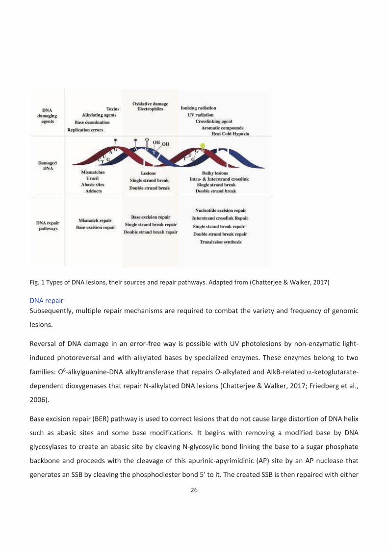
26
Fig. 1 Types of DNA lesions, their sources and repair pathways. Adapted from (Chatterjee & Walker, 2017)
DNA repair
Subsequently, multiple repair mechanisms are required to combat the variety and frequency of genomic
lesions.
Reversal of DNA damage in an error-free way is possible with UV photolesions by non-enzymatic light-
induced photoreversal and with alkylated bases by specialized enzymes. These enzymes belong to two
families: O6-alkylguanine-DNA alkyltransferase that repairs O-alkylated and AlkB-related a-ketoglutarate-
dependent dioxygenases that repair N-alkylated DNA lesions (Chatterjee & Walker, 2017; Friedberg et al.,
2006).
Base excision repair (BER) pathway is used to correct lesions that do not cause large distortion of DNA helix
such as abasic sites and some base modifications. It begins with removing a modified base by DNA
glycosylases to create an abasic site by cleaving N-glycosylic bond linking the base to a sugar phosphate
backbone and proceeds with the cleavage of this apurinic-apyrimidinic (AP) site by an AP nuclease that
generates an SSB by cleaving the phosphodiester bond 5’ to it. The created SSB is then repaired with either
27
the short-patch or long-patch repair pathway. An abasic site gets removed and the gap is filled by DNA
polymerase beta (Polb) and is followed by DNA ligase 1 (LIG1) -mediated ligation. BER is predominantly
used in the G1 phase of the cell cycle (Chatterjee & Walker, 2017; Dianov & Hübscher, 2013).
Nucleotide excision repair (NER) acts to repair bulky lesions and is divided into two mechanisms: global
genome NER (GG-NER) and transcription-coupled NER (TC-NER). They differ in a recognition step: while in
GG-NER a complex of XPC (Xeroderma Pigmentosum, complementation group C), RAD23b and CETN2
(Centrin2) scans genome for the presence of transient ssDNA caused by DNA helix unwinding at the place
of damage (Chatterjee & Walker, 2017; Nishi et al., 2005), TC-NER is initiated by PolII and involves CSA and
CSB proteins (Cockayne Syndrome proteins A and B) (Chatterjee & Walker, 2017; Friedberg et al., 2006;
Marteijn et al., 2014). Following repair process is the same for both pathways and involves pre-incision
complex (consisting of TFIIH (Transcription factor II H), XPA (Xeroderma Pigmentosum, complementation
group A), RPA (Replication protein A), and XPG (Xeroderma Pigmentosum, complementation group G))
formation, XPF (Excision repair cross-complementing rodent repair deficiency, complementation group 4)-
ERCC1 and XPC-induced cleavage, gap filling by Pold,e or k, and ligation by Lig1 of XRCC1-Lig3 (Chatterjee
& Walker, 2017; Friedberg et al., 2006).
Mismatch repair (MMR) is a post-replicative repair mechanism active in S and G2 phases of the cell cycle. It
is used to correct replication errors and thus plays a role in replication fidelity and genome maintenance
through generations (Chatterjee & Walker, 2017; T.A. Kunkel, 2009). It acts to correct mismatches that
occur due to replication errors as well as insertion-deletion loops at repetitive regions(Chatterjee & Walker,
2017; Friedberg et al., 2006). Various MSH (MutS homolog) proteins act to recognise lesions and initiate
Exonuclease 1 (EXO1)-mediated excision. Resulting gaps are processed by Pold, RFC (Replication factor C),
HMGB1 (high mobility group box 1 protein), and Lig1 (Chatterjee & Walker, 2017).
Interstrand crosslink (ICL) repair is required when a covalent bond is formed between bases of two
complementary strands. It is mediated by FA (Fanconi anemia) proteins. FA family contains 21 functional
complementation groups, which are involved in ICL resistance (Chatterjee & Walker, 2017; Clauson et al.,
2013). Damage sites are recognised by FANCM protein together with FAAP24 (FA associated protein of 24
kDA) and Forkhead box protein C2 (MFH) (Chatterjee & Walker, 2017; Ciccia et al., 2007; Clauson et al.,
2013). MFH stimulates fork remodelling and FANCM is responsible for Holliday junction migration and
28
creation of ssDNA gaps (Chatterjee & Walker, 2017; Clauson et al., 2013; Gari et al., 2008; Huang et al.,
2010). Presence of ssDNA results in RPA recruitment and ATR (Ataxia telangiectasia and Rad3-related
protein) signalling activation. In the context of the FA pathway, it leads to FANCE, FANCD2, and FANCI
activation by ATR target checkpoint kinase 1 (CHK1) as well as Mre11-Rad50-Nbs1 (MRN) complex assembly
(discussed in details later) (Chatterjee & Walker, 2017; Clauson et al., 2013; Duquette et al., 2012;
Smogorzewska et al., 2007; X. Wang et al., 2007). Other FA pathway core components get recruited to the
lesion and stimulate the excision of the DNA strand of the lesion by structure-specific endonucleases
(Chatterjee & Walker, 2017; Clauson et al., 2013). In replicating cells following repair is carried out by
translesion synthesis polymerases Pol i,k,n, and Rev1-like terminal deoxycytidyl transferase (REV1). These
polymerases are capable of carrying synthesis through aberrant DNA fragments, although with lower
fidelity (Chatterjee & Walker, 2017; Clauson et al., 2013; Minko et al., 2008; Räschle et al., 2008; Yamanaka
et al., 2010). In non-replicating cells, it depends on both GG-NER and TC-NER pathways and TLS polymerases
(Chatterjee & Walker, 2017; Clauson et al., 2013).
Translesion synthesis (TLS) is performed by a highly-conserved Y-family of DNA polymerases (consisting of
Pol i,k,n, and REV1) or some polymerases belonging to other families (B, X or A), such as Pol q,m,l, or z.
These polymerases are capable of carrying replication through DNA lesions but have considerably lower
fidelity as damaged bases often provide a misleading template (Chatterjee & Walker, 2017; Sale, 2013). It
has been shown that despite in some cases it is possible for a single polymerase to bypass a lesion (Johnson
et al., 1999; Sale, 2013) the bypass might also involve cooperation of different polymerases (Sale, 2013;
Shachar et al., 2009). Two models have been proposed for this phenomenon. The first one, the polymerase
switch model, suggests that TLS polymerases come sequentially in a two-step process, where an inserter
enzyme (usually Pol h, i, or k) incorporates a nucleotide at the place of the DNA lesion and then is replaced
by an extender enzyme (Pol z) (Chatterjee & Walker, 2017; Korzhnev & Hadden, 2016; Washington et al.,
2002). The second one, the gap-filling model, implies that ssDNA stretches are left by replicative
polymerases and are subsequently filled by TLS polymerases (Chatterjee & Walker, 2017; Quinet et al.,
2016; Sale et al., 2009). As translesion synthesis is a highly mutagenic process it must be tightly regulated.
In mammalian cells it is achieved by concentrating them in replication factories (Sabbioneda et al., 2008;
Sale, 2013). As previously mentioned, TLS polymerases are also known to play a role in other repair
29
pathways including NER, BER, and FA pathway, which further emphasises the importance of this
phenomenon (Chatterjee & Walker, 2017).
SSBs are repaired via three different pathways. First is the long patch single strand break repair (SSBR)
pathway where SSBs are detected by poly (ADP-ribose) polymerase 1 (PARP1) which is poly(ADP)-
ribosylated and quickly dissociates (Chatterjee & Walker, 2017; D’Amours et al., 1999), and the ends are
further processed by APE1 (apurinic-apyrimidinic endonuclease 1), PNKP (polynucleotide kinase 3’-
phosphate) and APTX (aprataxin). Subsequently, Flap endonuclease 1 (FEN1) removes the mutilated 5’ end
and the resulting ssDNA gap is filled by Polb and Pold/e and ligated by Lig1. In the short patch SSBR
pathways, breaks are recognised by APE1, and following steps converge with the long patch SSB pathway.
Another particularity of this pathway is the fact that gap-filling is performed exclusively by Polb and ligation
by Lig3 (Chatterjee & Walker, 2017; McKinnon & Caldecott, 2007). And the third one, the DNA
topoisomerase 1 (TOP1)-SSB pathway is a modification of the long-patch SSB repair where end processing
is performed by the TDP1, which acts to remove TOP1 (Keith W. Caldecott, 2008; Chatterjee & Walker,
2017).
Double-strand breaks are considered to be among the most deleterious and toxic kinds of DNA lesions as
in that case the second strand is not available as a repair template. Failure to repair them might lead to
severe consequences such as cancer or ageing, and it is therefore absolutely essential for cells to mend
them efficiently (Chatterjee & Walker, 2017; Mladenov et al., 2016; Thompson, 2012). Despite they pose a
serious threat to a cell in particular and to an organism in general, DSBs are quite abundant. They could
result from exposure to IR, UV, or small molecules (for example chemotherapy drugs), from environmental
stresses such as hyperosmotic stress, hypoxia or heat shock, and from replication stress, or be created on
purpose during lymphocyte maturation (V(D)J recombination of class-switch recombination) (Fillingham et
al., 2006; Lamarche et al., 2010; Ohnishi et al., 2009). As my work was focusing on DSB repair, I will discuss
mechanisms of their recognition and repair in more details in the next section.

30
DSB recognition and DNA damage response
The process of DSB repair can be conceptually divided into three sequential steps: DSB recognition and DNA
damage response (DDR) signalling activation, repair pathway choice, and repair itself. We shall consider all
of them one by one.
DSB sensing and DDR activation
Unlike other kinds of DNA lesions, DSB recognition is thought to be based on the altered chromatin
structure rather than on the recognition of mutilated DNA by sensor proteins. Chromatin relaxation, an
essential step for DSB repair, is promoted by certain covalent histone modifications, as well as by ATP-
dependent chromatin remodelling allowing repair factors to assess the damage site (Murr et al., 2006;
Thompson, 2012; van Attikum & Gasser, 2009). ATM (Ataxia telangiectasia mutated) activation seems to be
uniformly seen as the initial step of DDR. However, the exact mechanism of this event remains controversial
(Blackford & Jackson, 2017; Thompson, 2012). It has been proposed that it may result from changes in
chromatin structure due to relieving topological constraints caused by supercoiling (Bakkenist & Kastan,
2003; Thompson, 2012).
It has been observed that in eukaryotes DDR proteins often accumulate in conglomerates called ionizing
radiation-induced foci (IRIF) as they were first observed in cells treated with IR. IRIFs are considered to be
an indication of ongoing repair of one or more DSB (Bekker-Jensen et al., 2006; Carney et al., 1998;
Fernandez-Capetillo et al., 2003; van Attikum & Gasser, 2009; Vignard et al., 2013). Among others,
components of MRN (MRE11-RAD50-NBS1) complex appear to accumulate at IRIFs (Fernandez-Capetillo et
al., 2003; van Attikum & Gasser, 2009).
It is widely accepted that in higher eukaryotic cells DSBs are sensed by MRN complex (Ji Hoon Lee & Paull,
2005; Ohnishi et al., 2009; Thompson, 2012; van Attikum & Gasser, 2009). Despite influencing the repair
pathway choice at later stages by favouring one of the pathways, homologous recombination (HR), it has
been shown that MRN complex is involved at the earliest steps of DSB recognition and is essential for DDR
activation in all repair pathways. It binds to free DNA ends at the break site and promotes ATM activation
(Blackford & Jackson, 2017; Carney et al., 1998; Dupré et al., 2006; Ji Hoon Lee & Paull, 2004; Lou et al.,
2006; Stucki et al., 2005; van Attikum & Gasser, 2009). Along with two other phosphoinositide 3-kinase
(PI3K)-related kinases (PIKKs), ATR (ATM and Rad3-related kinase), and DNA-PKcs (DNA-dependent protein
kinase catalytic subunit), ATM plays a major role in DSB repair (Blackford & Jackson, 2017).
31
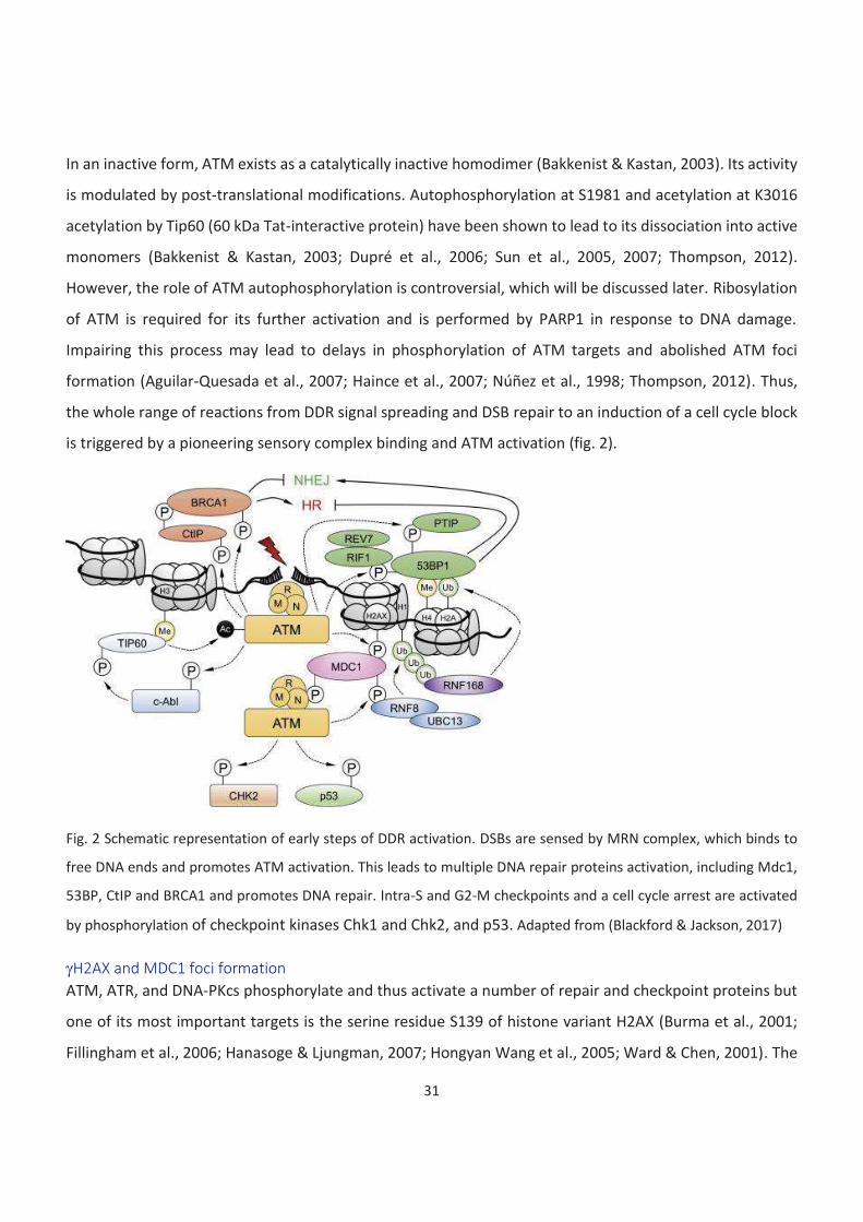
In an inactive form, ATM exists as a catalytically inactive homodimer (Bakkenist & Kastan, 2003). Its activity
is modulated by post-translational modifications. Autophosphorylation at S1981 and acetylation at K3016
acetylation by Tip60 (60 kDa Tat-interactive protein) have been shown to lead to its dissociation into active
monomers (Bakkenist & Kastan, 2003; Dupré et al., 2006; Sun et al., 2005, 2007; Thompson, 2012).
However, the role of ATM autophosphorylation is controversial, which will be discussed later. Ribosylation
of ATM is required for its further activation and is performed by PARP1 in response to DNA damage.
Impairing this process may lead to delays in phosphorylation of ATM targets and abolished ATM foci
formation (Aguilar-Quesada et al., 2007; Haince et al., 2007; Núñez et al., 1998; Thompson, 2012). Thus,
the whole range of reactions from DDR signal spreading and DSB repair to an induction of a cell cycle block
is triggered by a pioneering sensory complex binding and ATM activation (fig. 2).
Fig. 2 Schematic representation of early steps of DDR activation. DSBs are sensed by MRN complex, which binds to
free DNA ends and promotes ATM activation. This leads to multiple DNA repair proteins activation, including Mdc1,
53BP, CtIP and BRCA1 and promotes DNA repair. Intra-S and G2-M checkpoints and a cell cycle arrest are activated
by phosphorylation of checkpoint kinases Chk1 and Chk2, and p53. Adapted from (Blackford & Jackson, 2017)
gH2AX and MDC1 foci formation
ATM, ATR, and DNA-PKcs phosphorylate and thus activate a number of repair and checkpoint proteins but
one of its most important targets is the serine residue S139 of histone variant H2AX (Burma et al., 2001;
Fillingham et al., 2006; Hanasoge & Ljungman, 2007; Hongyan Wang et al., 2005; Ward & Chen, 2001). The

32
roles of ATM and DNA-PKcs in phosphorylating H2AX are largely overlapping. However, there some aspects
specific for each kinase (to be discussed later) (Stiff et al., 2004; Thompson, 2012). This phosphorylated
form is called gH2AX and it is one of the main signalling hallmarks of a DSB, independently of the way of its
induction and on whether it was caused by a hostile environment or induced in a controlled manner as a
part of cell homeostasis (Fillingham et al., 2006; Hua Tang Chen et al., 2000; Nazarov et al., 2003; Petersen
et al., 2001; Rogakou et al., 1998, 1999; Tomilin et al., 2001). In mammalian cells, it forms large domains,
up to megabases size (Rogakou et al., 1998; van Attikum & Gasser, 2009). gH2AX foci arise quickly, within
minutes after DNA damaging event has taken place, but continue to expand further for one to several hours
according to different studies (Y. Lee et al., 2019; Löbrich et al., 2010; Sharma et al., 2012; Staszewski et al.,
2008). Although H2AX-deficient mice are viable, they show increased genome instability and phenotypical
abnormalities, underlining its importance for DNA repair (Celeste et al., 2002, 2003; Thompson, 2012;
Weyemi et al., 2018). Another histone that is phosphorylated in the course of DDR signalling activation is
H2B. H2B phosphorylation on S14 is induced by IR and progresses to form foci. They colocalise with those
of gH2AX, although accumulating slower (Fernandez-Capetillo et al., 2004; Thompson, 2012). It has recently
been shown that kinases responsible for this process are Mst1 and 2 (Bitra et al., 2017; Pefani et al., 2018).
As DDR progresses, gH2AX is bound by the Mediator of DNA damage checkpoint protein 1 (MDC1), which
serves as a platform for a repair machinery assembly (Thompson, 2012). It directly interacts with multiple
proteins involved in DSB repair, such as ATM, MRN complex, 53BP1, and Structural maintenance of
chromosomes protein 1 (Smc1) (Stewart et al., 2003; Thompson, 2012). H2AX phosphorylation, as well as
MDC1 recruitment, also acts as a signal for activation of intra-S and G2-M checkpoints and a cell cycle arrest,
notably for phosphorylation of checkpoint kinases Chk1 and Chk2, KRAB-associated protein 1 (Kap1) and
Smc1 (Celeste et al., 2003; Fernandez-Capetillo et al., 2002; Stewart et al., 2003; Thompson, 2012). It has
been shown that phosphorylation and foci formation of H2AX and MDC1 is interdependent (Stewart et al.,
2003; Thompson, 2012). Direct interaction of MDC1 and gH2AX has been proven by co-crystallization, and
the relevance of this interaction has been demonstrated by analysing MDC1 mutant or null cells, which
have impaired accumulation of other DDR factors including 53BP1, NBS1 (Nijmegen breakage syndrome
protein 1), and phosphorylated ATM (Lou et al., 2006; Stucki et al., 2005; van Attikum & Gasser, 2009).
Therefore, MDC1 can be considered an early DDR factor, important for both recruitment and retention of
other players (Thompson, 2012; van Attikum & Gasser, 2009).
33
Role of parylation in DDR
Another important process involved in DDR is poly-ADP-ribosylation, or parylation, a post-translational
modification crucial for DSB recognition. This modification is catalysed by PARP (Poly-ADP-Ribosyl
Polymerase) enzymes that covalently link poly-ADP-ribosyl chains to protein substrates. PARP1 and PARP2
are involved in both SSB and DSB recognition, whereas PARP3 specifically responds to DSBs (Beck, Robert,
et al., 2014; Boehler et al., 2011; Boulton et al., 1999; K. W. Caldecott, 2014; Rulten et al., 2011). PARP
enzymes mediate DDR activation in several ways. First, some DDR factors have a PAR motif and are
therefore recruited to damage sites in a PAR-dependent manner. Second, parylation of DDR and
downstream repair factors might influence their catalytic activity or capability to bind to DNA or their
interaction factors. In particular, parylation facilitates the recruitment of MRN complex and ATM kinase to
a break site (Beck, Robert, et al., 2014; Bryant et al., 2009). Further on repair process, parylation plays a
role in a pathway choice by promoting end resection, and therefore favouring homologous recombination
and microhomology-mediated end joining (Beck, Boehler, et al., 2014; Langelier et al., 2014; Luijsterburg et
al., 2016). However, PARP3 is involved in non-homologous end-joining by limiting end-resection as well as
by promoting X-ray repair cross-complementing protein 1 (XRCC4)-Lig4 mediated ligation (Beck, Boehler,
et al., 2014; K. W. Caldecott, 2014; Fenton et al., 2013; Rulten et al., 2011). Therefore, PARP enzymes play
an important and complex role in DSB repair from the first to the last steps of this process.
Signal transduction and amplification. DDR kinases
As I mentioned before, break recognition leads to an activation of ATM, ATR, and DNA-PKcs kinases that all
belong to the phosphatidylinositol-3-kinase family. These kinases mediate signal transduction and
amplification via gH2AX foci formation and play a role in downstream DNA repair proteins activation.
ATM is the central kinase involved in DDR. It was identified in 1995 a single mutated gene responsible for a
rare genetic disorder, ataxia telangiectasia occurrence (Blackford & Jackson, 2017; K. Savitsky et al., 1995).
Ataxia telangiectasia is characterised by dilated blood vessels, progressive neurodegeneration, and
immunodeficiency (Blackford & Jackson, 2017). The activation of ATM proceeds as dissociation of
catalytically inactive homodimers into active monomers (Bakkenist & Kastan, 2003; Blackford & Jackson,
2017; Thompson, 2012). Among post-translational modifications that lead to its activation
autophosphorylation at S1981 was the first to be discovered (Bakkenist & Kastan, 2003). However, its role
remains controversial as on the one hand it has been reported to be the process initiated as the very first
34
step of DDR stimulated by chromatin changes upon the DSB occurrence, and on the other hand, it has been
shown dispensable for ATM functioning (Bakkenist & Kastan, 2003; Blackford & Jackson, 2017; Kozlov et al.,
2006; Pellegrini et al., 2006; Thompson, 2012). Another possible mechanism for early ATM activation is
acetylation by Tip60, which is attracted to the break site by exposed H3K9me3 nucleosomes, on K3016
(Blackford & Jackson, 2017; Sun et al., 2005, 2007). It is certain, however, that whatever causes its initial
activation, it is later enhanced by ATM direct interaction with the C-terminal domain of Nbs1, a component
of MRN complex. This interaction is stabilised in the presence of free DNA ends (Blackford & Jackson, 2017;
Falck et al., 2005). Nevertheless, MRN-independent mechanisms of ATM activation have also been shown
in multiple studies (Blackford & Jackson, 2017; Guo et al., 2010; Olcina et al., 2013). Activated ATM
phosphorylates a vast variety of targets, influencing the process of DSB repair at all levels: break recognition
and DDR activation, DDR enhancement, repair pathway choice and stimulation, cell cycle checkpoint
activation, and apoptosis. Its role in break recognition and DDR activation has already been discussed.
Further on ATM promotes DDR by phosphorylating H2AX into gH2AX and MDC1, the major gH2AX reader,
promoting its dimerization and stabilisation (Blackford & Jackson, 2017; Burma et al., 2001; Jungmichel et
al., 2012; Jinping Liu et al., 2012; Thompson, 2012). MDC1 further recruits the MRN complex, thus
promoting additional ATM accumulation and H2AX phosphorylation, thus providing a positive feedback
loop for repair foci formation (Blackford & Jackson, 2017). Moreover, ATM has also been shown to be
involved in chromatin relaxation and remodelling around the break (Goodarzi et al., 2008; Moyal et al.,
2011; Ziv et al., 2006). ATM also promotes recruitment downstream repair proteins to the break site. It was
shown to predominantly stimulate HR by phosphorylating CtBP-interacting protein (CtIP), a major MRN
interactor that promotes end-resection (Blackford & Jackson, 2017; Sartori et al., 2007; Shibata et al., 2011).
However, ATM is also able to stimulate DSB repair in G1 in a way that requires ATM, DNA-PK, MRN complex
and Artemis activity and by promoting NHEJ due to its redundant functions with XRCC4-like factor (XLF)
(Blackford & Jackson, 2017; Riballo et al., 2004; Zha et al., 2011). Additionally, it phosphorylates Chk2 and
p53 causing a cell cycle block at G2/M transition (Banin et al., 1998; Blackford & Jackson, 2017; Matsumura
et al., 2015; Siliciano et al., 1997).
The existence of DNA-PK was presupposed in 1985 when it was observed that the addition of double-
stranded DNA (dsDNA) into Xenopus and Arbacia egg extracts promoted phosphorylation of several
proteins and named DNA-dependent protein kinase (Blackford & Jackson, 2017; Walker et al., 1985). This

35
kinase was purified five years later (Blackford & Jackson, 2017; Carter et al., 1990; Jackson et al., 1990; Lees-
Miller et al., 1990). DNA-PKcs is recruited to DNA damage sites by Ku70/Ku80 (Ku autoantigen protein p70/80
homolog) heterodimers and form DNA-PK complex that enhances the catalytic activity of the kinase
(Blackford & Jackson, 2017; Gell & Jackson, 1999; Singleton et al., 1999). DNA-PKcs acts cooperatively with
ATM to promote gH2AX signal spreading and DDR activation (Firsanov et al., 2011; Stiff et al., 2004; Hongyan
Wang et al., 2005). It also plays a crucial role in NHEJ, and multiple target proteins have been suggested
(Blackford & Jackson, 2017; Enriquez-Rios et al., 2017). Some prominent NHEJ players as Artemis, are
among the confirmed targets (Blackford & Jackson, 2017; W. Jiang et al., 2015; Malu et al., 2012). While
DNA-PKcs phosphorylation by ATM leads to end-processing by promoting Artemis recruitment, additional
autophosphorylation is necessary for it to act on other targets. The exact target proteins are unknown but
the importance of DNA-PKcs is affirmed by the fact that its knock-out is lethal in mice during embryonic
development by E16.5 and on the cellular level leads to an inability to multiple NHEJ defects similar to
XRCC4 and XLF deficient cells (Blackford & Jackson, 2017; W. Jiang et al., 2015). Particularly, it was shown
to play an important role in regulating end-ligation by promoting the transition between two synaptic
complexes (Blackford & Jackson, 2017; Graham et al., 2016; W. Jiang et al., 2015). At the same time, DNA-
PKcs phosphorylates RPA and activates the S-phase cell cycle checkpoint (Ashley et al., 2014). Activated RPA
mediates the recruitment of PALB2 (Partner and localizer of BRCA2), thus promoting DSB repair progression
(Murphy et al., 2014). On the other hand, it has also been shown that Chk1 promotes DNA-PK activity in
order to stimulate fast repair by NHEJ and prevent cell cycle block (Goudelock et al., 2003; Shimura et al.,
2007).
ATR is an analogue of the yeast protein Mec1 (Mitosis entry checkpoint 1) that was first discovered as Rad3
in a radiosensitivity screen and later as being essential for cell cycle checkpoints (Blackford & Jackson, 2017;
Nasim & Smith, 1975; Weinert et al., 1994). A human ATR protein was cloned independently by two
different groups (Bentley et al., 1996; Blackford & Jackson, 2017; Cimprich et al., 1996). ATR kinase plays a
crucial role in replication stress response. It gets recruited ssDNA in response to various kinds of genotoxic
stress. Therefore, it accumulates at DSB sites after resection but also at stalled replication forks. Interaction
of ATR with RPA allows an ATRIP (ATR-interacting protein) cofactor recruitment and binding to ATR
(Blackford & Jackson, 2017; Zou & Elledge, 2003). ATR-ATRIP heterodimer formation is essential for ATR
activation. ATR recruitment alone is not sufficient for the optimal response and requires cofactors such as
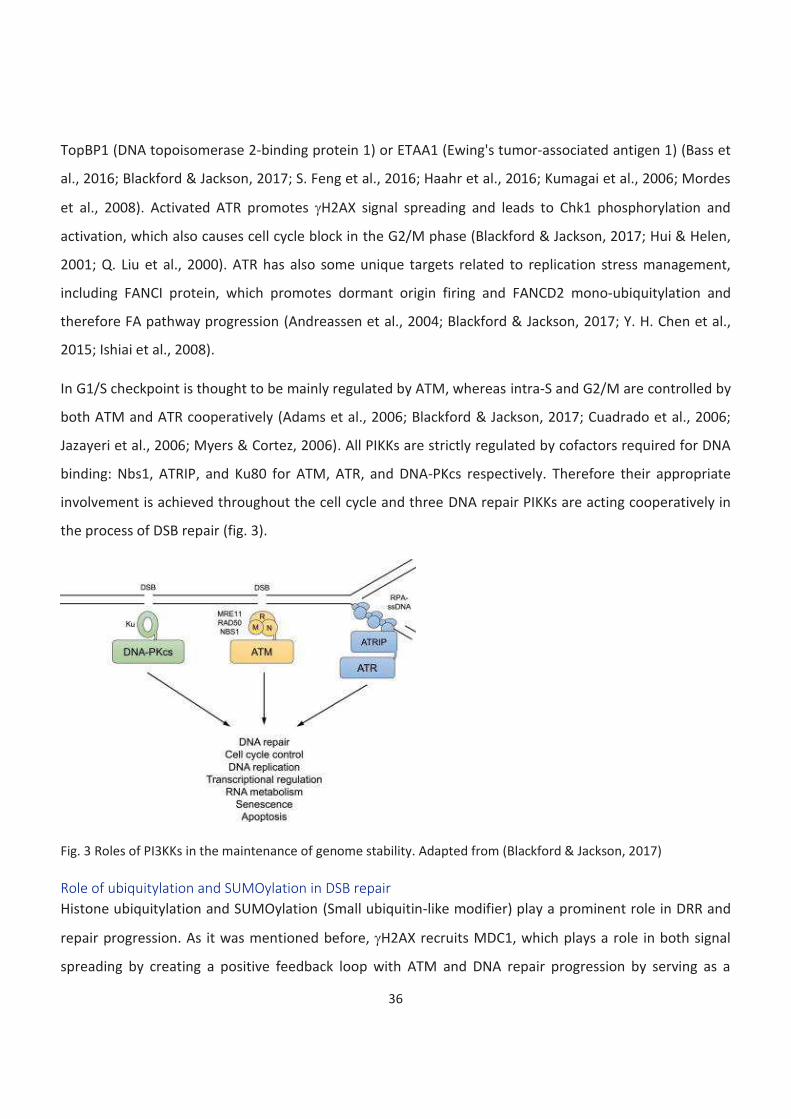
36
TopBP1 (DNA topoisomerase 2-binding protein 1) or ETAA1 (Ewing's tumor-associated antigen 1) (Bass et
al., 2016; Blackford & Jackson, 2017; S. Feng et al., 2016; Haahr et al., 2016; Kumagai et al., 2006; Mordes
et al., 2008). Activated ATR promotes gH2AX signal spreading and leads to Chk1 phosphorylation and
activation, which also causes cell cycle block in the G2/M phase (Blackford & Jackson, 2017; Hui & Helen,
2001; Q. Liu et al., 2000). ATR has also some unique targets related to replication stress management,
including FANCI protein, which promotes dormant origin firing and FANCD2 mono-ubiquitylation and
therefore FA pathway progression (Andreassen et al., 2004; Blackford & Jackson, 2017; Y. H. Chen et al.,
2015; Ishiai et al., 2008).
In G1/S checkpoint is thought to be mainly regulated by ATM, whereas intra-S and G2/M are controlled by
both ATM and ATR cooperatively (Adams et al., 2006; Blackford & Jackson, 2017; Cuadrado et al., 2006;
Jazayeri et al., 2006; Myers & Cortez, 2006). All PIKKs are strictly regulated by cofactors required for DNA
binding: Nbs1, ATRIP, and Ku80 for ATM, ATR, and DNA-PKcs respectively. Therefore their appropriate
involvement is achieved throughout the cell cycle and three DNA repair PIKKs are acting cooperatively in
the process of DSB repair (fig. 3).
Fig. 3 Roles of PI3KKs in the maintenance of genome stability. Adapted from (Blackford & Jackson, 2017)
Role of ubiquitylation and SUMOylation in DSB repair
Histone ubiquitylation and SUMOylation (Small ubiquitin-like modifier) play a prominent role in DRR and
repair progression. As it was mentioned before, gH2AX recruits MDC1, which plays a role in both signal
spreading by creating a positive feedback loop with ATM and DNA repair progression by serving as a
37
platform for downstream repair complexes assembly. Multiple ubiquitylation steps are performed to
ensure this process. As a first step, H2AX acetylation enabled by Tip60 facilitated H2AX monoubiquitylation
at K118/119. Not all players involved in this process are known to date but Ubc13 (ubiquitin-conjugating
enzyme 13) E2 ligase have been shown to play a role in it (Ikura et al., 2007; X. Jiang et al., 2010; Thompson,
2012; G. Y. Zhao et al., 2007).
Also, PRC1 (Polycomp repressive complex 1) gets recruited to DSBs and its subunits, Rnf2 (RING finger
protein 2) and Bmi1 (B cell-specific Moloney murine leukemia virus integration site 1), have been shown to
mediate the monoubiquitylation of H2A and H2AX on K119 (Ismail et al., 2010; Thompson, 2012). This
modification plays a role in the recruitment of DDR proteins and IRIFs formation. RNF2/BMI1 complex
controls DDR in an ATM-dependent manner and their knockdown leads to increased sensitivity to IR and
compromised DSB repair (Facchino et al., 2010; Pan et al., 2011; Thompson, 2012).
The best-established E3 ligases involved in DSB repair are RNF8 (RING finger protein 8), CHFR (checkpoint
protein with FHA and RING domain), and RNF168 (RING finger protein 168). All three of them use Ubc13 an
E2 ligase (Panier & Durocher, 2009; Thompson, 2012). RNF8 recruitment is facilitated by MDC1 (Huen et
al., 2007; Mailand et al., 2007; Thompson, 2012). RNF8 ubiquitylates histones H1 and H2A, H2AX, and H2B
to promote binding of repair factors such as 53BP1 (TP53-binding protein1), RAP80 (Receptor-associated
protein 80) and BRCA1 (Breast cancer type 1 susceptibility protein) (Huen et al., 2007; Mandemaker et al.,
2017; Sobhian et al., 2007; Thompson, 2012).
Another E3-ligase, RNF168, is recruited in an RNF8-dependent manner to enhance the signal by
ubiquitylation of H1 and H2A histones (Mandemaker et al., 2017; Thompson, 2012). Upon irradiation,
RNF168 gets stabilized by HERC2 (HECT domain and RCC1-like domain-containing protein 2) (Bekker-Jensen
& Mailand, 2010; Thompson, 2012). It colocalizes with gH2AX at damage sites and promotes amplification
of the ubiquitylation established by RNF8 and PRC1 complex (Doil et al., 2009; Thompson, 2012).
Finally, CHFR (checkpoint protein with FHA and RING domain) E3 ligase, which has considerable structural
similarity to RNF8, has been shown to act synergistically with it in promoting ATM activation and cell cycle
checkpoints activation (Bothos et al., 2003; Thompson, 2012; J. Wu et al., 2011).
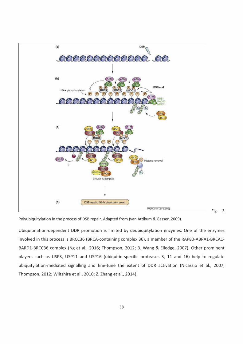
38
Fig. 3
Polyubiquitylation in the process of DSB repair. Adapted from (van Attikum & Gasser, 2009).
Ubiquitination-dependent DDR promotion is limited by deubiquitylation enzymes. One of the enzymes
involved in this process is BRCC36 (BRCA-containing complex 36), a member of the RAP80-ABRA1-BRCA1-
BARD1-BRCC36 complex (Ng et al., 2016; Thompson, 2012; B. Wang & Elledge, 2007), Other prominent
players such as USP3, USP11 and USP16 (ubiquitin-specific proteases 3, 11 and 16) help to regulate
ubiquitylation-mediated signalling and fine-tune the extent of DDR activation (Nicassio et al., 2007;
Thompson, 2012; Wiltshire et al., 2010; Z. Zhang et al., 2014).
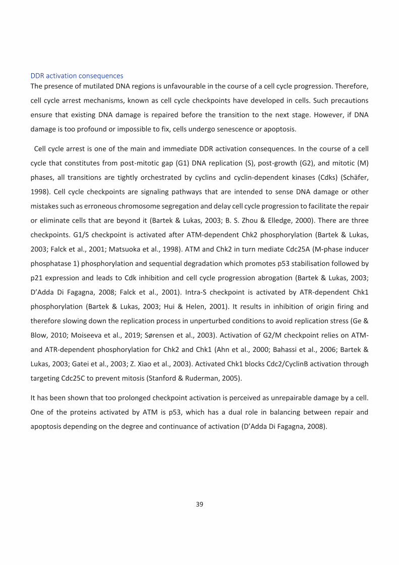
39
DDR activation consequences
The presence of mutilated DNA regions is unfavourable in the course of a cell cycle progression. Therefore,
cell cycle arrest mechanisms, known as cell cycle checkpoints have developed in cells. Such precautions
ensure that existing DNA damage is repaired before the transition to the next stage. However, if DNA
damage is too profound or impossible to fix, cells undergo senescence or apoptosis.
Cell cycle arrest is one of the main and immediate DDR activation consequences. In the course of a cell
cycle that constitutes from post-mitotic gap (G1) DNA replication (S), post-growth (G2), and mitotic (M)
phases, all transitions are tightly orchestrated by cyclins and cyclin-dependent kinases (Cdks) (Schäfer,
1998). Cell cycle checkpoints are signaling pathways that are intended to sense DNA damage or other
mistakes such as erroneous chromosome segregation and delay cell cycle progression to facilitate the repair
or eliminate cells that are beyond it (Bartek & Lukas, 2003; B. S. Zhou & Elledge, 2000). There are three
checkpoints. G1/S checkpoint is activated after ATM-dependent Chk2 phosphorylation (Bartek & Lukas,
2003; Falck et al., 2001; Matsuoka et al., 1998). ATM and Chk2 in turn mediate Cdc25A (M-phase inducer
phosphatase 1) phosphorylation and sequential degradation which promotes p53 stabilisation followed by
p21 expression and leads to Cdk inhibition and cell cycle progression abrogation (Bartek & Lukas, 2003;
D’Adda Di Fagagna, 2008; Falck et al., 2001). Intra-S checkpoint is activated by ATR-dependent Chk1
phosphorylation (Bartek & Lukas, 2003; Hui & Helen, 2001). It results in inhibition of origin firing and
therefore slowing down the replication process in unperturbed conditions to avoid replication stress (Ge &
Blow, 2010; Moiseeva et al., 2019; Sørensen et al., 2003). Activation of G2/M checkpoint relies on ATM-
and ATR-dependent phosphorylation for Chk2 and Chk1 (Ahn et al., 2000; Bahassi et al., 2006; Bartek &
Lukas, 2003; Gatei et al., 2003; Z. Xiao et al., 2003). Activated Chk1 blocks Cdc2/CyclinB activation through
targeting Cdc25C to prevent mitosis (Stanford & Ruderman, 2005).
It has been shown that too prolonged checkpoint activation is perceived as unrepairable damage by a cell.
One of the proteins activated by ATM is p53, which has a dual role in balancing between repair and
apoptosis depending on the degree and continuance of activation (D’Adda Di Fagagna, 2008).
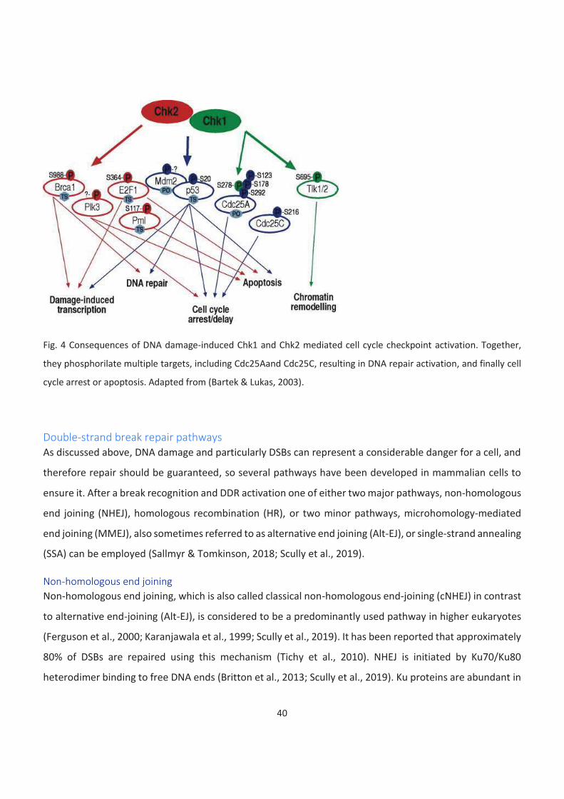
40
Fig. 4 Consequences of DNA damage-induced Chk1 and Chk2 mediated cell cycle checkpoint activation. Together,
they phosphorilate multiple targets, including Cdc25Aand Cdc25C, resulting in DNA repair activation, and finally cell
cycle arrest or apoptosis. Adapted from (Bartek & Lukas, 2003).
Double-strand break repair pathways
As discussed above, DNA damage and particularly DSBs can represent a considerable danger for a cell, and
therefore repair should be guaranteed, so several pathways have been developed in mammalian cells to
ensure it. After a break recognition and DDR activation one of either two major pathways, non-homologous
end joining (NHEJ), homologous recombination (HR), or two minor pathways, microhomology-mediated
end joining (MMEJ), also sometimes referred to as alternative end joining (Alt-EJ), or single-strand annealing
(SSA) can be employed (Sallmyr & Tomkinson, 2018; Scully et al., 2019).
Non-homologous end joining
Non-homologous end joining, which is also called classical non-homologous end-joining (cNHEJ) in contrast
to alternative end-joining (Alt-EJ), is considered to be a predominantly used pathway in higher eukaryotes
(Ferguson et al., 2000; Karanjawala et al., 1999; Scully et al., 2019). It has been reported that approximately
80% of DSBs are repaired using this mechanism (Tichy et al., 2010). NHEJ is initiated by Ku70/Ku80
heterodimer binding to free DNA ends (Britton et al., 2013; Scully et al., 2019). Ku proteins are abundant in
41
the nucleus and have a high affinity to free DNA ends which allows them to bind to a break site within 5
seconds after its occurrence (Fell & Schild-Poulter, 2015; Mari et al., 2006). It serves a dual role of, on the
one hand, protecting DNA ends from degradation and keeping them together for further repair, and on the
other hand, preventing resection (Fell & Schild-Poulter, 2015; Mimitou & Keeney, 2018). It also serves as a
platform for DNA repair machinery assembly (Costantini et al., 2007; Fell & Schild-Poulter, 2015; Hsu et al.,
2002; Rivera-Calzada et al., 2007; Yano et al., 2008). Another important function of Ku70/80 heterodimer
is in DDR regulation by modulating ATM activity and in preventing apoptosis by binding and inhibiting a pro-
apoptotic factor Bax (Amsel et al., 2008; Fell & Schild-Poulter, 2015; Tomimatsu et al., 2007; X. Y. Zhou et
al., 2002). DNA-PKcs binds to Ku70/80 heterodimer to form an active DNA-PK complex. DNA-PKcs mediates
tethering of broken ends (Fell & Schild-Poulter, 2015; Hammel et al., 2010). DNA-PK autophosphorylates its
own subunits and phosphorylates several other target molecules including Artemis, PNKP, XRCC4, XLF, and
DNA ligase IV (Fell & Schild-Poulter, 2015; Goodarzi et al., 2006; Hammel et al., 2010; Y. G. Wang et al.,
2004; Y. Yu et al., 2003, 2008). In case end-processing is required before ligation it can be performed by
various enzymes including PNKP (polynucleotide kinase/phosphatase), Artemis, Exo1, Tdp1, and others in
order to remove 3’ phosphate ends, 3ʹ-phosphoglycolates, or 5ʹ-hydroxyl groups and generate 5’ phosphate
ends required for further ligation (Fell & Schild-Poulter, 2015; Mahaney et al., 2009). The next step is
ligation, which is performed by a ligase complex of DNA Ligase IV with XRCC4 and XLF, co-factors required
for its stabilisation and stimulation (Ahnesorg et al., 2006; Fell & Schild-Poulter, 2015; Grawunder et al.,
1997; Nick McElhinny et al., 2000). However, it has recently been shown that a small subset of breaks
undergoes resection-dependent NHEJ repair enabled by Artemis and CtIP cooperation and resection
performed by MRE11 (Meiotic recombination 11 homolog 1) and EXO1 (Biehs et al., 2017; Shibata et al.,
2018). Finally, Ku dimers have to be removed from repaired DNA. The exact mechanism of how that
happens remains unclear. However, two mechanisms have been proposed: ubiquitylation-driven
degradation or DNA nicking that would allow Ku to escape (Fell & Schild-Poulter, 2015; Langerak et al.,
2011; Postow et al., 2008).
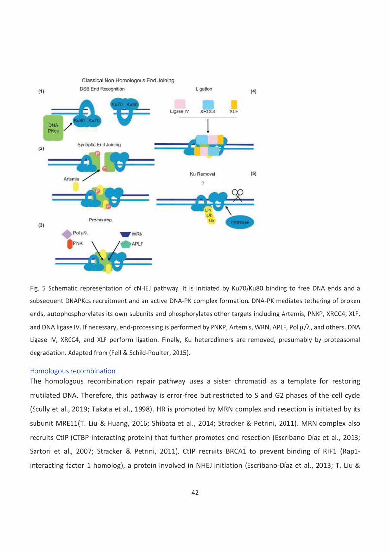
42
Fig. 5 Schematic representation of cNHEJ pathway. It is initiated by Ku70/Ku80 binding to free DNA ends and a
subsequent DNAPKcs recruitment and an active DNA-PK complex formation. DNA-PK mediates tethering of broken
ends, autophosphorylates its own subunits and phosphorylates other targets including Artemis, PNKP, XRCC4, XLF,
and DNA ligase IV. If necessary, end-processing is performed by PNKP, Artemis, WRN, APLF, Pol m/l, and others. DNA
Ligase IV, XRCC4, and XLF perform ligation. Finally, Ku heterodimers are removed, presumably by proteasomal
degradation. Adapted from (Fell & Schild-Poulter, 2015).
Homologous recombination
The homologous recombination repair pathway uses a sister chromatid as a template for restoring
mutilated DNA. Therefore, this pathway is error-free but restricted to S and G2 phases of the cell cycle
(Scully et al., 2019; Takata et al., 1998). HR is promoted by MRN complex and resection is initiated by its
subunit MRE11(T. Liu & Huang, 2016; Shibata et al., 2014; Stracker & Petrini, 2011). MRN complex also
recruits CtIP (CTBP interacting protein) that further promotes end-resection (Escribano-Díaz et al., 2013;
Sartori et al., 2007; Stracker & Petrini, 2011). CtIP recruits BRCA1 to prevent binding of RIF1 (Rap1-
interacting factor 1 homolog), a protein involved in NHEJ initiation (Escribano-Díaz et al., 2013; T. Liu &
43
Huang, 2016). End-resection is then performed by EXO1 (Exonuclease1) by 5’ to 3’ digestion of DNA ends,
which leads to the formation of single-strand DNA (ssDNA) overhangs (Cannavo et al., 2013; Eid et al., 2010;
T. Liu & Huang, 2016). ssDNA is then bound by RPA (replication protein A) heterotrimer that protects it from
degradation (San Filippo et al., 2008; Van Komen et al., 2002). MRN complex promotes Bloom syndrome
helicase (BLM) recruitment, which together with DNA2 (DNA replication ATP-dependent helicase/nuclease
DNA2) promotes extended resection (Daley et al., 2017; Nimonkar et al., 2011; Scully et al., 2019). Rad51
recombinase must be recruited to a break site by interacting with RPA, and later replace it as to HR could
proceed (San Filippo et al., 2008; Scully et al., 2019). To that end, BRCA2 competes with RPA for binding
ssDNA and interacts with RAD51 monomers and BRCA1 via its partner PALB2 to promote RAD51 filament
formation (Jensen et al., 2010, 2013; Prakash et al., 2015; Scully et al., 2019; Stark et al., 2004; Wright et
al., 2018; Xia et al., 2006; Yang et al., 2005; F. Zhang et al., 2009). RAD51 nucleoprotein filaments stabilized
by RAD54 are involved in the homology search process that is a key step of HR facilitated by BRCA1-BARD1
(BRCA1-associated RING domain protein 1) (Sanchez et al., 2013; Scully et al., 2019; Wright et al., 2018; W.
Zhao et al., 2017). RAD51 nucleofilaments invade a sister chromatid in a RAD54-dependent manner and
form a three-strand helix intermediate which is further resolved into a heteroduplex formed by an invading
strand and a complementary strand (Z. Chen et al., 2008; Mazina & Mazin, 2004; van der Heijden et al.,
2008; Wright et al., 2018). The dissociated DNA strand is displaced into a displacement-loop (D-loop) and
protected by RPA (Daley et al., 2014; Wright et al., 2018). At this step RAD51 filament is disassembled in an
ATP-dependent manner (Scully et al., 2019; van der Heijden et al., 2008). Invading strand extension is
performed by DNA polymerase d, although translesion synthesis DNA polymerases could also be involved
in this process (McVey et al., 2016; Scully et al., 2019). PCNA (Proliferating cell nuclear antigen) and its
loader RFC1-5 has been shown to be required for the initiation of DNA synthesis by Pold during HR (X. Li et
al., 2009; Wright et al., 2018). The final steps of HR can be conducted in three different ways. Synthesis-
dependent strand annealing pathway (SDSA), where only one strand undergoes invasion. This results in the
annealing of the non-invading strand with the displaced strand once D-loop is unwound and promotes HR
termination. This is a non-crossover and therefore preferable pathway as it does not involve a Holliday
junction formation (Scully et al., 2019; Westmoreland & Resnick, 2013; Wright et al., 2018). Another
pathway is double Holliday junction formation which happens due to the second strand invasion and can
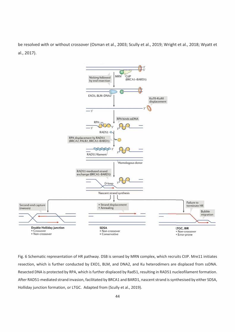
44
be resolved with or without crossover (Osman et al., 2003; Scully et al., 2019; Wright et al., 2018; Wyatt et
al., 2017).
Fig. 6 Schematic representation of HR pathway. DSB is sensed by MRN complex, which recruits CtIP. Mre11 initiates
resection, which is further conducted by EXO1, BLM, and DNA2, and Ku heterodimers are displaced from ssDNA.
Resected DNA is protected by RPA, which is further displaced by Rad51, resulting in RAD51 nucleofilament formation.
After RAD51-mediated strand invasion, facilitated by BRCA1 and BARD1, nascent strand is synthesised by either SDSA,
Holliday junction formation, or LTGC. Adapted from (Scully et al., 2019).
45
Finally, during replication long-tract gene conversion (LTGC) and break-induced replication (BIR) might be
deployed, in which DNA synthesis induced by HR proceeds to a large distance from the break sites. Those
pathways are highly mutagenic and are therefore suppressed in mammals and could mostly be observed if
one of the key HR players is dysfunctional (Chandramouly et al., 2013; Scully et al., 2019; Willis et al., 2017).
Microhomology-mediated end joining
MMEJ (or Alt-EJ) pathway was for a long time considered as a backup for the major ones. It is initiated by
end resection and requires short (2-20 bp) 3’ ssDNA overhangs (Seol et al., 2018; Welcker et al., 2000;
Wright et al., 2018). It is Ku-independent and require proteins known to be a part of HR end resection
machinery and is more prominent if NHEJ or HR is impaired (e.g. in KU or BRCA1-deficient cells) and utilizes
microhomologies in close proximity to a break site (Boboila et al., 2010; Ceccaldi et al., 2015; Scully et al.,
2019; Wright et al., 2018). This pathway leads to small deletions so it was also assumed that other
mechanisms, which are error-free or cause minimal error would be preferable. Break recognition is thought
to be mediated by PARP1 that competes with Ku dimer (Badie et al., 2015; Mansour et al., 2010, 2013).
After break recognition, the initial steps of MMEJ are common with HR. It is also dependent on Mre11 and
CtIP resection activity (Badie et al., 2015; Truong et al., 2013; Wright et al., 2018; Xie et al., 2009). However,
BLM/Exo1 complex that promotes long-range resection is inhibiting MMEJ (Daley et al., 2015; Wright et al.,
2018; Y. Wu et al., 2015). Mechanisms of microhomology search are not yet clear, but once it is found ssDNA
gaps are filled by Polq (Kent et al., 2015; Mateos-Gomez et al., 2017; Scully et al., 2019; Wright et al., 2018).
Polq contains a Rad51 domain that allows it to inhibit Rad51-mediated recombination as well as misplaces
RPA that is known to promote HR and negatively regulate MMEJ (Ahrabi et al., 2016; Ceccaldi et al., 2015;
S. K. Deng et al., 2014; Mateos-Gomez et al., 2015, 2017; Scully et al., 2019; Wright et al., 2018). The final
ligation step is performed by the XRCC1-Lig3 complex (Audebert et al., 2004; Okano et al., 2003, 2005;
Wright et al., 2018).
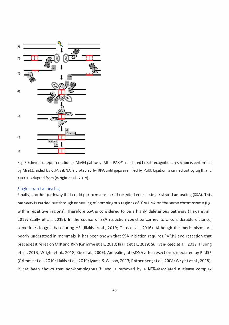
46
Fig. 7 Schematic representation of MMEJ pathway. After PARP1-mediated break recognition, resection is performed
by Mre11, aided by CtIP. ssDNA is protected by RPA until gaps are filled by Polq. Ligation is carried out by Lig III and
XRCC1. Adapted from (Wright et al., 2018).
Single-strand annealing
Finally, another pathway that could perform a repair of resected ends is single-strand annealing (SSA). This
pathway is carried out through annealing of homologous regions of 3’ ssDNA on the same chromosome (i.g.
within repetitive regions). Therefore SSA is considered to be a highly deleterious pathway (Iliakis et al.,
2019; Scully et al., 2019). In the course of SSA resection could be carried to a considerable distance,
sometimes longer than during HR (Iliakis et al., 2019; Ochs et al., 2016). Although the mechanisms are
poorly understood in mammals, it has been shown that SSA initiation requires PARP1 and resection that
precedes it relies on CtIP and RPA (Grimme et al., 2010; Iliakis et al., 2019; Sullivan-Reed et al., 2018; Truong
et al., 2013; Wright et al., 2018; Xie et al., 2009). Annealing of ssDNA after resection is mediated by Rad52
(Grimme et al., 2010; Iliakis et al., 2019; Iyama & Wilson, 2013; Rothenberg et al., 2008; Wright et al., 2018).
It has been shown that non-homologous 3’ end is removed by a NER-associated nuclease complex
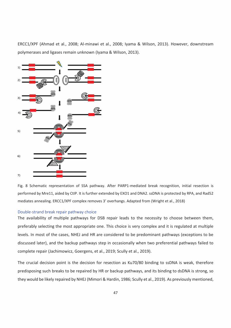
47
ERCC1/XPF (Ahmad et al., 2008; Al-minawi et al., 2008; Iyama & Wilson, 2013). However, downstream
polymerases and ligases remain unknown (Iyama & Wilson, 2013).
Fig. 8 Schematic representation of SSA pathway. After PARP1-mediated break recognition, initial resection is
performed by Mre11, aided by CtIP. It is further extended by EXO1 and DNA2. ssDNA is protected by RPA, and Rad52
mediates annealing. ERCC1/XPF complex removes 3’ overhangs. Adapted from (Wright et al., 2018)
Double-strand break repair pathway choice
The availability of multiple pathways for DSB repair leads to the necessity to choose between them,
preferably selecting the most appropriate one. This choice is very complex and it is regulated at multiple
levels. In most of the cases, NHEJ and HR are considered to be predominant pathways (exceptions to be
discussed later), and the backup pathways step in occasionally when two preferential pathways failed to
complete repair (Jachimowicz, Goergens, et al., 2019; Scully et al., 2019).
The crucial decision point is the decision for resection as Ku70/80 binding to ssDNA is weak, therefore
predisposing such breaks to be repaired by HR or backup pathways, and its binding to dsDNA is strong, so
they would be likely repaired by NHEJ (Mimori & Hardin, 1986; Scully et al., 2019). As previously mentioned,
48
one of the key factors in the repair pathway choice is the cell cycle state. This arises from HR requiring a
sister chromatid and therefore is limited to S and G2 phases of the cell cycle. Therefore many of the
resection-initiating factors that are essential for commitment to HR are regulated by CDKs (Aylon et al.,
2004; Ira et al., 2004; Scully et al., 2019; Tomimatsu et al., 2014). The competition between Ku70/80 binding
and repair machinery is ensured by multiple proteins, the 53BP1 and CtIP being the major players. CtIP
phosphorylation is essential for MRE11 endonuclease activity initiation and BRCA1 binding, whereas 53BP1
suppresses resection and promotes NHEJ (Anand et al., 2016; Bunting et al., 2010; Jachimowicz, Goergens,
et al., 2019; Scully et al., 2019; Xie et al., 2007; X. Yu & Chen, 2004). UBQLN4 (Ubiquilin-4) promotes MRE11
proteasomal degradation and DYNLL1 (Dynein light chain 1) blocks its function via direct interaction,
therefore inhibiting HR activation (He et al., 2018; Jachimowicz, Beleggia, et al., 2019; Jachimowicz,
Goergens, et al., 2019). 53BP1 acts in cooperation with its interactor PTIP (PAX transactivation activation
domain-interacting protein) and effectors RIF1 and REV7 (Rev7-like terminal deoxycytidyl transferase)
(Bunting et al., 2010; Escribano-Díaz et al., 2013; L. Feng et al., 2013; Jachimowicz, Goergens, et al., 2019;
Munoz et al., 2007; J. Wang et al., 2014; G. Xu et al., 2015; Zimmermann et al., 2013). PTIP directly interacts
with Artemis and ensures its retention at the break site (Jachimowicz, Goergens, et al., 2019; J. Wang et al.,
2014). The mechanism by which the 53BP1-RIF1-REV7 pathway promotes NHEJ was recently identified to
be via the Shieldin complex, which consists of REV7, SHLD1, SHLD2, and SHLD3 (Shieldin complex subunit
1-3). This complex is recruited to break sites in 53BP1 and RIF1 dependent way and protects them from
extensive resection. The loss of its components leads to PARP inhibition resistance in cancers due to HR
reinstatement (Findlay et al., 2018; Ghezraoui et al., 2018; Gupta et al., 2018; Jachimowicz, Goergens, et
al., 2019; Noordermeer et al., 2018; Tomida et al., 2018). It has also been proposed that it fills in ssDNA
ends to enable them for NHEJ (Mirman et al., 2018; Scully et al., 2019). REV7 and SHLD3 have been shown
to inhibit CtIP-dependent resection and therefore represses HR (Gupta et al., 2018; Jachimowicz, Goergens,
et al., 2019; G. Xu et al., 2015).
Misregulation of the repair pathway choice leads to severe consequences, including various syndromes and
cancer (Jachimowicz, Goergens, et al., 2019; Stewart et al., 2009; Terabayashi & Hanada, 2018).
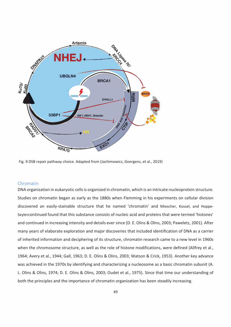
49
Fig. 9 DSB repair pathway choice. Adapted from (Jachimowicz, Goergens, et al., 2019)
Chromatin
DNA organization in eukaryotic cells is organized in chromatin, which is an intricate nucleoprotein structure.
Studies on chromatin began as early as the 1880s when Flemming in his experiments on cellular division
discovered an easily-stainable structure that he named ‘chromatin’ and Miescher, Kossel, and Hoppe-
Seylercontinued found that this substance consists of nucleic acid and proteins that were termed ‘histones’
and continued in increasing intensity and details ever since (D. E. Olins & Olins, 2003; Paweletz, 2001). After
many years of elaborate exploration and major discoveries that included identification of DNA as a carrier
of inherited information and deciphering of its structure, chromatin research came to a new level in 1960s
when the chromosome structure, as well as the role of histone modifications, were defined (Allfrey et al.,
1964; Avery et al., 1944; Gall, 1963; D. E. Olins & Olins, 2003; Watson & Crick, 1953). Another key advance
was achieved in the 1970s by identifying and characterizing a nucleosome as a basic chromatin subunit (A.
L. Olins & Olins, 1974; D. E. Olins & Olins, 2003; Oudet et al., 1975). Since that time our understanding of
both the principles and the importance of chromatin organization has been steadily increasing.
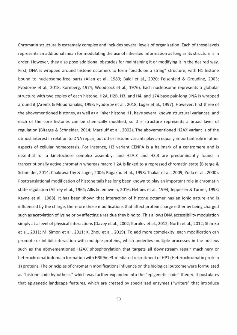
50
Chromatin structure is extremely complex and includes several levels of organization. Each of these levels
represents an additional mean for modulating the use of inherited information as long as its structure is in
order. However, they also pose additional obstacles for maintaining it or modifying it in the desired way.
First, DNA is wrapped around histone octamers to form “beads on a string” structure, with H1 histone
bound to nucleosome-free parts (Allan et al., 1980; Baldi et al., 2020; Felsenfeld & Groudine, 2003;
Fyodorov et al., 2018; Kornberg, 1974; Woodcock et al., 1976). Each nucleosome represents a globular
structure with two copies of each histone, H2A, H2B, H3, and H4, and 174 base pair-long DNA is wrapped
around it (Arents & Moudrianakis, 1993; Fyodorov et al., 2018; Luger et al., 1997). However, first three of
the abovementioned histones, as well as a linker histone H1, have several known structural variances, and
each of the core histones can be chemically modified, so this structure represents a broad layer of
regulation (Biterge & Schneider, 2014; Marzluff et al., 2002). The abovementioned H2AX variant is of the
utmost interest in relation to DNA repair, but other histone variants play an equally important role in other
aspects of cellular homeostasis. For instance, H3 variant CENPA is a hallmark of a centromere and is
essential for a kinetochore complex assembly, and H2A.Z and H3.3 are predominantly found in
transcriptionally active chromatin whereas macro H2A is linked to a repressed chromatin state (Biterge &
Schneider, 2014; Chakravarthy & Luger, 2006; Rogakou et al., 1998; Thakar et al., 2009; Yoda et al., 2000).
Posttranslational modification of histone tails has long been known to play an important role in chromatin
state regulation (Allfrey et al., 1964; Allis & Jenuwein, 2016; Hebbes et al., 1994; Jeppesen & Turner, 1993;
Kayne et al., 1988). It has been shown that interaction of histone octamer has an ionic nature and is
influenced by the charge, therefore those modifications that affect protein charge either by being charged
such as acetylation of lysine or by affecting a residue they bind to. This allows DNA accessibility modulation
simply at a level of physical interactions (Davey et al., 2002; Korolev et al., 2012; North et al., 2012; Shimko
et al., 2011; M. Simon et al., 2011; K. Zhou et al., 2019). To add more complexity, each modification can
promote or inhibit interaction with multiple proteins, which underlies multiple processes in the nucleus
such as the abovementioned H2AX phosphorylation that targets all downstream repair machinery or
heterochromatic domain formation with H3K9me3-mediated recruitment of HP1 (Heterochromatin protein
1) proteins. The principles of chromatin modifications influence on the biological outcome were formulated
as “histone code hypothesis” which was further expanded into the “epigenetic code” theory. It postulates
that epigenetic landscape features, which are created by specialized enzymes (“writers” that introduce
51
modifications and “erasers” that remove them), are recognized by an array of dedicated proteins
(“readers”) in order to control biological functions (Allis & Jenuwein, 2016; Jenuwein & Allis, 2001; Strahl &
Allis, 2000).
For many years the consensus was that “beads on a string“ structures further fold into 30 nm fibers
(Fyodorov et al., 2018; Ghirlando & Felsenfeld, 2008). However, recent studies using super-resolution
microscopy have shown that in vivo nucleosomes do not form a regular fiber but are arranged into
heterogeneous clusters of various sizes (Fyodorov et al., 2018; Ou et al., 2017; Ricci et al., 2015). This is in
accordance with another recent study where relaxed nucleosome zigzag chains rather than 30 nm fibers
were observed in vivo using electron microscopy (Fyodorov et al., 2018; Grigoryev et al., 2016).
Organized or not, chromatin fibers are further looped into topologically associated domains (TADs). TADs
represent chromatin regions with a high frequency of interactions as demonstrated by Hi-C experiments
(Bonev & Cavalli, 2016; Dixon et al., 2012; Nora et al., 2012; Sexton et al., 2012). It has been shown that
TADs are correlating with many genomic features such as transcription or chromatin marks, and that
promoter-enhancer interactions are mostly occurring within a TAD (Bonev & Cavalli, 2016; Dixon et al.,
2012; Shen et al., 2012). They are separated by boundaries established by architectural proteins, the most
prominent of which are CTCF (CCCTC-binding factor) and Cohesin (Bonev & Cavalli, 2016; Dixon et al., 2012;
Rao et al., 2014). In mammalian cells, TADs have a complex structure containing smaller domains called
subTADs (Bonev & Cavalli, 2016; Phillips-Cremins et al., 2013; Wijchers et al., 2016). Two chromatin
compartments have been specified based on inter-TAD interactions, active A compartment and inactive B
compartment (Bonev & Cavalli, 2016; Lieberman-Aiden et al., 2009). However, it has later been shown that
these compartments can be further divided into subtypes (Bonev & Cavalli, 2016; S. Wang et al., 2016;
Wijchers et al., 2016). TADs were shown to be predominantly conserved between cell types (Bonev &
Cavalli, 2016; Dixon et al., 2012; Rao et al., 2014). However, this is not the case regarding compartments,
which tend to switch in a cell type-specific manner (Bonev & Cavalli, 2016; Dixon et al., 2015; Lieberman-
Aiden et al., 2009). Other well-defined domains, such as lamina-associated domains (LADs), nucleolus-
associated domains, pericentromere-associated domains as well as the nucleolus itself and chromocenters
can be defined within the nucleus (Guelen et al., 2008; Németh et al., 2010; Solovei et al., 2016; Wijchers
et al., 2015). Finally, at larger scale chromatin is organized into chromosome territories (Bonev & Cavalli,
2016; Lieberman-Aiden et al., 2009; P et al., 1988; Pinkel et al., 1988).
52
Fig. 9 Levels of chromatin organization. Adapted from (Fyodorov et al., 2018)
Such a complex structure obviously leads to a complicated hierarchy of interactions and interconnections
but in the simplest and the most intuitive classification would be discerning active and inactive parts,
accessible and not accessible. And this, with a huge simplification, is a division to euchromatin and
heterochromatin.
Euchromatin and heterochromatin
Euchromatin is gene-rich, transcriptionally active, and belongs to early replicating parts of a genome. As a
contrast, heterochromatin is transcriptionally inactive, gene-poor, and late-replicating (Dileep & Gilbert,
2018; Hildebrand & Dekker, 2020; Jing Liu et al., 2020; Passarge, 1979; Schultz & Dobzhansky, 1934; Solovei
et al., 2016; Wakimoto & Hearn, 1990). Nucleosomes within euchromatin carry post-translational histone
modifications (histone marks) characteristic for transcription (Hildebrand & Dekker, 2020; Lawrence et al.,
2016). Prominent among these marks are H4K16ac that enhances transcription, H3K4me3 that marks active
TSS (transcription start sites), H3K36me3 that is present throughout the hole actively transcribed region,
H3K27ac that is a part of active enhancers and directly opposes establishing a repressive H3K27me3
modification, H3K9ac that marks active promoters and many others (Akhtar et al., 2000; Bannister et al.,
2005; Bannister & Kouzarides, 2011; Creyghton et al., 2010; Karmodiya et al., 2012; Lawrence et al., 2016;
53
Noma et al., 2001; Schneider et al., 2004; Stepanik & Harte, 2012; Tie et al., 2009). It is not compacted and
easily accessible, which is largely promoted by histone acetylation (Bassett et al., 2009; Görisch et al., 2005;
Lawrence et al., 2016; Otterstrom et al., 2019; Shogren-Knaak et al., 2006). As long as euchromatin is in
focus, the matter seems to be relatively straightforward. But heterochromatin comes to the spotlight, no
simplicity is left. Heterochromatin is a very complex and complicated concept, as there is no such thing as
just heterochromatin. Heterochromatin is often claimed to be compacted but not all data support this
theory, and it is possible that heterochromatic regions are inaccessible, not due to physical compaction but
rather an environment created by associated proteins (Boettiger et al., 2016; Hildebrand & Dekker, 2020;
Jing Liu et al., 2020; Otterstrom et al., 2019; Ou et al., 2017; J. Xu et al., 2018). There are two distinct
heterochromatin types, constitutive and facultative, LADs are considered separately as they are distinct
from both of them (Brown, 1966; Fu et al., 2020; Hildebrand & Dekker, 2020; Nagano et al., 2013).
Constitutive heterochromatin, marked with H3K9me3, is predominantly formed around genomic regions
that have to be strictly inhibited, such as repetitive elements such as satellite DNA, ribosomal DNA, and
transposable elements, elements of viral origin or inactivated X chromosome (Litt et al., 2001; Jing Liu et
al., 2020; Nakayama et al., 2001; Noma et al., 2001; M. Savitsky et al., 2002; Whitehead & Moran, 1949).
H3K9me3 promotes recruitment of HP1, which silences transcription and ensures formation and
maintenance of heterochromatic domains (Bannister et al., 2001; Lachner et al., 2001; Lehnertz et al., 2003;
Peters et al., 2003; Schotta et al., 2004; Zeng et al., 2009). Constitutive heterochromatin plays an important
role in regulating genome stability by protecting telomeres and pericentromeres from inappropriate
treatment by DNA repair machinery, recombination, and chromosomal rearrangements and by preventing
transposable elements activation (Eberhart et al., 2013; Fu et al., 2020; Peng & Karpen, 2007, 2009; M.
Savitsky et al., 2002; Sentmanat & Elgin, 2012).
On the contrary, facultative heterochromatin is formed in those parts of a genome that might be
transcribed, but have to be inactivated in a particular cell type (Hübner et al., 2015; Lewis, 1978; Jing Liu et
al., 2020; Ou et al., 2017; Solovei et al., 2016; Jiang Zhu et al., 2013). A hallmark of facultative
heterochromatin is H3K27me3 histone modification, which is established and maintained by Polycomb
repressive complexes, PRC1 and PRC2 complexes (Boyer et al., 2006; Bracken et al., 2006; Czermin et al.,
2002; Kuzmichev et al., 2002; Jing Liu et al., 2020; Müller et al., 2002; J. A. Simon & Kingston, 2013; Wiles &
Selker, 2017). The only methyltransferase that is capable of creating this mark is a PRC2 catalytic subunit

54
EZH2 (Enhancer of zeste homolog 2) (Kuzmichev et al., 2002; J. A. Simon & Kingston, 2013). Once marked,
facultative heterochromatic regions are recognised and bound by PRC1 or PRC2 complex to maintain
transcriptional repression (Boyer et al., 2006; Bracken et al., 2006; Cao et al., 2002; T. I. Lee et al., 2006;
Hengbin Wang et al., 2004; Wiles & Selker, 2017). Polycomb complexes play a crucial role in establishing
and maintaining expression and silencing patterns in the course of development, and either their loss or
malfunction leads to developmental abnormalities or cancer (Akasaka et al., 1996; Ben-Porath et al., 2008;
Bracken et al., 2006; Conway et al., 2015; Nikoloski et al., 2010; Ohm & Baylin, 2007; Sneeringer et al.,
2010).
And finally, LADs are the most repressed part of a genome. They are known to be strictly repressive for
transcription, and were proven to be physically compacted (Dixon et al., 2012; Ou et al., 2017; Reddy et al.,
2008; Solovei et al., 2016). LADs are enriched for repressive chromatin marks like H3K9me2/3 and
H3K27me3 and directly interact with nuclear lamins (Guelen et al., 2008; Harr et al., 2015; Kind et al., 2013;
Lochs et al., 2019; Meuleman et al., 2013; Solovei et al., 2016; F. Wu & Yao, 2017).
All these levels of the organization are interconnected and influence all processes that take place in the
nucleus, including DNA repair.
DNA repair in the chromatin context
It has been shown multiple times by many researchers that chromatin context surrounding a break site has
a strong influence over the flow of repair.
Chromatin sensitively responds to DNA damage. DDR activation leads to immediate chromatin relaxation
that results from PARP1 activity and chromatin remodelers (Burgess et al., 2014; Clouaire & Legube, 2019;
Luijsterburg et al., 2016; Sellou et al., 2016; Smith et al., 2018). However, this relaxation is only transient, it
is followed by an ATM-dependent re-condensation, and this re-condensation is crucial for normal DDR
propagation (Burgess et al., 2014; Clouaire & Legube, 2019). Even the most prominent DNA damage marker,
gH2AX, is itself a chromatin modification, and therefore chromatin is an effector for DDR. Genome-wide
studies of chromatin response to double-strand break introduction revealed a plethora of induced
chromatin changes, some local and some ranging megabases (Clouaire et al., 2018; Clouaire & Legube,
2019). Large-scale modifications are common for all mechanisms of repair include gH2AX foci formation
(discussed above) linked with de novo H2AX deposition and the loss of H1 (Clouaire & Legube, 2019; Piquet
55
et al., 2018; Sellou et al., 2016; Strickfaden et al., 2016). Conversely, local chromatin changes are repair
pathway specific (Clouaire & Legube, 2019). H3.3 histone variant deposition, were reported to contribute
to Ku and XRCC4 recruitment and thus promote NHEJ (Clouaire & Legube, 2019; Luijsterburg et al., 2016).
H2A.Z loading also plays a role in the Ku70 deposition albeit it has to be removed later in order for repair
the process to proceed (Clouaire & Legube, 2019; Gursoy-Yuzugullu et al., 2015; Y. Xu et al., 2012). Histone
modifications that promote NHEJ include H4Y51 phosphorylation and H2BK120 acetyl to monoubiquitin
transition and H3K36 dimethylation (Clouaire et al., 2018; Clouaire & Legube, 2019; Fnu et al., 2011; Hossain
et al., 2016). H2AK15 monoubiquitylation by RNF168 and H4K20me1/2 facilitate recruitment of resection
restraining factors including 53BP1 (Clouaire & Legube, 2019; Fradet-Turcotte et al., 2013; Wilson et al.,
2016). Histone variant loaded in order to promote HR is macroH2AX, and H2A.Z is depleted from the break
vicinity (Clouaire et al., 2018; Clouaire & Legube, 2019; Khurana et al., 2014; C. Xu et al., 2012). Changes in
histone modification include H2BK120ac and H2AK127/129ub establishment and H2BK120ub, H3K4me3,
H3K79me2, and H4K12ac decrease (Clouaire et al., 2018; Clouaire & Legube, 2019; Densham et al., 2016;
Uckelmann & Sixma, 2017). H2AK15 and H4K16 acetylation performed by TIP60 has been shown to promote
resection (Clouaire & Legube, 2019; Jacquet et al., 2016; Tang et al., 2013).
However, not only is the chromatin environment affected DNA damage, but it can also influence the way
this damage would be repaired. It has been proposed that euchromatic regions are more prone to HR
(Aymard et al., 2014; Mitrentsi et al., 2020). It is achieved by the affinity of HR promoting proteins, namely
BRCA1 and CtIP interactor LEDGF (Lens epithelium-derived growth factor), to active chromatin marks
(Daugaard et al., 2012; Mitrentsi et al., 2020; Tamburini & Tyler, 2005; Wilson & Durocher, 2017). It has also
been shown that proteins involved in R-loops resolution can recruit RAD51 and BRCA2 (Cohen et al., 2018;
D’Alessandro et al., 2018; Mitrentsi et al., 2020). It has even been suggested that if DSBs are acquired at
transcriptionally active regions during G1 when HR cannot be engaged they are held unrepaired until S or
G2 phase (Aymard et al., 2017; Mitrentsi et al., 2020). Another hypothesis implies the use of RNA as a
template for DSB repair (Meers et al., 2016; Mitrentsi et al., 2020). At the same time, heterochromatin has
been shown to be not a very HR-friendly environment and rather promotes NHEJ (Kalousi & Soutoglou,
2016; Pfister et al., 2014). Despite its compaction might represent a barrier to repair factor recruitment, it
is unclear whether heterochromatin proteins such as HP1 are dismissed or the repair process can proceed
without it (Kalousi et al., 2015; Mitrentsi et al., 2020; Noon et al., 2010; Tsouroula et al., 2016). Some studies
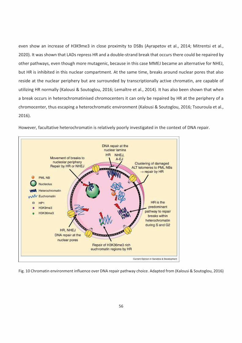
56
even show an increase of H3K9me3 in close proximity to DSBs (Ayrapetov et al., 2014; Mitrentsi et al.,
2020). It was shown that LADs repress HR and a double-strand break that occurs there could be repaired by
other pathways, even though more mutagenic, because in this case MMEJ became an alternative for NHEJ,
but HR is inhibited in this nuclear compartment. At the same time, breaks around nuclear pores that also
reside at the nuclear periphery but are surrounded by transcriptionally active chromatin, are capable of
utilizing HR normally (Kalousi & Soutoglou, 2016; Lemaître et al., 2014). It has also been shown that when
a break occurs in heterochromatinised chromocenters it can only be repaired by HR at the periphery of a
chromocenter, thus escaping a heterochromatic environment (Kalousi & Soutoglou, 2016; Tsouroula et al.,
2016).
However, facultative heterochromatin is relatively poorly investigated in the context of DNA repair.
Fig. 10 Chromatin environment influence over DNA repair pathway choice. Adapted from (Kalousi & Soutoglou, 2016)
57
Bivalency
Another type of chromatin that has never to my knowledge been investigated in the context of DNA repair
is bivalent chromatin. Generally, any kind of state that combines active and repressive chromatin marks is
bivalent chromatin. To date, these include a simultaneous presence of H3K4me3 and H3K27me3, H3K4me3
and H3K9me3, H3K36me2/3 and H3K9me3, or a combination of H3K9acK14ac, H3K4me3 and H3K27me3
(Azuara et al., 2006; Bernstein et al., 2006; Harikumar & Meshorer, 2015; Matsumura et al., 2015; Mauser
et al., 2017; Roh et al., 2006; Vakoc et al., 2005). But the first kind of such chromatin state that has been
observed (currently termed ‘classic’ bivalent domains) is a combination of H3K4me3 and H3K27 me3. They
were initially found in mouse embryonic stem (ES) cells in culture and for the first several years it was rather
common to consider them a culture artifact (Azuara et al., 2006; Bernstein et al., 2006; Harikumar &
Meshorer, 2015). However, later bivalent domains has been observed in other cell types, and even in vivo
during development and in cancer (Béguelin et al., 2016; Kampilafkos et al., 2015; Matsumura et al., 2015;
Minoux et al., 2017; Sachs et al., 2013; Sin et al., 2015). In the course of differentiation, one of the marks is
normally lost from bivalent promoters (Bernstein et al., 2006). Another question was addressed, whether
real bivalency exists, it is only resulted from a heterogeneity of cells in the population or from the close
proximity of two differentially marked genomic region. However, the existence of bivalency has later been
proven by genome-wide assessing individual bivalent nucleosomes. Sequential immunoprecipitation of
single nucleosomes confirmed that both marks can coexist on the same nucleosome, although on different
histone H3 tails (Sen et al., 2016).
A currently accepted explanation of this phenomenon is based on the observation that many of the
bivalently marked regions found in ES cells are promoters of developmental genes. The hypothesis is that
such a chromatin state is needed to enable rapid activation or complete inactivation of a respective gene
(Harikumar & Meshorer, 2015). However, RNA polymerase II (RNAPII) pausing was not observed on these
promoters in accordance with RNAPII association with actively transcribed and not bivalent genes
(Mantsoki et al., 2018). Additionally, it has also been shown that bivalency is linked to the cell cycle. MLL2
(Myeloid/lymphoid or mixed-lineage leukemia protein 2), as a part HMT (Histone methyltransferases)
complex, establishes H3K4me3 at developmental genes and thus real bivalency, and during the rest of the
cell cycle genes are robustly repressed (Singh et al., 2015). According to other studies, exit from
58
pluripotency accrues in a cell cycle-dependent manner, so these two studies go in line (Boward et al., 2016;
Pauklin & Vallier, 2013).
This chromatin type has not been studied extensively yet and the best model for it are embryonic stem cells
where this type of chromatin is rather abundant.
Embryonic stem cells
Stem cells are defined by the ability of both self-renewal and differentiation. There are three classes of stem
cells (although the last one is sometimes divided into two). The first one, totipotent stem cells, are capable
of differentiation into any type of adult, embryonic, or extraembryonic tissue. The second one, pluripotent
stem cells, keeps the potential and can give rise to any adult or embryonic cell type but not some of the
extraembryonic tissues. Finally, multipotent stem cells are tissue-specific and usually reside in mouse
embryonic stem cells. Unipotent stem cells, that are sometimes considered separately from multipotent
ones, can only differentiate on one cell type (Alison & Islam, 2009). Embryonic stem (ES) cells represent
pluripotent stem cells derived from a preimplantation blastocyst (Martello & Smith, 2014). Mouse ES cells
were first obtained by Evans and Kaufman in 1981 and until now stay a very attractive research model and
potentially powerful therapeutic tool as well as a fascinating research subject (Evans & Kaufman, 1981).
The pluripotent state is maintained by a regulatory network of transcription factors. The core factors are
Oct4 (Octamer-binding protein 4), Sox2 (SRY-box 2), and Nanog, and their key role has been proven many
times both by demonstrating their importance for stemness and viability and by their capacity to initiate
somatic cell reprogramming (Kehler et al., 2004; M. Li & Izpisua Belmonte, 2018; Mitsui et al., 2003; Nichols
et al., 1998; Takahashi & Yamanaka, 2006). Oct4 and Sox2 form heterodimers and induce numerous targets
including their own genes (Chew et al., 2005; M. Li & Izpisua Belmonte, 2018; Rodda et al., 2005). However,
change of expression levels or interaction partners of Oct4 and Sox2 drive mesendodermal or ectodermal
differentiation respectively (Avilion et al., 2003; Ferri et al., 2004; M. Li & Izpisua Belmonte, 2018; Niwa et
al., 2000; M. Thomson et al., 2011). Nanog is required for maintenance of pluripotency and appear to be
the key LIF/STAT3 (Leukemia inhibitory factor 1, Signal transducer and activator of transcription 3) pathway
as its overexpression makes LIF redundant (Chambers et al., 2003; M. Li & Izpisua Belmonte, 2018; Mitsui
et al., 2003). Together Oct4, Sox2, and Nanog binding cover the majority of pluripotency regulatory network
59
transcription factors (X. Chen et al., 2008; M. Li & Izpisua Belmonte, 2018; Loh et al., 2006). Their synergistic
action plays a role in both propagating pluripotency by activating transcription of pluripotency-related
genes and preventing differentiation by counter-balancing each other and blocking transcription of
developmental regulators (Boyer et al., 2005; Loh et al., 2006; Young, 2011).
Another curious feature of ES cells should be mentioned. They have a distinct cell cycle profile with a very
short G1 phase, which is tightly controlled by the pluripotency regulatory network and, in turn, has been
shown to suppress differentiation (Coronado et al., 2013; Kareta et al., 2015; V. C. Li & Kirschner, 2014;
Savatier et al., 2002). One of the core network targets, Myc (Myc proto-oncogene protein), enhances
proliferation by activating transcription of cell-cycle related genes as well as directly represses
differentiation by inhibiting FGF/ERK (Fibroblast growth factor, Extracellular signal-regulated kinase)
signaling pathway (Cartwright et al., 2005; Chappell et al., 2013; Kareta et al., 2015; Kim et al., 2008; Loh et
al., 2006). A collective action of Myc and core pluripotency factors leads to a compromised G1/S cell cycle
checkpoint that contributes to G1 shortening (Kanai et al., 2015; Kareta et al., 2015; Kim et al., 2010;
Suvorova et al., 2016; F. Zhang et al., 2009).
Among multiple regulatory measures than ensure stem cell proficiency for pluripotency and differentiation
of them unique chromatin organization is a prominent one.
Chromatin in embryonic stem cells
As previously mentioned, one of the prominent features of stem cells is the enrichment for bivalent
domains. However, it is not the only unique feature of ES cell chromatin.
Pluripotent stem cells have less compacted chromatin and higher dynamics of interaction with chromatin-
associated proteins and increased turnover of a linker histone H1, which most likely results from
overexpression of chromatin remodelers. Even the distribution of chromatin in the nucleus is more even in
pluripotent than in somatic cells. These features have been observed both in vitro and in vivo (Ahmed et
al., 2010; Azuara et al., 2006; Bouwman & De Laat, 2015; Efroni et al., 2008; Fussner et al., 2011; Gaspar-
Maia et al., 2011; Meshorer et al., 2006; Meshorer & Misteli, 2006; Otterstrom et al., 2019; Ricci et al.,
2015; Schlesinger & Meshorer, 2019). As a result, basal transcription is also elevated in pluripotent cells
(Efroni et al., 2008; Kobayashi & Kikyo, 2015; Ramalho-Santos et al., 2002). This includes exonic, intronic,
and non-genic regions, causing a considerable profusion of nascent RNA as compared to differentiated cells
60
(Bouwman & De Laat, 2015; Efroni et al., 2008). Reduced levels of heterochromatic histone marks and
increased levels of those associated with active transcription characteristic for ES cells fall in line with these
observations (Efroni et al., 2008; Kobayashi & Kikyo, 2015; Jeong Heon Lee et al., 2004; Meshorer et al.,
2006; Schlesinger & Meshorer, 2019; Wen et al., 2009). PRC2 complex plays an important role in
maintaining pluripotency as in ES cells it is predominantly bound to promoters of genes related to
differentiation (Boyer et al., 2006; Bracken et al., 2006; Denholtz et al., 2013; Kobayashi & Kikyo, 2015).
Lower levels of DNA methylation, which is largely associated with repressed chromatin state, were observed
in ES cells (Leitch et al., 2013; Marks et al., 2012; Schlesinger & Meshorer, 2019). Finally, one of the
prominent features observed both in early embryos and in ES cells is a reactivation of an imprinted X
chromosome (Mak et al., 2004; Okamoto et al., 2004; Surani et al., 2007; Wutz & Jaenisch, 2000).
Unraveling a great number of pluripotent stem cell-specific long-range interactions resulted from genome
architecture studies (Apostolou et al., 2013; De Wit et al., 2013; Dixon et al., 2012; Kobayashi & Kikyo, 2015;
Wei et al., 2013). Moreover, whereas the genome of differentiated cells is predominantly shaped in 3D
around repressed heterochromatic regions, in ES cells the main structure-forming entities are hubs of active
transcription dependent on core pluripotency factors (de Wit et al., 2013; Dixon et al., 2012; Kobayashi &
Kikyo, 2015). These changes are notable even on a whole-nucleus level. For example, chromocenters,
prominent in differentiated cells, show higher numbers and are less distinct in embryonic stem cells (Aoto
et al., 2006; Bouwman & De Laat, 2015; Mayer et al., 2005; Schlesinger & Meshorer, 2019; Wiblin et al.,
2005). Heterochromatin protein HP1b is almost diffused in ES cells but becomes accumulated at
heterochromatic foci in the course of differentiation (Mattout et al., 2015; Schlesinger & Meshorer, 2019).
Even interactions with nuclear lamina are massively reorganized between ES and differentiated cells
(Kobayashi & Kikyo, 2015; Peric-Hupkes et al., 2010). Nuclear lamina itself lacks Lamin A and is less
organized in pluripotent cells (Melcer et al., 2012; Pagliara et al., 2014; Schlesinger & Meshorer, 2019).
All in all, the evidence point that chromatin structure in pluripotent stem cells is distinct and is tightly
interconnected with their undifferentiated state.
DNA repair in embryonic stem cells
ES cells represent a fascinating model for us not only for having unique chromatin features but also for their
special mechanisms for maintaining genome integrity. Similar to other cell types, they are permanently
61
exposed to DNA damaging agents of both exogenous and endogenous origin. However, due to their rapid
proliferation (as discussed above) they also undergo constant replication stress. These factors combined
lead to constant activation of DDR (Ahuja et al., 2016; Juan et al., 2016; Nagaria et al., 2013; Savatier et al.,
2002). However, ES cells are highly tolerant of this stress and show a lower level of spontaneously induced
mutations and karyotypic aberrations than somatic cells (Brimble et al., 2004; Y. Hong et al., 2007; Nagaria
et al., 2013; J. A. Thomson, 1998). Enhanced maintenance of genome integrity appears to be a characteristic
of pluripotent cells (Y. Hong et al., 2007; Nagaria et al., 2013). In order to protect the genome of the whole
pool stem cells readily sacrifice themselves by initiating apoptosis or differentiation (Aladjem et al., 1998;
Yiling Hong & Stambrook, 2004; Lin et al., 2005; Jinping Liu et al., 2012; Nagaria et al., 2013). Of note, p53
plays a less role in ES cells, particularly it does not activate G1/S or intra-S checkpoint, and processes related
DNA damage response and repair are regulated by a stem-cell specific protein Filia (Aladjem et al., 1998;
Chuykin et al., 2008; Nagaria et al., 2013; Suvorova et al., 2016; vanderLaan et al., 2013; Vitale et al., 2017;
B. Zhao et al., 2015). It has also been shown that some known pluripotency markers such as Sall4 (Sal-like
protein 4) are directly involved in the process of DNA repair. It gets recruited to DSBs and helps to stabilize
MRN complex and enhance ATM activation (Xiong et al., 2015). ES cells have enriched levels of DNA repair
proteins, including HR and NHEJ factors, and enhanced levels of pro-apoptotic factors that are lost upon
differentiation (Cooper et al., 2014). Together with constant activation of DDR, it is capable of sufficiently
safeguarding their genome (Ahuja et al., 2016). Mouse ES cells were shown to have a strong preference
towards HR and NHEJ pathway in DSB repair (Bañuelos et al., 2008; Nagaria et al., 2013; Serrano et al., 2011;
Tichy et al., 2010). They have been shown to repair 80% of DSBs by HR and only 20% by NHEJ, whereas in
somatic cells the ratio is inverted. Alt-EJ was equally both cases, which means a shifted balance towards the
precise repair pathway choice (Nagaria et al., 2013; Serrano et al., 2011; Tichy et al., 2010). Therefore, it is
of no surprise that knockouts of key HR players usually result in embryonic lethality (Lim & Hasty, 1996;
Nagaria et al., 2013; Tsuzuki et al., 1996; Y. Xiao & Weaver, 1997; Jie Zhu et al., 2001).
Taken together, the presence of unique mechanisms of replications stress and DNA damage tolerance
makes ES cells a fascinating model for investigation.
62
Genome editing
Artificial genome editing in a variety of species has long been an important task for both medical and
research purposes. Medical applications include perspectives of gene editing for correction of disease-
related mutations, disruption of disease-promoting genes or even introducing novel genes (e.g. for
sensitising the immune system to tumour cells). The research application range is even broader: it includes
creating knock-outs, knock-ins, and introducing mutations to study the role of particular proteins,
processes, create disease models, and more.
Given tempting perspectives of practical use, it is of no surprise that the development of methods that
would allow gene editing has been going on for a considerable time. At first, the strategy implied the use
of random recombination, either to change the sequence or to allow further use of nucleases (such as Cre)
(Carroll, 2017; ROTHSTEIN, 1989; Scherer & Davis, 1979; Smithies et al., 1985; Thomas et al., 1986). The
discovery of zinc finger nucleases (ZFNs) and transcription activator-like effector nucleases (TALENs)
allowed a considerable advance if the field. However, the largest step has been done with the discovery of
CRISPR-Cas9.
Clustered regularly interspaced short palindromic repeats (CRISPR) and CRISPR-associated protein 9 (Cas9)
are parts of bacterial mechanisms of antiviral defence (Carroll, 2014; Jansen et al., 2002; Sorek et al., 2013).
Shortly after their discovery, the system was adopted as a tool of genome editing in other organisms, such
as plants, fish, human, mice, and many more (Carroll, 2014; Cho et al., 2013; Hall et al., 2018; Hwang et al.,
2013; Jinek et al., 2012; Ma et al., 2015).
The editing system consists of Cas9 protein and a short non-coding RNA that contains two parts, a target-
specific CRISPR RNA (crRNA) and a helper trans-activating RNA (tracrRNA) (Karvelis et al., 2013; Memi et al.,
2018). In the guide RNA, tracrRNA component is responsible for binding Cas9 and crRNA acts in sequence
recognition by pairing to the target sequence (also called protospacer) (Memi et al., 2018; Pattanayak et
al., 2013). However, Cas9 protein only performs catalytic reaction if a protospacer-adjacent motif (PAM) is
present at the 5’ side of the protospacer (Memi et al., 2018; O’Connell et al., 2014). Later on single guide
RNAs containing both crRNA and tracRNA sequences were designed to simplify the practical use (Mali et
al., 2013; Memi et al., 2018).
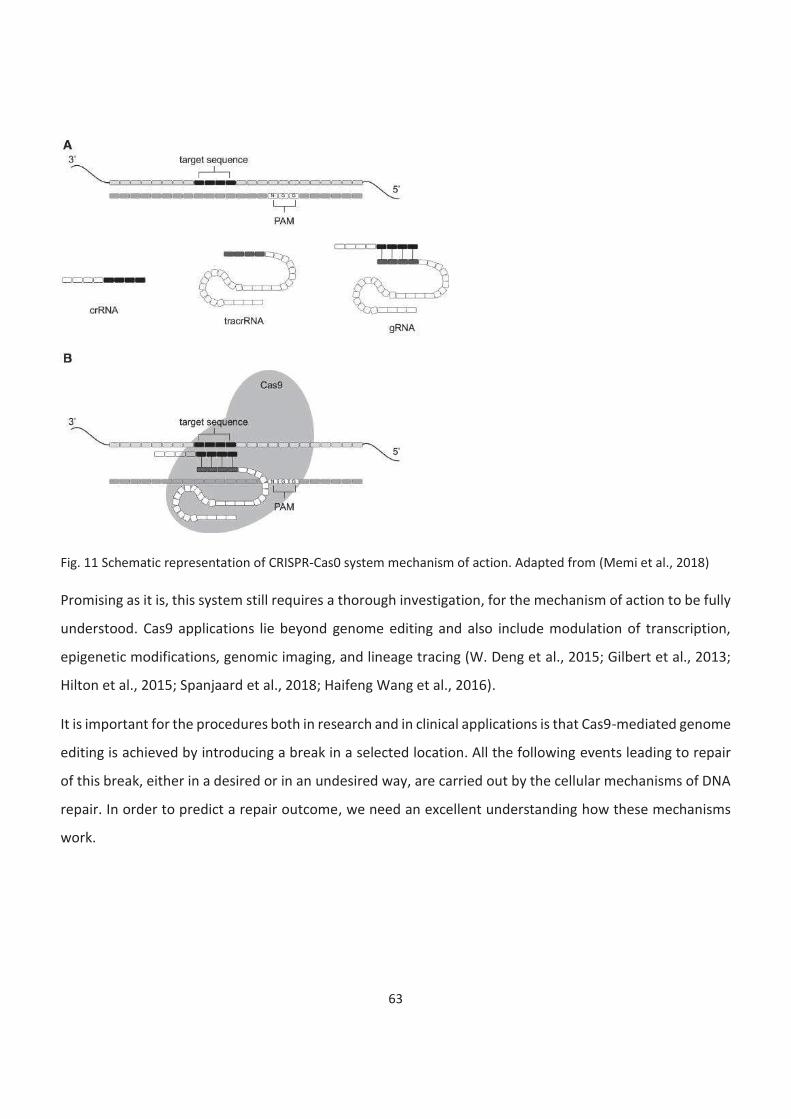
63
Fig. 11 Schematic representation of CRISPR-Cas0 system mechanism of action. Adapted from (Memi et al., 2018)
Promising as it is, this system still requires a thorough investigation, for the mechanism of action to be fully
understood. Cas9 applications lie beyond genome editing and also include modulation of transcription,
epigenetic modifications, genomic imaging, and lineage tracing (W. Deng et al., 2015; Gilbert et al., 2013;
Hilton et al., 2015; Spanjaard et al., 2018; Haifeng Wang et al., 2016).
It is important for the procedures both in research and in clinical applications is that Cas9-mediated genome
editing is achieved by introducing a break in a selected location. All the following events leading to repair
of this break, either in a desired or in an undesired way, are carried out by the cellular mechanisms of DNA
repair. In order to predict a repair outcome, we need an excellent understanding how these mechanisms
work.
64
Genome editing fidelity studies
65
66
67
68
69
70
71
72
Goals
In my project, I intended to investigate two different questions.
First, I wanted to elucidate the influence of the pluripotent state on the fidelity of DSB repair and a
pathway choice. It has been previously reported that HR is enhanced in ES cells, which provides the
necessary level of genome stability. However, current knowledge is mostly based on either a study of
indirect factors, such as proteins involved in certain DNA repair pathways, or studying single loci, which
cannot always be extrapolated to the whole genome. Therefore, we found it relevant to perform a study
tackling several targets and relying on repair outcomes rather than factors recruiting.
Second, I aimed to investigate the role of a local chromatin structure on a repair outcome and a balance
between different repair pathways. Bivalent chromatin was one of the types of particular interest for me
as its influence on the process of DNA repair has never been studied yet. Other chromatin types, like
euchromatin and facultative heterochromatin, appeared important to study as well. On the one hand,
they could serve as a reference to compare behaviour of bivalent chromatin to. On the other hand,
existing studies provided incomplete information, as they were based on indirect detection of a pathway
use rather than assessing a direct repair outcome.
Therefore, the goals of my work were as following:
1. Compare fidelity of double-strand break repair between different chromatin contexts, and
particularly in bivalent chromatin
2. Compare fidelity of double-strand break repair between pluripotent and differentiated cells.
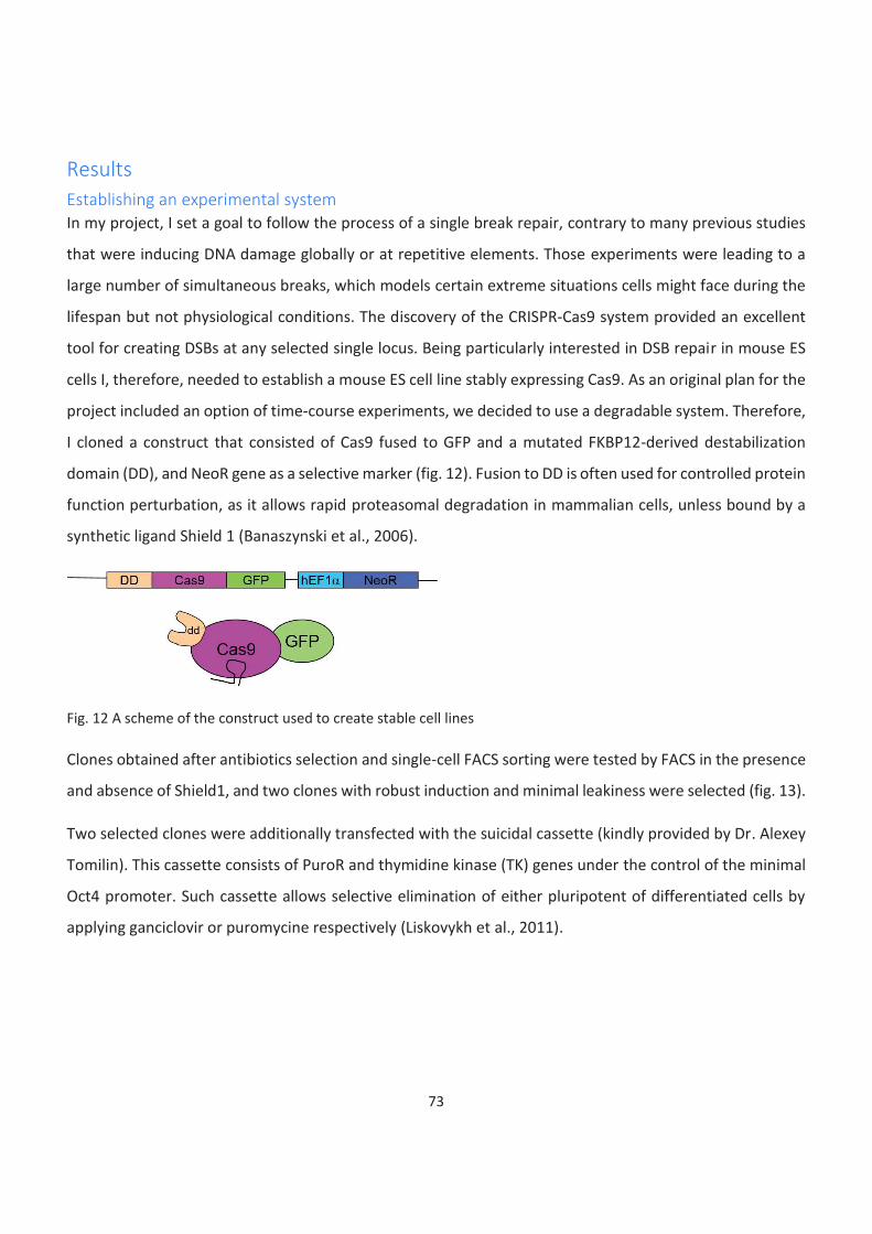
73
Results
Establishing an experimental system
In my project, I set a goal to follow the process of a single break repair, contrary to many previous studies
that were inducing DNA damage globally or at repetitive elements. Those experiments were leading to a
large number of simultaneous breaks, which models certain extreme situations cells might face during the
lifespan but not physiological conditions. The discovery of the CRISPR-Cas9 system provided an excellent
tool for creating DSBs at any selected single locus. Being particularly interested in DSB repair in mouse ES
cells I, therefore, needed to establish a mouse ES cell line stably expressing Cas9. As an original plan for the
project included an option of time-course experiments, we decided to use a degradable system. Therefore,
I cloned a construct that consisted of Cas9 fused to GFP and a mutated FKBP12-derived destabilization
domain (DD), and NeoR gene as a selective marker (fig. 12). Fusion to DD is often used for controlled protein
function perturbation, as it allows rapid proteasomal degradation in mammalian cells, unless bound by a
synthetic ligand Shield 1 (Banaszynski et al., 2006).
Fig. 12 A scheme of the construct used to create stable cell lines
Clones obtained after antibiotics selection and single-cell FACS sorting were tested by FACS in the presence
and absence of Shield1, and two clones with robust induction and minimal leakiness were selected (fig. 13).
Two selected clones were additionally transfected with the suicidal cassette (kindly provided by Dr. Alexey
Tomilin). This cassette consists of PuroR and thymidine kinase (TK) genes under the control of the minimal
Oct4 promoter. Such cassette allows selective elimination of either pluripotent of differentiated cells by
applying ganciclovir or puromycine respectively (Liskovykh et al., 2011).
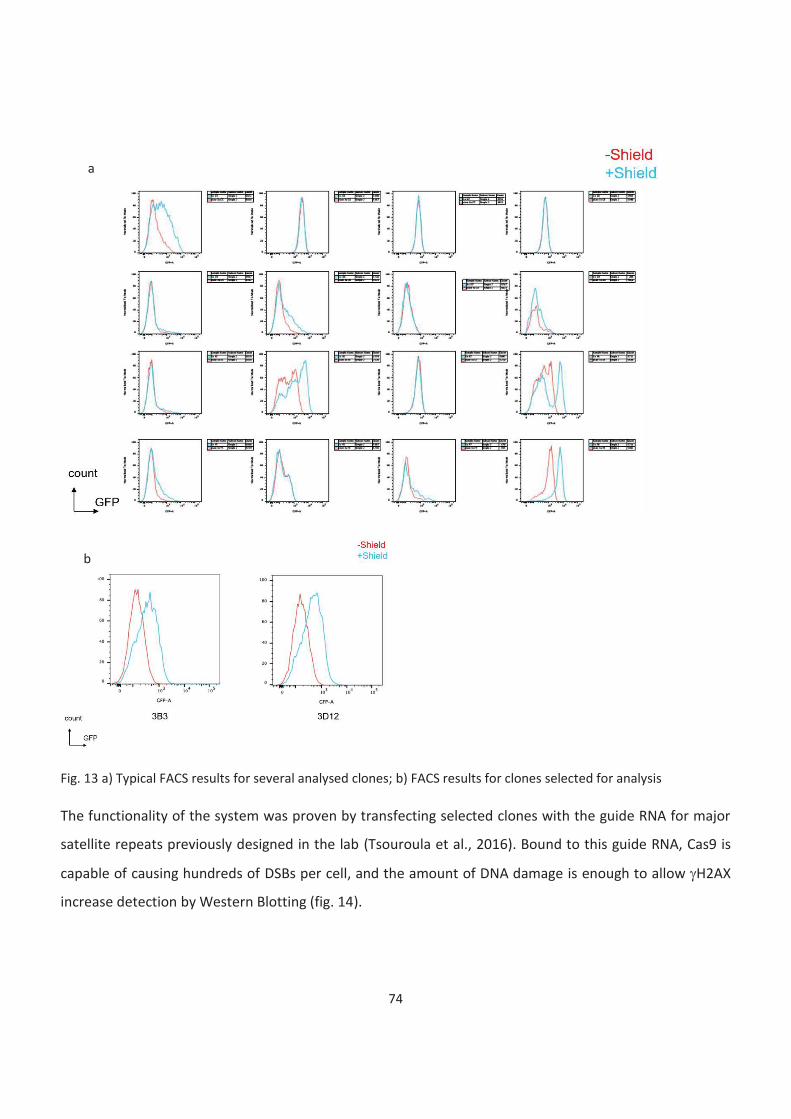
74
Fig. 13 a) Typical FACS results for several analysed clones; b) FACS results for clones selected for analysis
The functionality of the system was proven by transfecting selected clones with the guide RNA for major
satellite repeats previously designed in the lab (Tsouroula et al., 2016). Bound to this guide RNA, Cas9 is
capable of causing hundreds of DSBs per cell, and the amount of DNA damage is enough to allow gH2AX
increase detection by Western Blotting (fig. 14).
a
b
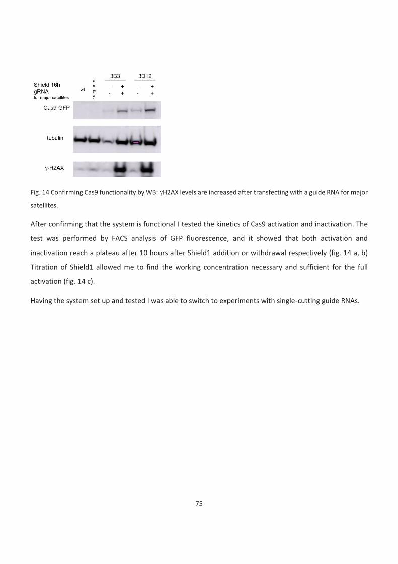
75
Fig. 14 Confirming Cas9 functionality by WB: gH2AX levels are increased after transfecting with a guide RNA for major
satellites.
After confirming that the system is functional I tested the kinetics of Cas9 activation and inactivation. The
test was performed by FACS analysis of GFP fluorescence, and it showed that both activation and
inactivation reach a plateau after 10 hours after Shield1 addition or withdrawal respectively (fig. 14 a, b)
Titration of Shield1 allowed me to find the working concentration necessary and sufficient for the full
activation (fig. 14 c).
Having the system set up and tested I was able to switch to experiments with single-cutting guide RNAs.
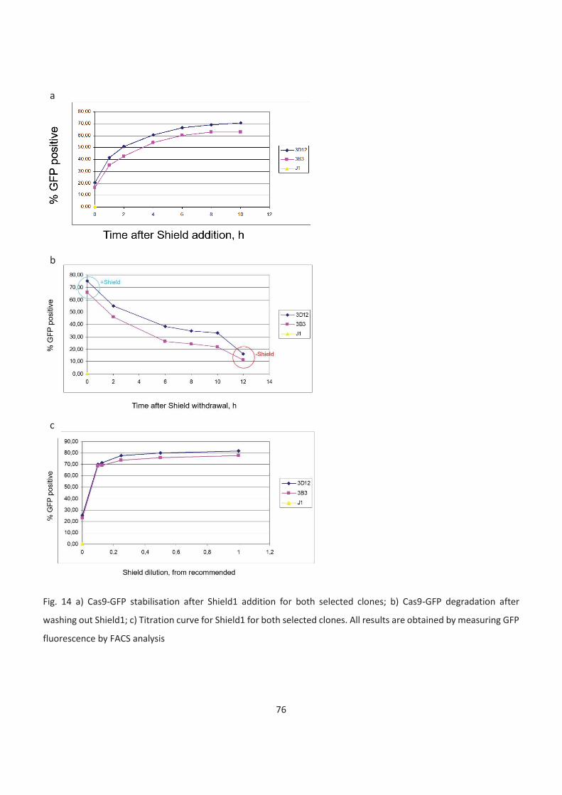
76
Fig. 14 a) Cas9-GFP stabilisation after Shield1 addition for both selected clones; b) Cas9-GFP degradation after
washing out Shield1; c) Titration curve for Shield1 for both selected clones. All results are obtained by measuring GFP
fluorescence by FACS analysis
a
b
c

77
Target choice and guide RNA design
The goal of my project was to compare DSB repair in different chromatin contexts. Therefore, targets were
selected so that they would represent different chromatin types. I chose several groups of genes depending
on their functionality and epigenetic status:
- housekeeping genes (Gapdh and Actin), further colourcoded as pink
- pluripotency regulators (Pou5f1, Nanog, and Tfcp2l1), further colourcoded as green
- developmental regulators that are bivalent in ES cells (Pax6, Ngn2, Zic1, Dlx2, Zfpm2, Foxa), further
colourcoded as yellow
- facultative heterochromatic genes (Hoxb1, Tdrd1, and Mc4r), further colourcoded as red
- genes belonging to LADs (Sox6, Ptn, and Nrp1), further colourcoded as purple
Given that one of the chromatin types of interest was bivalent, and these domains are typically found at
promoter regions, most of the guide RNAs were designed within a proximal promoter of a corresponding
gene. The only exception was the group of genes located in LADs, as this part was connected to another
project running at the lab, and introducing breaks at the intrones was required. Proximal promoters were
defined as 1 kb up- and downstream of the TSS according to the BindDB web tool (Livyatan et al., 2015).
Guide RNAs were designed using the Benchling website (https://www.benchling.com/). guide RNA
sequences are provided in Table 1 in the Materials and methods section.
Chromatin status at targeted sites was confirmed by ChIP. I used H3K4me3 as a mark of active chromatin,
H3K27me3 as a mark of facultative heterochromatin, and H3K9me2 as a mark for LADs. I also tested
H3K9me3 as a marker of constitutive heterochromatin. I performed ChIP in ES cells. 3T3 cells were used as
a differentiated control. I could observe an expected chromatin pattern in ES cells. In 3T3 cells only
housekeeping genes of all tested were active, and the rest represented facultative heterochromatin (fig.
15).
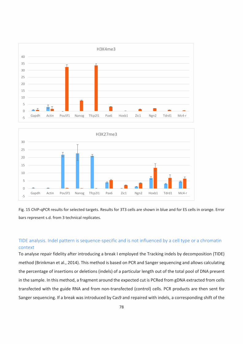
78
Fig. 15 ChIP-qPCR results for selected targets. Results for 3T3 cells are shown in blue and for ES cells in orange. Error
bars represent s.d. from 3 technical replicates.
TIDE analysis. Indel pattern is sequence-specific and is not influenced by a cell type or a chromatin
context
To analyse repair fidelity after introducing a break I employed the Tracking indels by decomposition (TIDE)
method (Brinkman et al., 2014). This method is based on PCR and Sanger sequencing and allows calculating
the percentage of insertions or deletions (indels) of a particular length out of the total pool of DNA present
in the sample. In this method, a fragment around the expected cut is PCRed from gDNA extracted from cells
transfected with the guide RNA and from non-transfected (control) cells. PCR products are then sent for
Sanger sequencing. If a break was introduced by Cas9 and repaired with indels, a corresponding shift of the
-5
0
5
10
15
20
25
30
35
40
Gapdh Actin Pou5f1 Nanog Tfcp2l1 Pax6 Hoxb1 Zic1 Ngn2 Tdrd1 Mc4-r
H3K4me3
-5
0
5
10
15
20
25
30
Gapdh Actin Pou5f1 Nanog Tfcp2l1 Pax6 Zic1 Ngn2 Hoxb1 Tdrd1 Mc4-r
H3K27me3
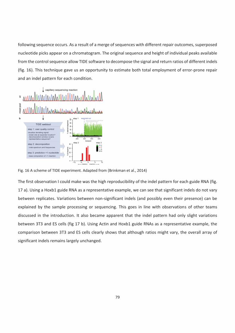
79
following sequence occurs. As a result of a merge of sequences with different repair outcomes, superposed
nucleotide picks appear on a chromatogram. The original sequence and height of individual peaks available
from the control sequence allow TIDE software to decompose the signal and return ratios of different indels
(fig. 16). This technique gave us an opportunity to estimate both total employment of error-prone repair
and an indel pattern for each condition.
Fig. 16 A scheme of TIDE experiment. Adapted from (Brinkman et al., 2014)
The first observation I could make was the high reproducibility of the indel pattern for each guide RNA (fig.
17 a). Using a Hoxb1 guide RNA as a representative example, we can see that significant indels do not vary
between replicates. Variations between non-significant indels (and possibly even their presence) can be
explained by the sample processing or sequencing. This goes in line with observations of other teams
discussed in the introduction. It also became apparent that the indel pattern had only slight variations
between 3T3 and ES cells (fig 17 b). Using Actin and Hoxb1 guide RNAs as a representative example, the
comparison between 3T3 and ES cells clearly shows that although ratios might vary, the overall array of
significant indels remains largely unchanged.
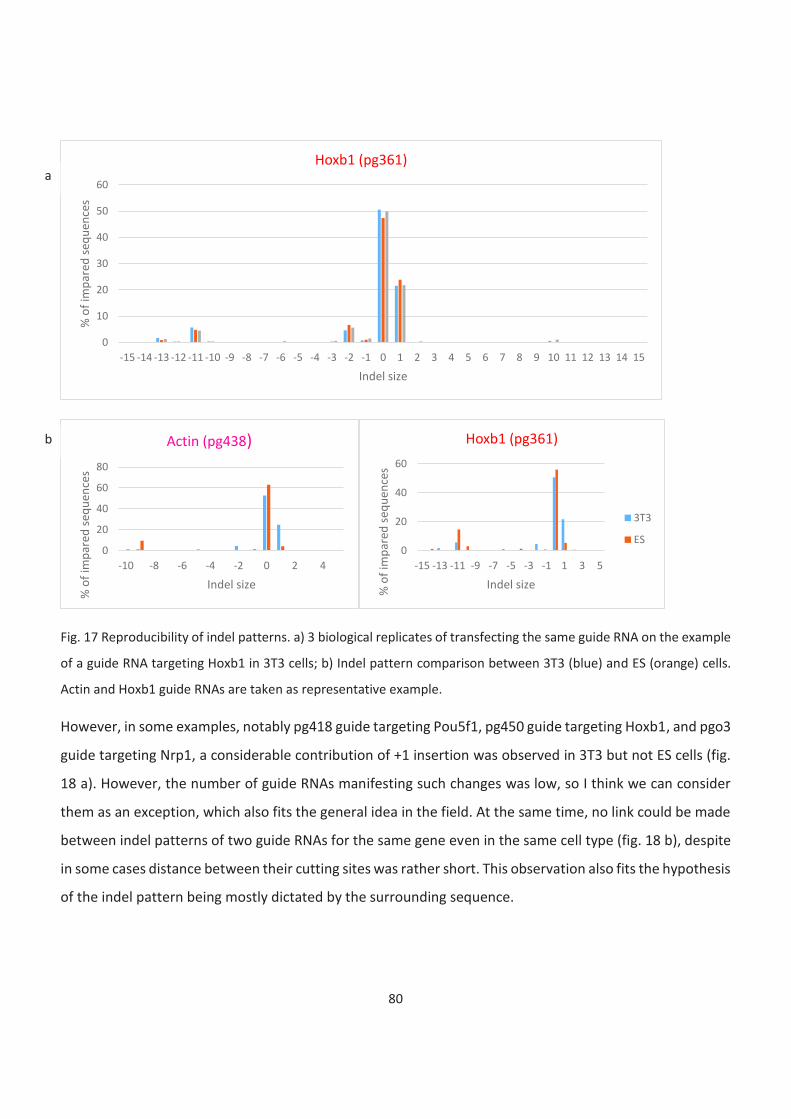
80
Fig. 17 Reproducibility of indel patterns. a) 3 biological replicates of transfecting the same guide RNA on the example
of a guide RNA targeting Hoxb1 in 3T3 cells; b) Indel pattern comparison between 3T3 (blue) and ES (orange) cells.
Actin and Hoxb1 guide RNAs are taken as representative example.
However, in some examples, notably pg418 guide targeting Pou5f1, pg450 guide targeting Hoxb1, and pgo3
guide targeting Nrp1, a considerable contribution of +1 insertion was observed in 3T3 but not ES cells (fig.
18 a). However, the number of guide RNAs manifesting such changes was low, so I think we can consider
them as an exception, which also fits the general idea in the field. At the same time, no link could be made
between indel patterns of two guide RNAs for the same gene even in the same cell type (fig. 18 b), despite
in some cases distance between their cutting sites was rather short. This observation also fits the hypothesis
of the indel pattern being mostly dictated by the surrounding sequence.
0
10
20
30
40
50
60
-15 -14 -13 -12 -11 -10 -9 -8 -7 -6 -5 -4 -3 -2 -1 0 1 2 3 4 5 6 7 8 9 10 11 12 13 14 15
% o
f im
par
ed s
equ
ence
s
Indel size
Hoxb1 (pg361)
0
20
40
60
80
-10 -8 -6 -4 -2 0 2 4
% o
f im
par
ed s
equ
ence
s
Indel size
Actin (pg438)
0
20
40
60
-15 -13 -11 -9 -7 -5 -3 -1 1 3 5
% o
f im
par
ed s
equ
ence
s
Indel size
Hoxb1 (pg361)
3T3
ES
a
b

81
Fig. 18 TIDE results: indel pattern. a) guide RNAs targeting Pou5f1 and Nrp1. +1 insertion can be seen in 3T3 but not
in ES cells; b) Comparison of indel patterns of two guide RNAs targeting the same locus in the same cell type.
The size of deletions has been largely debated in the context of the balance between error-prone repair
pathways, NHEJ and MMEJ. While it remains under debate which of these pathways could be the source of
relatively small indels, there is no doubt that deletions larger than 10 b.p. are generated by MMEJ.
Therefore, I tried to estimate MMEJ recruitment in different cell types by analysing deletions larger than 10
b.p (fig. 19 a). It can be seen from comparison between 3T3 and ES cells for the same guide RNA that most
of the time large deletions either occur in both cell types or are absent in either. Moreover, sometimes
even two guide RNAs for the same gene (fig. 19 b) can demonstrate these two different patterns.
0
20
40
60
80
100
-10 -8 -6 -4 -2 0 2 4
% o
f im
par
ed s
equ
ence
s
Indel size
Pou5f1 (pg418)
0
20
40
60
80
-10 -8 -6 -4 -2 0 2 4
% o
f im
par
ed s
equ
ence
s
Indel size
Nrp1 (pgo3)
3T3
ES
0
10
20
30
40
50
60
70
-10 -9 -8 -7 -6 -5 -4 -3 -2 -1 0 1 2 3 4 5% o
f im
par
ed s
equ
ence
s
Indel size
Gapdh
pg313 pg439
0
10
20
30
40
50
60
-15 -13 -11 -9 -7 -5 -3 -1 1 3 5% o
f im
par
ed s
equ
ence
s
Indel size
Pax6
pg445 pg446
a
b
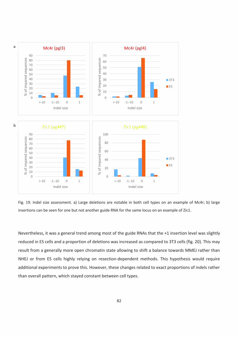
82
Fig. 19. Indel size assessment. a) Large deletions are notable in both cell types on an example of Mc4r; b) large
insertions can be seen for one but not another guide RNA for the same locus on an example of Zic1.
Nevertheless, it was a general trend among most of the guide RNAs that the +1 insertion level was slightly
reduced in ES cells and a proportion of deletions was increased as compared to 3T3 cells (fig. 20). This may
result from a generally more open chromatin state allowing to shift a balance towards MMEJ rather than
NHEJ or from ES cells highly relying on resection-dependent methods. This hypothesis would require
additional experiments to prove this. However, these changes related to exact proportions of indels rather
than overall pattern, which stayed constant between cell types.
0
10
20
30
40
50
60
70
80
90
>-10 -1:-10 0 1
% o
f im
par
ed s
equ
ence
s
Indel size
Mc4r (pgl3)
0
10
20
30
40
50
60
70
>-10 -1:-10 0 1%
of
imp
ared
seq
uen
ces
Indel size
Mc4r (pgl4)
3T3
ES
0
10
20
30
40
50
60
70
80
90
>-10 -1:-10 0 1
% o
f im
par
ed s
equ
ence
s
Indel size
Zic1 (pg447)
0
20
40
60
80
100
>-10 -1:-10 0 1
% o
f im
par
ed s
equ
ence
s
Indel size
Zic1 (pg448)
3T3
ES
a
b
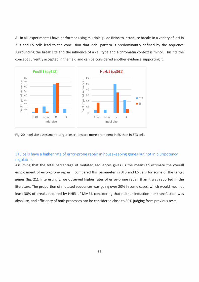
83
All in all, experiments I have performed using multiple guide RNAs to introduce breaks in a variety of loci in
3T3 and ES cells lead to the conclusion that indel pattern is predominantly defined by the sequence
surrounding the break site and the influence of a cell type and a chromatin context is minor. This fits the
concept currently accepted in the field and can be considered another evidence supporting it.
Fig. 20 Indel size assessment. Larger insertions are more prominent in ES than in 3T3 cells
3T3 cells have a higher rate of error-prone repair in housekeeping genes but not in pluripotency
regulators
Assuming that the total percentage of mutated sequences gives us the means to estimate the overall
employment of error-prone repair, I compared this parameter in 3T3 and ES cells for some of the target
genes (fig. 21). Interestingly, we observed higher rates of error-prone repair than it was reported in the
literature. The proportion of mutated sequences was going over 20% in some cases, which would mean at
least 30% of breaks repaired by NHEJ of MMEJ, considering that neither induction nor transfection was
absolute, and efficiency of both processes can be considered close to 80% judging from previous tests.
0
10
20
30
40
50
60
70
80
>-10 -1:-10 0 1
% o
f im
par
ed s
equ
ence
s
Indel size
Pou5f1 (pg418)
0
10
20
30
40
50
60
>-10 -1:-10 0 1
% o
f im
par
ed s
equ
ence
s
Indel size
Hoxb1 (pg361)
3T3
ES
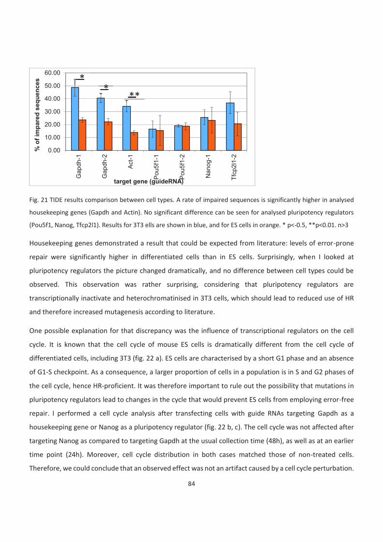
84
Fig. 21 TIDE results comparison between cell types. A rate of impaired sequences is significantly higher in analysed
housekeeping genes (Gapdh and Actin). No significant difference can be seen for analysed pluripotency regulators
(Pou5f1, Nanog, Tfcp2l1). Results for 3T3 ells are shown in blue, and for ES cells in orange. * p<-0.5, **p<0.01. n>3
Housekeeping genes demonstrated a result that could be expected from literature: levels of error-prone
repair were significantly higher in differentiated cells than in ES cells. Surprisingly, when I looked at
pluripotency regulators the picture changed dramatically, and no difference between cell types could be
observed. This observation was rather surprising, considering that pluripotency regulators are
transcriptionally inactivate and heterochromatinised in 3T3 cells, which should lead to reduced use of HR
and therefore increased mutagenesis according to literature.
One possible explanation for that discrepancy was the influence of transcriptional regulators on the cell
cycle. It is known that the cell cycle of mouse ES cells is dramatically different from the cell cycle of
differentiated cells, including 3T3 (fig. 22 a). ES cells are characterised by a short G1 phase and an absence
of G1-S checkpoint. As a consequence, a larger proportion of cells in a population is in S and G2 phases of
the cell cycle, hence HR-proficient. It was therefore important to rule out the possibility that mutations in
pluripotency regulators lead to changes in the cycle that would prevent ES cells from employing error-free
repair. I performed a cell cycle analysis after transfecting cells with guide RNAs targeting Gapdh as a
housekeeping gene or Nanog as a pluripotency regulator (fig. 22 b, c). The cell cycle was not affected after
targeting Nanog as compared to targeting Gapdh at the usual collection time (48h), as well as at an earlier
time point (24h). Moreover, cell cycle distribution in both cases matched those of non-treated cells.
Therefore, we could conclude that an observed effect was not an artifact caused by a cell cycle perturbation.
0.00
10.00
20.00
30.00
40.00
50.00
60.00G
ap
dh-1
Gap
dh-2
Act-
1
Pou5
f1-1
Pou5
f1-2
Na
nog
-1
Tfc
p2l1
-2
% o
f im
pare
d s
eq
uen
ces
target gene (guideRNA)
*
* **
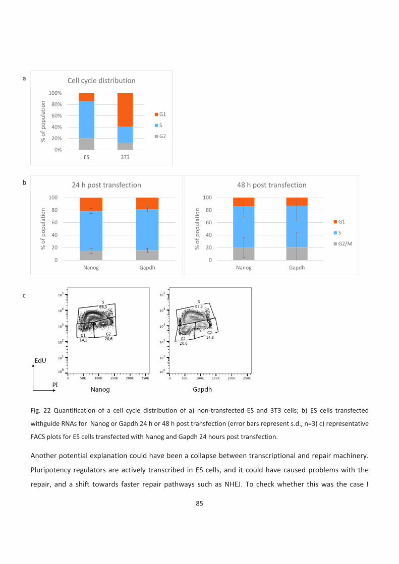
85
Fig. 22 Quantification of a cell cycle distribution of a) non-transfected ES and 3T3 cells; b) ES cells transfected
withguide RNAs for Nanog or Gapdh 24 h or 48 h post transfection (error bars represent s.d., n=3) c) representative
FACS plots for ES cells transfected with Nanog and Gapdh 24 hours post transfection.
Another potential explanation could have been a collapse between transcriptional and repair machinery.
Pluripotency regulators are actively transcribed in ES cells, and it could have caused problems with the
repair, and a shift towards faster repair pathways such as NHEJ. To check whether this was the case I
0%
20%
40%
60%
80%
100%
ES 3T3
% o
f p
op
ula
tio
n
Cell cycle distribution
G1
S
G2
0
20
40
60
80
100
Nanog Gapdh
% o
f p
op
ula
tio
n
24 h post transfection
0
20
40
60
80
100
Nanog Gapdh
% o
f p
op
ula
tio
n
48 h post transfection
G1
S
G2/M
b
c
a
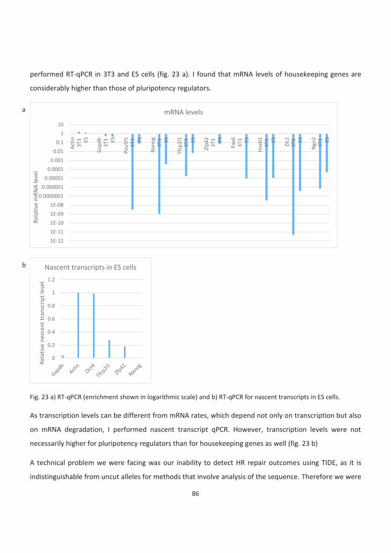
86
performed RT-qPCR in 3T3 and ES cells (fig. 23 a). I found that mRNA levels of housekeeping genes are
considerably higher than those of pluripotency regulators.
Fig. 23 a) RT-qPCR (enrichment shown in logarithmic scale) and b) RT-qPCR for nascent transcripts in ES cells.
As transcription levels can be different from mRNA rates, which depend not only on transcription but also
on mRNA degradation, I performed nascent transcript qPCR. However, transcription levels were not
necessarily higher for pluripotency regulators than for housekeeping genes as well (fig. 23 b)
A technical problem we were facing was our inability to detect HR repair outcomes using TIDE, as it is
indistinguishable from uncut alleles for methods that involve analysis of the sequence. Therefore we were
1E-12
1E-11
1E-10
1E-09
1E-08
0.0000001
0.000001
0.00001
0.0001
0.001
0.01
0.1
1
10
Act
in3T
3 ES
Gap
dh
3T3 ES
Po
u5f
13T
3 ES
Nan
og
3T3 ES
Tfcp
2l1
3T3 ES
Zfp
423T
3 ES
Pax
63T
3 ES
Ho
xb1
3T3 ES
Zic1
3T3 ES
Ngn
23T
3 ES
Rel
ativ
e m
RN
A le
vel
mRNA levels
0
0.2
0.4
0.6
0.8
1
1.2
Rel
ativ
e n
esce
nt
tran
scri
pt
leve
l
Nascent transcripts in ES cells
a
b
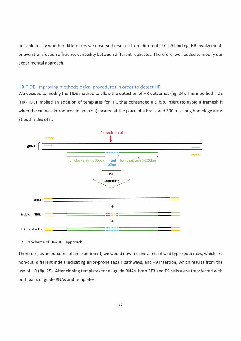
87
not able to say whether differences we observed resulted from differential Cas9 binding, HR involvement,
or even transfection efficiency variability between different replicates. Therefore, we needed to modify our
experimental approach.
HR-TIDE: improving methodological procedures in order to detect HR
We decided to modify the TIDE method to allow the detection of HR outcomes (fig. 24). This modified TIDE
(HR-TIDE) implied an addition of templates for HR, that contended a 9 b.p. insert (to avoid a frameshift
when the cut was introduced in an exon) located at the place of a break and 500 b.p.-long homology arms
at both sides of it.
Fig. 24 Scheme of HR-TIDE approach
Therefore, as an outcome of an experiment, we would now receive a mix of wild type sequences, which are
non-cut, different indels indicating error-prone repair pathways, and +9 insertion, which results from the
use of HR (fig. 25). After cloning templates for all guide RNAs, both 3T3 and ES cells were transfected with
both pairs of guide RNAs and templates.
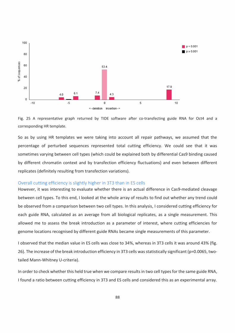
88
Fig. 25 A representative graph returned by TIDE software after co-transfecting guide RNA for Oct4 and a
corresponding HR template.
So as by using HR templates we were taking into account all repair pathways, we assumed that the
percentage of perturbed sequences represented total cutting efficiency. We could see that it was
sometimes varying between cell types (which could be explained both by differential Cas9 binding caused
by different chromatin context and by transfection efficiency fluctuations) and even between different
replicates (definitely resulting from transfection variations).
Overall cutting efficiency is slightly higher in 3T3 than in ES cells
However, it was interesting to evaluate whether there is an actual difference in Cas9-mediated cleavage
between cell types. To this end, I looked at the whole array of results to find out whether any trend could
be observed from a comparison between two cell types. In this analysis, I considered cutting efficiency for
each guide RNA, calculated as an average from all biological replicates, as a single measurement. This
allowed me to assess the break introduction as a parameter of interest, where cutting efficiencies for
genome locations recognised by different guide RNAs became single measurements of this parameter.
I observed that the median value in ES cells was close to 34%, whereas in 3T3 cells it was around 43% (fig.
26). The increase of the break introduction efficiency in 3T3 cells was statistically significant (p=0.0065, two-
tailed Mann-Whitney U-criteria).
In order to check whether this held true when we compare results in two cell types for the same guide RNA,
I found a ratio between cutting efficiency in 3T3 and ES cells and considered this as an experimental array.
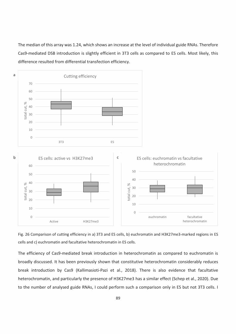
89
The median of this array was 1.24, which shows an increase at the level of individual guide RNAs. Therefore
Cas9-mediated DSB introduction is slightly efficient in 3T3 cells as compared to ES cells. Most likely, this
difference resulted from differential transfection efficiency.
Fig. 26 Comparison of cutting efficiency in a) 3T3 and ES cells, b) euchromatin and H3K27me3-marked regions in ES
cells and c) euchromatin and facultative heterochromatin in ES cells.
The efficiency of Cas9-mediated break introduction in heterochromatin as compared to euchromatin is
broadly discussed. It has been previously shown that constitutive heterochromatin considerably reduces
break introduction by Cas9 (Kallimasioti-Pazi et al., 2018). There is also evidence that facultative
heterochromatin, and particularly the presence of H3K27me3 has a similar effect (Schep et al., 2020). Due
to the number of analysed guide RNAs, I could perform such a comparison only in ES but not 3T3 cells. I
0
10
20
30
40
50
60
70
3T3 ES
tota
l cu
t, %
Cutting efficiency
0
10
20
30
40
50
60
Active H3K27me3
tota
l cu
t, %
ES cells: active vs H3K27me3
0
10
20
30
40
50
euchromatin facultativeheterochromatin
tota
l cu
t, %
ES cells: euchromatin vs facultative heterochromatin
a
b c
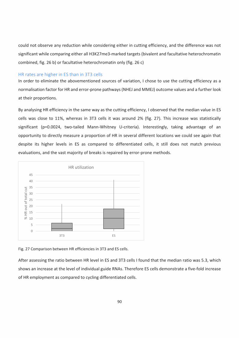
90
could not observe any reduction while considering either in cutting efficiency, and the difference was not
significant while comparing either all H3K27me3-marked targets (bivalent and facultative heterochromatin
combined, fig. 26 b) or facultative heterochromatin only (fig. 26 c)
HR rates are higher in ES than in 3T3 cells
In order to eliminate the abovementioned sources of variation, I chose to use the cutting efficiency as a
normalisation factor for HR and error-prone pathways (NHEJ and MMEJ) outcome values and a further look
at their proportions.
By analysing HR efficiency in the same way as the cutting efficiency, I observed that the median value in ES
cells was close to 11%, whereas in 3T3 cells it was around 2% (fig. 27). This increase was statistically
significant (p=0.0024, two-tailed Mann-Whitney U-criteria). Interestingly, taking advantage of an
opportunity to directly measure a proportion of HR in several different locations we could see again that
despite its higher levels in ES as compared to differentiated cells, it still does not match previous
evaluations, and the vast majority of breaks is repaired by error-prone methods.
Fig. 27 Comparison between HR efficiencies in 3T3 and ES cells.
After assessing the ratio between HR level in ES and 3T3 cells I found that the median ratio was 5.3, which
shows an increase at the level of individual guide RNAs. Therefore ES cells demonstrate a five-fold increase
of HR employment as compared to cycling differentiated cells.
0
5
10
15
20
25
30
35
40
45
3T3 ES
% H
R o
ut
of
tota
l cu
t
HR utilization
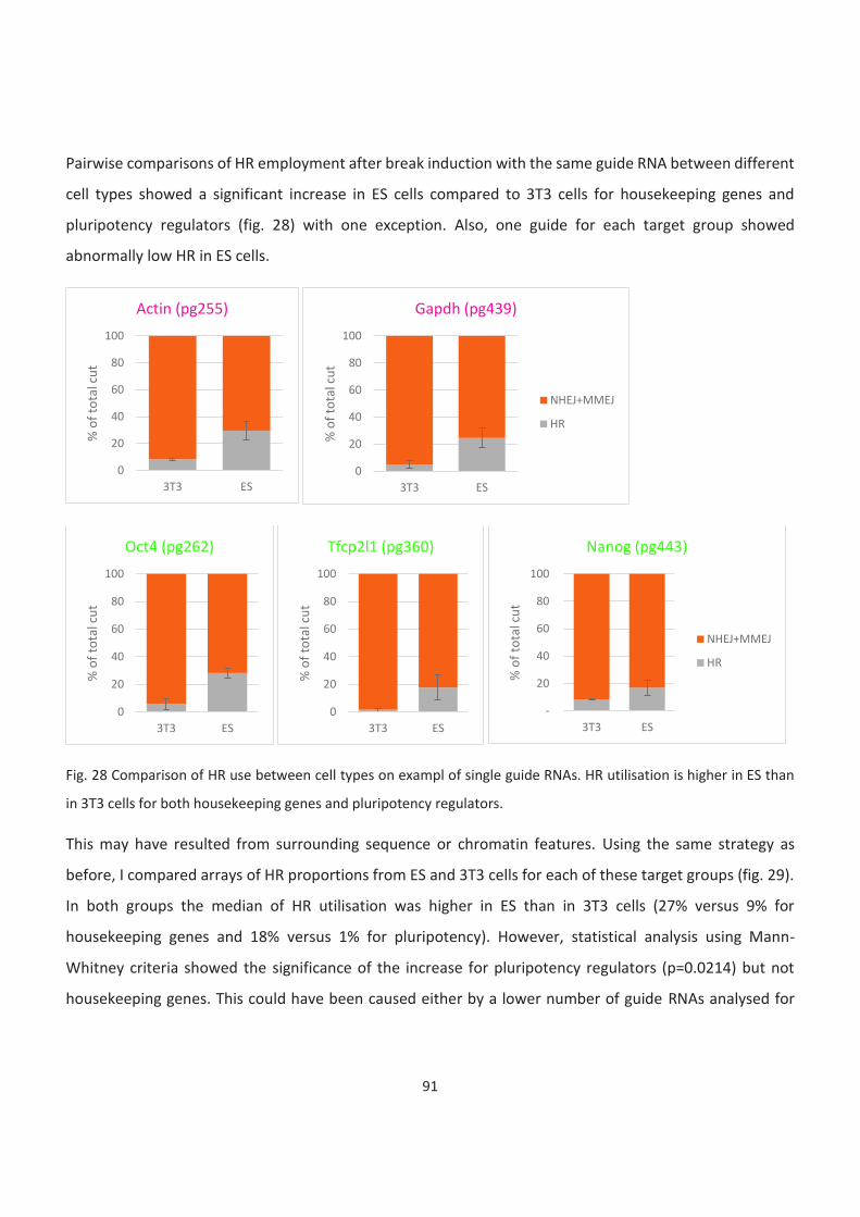
91
Pairwise comparisons of HR employment after break induction with the same guide RNA between different
cell types showed a significant increase in ES cells compared to 3T3 cells for housekeeping genes and
pluripotency regulators (fig. 28) with one exception. Also, one guide for each target group showed
abnormally low HR in ES cells.
Fig. 28 Comparison of HR use between cell types on exampl of single guide RNAs. HR utilisation is higher in ES than
in 3T3 cells for both housekeeping genes and pluripotency regulators.
This may have resulted from surrounding sequence or chromatin features. Using the same strategy as
before, I compared arrays of HR proportions from ES and 3T3 cells for each of these target groups (fig. 29).
In both groups the median of HR utilisation was higher in ES than in 3T3 cells (27% versus 9% for
housekeeping genes and 18% versus 1% for pluripotency). However, statistical analysis using Mann-
Whitney criteria showed the significance of the increase for pluripotency regulators (p=0.0214) but not
housekeeping genes. This could have been caused either by a lower number of guide RNAs analysed for
0
20
40
60
80
100
3T3 ES
% o
f to
tal c
ut
Actin (pg255)
0
20
40
60
80
100
3T3 ES
% o
f to
tal c
ut
Gapdh (pg439)
NHEJ+MMEJ
HR
0
20
40
60
80
100
3T3 ES
% o
f to
tal c
ut
Oct4 (pg262)
0
20
40
60
80
100
3T3 ES
% o
f to
tal c
ut
Tfcp2l1 (pg360)
-
20
40
60
80
100
3T3 ES
% o
f to
tal c
ut
Nanog (pg443)
NHEJ+MMEJ
HR
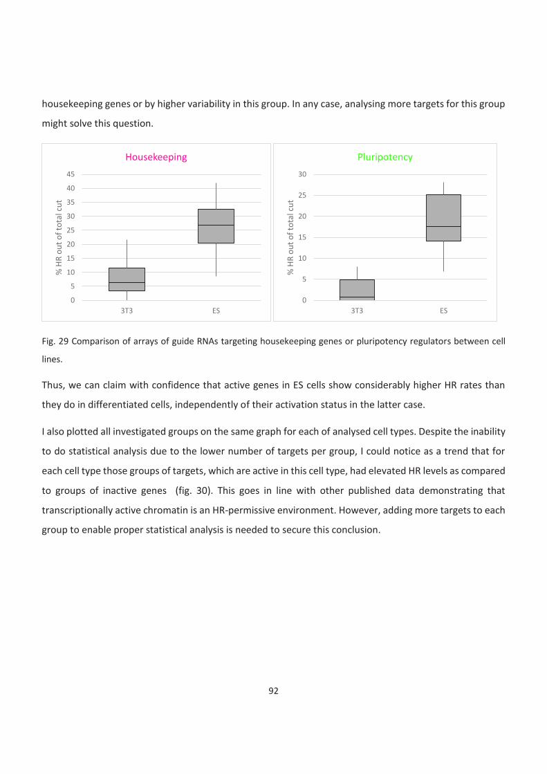
92
housekeeping genes or by higher variability in this group. In any case, analysing more targets for this group
might solve this question.
Fig. 29 Comparison of arrays of guide RNAs targeting housekeeping genes or pluripotency regulators between cell
lines.
Thus, we can claim with confidence that active genes in ES cells show considerably higher HR rates than
they do in differentiated cells, independently of their activation status in the latter case.
I also plotted all investigated groups on the same graph for each of analysed cell types. Despite the inability
to do statistical analysis due to the lower number of targets per group, I could notice as a trend that for
each cell type those groups of targets, which are active in this cell type, had elevated HR levels as compared
to groups of inactive genes (fig. 30). This goes in line with other published data demonstrating that
transcriptionally active chromatin is an HR-permissive environment. However, adding more targets to each
group to enable proper statistical analysis is needed to secure this conclusion.
0
5
10
15
20
25
30
35
40
45
3T3 ES
% H
R o
ut
of
tota
l cu
t
Housekeeping
0
5
10
15
20
25
30
3T3 ES
% H
R o
ut
of
tota
l cu
t
Pluripotency
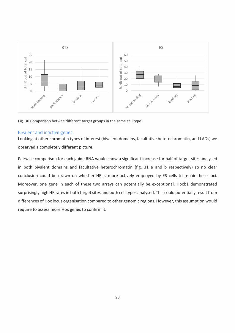
93
Fig. 30 Comparison betwee different target groups in the same cell type.
Bivalent and inactive genes
Looking at other chromatin types of interest (bivalent domains, facultative heterochromatin, and LADs) we
observed a completely different picture.
Pairwise comparison for each guide RNA would show a significant increase for half of target sites analysed
in both bivalent domains and facultative heterochromatin (fig. 31 a and b respectively) so no clear
conclusion could be drawn on whether HR is more actively employed by ES cells to repair these loci.
Moreover, one gene in each of these two arrays can potentially be exceptional. Hoxb1 demonstrated
surprisingly high HR rates in both target sites and both cell types analysed. This could potentially result from
differences of Hox locus organisation compared to other genomic regions. However, this assumption would
require to assess more Hox genes to confirm it.
0
5
10
15
20
25
% H
R o
ut
of
tota
l cu
t
3T3
0
10
20
30
40
50
60
% H
R o
ut
of
tota
l cu
t
ES

94
Fig. 31 Comparison of a) bivalent and b) heterochromatic targets between two cell lines on single guide RNAs and c)
on combined arrays. There is no significant difference in HR efficiency in different cell types.
Considering bivalent regions and facultative heterochromatic targets as independent arrays (fig. 31 c) we
found no significant difference between analysed cell types. Moreover, medians of an array of one cell type
lied within a confident interval of another cell type for both chromatin environment, and the increase of
HR utilisation in ES cells was approximately two-fold (close to 2 and 2.16 respectively). This led us to a
0
20
40
60
80
100
3T3 ES
% o
f to
tal c
ut
Ngn2 (pg316)
0
20
40
60
80
100
3T3 ES
% o
f to
tal c
ut
Pax6 (pg445)
0
20
40
60
80
100
3T3 ES
% o
f to
tal c
ut
Zic1 (pg447)
NHEJ+MMEJ
HR
0
20
40
60
80
100
3T3 ES
% o
f to
tal c
ut
Hoxb1 (pg450)
0
20
40
60
80
100
3T3 ES
% o
f to
tal c
ut
Tdrd1 (pgl1)
0
20
40
60
80
100
3T3 ES
% o
f to
tal c
ut
Mc4r (pgl3)
NHEJ+MMEJ
HR
0
5
10
15
20
25
3T3 ES
% H
R o
ut
of
tota
l cu
t
Bivalent
0
5
10
15
20
25
30
35
3T3 ES
% H
R o
ut
of
tota
l cu
t
Inactive
b
c
a
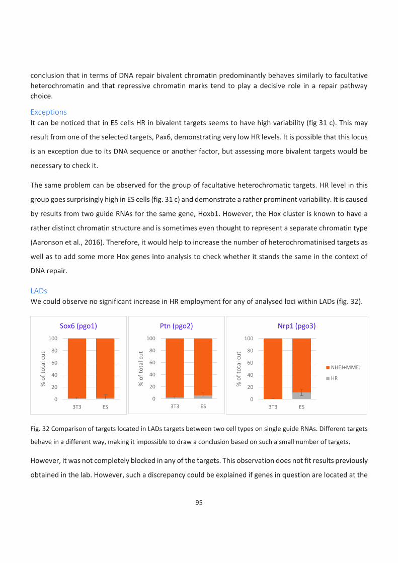
95
conclusion that in terms of DNA repair bivalent chromatin predominantly behaves similarly to facultative
heterochromatin and that repressive chromatin marks tend to play a decisive role in a repair pathway
choice.
Exceptions
It can be noticed that in ES cells HR in bivalent targets seems to have high variability (fig 31 c). This may
result from one of the selected targets, Pax6, demonstrating very low HR levels. It is possible that this locus
is an exception due to its DNA sequence or another factor, but assessing more bivalent targets would be
necessary to check it.
The same problem can be observed for the group of facultative heterochromatic targets. HR level in this
group goes surprisingly high in ES cells (fig. 31 c) and demonstrate a rather prominent variability. It is caused
by results from two guide RNAs for the same gene, Hoxb1. However, the Hox cluster is known to have a
rather distinct chromatin structure and is sometimes even thought to represent a separate chromatin type
(Aaronson et al., 2016). Therefore, it would help to increase the number of heterochromatinised targets as
well as to add some more Hox genes into analysis to check whether it stands the same in the context of
DNA repair.
LADs
We could observe no significant increase in HR employment for any of analysed loci within LADs (fig. 32).
Fig. 32 Comparison of targets located in LADs targets between two cell types on single guide RNAs. Different targets
behave in a different way, making it impossible to draw a conclusion based on such a small number of targets.
However, it was not completely blocked in any of the targets. This observation does not fit results previously
obtained in the lab. However, such a discrepancy could be explained if genes in question are located at the
0
20
40
60
80
100
3T3 ES
% o
f to
tal c
ut
Sox6 (pgo1)
0
20
40
60
80
100
3T3 ES
% o
f to
tal c
ut
Ptn (pgo2)
0
20
40
60
80
100
3T3 ES
% o
f to
tal c
ut
Nrp1 (pgo3)
NHEJ+MMEJ
HR
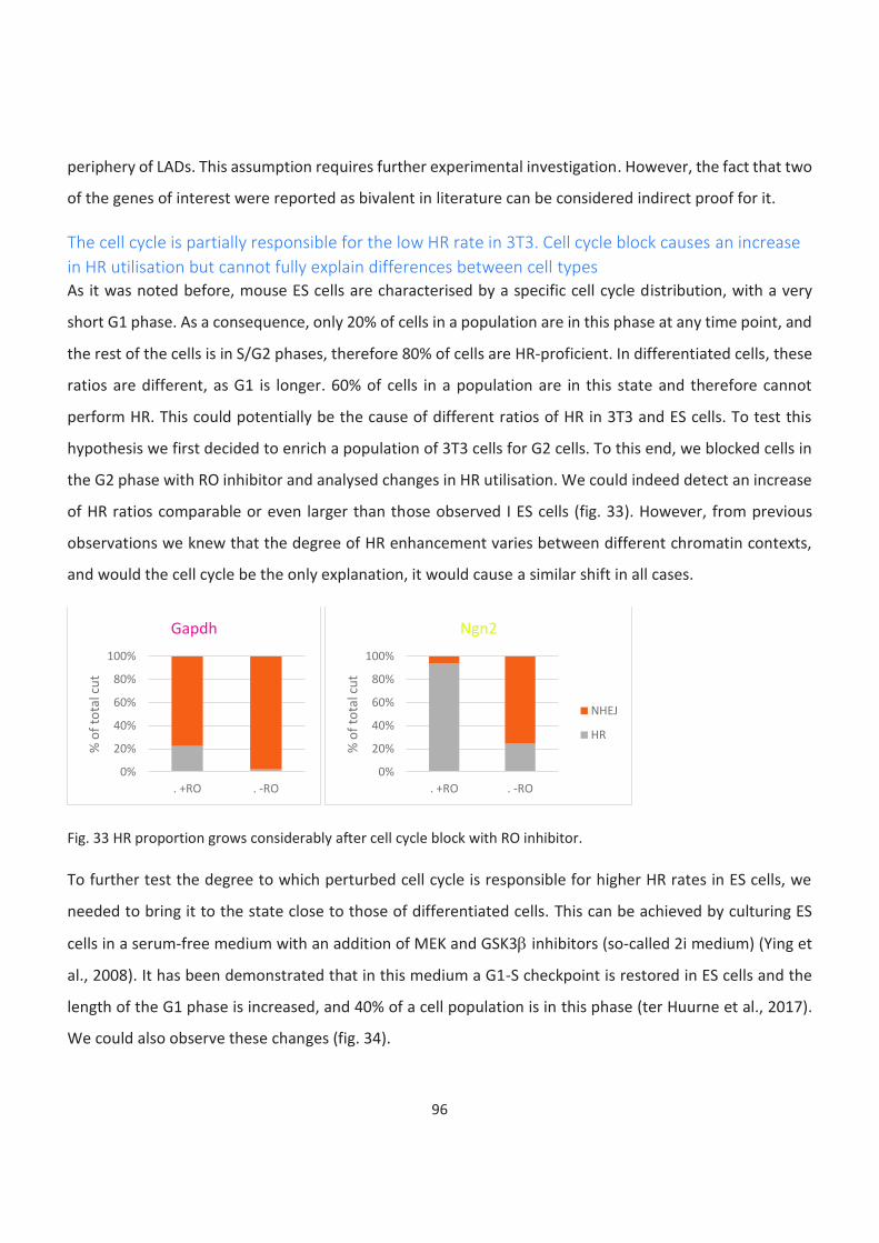
96
periphery of LADs. This assumption requires further experimental investigation. However, the fact that two
of the genes of interest were reported as bivalent in literature can be considered indirect proof for it.
The cell cycle is partially responsible for the low HR rate in 3T3. Cell cycle block causes an increase
in HR utilisation but cannot fully explain differences between cell types
As it was noted before, mouse ES cells are characterised by a specific cell cycle distribution, with a very
short G1 phase. As a consequence, only 20% of cells in a population are in this phase at any time point, and
the rest of the cells is in S/G2 phases, therefore 80% of cells are HR-proficient. In differentiated cells, these
ratios are different, as G1 is longer. 60% of cells in a population are in this state and therefore cannot
perform HR. This could potentially be the cause of different ratios of HR in 3T3 and ES cells. To test this
hypothesis we first decided to enrich a population of 3T3 cells for G2 cells. To this end, we blocked cells in
the G2 phase with RO inhibitor and analysed changes in HR utilisation. We could indeed detect an increase
of HR ratios comparable or even larger than those observed I ES cells (fig. 33). However, from previous
observations we knew that the degree of HR enhancement varies between different chromatin contexts,
and would the cell cycle be the only explanation, it would cause a similar shift in all cases.
Fig. 33 HR proportion grows considerably after cell cycle block with RO inhibitor.
To further test the degree to which perturbed cell cycle is responsible for higher HR rates in ES cells, we
needed to bring it to the state close to those of differentiated cells. This can be achieved by culturing ES
cells in a serum-free medium with an addition of MEK and GSK3b inhibitors (so-called 2i medium) (Ying et
al., 2008). It has been demonstrated that in this medium a G1-S checkpoint is restored in ES cells and the
length of the G1 phase is increased, and 40% of a cell population is in this phase (ter Huurne et al., 2017).
We could also observe these changes (fig. 34).
0%
20%
40%
60%
80%
100%
. +RO . -RO
% o
f to
tal c
ut
Gapdh
0%
20%
40%
60%
80%
100%
. +RO . -RO
% o
f to
tal c
ut
Ngn2
NHEJ
HR
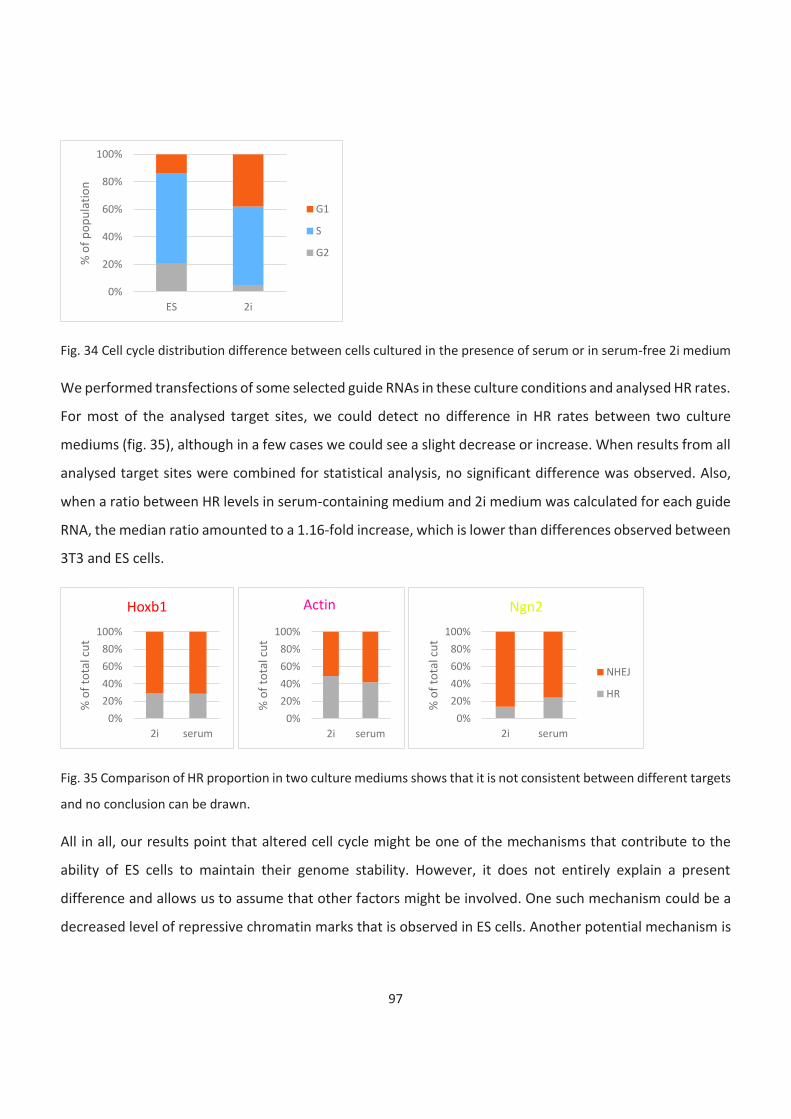
97
Fig. 34 Cell cycle distribution difference between cells cultured in the presence of serum or in serum-free 2i medium
We performed transfections of some selected guide RNAs in these culture conditions and analysed HR rates.
For most of the analysed target sites, we could detect no difference in HR rates between two culture
mediums (fig. 35), although in a few cases we could see a slight decrease or increase. When results from all
analysed target sites were combined for statistical analysis, no significant difference was observed. Also,
when a ratio between HR levels in serum-containing medium and 2i medium was calculated for each guide
RNA, the median ratio amounted to a 1.16-fold increase, which is lower than differences observed between
3T3 and ES cells.
Fig. 35 Comparison of HR proportion in two culture mediums shows that it is not consistent between different targets
and no conclusion can be drawn.
All in all, our results point that altered cell cycle might be one of the mechanisms that contribute to the
ability of ES cells to maintain their genome stability. However, it does not entirely explain a present
difference and allows us to assume that other factors might be involved. One such mechanism could be a
decreased level of repressive chromatin marks that is observed in ES cells. Another potential mechanism is
0%
20%
40%
60%
80%
100%
ES 2i
% o
f p
op
ula
tio
n
G1
S
G2
0%
20%
40%
60%
80%
100%
2i serum
% o
f to
tal c
ut
Hoxb1
0%
20%
40%
60%
80%
100%
2i serum
% o
f to
tal c
ut
Actin
0%
20%
40%
60%
80%
100%
2i serum
% o
f to
tal c
ut
Ngn2
NHEJ
HR

98
an increased level of proteins involved in DNA repair and particularly in HR that was previously reported.
However, a precise answer to this question would require further investigation.
99
Discussion
In my project, I investigated the role of sequence and chromatin context in the process of DNA double-
strand break repair and the influence of the cell type on it. Cell types in the focus were mouse ES cells and
3T3 cells. ES cells were selected as a model because they were reported to have unique pluripotency-
specific features of DNA repair, and because of the abundance of bivalent chromatin. 3T3 cells were used
as a model of differentiated yet cycling cells as a line stably expressing Cas9 was previously acquired in the
team. Targets for a break induction were selected at euchromatic, bivalent regions, facultative
heterochromatin, and LADs. The first three target types were dictated by our wish to compare bivalent
regions to chromatin types a mix of which they represent. I performed TIDE and HR-TIDE experiments for
all selected targets in both cell types, in normal conditions or with cell cycle perturbations. I could confirm
existing hypothesis of error-prone repair outcome being greatly dependent on the sequence around the
break site, and having variations depending on a cell type.
The tendency seems to be in accordance with previous observations that euchromatin seems to shift the
balance between repair pathway towards HR, as compared to transcriptionally inactive regions such as
facultative heterochromatin and bivalent domains, although due to the small number of investigated
targets I cannot claim statistical significance. However, these observations come into contradiction with
the results of a study by Schep et al. that was recently published on BioRxiv (Schep et al., 2020). They
addressed the question of the role of the chromatin environment in repair pathway choice by inserting a
short barcoded reporter cassette with a well-characterised Cas9 target site into multiple genomic
locations in a human immortalised cancer cell line. Analysing thousands of target sites, they observed a
negative correlation between the presence of H3K27me3 chromatin mark and NHEJ employment and
could also see that the euchromatic environment promotes the use of NHEJ. In our investigation we could
see prominent recruitment of NHEJ in euchromatic targets in 3T3 but not in ES cells. However, somatic
3T3 cells seem to be a more proper line to compare to another differentiated cell line, such as K562
utilised by Schep et al. At the same time, we did not see the anticorrelation between H3K27
trimethylation and the use of NHEJ in either of the model cell types. However, a small number of targets
does not allow us to draw such broad conclusions.
In the same study, a template-dependent repair has been tackled. It was observed that the chromatin
environment influences it the same way as it does for NHEJ. However, the pathway chosen for that

100
investigation was single-strand templated repair, so we could not directly compare it to our results as we
were interested in HR. We could see that HR utilization was influenced both by a chromatin type and a
cell type. Heterochromatic marks appeared to dominate over euchromatic in control of a repair pathway
choice and to be repressive for HR. I found that cell cycle characteristics are one of the factors leading to
the abovementioned differences between pluripotent and differentiated cells but they are unlikely to be
the only factor involved. Therefore, further experiments to dissect this issue might be necessary.
Altogether, despite certain similarities in the experimental setup, we mostly addressed different
questions from those of Schep et al. Our studies could therefore be considered as complementary for
clarification of the role of chromatin features in defining DSB repair strategy.
Sequence predominantly defines the DSB repair outcome. Chromatin and cell type might have no
more than moderate influence on the DSB repair outcome
After analysing indel patterns for all target sites (26 guide RNAs) designed to induce DSBs in different
chromatin contexts in two different cell types I could see no correlation with any of these factors. All
types of repair outcomes, including small insertions and deletions, thought to be resulting from NHEJ
employment, and larger deletions normally connected to MMEJ could be observed in all chromatin types.
Indel patterns between different cell types bore strong similarity for every guide RNA, whereas no
resemblance could be detected for two different guide RNAs even in a case of a short distance between
break sites. This goes in line with previously published data indicating that DNA sequence around the
break site, particularly a few nucleotides surrounding it, and a presence of microhomologies define
specific indels found after a break repair.
However, an overall proportion of larger deletions, presumably associated with MMEJ, appeared to be
higher in ES cells than in 3T3 cells. This is likely to indicate a shift in a balance between NHEJ and MMEJ.
One possible explanation for this effect could be a more open chromatin organisation in ES cells,
considering that such a state is generally reported to be prone to HR and at the same time MMEJ being
also resection-dependent. This shift of balance could also be a consequence of an increased level of some
proteins simultaneously involved in both HR and MMEJ repair pathways, such as CtIP. Nevertheless, this
would require additional experiments to prove.
101
ES cells use HR more actively than differentiated cells
Focusing more on the balance between error-free and error-prone repair pathways I performed HR-TIDE
experiments and analyzed a proportion of sequences that incorporated a provided template for HR.
Despite numbers varying for individual guide RNAs, analysis on the whole array revealed that HR is
significantly higher used across the genome in ES cells as compared to 3T3 cells. This corresponds to the
current paradigm in the field. However, unlike some other cases, in my setup, I could still see a vast
contribution of error-prone pathways in the repair process. Therefore, my results support the hypothesis
of HR being more actively employed in ES cells but do not confirm the degree of its mobilization
previously claimed. This is likely to result from the difference between experimental setups. Studies
leading to conclusions that HR is utilised to repair up to 80% of breaks in ES cells were performed on one
single break on a transcriptionally active gene, and I checked several target sites across the genome, in
both active and inactive states. Another possible explanation could be the use of Cas9 to introduce breaks
in my experiments, as it has previously been suggested that the presence of Cas9 at the DNA after a break
was introduced might potentially influence the pathway choice.
HR is enhanced in transcriptionally active genes. It is more prominent in ES than in differentiated
cells
A comparison between transcriptionally active regions and other types of chromatin led me to the
conclusion that the euchromatic environment is promoting HR. Despite I cannot prove this observation
statistically due to a low number of loci explored for each group, it is supported by several other studies,
which makes it likely to be correct. Nevertheless, it would be preferable to increase the number of targets
to enable a solid conclusion. Therefore, finding that pluripotency regulators demonstrate significantly
higher levels of HR in ES than 3T3 cells is not surprising. However, I could also see a threefold increase for
housekeeping genes, which supports the previous conclusion that HR levels are generally elevated in
pluripotent cells.
Despite both conclusions fit within currently existing views, I think they add considerably to previously
developed theories as to my knowledge it is the first time when HR contribution is assessed directly at the
level of sequence and not based on the recruitment of different factors, which could also be involved into
some other processes. Moreover, it disconfirms a recent study by Janssen et al. that claimed no influence
102
of the chromatin state on the process of HR and therefore sheds light on a still controversial topic
(Janssen et al., 2019).
HR levels are low in bivalent domains and facultative heterochromatin
Of different types of heterochromatin constitutive heterochromatin is the most studied and it has been
shown that DSBs in such an environment are less likely to be repaired by HR. However, facultative
heterochromatin has been less studied. We assessed several loci marked with H3K27me3 and found that
HR levels, in this case, were low, and more interestingly, they were not significantly increased in ES cells as
compared to 3T3 cells. Therefore, we can conclude that facultative heterochromatin represents an
environment predominantly repressive for HR.
As euchromatin represents an HR-promoting environment and heterochromatin is repressive to it, it was
especially interesting for us to see what results we might observe in the case of bivalent domains, where
marks of both are combined. Interestingly, we could see that HR levels stay low and there is no significant
difference between cell types. Therefore, the same outcome is observed for bivalent domains behave as
for facultative heterochromatin. In other words, in regulating the process of DSB repair repressive
chromatin marks appear to be a stronger driver than permissive ones. It is an interesting observation as to
our knowledge no one has yet studied the influence of this type of chromatin on the process of DNA
repair.
Influence of the cell cycle on the enhanced HR utilization in ES cells
As a cell cycle is known to strongly influence the process and it is also changed in ES cells as compared to
differentiated cells, we decided to check to what extent it contributes to the difference in repair pathway
choice we see between cell types. For this we blocked 3T3 cells in the G2 phase to check if increasing a
proportion of HR-proficient cells in the population would change the balance between repair pathways. It
is hardly surprising that we found it to lead to an increase in the proportion of HR products. Therefore, in
consonance with existing knowledge, we could see that blocking cells in a cell cycle stage permissive for
HR would lead to increase utilisation of this pathway up to levels similar or even exceeding observed in ES
cells.
Another apparent question was whether modifying the cell cycle of ES cells to resemble one of the
differentiated cells would cause the opposite effect. Fortunately, we had a way to do it, as it has been
103
reported that in 2i medium ES cells acquire a cell cycle that resembles one of the differentiated cells, and
what was of particular interest for us, in such culture conditions larger proportion of cells in a population
stays in G1 phase. After introducing DSBs using several selected guide RNAs we only found a moderate
drop in HR efficiency, which was not statistically significant. However, it has to be taken into account that
the share of cells in the G1 phase is smaller in ES cells even in 2i than in 3T3 cells.
All in all, we came to the conclusion that it is very likely that a distinct cell cycle that is known to be
characteristic for ES cells is likely to be one of the mechanisms to ensure higher genome stability.
Nevertheless, it is highly doubtful it is the only underlying mechanism, and further experiments would be
required to uncover other ways utilised by ES cells to ensure their genome is preserved through
generations.
104
Perspectives
It has to be noted that I performed all experiments in my project on a relatively small number of targets,
which limits our ability to generalise conclusions. Therefore, my perspectives could be divided into short-
term, aiming to prove established results without the addition of a large number of other target sites or a
major change in the experimental setup, and long-term, aiming to broaden our observations to a genome-
wide level.
I would like to start discussing the suggested experiments with short-term perspectives. First, considering
the heterogeneity of results for bivalent domains and facultative heterochromatin target groups I think
adding several more targets for each group to ensure that none of the results for selected targets was
impaired by any factor other than chromatin structure (in other words, that we do not deal with any
target representing an exception from the general trend) and reduce the influence of outliers. Adding 3 or
4 more targets should be sufficient to complement already performed HR-TIDE-based experiments.
Considering the fact that most of the time using two guide RNAs designed for the same region leads to
very similar results, it might be profitable to design one guide RNA per each selected target. Choosing
some additional targets within the Hox cluster would also allow evaluating the hypothesis of exceptional
DSB repair pathway preference (and therefore indirectly confirm an exceptional chromatin organisation in
this part of a genome). Considering the differences observed between publications it would be more
tenable to perform ChIP-qPCR for all newly selected targets to confirm their chromatin status.
Second, adding several more guide RNAs for housekeeping genes would enable us to perform a proper
statistical analysis to compare different target groups within the same cell type. It ought to be remarked
that the target group of LADs was excluded from all comparisons with other chromatin types due to a
small size of the group. Therefore, it also requires enlargement. However, considering highly heterogenic
results observed for this target group it is also necessary to perform FISH to prove that all of the targeted
regions indeed belong to LADs in the utilised cell line.
In my analysis, I have set the NHEJ/MMEJ border voluntary. To stratify this question as well as to prove
that +9 incorporation is indeed facilitated by HR, it would be necessary to perform TIDE or HR-TIDE
experiments while knocking down or chemically inhibiting key players of investigated pathways, such as
105
DNA-PKcs, DNA Ligase IV, or XRCC4 for NHEJ, Polq, DNA Ligase III, or XRCC1 for MMEJ, and Rad51 or
BRCA1 for HR.
According to the obtained results, we could formulate a hypothesis that in case of bivalency, i.e.
coexistence between euchromatic and heterochromatic marks DSB pathway balance is kept similar to
those of facultative heterochromatin. However, this assumption requires direct confirmation. In order to
achieve it, we plan to remove H3K27me3 from bivalent genes by recruitment of histone demethylases
such as JMJD3 or UTX to target sites by fusing them to dCas9 (a catalytically inactive form of Cas9) prior to
performing HR-TIDE experiments. Demethylases recruitment can alternatively be substituted by chemical
inhibition or knock-down of PRC2 complex subunits. However, it would cause genome-wide
perturbations, therefore, targeted demethylation is preferable. On the other hand, targeting PRC2
complex to transcriptionally active targets to form bivalent domains de novo would complement
proposed experiments in order to prove a proposed theory. However, if this strategy is chosen, it would
be important to confirm that bivalent domain formation and not replacement of euchromatic marks by
heterochromatic follows such a recruitment. Therefore, a confirmation by ChIP-qPCR would be required.
We could conclude from our results that HR rates are higher in actively transcribed regions marked by
H3K4me3. However, we cannot draw a line between roles transcriptional activity and chromatin status.
Performing HR-TIDE experiments after chemical transcription inhibition would allow us to dissect
between these two factors. Triptolide use would be preferable compared to other commonly used
compounds, such as α-amanitin, actinomycin D, DRB, or flavopiridol, due to the fact that it inhibits
transcription initiation, while other inhibitors block elongation. Considering that selected targets lie within
proximal promoter regions, an attitude that blocks transcription inhibition should preferentially be used.
Potential downsides of such an approach are massive perturbation caused in a cell by a prolonged
transcription block. One cannot exclude that effects caused by such a block might mask analyzed
influence on DNA repair.
Regarding long-term perspectives, I would like to mention a potential practical application of the
developed approach. In my opinion, it could be used for predicting knock-in efficiency. Analysing obtained
results I have noticed that in some cases HR efficiency differs from what can be expected from the
chromatin environment surrounding the introduced break. Knocking-in large fragments, such as
106
fluorescent reporters, is often necessary for research. However, the introduction of large fragments often
results in reduced efficiency compared to smaller ones. Therefore, I think that prior testing of designed
homology arms with smaller insert might be a good strategy to predict their efficiency before engaging in
a large experiment to enable the optimal setup. To allow this, homology arms should be cloned with a
small insert containing digestion sites that would enable further replacement by a larger insert of interest.
This way, knock-in efficiency could be tested by HR-TIDE prior to the real experiment. To verify that HR-
TIDE results are a good predictor for a larger fragment, it would be good to replace an insert in several of
previously used templates with mCherry and evaluate knock-in efficiency by FACS to test whether it
correlates with HR proportion observed in HR-TIDE experiments.
Finally, all experiments performed in the course of my doctoral project can be used as a basis to design a
genome-wide test of HR utilization. Selecting one or two hundreds of well-cutting guide RNAs targeting
different genome regions based on available data and cloning them together with corresponding HR
templates into a viral library based on an integration-deficient lentiviral vector could be a solution for
such a goal. This library should be used for infecting cells of interest and further submitted to NGS
analysis. Such an experiment would provide a map of HR utilisation efficiency and allow assessment of its
dependence on a chromatin state as well as other factors. It would also help to select a group of targets
for further dissection of mechanisms underlying observed correlations.
107
Materials and methods
Cell culture
Mouse ES cells were cultured on a feeder layer of mitotically inactive MEFs, growing conditions were set
to 37°C, 5% CO2. Cells were passaged every 2-3 days and kept in culture for no more than 10 passages.
Passaging involved trypsinisation for 5 minutes at 37°C, trypsin inactivation with at least equal volume of
medium, achieving a state of single-cell suspension, separation from feeders by gravity (after 10 minutes
incubation in a 15 ml Falcon tube, top fraction was used for plating) and plating on a fresh dish at 1:10 to
1:20 rate depending on the desired final confluency.
Standard ES cell medium composition:
High glucose DMEM
15% ES-grade FCS
1mM NEAA
1 mM Sodium Pyruvate
40mg/ml Gentamycin
100 mM b-mercaptoethanol
2x recombinant LIF
For creating stable cell lines, antibiotics selection begun 24 h post transfection and continued for 2 days
with Puromycin (at a final concentration of 2mM) or for 10 days with G418 (at a final concentration of 400
mM).
Suicidal cassette: for all following experiments cells were treated with puromycine for 2 days starting 24h
after thawing.
For experiments in serum-free conditions 2i medium was used.
108
2i medium composition:
DMEM/F12 + Neurobasal mixed 1:1
1x N2
1x B27
25 mg/ml BSA
100 mM b-mercaptoethanol
2x recombinant LIF
1 mM PD 98059
3 mM SB 216763
For a plasmid transfection, reverse transfection protocol using Lipofectamine 2000 was applied. Cells
were pre-treated with Shield1 (at a final concentration of 1:2000) overnight prior transfection. For the
transfection, cells were trypsinised and separated from feeders by gravity in 15 ml Falcon tubes for 15
minutes. ES cell containing supernatant was separated from a pellet of feeders and transferred into fresh
Falcon tubes. 0.5*106 cells per sample were taken if transfecting cells cultured in the standard ES cell
medium. In the time of incubation, transfection mixes were prepared. Transfection mix 1 contained total
of 5 mg of DNA (when transfecting for HR-TIDE experiments, HR template and a guide RNA were co-
transfected at a 3:2 ratio, for template only or guide samples a corresponding plasmid was replaced by
the same amount of the empty vector), 10 ml of Lipofectamine 2000, and 250 ml of OptiMEM medium.
Transfection mix 2 contained 12,5 ml of Lipofectamine 2000, and 250 ml of OptiMEM medium.
Transfection mixes 1 and 2 were incubated for 5 min at room temperature, then mixed and incubated for
10 more min at room temperature. For transfection, cells were centrifuged for 3 min at 1200 rpm, and the
supernatant removed. Resulting pellets were carefully disloged, and then gently resuspended in
corresponding transfection mixes. Transfection was performed with 15 minutes of incubation in
suspension at 37°C. In order to prevent a pellet formation, tubes were gently shaken every 5 min. Cells
109
were plated on 6-well plates pre-coated with gelatine with addition of 0,5 ml of OptiMEM per well for 4
hours, than medium was replaced by standard ES cell culture medium supplemented with Shield1.
For transfections of cells cultured in 2i medium 1*10^6 cells was taken per sample and transfected with
total of 2 mg of DNA. Reverse transfection was performed with reducing incubation time to 7 minutes.
Cells were plated in 2i medium supplemented with 2% serum.
Growing conditions for 3T3 cells were set to 37°C, 5% CO2. Cells were passaged every 3-4 days and kept in
culture for no more than 8 passages.
3T3 cell medium composition:
High glucose DMEM
10% NCS
40mg/ml Gentamycin
Transfection of 3T3 cells was performed using Lipofectamine 2000. Cells were plated at a confluency of
1*105 cells per sample on a well of a 6-well plate 18 h prior transfection. On the day of transfection,
medium in each well was replaced by 1 ml of a fresh 3T3 cell medium and transfection mixes were
prepared. Transfection mix 1 contained total of 2 mg of DNA (when transfecting for HR-TIDE experiments,
HR template and a guide RNA were co-transfected at a 3:2 ratio, for template only or guide samples a
corresponding plasmid was replaced by the same amount of the empty vector) and 150 ml of OptiMEM
medium. Transfection mix 2 contained 10 ml of Lipofectamine 2000 and 150 ml of OptiMEM medium.
Transfection mixes 1 and 2 were incubated for 5 min at room temperature, then mixed and incubated for
10 more min. Next, each transfection mix was added to a corresponding well.
110
Cell cycle analysis
Cell cycle analysis was performed using Click-iT EdU incorporation kit (Invitrogen) according to a slightly
modified manufacturer’s protocol. Cell culture medium was changed to fresh medium pre-warmed to
37°C and supplemented with EdU at a final concentration of 50 mM. After 30 min, cells were harvested by
trypsinisation, achieving a state of single-cell suspension, washed with 1% BSA in PBS, and fixed with a 100
ml of a provided fixative solution (Component D) for 15 min at room temperature. Then cells were washed
with 1% BSA in PBS and incubated with a 100 ml of a provided permeabilization and wash reagent
(prepared according to manufacturer’s protocol) for 15 min at room temperature. Next, cells were
washed with 1% BSA in PBS and a total 200 ml a reaction cocktail was added per sample, followed by a 30
min incubation. Then samples were washed with 1,2 ml per sample of a permeabilization and wash
reagent and pelleted, and supernatant was removed. Resulting pellets were resuspended in 200 ml of a
permeabilization and wash reagent, supplemented by RNaseA at a final concentration of 4 mg/ml, and PI
at a final concentration of 4 mg/ml. Samples were further analysed by FACS.
Reaction cocktail composition (per sample):
175,2 ml of PBS
4 ml of CuSO4 (Component F)
2 ml of Fluorescent dye azide (prepared according to manufacturer’s protocol)
20 ml of Reaction Buffer Additive (prepared according to manufacturer’s protocol)
Western blotting
Cells were harvested by trypsinisation and lysed in RIPA buffer containing HEPES pH7.6 50mM, Sodium
dexicholate 0.5%, NP40 1%, EDTA 1mM, LiCl 0.5M. Quantification was performed with Bradford assay
according to manufacturer’s protocol. Samples were mixed with NuPAGE lysing buffer and pre-heated for
20 minutes. Samples were loaded on pre-cast 4-12% gradient gels for migration. Wet transfer was
performed on nitrocellulose membrane at 400 mA for 90 minute. Membrane was blocked in 5% non-fat
dry milk in PBS for 1 hour at room temperature. Primary antibodies were diluted in 3% non-fat dry milk in
PBS-0.1% Tween20 and incubated at 4°C overnight. Primary antibodies used were gH2AX (Abcam
111
ab2893) diluted 1:1000, tubulin (Sigma T9026) diluted 1:10000 and GFP (Santa Cruz sc-9,996 (clone B-2))
diluted 1:500. Membranes were washed with PBS-0.1% Tween20 and incubated with secondary
antibodies for 1 hour at room temperature. After incubations were washed with PBS-0.1% Tween20 and
developed using ECL reagent.
RIPA buffer composition:
50mM HEPES pH7.6
0,5% Sodium dexicholate
1% NP40
1mM EDTA
0.5M LiCl
Chromatin Immunoprecipitation (ChIP)
Cells were rinsed with PNS and fixed with 1% PFA in PBS for 15 minutes at 37°C. Cells were washed twice
with ice-cold PBS and collected by scraping on ice in PBS with PIC and PhoSTOP. Scraped cells were pulled
down at 4°C for 5 minutes at 1200 rpm. Starting from scraping, all samples and buffers were kept on ice,
except for SDS-containing sonication buffer. After removing supernatant cells were incubated for 10
minutes of ice in Lysis buffer 1 and centrifuged at 4°C for 2 minutes at 6000 rpm. After removing
supernatant cells were incubated for 10 minutes in Lysis buffer 2 and centrifuged at 4°C for 2 minutes at
6000 rpm. Supernatant was removed and after addition of Sonication buffer cells were transferred into
Covaris sonication tubes and sonicated for 15 minutes. 50 ml of Protein G Dynabeads was used for each
sample. Beads were washed 3 times with 5 mg/ml BSA in PBS, blocked in 5 mg/ml BSA in PBS for 30
minutes at 4°C on a rotating wheel and coated with antibodies for 4 hours. Immunoprecipitation was
performed with 20 mg of sonication materiel. Samples were diluted with ChIP dilution buffer and
immunoprecipitated overnight at 4°C. Samples were washed twice with SDS wash buffer, once in high salt
buffer and once in TE buffer. Samples were eluted with Elution buffer and decrosslinked overnight at 65°C
with vigorous shaking.
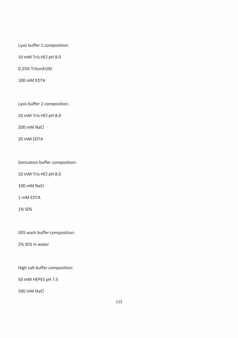
112
Lysis buffer 1 composition:
10 mM Tris-HCl pH 8.0
0.25% TritonX100
100 mM EDTA
Lysis buffer 2 composition:
10 mM Tris-HCl pH 8.0
200 mM NaCl
20 mM EDTA
Sonication buffer composition:
10 mM Tris-HCl pH 8.0
100 mM NaCl
1 mM EDTA
1% SDS
SDS wash buffer composition:
2% SDS in water
High salt buffer composition:
50 mM HEPES pH 7.5
500 mM NaCl
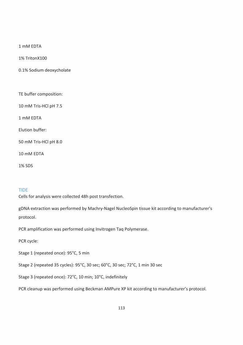
113
1 mM EDTA
1% TritonX100
0.1% Sodium deoxycholate
TE buffer composition:
10 mM Tris-HCl pH 7.5
1 mM EDTA
Elution buffer:
50 mM Tris-HCl pH 8.0
10 mM EDTA
1% SDS
TIDE
Cells for analysis were collected 48h post transfection.
gDNA extraction was performed by Machry-Nagel NucleoSpin tissue kit according to manufacturer’s
protocol.
PCR amplification was performed using Invitrogen Taq Polymerase.
PCR cycle:
Stage 1 (repeated once): 95°C, 5 min
Stage 2 (repeated 35 cycles): 95°C, 30 sec; 60°C, 30 sec; 72°C, 1 min 30 sec
Stage 3 (repeated once): 72°C, 10 min; 10°C, indefinitely
PCR cleanup was performed using Beckman AMPure XP kit according to manufacturer’s protocol.
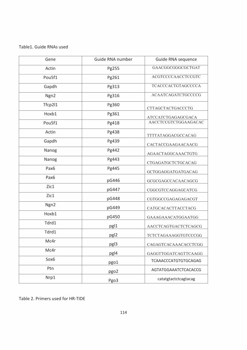
114
Table1. Guide RNAs used
Gene Guide RNA number Guide RNA sequence
Actin Pg255 GAACGGCGGGCGCTGAT
Pou5f1 Pg261 ACGTCCCCAACCTCCGTC
Gapdh Pg313 TCACCCACTGTAGCCCCA
Ngn2 Pg316 ACAATCAGATCTGCCCCG
Tfcp2l1 Pg360 CTTAGCTACTGACCCTG
Hoxb1 Pg361 ATCCATCTGAGAGCGACA
Pou5f1 Pg418 AACCTCCGTCTGGAAGACAC
Actin Pg438 TTTTATAGGACGCCACAG
Gapdh Pg439 CACTACCGAAGAACAACG
Nanog Pg442 AGAACTAGGCAAACTGTG
Nanog Pg443 CTGAGATGCTCTGCACAG
Pax6 Pg445 GCTGGAGGATGATGACAG
Pax6 pG446 GCGCGAGCCACAACAGCG
Zic1 pG447 CGGCGTCCAGGAGCATCG
Zic1 pG448 CGTGGCCGAGAGAGACGT
Ngn2 pG449 CATGCACACTTACCTACG
Hoxb1 pG450 GAAAGAAACATGGAATGG
Tdrd1 pgl1 AACCTCAGTGACTCTCAGCG
Tdrd1 pgl2 TCTCTAGAAAGGTGTCCCGG
Mc4r pgl3 CAGAGTCACAAACACCTCGG
Mc4r pgl4 GAGGTTGGATCAGTTCAAGG
Sox6 pgo1 TCAAACCCATGTGTGCAGAG
Ptn pgo2 AGTATGGAAATCTCACACCG
Nrp1 Pgo3 catatgtactctcagtacag
Table 2. Primers used for HR-TIDE
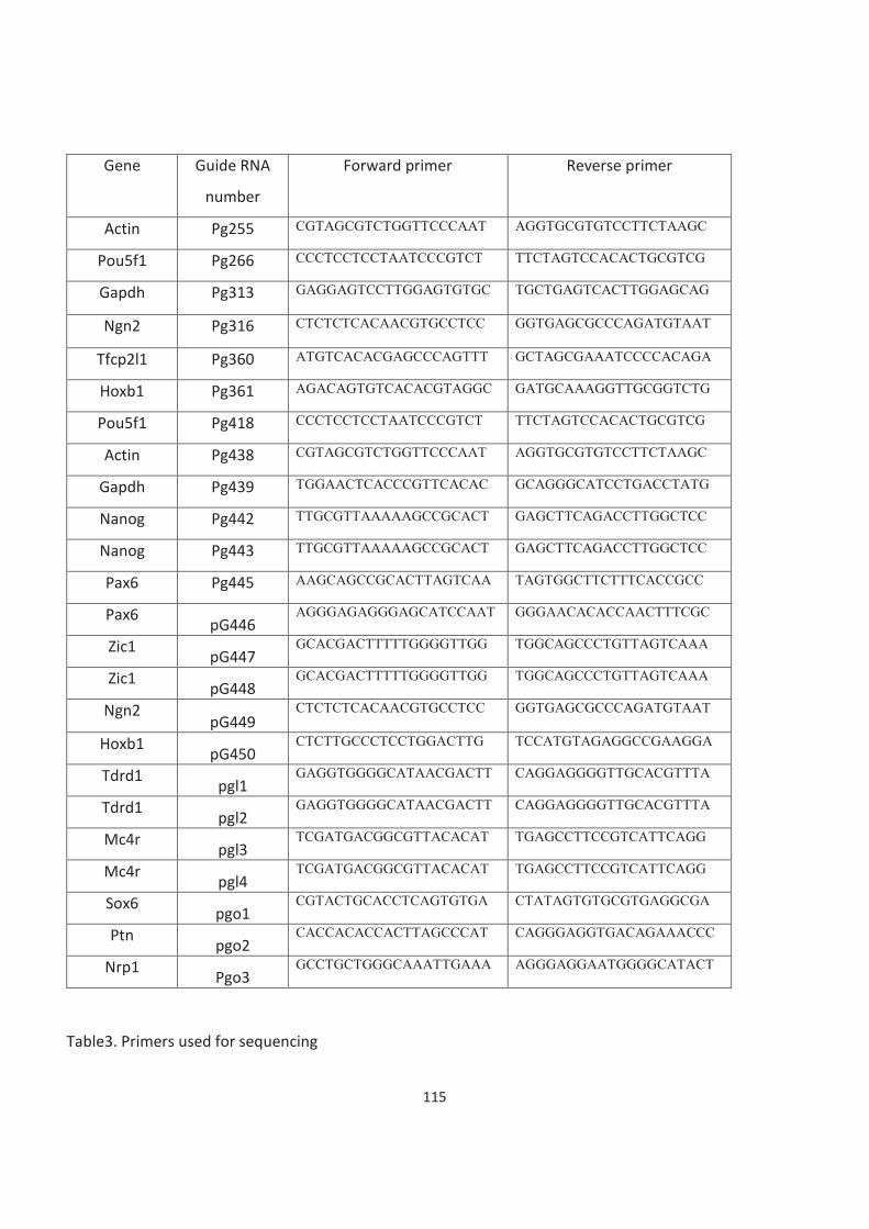
115
Gene Guide RNA
number
Forward primer Reverse primer
Actin Pg255 CGTAGCGTCTGGTTCCCAAT AGGTGCGTGTCCTTCTAAGC
Pou5f1 Pg266 CCCTCCTCCTAATCCCGTCT TTCTAGTCCACACTGCGTCG
Gapdh Pg313 GAGGAGTCCTTGGAGTGTGC
TGCTGAGTCACTTGGAGCAG
Ngn2 Pg316 CTCTCTCACAACGTGCCTCC
GGTGAGCGCCCAGATGTAAT
Tfcp2l1 Pg360 ATGTCACACGAGCCCAGTTT GCTAGCGAAATCCCCACAGA
Hoxb1 Pg361 AGACAGTGTCACACGTAGGC GATGCAAAGGTTGCGGTCTG
Pou5f1 Pg418 CCCTCCTCCTAATCCCGTCT TTCTAGTCCACACTGCGTCG
Actin Pg438 CGTAGCGTCTGGTTCCCAAT AGGTGCGTGTCCTTCTAAGC
Gapdh Pg439 TGGAACTCACCCGTTCACAC GCAGGGCATCCTGACCTATG
Nanog Pg442 TTGCGTTAAAAAGCCGCACT GAGCTTCAGACCTTGGCTCC
Nanog Pg443 TTGCGTTAAAAAGCCGCACT GAGCTTCAGACCTTGGCTCC
Pax6 Pg445 AAGCAGCCGCACTTAGTCAA TAGTGGCTTCTTTCACCGCC
Pax6 pG446
AGGGAGAGGGAGCATCCAAT GGGAACACACCAACTTTCGC
Zic1 pG447
GCACGACTTTTTGGGGTTGG TGGCAGCCCTGTTAGTCAAA
Zic1 pG448
GCACGACTTTTTGGGGTTGG TGGCAGCCCTGTTAGTCAAA
Ngn2 pG449
CTCTCTCACAACGTGCCTCC
GGTGAGCGCCCAGATGTAAT
Hoxb1 pG450
CTCTTGCCCTCCTGGACTTG TCCATGTAGAGGCCGAAGGA
Tdrd1 pgl1
GAGGTGGGGCATAACGACTT CAGGAGGGGTTGCACGTTTA
Tdrd1 pgl2
GAGGTGGGGCATAACGACTT CAGGAGGGGTTGCACGTTTA
Mc4r pgl3
TCGATGACGGCGTTACACAT TGAGCCTTCCGTCATTCAGG
Mc4r pgl4
TCGATGACGGCGTTACACAT TGAGCCTTCCGTCATTCAGG
Sox6 pgo1
CGTACTGCACCTCAGTGTGA CTATAGTGTGCGTGAGGCGA
Ptn pgo2
CACCACACCACTTAGCCCAT CAGGGAGGTGACAGAAACCC
Nrp1 Pgo3
GCCTGCTGGGCAAATTGAAA AGGGAGGAATGGGGCATACT
Table3. Primers used for sequencing
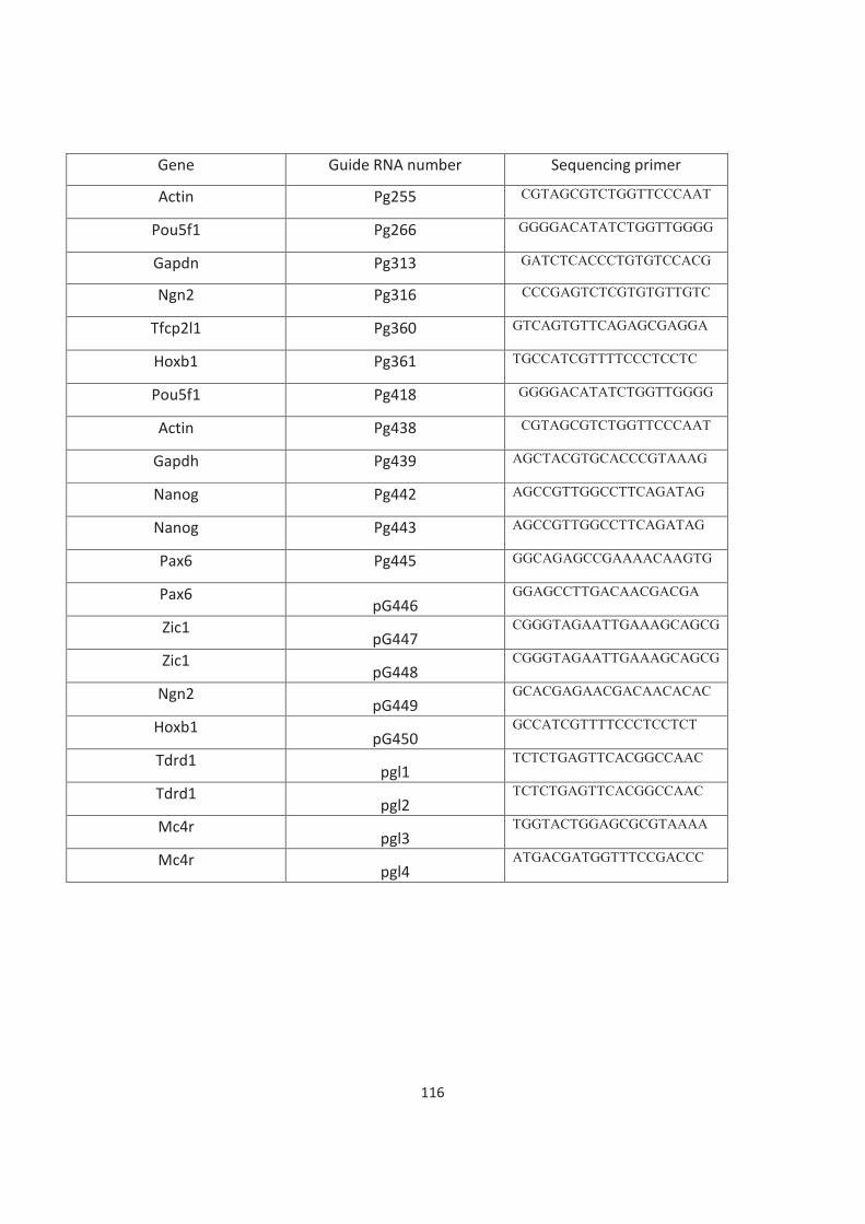
116
Gene Guide RNA number Sequencing primer
Actin Pg255 CGTAGCGTCTGGTTCCCAAT
Pou5f1 Pg266 GGGGACATATCTGGTTGGGG
Gapdn Pg313 GATCTCACCCTGTGTCCACG
Ngn2 Pg316 CCCGAGTCTCGTGTGTTGTC
Tfcp2l1 Pg360 GTCAGTGTTCAGAGCGAGGA
Hoxb1 Pg361 TGCCATCGTTTTCCCTCCTC
Pou5f1 Pg418 GGGGACATATCTGGTTGGGG
Actin Pg438 CGTAGCGTCTGGTTCCCAAT
Gapdh Pg439 AGCTACGTGCACCCGTAAAG
Nanog Pg442 AGCCGTTGGCCTTCAGATAG
Nanog Pg443 AGCCGTTGGCCTTCAGATAG
Pax6 Pg445 GGCAGAGCCGAAAACAAGTG
Pax6 pG446
GGAGCCTTGACAACGACGA
Zic1 pG447
CGGGTAGAATTGAAAGCAGCG
Zic1 pG448
CGGGTAGAATTGAAAGCAGCG
Ngn2 pG449
GCACGAGAACGACAACACAC
Hoxb1 pG450
GCCATCGTTTTCCCTCCTCT
Tdrd1 pgl1
TCTCTGAGTTCACGGCCAAC
Tdrd1 pgl2
TCTCTGAGTTCACGGCCAAC
Mc4r pgl3
TGGTACTGGAGCGCGTAAAA
Mc4r pgl4
ATGACGATGGTTTCCGACCC
117
Bibliography
Aaronson, Y., Livyatan, I., Gokhman, D., & Meshorer, E. (2016). Systematic identification of gene family regulators in mouse and human embryonic stem cells. Nucleic Acids Research, 44(9), 4080–4089. https://doi.org/10.1093/nar/gkw259
Adams, K. E., Medhurst, A. L., Dart, D. A., & Lakin, N. D. (2006). Recruitment of ATR to sites of ionising radiation-induced DNA damage requires ATM and components of the MRN protein complex. Oncogene, 25(28), 3894–
3904. https://doi.org/10.1038/sj.onc.1209426
Aguilar-Quesada, R., Muñoz-Gámez, J. A., Martín-Oliva, D., Peralta, A., Valenzuela, M. T., Matínez-Romero, R., Quiles-Pérez, R., Menissier-de Murcia, J., de Murcia, G., Ruiz de Almodóvar, M., & Javier, F. J. (2007). Interaction between ATM and PARP-1 in response to DNA damage and sensitization of ATM deficient cells through PARP inhibition. BMC Molecular Biology, 8, 1–8. https://doi.org/10.1186/1471-2199-8-29
Ahmad, A., Robinson, A. R., Duensing, A., van Drunen, E., Beverloo, H. B., Weisberg, D. B., Hasty, P., Hoeijmakers, J. H. J., & Niedernhofer, L. J. (2008). ERCC1-XPF Endonuclease Facilitates DNA Double-Strand Break Repair. Molecular and Cellular Biology, 28(16), 5082–5092. https://doi.org/10.1128/mcb.00293-08
Ahmed, K., Dehghani, H., Rugg-Gunn, P., Fussner, E., Rossant, J., & Bazett-Jones, D. P. (2010). Global chromatin architecture reflects pluripotency and lineage commitment in the early mouse embryo. PLoS ONE, 5(5). https://doi.org/10.1371/journal.pone.0010531
Ahn, J. Y., Schwarz, J. K., Piwnica-Worms, H., & Canman, C. E. (2000). Threonine 68 phosphorylation by ataxia telangiectasia mutated is required for efficient activation of Chk2 in response to ionizing radiation. Cancer
Research, 60(21), 5934–5936.
Ahnesorg, P., Smith, P., & Jackson, S. P. (2006). XLF interacts with the XRCC4-DNA Ligase IV complex to promote DNA nonhomologous end-joining. Cell, 124(2), 301–313. https://doi.org/10.1016/j.cell.2005.12.031
Ahrabi, S., Sarkar, S., Pfister, S. X., Pirovano, G., Higgins, G. S., Porter, A. C. G., & Humphrey, T. C. (2016). A role for human homologous recombination factors in suppressing microhomology-mediated end joining. Nucleic Acids
Research, 44(12), 5743–5757. https://doi.org/10.1093/nar/gkw326
Ahuja, A. K., Jodkowska, K., Teloni, F., Bizard, A. H., Zellweger, R., Herrador, R., Ortega, S., Hickson, I. D., Altmeyer, M., Mendez, J., & Lopes, M. (2016). A short G1 phase imposes constitutive replication stress and fork remodelling in mouse embryonic stem cells. Nature Communications, 7(May 2015). https://doi.org/10.1038/ncomms10660
Akasaka, T., Kanno, M., Balling, R., Mieza, M. A., Taniguchi, M., & Koseki, H. (1996). A role for mel-18, a Polycomb group-related vertebrate gene, during the anteroposterior specification of the axial skeleton. Development, 122(5), 1513–1522.
Akhtar, A., Zink, D., & Becker, P. B. (2000). Chromodomains are protein-RNA interaction modules. Nature, 407(6802), 405–409. https://doi.org/10.1038/35030169
Al-minawi, A. Z., Saleh-gohari, N., & Helleday, T. (2008). The ERCC1/XPF endonuclease is required for efficient single-strand annealing and gene conversion in mammalian cells. Nucleic Acids Research, 36(1), 1–9. https://doi.org/10.1093/nar/gkm888
Aladjem, M. I., Spike, B. T., Rodewald, L. W., Hope, T. J., Klemm, M., Jaenisch, R., & Wahl, G. M. (1998). ES cells do
118
not activate p53-dependent stress responses and undergo p53-independent apoptosis in response to DNA damage. Current Biology, 8(3), 145–155. https://doi.org/10.1016/S0960-9822(98)70061-2
Alison, M., & Islam, S. (2009). Attributes of adult stem cells. Journal of Pathology, 217(November), 144–160. https://doi.org/10.1002/path
Allan, J., Hartman, P. G., Crane-Robinson, C., & Aviles, F. X. (1980). The structure of histone H1 and its location in chromatin. Nature, 288(18), 675–679. https://www.nature.com/articles/288675a0.pdf
Allfrey, V. G., Faulkner, R., & Mirsky, A. E. (1964). Acetylation and Methylation of Histones and Their Possible Role in the. Proceedings of the National Academy of Sciences of the United States Of, 51(1938), 786–794. https://doi.org/10.1073/pnas.51.5.786
Allis, C. D., & Jenuwein, T. (2016). The molecular hallmarks of epigenetic control. Nature Reviews Genetics, 17(8), 487–500. https://doi.org/10.1038/nrg.2016.59
Amsel, A. D., Rathaus, M., Kronman, N., & Cohen, H. Y. (2008). Regulation of the proapoptotic factor Bax by Ku70-dependent deubiquitylation. Proceedings of the National Academy of Sciences of the United States of America, 105(13), 5117–5122. https://doi.org/10.1073/pnas.0706700105
Anand, R., Ranjha, L., Cannavo, E., & Cejka, P. (2016). Phosphorylated CtIP Functions as a Co-factor of the MRE11-RAD50-NBS1 Endonuclease in DNA End Resection. Molecular Cell, 64(5), 940–950. https://doi.org/10.1016/j.molcel.2016.10.017
Andreassen, P. R., D’Andrea, A. D., & Taniguchi, T. (2004). ATR couples FANCD2 monoubiquitination to the DNA-damage responsey. Genes and Development, 18(16), 1958–1963. https://doi.org/10.1101/gad.1196104
Aoto, T., Saitoh, N., Ichimura, T., Niwa, H., & Nakao, M. (2006). Nuclear and chromatin reorganization in the MHC-Oct3/4 locus at developmental phases of embryonic stem cell differentiation. Developmental Biology, 298(2), 354–367. https://doi.org/10.1016/j.ydbio.2006.04.450
Apostolou, E., Ferrari, F., Walsh, R. M., Bar-Nur, O., Stadtfeld, M., Cheloufi, S., Stuart, H. T., Polo, J. M., Ohsumi, T. K., Borowsky, M. L., Kharchenko, P. V., Park, P. J., & Hochedlinger, K. (2013). Genome-wide chromatin interactions of the nanog locus in pluripotency, differentiation, and reprogramming. Cell Stem Cell, 12(6), 699–
712. https://doi.org/10.1016/j.stem.2013.04.013
Arents, G., & Moudrianakis, E. N. (1993). Topography of the histone octamer surface: Repeating structural motifs utilized in the docking of nucleosomal DNA. Proceedings of the National Academy of Sciences of the United
States of America, 90(22), 10489–10493. https://doi.org/10.1073/pnas.90.22.10489
Ashley, A. K., Shrivastav, M., Nie, J., Amerin, C., Troksa, K., Glanzer, J. G., Liu, S., Opiyo, S. O., Dimitrova, D. D., Le, P., Sishc, B., Bailey, S. M., Oakley, G. G., & Nickoloff, J. A. (2014). DNA-PK phosphorylation of RPA32 Ser4/Ser8 regulates replication stress checkpoint activation, fork restart, homologous recombination and mitotic catastrophe. DNA Repair, 21, 131–139. https://doi.org/10.1016/j.dnarep.2014.04.008
Audebert, M., Salles, B., & Calsou, P. (2004). Involvement of poly(ADP-ribose) polymerase-1 and XRCC1/DNA ligase III in an alternative route for DNA double-strand breaks rejoining. Journal of Biological Chemistry, 279(53), 55117–55126. https://doi.org/10.1074/jbc.M404524200
Avery, O. T., MacLeod, C. M., & McCarty, M. (1944). Studies on the Chemical Nature of the Substance Inducing Transformation of Pneumococcal Types. Journal of Experimental Medicine, 79, 137–158.
Avilion, A. A., Nicolis, S. K., Pevny, L. H., Perez, L., Vivian, N., & Lovell-Badge, R. (2003). Multipotent cell lineages in
119
early mouse development depend on SOX2 function. Genes and Development, 17(1), 126–140. https://doi.org/10.1101/gad.224503
Aylon, Y., Liefshitz, B., & Kupiec, M. (2004). The CDK regulates repair of double-strand breaks by homologous recombination during the cell cycle. EMBO Journal, 23(24), 4868–4875. https://doi.org/10.1038/sj.emboj.7600469
Aymard, F., Aguirrebengoa, M., Guillou, E., Javierre, B. M., Bugler, B., Arnould, C., Rocher, V., Iacovoni, J. S., Biernacka, A., Skrzypczak, M., Ginalski, K., Rowicka, M., Fraser, P., & Legube, G. (2017). Genome-wide mapping of long-range contacts unveils clustering of DNA double-strand breaks at damaged active genes. Nature
Structural and Molecular Biology, 24(4), 353–361. https://doi.org/10.1038/nsmb.3387
Aymard, F., Bugler, B., Schmidt, C. K., Guillou, E., Caron, P., Briois, S., Iacovoni, J. S., Daburon, V., Miller, K. M., Jackson, S. P., & Legube, G. (2014). Transcriptionally active chromatin recruits homologous recombination at DNA double-strand breaks. Nature Structural and Molecular Biology, 21(4), 366–374. https://doi.org/10.1038/nsmb.2796
Ayrapetov, M. K., Gursoy-Yuzugullu, O., Xu, C., Xu, Y., & Price, B. D. (2014). DNA double-strand breaks promote methylation of histone H3 on lysine 9 and transient formation of repressive chromatin. Proceedings of the
National Academy of Sciences of the United States of America, 111(25), 9169–9174. https://doi.org/10.1073/pnas.1403565111
Azuara, V., Perry, P., Sauer, S., Spivakov, M., Jørgensen, H. F., John, R. M., Gouti, M., Casanova, M., Warnes, G., Merkenschlager, M., & Fisher, A. G. (2006). Chromatin signatures of pluripotent cell lines. Nature Cell Biology, 8(5), 532–538. https://doi.org/10.1038/ncb1403
Badie, S., Carlos, A. R., Folio, C., Okamoto, K., Bouwman, P., Jonkers, J., & Tarsounas, M. (2015). BRCA 1 and Ct IP promote alternative non-homologous end-joining at uncapped telomeres . The EMBO Journal, 34(6), 828–828. https://doi.org/10.15252/embj.201570610
Bahassi, E. M., Myer, D. L., McKenney, R. J., Hennigan, R. F., & Stambrook, P. J. (2006). Priming phosphorylation of Chk2 by polo-like kinase 3 (Plk3) mediates its full activation by ATM and a downstream checkpoint in response to DNA damage. Mutation Research - Fundamental and Molecular Mechanisms of Mutagenesis, 596(1-2 SPEC. ISS.), 166–176. https://doi.org/10.1016/j.mrfmmm.2005.12.002
Bakkenist, C., & Kastan, M. (2003). DNA damage activates ATM through intermolecular autophosphorylation and dimmer association. Nature, 421, 499–506.
Baldi, S., Korber, P., & Becker, P. B. (2020). Beads on a string—nucleosome array arrangements and folding of the chromatin fiber. Nature Structural and Molecular Biology, 27(2), 109–118. https://doi.org/10.1038/s41594-019-0368-x
Banaszynski, L. A., Chen, L. chun, Maynard-Smith, L. A., Ooi, A. G. L., & Wandless, T. J. (2006). A Rapid, Reversible, and Tunable Method to Regulate Protein Function in Living Cells Using Synthetic Small Molecules. Cell, 126(5), 995–1004. https://doi.org/10.1016/j.cell.2006.07.025
Banin, S., Moyal, L., Shieh, S. Y., Taya, Y., Anderson, C. W., Chessa, L., Smorodinsky, N. I., Prives, C., Reiss, Y., Shiloh, Y., & Ziv, Y. (1998). Enhanced phosphorylation of p53 by ATM in response to DNA damage. Science, 281(5383), 1674–1677. https://doi.org/10.1126/science.281.5383.1674
Bannister, A. J., & Kouzarides, T. (2011). Regulation of chromatin by histone modifications. Nature Publishing Group, 21, 381–395. https://doi.org/10.1038/cr.2011.22
120
Bannister, A. J., Schneider, R., Myers, F. A., Thorne, A. W., Crane-Robinson, C., & Kouzarides, T. (2005). Spatial distribution of di- and tri-methyl lysine 36 of histone H3 at active genes. Journal of Biological Chemistry, 280(18), 17732–17736. https://doi.org/10.1074/jbc.M500796200
Bannister, A. J., Zegerman, P., Partridge, J. F., Miska, E. A., Thomas, J. O., Allshire, R. C., & Kouzarides, T. (2001). Selective recognition of methylated lysine 9 on histone H3 by the HP1 chromo domain. Nature, 410(6824), 120–124. https://doi.org/10.1038/35065138
Bañuelos, C. A., Banáth, J. P., MacPhail, S. H., Zhao, J., Eaves, C. A., O’Connor, M. D., Lansdorp, P. M., & Olive, P. L.
(2008). Mouse but not human embryonic stem cells are deficient in rejoining of ionizing radiation-induced DNA double-strand breaks. DNA Repair, 7(9), 1471–1483. https://doi.org/10.1016/j.dnarep.2008.05.005
Bartek, J., & Lukas, J. (2003). Chk1 and Chk2 kinases in checkpoint control and cancer. Cancer Cell, 3(5), 421–429. https://doi.org/10.1016/S1535-6108(03)00110-7
Bass, T. E., Luzwick, J. W., Kavanaugh, G., Carroll, C., Dungrawala, H., Glick, G. G., Feldkamp, M. D., Putney, R., Chazin, W. J., & Cortez, D. (2016). ETAA1 acts at stalled replication forks to maintain genome integrity. Nature
Cell Biology, 18(11), 1185–1195. https://doi.org/10.1038/ncb3415
Bassett, A., Cooper, S., Wu, C., & Travers, A. (2009). The folding and unfolding of eukaryotic chromatin. Current
Opinion in Genetics and Development, 19(2), 159–165. https://doi.org/10.1016/j.gde.2009.02.010
Beck, C., Boehler, C., Guirouilh Barbat, J., Bonnet, M. E., Illuzzi, G., Ronde, P., Gauthier, L. R., Magroun, N., Rajendran, A., Lopez, B. S., Scully, R., Boussin, F. D., Schreiber, V., & Dantzer, F. (2014). PARP3 affects the relative contribution of homologous recombination and nonhomologous end-joining pathways. Nucleic Acids
Research, 42(9), 5616–5632. https://doi.org/10.1093/nar/gku174
Beck, C., Robert, I., Reina-San-Martin, B., Schreiber, V., & Dantzer, F. (2014). Poly(ADP-ribose) polymerases in double-strand break repair: Focus on PARP1, PARP2 and PARP3. Experimental Cell Research, 329(1), 18–25. https://doi.org/10.1016/j.yexcr.2014.07.003
Béguelin, W., Teater, M., Gearhart, M. D., Calvo Fernández, M. T., Goldstein, R. L., Cárdenas, M. G., Hatzi, K., Rosen, M., Shen, H., Corcoran, C. M., Hamline, M. Y., Gascoyne, R. D., Levine, R. L., Abdel-Wahab, O., Licht, J. D., Shaknovich, R., Elemento, O., Bardwell, V. J., & Melnick, A. M. (2016). EZH2 and BCL6 Cooperate to Assemble CBX8-BCOR Complex to Repress Bivalent Promoters, Mediate Germinal Center Formation and Lymphomagenesis. Cancer Cell, 30(2), 197–213. https://doi.org/10.1016/j.ccell.2016.07.006
Bekker-Jensen, S., Lukas, C., Kitagawa, R., Melander, F., Kastan, M. B., Bartek, J., & Lukas, J. (2006). Spatial organization of the mammalian genome surveillance machinery in response to DNA strand breaks. Journal of
Cell Biology, 173(2), 195–206. https://doi.org/10.1083/jcb.200510130
Bekker-Jensen, S., & Mailand, N. (2010). Assembly and function of DNA double-strand break repair foci in mammalian cells. DNA Repair, 9(12), 1219–1228. https://doi.org/10.1016/j.dnarep.2010.09.010
Ben-Porath, I., Thomson, M. W., Carey, V. J., Ge, R., Bell, G. W., Regev, A., & Weinberg, R. A. (2008). An embryonic stem cell-like gene expression signature in poorly differentiated aggressive human tumors. Nature Genetics, 40(5), 499–507. https://doi.org/10.1038/ng.127
Bentley, N. J., Holtzman, D. A., Flaggs, G., Keegan, K. S., DeMaggio, A., Ford, J. C., Hoekstra, M., & Carr, A. M. (1996). The schizosaccharomyces pombe rad3 checkpoint gene. EMBO Journal, 15(23), 6641–6651. https://doi.org/10.1002/j.1460-2075.1996.tb01054.x
Bernstein, B. E., Mikkelsen, T. S., Xie, X., Kamal, M., Huebert, D. J., Cuff, J., Fry, B., Meissner, A., Wernig, M., Plath,
121
K., Jaenisch, R., Wagschal, A., Feil, R., Schreiber, S. L., & Lander, E. S. (2006). A Bivalent Chromatin Structure Marks Key Developmental Genes in Embryonic Stem Cells. Cell, 125(2), 315–326. https://doi.org/10.1016/j.cell.2006.02.041
Biehs, R., Steinlage, M., Barton, O., Juhasz, S., Kuenzel, J., Spies, J., Shibata, A., Jeggo, P. A., & Loebrich, M. (2017). DNA Double-Strand Break Resection Occurs during Non-homologous End Joining in G1 but Is Distinct from Resection during Homologous Recombination. Molecular Cell, 65(4), 671-684.e5. https://doi.org/10.1016/j.molcel.2016.12.016
Biterge, B., & Schneider, R. (2014). Histone variants: Key players of chromatin. Cell and Tissue Research, 356(3), 457–466. https://doi.org/10.1007/s00441-014-1862-4
Bitra, A., Sistla, S., Mariam, J., Malvi, H., & Anand, R. (2017). Rassf proteins as modulators of Mst1 kinase activity. Scientific Reports, 7(February), 1–11. https://doi.org/10.1038/srep45020
Blackford, A. N., & Jackson, S. P. (2017). ATM, ATR, and DNA-PK: The Trinity at the Heart of the DNA Damage Response. Molecular Cell, 66(6), 801–817. https://doi.org/10.1016/j.molcel.2017.05.015
Boboila, C., Yan, C., Wesemann, D. R., Jankovic, M., Wang, J. H., Manis, J., Nussenzweig, A., Nussenzweig, M., & Alt, F. W. (2010). Alternative end-joining catalyzes class switch recombination in the absence of both Ku70 and DNA ligase. Journal of Experimental Medicine, 207(2), 417–427. https://doi.org/10.1084/jem.20092449
Boehler, C., Gauthier, L. R., Mortusewicz, O., Biard, D. S., Saliou, J. M., Bresson, A., Sanglier-Cianferani, S., Smith, S., Schreiber, V., Boussin, F., & Dantzer, F. (2011). Poly(ADP-ribose) polymerase 3 (PARP3), a newcomer in cellular response to DNA damage and mitotic progression. Proceedings of the National Academy of Sciences of the
United States of America, 108(7), 2783–2788. https://doi.org/10.1073/pnas.1016574108
Boettiger, A. N., Bintu, B., Moffitt, J. R., Wang, S., Beliveau, B. J., Fudenberg, G., Imakaev, M., Mirny, L. A., Wu, C. T., & Zhuang, X. (2016). Super-resolution imaging reveals distinct chromatin folding for different epigenetic states. Nature, 529(7586), 418–422. https://doi.org/10.1038/nature16496
Bonev, B., & Cavalli, G. (2016). Organization and function of the 3D genome. Nature Reviews Genetics, 17(11), 661–
678. https://doi.org/10.1038/nrg.2016.112
Bothos, J., Summers, M. K., Venere, M., Scolnick, D. M., & Halazonetis, T. D. (2003). The Chfr mitotic checkpoint protein functions with Ubc13-Mms2 to form Lys63-linked polyubiquitin chains. Oncogene, 22(46), 7101–7107. https://doi.org/10.1038/sj.onc.1206831
Boulton, S., Kyle, S., & Durkacz, B. W. (1999). Interactive effects of inhibitors of poly(ADP-ribose) polymerase and DNA-dependent protein kinase on cellular responses to DNA damage. Carcinogenesis, 20(2), 199–203. https://doi.org/10.1093/carcin/20.2.199
Bouwman, B. A. M., & De Laat, W. (2015). Architectural hallmarks of the pluripotent genome. FEBS Letters, 589(20), 2905–2913. https://doi.org/10.1016/j.febslet.2015.04.055
Boward, B., Wu, T., & Dalton, S. (2016). Concise Review: Control of Cell Fate Through Cell Cycle and Pluripotency Networks. Stem Cells, 34(6), 1427–1436. https://doi.org/10.1002/stem.2345
Boyer, L. A., Plath, K., Zeitlinger, J., Brambrink, T., Medeiros, L. A., Lee, T. I., Levine, S. S., Wernig, M., Tajonar, A., Ray, M. K., Bell, G. W., Otte, A. P., Vidal, M., Gifford, D. K., Young, R. A., & Jaenisch, R. (2006). Polycomb complexes repress developmental regulators in murine embryonic stem cells. Nature, 441(7091), 349–353. https://doi.org/10.1038/nature04733
122
Boyer, L. A., Tong, I. L., Cole, M. F., Johnstone, S. E., Levine, S. S., Zucker, J. P., Guenther, M. G., Kumar, R. M., Murray, H. L., Jenner, R. G., Gifford, D. K., Melton, D. A., Jaenisch, R., & Young, R. A. (2005). Core transcriptional regulatory circuitry in human embryonic stem cells. Cell, 122(6), 947–956. https://doi.org/10.1016/j.cell.2005.08.020
Bracken, A. P., Dietrich, N., Pasini, D., Hansen, K. H., & Helin, K. (2006). Genome-wide mapping of polycomb target genes unravels their roles in cell fate transitions. Genes and Development, 20(9), 1123–1136. https://doi.org/10.1101/gad.381706
Brimble, S. N., Zeng, X., Weiler, D. A., Luo, Y., Liu, Y., LYONS, I. G., FREED, W. J., ROBINS, A. J., RAO, M. S., & SCHULZ, T. C. (2004). Karyotypic Stability, Genotyping, Differentiation, Feeder-Free Maintenance, and Gene Expression Sampling in Three Human Embryonic Stem Cell Lines Derived Prior to August 9, 2001. Stem Cells and
Development, 13, 585–596. https://doi.org/10.1057/9781137012883_1
Brinkman, E. K., Chen, T., Amendola, M., & Van Steensel, B. (2014). Easy quantitative assessment of genome editing by sequence trace decomposition. Nucleic Acids Research, 42(22), 1–8. https://doi.org/10.1093/nar/gku936
Britton, S., Coates, J., & Jackson, S. P. (2013). A new method for high-resolution imaging of Ku foci to decipher mechanisms of DNA double-strand break repair. Journal of Cell Biology, 202(3), 579–595. https://doi.org/10.1083/jcb.201303073
Brown, S. W. (1966). Heterochromatin. Science, 151(January), 417–425.
Bryant, H. E., Petermann, E., Schultz, N., Jemth, A. S., Loseva, O., Issaeva, N., Johansson, F., Fernandez, S., McGlynn, P., & Helleday, T. (2009). PARP is activated at stalled forks to mediate Mre11-dependent replication restart and recombination. EMBO Journal, 28(17), 2601–2615. https://doi.org/10.1038/emboj.2009.206
Bunting, S. F., Callén, E., Wong, N., Chen, H. T., Polato, F., Gunn, A., Bothmer, A., Feldhahn, N., Fernandez-Capetillo, O., Cao, L., Xu, X., Deng, C. X., Finkel, T., Nussenzweig, M., Stark, J. M., & Nussenzweig, A. (2010). 53BP1 inhibits homologous recombination in brca1-deficient cells by blocking resection of DNA breaks. Cell, 141(2), 243–254. https://doi.org/10.1016/j.cell.2010.03.012
Burgess, R. C., Burman, B., Kruhlak, M. J., & Misteli, T. (2014). Activation of DNA Damage Response Signaling by Condensed Chromatin. Cell Reports, 9(5), 1703–1717. https://doi.org/10.1016/j.celrep.2014.10.060
Burma, S., Chen, B. P., Murphy, M., Kurimasa, A., & Chen, D. J. (2001). ATM Phosphorylates Histone H2AX in Response to DNA Double-strand Breaks. Journal of Biological Chemistry, 276(45), 42462–42467. https://doi.org/10.1074/jbc.C100466200
Caldecott, K. W. (2014). Protein ADP-ribosylation and the cellular response to DNA strand breaks. DNA Repair, 19, 108–113. https://doi.org/10.1016/j.dnarep.2014.03.021
Caldecott, Keith W. (2008). Single-strand break repair and genetic disease. Nature Reviews Genetics, 9(8), 619–631. https://doi.org/10.1038/nrg2380
Cannavo, E., Cejka, P., & Kowalczykowski, S. C. (2013). Relationship of DNA degradation by Saccharomyces cerevisiae Exonuclease 1 and its stimulation by RPA and Mre11-Rad50-Xrs2 to DNA end resection. Proceedings
of the National Academy of Sciences of the United States of America, 110(18), 1661–1668. https://doi.org/10.1073/pnas.1305166110
Cao, R., Wang, L., Wang, H., Xia, L., Erdjument-Bromage, H., Tempst, P., Jones, R. S., & Zhang, Y. (2002). Role of histone H3 lysine 27 methylation in polycomb-group silencing. Science, 298(5595), 1039–1043. https://doi.org/10.1126/science.1076997
123
Carney, J. P., Maser, R. S., Olivares, H., Davis, E. M., Le Beau, M., Yates, J. R., Hays, L., Morgan, W. F., & Petrini, J. H. J. (1998). The hMre11/hRad50 protein complex and Nijmegen breakage syndrome: Linkage of double-strand break repair to the cellular DNA damage response. Cell, 93(3), 477–486. https://doi.org/10.1016/S0092-8674(00)81175-7
Carroll, D. (2014). Genome engineering with targetable nucleases. Annual Review of Biochemistry, 83, 409–439. https://doi.org/10.1146/annurev-biochem-060713-035418
Carroll, D. (2017). Genome editing: Past, present, and future. Yale Journal of Biology and Medicine, 90(4), 653–659.
Carter, T., Vancurová, I., Sun, I., Lou, W., & DeLeon, S. (1990). A DNA-activated protein kinase from HeLa cell nuclei. Molecular and Cellular Biology, 10(12), 6460–6471. https://doi.org/10.1128/mcb.10.12.6460
Cartwright, P., McLean, C., Sheppard, A., Rivett, D., Jones, K., & Dalton, S. (2005). LIF/STAT3 controls ES cell self-renewal and pluripotency by a Myc-dependent mechanism. Development, 132(5), 885–896. https://doi.org/10.1242/dev.01670
Ceccaldi, R., Liu, J. C., Amunugama, R., Hajdu, I., Primack, B., Petalcorin, M. I. R., O’Connor, K. W.,
Konstantinopoulos, P. A., Elledge, S. J., Boulton, S. J., Yusufzai, T., & D’Andrea, A. D. (2015). Homologous-recombination-deficient tumours are dependent on Polθ -mediated repair. Nature, 518(7538), 258–262. https://doi.org/10.1038/nature14184
Celeste, A., Fernandez-Capetillo, O., Kruhlak, M. J., Pilch, D. R., Staudt, D. W., Lee, A., Bonner, R. F., Bonner, W. M., & Nussenzweig, A. (2003). Histone H2AX phosphorylation is dispensable for the initial recognition of DNA breaks. Nature Cell Biology, 5(7), 675–679. https://doi.org/10.1038/ncb1004
Celeste, A., Petersen, S., Romanienko, P. J., Fernandez-Capetillo, O., Chen, H. T., Sedelnikova, O. A., Reina-San-Martin, B., Coppola, V., Meffre, E., Difilippantonio, M. J., Redon, C., Pilch, D. R., Olaru, A., Eckhaus, M., Camerini-Otero, R. D., Tessarollo, L., Livak, F., Manova, K., Bonner, W. M., … Nussenzweig, A. (2002). Genomic
instability in mice lacking histone H2AX. Science, 296(5569), 922–927. https://doi.org/10.1126/science.1069398
Chakravarthy, S., & Luger, K. (2006). The histone variant macro-H2A preferentially forms “hybrid nucleosomes.”
Journal of Biological Chemistry, 281(35), 25522–25531. https://doi.org/10.1074/jbc.M602258200
Chambers, I., Colby, D., Robertson, M., Nichols, J., Lee, S., Tweedie, S., & Smith, A. (2003). Functional expression cloning of Nanog, a pluripotency sustaining factor in embryonic stem cells. Cell, 113(5), 643–655. https://doi.org/10.1016/S0092-8674(03)00392-1
Chandramouly, G., Kwok, A., Huang, B., Willis, N. A., Xie, A., & Scully, R. (2013). BRCA1 and CtIP suppress long-tract gene conversion between sister chromatids. Nature Communications, 4, 1–12. https://doi.org/10.1038/ncomms3404
Chappell, J., Sun, Y., Singh, A., & Dalton, S. (2013). MYC/MAX control ERK signaling and pluripotency by regulation of dual-specificity phosphatases 2 and 7. Genes and Development, 27(7), 725–733. https://doi.org/10.1101/gad.211300.112
Chatterjee, N., & Walker, G. C. (2017). Mechanisms of DNA Damage, Repair and Mutagenesis. Environmental and
Molecular Mutagenesis, 405(April), 391–405. https://doi.org/10.1002/em
Chen, X., Xu, H., Yuan, P., Fang, F., Huss, M., Vega, V. B., Wong, E., Orlov, Y. L., Zhang, W., Jiang, J., Loh, Y.-H., Yeo, H. C., Yeo, Z. X., Narang, V., Govindarajan, K. R., Leong, B., Shahab, A., Ruan, Y., Bourque, G., … Ng, H.-H. (2008). Integration of External Signaling Pathways with the Core Transcriptional Network in Embryonic Stem Cells.
124
Cell, 133(6), 1106–1117. https://doi.org/10.1016/j.cell.2008.04.043
Chen, Y. H., Jones, M. J. K., Yin, Y., Crist, S. B., Colnaghi, L., Sims, R. J., Rothenberg, E., Jallepalli, P. V., & Huang, T. T. (2015). ATR-Mediated Phosphorylation of FANCI Regulates Dormant Origin Firing in Response to Replication Stress. Molecular Cell, 58(2), 323–338. https://doi.org/10.1016/j.molcel.2015.02.031
Chen, Z., Yang, H., & Pavletich, N. P. (2008). Mechanism of homologous recombination from the RecA-ssDNA/dsDNA structures. Nature, 453(7194), 489–494. https://doi.org/10.1038/nature06971
Chew, J.-L., Loh, Y.-H., Zhang, W., Chen, X., Tam, W.-L., Yeap, L.-S., Li, P., Ang, Y.-S., Lim, B., Robson, P., & Ng, H.-H. (2005). Reciprocal Transcriptional Regulation of Pou5f1 and Sox2 via the Oct4/Sox2 Complex in Embryonic Stem Cells. Molecular and Cellular Biology, 25(14), 6031–6046. https://doi.org/10.1128/mcb.25.14.6031-6046.2005
Cho, S. W., Kim, S., Kim, J. M., & Kim, J. S. (2013). Targeted genome engineering in human cells with the Cas9 RNA-guided endonuclease. Nature Biotechnology, 31(3), 230–232. https://doi.org/10.1038/nbt.2507
Chuykin, I. A., Lianguzova, M. S., Pospelova, T. V., & Pospelov, V. A. (2008). Activation of DNA damage response signaling in mouse embryonic stem cells. Cell Cycle, 7(18), 2922–2928. https://doi.org/10.4161/cc.7.18.6699
Ciccia, A., Ling, C., Coulthard, R., Yan, Z., Xue, Y., Meetei, A. R., Laghmani, E. H., Joenje, H., McDonald, N., de Winter, J. P., Wang, W., & West, S. C. (2007). Identification of FAAP24, a Fanconi Anemia Core Complex Protein that Interacts with FANCM. Molecular Cell, 25(3), 331–343. https://doi.org/10.1016/j.molcel.2007.01.003
Cimprich, K. A., Shin, T. B., Keith, C. T., & Schreiber, S. L. (1996). cDNA cloning and gene mapping of a candidate human cell cycle checkpoint protein. Proceedings of the National Academy of Sciences of the United States of
America, 93(7), 2850–2855. https://doi.org/10.1073/pnas.93.7.2850
Clauson, C., Schärer, O. D., & Niedernhofer, L. (2013). Advances in understanding the complex mechanisms of DNA inter strand cross-link repair. Cold Spring Harbor Perspectives in Medicine, 3(10), 1–26. https://doi.org/10.1101/cshperspect.a012732
Clouaire, T., & Legube, G. (2019). A Snapshot on the Cis Chromatin Response to DNA Double-Strand Breaks. Trends
in Genetics, 35(5), 330–345. https://doi.org/10.1016/j.tig.2019.02.003
Clouaire, T., Rocher, V., Lashgari, A., Arnould, C., Aguirrebengoa, M., Biernacka, A., Skrzypczak, M., Aymard, F., Fongang, B., Dojer, N., Iacovoni, J. S., Rowicka, M., Ginalski, K., Côté, J., & Legube, G. (2018). Comprehensive Mapping of Histone Modifications at DNA Double-Strand Breaks Deciphers Repair Pathway Chromatin Signatures. Molecular Cell, 72(2), 250-262.e6. https://doi.org/10.1016/j.molcel.2018.08.020
Cohen, S., Puget, N., Lin, Y. L., Clouaire, T., Aguirrebengoa, M., Rocher, V., Pasero, P., Canitrot, Y., & Legube, G. (2018). Senataxin resolves RNA:DNA hybrids forming at DNA double-strand breaks to prevent translocations. Nature Communications, 9(1). https://doi.org/10.1038/s41467-018-02894-w
Conway, E., Healy, E., & Bracken, A. P. (2015). PRC2 mediated H3K27 methylations in cellular identity and cancer. Current Opinion in Cell Biology, 37, 42–48. https://doi.org/10.1016/j.ceb.2015.10.003
Cooper, D. J., Walter, C. A., & McCarrey, J. R. (2014). Co-regulation of pluripotency and genetic integrity at the genomic level. Stem Cell Research, 13(3), 508–519. https://doi.org/10.1016/j.scr.2014.09.006
Coronado, D., Godet, M., Bourillot, P. Y., Tapponnier, Y., Bernat, A., Petit, M., Afanassieff, M., Markossian, S., Malashicheva, A., Iacone, R., Anastassiadis, K., & Savatier, P. (2013). A short G1 phase is an intrinsic determinant of naïve embryonic stem cell pluripotency. Stem Cell Research, 10(1), 118–131.
125
https://doi.org/10.1016/j.scr.2012.10.004
Costantini, S., Woodbine, L., Andreoli, L., Jeggo, P. A., & Vindigni, A. (2007). Interaction of the Ku heterodimer with the DNA ligase IV/Xrcc4 complex and its regulation by DNA-PK. DNA Repair, 6(6), 712–722. https://doi.org/10.1016/j.dnarep.2006.12.007
Creyghton, M. P., Cheng, A. W., Welstead, G. G., Kooistra, T., Carey, B. W., Steine, E. J., Hanna, J., Lodato, M. A., Frampton, G. M., Sharp, P. A., Boyer, L. A., Young, R. A., & Jaenisch, R. (2010). Histone H3K27ac separates active from poised enhancers and predicts developmental state. Proceedings of the National Academy of
Sciences of the United States of America, 107(50), 21931–21936. https://doi.org/10.1073/pnas.1016071107
Cuadrado, M., Martinez-Pastor, B., Murga, M., Toledo, L. I., Gutierrez-Martinez, P., Lopez, E., & Fernandez-Capetillo, O. (2006). ATM regulates ATR chromatin loading in response to DNA double-strand breaks. Journal of
Experimental Medicine, 203(2), 297–303. https://doi.org/10.1084/jem.20051923
Czermin, B., Melfi, R., McCabe, D., Seitz, V., Imhof, A., & Pirrotta, V. (2002). Drosophila enhancer of Zeste/ESC complexes have a histone H3 methyltransferase activity that marks chromosomal Polycomb sites. Cell, 111(2), 185–196. https://doi.org/10.1016/S0092-8674(02)00975-3
D’Adda Di Fagagna, F. (2008). Living on a break: Cellular senescence as a DNA-damage response. Nature Reviews
Cancer, 8(7), 512–522. https://doi.org/10.1038/nrc2440
D’Alessandro, G., Whelan, D. R., Howard, S. M., Vitelli, V., Renaudin, X., Adamowicz, M., Iannelli, F., Jones-Weinert, C. W., Lee, M. Y., Matti, V., Lee, W. T. C., Morten, M. J., Venkitaraman, A. R., Cejka, P., Rothenberg, E., & d’Adda di Fagagna, F. (2018). BRCA2 controls DNA:RNA hybrid level at DSBs by mediating RNase H2
recruitment. Nature Communications, 9(1). https://doi.org/10.1038/s41467-018-07799-2
D’Amours, D., Desnoyers, S., D’Silva, I., & Poirier, G. G. (1999). Poly(ADP-ribosyl)ation reactions in the regulation of nuclear functions. Biochemical Journal, 342(2), 249–268. https://doi.org/10.1042/0264-6021:3420249
Daley, J. M., Gaines, W. A., Kwon, Y., & Sung, P. (2014). Regulation of DNA Pairing in Homologous Recombination. Cold Spring Harbor Perspectives in Biology, 6(11), 1–15. https://doi.org/10.1101/cshperspect.a017954
Daley, J. M., Jimenez-Sainz, J., Wang, W., Miller, A. S., Xue, X., Nguyen, K. A., Jensen, R. B., & Sung, P. (2017). Enhancement of BLM-DNA2-Mediated Long-Range DNA End Resection by CtIP. Cell Reports, 21(2), 324–332. https://doi.org/10.1016/j.celrep.2017.09.048
Daley, J. M., Niu, H., Miller, A. S., & Sung, P. (2015). Biochemical mechanism of DSB end resection and its regulation. DNA Repair, 32, 66–74. https://doi.org/10.1016/j.dnarep.2015.04.015
Daugaard, M., Baude, A., Fugger, K., Povlsen, L. K., Beck, H., Sørensen, C. S., Petersen, N. H. T., Sorensen, P. H. B., Lukas, C., Bartek, J., Lukas, J., Rohde, M., & Jäättelä, M. (2012). LEDGF (p75) promotes DNA-end resection and homologous recombination. Nature Structural and Molecular Biology, 19(8), 803–810. https://doi.org/10.1038/nsmb.2314
Davey, C. A., Sargent, D. F., Luger, K., Maeder, A. W., & Richmond, T. J. (2002). Solvent mediated interactions in the structure of the nucleosome core particle at 1.9 Å resolution. Journal of Molecular Biology, 319(5), 1097–
1113. https://doi.org/10.1016/S0022-2836(02)00386-8
Davies, R. J. H. (1995). Ultraviolet radiation damage in DNA. Biochemical Society Transactions, 23(2), 407–418. https://doi.org/10.1042/bst0230407
de Wit, E., Bouwman, B. a. M., Zhu, Y., Klous, P., Splinter, E., Verstegen, M. J. a. M., Krijger, P. H. L., Festuccia, N.,
126
Nora, E. P., Welling, M., Heard, E., Geijsen, N., Poot, R. a., Chambers, I., & de Laat, W. (2013). The pluripotent genome in three dimensions is shaped around pluripotency factors. Nature, 501(7466), 227–231. https://doi.org/10.1038/nature12420
De Wit, E., Bouwman, B. A. M., Zhu, Y., Klous, P., Splinter, E., Verstegen, M. J. A. M., Krijger, P. H. L., Festuccia, N., Nora, E. P., Welling, M., Heard, E., Geijsen, N., Poot, R. A., Chambers, I., & De Laat, W. (2013). The pluripotent genome in three dimensions is shaped around pluripotency factors. Nature, 501(7466), 227–231. https://doi.org/10.1038/nature12420
Deng, S. K., Gibb, B., De Almeida, M. J., Greene, E. C., & Symington, L. S. (2014). RPA antagonizes microhomology-mediated repair of DNA double-strand breaks. Nature Structural and Molecular Biology, 21(4), 405–412. https://doi.org/10.1038/nsmb.2786
Deng, W., Shi, X., Tjian, R., Lionnet, T., & Singer, R. H. (2015). CASFISH: CRISPR/Cas9-mediated in situ labeling of genomic loci in fixed cells. Proceedings of the National Academy of Sciences of the United States of America, 112(38), 11870–11875. https://doi.org/10.1073/pnas.1515692112
Denholtz, M., Bonora, G., Chronis, C., Splinter, E., Laat, W. de, Ernst, J., Pellegrini, M., & Plath, K. (2013). Long-range chromatin contacts in embryonic stem cells reveal a role for pluripotency factors and Polycomb proteins in genome organization. Cell Stem Cell, 13(5), 1–28. https://doi.org/10.1016/j.stem.2013.08.013.Long-range
Densham, R. M., Garvin, A. J., Stone, H. R., Strachan, J., Baldock, R. A., Daza-Martin, M., Fletcher, A., Blair-Reid, S., Beesley, J., Johal, B., Pearl, L. H., Neely, R., Keep, N. H., Watts, F. Z., & Morris, J. R. (2016). Human BRCA1-BARD1 ubiquitin ligase activity counteracts chromatin barriers to DNA resection. Nature Structural and
Molecular Biology, 23(7), 647–655. https://doi.org/10.1038/nsmb.3236
Desouky, O., Ding, N., & Zhou, G. (2015). Targeted and non-targeted effects of ionizing radiation. Journal of
Radiation Research and Applied Sciences, 8(2), 247–254. https://doi.org/10.1016/j.jrras.2015.03.003
Dianov, G. L., & Hübscher, U. (2013). Mammalian base excision repair: The forgotten archangel. Nucleic Acids
Research, 41(6), 3483–3490. https://doi.org/10.1093/nar/gkt076
Dileep, V., & Gilbert, D. M. (2018). Single-cell replication profiling to measure stochastic variation in mammalian replication timing. Nature Communications, 9(1). https://doi.org/10.1038/s41467-017-02800-w
Dixon, J. R., Jung, I., Selvaraj, S., Shen, Y., Antosiewicz-Bourget, J. E., Lee, A. Y., Ye, Z., Kim, A., Rajagopal, N., Xie, W., Diao, Y., Liang, J., Zhao, H., Lobanenkov, V. V., Ecker, J. R., Thomson, J. A., & Ren, B. (2015). Chromatin architecture reorganization during stem cell differentiation. Nature, 518(7539), 331–336. https://doi.org/10.1038/nature14222
Dixon, J. R., Selvaraj, S., Yue, F., Kim, A., Li, Y., Shen, Y., Hu, M., Liu, J. S., & Ren, B. (2012). Topological domains in mammalian genomes identified by analysis of chromatin interactions. Nature, 485(7398), 376–380. https://doi.org/10.1038/nature11082
Doil, C., Mailand, N., Bekker-Jensen, S., Menard, P., Larsen, D. H., Pepperkok, R., Ellenberg, J., Panier, S., Durocher, D., Bartek, J., Lukas, J., & Lukas, C. (2009). RNF168 Binds and Amplifies Ubiquitin Conjugates on Damaged Chromosomes to Allow Accumulation of Repair Proteins. Cell, 136(3), 435–446. https://doi.org/10.1016/j.cell.2008.12.041
Dupré, A., Boyer-Chatenet, L., & Gautier, J. (2006). Two-step activation of ATM by DNA and the Mre11-Rad50-Nbs1 complex. Nature Structural and Molecular Biology, 13(5), 451–457. https://doi.org/10.1038/nsmb1090
Duquette, M. L., Zhu, Q., Taylor, E. R., Tsay, A. J., Shi, L. Z., Berns, M. W., & McGowan, C. H. (2012). CtIP Is Required
127
to Initiate Replication-Dependent Interstrand Crosslink Repair. PLoS Genetics, 8(11), 26–28. https://doi.org/10.1371/journal.pgen.1003050
Eberhart, A., Feodorova, Y., Song, C., Wanner, G., Kiseleva, E., Furukawa, T., Kimura, H., Schotta, G., Leonhardt, H., Joffe, B., & Solovei, I. (2013). Epigenetics of eu- and heterochromatin in inverted and conventional nuclei from mouse retina. Chromosome Research, 21(5), 535–554. https://doi.org/10.1007/s10577-013-9375-7
Efroni, S., Duttagupta, R., Cheng, J., Dehghani, H., Hoeppner, D. J., Dash, C., Bazett-Jones, D. P., Le Grice, S., McKay, R. D. G., Buetow, K. H., Gingeras, T. R., Misteli, T., & Meshorer, E. (2008). Global Transcription in Pluripotent Embryonic Stem Cells. Cell Stem Cell, 2(5), 437–447. https://doi.org/10.1016/j.stem.2008.03.021
Eid, W., Steger, M., El-Shemerly, M., Ferretti, L. P., Peña-Diaz, J., König, C., Valtorta, E., Sartori, A. A., & Ferrari, S. (2010). DNA end resection by CtIP and exonuclease 1 prevents genomic instability. EMBO Reports, 11(12), 962–968. https://doi.org/10.1038/embor.2010.157
El-Khamisy, S. F., Hartsuiker, E., & Caldecott, K. W. (2007). TDP1 facilitates repair of ionizing radiation-induced DNA single-strand breaks. DNA Repair, 6(10), 1485–1495. https://doi.org/10.1016/j.dnarep.2007.04.015
Enriquez-Rios, V., Dumitrache, L. C., Downing, S. M., Li, Y., Brown, E. J., Russell, H. R., & McKinnon, P. J. (2017). DNA-PKcs, ATM, and ATR interplay maintains genome integrity during neurogenesis. Journal of Neuroscience, 37(4), 893–905. https://doi.org/10.1523/JNEUROSCI.4213-15.2016
Escribano-Díaz, C., Orthwein, A., Fradet-Turcotte, A., Xing, M., Young, J. T. F., Tkáč, J., Cook, M. A., Rosebrock, A. P.,
Munro, M., Canny, M. D., Xu, D., & Durocher, D. (2013). A Cell Cycle-Dependent Regulatory Circuit Composed of 53BP1-RIF1 and BRCA1-CtIP Controls DNA Repair Pathway Choice. Molecular Cell, 49(5), 872–883. https://doi.org/10.1016/j.molcel.2013.01.001
Evans, M. J., & Kaufman, M. H. (1981). Establishment in culture of pluripotential cells from mouse embryos. Nature, 292(July), 154–156.
Facchino, S., Abdouh, M., Chatoo, W., & Bernier, G. (2010). BMI1 confers radioresistance to normal and cancerous neural stem cells through recruitment of the DNA damage response machinery. Journal of Neuroscience, 30(30), 10096–10111. https://doi.org/10.1523/JNEUROSCI.1634-10.2010
Falck, J., Coates, J., & Jackson, S. P. (2005). Conserved modes of recruitment of ATM, ATR and DNA-PKcs to sites of DNA damage. Nature, 434(7033), 605–611. https://doi.org/10.1038/nature03442
Falck, J., Mailand, N., Syljua, R. G., Bartek, J., & Lukas, J. (2001). The ATM-Chk2-Cdc25A checkpoint pathway guards against radioresistant DNA synthesis. Nature, 410(April), 842–847. http://www.sanger.ac.uk/Projects/P_vivax.
Fell, V. L., & Schild-Poulter, C. (2015). The Ku heterodimer : Function in DNA repair and beyond. Mutation Research-
Reviews in Mutation Research, 763, 15–29. https://doi.org/10.1016/j.mrrev.2014.06.002
Felsenfeld, G., & Groudine, M. (2003). Controlling the double helix. Nature, 421(6921), 448–453. https://doi.org/10.1038/nature01410
Feng, L., Fong, K. W., Wang, J., Wang, W., & Chen, J. (2013). RIF1 counteracts BRCA1-mediated end resection during DNA repair. Journal of Biological Chemistry, 288(16), 11135–11143. https://doi.org/10.1074/jbc.M113.457440
Feng, S., Zhao, Y., Xu, Y., Ning, S., Huo, W., Hou, M., Gao, G., Ji, J., Guo, R., & Xu, D. (2016). Ewing tumor-associated antigen 1 interacts with replication protein A to promote restart of stalled replication forks. Journal of
Biological Chemistry, 291(42), 21956–21962. https://doi.org/10.1074/jbc.C116.747758
Fenton, A. L., Shirodkar, P., MacRae, C. J., Meng, L., & Anne Koch, C. (2013). The PARP3-and ATM-dependent
128
phosphorylation of APLF facilitates DNA double-strand break repair. Nucleic Acids Research, 41(7), 4080–4092. https://doi.org/10.1093/nar/gkt134
Ferguson, D. O., Sekiguchi, J. M., Chang, S., Frank, K. M., Gao, Y., DePinho, R. A., & Alt, F. W. (2000). The nonhomologous end-joining pathway of DNA repair is required for genomic stability and the suppression of translocations. Proceedings of the National Academy of Sciences of the United States of America, 97(12), 6630–6633. https://doi.org/10.1073/pnas.110152897
Fernandez-Capetillo, O., Allis, C. D., & Nussenzweig, A. (2004). Phosphorylation of histone H2B at DNA double-strand breaks. Journal of Experimental Medicine, 199(12), 1671–1677. https://doi.org/10.1084/jem.20032247
Fernandez-Capetillo, O., Celeste, A., & Nussenzweig, A. (2003). Focusing on foci. Cell Cycle, 2(5), 426–427.
Fernandez-Capetillo, O., Chen, H. T., Celeste, A., Ward, I., Romanienko, P. J., Morales, J. C., Naka, K., Xia, Z., Camerini-Otero, R. D., Motoyama, N., Carpenter, P. B., Bonner, W. M., Chen, J., & Nussenzweig, A. (2002). DNA damage-induced G2-M checkpoint activation by histone H2AX and 53BP1. Nature Cell Biology, 4(12), 993–997. https://doi.org/10.1038/ncb884
Ferri, A. L. M., Cavallaro, M., Braida, D., Di Cristofano, A., Canta, A., Vezzani, A., Ottolenghi, S., Pandolfi, P. P., Sala, M., DeBiasi, S., & Nicolis, S. K. (2004). Sox2 deficiency causes neurodegeneration and impaired neurogenesis in the adult mouse brain. Development, 131(15), 3805–3819. https://doi.org/10.1242/dev.01204
Fillingham, J., Keogh, M. C., & Krogan, N. J. (2006). γH2AX and its role in DNA double-strand break repair. Biochemistry and Cell Biology, 84(4), 568–577. https://doi.org/10.1139/O06-072
Findlay, S., Heath, J., Luo, V. M., Malina, A., Morin, T., Coulombe, Y., Djerir, B., Li, Z., Samiei, A., Simo-Cheyou, E., Karam, M., Bagci, H., Rahat, D., Grapton, D., Lavoie, E. G., Dove, C., Khaled, H., Kuasne, H., Mann, K. K., …
Orthwein, A. (2018). SHLD 2/ FAM 35A co-operates with REV 7 to coordinate DNA double-strand break repair
pathway choice . The EMBO Journal, 37(18), 1–20. https://doi.org/10.15252/embj.2018100158
Firsanov, D. V., Solovjeva, L. V., & Svetlova, M. P. (2011). H2AX phosphorylation at the sites of DNA double-strand breaks in cultivated mammalian cells and tissues. Clinical Epigenetics, 2(2), 283–297. https://doi.org/10.1007/s13148-011-0044-4
Fnu, S., Williamson, E. A., De Haro, L. P., Brenneman, M., Wray, J., Shaheen, M., Radhakrishnan, K., Lee, S. H., Nickoloff, J. A., & Hromas, R. (2011). Methylation of histone H3 lysine 36 enhances DNA repair by nonhomologous end-joining. Proceedings of the National Academy of Sciences of the United States of America, 108(2), 540–545. https://doi.org/10.1073/pnas.1013571108
Fradet-Turcotte, A., Canny, M. D., Escribano-Díaz, C., Orthwein, A., Leung, C. C. Y., Huang, H., Landry, M. C., Kitevski-Leblanc, J., Noordermeer, S. M., Sicheri, F., & Durocher, D. (2013). 53BP1 is a reader of the DNA-damage-induced H2A Lys 15 ubiquitin mark. Nature, 499(7456), 50–54. https://doi.org/10.1038/nature12318
Friedberg, E. C., Walker, G. C., Siede, W., Wood, R. D., Schultz, R. A., & Ellenberger, T. (2006). DNA repair and mutagenesis. In ASM Press (Vol. 12, Issue 2). https://doi.org/10.1016/0168-9525(96)81408-9
Fu, X., Wu, S., Li, B., Xu, Y., & Liu, J. (2020). Functions of p53 in pluripotent stem cells. Protein and Cell, 11(1), 71–78. https://doi.org/10.1007/s13238-019-00665-x
Fussner, E., Djuric, U., Strauss, M., Hotta, A., Perez-Iratxeta, C., Lanner, F., Dilworth, F. J., Ellis, J., & Bazett-Jones, D. P. (2011). Constitutive heterochromatin reorganization during somatic cell reprogramming. EMBO Journal, 30(9), 1778–1789. https://doi.org/10.1038/emboj.2011.96
129
Fyodorov, D. V., Zhou, B. R., Skoultchi, A. I., & Bai, Y. (2018). Emerging roles of linker histones in regulating chromatin structure and function. Nature Reviews Molecular Cell Biology, 19(3), 192–206. https://doi.org/10.1038/nrm.2017.94
Gafter-Gvili, A., Zingerman, B., Rozen-Zvi, B., Ori, Y., Green, H., Lubin, I., Malachi, T., Gafter, U., & Herman-Edelstein, M. (2013). Oxidative Stress-Induced DNA Damage and Repair in Human Peripheral Blood Mononuclear Cells: Protective Role of Hemoglobin. PLoS ONE, 8(7), 1–7. https://doi.org/10.1371/journal.pone.0068341
Gall, J. (1963). Chromosome Fibers from an Interphase Nucleus. Science, 139(2), 2–4.
Gari, K., Décaillet, C., Stasiak, A. Z., Stasiak, A., & Constantinou, A. (2008). The Fanconi Anemia Protein FANCM Can Promote Branch Migration of Holliday Junctions and Replication Forks. Molecular Cell, 29(1), 141–148. https://doi.org/10.1016/j.molcel.2007.11.032
Gaspar-Maia, A., Alajem, A., Meshorer, E., & Ramalho-Santos, M. (2011). Open chromatin in pluripotency and reprogramming. Nature Reviews Molecular Cell Biology, 12(1), 36–47. https://doi.org/10.1038/nrm3036
Gatei, M., Sloper, K., Sörensen, C., Syljuäsen, R., Falck, J., Hobson, K., Savage, K., Lukas, J., Zhou, B. B., Bartek, J., & Khanna, K. K. (2003). Ataxia-telangiectasia-mutated (ATM) and NBS1-dependent phosphorylation of Chk1 on Ser-317 in response to ionizing radiation. Journal of Biological Chemistry, 278(17), 14806–14811. https://doi.org/10.1074/jbc.M210862200
Ge, X. Q., & Blow, J. J. (2010). Chk1 inhibits replication factory activation but allows dormant origin firing in existing factories. Journal of Cell Biology, 191(7), 1285–1297. https://doi.org/10.1083/jcb.201007074
Gell, D., & Jackson, S. P. (1999). Mapping of protein-protein interactions within the DNA-dependent protein kinase complex. Nucleic Acids Research, 27(17), 3494–3502. https://doi.org/10.1093/nar/27.17.3494
Ghezraoui, H., Oliveira, C., Becker, J. R., Bilham, K., Moralli, D., Anzilotti, C., Fischer, R., Deobagkar-Lele, M., Sanchiz-Calvo, M., Fueyo-Marcos, E., Bonham, S., Kessler, B. M., Rottenberg, S., Cornall, R. J., Green, C. M., & Chapman, J. R. (2018). 53BP1 cooperation with the REV7–shieldin complex underpins DNA structure-specific NHEJ. Nature, 560(7716), 122–127. https://doi.org/10.1038/s41586-018-0362-1
Ghirlando, R., & Felsenfeld, G. (2008). Hydrodynamic Studies on Defined Heterochromatin Fragments Support a 30-nm Fiber Having Six Nucleosomes per Turn. Journal of Molecular Biology, 376(5), 1417–1425. https://doi.org/10.1016/j.jmb.2007.12.051
Gilbert, L. A., Larson, M. H., Morsut, L., Liu, Z., Brar, G. A., Torres, S. E., Stern-Ginossar, N., Brandman, O., Whitehead, E. H., Doudna, J. A., Lim, W. A., Weissman, J. S., & Qi, L. S. (2013). XCRISPR-mediated modular RNA-guided regulation of transcription in eukaryotes. Cell, 154(2), 442. https://doi.org/10.1016/j.cell.2013.06.044
Goodarzi, A. A., Noon, A. T., Deckbar, D., Ziv, Y., Shiloh, Y., Löbrich, M., & Jeggo, P. A. (2008). ATM Signaling Facilitates Repair of DNA Double-Strand Breaks Associated with Heterochromatin. Molecular Cell, 31(2), 167–
177. https://doi.org/10.1016/j.molcel.2008.05.017
Goodarzi, A. A., Yu, Y., Riballo, E., Douglas, P., Walker, S. A., Ye, R., Härer, C., Marchetti, C., Morrice, N., Jeggo, P. A., & Lees-Miller, S. P. (2006). DNA-PK autophosphorylation facilitates Artemis endonuclease activity. EMBO
Journal, 25(16), 3880–3889. https://doi.org/10.1038/sj.emboj.7601255
Görisch, S. M., Wachsmuth, M., Tóth, K. F., Lichter, P., & Rippe, K. (2005). Histone acetylation increases chromatin accessibility. Journal of Cell Science, 118(24), 5825–5834. https://doi.org/10.1242/jcs.02689
130
Goudelock, D. M., Jiang, K., Pereira, E., Russell, B., & Sanchez, Y. (2003). Regulatory interactions between the checkpoint kinase Chk1 and the proteins of the DNA-dependent protein kinase complex. Journal of Biological
Chemistry, 278(32), 29940–29947. https://doi.org/10.1074/jbc.M301765200
Graham, T. G. W., Walter, J. C., & Loparo, J. J. (2016). Two-Stage Synapsis of DNA Ends during Non-homologous End Joining. Molecular Cell, 61(6), 850–858. https://doi.org/10.1016/j.molcel.2016.02.010
Grawunder, U., Wilm, M., Wu, X., Kulesza, P., Wilson, T. E., Mann, M., & Lieber, M. R. (1997). Activity of DNA ligase IV stimulated by complex formation with XRCC4 protein in mammalian cells. Nature, 388(6641), 492–495. https://doi.org/10.1038/41358
Grigoryev, S. A., Bascom, G., Buckwalter, J. M., Schubert, M. B., Woodcock, C. L., & Schlick, T. (2016). Hierarchical looping of zigzag nucleosome chains in metaphase chromosomes. Proceedings of the National Academy of
Sciences of the United States of America, 113(5), 1238–1243. https://doi.org/10.1073/pnas.1518280113
Grimme, J. M., Honda, M., Wright, R., Okuno, Y., Rothenberg, E., Mazin, A. V., Ha, T., & Spies, M. (2010). Human Rad52 binds and wraps single-stranded DNA and mediates annealing via two hRad52-ssDNA complexes. Nucleic Acids Research, 38(9), 2917–2930. https://doi.org/10.1093/nar/gkp1249
Guelen, L., Pagie, L., Brasset, E., Meuleman, W., Faza, M. B., Talhout, W., Eussen, B. H., De Klein, A., Wessels, L., De Laat, W., & Van Steensel, B. (2008). Domain organization of human chromosomes revealed by mapping of nuclear lamina interactions. Nature, 453(7197), 948–951. https://doi.org/10.1038/nature06947
Guo, Z., Kozlov, S., Lavin, M. F., Person, M. D., & Paull, T. T. (2010). ATM activation by oxidative stress. Science, 330(6003), 517–521. https://doi.org/10.1126/science.1192912
Gupta, R., Somyajit, K., Narita, T., Maskey, E., Stanlie, A., Kremer, M., Typas, D., Lammers, M., Mailand, N., Nussenzweig, A., Lukas, J., & Choudhary, C. (2018). DNA Repair Network Analysis Reveals Shieldin as a Key Regulator of NHEJ and PARP Inhibitor Sensitivity. Cell, 173(4), 972-988.e23. https://doi.org/10.1016/j.cell.2018.03.050
Gursoy-Yuzugullu, O., Ayrapetov, M. K., & Price, B. D. (2015). Correction for “Histone chaperone Anp32e removes
H2A.Z from DNA double-strand breaks and promotes nucleosome reorganization and DNA repair.”
Proceedings of the National Academy of Sciences of the United States of America, 112(28), E3750. https://doi.org/10.1073/pnas.1511770112
Haahr, P., Hoffmann, S., Tollenaere, M. A. X., Ho, T., Toledo, L. I., Mann, M., Bekker-Jensen, S., Räschle, M., & Mailand, N. (2016). Activation of the ATR kinase by the RPA-binding protein ETAA1. Nature Cell Biology, 18(11), 1196–1207. https://doi.org/10.1038/ncb3422
Haince, J. F., Kozlov, S., Dawson, V. L., Dawson, T. M., Hendzel, M. J., Lavin, M. F., & Poirier, G. G. (2007). Ataxia telangiectasia mutated (ATM) signaling network is modulated by a novel poly(ADP-ribose)-dependent pathway in the early response to DNA-damaging agents. Journal of Biological Chemistry, 282(22), 16441–
16453. https://doi.org/10.1074/jbc.M608406200
Hall, B., Cho, A., Limaye, A., Cho, K., Khillan, J., & Kulkarni, A. B. (2018). Genome Editing in Mice Using CRISPR/Cas9 Technology. Current Protocols in Cell Biology, 81(1), 1–31. https://doi.org/10.1002/cpcb.57
Hammel, M., Yu, Y., Mahaney, B. L., Cai, B., Ye, R., Phipps, B. M., Rambo, R. P., Hura, G. L., Pelikan, M., So, S., Abolfath, R. M., Chen, D. J., Lees-Miller, S. P., & Tainer, J. A. (2010). Ku and DNA-dependent protein kinase dynamic conformations and assembly regulate DNA binding and the initial non-homologous end joining complex. Journal of Biological Chemistry, 285(2), 1414–1423. https://doi.org/10.1074/jbc.M109.065615

131
Hanasoge, S., & Ljungman, M. (2007). H2AX phosphorylation after UV irradiation is triggered by DNA repair intermediates and is mediated by the ATR kinase. Carcinogenesis, 28(11), 2298–2304. https://doi.org/10.1093/carcin/bgm157
Harikumar, A., & Meshorer, E. (2015). Chromatin remodeling and bivalent histone modifications in embryonic stem cells. EMBO Reports, 16(12), 1609–1619. https://doi.org/10.15252/embr.201541011
Harr, J. C., Luperchio, T. R., Wong, X., Cohen, E., Wheelan, S. J., & Reddy, K. L. (2015). Directed targeting of chromatin to the nuclear lamina is mediated by chromatin state and A-type lamins. Journal of Cell Biology, 208(1), 33–52. https://doi.org/10.1083/jcb.201405110
He, Y. J., Meghani, K., Caron, M. C., Yang, C., Ronato, D. A., Bian, J., Sharma, A., Moore, J., Niraj, J., Detappe, A., Doench, J. G., Legube, G., Root, D. E., D’Andrea, A. D., Drané, P., De, S., Konstantinopoulos, P. A., Masson, J. Y.,
& Chowdhury, D. (2018). DYNLL1 binds to MRE11 to limit DNA end resection in BRCA1-deficient cells. Nature, 563(7732), 522–526. https://doi.org/10.1038/s41586-018-0670-5
Hebbes, T. R., Clayton, A. L., Thorne, A. W., & Crane-Robinson, C. (1994). Core histone hyperacetylation co-maps with generalized DNase I sensitivity in the chicken β-globin chromosomal domain. EMBO Journal, 13(8), 1823–
1830. https://doi.org/10.1002/j.1460-2075.1994.tb06451.x
Henner, W. D., Rodriguez, L. O., Hecht, S. M., & Haseltine, W. A. (1983). gamma Ray induced deoxyribonucleic acid strand breaks. 3’ Glycolate termini. Journal of Biological Chemistry, 258(2), 711–713.
Hildebrand, E. M., & Dekker, J. (2020). Mechanisms and Functions of Chromosome Compartmentalization. Trends in
Biochemical Sciences, 45(5), 385–396. https://doi.org/10.1016/j.tibs.2020.01.002
Hilton, I. B., D’Ippolito, A. M., Vockley, C. M., Thakore, P. I., Crawford, G. E., Reddy, T. E., & Gersbach, C. A. (2015). Epigenome editing by a CRISPR-Cas9-based acetyltransferase activates genes from promoters and enhancers. Nature Biotechnology, 33(5), 510–517. https://doi.org/10.1038/nbt.3199
Hong, Y., Cervantes, R. B., Tichy, E., Tischfield, J. A., & Stambrook, P. J. (2007). Protecting genomic integrity in somatic cells and embryonic stem cells. Mutation Research - Fundamental and Molecular Mechanisms of
Mutagenesis, 614(1–2), 48–55. https://doi.org/10.1016/j.mrfmmm.2006.06.006
Hong, Yiling, & Stambrook, P. J. (2004). Restoration of an absent G1 arrest and protection from apoptosis in embryonic stem cells after ionizing radiation. Proceedings of the National Academy of Sciences of the United
States of America, 101(40), 14443–14448. https://doi.org/10.1073/pnas.0401346101
Hossain, M. B., Shifat, R., Johnson, D. G., Bedford, M. T., Gabrusiewicz, K. R., Cortes-Santiago, N., Luo, X., Lu, Z., Ezhilarasan, R., Sulman, E. P., Jiang, H., Li, S. S. C., Lang, F. F., Tyler, J., Hung, M. C., Fueyo, J., & Gomez-Manzano, C. (2016). TIE2-mediated tyrosine phosphorylation of H4 regulates DNA damage response by recruiting ABL1. Science Advances, 2(4). https://doi.org/10.1126/sciadv.1501290
Hsu, H. L., Yannone, S. M., & Chen, D. J. (2002). Defining interactions between DNA-PK and ligase IV/XRCC4. DNA
Repair, 1(3), 225–235. https://doi.org/10.1016/S1568-7864(01)00018-0
Hua Tang Chen, Bhandoola, A., Difilippantonio, M. J., Zhu, J., Brown, M. J., Tai, X., Rogakou, E. P., Brotz, T. M., Bonner, W. M., Ried, T., & Nussenzweig, A. (2000). Response to RAG-mediated V(D)J cleavage by NBS1 and γ-H2AX. Science, 290(5498), 1962–1964. https://doi.org/10.1126/science.290.5498.1962
Huang, M., Kim, J. M., Shiotani, B., Yang, K., Zou, L., & D’Andrea, A. D. (2010). The FANCM/FAAP24 complex is
required for the DNA interstrand crosslink-induced checkpoint response. Molecular Cell, 39(2), 259–268. https://doi.org/10.1016/j.molcel.2010.07.005
132
Hübner, B., Lomiento, M., Mammoli, F., Illner, D., Markaki, Y., Ferrari, S., Cremer, M., & Cremer, T. (2015). Remodeling of nuclear landscapes during human myelopoietic cell differentiation maintains co-aligned active and inactive nuclear compartments. Epigenetics and Chromatin, 8(1), 1–21. https://doi.org/10.1186/s13072-015-0038-0
Huen, M. S. Y., Grant, R., Manke, I., Minn, K., Yu, X., Yaffe, M. B., & Chen, J. (2007). RNF8 Transduces the DNA-Damage Signal via Histone Ubiquitylation and Checkpoint Protein Assembly. Cell, 131(5), 901–914. https://doi.org/10.1016/j.cell.2007.09.041
Hui, Z., & Helen, P.-W. (2001). ATR-Mediated Checkpoint Pathways Regulate Phosphorylation and Activation of Human Chk1. Molecular and Cellular Biology, 21(13), 4129–4139. https://doi.org/10.1128/MCB.21.13.4129
Hutchinson, F. (1985). Chemical Changes Induced in DNA by Ionizing Radiation. Progress in Nucleic Acid Research
and Molecular Biology, 32(C), 115–154. https://doi.org/10.1016/S0079-6603(08)60347-5
Hwang, W. Y., Fu, Y., Reyon, D., Maeder, M. L., Tsai, S. Q., Sander, J. D., Peterson, R. T., Yeh, J. R. J., & Joung, J. K. (2013). Efficient genome editing in zebrafish using a CRISPR-Cas system. Nature Biotechnology, 31(3), 227–
229. https://doi.org/10.1038/nbt.2501
Ikura, T., Tashiro, S., Kakino, A., Shima, H., Jacob, N., Amunugama, R., Yoder, K., Izumi, S., Kuraoka, I., Tanaka, K., Kimura, H., Ikura, M., Nishikubo, S., Ito, T., Muto, A., Miyagawa, K., Takeda, S., Fishel, R., Igarashi, K., & Kamiya, K. (2007). DNA Damage-Dependent Acetylation and Ubiquitination of H2AX Enhances Chromatin Dynamics. Molecular and Cellular Biology, 27(20), 7028–7040. https://doi.org/10.1128/mcb.00579-07
Iliakis, G., Mladenov, E., & Mladenova, V. (2019). Necessities in the processing of DNA double strand breaks and their effects on genomic instability and cancer. Cancers, 11(11). https://doi.org/10.3390/cancers11111671
Ira, G., Pellicioll, A., Balijja, A., Wang, X., Florani, S., Carotenuto, W., Liberi, G., Bressan, D., Wan, L., Hollingsworth, N. M., Haber, J. E., & Folani, M. (2004). DNA end resection, homologous recombination and DNA damage checkpoint activation require CDK1. Nature, 431(7011), 1011–1017. https://doi.org/10.1038/nature02964
Ishiai, M., Kitao, H., Smogorzewska, A., Tomida, J., Kinomura, A., Uchida, E., Saberi, A., Kinoshita, E., Kinoshita-Kikuta, E., Koike, T., Tashiro, S., Elledge, S. J., & Takata, M. (2008). FANCI phosphorylation functions as a molecular switch to turn on the Fanconi anemia pathway. Nature Structural and Molecular Biology, 15(11), 1138–1146. https://doi.org/10.1038/nsmb.1504
Ismail, H., Andrin, C., McDonald, D., & Hendzel, M. J. (2010). BMI1-mediated histone ubiquitylation promotes DNA double-strand break repair. Journal of Cell Biology, 191(1), 45–60. https://doi.org/10.1083/jcb.201003034
Iyama, T., & Wilson, D. M. (2013). DNA repair mechanisms in dividing and non-dividing cells. DNA Repair, 12(8), 620–636. https://doi.org/10.1016/j.dnarep.2013.04.015
Jachimowicz, R. D., Beleggia, F., Isensee, J., Velpula, B. B., Goergens, J., Bustos, M. A., Doll, M. A., Shenoy, A., Checa-Rodriguez, C., Wiederstein, J. L., Baranes-Bachar, K., Bartenhagen, C., Hertwig, F., Teper, N., Nishi, T., Schmitt, A., Distelmaier, F., Lüdecke, H. J., Albrecht, B., … Shiloh, Y. (2019). UBQLN4 Represses Homologous
Recombination and Is Overexpressed in Aggressive Tumors. Cell, 176(3), 505-519.e22. https://doi.org/10.1016/j.cell.2018.11.024
Jachimowicz, R. D., Goergens, J., & Reinhardt, H. C. (2019). DNA double-strand break repair pathway choice - from basic biology to clinical exploitation. Cell Cycle, 18(13), 1423–1434. https://doi.org/10.1080/15384101.2019.1618542
Jackson, S. P., MacDonald, J. J., Lees-Miller, S., & Tjian, R. (1990). GC box binding induces phosphorylation of Sp1 by
133
a DNA-dependent protein kinase. Cell, 63(1), 155–165. https://doi.org/10.1016/0092-8674(90)90296-Q
Jacquet, K., Fradet-Turcotte, A., Avvakumov, N., Lambert, J. P., Roques, C., Pandita, R. K., Paquet, E., Herst, P., Gingras, A. C., Pandita, T. K., Legube, G., Doyon, Y., Durocher, D., & Côté, J. (2016). The TIP60 Complex Regulates Bivalent Chromatin Recognition by 53BP1 through Direct H4K20me Binding and H2AK15 Acetylation. Molecular Cell, 62(3), 409–421. https://doi.org/10.1016/j.molcel.2016.03.031
Jansen, R., Van Embden, J. D. A., Gaastra, W., & Schouls, L. M. (2002). Identification of genes that are associated with DNA repeats in prokaryotes. Molecular Microbiology, 43(6), 1565–1575. https://doi.org/10.1046/j.1365-2958.2002.02839.x
Janssen, J. M., Chen, X., Liu, J., & Gonçalves, M. A. F. V. (2019). The Chromatin Structure of CRISPR-Cas9 Target DNA Controls the Balance between Mutagenic and Homology-Directed Gene-Editing Events. Molecular Therapy -
Nucleic Acids, 16(June), 141–154. https://doi.org/10.1016/j.omtn.2019.02.009
Jazayeri, A., Falck, J., Lukas, C., Bartek, J., Smith, G. C. M., Lukas, J., & Jackson, S. P. (2006). ATM- and cell cycle-dependent regulation of ATR in response to DNA double-strand breaks. Nature Cell Biology, 8(1), 37–45. https://doi.org/10.1038/ncb1337
Jensen, R. B., Carreira, A., & Kowalczykowski, S. C. (2010). Purified human BRCA2 stimulates RAD51-mediated recombination. Nature, 467(7316), 678–683. https://doi.org/10.1038/nature09399
Jensen, R. B., Ozes, A., Kim, T., Estep, A., & Kowalczykowski, S. C. (2013). BRCA2 is epistatic to the RAD51 paralogs in response to DNA damage. DNA Repair, 12(4), 306–311. https://doi.org/10.1016/j.dnarep.2012.12.007
Jenuwein, T., & Allis, C. D. (2001). Translating the histone code. Science, 293(5532), 1074–1080. https://doi.org/10.1126/science.1063127
Jeppesen, P., & Turner, B. M. (1993). The inactive X chromosome in female mammals is distinguished by a lack of histone H4 acetylation, a cytogenetic marker for gene expression. Cell, 74(2), 281–289. https://doi.org/10.1016/0092-8674(93)90419-Q
Jiang, W., Crowe, J. L., Liu, X., Nakajima, S., Wang, Y., Li, C., Lee, B. J., Dubois, R. L., Liu, C., Yu, X., Lan, L., & Zha, S. (2015). Differential phosphorylation of DNA-PKcs regulates the interplay between end-processing and end-ligation during nonhomologous end-joining. Molecular Cell, 58(1), 172–185. https://doi.org/10.1016/j.molcel.2015.02.024
Jiang, X., Xu, Y., & Price, B. D. (2010). Acetylation of H2AX on lysine 36 plays a key role in the DNA double-strand break repair pathway. FEBS Letters, 584(13), 2926–2930. https://doi.org/10.1016/j.febslet.2010.05.017
Jilani, A., Ramotar, D., Slack, C., Ong, C., Yang, X. M., Scherer, S. W., & Lasko, D. D. (1999). Molecular cloning of the human gene, PNKP, encoding a polynucleotide kinase 3’-phosphatase and evidence for its role in repair of DNA strand breaks caused by oxidative damage. Journal of Biological Chemistry, 274(34), 24176–24186. https://doi.org/10.1074/jbc.274.34.24176
Jinek, M., Chylinski, K., Fonfara, I., Hauer, M., Doudna, J. A., & Charpentier, E. (2012). A Programmable Dual-RNA –
Guided. 337(August), 816–822.
Johnson, R. E., Prakash, S., & Prakash, L. (1999). Efficient bypass of a thymine-thymine dimer by yeast DNA polymerase, Polη. Science, 283(5404), 1001–1004. https://doi.org/10.1126/science.283.5404.1001
Juan, H. C., Lin, Y., Chen, H. R., & Fann, M. J. (2016). Cdk12 is essential for embryonic development and the maintenance of genomic stability. Cell Death and Differentiation, 23(6), 1038–1048.
134
https://doi.org/10.1038/cdd.2015.157
Jungmichel, S., Clapperton, J. A., Lloyd, J., Hari, F. J., Spycher, C., Pavic, L., Li, J., Haire, L. F., Bonalli, M., Larsen, D. H., Lukas, C., Lukas, J., MacMillan, D., Nielsen, M. L., Stucki, M., & Smerdon, S. J. (2012). The molecular basis of ATM-dependent dimerization of the Mdc1 DNA damage checkpoint mediator. Nucleic Acids Research, 40(9), 3913–3928. https://doi.org/10.1093/nar/gkr1300
Kallimasioti-Pazi, E., Thelakkad Chathoth, K., Taylor, G., Meynert, A., Ballinger, T., Kelder, M., Lalevée, S., Sanli, I., Feil, R., & Wood, A. (2018). Heterochromatin delays CRISPR-Cas9 mutagenesis but does not influence repair outcome. Heterochromatin Delays CRISPR-Cas9 Mutagenesis but Does Not Influence the Outcome of
Mutagenic DNA Repair, 267690. https://doi.org/10.1101/267690
Kalousi, A., Hoffbeck, A. S., Selemenakis, P. N., Pinder, J., Savage, K. I., Khanna, K. K., Brino, L., Dellaire, G., Gorgoulis, V. G., & Soutoglou, E. (2015). The nuclear oncogene SET controls DNA repair by KAP1 and HP1 retention to chromatin. Cell Reports, 11(1), 149–163. https://doi.org/10.1016/j.celrep.2015.03.005
Kalousi, A., & Soutoglou, E. (2016). Nuclear compartmentalization of DNA repair. Current Opinion in Genetics and
Development, 37, 148–157. https://doi.org/10.1016/j.gde.2016.05.013
Kampilafkos, P., Melachrinou, M., Kefalopoulou, Z., Lakoumentas, J., & Sotiropoulou-Bonikou, G. (2015). Epigenetic modifications in cutaneous malignant melanoma: EZH2, H3K4me2, and H3K27me3 immunohistochemical expression is enhanced at the invasion front of the tumor. American Journal of Dermatopathology, 37(2), 138–
144. https://doi.org/10.1097/DAD.0b013e31828a2d54
Kanai, D., Ueda, A., Akagi, T., Yokota, T., & Koide, H. (2015). Oct3/4 directly regulates expression of E2F3a in mouse embryonic stem cells. Biochemical and Biophysical Research Communications, 459(3), 374–378. https://doi.org/10.1016/j.bbrc.2015.02.105
Kantidze, O., Velichko, A. K., Luzhin, A., & Razin, S. V. (2016). Heat Stress-Induced DNA Damage. June.
Karanjawala, Z. E., Grawunder, U., Hsieh, C. L., & Lieber, M. R. (1999). The nonhomologous DNA end joining pathway is important for chromosome stability in primary fibroblasts. Current Biology, 9(24), 1501–1506. https://doi.org/10.1016/S0960-9822(00)80123-2
Kareta, M. S., Sage, J., & Wernig, M. (2015). Crosstalk between stem cell and cell cycle machineries. Current Opinion
in Cell Biology, 37, 68–74. https://doi.org/10.1016/j.ceb.2015.10.001
Karmodiya, K., Krebs, A. R., Oulad-Abdelghani, M., Kimura, H., & Tora, L. (2012). H3K9 and H3K14 acetylation co-occur at many gene regulatory elements, while H3K14ac marks a subset of inactive inducible promoters in mouse embryonic stem cells. BMC Genomics, 13(1), 1. https://doi.org/10.1186/1471-2164-13-424
Karvelis, T., Gasiunas, G., Miksys, A., Barrangou, R., Horvath, P., & Siksnys, V. (2013). crRNA and tracrRNA guide Cas9-mediated DNA interference in Streptococcus thermophilus. RNA Biology, 10(5), 841–851. https://doi.org/10.4161/rna.24203
Kasai, H., & Mishimura, S. (1983). Hydroxylation of deoxyguanosine at the C-8 position by ascorbic acid and other reducing agents. Nucleic Acids Research, 11(5), 1475–1489. https://doi.org/10.1093/nar/11.7.1997
Kayne, P. S., Kim, U. J., Han, M., Mullen, J. R., Yoshizaki, F., & Grunstein, M. (1988). Extremely conserved histone H4 N terminus is dispensable for growth but essential for repressing the silent mating loci in yeast. Cell, 55(1), 27–
39. https://doi.org/10.1016/0092-8674(88)90006-2
Kehler, J., Tolkunova, E., Koschorz, B., Pesce, M., Gentile, L., Boiani, M., Lomelí, H., Nagy, A., McLaughlin, K. J.,
135
Schöler, H. R., & Tomilin, A. (2004). Oct4 is required for primordial germ cell survival. EMBO Reports, 5(11), 1078–1083. https://doi.org/10.1038/sj.embor.7400279
Kent, T., Chandramouly, G., Mcdevitt, S. M., Ozdemir, A. Y., & Pomerantz, R. T. (2015). Mechanism of microhomology-mediated end-joining promoted by human DNA polymerase θ. Nature Structural and
Molecular Biology, 22(3), 230–237. https://doi.org/10.1038/nsmb.2961
Khurana, S., Kruhlak, M. J., Kim, J., Tran, A. D., Liu, J., Nyswaner, K., Shi, L., Jailwala, P., Sung, M. H., Hakim, O., & Oberdoerffer, P. (2014). A macrohistone variant links dynamic chromatin compaction to BRCA1-dependent genome maintenance. Cell Reports, 8(4), 1049–1062. https://doi.org/10.1016/j.celrep.2014.07.024
Kim, J., Chu, J., Shen, X., Wang, J., & Orkin, S. H. (2008). An Extended Transcriptional Network for Pluripotency of Embryonic Stem Cells. Cell, 132(6), 1049–1061. https://doi.org/10.1016/j.cell.2008.02.039
Kim, J., Woo, A. J., Chu, J., Snow, J. W., Fujiwara, Y., Kim, C. G., Cantor, A. B., & Orkin, S. H. (2010). A Myc Network Accounts for Similarities between Embryonic Stem and Cancer Cell Transcription Programs. Cell, 143(2), 313–
324. https://doi.org/10.1016/j.cell.2010.09.010
Kind, J., Pagie, L., Ortabozkoyun, H., Boyle, S., De Vries, S. S., Janssen, H., Amendola, M., Nolen, L. D., Bickmore, W. A., & Van Steensel, B. (2013). Single-cell dynamics of genome-nuclear lamina interactions. Cell, 153(1), 178–
192. https://doi.org/10.1016/j.cell.2013.02.028
Kobayashi, H., & Kikyo, N. (2015). Epigenetic regulation of open chromatin in pluripotent stem cells. In Translational
Research. https://doi.org/10.1016/j.trsl.2014.03.004
Kornberg, R. (1974). Chromatin Structure : A Repeating Unit of Histones and DNA Chromatin structure is based on a
repeating unit of eight. Science, 184, 868–871.
Korolev, N., Allahverdi, A., Lyubartsev, A. P., & Nordenskiöld, L. (2012). The polyelectrolyte properties of chromatin. Soft Matter, 8(36), 9322–9333. https://doi.org/10.1039/c2sm25662b
Korzhnev, D. M., & Hadden, M. K. (2016). Targeting the Translesion Synthesis Pathway for the Development of Anti-Cancer Chemotherapeutics. Journal of Medicinal Chemistry, 59(20), 9321–9336. https://doi.org/10.1021/acs.jmedchem.6b00596
Kozlov, S. V., Graham, M. E., Peng, C., Chen, P., Robinson, P. J., & Lavin, M. F. (2006). Involvement of novel autophosphorylation sites in ATM activation. EMBO Journal, 25(15), 3504–3514. https://doi.org/10.1038/sj.emboj.7601231
Kumagai, A., Lee, J., Yoo, H. Y., & Dunphy, W. G. (2006). TopBP1 activates the ATR-ATRIP complex. Cell, 124(5), 943–
955. https://doi.org/10.1016/j.cell.2005.12.041
Kunkel, T.A. (2009). Evolving Views of DNA Replication (In) Fidelity. Cold Spring Harbor Laboratory Press, LXXIV, 1–
11.
Kunkel, Thomas A. (2004). DNA Replication Fidelity. Journal of Biological Chemistry, 279(17), 16895–16898. https://doi.org/10.1074/jbc.R400006200
Kuzmichev, A., Nishioka, K., Erdjument-Bromage, H., Tempst, P., & Reinberg, D. (2002). Histone methyltransferase activity associated with a human multiprotein complex containing the enhancer of zeste protein. Genes and
Development, 16(22), 2893–2905. https://doi.org/10.1101/gad.1035902
Lachner, M., O’Carroll, D., Rea, S., Mechtler, K., & Jenuwein, T. (2001). Methylation of histone H3 lysine 9 creates a
binding site for HP1 proteins. Na, 410(March), 116–120.
136
Lamarche, B. J., Orazio, N. I., & Weitzman, M. D. (2010). The MRN complex in double-strand break repair and telomere maintenance. FEBS Letters, 584(17), 3682–3695. https://doi.org/10.1016/j.febslet.2010.07.029
Langelier, M. F., Riccio, A. A., & Pascal, J. M. (2014). PARP-2 and PARP-3 are selectively activated by 5’
phosphorylated DNA breaks through an allosteric regulatory mechanism shared with PARP-1. Nucleic Acids
Research, 42(12), 7762–7775. https://doi.org/10.1093/nar/gku474
Langerak, P., Mejia-Ramirez, E., Limbo, O., & Russell, P. (2011). Release of Ku and MRN from DNA ends by Mre11 nuclease activity and Ctp1 is required for homologous recombination repair of double-strand breaks. PLoS
Genetics, 7(9). https://doi.org/10.1371/journal.pgen.1002271
Lawrence, M., Daujat, S., & Schneider, R. (2016). Lateral Thinking: How Histone Modifications Regulate Gene Expression. Trends in Genetics, 32(1), 42–56. https://doi.org/10.1016/j.tig.2015.10.007
Lee, Jeong Heon, Hart, S. R. L., & Skalnik, D. G. (2004). Histone Deacetylase Activity Is Required for Embryonic Stem Cell Differentiation. Genesis, 38(1), 32–38. https://doi.org/10.1002/gene.10250
Lee, Ji Hoon, & Paull, T. T. (2004). Direct Activation of the ATM Protein Kinase by the Mre11/Rad50/Nbs1 Complex. Science, 304(5667), 93–96. https://doi.org/10.1126/science.1091496
Lee, Ji Hoon, & Paull, T. T. (2005). ATM activation by DNA double-strand breaks through the Mre11-Rad50-Nbs1 complex. Science, 308(5721), 551–554. https://doi.org/10.1126/science.1108297
Lee, T. I., Jenner, R. G., Boyer, L. A., Guenther, M. G., Levine, S. S., Kumar, R. M., Chevalier, B., Johnstone, S. E., Cole, M. F., Isono, K. ichi, Koseki, H., Fuchikami, T., Abe, K., Murray, H. L., Zucker, J. P., Yuan, B., Bell, G. W., Herbolsheimer, E., Hannett, N. M., … Young, R. A. (2006). Control of Developmental Regulators by Polycomb in
Human Embryonic Stem Cells. Cell, 125(2), 301–313. https://doi.org/10.1016/j.cell.2006.02.043
Lee, Y., Wang, Q., Shuryak, I., Brenner, D. J., & Turner, H. C. (2019). Development of a high-throughput γ-H2AX assay based on imaging flow cytometry. Radiation Oncology, 14(1), 1–10. https://doi.org/10.1186/s13014-019-1344-7
Lees-Miller, S. P., Chen, Y. R., & Anderson, C. W. (1990). Human cells contain a DNA-activated protein kinase that phosphorylates simian virus 40 T antigen, mouse p53, and the human Ku autoantigen. Molecular and Cellular
Biology, 10(12), 6472–6481. https://doi.org/10.1128/mcb.10.12.6472
Lehnertz, B., Ueda, \Yoshihide, Derijck, A. A. H. A., Braunschweig, U., Perez-Burgos, L., Kubicek, S., Chen, T., Li, E., Jenuwein, T., & Peters, A. H. F. M. (2003). Suv39h-Mediated Histone H3 Lysine 9 Methylation Directs DNA Methylation to Major Satellite Repeats at Pericentric Heterochromatin. Current Biology, 13, 654–658. https://doi.org/10.1016/S
Leitch, H. G., Mcewen, K. R., Turp, A., Encheva, V., Carroll, T., Grabole, N., Mansfield, W., Nashun, B., Knezovich, J. G., Smith, A., Surani, M. A., & Hajkova, P. (2013). Naive pluripotency is associated with global DNA hypomethylation. Nature Structural and Molecular Biology, 20(3), 311–316. https://doi.org/10.1038/nsmb.2510
Lemaître, C., Grabarz, A., Tsouroula, K., Andronov, L., Furst, A., Pankotai, T., Heyer, V., Rogier, M., Attwood, K. M., Kessler, P., Dellaire, G., Klaholz, B., Reina-San-Martin, B., & Soutoglou, E. (2014). Nuclear position dictates DNA repair pathway choice. Genes and Development, 28(22), 2450–2463. https://doi.org/10.1101/gad.248369.114
Lewis, E. B. (1978). A gene complex controlling segmentation in Drosophila. Nature, 276(5688), 565–570. https://doi.org/10.1038/276565a0
137
Li, M., & Izpisua Belmonte, J. C. (2018). Deconstructing the pluripotency gene regulatory network. Nature Cell
Biology, 20(4), 382–392. https://doi.org/10.1038/s41556-018-0067-6
Li, V. C., & Kirschner, M. W. (2014). Molecular ties between the cell cycle and differentiation in embryonic stem cells. Proceedings of the National Academy of Sciences of the United States of America, 111(26), 9503–9508. https://doi.org/10.1073/pnas.1408638111
Li, X., Stith, C. M., Burgers, P. M., & Heyer, W. D. (2009). PCNA Is Required for Initiation of Recombination-Associated DNA Synthesis by DNA Polymerase δ. Molecular Cell, 36(4), 704–713. https://doi.org/10.1016/j.molcel.2009.09.036
Lieberman-Aiden, E., Van Berkum, N. L., Williams, L., Imakaev, M., Ragoczy, T., Telling, A., Amit, I., Lajoie, B. R., Sabo, P. J., Dorschner, M. O., Sandstrom, R., Bernstein, B., Bender, M. A., Groudine, M., Gnirke, A., Stamatoyannopoulos, J., Mirny, L. A., Lander, E. S., & Dekker, J. (2009). Comprehensive mapping of long-range interactions reveals folding principles of the human genome. Science, 326(5950), 289–293. https://doi.org/10.1126/science.1181369
Lim, D. S., & Hasty, P. (1996). A mutation in mouse rad51 results in an early embryonic lethal that is suppressed by a mutation in p53. Molecular and Cellular Biology, 16(12), 7133–7143. https://doi.org/10.1128/mcb.16.12.7133
Lin, T., Chao, C., Saito, S., Mazur, S. J., Murphy, M. E., Appella, E., & Xu, Y. (2005). p53 induces differentiation of mouse embryonic stem cells by suppressing Nanog expression. Nature Cell Biology, 7(2), 165–171. https://doi.org/10.1038/ncb1211
Lindahl, T., & Barnes, D. E. (2000). Repair of endogenous DNA damage. Cold Spring Harbor Symposia on
Quantitative Biology, 65, 127–133. https://doi.org/10.1101/sqb.2000.65.127
Lindahl, Tomas. (1993). Instability and decay of the primary structure of DNA. Nature, 362(6422), 709–715. https://doi.org/10.1038/362709a0
Liskovykh, M., Chuykin, I., Ranjan, A., Safina, D., Popova, E., Tolkunova, E., Mosienko, V., Minina, J. M., Zhdanova, N. S., Mullins, J. J., Bader, M., Alenina, N., & Tomilin, A. (2011). Derivation, characterization, and stable transfection of induced pluripotent stem cells from fischer344 rats. PLoS ONE, 6(11). https://doi.org/10.1371/journal.pone.0027345
Litt, M. D., Simpson, M., Gaszner, M., Allis, C. D., & Felsenfeld, G. (2001). Correlation between histone lysine methylation and developmental changes at the chicken β-globin locus. Science, 293(5539), 2453–2455. https://doi.org/10.1126/science.1064413
Liu, Jing, Ali, M., & Zhou, Q. (2020). Establishment and evolution of heterochromatin. Annals of the New York
Academy of Sciences, 1476(1), 59–77. https://doi.org/10.1111/nyas.14303
Liu, Jinping, Luo, S., Zhao, H., Liao, J., Li, J., Yang, C., Xu, B., Stern, D. F., Xu, X., & Ye, K. (2012). Structural mechanism of the phosphorylation-dependent dimerization of the MDC1 forkhead-associated domain. Nucleic Acids
Research, 40(9), 3898–3912. https://doi.org/10.1093/nar/gkr1296
Liu, Q., Guntuku, S., Cui, X. S., Matsuoka, S., Cortez, D., Tamai, K., Luo, G., Carattini-Rivera, S., DeMayo, F., Bradley, A., Donehower, L. A., & Elledge, S. J. (2000). Chk1 is an essential kinase that is regulated by Atr and required for the G2/M DNA damage checkpoint. Genes and Development, 14(12), 1448–1459. https://doi.org/10.1101/gad.14.12.1448
Liu, T., & Huang, J. (2016). DNA End Resection: Facts and Mechanisms. Genomics, Proteomics and Bioinformatics, 14(3), 126–130. https://doi.org/10.1016/j.gpb.2016.05.002
138
Livyatan, I., Aaronson, Y., Gokhman, D., Ashkenazi, R., & Meshorer, E. (2015). BindDB: An Integrated Database and Webtool Platform for “reverse-ChIP” Epigenomic Analysis. Cell Stem Cell, 17(6), 647–648. https://doi.org/10.1016/j.stem.2015.11.015
Löbrich, M., Shibata, A., Beucher, A., Fisher, A., Ensminger, M., Goodarzi, A. A., Barton, O., & Jeggo, P. A. (2010). γH2AX foci analysis for monitoring DNA double-strand break repair: Strengths, limitations and optimization. Cell Cycle, 9(4), 662–669. https://doi.org/10.4161/cc.9.4.10764
Lochs, S. J. A., Kefalopoulou, S., & Kind, J. (2019). Lamina Associated Domains and Gene Regulation in Development and Cancer. Cells, 8(3), 271. https://doi.org/10.3390/cells8030271
Loh, Y. H., Wu, Q., Chew, J. L., Vega, V. B., Zhang, W., Chen, X., Bourque, G., George, J., Leong, B., Liu, J., Wong, K. Y., Sung, K. W., Lee, C. W. H., Zhao, X. D., Chiu, K. P., Lipovich, L., Kuznetsov, V. A., Robson, P., Stanton, L. W., …
Ng, H. H. (2006). The Oct4 and Nanog transcription network regulates pluripotency in mouse embryonic stem cells. Nature Genetics, 38(4), 431–440. https://doi.org/10.1038/ng1760
Lou, Z., Minter-Dykhouse, K., Franco, S., Gostissa, M., Rivera, M. A., Celeste, A., Manis, J. P., Van Deursen, J., Nussenzweig, A., Paull, T. T., Alt, F. W., & Chen, J. (2006). MDC1 maintains genomic stability by participating in the amplification of ATM-dependent DNA damage signals. Molecular Cell, 21(2), 187–200. https://doi.org/10.1016/j.molcel.2005.11.025
Luger, K., Mäder, A. W., Richmond, R. K., Sargent, D. F., & Richmond, T. J. (1997). Crystal structure of the nucleosome core particle at 2.8 Å resolution. Nature, 389(6648), 251–260. https://doi.org/10.1038/38444
Luijsterburg, M. S., de Krijger, I., Wiegant, W. W., Shah, R. G., Smeenk, G., de Groot, A. J. L., Pines, A., Vertegaal, A. C. O., Jacobs, J. J. L., Shah, G. M., & van Attikum, H. (2016). PARP1 Links CHD2-Mediated Chromatin Expansion and H3.3 Deposition to DNA Repair by Non-homologous End-Joining. Molecular Cell, 61(4), 547–562. https://doi.org/10.1016/j.molcel.2016.01.019
Luoto, K. R., Kumareswaran, R., & Bristow, R. G. (2013). Tumor hypoxia as a driving force in genetic instability. Genome Integrity, 4(1), 1. https://doi.org/10.1186/2041-9414-4-5
Ma, X., Zhang, Q., Zhu, Q., Liu, W., Chen, Y., Qiu, R., Wang, B., Yang, Z., Li, H., Lin, Y., Xie, Y., Shen, R., Chen, S., Wang, Z., Chen, Y., Guo, J., Chen, L., Zhao, X., Dong, Z., & Liu, Y. G. (2015). A Robust CRISPR/Cas9 System for Convenient, High-Efficiency Multiplex Genome Editing in Monocot and Dicot Plants. Molecular Plant, 8(8), 1274–1284. https://doi.org/10.1016/j.molp.2015.04.007
Mahaney, B. L., Meek, K., & Lees-Miller, S. P. (2009). Repair of ionizing radiation-induced DNA double-strand breaks by non-homologous end-joining. Biochemical Journal, 417(3), 639–650. https://doi.org/10.1042/BJ20080413
Mailand, N., Bekker-Jensen, S., Faustrup, H., Melander, F., Bartek, J., Lukas, C., & Lukas, J. (2007). RNF8 Ubiquitylates Histones at DNA Double-Strand Breaks and Promotes Assembly of Repair Proteins. Cell, 131(5), 887–900. https://doi.org/10.1016/j.cell.2007.09.040
Mak, W., Nesterova, T. B., De Napoles, M., Appanah, R., Yamanaka, S., Otte, A. P., & Brockdorff, N. (2004). Reactivation of the Paternal X Chromosome in Early Mouse Embryos. Science, 303(5658), 666–669. https://doi.org/10.1126/science.1092674
Mali, P., Yang, L., Esvelt, K. M., Aach, J., Guell, M., DiCarlo, J. E., Norville, J. E., & Church, G. M. (2013). RNA-guided human genome engineering via Cas9. Science, 339(6121), 823–826. https://doi.org/10.1126/science.1232033
Malu, S., De Ioannes, P., Kozlov, M., Greene, M., Francis, D., Hanna, M., Pena, J., Escalante, C. R., Kurosawa, A., Erdjument-Bromage, H., Tempst, P., Adachi, N., Vezzoni, P., Villa, A., Aggarwal, A. K., & Cortes, P. (2012).
139
Artemis C-terminal region facilitates V(D)J recombination through its interactions with DNA Ligase IV and DNA-pkcs. Journal of Experimental Medicine, 209(5), 955–963. https://doi.org/10.1084/jem.20111437
Mandemaker, I. K., Van Cuijk, L., Janssens, R. C., Lans, H., Bezstarosti, K., Hoeijmakers, J. H., Demmers, J. A., Vermeulen, W., & Marteijn, J. A. (2017). DNA damage-induced histone H1 ubiquitylation is mediated by HUWE1 and stimulates the RNF8-RNF168 pathway. Scientific Reports, 7(1), 1–11. https://doi.org/10.1038/s41598-017-15194-y
Mansour, W. Y., Borgmann, K., Petersen, C., Dikomey, E., & Dahm-Daphi, J. (2013). The absence of Ku but not defects in classical non-homologous end-joining is required to trigger PARP1-dependent end-joining. DNA
Repair, 12(12), 1134–1142. https://doi.org/10.1016/j.dnarep.2013.10.005
Mansour, W. Y., Rhein, T., & Dahm-Daphi, J. (2010). The alternative end-joining pathway for repair of DNA double-strand breaks requires PARP1 but is not dependent upon microhomologies. Nucleic Acids Research, 38(18), 6065–6077. https://doi.org/10.1093/nar/gkq387
Mantsoki, A., Devailly, G., & Joshi, A. (2018). Dynamics of promoter bivalency and RNAP II pausing in mouse stem and differentiated cells. BMC Developmental Biology, 18(1). https://doi.org/10.1186/s12861-018-0163-7
Mari, P. O., Florea, B. I., Persengiev, S. P., Verkaik, N. S., Brüggenwirth, H. T., Modesti, M., Giglia-Mari, G., Bezstarosti, K., Demmers, J. A. A., Luider, T. M., Houtsmuller, A. B., & Van Gent, D. C. (2006). Dynamic assembly of end-joining complexes requires interaction between Ku70/80 and XRCC4. Proceedings of the
National Academy of Sciences of the United States of America, 103(49), 18597–18602. https://doi.org/10.1073/pnas.0609061103
Marks, H., Kalkan, T., Menafra, R., Denissov, S., Jones, K., Hofemeister, H., Nichols, J., Kranz, A., Francis Stewart, A., Smith, A., & Stunnenberg, H. G. (2012). The transcriptional and epigenomic foundations of ground state pluripotency. Cell, 149(3), 590–604. https://doi.org/10.1016/j.cell.2012.03.026
Marteijn, J. A., Lans, H., Vermeulen, W., & Hoeijmakers, J. H. J. (2014). Understanding nucleotide excision repair and its roles in cancer and ageing. Nature Reviews Molecular Cell Biology, 15(7), 465–481. https://doi.org/10.1038/nrm3822
Martello, G., & Smith, A. (2014). The Nature of Embryonic Stem Cells. Annual Review of Cell and Developmental
Biology, 30(1), 647–675. https://doi.org/10.1146/annurev-cellbio-100913-013116
Marzluff, W. F., Gongidi, P., Woods, K. R., Jin, J., & Maltais, L. J. (2002). The Human and Mouse Replication-Dependent Histone Genes. Genomics, 80(5), 487–498. https://doi.org/10.1006/geno.2002.6850
Mateos-Gomez, P. A., Gong, F., Nair, N., Miller, K. M., Lazzerini-Denchi, E., & Sfeir, A. (2015). Mammalian polymerase θ promotes alternative NHEJ and suppresses recombination. Nature, 518(7538), 254–257. https://doi.org/10.1038/nature14157
Mateos-Gomez, P. A., Kent, T., Deng, S. K., Mcdevitt, S., Kashkina, E., Hoang, T. M., Pomerantz, R. T., & Sfeir, A. (2017). The helicase domain of Polθ counteracts RPA to promote alt-NHEJ. Nature Structural and Molecular
Biology, 24(12), 1116–1123. https://doi.org/10.1038/nsmb.3494
Matsumura, Y., Nakaki, R., Inagaki, T., Yoshida, A., Kano, Y., Kimura, H., Tanaka, T., Tsutsumi, S., Nakao, M., Doi, T., Fukami, K., Osborne, T. F., Kodama, T., Aburatani, H., & Sakai, J. (2015). H3K4/H3K9me3 Bivalent Chromatin Domains Targeted by Lineage-Specific DNA Methylation Pauses Adipocyte Differentiation. Molecular Cell, 60(4), 584–596. https://doi.org/10.1016/j.molcel.2015.10.025
Matsuoka, S., Huang, M., & Elledge, S. J. (1998). Linkage of ATM to cell cycle regulation by the Chk2 protein kinase.
140
Science, 282(5395), 1893–1897. https://doi.org/10.1126/science.282.5395.1893
Mattout, A., Aaronson, Y., Sailaja, B. S., Raghu Ram, E. V., Harikumar, A., Mallm, J. P., Sim, K. H., Nissim-Rafinia, M., Supper, E., Singh, P. B., Sze, S. K., Gasser, S. M., Rippe, K., & Meshorer, E. (2015). Heterochromatin Protein 1β
(HP1β) has distinct functions and distinct nuclear distribution in pluripotent versus differentiated cells. Genome Biology, 16(1), 7–9. https://doi.org/10.1186/s13059-015-0760-8
Mauser, R., Kungulovski, G., Keup, C., Reinhardt, R., & Jeltsch, A. (2017). Application of dual reading domains as novel reagents in chromatin biology reveals a new H3K9me3 and H3K36me2/3 bivalent chromatin state. Epigenetics and Chromatin, 10(1), 1–19. https://doi.org/10.1186/s13072-017-0153-1
Mayer, R., Brero, A., von Hase, J., Schroeder, T., Cremer, T., & Dietzel, S. (2005). Common themes and cell type specific variations of higher order chromatin arrangements in the mouse. BMC Cell Biology, 6, 1–22. https://doi.org/10.1186/1471-2121-6-44
Mazina, O. M., & Mazin, A. V. (2004). Human Rad54 protein stimulates DNA strand exchange activity of hRad51 protein in the presence of Ca2+. Journal of Biological Chemistry, 279(50), 52042–52051. https://doi.org/10.1074/jbc.M410244200
McKinnon, P. J., & Caldecott, K. W. (2007). DNA strand break repair and human genetic disease. Annual Review of
Genomics and Human Genetics, 8, 37–55. https://doi.org/10.1146/annurev.genom.7.080505.115648
McVey, M., Khodaverdian, V. Y., Meyer, D., Cerqueira, P. G., & Heyer, W. D. (2016). Eukaryotic DNA Polymerases in Homologous Recombination. Annual Review of Genetics, 50, 393–421. https://doi.org/10.1146/annurev-genet-120215-035243
Meers, C., Keskin, H., & Storici, F. (2016). DNA repair by RNA: Templated, or not templated, that is the question. DNA Repair, 44, 17–21. https://doi.org/10.1016/j.dnarep.2016.05.002
Melcer, S., Hezroni, H., Rand, E., Nissim-Rafinia, M., Skoultchi, A., Stewart, C. L., Bustin, M., & Meshorer, E. (2012). Histone modifications and lamin A regulate chromatin protein dynamics in early embryonic stem cell differentiation. Nature Communications, 3(May). https://doi.org/10.1038/ncomms1915
Memi, F., Ntokou, A., & Papangeli, I. (2018). CRISPR/Cas9 gene-editing: Research technologies, clinical applications and ethical considerations. Seminars in Perinatology, 42(8), 487–500. https://doi.org/10.1053/j.semperi.2018.09.003
Meshorer, E., & Misteli, T. (2006). Chromatin in pluripotent embryonic stem cells and differentiation. Nature
Reviews Molecular Cell Biology, 7(7), 540–546. https://doi.org/10.1038/nrm1938
Meshorer, E., Yellajoshula, D., George, E., Scambler, P. J., Brown, D. T., & Misteli, T. (2006). Hyperdynamic plasticity of chromatin proteins in pluripotent embryonic stem cells. Developmental Cell, 10(1), 105–116. https://doi.org/10.1016/j.devcel.2005.10.017
Meuleman, W., Peric-Hupkes, D., Kind, J., Beaudry, J. B., Pagie, L., Kellis, M., Reinders, M., Wessels, L., & Van Steensel, B. (2013). Constitutive nuclear lamina-genome interactions are highly conserved and associated with A/T-rich sequence. Genome Research, 23(2), 270–280. https://doi.org/10.1101/gr.141028.112
Mimitou, E. P., & Keeney, S. (2018). S1-seq Assay for Mapping Processed DNA Ends. In Methods in Enzymology (1st ed., Vol. 601). Elsevier Inc. https://doi.org/10.1016/bs.mie.2017.11.031
Mimori, T., & Hardin, J. A. (1986). Mechanism of interaction between Ku protein and DNA. Journal of Biological
Chemistry, 261(22), 10375–10379.
141
Minko, I. G., Harbut, M. B., Kozekov, I. D., Kozekova, A., Jakobs, P. M., Olson, S. B., Moses, R. E., Harris, T. M., Rizzo, C. J., & Lloyd, R. S. (2008). Role for DNA polymerase κ in the processing of N2-N 2-guanine interstrand cross-links. Journal of Biological Chemistry, 283(25), 17075–17082. https://doi.org/10.1074/jbc.M801238200
Minoux, M., Holwerda, S., Vitobello, A., Kitazawa, T., Kohler, H., Stadler, M. B., & Rijli, F. M. (2017). Gene bivalency at Polycomb domains regulates cranial neural crest positional identity. Science, 355(6332). https://doi.org/10.1126/science.aal2913
Mirman, Z., Lottersberger, F., Takai, H., Kibe, T., Gong, Y., Takai, K., Bianchi, A., Zimmermann, M., Durocher, D., & de Lange, T. (2018). 53BP1–RIF1–shieldin counteracts DSB resection through CST- and Polα-dependent fill-in. Nature, 560(7716), 112–116. https://doi.org/10.1038/s41586-018-0324-7
Mitrentsi, I., Yilmaz, D., & Soutoglou, E. (2020). How to maintain the genome in nuclear space. Current Opinion in
Cell Biology, 64, 58–66. https://doi.org/10.1016/j.ceb.2020.02.014
Mitsui, K., Tokuzawa, Y., Itoh, H., Segawa, K., Murakami, M., Takahashi, K., Maruyama, M., Maeda, M., & Yamanaka, S. (2003). The homeoprotein nanog is required for maintenance of pluripotency in mouse epiblast and ES cells. Cell, 113(5), 631–642. https://doi.org/10.1016/S0092-8674(03)00393-3
Mladenov, E., Magin, S., Soni, A., & Iliakis, G. (2016). DNA double-strand-break repair in higher eukaryotes and its role in genomic instability and cancer: Cell cycle and proliferation-dependent regulation. Seminars in Cancer
Biology, 37–38, 51–64. https://doi.org/10.1016/j.semcancer.2016.03.003
Moiseeva, T. N., Yin, Y., Calderon, M. J., Qian, C., Schamus-Haynes, S., Sugitani, N., Osmanbeyoglu, H. U., Rothenberg, E., Watkins, S. C., & Bakkenist, C. J. (2019). An ATR and CHK1 kinase signaling mechanism that limits origin firing during unperturbed DNA replication. Proceedings of the National Academy of Sciences of the
United States of America, 116(27), 13374–13383. https://doi.org/10.1073/pnas.1903418116
Mordes, D. A., Glick, G. G., Zhao, R., & Cortez, D. (2008). TopBP1 activates ATR through ATRIP and a PIKK regulatory domain. Genes and Development, 22(11), 1478–1489. https://doi.org/10.1101/gad.1666208
Moyal, L., Lerenthal, Y., Gana-Weisz, M., Mass, G., So, S., Wang, S. Y., Eppink, B., Chung, Y. M., Shalev, G., Shema, E., Shkedy, D., Smorodinsky, N. I., van Vliet, N., Kuster, B., Mann, M., Ciechanover, A., Dahm-Daphi, J., Kanaar, R., Hu, M. C. T., … Shiloh, Y. (2011). Requirement of ATM-Dependent Monoubiquitylation of Histone H2B for Timely Repair of DNA Double-Strand Breaks. Molecular Cell, 41(5), 529–542. https://doi.org/10.1016/j.molcel.2011.02.015
Müller, J., Hart, C. M., Francis, N. J., Vargas, M. L., Sengupta, A., Wild, B., Miller, E. L., O’Connor, M. B., Kingston, R.
E., & Simon, J. A. (2002). Histone methyltransferase activity of a Drosophila Polycomb group repressor complex. Cell, 111(2), 197–208. https://doi.org/10.1016/S0092-8674(02)00976-5
Munoz, I. M., Jowsey, P. A., Toth, R., & Rouse, J. (2007). Phospho-epitope binding by the BRCT domains of hPTIP controls multiple aspects of the cellular response to DNA damage. Nucleic Acids Research, 35(16), 5312–5322. https://doi.org/10.1093/nar/gkm493
Murphy, A. K., Fitzgerald, M., Ro, T., Kim, J. H., Rabinowitsch, A. I., Chowdhury, D., Schildkraut, C. L., & Borowiec, J. A. (2014). Phosphorylated RPA recruits PALB2 to stalled DNA replication forks to facilitate fork recovery. Journal of Cell Biology, 206(4), 493–507. https://doi.org/10.1083/jcb.201404111
Murr, R., Loizou, J. I., Yang, Y. G., Cuenin, C., Li, H., Wang, Z. Q., & Herceg, Z. (2006). Histone acetylation by Trrap-Tip60 modulates loading of repair proteins and repair of DNA double-strand breaks. Nature Cell Biology, 8(1), 91–99. https://doi.org/10.1038/ncb1343
142
Myers, J. S., & Cortez, D. (2006). Rapid activation of ATR by ionizing radiation requires ATM and Mre11. Journal of
Biological Chemistry, 281(14), 9346–9350. https://doi.org/10.1074/jbc.M513265200
Nagano, T., Lubling, Y., Stevens, T. J., Schoenfelder, S., Yaffe, E., Dean, W., Laue, E. D., Tanay, A., & Fraser, P. (2013). Single-cell Hi-C reveals cell-to-cell variability in chromosome structure. Nature, 502(7469), 59–64. https://doi.org/10.1038/nature12593
Nagaria, P., Robert, C., & Rassool, F. V. (2013). DNA double-strand break response in stem cells: Mechanisms to maintain genomic integrity. Biochimica et Biophysica Acta - General Subjects, 1830(2), 2345–2353. https://doi.org/10.1016/j.bbagen.2012.09.001
Nakayama, J., Rice, J. C., Strahl, B. D., Allis, C. D., & Grewal, S. I. S. (2001). Role of histone H3 lysine 9 methylation in epigenetic control of heterochromatin assembly. Science, 292(5514), 110–113. https://doi.org/10.1126/science.1060118
Nasim, A., & Smith, B. P. (1975). Genetic control of radiation sensitivity in diploid yeast. Genetics, 79, 573–582.
Nazarov, I. B., Smirnova, A. N., Krutilina, R. I., Svetlova, M. P., Solovjeva, L. V., Nikiforov, A. A., Oei, S. L., Zalenskaya, I. A., Yau, P. M., Bradbury, E. M., & Tomilin, N. V. (2003). Dephosphorylation of histone γ-H2AX during repair of DNA double-strand breaks in mammalian cells and its inhibition by calyculin A. Radiation Research, 160(3), 309–317. https://doi.org/10.1667/RR3043
Németh, A., Conesa, A., Santoyo-Lopez, J., Medina, I., Montaner, D., Péterfia, B., Solovei, I., Cremer, T., Dopazo, J., & Längst, G. (2010). Initial genomics of the human nucleolus. PLoS Genetics, 6(3). https://doi.org/10.1371/journal.pgen.1000889
Neutelings, T., Lambert, C. A., Nusgens, B. V., & Colige, A. C. (2013). Effects of Mild Cold Shock (25°C) Followed by Warming Up at 37°C on the Cellular Stress Response. PLoS ONE, 8(7), 1–15. https://doi.org/10.1371/journal.pone.0069687
Ng, H. M., Wei, L., Lan, L., & Huen, M. S. Y. (2016). The Lys63-deubiquitylating enzyme BRCC36 limits DNA break processing and repair. Journal of Biological Chemistry, 291(31), 16197–16207. https://doi.org/10.1074/jbc.M116.731927
Nicassio, F., Corrado, N., Vissers, J. H. A., Areces, L. B., Bergink, S., Marteijn, J. A., Geverts, B., Houtsmuller, A. B., Vermeulen, W., Di Fiore, P. P., & Citterio, E. (2007). Human USP3 Is a Chromatin Modifier Required for S Phase Progression and Genome Stability. Current Biology, 17(22), 1972–1977. https://doi.org/10.1016/j.cub.2007.10.034
Nichols, J., Zevnik, B., Anastassiadis, K., Niwa, H., Klewe-Nebenius, D., Chambers, I., Schöler, H., & Smith, A. (1998). Formation of pluripotent stem cells in the mammalian embryo depends on the POU transcription factor Oct4. Cell, 95(3), 379–391. https://doi.org/10.1016/S0092-8674(00)81769-9
Nick McElhinny, S. A., Snowden, C. M., McCarville, J., & Ramsden, D. A. (2000). Ku Recruits the XRCC4-Ligase IV Complex to DNA Ends. Molecular and Cellular Biology, 20(9), 2996–3003. https://doi.org/10.1128/mcb.20.9.2996-3003.2000
Nikoloski, G., Langemeijer, S. M. C., Kuiper, R. P., Knops, R., Massop, M., Tönnissen, E. R. L. T. M., Van Der Heijden, A., Scheele, T. N., Vandenberghe, P., De Witte, T., Van Der Reijden, B. A., & Jansen, J. H. (2010). Somatic mutations of the histone methyltransferase gene EZH2 in myelodysplastic syndromes. Nature Genetics, 42(8), 665–667. https://doi.org/10.1038/ng.620
Nimonkar, A. V., Genschel, J., Kinoshita, E., Polaczek, P., Campbell, J. L., Wyman, C., Modrich, P., & Kowalczykowski,
143
S. C. (2011). BLM-DNA2-RPA-MRN and EXO1-BLM-RPA-MRN constitute two DNA end resection machineries for human DNA break repair. Genes and Development, 25(4), 350–362. https://doi.org/10.1101/gad.2003811
Nishi, R., Okuda, Y., Watanabe, E., Mori, T., Iwai, S., Masutani, C., Sugasawa, K., & Hanaoka, F. (2005). Centrin 2 Stimulates Nucleotide Excision Repair by Interacting with Xeroderma Pigmentosum Group C Protein. Molecular and Cellular Biology, 25(13), 5664–5674. https://doi.org/10.1128/mcb.25.13.5664-5674.2005
Niwa, H., Miyazaki, J. I., & Smith, A. G. (2000). Quantitative expression of Oct-3/4 defines differentiation, dedifferentiation or self-renewal of ES cells. Nature Genetics, 24(4), 372–376. https://doi.org/10.1038/74199
Noma, K. I., Allis, C. D., & Grewal, S. I. S. (2001). Transitions in distinct histone H3 methylation patterns at the heterochromatin domain boundaries. Science, 293(5532), 1150–1155. https://doi.org/10.1126/science.1064150
Noon, A. T., Shibata, A., Rief, N., Löbrich, M., Stewart, G. S., Jeggo, P. A., & Goodarzi, A. A. (2010). 53BP1-dependent robust localized KAP-1 phosphorylation is essential for heterochromatic DNA double-strand break repair. Nature Cell Biology, 12(2), 177–184. https://doi.org/10.1038/ncb2017
Noordermeer, S. M., Adam, S., Setiaputra, D., Barazas, M., Pettitt, S. J., Ling, A. K., Olivieri, M., Álvarez-Quilón, A., Moatti, N., Zimmermann, M., Annunziato, S., Krastev, D. B., Song, F., Brandsma, I., Frankum, J., Brough, R., Sherker, A., Landry, S., Szilard, R. K., … Durocher, D. (2018). The shieldin complex mediates 53BP1-dependent DNA repair. Nature, 560(7716), 117–121. https://doi.org/10.1038/s41586-018-0340-7
Nora, E. P., Lajoie, B. R., Schulz, E. G., Giorgetti, L., Okamoto, I., Servant, N., Piolot, T., Van Berkum, N. L., Meisig, J., Sedat, J., Gribnau, J., Barillot, E., Blüthgen, N., Dekker, J., & Heard, E. (2012). Spatial partitioning of the regulatory landscape of the X-inactivation centre. Nature, 485(7398), 381–385. https://doi.org/10.1038/nature11049
North, J. A., Shimko, J. C., Javaid, S., Mooney, A. M., Shoffner, M. A., Rose, S. D., Bundschuh, R., Fishel, R., Ottesen, J. J., & Poirier, M. G. (2012). Regulation of the nucleosome unwrapping rate controls DNA accessibility. Nucleic
Acids Research, 40(20), 10215–10227. https://doi.org/10.1093/nar/gks747
Núñez, M. I., Guerrero, M. R., López, E., Del Moral, M. R., Valenzuela, M. T., Siles, E., Villalobos, M., Pedraza, V., Peacock, J. H., & Ruiz de Almodóvar, J. M. (1998). DNA damage and prediction of radiation response in lymphocytes and epidermal skin human cells. International Journal of Cancer, 76(3), 354–361. https://doi.org/10.1002/(SICI)1097-0215(19980504)76:3<354::AID-IJC12>3.0.CO;2-B
O’Connell, M. R., Oakes, B. L., Sternberg, S. H., East-Seletsky, A., Kaplan, M., & Doudna, J. A. (2014). Programmable RNA recognition and cleavage by CRISPR/Cas9. Nature, 516(7530), 263–266. https://doi.org/10.1038/nature13769
Ochs, F., Somyajit, K., Altmeyer, M., Rask, M. B., Lukas, J., & Lukas, C. (2016). 53BP1 fosters fidelity of homology-directed DNA repair. Nature Structural and Molecular Biology, 23(8), 714–721. https://doi.org/10.1038/nsmb.3251
Ohm, J. E., & Baylin, S. B. (2007). Stem cell chromatin patterns: An instructive mechanism for DNA hypermethylation? Cell Cycle, 6(9), 1040–1043. https://doi.org/10.4161/cc.6.9.4210
Ohnishi, T., Mori, E., & Takahashi, A. (2009). DNA double-strand breaks: Their production, recognition, and repair in eukaryotes. Mutation Research - Fundamental and Molecular Mechanisms of Mutagenesis, 669(1–2), 8–12. https://doi.org/10.1016/j.mrfmmm.2009.06.010
Okamoto, I., Otte, A. P., Allis, C. D., Reinberg, D., & Heard, E. (2004). Epigenetic Dynamics of Imprinted X
144
Inactivation during Early Mouse Development. Science, 303(5658), 644–649. https://doi.org/10.1126/science.1092727
Okano, S., Lan, L., Caldecott, K. W., Mori, T., & Yasui, A. (2003). Spatial and Temporal Cellular Responses to Single-Strand Breaks in Human Cells. Molecular and Cellular Biology, 23(15), 5472–5472. https://doi.org/10.1128/mcb.23.15.5472.2003
Okano, S., Lan, L., Tomkinson, A. E., & Yasui, A. (2005). Translocation of XRCC1 and DNA ligase IIIα from
centrosomes to chromosomes in response to DNA damage in mitotic human cells. Nucleic Acids Research, 33(1), 422–429. https://doi.org/10.1093/nar/gki190
Olcina, M. M., Foskolou, I. P., Anbalagan, S., Senra, J. M., Pires, I. M., Jiang, Y., Ryan, A. J., & Hammond, E. M. (2013). Replication stress and chromatin context link ATM activation to a role in DNA replication. Molecular Cell, 52(5), 758–766. https://doi.org/10.1016/j.molcel.2013.10.019
Olins, A. L., & Olins, D. E. (1974). Spheroid chromatin units (v bodies). Science, 183(4122), 330–332. https://doi.org/10.1126/science.183.4122.330
Olins, D. E., & Olins, A. L. (2003). Olins-2003-Chromatin-history-our-view-from-the. 4(October).
Osman, F., Dixon, J., Doe, C. L., & Whitby, M. C. (2003). Generating crossovers by resolution of nicked Holliday junctions: A role for Mus81-Eme1 in meiosis. Molecular Cell, 12(3), 761–774. https://doi.org/10.1016/S1097-2765(03)00343-5
Otterstrom, J., Castells-Garcia, A., Vicario, C., Gomez-Garcia, P. A., Cosma, M. P., & Lakadamyali, M. (2019). Super-resolution microscopy reveals how histone tail acetylation affects DNA compaction within nucleosomes in vivo. Nucleic Acids Research, 47(16), 8470–8484. https://doi.org/10.1093/nar/gkz593
Ou, H. D., Phan, S., Deerinck, T. J., Thor, A., Ellisman, M. H., & O’Shea, C. C. (2017). ChromEMT: Visualizing 3D
chromatin structure and compaction in interphase and mitotic cells. Science, 357(6349). https://doi.org/10.1126/science.aag0025
Oudet, P., Gross-Bellard, M., & Chambon, P. (1975). Electron Microscopic and Biochemical that Chromatin Structure Is a Repeating Unit. Cell, 4(April), 281–300.
P, L., T, C., J, B., L, M., & DC., W. (1988). Delineation of individual human chromosomes in metaphase and interphase cells by in situ suppression hybridization using recombinant DNA libraries. Human Genetics, 80(3), 224–234.
Pagliara, S., Franze, K., McClain, C. R., Wylde, G. W., Fisher, C. L., Franklin, R. J. M., Kabla, A. J., Keyser, U. F., & Chalut, K. J. (2014). Auxetic nuclei in embryonic stem cells exiting pluripotency. Nature Materials, 13(6), 638–
644. https://doi.org/10.1038/nmat3943
Pan, M. R., Peng, G., Hungs, W. C., & Lin, S. Y. (2011). Monoubiquitination of H2AX protein regulates DNA damage response signaling. Journal of Biological Chemistry, 286(32), 28599–28607. https://doi.org/10.1074/jbc.M111.256297
Panier, S., & Durocher, D. (2009). Regulatory ubiquitylation in response to DNA double-strand breaks. DNA Repair, 8(4), 436–443. https://doi.org/10.1016/j.dnarep.2009.01.013
Passarge, E. (1979). Emil Heitz and the concept of heterochromatin: Longitudinal chromosome differentiation was recognized fifty years ago. American Journal of Human Genetics, 31(2), 106–115.
Pattanayak, V., Lin, S., Guilinger, J. P., Ma, E., Doudna, J. A., & Liu, D. R. (2013). High-throughput profiling of off-

145
target DNA cleavage reveals RNA-programmed Cas9 nuclease specificity. Nature Biotechnology, 31(9), 839–
843. https://doi.org/10.1038/nbt.2673
Pauklin, S., & Vallier, L. (2013). XThe cell-cycle state of stem cells determines cell fate propensity. Cell, 155(1), 135. https://doi.org/10.1016/j.cell.2013.08.031
Paweletz, N. (2001). Walther Flemming: Pioneer of mitosis research. Nature Reviews Molecular Cell Biology, 2(1), 72–75. https://doi.org/10.1038/35048077
Pefani, D. E., Tognoli, M. L., Pirincci Ercan, D., Gorgoulis, V., & O’Neill, E. (2018). MST2 kinase suppresses rDNA
transcription in response to DNA damage by phosphorylating nucleolar histone H2B. The EMBO Journal, 37(15), 1–16. https://doi.org/10.15252/embj.201798760
Pellegrini, M., Celeste, A., Difilippantonio, S., Guo, R., Wang, W., Feigenbaum, L., & Nussenzweig, A. (2006). Autophosphorylation at serine 1987 is dispensable for murine Atm activation in vivo. Nature, 443(7108), 222–
225. https://doi.org/10.1038/nature05112
Peng, J. C., & Karpen, G. H. (2007). H3K9 methylation and RNA interference regulate nucleolar organization and repeated DNA stability. Nature Cell Biology, 9(1), 25–35. https://doi.org/10.1038/ncb1514
Peng, J. C., & Karpen, G. H. (2009). Heterochromatic genome stability requires regulators of histone H3 K9 methylation. PLoS Genetics, 5(3). https://doi.org/10.1371/journal.pgen.1000435
Peric-Hupkes, D., Meuleman, W., Pagie, L., Bruggeman, S. W. M., Solovei, I., Brugman, W., Gräf, S., Flicek, P., Kerkhoven, R. M., van Lohuizen, M., Reinders, M., Wessels, L., & van Steensel, B. (2010). Molecular Maps of the Reorganization of Genome-Nuclear Lamina Interactions during Differentiation. Molecular Cell, 38(4), 603–
613. https://doi.org/10.1016/j.molcel.2010.03.016
Peters, A. H. F. M., Kubicek, S., Mechtler, K., O’Sullivan, R. J., Derijck, A. A. H. A., Perez-Burgos, L., Kohlmaier, A., Opravil, S., Tachibana, M., Shinkai, Y., Martens, J. H. A., & Jenuwein, T. (2003). Partitioning and Plasticity of Repressive Histone Methylation States in Mammalian Chromatin. Molecular Cell, 12(6), 1577–1589. https://doi.org/10.1016/S1097-2765(03)00477-5
Petersen, S., Casellas, R., Reina-San-Martin, B., Chen, H. T., Difilippantonio, M. J., Wilson, P. C., Hanitsch, L., Celeste, A., Muramatsu, M., Pilch, D. R., Redon, C., Ried, T., Bonner, W. M., Honjo, T., Nussenzweig, M. C., & Nussenzweig, A. (2001). AID is required to initiate Nbs1/γ-H2AX focus formation and mutations at sites of class switching. Nature, 414(6864), 660–665. https://doi.org/10.1038/414660a
Pfister, S. X., Ahrabi, S., Zalmas, L. P., Sarkar, S., Aymard, F., Bachrati, C. Z., Helleday, T., Legube, G., LaThangue, N. B., Porter, A. C. G., & Humphrey, T. C. (2014). SETD2-Dependent Histone H3K36 Trimethylation Is Required for Homologous Recombination Repair and Genome Stability. Cell Reports, 7(6), 2006–2018. https://doi.org/10.1016/j.celrep.2014.05.026
Phillips-Cremins, J. E., Sauria, M. E. G., Sanyal, A., Gerasimova, T. I., Lajoie, B. R., Bell, J. S. K., Ong, C. T., Hookway, T. A., Guo, C., Sun, Y., Bland, M. J., Wagstaff, W., Dalton, S., McDevitt, T. C., Sen, R., Dekker, J., Taylor, J., & Corces, V. G. (2013). Architectural protein subclasses shape 3D organization of genomes during lineage commitment. Cell, 153(6), 1281–1295. https://doi.org/10.1016/j.cell.2013.04.053
Pinkel, D., Landegent, J., Collins, C., Fuscoe, J., Segraves, R., Lucas, J., & Gray, J. (1988). Fluorescence in situ hybridization with human chromosome-specific libraries: Detection of trisomy 21 and translocations of chromosome 4. Proceedings of the National Academy of Sciences of the United States of America, 85(23), 9138–9142. https://doi.org/10.1073/pnas.85.23.9138
146
Piquet, S., Le Parc, F., Bai, S. K., Chevallier, O., Adam, S., & Polo, S. E. (2018). The Histone Chaperone FACT Coordinates H2A.X-Dependent Signaling and Repair of DNA Damage. Molecular Cell, 72(5), 888-901.e7. https://doi.org/10.1016/j.molcel.2018.09.010
Pommier, Y., Barcelo, J. M., Rao, V. A., Sordet, O., Jobson, A. G., Thibaut, L., Miao, Z. H., Seiler, J. A., Zhang, H., Marchand, C., Agama, K., Nitiss, J. L., & Redon, C. (2006). Repair of Topoisomerase I-Mediated DNA Damage. Progress in Nucleic Acid Research and Molecular Biology, 81(06), 179–229. https://doi.org/10.1016/S0079-6603(06)81005-6
Postow, L., Ghenoiu, C., Woo, E. M., Krutchinsky, A. N., Chait, B. T., & Funabiki, H. (2008). Ku80 removal from DNA through double strand break-induced ubiquitylation. Journal of Cell Biology, 182(3), 467–479. https://doi.org/10.1083/jcb.200802146
Prakash, R., Zhang, Y., Feng, W., & Jasin, M. (2015). Homologous Recombination and Human Health. Perspectives in
Biology, 1–29. https://doi.org/10.1101/cshperspect.a016600
Quinet, A., Martins, D. J., Vessoni, A. T., Biard, D., Sarasin, A., Stary, A., & Menck, C. F. M. (2016). Translesion synthesis mechanisms depend on the nature of DNA damage in UV-irradiated human cells. Nucleic Acids
Research, 44(12), 5717–5731. https://doi.org/10.1093/nar/gkw280
Ramalho-Santos, M., Yoon, S., Matsuzaki, Y., Mulligan, R. C., & Melton, D. A. (2002). “Stemness”: Transcriptional
profiling of embryonic and adult stem cells. Science, 298(5593), 597–600. https://doi.org/10.1126/science.1072530
Rao, S. S. P., Huntley, M. H., Durand, N. C., Stamenova, E. K., Bochkov, I. D., Robinson, J. T., Sanborn, A. L., Machol, I., Omer, A. D., Lander, E. S., & Aiden, E. L. (2014). A 3D map of the human genome at kilobase resolution reveals principles of chromatin looping. Cell, 159(7), 1665–1680. https://doi.org/10.1016/j.cell.2014.11.021
Räschle, M., Knipsheer, P., Enoiu, M., Angelov, T., Sun, J., Griffith, J. D., Ellenberger, T. E., Schärer, O. D., & Walter, J. C. (2008). Mechanism of Replication-Coupled DNA Interstrand Crosslink Repair. Cell, 134(6), 969–980. https://doi.org/10.1016/j.cell.2008.08.030
Reddy, K. L., Zullo, J. M., Bertolino, E., & Singh, H. (2008). Transcriptional repression mediated by repositioning of genes to the nuclear lamina. Nature, 452(7184), 243–247. https://doi.org/10.1038/nature06727
Riballo, E., Kühne, M., Rief, N., Doherty, A., Smith, G. C. M., Recio, M. J., Reis, C., Dahm, K., Fricke, A., Krempler, A., Parker, A. R., Jackson, S. P., Gennery, A., Jeggo, P. A., & Löbrich, M. (2004). A pathway of double-strand break rejoining dependent upon ATM, Artemis, and proteins locating to γ-H2AX foci. Molecular Cell, 16(5), 715–724. https://doi.org/10.1016/j.molcel.2004.10.029
Ricci, M. A., Manzo, C., García-Parajo, M. F., Lakadamyali, M., & Cosma, M. P. (2015). Chromatin fibers are formed by heterogeneous groups of nucleosomes in vivo. Cell, 160(6), 1145–1158. https://doi.org/10.1016/j.cell.2015.01.054
Rivera-Calzada, A., Spagnolo, L., Pearl, L. H., & Llorca, O. (2007). Structural model of full-length human Ku70-Ku80 heterodimer and its recognition of DNA and DNA-PKcs. EMBO Reports, 8(1), 56–62. https://doi.org/10.1038/sj.embor.7400847
Rodda, D. J., Chew, J. L., Lim, L. H., Loh, Y. H., Wang, B., Ng, H. H., & Robson, P. (2005). Transcriptional regulation of Nanog by OCT4 and SOX2. Journal of Biological Chemistry, 280(26), 24731–24737. https://doi.org/10.1074/jbc.M502573200
Rogakou, E. P., Boon, C., Redon, C., & Bonner, W. M. (1999). Megabase chromatin domains involved in DNA double-
147
strand breaks in vivo. Journal of Cell Biology, 146(5), 905–915. https://doi.org/10.1083/jcb.146.5.905
Rogakou, E. P., Pilch, D. R., Orr, A. H., Ivanova, V. S., & Bonner, W. M. (1998). DNA double-stranded breaks induce histone H2AX phosphorylation on serine 139. Journal of Biological Chemistry, 273(10), 5858–5868. https://doi.org/10.1074/jbc.273.10.5858
Roh, T. Y., Cuddapah, S., Cui, K., & Zhao, K. (2006). The genomic landscape of histone modifications in human T cells. Proceedings of the National Academy of Sciences of the United States of America, 103(43), 15782–15787. https://doi.org/10.1073/pnas.0607617103
Rothenberg, E., Grimme, J. M., Spies, M., & Ha, T. (2008). Human Rad52-mediated homology search and annealing occurs by continuous interactions between overlapping nucleoprotein complexes. Proceedings of the National
Academy of Sciences of the United States of America, 105(51), 20274–20279. https://doi.org/10.1073/pnas.0810317106
ROTHSTEIN, R. J. (1989). One-Step Gene Disruption in Yeast. In Recombinant DNA Methodology (Vol. 101, Issue 1981). Academic Press, Inc. https://doi.org/10.1016/b978-0-12-765560-4.50024-1
Rulten, S. L., Fisher, A. E. O., Robert, I., Zuma, M. C., Rouleau, M., Ju, L., Poirier, G., Reina-San-Martin, B., & Caldecott, K. W. (2011). PARP-3 and APLF function together to accelerate nonhomologous end-joining. Molecular Cell, 41(1), 33–45. https://doi.org/10.1016/j.molcel.2010.12.006
Sabbioneda, S., Gourdin, A. M., Green, C. M., Zotter, A., Giglia-Mari, G., Houtsmuller, A., Vermeulen, W., & Lehmann, A. R. (2008). Effect of Proliferating Cell Nuclear Antigen Ubiquitination and Chromatin Structure on the Dynamic Properties of the Y-family DNA Polymerases. Molecular Biology of the Cell, 19(December), 2193–
5202. https://doi.org/10.1091/mbc.E08
Sachs, M., Onodera, C., Blaschke, K., Ebata, K. T., Song, J. S., & Ramalho-Santos, M. (2013). Bivalent Chromatin Marks Developmental Regulatory Genes in the Mouse Embryonic Germline InVivo. Cell Reports, 3(6), 1777–
1784. https://doi.org/10.1016/j.celrep.2013.04.032
Sale, J. E. (2013). Translesion DNA synthesis and mutagenesis in prokaryotes. Cold Spring Harbor Perspectives in
Biology, 5(12). https://doi.org/10.1101/cshperspect.a012682
Sale, J. E., Batters, C., Edmunds, C. E., Phillips, L. G., Simpson, L. J., & Szüts, D. (2009). Timing matters: Error-prone gap filling and translesion synthesis in immunoglobulin gene hypermutation. Philosophical Transactions of the
Royal Society B: Biological Sciences, 364(1517), 595–603. https://doi.org/10.1098/rstb.2008.0197
Sallmyr, A., & Tomkinson, A. E. (2018). Repair of DNA double-strand breaks by mammalian alternative end-joining pathways. Journal of Biological Chemistry, 293(27), 10536–10549. https://doi.org/10.1074/jbc.TM117.000375
San Filippo, J., Sung, P., & Klein, H. (2008). Mechanism of eukaryotic homologous recombination. Annual Review of
Biochemistry, 77, 229–257. https://doi.org/10.1146/annurev.biochem.77.061306.125255
Sanchez, H., Kertokalio, A., Van Rossum-Fikkert, S., Kanaar, R., & Wyman, C. (2013). Combined optical and topographic imaging reveals different arrangements of human RAD54 with presynaptic and postsynaptic RAD51-DNA filaments. Proceedings of the National Academy of Sciences of the United States of America, 110(28), 11385–11390. https://doi.org/10.1073/pnas.1306467110
Sartori, A. A., Lukas, C., Coates, J., Mistrik, M., Fu, S., Bartek, J., Baer, R., Lukas, J., & Jackson, S. P. (2007). Human CtIP promotes DNA end resection. Nature, 450(7169), 509–514. https://doi.org/10.1038/nature06337
Savatier, P., Lapillonne, H., Jirmanova, L., Vitelli, L., & Samarut, J. (2002). Analysis of the cell cycle in mouse
148
embryonic stem cells. Methods in Molecular Biology (Clifton, N.J.), 185(3), 27–33. https://doi.org/10.1385/1-59259-241-4:27
Savitsky, K., Bar-Shira, A., Gilad, S., Rotman, G., Ziv, Y., Vanagaite, L., Tagle, D. A., Smith, S., Uziel, T., Sfez, S., Ashkenazi, M., Pecker, I., Frydman, M., Harnik, R., Patanjali, S. R., Simmons, A., Clines, G. A., Sartiel, A., Gatti, R. A., … Shiloh, Y. (1995). A single ataxia telangiectasia gene with a product similar to PI-3 kinase. Science, 268(5218), 1749–1753. https://doi.org/10.1126/science.7792600
Savitsky, M., Kravchuk, O., Melnikova, L., & Georgiev, P. (2002). Heterochromatin Protein 1 Is Involved in Control of Telomere Elongation in Drosophila melanogaster. Molecular and Cellular Biology, 22(9), 3204–3218. https://doi.org/10.1128/mcb.22.9.3204-3218.2002
Schäfer, K. A. (1998). The Cell Cycle: A Review. Vet Pathol., 35(6), 461–478.
Schep, R., Brinkman, E. K., Leemans, C., Vergara, X., Morris, B., Schaik, T. van, Manzo, S. G., Hupkes, D. P., Berg, J. van den, Beijersbergen, R., Medema, R. H., & Steensel, B. van. (2020). Impact of chromatin context on Cas9-induced DNA double-strand break repair pathway balance. BioRxiv, 2020.05.05.078436. https://doi.org/10.1101/2020.05.05.078436
Scherer, S., & Davis, R. W. (1979). Replacement of chromosome segments with altered DNA sequences constructed in vitro. Proceedings of the National Academy of Sciences of the United States of America, 76(10), 4951–4955. https://doi.org/10.1073/pnas.76.10.4951
Schlesinger, S., & Meshorer, E. (2019). Open Chromatin, Epigenetic Plasticity, and Nuclear Organization in Pluripotency. Developmental Cell, 48(2), 135–150. https://doi.org/10.1016/j.devcel.2019.01.003
Schneider, R., Bannister, A. J., Myers, F. A., Thorne, A. W., Crane-Robinson, C., & Kouzarides, T. (2004). Histone H3 lysine 4 methylation patterns in higher eukaryotic genes. Nature Cell Biology, 6(1), 73–77. https://doi.org/10.1038/ncb1076
Schotta, G., Lachner, M., Sarma, K., Ebert, A., Sengupta, R., Reuter, G., Reinberg, D., & Jenuwein, T. (2004). A silencing pathway to induce H3-K9 and H4-K20 trimethylation at constitutive heterochromatin. Genes and
Development, 18(11), 1251–1262. https://doi.org/10.1101/gad.300704
Schultz, J., & Dobzhansky, T. (1934). The Relation of a Dominant Eye Color in Drosophila Melanogaster to the Associated Chromosome Rearrangement. Genetics, 19(4), 344–34464.
Scully, R., Panday, A., Elango, R., & Willis, N. A. (2019). DNA double-strand break repair-pathway choice in somatic mammalian cells. Nature Reviews Molecular Cell Biology, 20(11), 698–714. https://doi.org/10.1038/s41580-019-0152-0
Sellou, H., Lebeaupin, T., Chapuis, C., Smith, R., Hegele, A., Singh, H. R., Kozlowski, M., Bultmann, S., Ladurner, A. G., Timinszky, G., & Huet, S. (2016). The poly(ADP-ribose)-dependent chromatin remodeler Alc1 induces local chromatin relaxation upon DNA damage. Molecular Biology of the Cell, 27(24), 3791–3799. https://doi.org/10.1091/mbc.E16-05-0269
Sen, S., Block, K. F., Pasini, A., Baylin, S. B., & Easwaran, H. (2016). Genome-wide positioning of bivalent mononucleosomes. BMC Medical Genomics, 9(1), 1–14. https://doi.org/10.1186/s12920-016-0221-6
Sentmanat, M. F., & Elgin, S. C. R. (2012). Ectopic assembly of heterochromatin in Drosophila melanogaster triggered by transposable elements. Proceedings of the National Academy of Sciences of the United States of
America, 109(35), 14104–14109. https://doi.org/10.1073/pnas.1207036109
149
Seol, J. H., Shim, E. Y., & Lee, S. E. (2018). Microhomology-mediated end joining: Good, bad and ugly. Mutation
Research - Fundamental and Molecular Mechanisms of Mutagenesis, 809(June 2017), 81–87. https://doi.org/10.1016/j.mrfmmm.2017.07.002
Serrano, L., Liang, L., Chang, Y., Deng, L., Maulion, C., Nguyen, S., & Tischfield, J. A. (2011). Homologous recombination conserves DNA sequence integrity throughout the cell cycle in embryonic stem cells. Stem Cells
and Development, 20(2), 363–374. https://doi.org/10.1089/scd.2010.0159
Sexton, T., Yaffe, E., Kenigsberg, E., Bantignies, F., Leblanc, B., Hoichman, M., Parrinello, H., Tanay, A., & Cavalli, G. (2012). Three-dimensional folding and functional organization principles of the Drosophila genome. Cell, 148(3), 458–472. https://doi.org/10.1016/j.cell.2012.01.010
Shachar, S., Ziv, O., Avkin, S., Adar, S., Wittschieben, J., Reißner, T., Chaney, S., Friedberg, E. C., Wang, Z., Carell, T., Geacintov, N., & Livneh, Z. (2009). Two-polymerase mechanisms dictate error-free and error-prone translesion DNA synthesis in mammals. EMBO Journal, 28(4), 383–393. https://doi.org/10.1038/emboj.2008.281
Sharma, A., Singh, K., & Almasan, A. (2012). Chapter 40 Histone H2AX Phosphorylation: A Marker for DNA Damage. In DNA Repair Protocols, Methods in Molecular Biology (Vol. 920, Issue D, pp. 613–626). https://doi.org/10.1007/978-1-61779-998-3
Shen, Y., Yue, F., Mc Cleary, D. F., Ye, Z., Edsall, L., Kuan, S., Wagner, U., Dixon, J., Lee, L., Ren, B., & Lobanenkov, V. V. (2012). A map of the cis-regulatory sequences in the mouse genome. Nature, 488(7409), 116–120. https://doi.org/10.1038/nature11243
Shibata, A., Conrad, S., Birraux, J., Geuting, V., Barton, O., Ismail, A., Kakarougkas, A., Meek, K., Taucher-Scholz, G., Löbrich, M., & Jeggo, P. A. (2011). Factors determining DNA double-strand break repair pathway choice in G2 phase. EMBO Journal, 30(6), 1079–1092. https://doi.org/10.1038/emboj.2011.27
Shibata, A., Jeggo, P., & Löbrich, M. (2018). The pendulum of the Ku-Ku clock. DNA Repair, 71(August), 164–171. https://doi.org/10.1016/j.dnarep.2018.08.020
Shibata, A., Moiani, D., Arvai, A. S., Perry, J., Harding, S. M., Genois, M. M., Maity, R., van Rossum-Fikkert, S., Kertokalio, A., Romoli, F., Ismail, A., Ismalaj, E., Petricci, E., Neale, M. J., Bristow, R. G., Masson, J. Y., Wyman, C., Jeggo, P. A., & Tainer, J. A. (2014). DNA Double-Strand Break Repair Pathway Choice Is Directed by Distinct MRE11 Nuclease Activities. Molecular Cell, 53(1), 7–18. https://doi.org/10.1016/j.molcel.2013.11.003
Shimko, J. C., North, J. A., Bruns, A. N., Poirier, M. G., & Ottesen, J. J. (2011). Preparation of fully synthetic histone H3 reveals that acetyl-lysine 56 facilitates protein binding within nucleosomes. Journal of Molecular Biology, 408(2), 187–204. https://doi.org/10.1016/j.jmb.2011.01.003
Shimura, T., Martin, M. M., Torres, M. J., Gu, C., Pluth, J. M., DiBernardi, M. A., McDonald, J. S., & Aladjem, M. I. (2007). DNA-PK Is Involved in Repairing a Transient Surge of DNA Breaks Induced by Deceleration of DNA Replication. Journal of Molecular Biology, 367(3), 665–680. https://doi.org/10.1016/j.jmb.2007.01.018
Shogren-Knaak, M., Ishii, H., Sun, J. M., Pazin, M. J., Davie, J. R., & Peterson, C. L. (2006). Histone H4-K16 acetylation controls chromatin structure and protein interactions. Science, 311(5762), 844–847. https://doi.org/10.1126/science.1124000
Siliciano, J. D., Canman, C. E., Taya, Y., Sakaguchi, K., Appella, E., & Kastan, M. B. (1997). DNA damage induces phosphorylation of the amino terminus of p53. Genes and Development, 11(24), 3471–3481. https://doi.org/10.1101/gad.11.24.3471
Simon, J. A., & Kingston, R. E. (2013). Occupying Chromatin: Polycomb Mechanisms for Getting to Genomic Targets,
150
Stopping Transcriptional Traffic, and Staying Put. Molecular Cell, 49(5), 808–824. https://doi.org/10.1016/j.molcel.2013.02.013
Simon, M., North, J. A., Shimko, J. C., Forties, R. A., Ferdinand, M. B., Manohar, M., Zhang, M., Fishel, R., Ottesen, J. J., & Poirier, M. G. (2011). Histone fold modifications control nucleosome unwrapping and disassembly. Proceedings of the National Academy of Sciences of the United States of America, 108(31), 12711–12716. https://doi.org/10.1073/pnas.1106264108
Sin, H. S., Kartashov, A. V., Hasegawa, K., Barski, A., & Namekawa, S. H. (2015). Poised chromatin and bivalent domains facilitate the mitosis-to-meiosis transition in the male germline. BMC Biology, 13(1), 1–15. https://doi.org/10.1186/s12915-015-0159-8
Singer, B., & Kusmierek, J. T. (1982). Chemical mutagenesis. Ann. Rev. Biochem, 52, 655–693. https://doi.org/10.1136/bmj.2.5752.55
Singh, A. M., Sun, Y., Li, L., Zhang, W., Wu, T., Zhao, S., Qin, Z., & Dalton, S. (2015). Cell-Cycle Control of Bivalent Epigenetic Domains Regulates the Exit from Pluripotency. Stem Cell Reports, 5(3), 323–336. https://doi.org/10.1016/j.stemcr.2015.07.005
Singleton, B. K., Torres-Arzayus, M. I., Rottinghaus, S. T., Taccioli, G. E., & Jeggo, P. A. (1999). The C Terminus of Ku80 Activates the DNA-Dependent Protein Kinase Catalytic Subunit. Molecular and Cellular Biology, 19(5), 3267–3277. https://doi.org/10.1128/mcb.19.5.3267
Skipper, P. L., Kim, M. Y., Patty Sun, H. L., Wogan, G. N., & Tannenbaum, S. R. (2010). Monocyclic aromatic amines as potential human carcinogens: Old is new again. Carcinogenesis, 31(1), 50–58. https://doi.org/10.1093/carcin/bgp267
Smith, R., Sellou, H., Chapuis, C., Huet, S., & Timinszky, G. (2018). CHD3 and CHD4 recruitment and chromatin remodeling activity at DNA breaks is promoted by early poly(ADP-ribose)-dependent chromatin relaxation. Nucleic Acids Research, 46(12), 6087–6098. https://doi.org/10.1093/nar/gky334
Smithies, O., Gregg, R. G., Boggst, S. S., Koralewski, M. A., & Kucherlapati, R. S. (1985). Insertion of DNA sequences into the locus by homologous recombination. Nature, 317, 230–236.
Smogorzewska, A., Matsuoka, S., Vinciguerra, P., McDonald, E. R., Hurov, K. E., Luo, J., Ballif, B. A., Gygi, S. P., Hofmann, K., D’Andrea, A. D., & Elledge, S. J. (2007). Identification of the FANCI Protein, a Monoubiquitinated FANCD2 Paralog Required for DNA Repair. Cell, 129(2), 289–301. https://doi.org/10.1016/j.cell.2007.03.009
Sneeringer, C. J., Scott, M. P., Kuntz, K. W., Knutson, S. K., Pollock, R. M., Richon, V. M., & Copeland, R. A. (2010). Coordinated activities of wild-type plus mutant EZH2 drive tumor-associated hypertrimethylation of lysine 27 on histone H3 (H3K27) in human B-cell lymphomas. Proceedings of the National Academy of Sciences of the
United States of America, 107(49), 20980–20985. https://doi.org/10.1073/pnas.1012525107
Sobhian, B., Shao, G., Lilli, D. R., Culhane, A. C., Moreau, L. A., Xia, B., Livingston, D. M., & Greenberg, R. A. (2007). RAP80 targets BRCA1 to specific ubiquitin structures at DNA damage sites. Science, 316(5828), 1198–1202. https://doi.org/10.1126/science.1139516
Solovei, I., Thanisch, K., & Feodorova, Y. (2016). How to rule the nucleus: divide et impera. Current Opinion in Cell
Biology, 40, 47–59. https://doi.org/10.1016/j.ceb.2016.02.014
Sorek, R., Lawrence, C. M., & Wiedenheft, B. (2013). CRISPR-mediated adaptive immune systems in bacteria and archaea. Annual Review of Biochemistry, 82, 237–266. https://doi.org/10.1146/annurev-biochem-072911-172315
151
Sørensen, C. S., Syljuåsen, R. G., Falck, J., Schroeder, T., Rönnstrand, L., Khanna, K. K., Zhou, B. B., Bartek, J., & Lukas, J. (2003). Chk1 regulates the S phase checkpoint by coupling the physiological turnover and ionizing radiation-induced accelerated proteolysis of Cdc25A. Cancer Cell, 3(3), 247–258. https://doi.org/10.1016/S1535-6108(03)00048-5
Spanjaard, B., Hu, B., Mitic, N., Olivares-Chauvet, P., Janjuha, S., Ninov, N., & Junker, J. P. (2018). Simultaneous lineage tracing and cell-type identification using CrIsPr-Cas9-induced genetic scars. Nature Biotechnology, 36(5), 469–473. https://doi.org/10.1038/nbt.4124
Stanford, J. S., & Ruderman, J. V. (2005). Changes in Regulatory Phosphorylation of Cdc25C Ser287 and Wee1 Ser549 during Normal Cell Cycle Progression and Checkpoint Arrests. Molecular Biology of the Cell, 16(December), 5749–5760. https://doi.org/10.1091/mbc.E05
Stark, J. M., Pierce, A. J., Oh, J., Pastink, A., & Jasin, M. (2004). Genetic Steps of Mammalian Homologous Repair with Distinct Mutagenic Consequences. Molecular and Cellular Biology, 24(21), 9305–9316. https://doi.org/10.1128/mcb.24.21.9305-9316.2004
Staszewski, O., Nikolova, T., & Kaina, B. (2008). Kinetics of c-H2AX Focus Formation Upon Treatment of Cells With UV Light and Alkylating Agents. Environmental and Molecular Mutagenesis, 49(April), 734–740. https://doi.org/10.1002/em
Stepanik, V. A., & Harte, P. J. (2012). A mutation in the E(Z) methyltransferase that increases trimethylation of histone H3 lysine 27 and causes inappropriate silencing of active Polycomb target genes. Developmental
Biology, 364(2), 249–258. https://doi.org/10.1016/j.ydbio.2011.12.007
Stewart, G. S., Panier, S., Townsend, K., Al-Hakim, A. K., Kolas, N. K., Miller, E. S., Nakada, S., Ylanko, J., Olivarius, S., Mendez, M., Oldreive, C., Wildenhain, J., Tagliaferro, A., Pelletier, L., Taubenheim, N., Durandy, A., Byrd, P. J., Stankovic, T., Taylor, A. M. R., & Durocher, D. (2009). The RIDDLE Syndrome Protein Mediates a Ubiquitin-Dependent Signaling Cascade at Sites of DNA Damage. Cell, 136(3), 420–434. https://doi.org/10.1016/j.cell.2008.12.042
Stewart, G. S., Wang, B., Bigneli, C. R., Taylor, A. M. R., & Elledge, S. J. (2003). MDC1 is a mediator of the mammalian DNA damage checkpoint. Nature, 421(6926), 961–966. https://doi.org/10.1038/nature01446
Stiff, T., O’Driscoll, M., Rief, N., Iwabuchi, K., Löbrich, M., & Jeggo, P. A. (2004). ATM and DNA-PK Function Redundantly to Phosphorylate H2AX after Exposure to Ionizing Radiation. Cancer Research, 64(7), 2390–2396. https://doi.org/10.1158/0008-5472.CAN-03-3207
Stracker, T. H., & Petrini, J. H. J. (2011). The MRE11 complex: Starting from the ends. Nature Reviews Molecular Cell
Biology, 12(2), 90–103. https://doi.org/10.1038/nrm3047
Strahl, B. D., & Allis, C. D. (2000). The language of covalent histone modifications. Nature, 403(6765), 41–45. https://doi.org/10.1038/47412
Strickfaden, H., McDonald, D., Kruhlak, M. J., Haince, J. F., Th’Ng, J. P. H., Rouleau, M., Ishibashi, T., Corry, G. N.,
Ausio, J., Underhill, D. A., Poirier, G. G., & Hendzel, M. J. (2016). Poly(ADP-ribosyl)ation-dependent transient chromatin decondensation and histone displacement following laser microirradiation. Journal of Biological
Chemistry, 291(4), 1789–1802. https://doi.org/10.1074/jbc.M115.694992
Stucki, M., Clapperton, J. A., Mohammad, D., Yaffe, M. B., Smerdon, S. J., & Jackson, S. P. (2005). MDC1 directly binds phosphorylated histone H2AX to regulate cellular responses to DNA double-strand breaks. Cell, 123(7), 1213–1226. https://doi.org/10.1016/j.cell.2005.09.038

152
Sullivan-Reed, K., Bolton-Gillespie, E., Dasgupta, Y., Langer, S., Siciliano, M., Nieborowska-Skorska, M., Hanamshet, K., Belyaeva, E. A., Bernhardy, A. J., Lee, J., Moore, M., Zhao, H., Valent, P., Matlawska-Wasowska, K., Müschen, M., Bhatia, S., Bhatia, R., Johnson, N., Wasik, M. A., … Skorski, T. (2018). Simultaneous Targeting of
PARP1 and RAD52 Triggers Dual Synthetic Lethality in BRCA-Deficient Tumor Cells. Cell Reports, 23(11), 3127–
3136. https://doi.org/10.1016/j.celrep.2018.05.034
Sun, Y., Jiang, X., Chen, S., Fernandes, N., & Price, B. D. (2005). A role for the Tip60 histone acetyltransferase in the acetylation and activation of ATM. Proceedings of the National Academy of Sciences of the United States of
America, 102(37), 13182–13187. https://doi.org/10.1073/pnas.0504211102
Sun, Y., Xu, Y., Roy, K., & Price, B. D. (2007). DNA Damage-Induced Acetylation of Lysine 3016 of ATM Activates ATM Kinase Activity. Molecular and Cellular Biology, 27(24), 8502–8509. https://doi.org/10.1128/mcb.01382-07
Surani, M. A., Hayashi, K., & Hajkova, P. (2007). Genetic and Epigenetic Regulators of Pluripotency. Cell, 128(4), 747–762. https://doi.org/10.1016/j.cell.2007.02.010
Suvorova, I. I., Grigorash, B. B., Chuykin, I. A., Pospelova, T. V., & Pospelov, V. A. (2016). G1 checkpoint is compromised in mouse ESCs due to functional uncoupling of p53-p21waf1 signaling. Cell Cycle, 15(1), 52–63. https://doi.org/10.1080/15384101.2015.1120927
Takahashi, K., & Yamanaka, S. (2006). Induction of Pluripotent Stem Cells from Mouse Embryonic and Adult Fibroblast Cultures by Defined Factors. Cell, 126(4), 663–676. https://doi.org/10.1016/j.cell.2006.07.024
Takata, M., Sasaki, M. S., Sonoda, E., Morrison, C., Hashimoto, M., Utsumi, H., Yamaguchi-Iwai, Y., Shinohara, A., & Takeda, S. (1998). Homologous recombination and non-homologous end-joining pathways of DNA double-strand break repair have overlapping roles in the maintenance of chromosomal integrity in vertebrate cells. EMBO Journal, 17(18), 5497–5508. https://doi.org/10.1093/emboj/17.18.5497
Tamburini, B. A., & Tyler, J. K. (2005). Localized Histone Acetylation and Deacetylation Triggered by the Homologous Recombination Pathway of Double-Strand DNA Repair. Molecular and Cellular Biology, 25(12), 4903–4913. https://doi.org/10.1128/mcb.25.12.4903-4913.2005
Tang, J., Cho, N. W., Cui, G., Manion, E. M., Shanbhag, N. M., Botuyan, M. V., Mer, G., & Greenberg, R. A. (2013). Acetylation limits 53BP1 association with damaged chromatin to promote homologous recombination. Nature
Structural and Molecular Biology, 20(3), 317–325. https://doi.org/10.1038/nsmb.2499
ter Huurne, M., Chappell, J., Dalton, S., & Stunnenberg, H. G. (2017). Distinct Cell-Cycle Control in Two Different States of Mouse Pluripotency. Cell Stem Cell, 21(4), 449-455.e4. https://doi.org/10.1016/j.stem.2017.09.004
Terabayashi, T., & Hanada, K. (2018). Genome instability syndromes caused by impaired DNA repair and aberrant DNA damage responses. Cell Biology and Toxicology, 34(5), 337–350. https://doi.org/10.1007/s10565-018-9429-x
Thakar, A., Gupta, P., Ishibashi, T., Finn, R., Silva-Moreno, B., Uchiyama, S., Fukui, K., Tomschik, M., Ausio, J., & Zlatanova, J. (2009). H2A.Z and H3.3 histone variants affect nucleosome structure: Biochemical and biophysical studies. Biochemistry, 48(46), 10852–10857. https://doi.org/10.1021/bi901129e
Thomas, K. R., Folger, K. R., & Capecchi, M. R. (1986). High frequency targeting of genes to specific sites in the mammalian genome. Cell, 44(3), 419–428. https://doi.org/10.1016/0092-8674(86)90463-0
Thompson, L. H. (2012). Recognition, signaling, and repair of DNA double-strand breaks produced by ionizing radiation in mammalian cells: The molecular choreography. Mutation Research - Reviews in Mutation
Research, 751(2), 158–246. https://doi.org/10.1016/j.mrrev.2012.06.002
153
Thomson, J. A. (1998). Embryonic stem cell lines derived from human blastocysts. Science, 282(5391), 1145–1147. https://doi.org/10.1126/science.282.5391.1145
Thomson, M., Liu, S. J., Zou, L. N., Smith, Z., Meissner, A., & Ramanathan, S. (2011). Pluripotency factors in embryonic stem cells regulate differentiation into germ layers. Cell, 145(6), 875–889. https://doi.org/10.1016/j.cell.2011.05.017
Tichy, E. D., Pillai, R., Deng, L., Liang, L., Tischfield, J., Schwemberger, S. J., Babcock, G. F., & Stambrook, P. J. (2010). Mouse embryonic stem cells, but not somatic cells, predominantly use homologous recombination to repair double-strand DNA breaks. Stem Cells and Development, 19(11), 1699–1711. https://doi.org/10.1089/scd.2010.0058
Tie, F., Banerjee, R., Stratton, C. A., Prasad-Sinha, J., Stepanik, V., Zlobin, A., Diaz, M. O., Scacheri, P. C., & Harte, P. J. (2009). CBP-mediated acetylation of histone H3 lysine 27 antagonizes Drosophila Polycomb silencing. Development, 136(18), 3131–3141. https://doi.org/10.1242/dev.037127
Tomida, J., Takata, K., Bhetawal, S., Person, M. D., Chao, H., Tang, D. G., & Wood, R. D. (2018). FAM 35A associates with REV 7 and modulates DNA damage responses of normal and BRCA 1-defective cells . The EMBO Journal, 37(12), 1–14. https://doi.org/10.15252/embj.201899543
Tomilin, N. V., Solovjeva, L. V., Svetlova, M. P., Pleskach, N. M., Zalenskaya, I. A., Yau, P. M., & Bradbury, E. M. (2001). Visualization of focal nuclear sites of DNA repair synthesis induced by bleomycin in human cells. Radiation Research, 156(4), 347–354. https://doi.org/10.1667/0033-7587(2001)156[0347:VOFNSO]2.0.CO;2
Tomimatsu, N., Mukherjee, B., Catherine Hardebeck, M., Ilcheva, M., Vanessa Camacho, C., Louise Harris, J., Porteus, M., Llorente, B., Khanna, K. K. u., & Burma, S. (2014). Phosphorylation of EXO1 by CDKs 1 and 2 regulates DNA end resection and repair pathway choice. Nature Communications, 5, 3561. https://doi.org/10.1038/ncomms4561
Tomimatsu, N., Tahimic, C. G. T., Otsuki, A., Burma, S., Fukuhara, A., Sato, K., Shiota, G., Oshimura, M., Chen, D. J., & Kurimasa, A. (2007). Ku70/80 modulates ATM and ATR signaling pathways in response to DNA double strand breaks. Journal of Biological Chemistry, 282(14), 10138–10145. https://doi.org/10.1074/jbc.M611880200
Truong, L. N., Li, Y., Shi, L. Z., Hwang, P. Y. H., He, J., Wang, H., Razavian, N., Berns, M. W., & Wu, X. (2013). Microhomology-mediated End Joining and Homologous Recombination share the initial end resection step to repair DNA double-strand breaks in mammalian cells. Proceedings of the National Academy of Sciences of the
United States of America, 110(19), 7720–7725. https://doi.org/10.1073/pnas.1213431110
Tsouroula, K., Furst, A., Rogier, M., Heyer, V., Maglott-Roth, A., Ferrand, A., Reina-San-Martin, B., & Soutoglou, E. (2016). Temporal and Spatial Uncoupling of DNA Double Strand Break Repair Pathways within Mammalian Heterochromatin. Molecular Cell, 63(2), 293–305. https://doi.org/10.1016/j.molcel.2016.06.002
Tsuzuki, T., Fujii, Y., Sakumi, K., Tominaga, Y., Nakao, K., Sekiguchi, M., Matsushiro, A., Yoshimura, Y., & Morita, T. (1996). Targeted disruption of the Rad51 gene leads to lethality in embryonic mice. Proceedings of the
National Academy of Sciences of the United States of America, 93(13), 6236–6240. https://doi.org/10.1073/pnas.93.13.6236
Uckelmann, M., & Sixma, T. K. (2017). Histone ubiquitination in the DNA damage response. DNA Repair, 56(June), 92–101. https://doi.org/10.1016/j.dnarep.2017.06.011
Vakoc, C. R., Mandat, S. A., Olenchock, B. A., & Blobel, G. A. (2005). Histone H3 lysine 9 methylation and HP1γ are
associated with transcription elongation through mammalian chromatin. Molecular Cell, 19(3), 381–391. https://doi.org/10.1016/j.molcel.2005.06.011
154
van Attikum, H., & Gasser, S. M. (2009). Crosstalk between histone modifications during the DNA damage response. Trends in Cell Biology, 19(5), 207–217. https://doi.org/10.1016/j.tcb.2009.03.001
van der Heijden, T., Modesti, M., Hage, S., Kanaar, R., Wyman, C., & Dekker, C. (2008). Homologous Recombination in Real Time: DNA Strand Exchange by RecA. Molecular Cell, 30(4), 530–538. https://doi.org/10.1016/j.molcel.2008.03.010
Van Komen, S., Petukhova, G., Sigurdsson, S., & Sung, P. (2002). Functional cross-talk among Rad51, Rad54, and replication protein A in heteroduplex DNA joint formation. Journal of Biological Chemistry, 277(46), 43578–
43587. https://doi.org/10.1074/jbc.M205864200
vanderLaan, S., Tsanov, N., Crozet, C., & Maiorano, D. (2013). High Dub3 Expression in Mouse ESCs Couples the G1/S Checkpoint to Pluripotency. Molecular Cell, 52(3), 366–379. https://doi.org/10.1016/j.molcel.2013.10.003
Vignard, J., Mirey, G., & Salles, B. (2013). Ionizing-radiation induced DNA double-strand breaks: A direct and indirect lighting up. Radiotherapy and Oncology, 108(3), 362–369. https://doi.org/10.1016/j.radonc.2013.06.013
Viguera, E., Canceill, D., & Ehrlich, S. D. (2001). Replication slippage involves DNA polymerase pausing and dissociation. EMBO Journal, 20(10), 2587–2595. https://doi.org/10.1093/emboj/20.10.2587
Vitale, I., Manic, G., De Maria, R., Kroemer, G., & Galluzzi, L. (2017). DNA Damage in Stem Cells. Molecular Cell, 66(3), 306–319. https://doi.org/10.1016/j.molcel.2017.04.006
Wakimoto, B. T., & Hearn, M. G. (1990). The effects of chromosome rearrangements on the expression of heterochromatic genes in chromosome 2L of Drosophila melanogaster. Genetics, 125(1), 141–154.
Walker, A. I., Hunt, T., Jackson, R. J., & Anderson, C. W. (1985). Double-stranded DNA induces the phosphorylation of several proteins including the 90 000 mol. wt. heat-shock protein in animal cell extracts. The EMBO Journal, 4(1), 139–145. https://doi.org/10.1002/j.1460-2075.1985.tb02328.x
Wang, B., & Elledge, S. J. (2007). Ubc13/Rnf8 ubiquitin ligases control foci formation of the Rap80/Abraxas/Brca1/Brcc36 complex in response to DNA damage. Proceedings of the National Academy of
Sciences of the United States of America, 104(52), 20759–20763. https://doi.org/10.1073/pnas.0710061104
Wang, Haifeng, La Russa, M., & Qi, L. S. (2016). CRISPR/Cas9 in Genome Editing and beyond. Annual Review of
Biochemistry, 85, 227–264. https://doi.org/10.1146/annurev-biochem-060815-014607
Wang, Hengbin, Wang, L., Erdjument-Bromage, H., Vidal, M., Tempst, P., Jones, R. S., & Zhang, Y. (2004). Role of histone H2A ubiquitination in Polycomb silencing. Nature, 431(7010), 873–878. https://doi.org/10.1038/nature02926
Wang, Hongyan, Wang, M., Wang, H., Böcker, W., & Iliakis, G. (2005). Complex H2AX phosphorylation patterns by multiple kinases including ATM and DNA-PK in human cells exposed to ionizing radiation and treated with kinase inhibitors. Journal of Cellular Physiology, 202(2), 492–502. https://doi.org/10.1002/jcp.20141
Wang, J., Aroumougame, A., Lobrich, M., Li, Y., Chen, D., Chenj, J., & Gong, Z. (2014). PTIP associates with artemis to dictate DNA repair pathway choice. Genes and Development, 28(24), 2693–2698. https://doi.org/10.1101/gad.252478.114
Wang, J. C. (2002). Cellular roles of DNA topoisomerases: A molecular perspective. Nature Reviews Molecular Cell
Biology, 3(6), 430–440. https://doi.org/10.1038/nrm831
Wang, S., Su, J., Beliveau, B. J., Bintu, B., Moffitt, J. R., & Wu, C. (2016). Single Chromosomes. 544(2011), 2955–2960.
155
Wang, X., Kennedy, R. D., Ray, K., Stuckert, P., Ellenberger, T., & D’Andrea, A. D. (2007). Chk1-Mediated Phosphorylation of FANCE Is Required for the Fanconi Anemia/BRCA Pathway. Molecular and Cellular Biology, 27(8), 3098–3108. https://doi.org/10.1128/mcb.02357-06
Wang, Y. G., Nnakwe, C., Lane, W. S., Modesti, M., & Frank, K. M. (2004). Phosphorylation and regulation of DNA ligase IV stability by DNA-dependent protein kinase. Journal of Biological Chemistry, 279(36), 37282–37290. https://doi.org/10.1074/jbc.M401217200
Ward, I. M., & Chen, J. (2001). Histone H2AX Is Phosphorylated in an ATR-dependent Manner in Response to Replicational Stress. Journal of Biological Chemistry, 276(51), 47759–47762. https://doi.org/10.1074/jbc.C100569200
Washington, M. T., Johnson, R. E., Prakash, L., & Prakash, S. (2002). Human DINB1-encoded DNA polymerase κ is a
promiscuous extender of mispaired primer termini. Proceedings of the National Academy of Sciences of the
United States of America, 99(4), 1910–1914. https://doi.org/10.1073/pnas.032594399
Watson, J., & Crick, F. (1953). Molecular Structure of Nucleic Acids. A Strucrure for Deoxyribose Nucleic Acid. Nature, 171(April), 737–738.
Wei, Z., Gao, F., Kim, S., Yang, H., Lyu, J., An, W., Wang, K., & Lu, W. (2013). Klf4 organizes long-range chromosomal interactions with the OCT4 locus inreprogramming andpluripotency. Cell Stem Cell, 13(1), 36–47. https://doi.org/10.1016/j.stem.2013.05.010
Weinert, T. A., Kiser, G. L., & Hartwell, L. H. (1994). Mitotic checkpoint genes in budding yeast and the dependence of mitosis on DNA replication and repair. Genes and Development, 8(6), 652–665. https://doi.org/10.1101/gad.8.6.652
Welcker, A. J., De Montigny, J., Potier, S., & Souciet, J. L. (2000). Involvement of very short DNA tandem repeats and the influence of the RAD52 gene on the occurrence of deletions in Saccharomyces cerevisiae. Genetics, 156(2), 549–557.
Wen, B., Wu, H., Shinkai, Y., Irizarry, R. A., & Feinberg, A. P. (2009). Large histone H3 lysine 9 dimethylated chromatin blocks distinguish differentiated from embryonic stem cells. Nature Genetics, 41(2), 246–250. https://doi.org/10.1038/ng.297
Westmoreland, J. W., & Resnick, M. A. (2013). Coincident Resection at Both Ends of Random, γ-Induced Double-Strand Breaks Requires MRX (MRN), Sae2 (Ctp1), and Mre11-Nuclease. PLoS Genetics, 9(3). https://doi.org/10.1371/journal.pgen.1003420
Weyemi, U., Paul, B. D., Snowman, A. M., Jailwala, P., Nussenzweig, A., Bonner, W. M., & Snyder, S. H. (2018). Histone H2AX deficiency causes neurobehavioral deficits and impaired redox homeostasis. Nature
Communications, 9(1), 1–10. https://doi.org/10.1038/s41467-018-03948-9
Whitehead, J. K., & Moran, T. (1949). Neurones of the Male and Female , and the Behaviour of the Nucleolar
Satellite during. 163, 676–677.
Wiblin, A. E., Cui, W., Clark, J. A., & Bickmore, W. A. (2005). Distinctive nuclear organisation of centromeres and regions involved in pluripotency in human embryonic stem cells. Journal of Cell Science, 118(17), 3861–3868. https://doi.org/10.1242/jcs.02500
Wijchers, P. J., Geeven, G., Eyres, M., Bergsma, A. J., Janssen, M., Verstegen, M., Zhu, Y., Schell, Y., Vermeulen, C., De Wit, E., & De Laat, W. (2015). Characterization and dynamics of pericentromere-associated domains in mice. Genome Research, 25(7), 958–969. https://doi.org/10.1101/gr.186643.114
156
Wijchers, P. J., Krijger, P. H. L., Geeven, G., Zhu, Y., Denker, A., Verstegen, M. J. A. M., Valdes-Quezada, C., Vermeulen, C., Janssen, M., Teunissen, H., Anink-Groenen, L. C. M., Verschure, P. J., & de Laat, W. (2016). Cause and Consequence of Tethering a SubTAD to Different Nuclear Compartments. Molecular Cell, 61(3), 461–473. https://doi.org/10.1016/j.molcel.2016.01.001
Wiles, E. T., & Selker, E. U. (2017). H3K27 methylation: a promiscuous repressive chromatin mark. Current Opinion
in Genetics and Development, 43, 31–37. https://doi.org/10.1016/j.gde.2016.11.001
Willis, N. A., Frock, R. L., Menghi, F., Duffey, E. E., Panday, A., Camacho, V., Hasty, E. P., Liu, E. T., Alt, F. W., & Scully, R. (2017). Mechanism of tandem duplication formation in BRCA1-mutant cells. Nature, 551(7682), 590–595. https://doi.org/10.1038/nature24477
Wilson, M. D., Benlekbir, S., Fradet-Turcotte, A., Sherker, A., Julien, J. P., McEwan, A., Noordermeer, S. M., Sicheri, F., Rubinstein, J. L., & Durocher, D. (2016). The structural basis of modified nucleosome recognition by 53BP1. Nature, 536(7614), 100–103. https://doi.org/10.1038/nature18951
Wilson, M. D., & Durocher, D. (2017). Reading chromatin signatures after DNA double-strand breaks. Philosophical
Transactions of the Royal Society B: Biological Sciences, 372(1731). https://doi.org/10.1098/rstb.2016.0280
Wiltshire, T. D., Lovejoy, C. A., Wang, T., Xia, F., O’Connor, M. J., & Cortez, D. (2010). Sensitivity to poly(ADP-ribose) polymerase (PARP) inhibition identifies ubiquitin-specific peptidase 11 (USP11) as a regulator of DNA double-strand break repair. Journal of Biological Chemistry, 285(19), 14565–14571. https://doi.org/10.1074/jbc.M110.104745
Winterbourn, C. C. (2008). Reconciling the chemistry and biology of reactive oxygen species. Nature Chemical
Biology, 4(5), 278–286. https://doi.org/10.1038/nchembio.85
Woodcock, C. L. F., Safer, J. P., & Stanchfield, J. E. (1976). Structural repeating units in chromatin. I. Evidence for their general occurrence. Experimental Cell Research, 97(1), 101–110. https://doi.org/10.1016/0014-4827(76)90659-5
Wright, W. D., Shah, S. S., & Heyer, W. D. (2018). Homologous recombination and the repair of DNA double-strand breaks. Journal of Biological Chemistry, 293(27), 10524–10535. https://doi.org/10.1074/jbc.TM118.000372
Wu, F., & Yao, J. (2017). Identifying novel transcriptional and epigenetic features of nuclear lamina-associated genes. Scientific Reports, 7(1), 1–15. https://doi.org/10.1038/s41598-017-00176-x
Wu, J., Chen, Y., Lu, L. Y., Wu, Y., Paulsen, M. T., Ljungman, M., Ferguson, D. O., & Yu, X. (2011). Chfr and RNF8 synergistically regulate ATM activation. Nature Structural and Molecular Biology, 18(7), 761–768. https://doi.org/10.1038/nsmb.2078
Wu, Y., Lee, S. H., Williamson, E. A., Reinert, B. L., Cho, J. H., Xia, F., Jaiswal, A. S., Srinivasan, G., Patel, B., Brantley, A., Zhou, D., Shao, L., Pathak, R., Hauer-Jensen, M., Singh, S., Kong, K., Wu, X., Kim, H. S., Beissbarth, T., …
Hromas, R. A. (2015). EEPD1 Rescues Stressed Replication Forks and Maintains Genome Stability by Promoting End Resection and Homologous Recombination Repair. PLoS Genetics, 11(12), 1–29. https://doi.org/10.1371/journal.pgen.1005675
Wutz, A., & Jaenisch, R. (2000). A shift from reversible to irreversible X inactivation is triggered during ES cell differentiation. Molecular Cell, 5(4), 695–705. https://doi.org/10.1016/S1097-2765(00)80248-8
Wyatt, H. D. M., Laister, R. C., Martin, S. R., Arrowsmith, C. H., & West, S. C. (2017). The SMX DNA Repair Tri-nuclease. Molecular Cell, 65(5), 848-860.e11. https://doi.org/10.1016/j.molcel.2017.01.031
157
Xia, B., Sheng, Q., Nakanishi, K., Ohashi, A., Wu, J., Christ, N., Liu, X., Jasin, M., Couch, F. J., & Livingston, D. M. (2006). Control of BRCA2 Cellular and Clinical Functions by a Nuclear Partner, PALB2. Molecular Cell, 22(6), 719–729. https://doi.org/10.1016/j.molcel.2006.05.022
Xiao, Y., & Weaver, D. T. (1997). Conditional gene targeted deletion by Cre recombinase demonstrates the requirement for the double-strand break repair Mre11 protein in murine embryonic stem cells. Nucleic Acids
Research, 25(15), 2985–2991. https://doi.org/10.1093/nar/25.15.2985
Xiao, Z., Chen, Z., Gunasekera, A. H., Sowin, T. J., Rosenberg, S. H., Fesik, S., & Zhang, H. (2003). Chk1 mediates S and G2 arrests through Cdc25A degradation in response to DNA-damaging agents. Journal of Biological
Chemistry, 278(24), 21767–21773. https://doi.org/10.1074/jbc.M300229200
Xie, A., Hartlerode, A., Stucki, M., Odate, S., Puget, N., Kwok, A., Nagaraju, G., Yan, C., Alt, F. W., Chen, J., Jackson, S. P., & Scully, R. (2007). Distinct Roles of Chromatin-Associated Proteins MDC1 and 53BP1 in Mammalian Double-Strand Break Repair. Molecular Cell, 28(6), 1045–1057. https://doi.org/10.1016/j.molcel.2007.12.005
Xie, A., Kwok, A., & Scully, R. (2009). Role of mammalian Mre11 in classical and alternative nonhomologous end joining. Nature Structural and Molecular Biology, 16(8), 814–818. https://doi.org/10.1038/nsmb.1640
Xiong, J., Todorova, D., Su, N. Y., Kim, J., Lee, P. J., Shen, Z., Briggs, S. P., & Xu, Y. (2015). Stemness factor Sall4 is required for DNA damage response in embryonic stem cells. Journal of Cell Biology, 208(5), 513–520. https://doi.org/10.1083/jcb.201408106
Xu, C., Xu, Y., Gursoy-Yuzugullu, O., & Price, B. D. (2012). The histone variant macroH2A1.1 is recruited to DSBs through a mechanism involving PARP1. FEBS Letters, 586(21), 3920–3925. https://doi.org/10.1016/j.febslet.2012.09.030
Xu, G., Ross Chapman, J., Brandsma, I., Yuan, J., Mistrik, M., Bouwman, P., Bartkova, J., Gogola, E., Warmerdam, D., Barazas, M., Jaspers, J. E., Watanabe, K., Pieterse, M., Kersbergen, A., Sol, W., Celie, P. H. N., Schouten, P. C., Van Den Broek, B., Salman, A., … Rottenberg, S. (2015). REV7 counteracts DNA double-strand break resection and affects PARP inhibition. Nature, 521(7553), 541–544. https://doi.org/10.1038/nature14328
Xu, J., Ma, H., Jin, J., Uttam, S., Fu, R., Huang, Y., & Liu, Y. (2018). Super-Resolution Imaging of Higher-Order Chromatin Structures at Different Epigenomic States in Single Mammalian Cells. Cell Reports, 24(4), 873–882. https://doi.org/10.1016/j.celrep.2018.06.085
Xu, Y., Ayrapetov, M. K., Xu, C., Gursoy-Yuzugullu, O., Hu, Y., & Price, B. D. (2012). Histone H2A.Z Controls a Critical Chromatin Remodeling Step Required for DNA Double-Strand Break Repair. Molecular Cell, 48(5), 723–733. https://doi.org/10.1016/j.molcel.2012.09.026
Yamanaka, K., Minko, I. G., Takata, K. I., Kolbanovskiy, A., Kozekov, I. D., Wood, R. D., Rizzo, C. J., & Lloyd, R. S. (2010). Novel enzymatic function of DNA Polymerase v in translesion DNA synthesis past major groove DNA-peptide and DNA-DNA cross-links. Chemical Research in Toxicology, 23(3), 689–695. https://doi.org/10.1021/tx900449u
Yang, H., Li, Q., Fan, J., Holloman, W. K., & Pavletich, N. P. (2005). The BRCA2 homologue Brh2 nucleates RAD51 filament formation at a dsDNA-ssDNA junction. Nature, 433(7026), 653–657. https://doi.org/10.1038/nature03234
Yano, K. I., Morotomi-Yano, K., Wang, S. Y., Uematsu, N., Lee, K. J., Asaithamby, A., Weterings, E., & Chen, D. J. (2008). Ku recruits XLF to DNA double-strand breaks. EMBO Reports, 9(1), 91–96. https://doi.org/10.1038/sj.embor.7401137

158
Ying, Q., Wray, J., Nichols, J., Batlle-Morera, L., Doble, B., Woodgett, J., Cohen, P., & Smith, A. (2008). The ground state of embryonic stem cell self-renewal. Nature, 453(7194), 519–524. https://doi.org/10.1038/nature06968
Yoda, K., Ando, S., Morishita, S., Houmura, K., Hashimoto, K., Takeyasu, K., & Okazaki, T. (2000). Human centromere protein A (CENP-A) can replace histone H3 in nucleosome reconstitution in vitro. Proceedings of the National
Academy of Sciences of the United States of America, 97(13), 7266–7271. https://doi.org/10.1073/pnas.130189697
Yonekura, S.-I., Nakamura, N., Yonei, S., & Zhang-Akiyama, Q.-M. (2009). Generation, Biological Consequences and Repair Mechanisms of Cytosine Deamination in DNA. Journal of Radiation Research, 50(1), 19–26. https://doi.org/10.1269/jrr.08080
Young, R. A. (2011). Control of the Embryonic Stem Cell State. Cell, 144(6), 940–954. https://doi.org/10.1016/j.cell.2011.01.032
Yu, X., & Chen, J. (2004). DNA Damage-Induced Cell Cycle Checkpoint Control Requires CtIP, a Phosphorylation-Dependent Binding Partner of BRCA1 C-Terminal Domains. Molecular and Cellular Biology, 24(21), 9478–9486. https://doi.org/10.1128/mcb.24.21.9478-9486.2004
Yu, Y., Mahaney, B. L., Yano, K. I., Ye, R., Fang, S., Douglas, P., Chen, D. J., & Lees-Miller, S. P. (2008). DNA-PK and ATM phosphorylation sites in XLF/Cernunnos are not required for repair of DNA double strand breaks. DNA
Repair, 7(10), 1680–1692. https://doi.org/10.1016/j.dnarep.2008.06.015
Yu, Y., Wang, W., Ding, Q., Ye, R., Chen, D., Merkle, D., Schriemer, D., Meek, K., & Lees-Miller, S. P. (2003). DNA-PK phosphorylation sites in XRCC4 are not required for survival after radiation or for V(D)J recombination. DNA
Repair, 2(11), 1239–1252. https://doi.org/10.1016/S1568-7864(03)00143-5
Zeng, W., De Greef, J. C., Chen, Y. Y., Chien, R., Kong, X., Gregson, H. C., Winokur, S. T., Pyle, A., Robertson, K. D., Schmiesing, J. A., Kimonis, V. E., Balog, J., Frants, R. R., Ball, A. R., Lock, L. F., Donovan, P. J., Van Der Maarel, S. M., & Yokomori, K. (2009). Specific loss of histone H3 lysine 9 trimethylation and HP1γ/cohesin binding at D4Z4 repeats is associated with facioscapulohumeral dystrophy (FSHD). PLoS Genetics, 5(7). https://doi.org/10.1371/journal.pgen.1000559
Zha, S., Guo, C., Boboila, C., Oksenych, V., Cheng, H. L., Zhang, Y., Wesemann, D. R., Yuen, G., Patel, H., Goff, P. H., Dubois, R. L., & Alt, F. W. (2011). ATM damage response and XLF repair factor are functionally redundant in joining DNA breaks. Nature, 469(7329), 250–254. https://doi.org/10.1038/nature09604
Zhang, F., Fan, Q., Ren, K., & Andreassen, P. R. (2009). PALB2 functionally connects the breast cancer susceptibility proteins BRCA1 and BRCA2. Molecular Cancer Research, 7(7), 1110–1118. https://doi.org/10.1158/1541-7786.MCR-09-0123
Zhang, Z., Yang, H., & Wang, H. (2014). The histone H2A deubiquitinase USP16 interacts with HERC2 and fine-tunes cellular response to DNA damage. Journal of Biological Chemistry, 289(47), 32883–32894. https://doi.org/10.1074/jbc.M114.599605
Zhao, B., Zhang, W. D., Duan, Y. L., Lu, Y. Q., Cun, Y. X., Li, C. H., Guo, K., Nie, W. H., Li, L., Zhang, R., & Zheng, P. (2015). Filia is an ESC-Specific Regulator of DNA Damage Response and Safeguards Genomic Stability. Cell
Stem Cell, 16(6), 684–698. https://doi.org/10.1016/j.stem.2015.03.017
Zhao, G. Y., Sonoda, E., Barber, L. J., Oka, H., Murakawa, Y., Yamada, K., Ikura, T., Wang, X., Kobayashi, M., Yamamoto, K., Boulton, S. J., & Takeda, S. (2007). A Critical Role for the Ubiquitin-Conjugating Enzyme Ubc13 in Initiating Homologous Recombination. Molecular Cell, 25(5), 663–675. https://doi.org/10.1016/j.molcel.2007.01.029
159
Zhao, W., Steinfeld, J. B., Liang, F., Chen, X., Maranon, D. G., Jian Ma, C., Kwon, Y., Rao, T., Wang, W., Sheng, C., Song, X., Deng, Y., Jimenez-Sainz, J., Lu, L., Jensen, R. B., Xiong, Y., Kupfer, G. M., Wiese, C., Greene, E. C., & Sung, P. (2017). BRCA1-BARD1 promotes RAD51-mediated homologous DNA pairing. Nature, 550(7676), 360–
365. https://doi.org/10.1038/nature24060
Zhou, B. S., & Elledge, S. J. (2000). The DNA damage response: putting checkpoints in perspective. Nature, 408(November), 433–439.
Zhou, K., Gaullier, G., & Luger, K. (2019). Nucleosome structure and dynamics are coming of age. Nature Structural
and Molecular Biology, 26(1), 3–13. https://doi.org/10.1038/s41594-018-0166-x
Zhou, T., Lee, J. W., Tatavarthi, H., Lupski, J. R., Valerie, K., & Povirk, L. F. (2005). Deficiency in 3ʹ-phosphoglycolate processing in human cells with a hereditary mutation in tyrosyl-DNA phosphodiesterase (TDP1). Nucleic Acids
Research, 33(1), 289–297. https://doi.org/10.1093/nar/gki170
Zhou, X. Y., Wang, X., Wang, H., Chen, D. J., Li, G. C., Iliakis, G., & Wang, Y. (2002). Ku affects the ATM-dependent S phase checkpoint following ionizing radiation. Oncogene, 21(41), 6377–6381. https://doi.org/10.1038/sj.onc.1205782
Zhu, Jiang, Adli, M., Zou, J. Y., Verstappen, G., Coyne, M., Zhang, X., Durham, T., Miri, M., Deshpande, V., De Jager, P. L., Bennett, D. A., Houmard, J. A., Muoio, D. M., Onder, T. T., Camahort, R., Cowan, C. A., Meissner, A., Epstein, C. B., Shoresh, N., & Bernstein, B. E. (2013). Genome-wide chromatin state transitions associated with developmental and environmental cues. Cell, 152(3), 642–654. https://doi.org/10.1016/j.cell.2012.12.033
Zhu, Jie, Petersen, S., Tessarollo, L., & Nussenzweig, A. (2001). Targeted disruption of the Nijmegen breakage syndrome gene NBS1 leads to early embryonic lethality in mice. Current Biology, 11(2), 105–109. https://doi.org/10.1016/S0960-9822(01)00019-7
Zimmermann, M., Lottersberger, F., & Buonomo, S. B. (2013). 53BP1 Regulates DSB Repair Using. Science, 339(February), 700–704.
Ziv, Y., Bielopolski, D., Galanty, Y., Lukas, C., Taya, Y., Schultz, D. C., Lukas, J., Bekker-Jensen, S., Bartek, J., & Shiloh, Y. (2006). Chromatin relaxation in response to DNA double-strand breaks is modulated by a novel ATM-and KAP-1 dependent pathway. Nature Cell Biology, 8(8), 870–876. https://doi.org/10.1038/ncb1446
Zou, L., & Elledge, S. J. (2003). Sensing DNA damage through ATRIP recognition of RPA-ssDNA complexes. Science, 300(5625), 1542–1548. https://doi.org/10.1126/science.1083430
160
!Liubov!CHECHIK!
The!role!of!the!chromatin!
organization!in!DNA!double!
strand!break!repair!in!mouse!
embryonic!stem!cells!
!
!
Résumé!
De!plusier!facteurs!influencent!la!réparation!de!l'ADN,!y!compris!l'organisation!locale!de!la!chromatine,!
l'état! de! différenciation! et! du! cycle! cellulaire.! En! développant! un! système! CRISPR/Cas9! dans! des!
cellules! souches! de! souris! pour! induire! des! cassures! des! doubles! brins! dans! divers! contextes!
chromatiniens,!et!HR-TIDE,!une!méthode!pour!détecter! le! résultat!de! la! réparation,!nous!avons!pu!
montrer!que!la!fréquence!de!l�utilisation!du!recombinaison!homologue!est!plus!élevée!chez!les!cellules!
souches!que!dans!les!cellules!différenciées.!Nous!avons!pu!voir!qu'il!est!partiellement!causé!par!des!
différences!de!cycle!cellulaire.!Nous!pourrions!également!confirmer!que!la!chromatine!active!favorise!
plutôt!la!recombinaison!homologue,!alors!que!l'hétérochromatine!facultative!et!les!domaines!bivalents!
la!répriment.!Dans!l'ensemble,!nos!données!ont!mis!en!lumière!le!rôle!de!la!chromatine!bivalente!et!de!
l'hétérochromatine!facultative!dans!le!processus!de!choix!de!la!voie!de!réparation!de!l'ADN.!!
Mots!clés!:!cellules!souches!de!souris,!l'hétérochromatine!facultative,!domaines!bivalents,!HR,!NHEJ,!
MMEJ,!fidélité!de!réparation,!CRISPR/Cas9!
!
Résumé!en!anglais!
Multiple! parameters! can! influence! DNA! repair,! including! local! chromatin! organization! around! the!
damage!site,! cell!differentiation! status,! and!a!cell! cycle! state.!Developing!a!CRISPR/Cas9!system! in!
mouse!embryonic!stem!cells!for!specific!targeting!chromatin!types!of!interest,!and!HR-TIDE,!a!method!
to!detect!repair!outcome,!we!were!able!to!show!that!homologuos!recombination!frequency,!despite!
often!being!low,!is!higher!in!embryonic!stem!cells!than!in!differentiated!cells.!However,!we!could!see!
that!it!is!at!least!partially!caused!by!cell!cycle!differences.!We!could!also!confirm!that!transcriptionally!
active! chromatin! is! rather! promoting! homologous! recombination,! whereas! facultative!
heterochromatin!and!bivalent!domains!represent!a!repressive!environment.!All!in!all,!our!data!shed!
light!on!the!role!of!bivalent!chromatin!and!facultative!heterochromatin!in!the!process!of!DNA!repair!
pathway!choice.!
Keywords! :!mouse!ES!cells,! facultative!heterochromatin,!bivalent!domains,!HR,!NHEJ,!MMEJ,! repair!
fidelity,!CRISPR/Cas9!