Änderungsnachweis Identification List Liste de modifications

Berta SunyerWeifei DiaoGert Lubec
Department of Pediatrics,Medical University of Vienna,Vienna, Austria
Received October 31, 2007Revised February 27, 2008Accepted February 27, 2008
Review
The role of post-translational modificationsfor learning and memory formation
Learning and memory depend on molecular mechanisms involving the protein machinery.Recent evidence proposes that post-translational modifications (PTMs) play a major role inthese cognitive processes. PTMs including phosphorylation of serine, threonine, and tyro-sine are already well-documented to play a role for synaptic plasticity of the brain, neuro-transmitter release, vesicle trafficking and synaptosomal or synaptosomal-associated pro-teins are substrates of a series of specific protein kinases and their counterparts, proteinphosphatases. But protein phosphorylation is only one out of many possible PTMs and firstwork shows a role of palmitoylation as well as glycosylation for proteins involved in memoryformation. Recent technology may now allow reliable detection and even quantification ofPTMs of proteins involved in the cognitive system. This will contribute to the under-standing of mechanisms for learning and memory formation at the chemical level and hasto complement determination of protein levels and indeed determination of proteinexpression per se generates limited information. The many other PTMs expected includingprotein nitrosylation and alkylation will even represent targets for pharmacological inter-ventions but in turn increase the complexity of the system. Nevertheless, determination ofthe presence and the function of PTMs is mandatory and promising cognitive research atthe protein chemical level.
Keywords:
Acetylation / Glycosylation / Learning and memory / Palmitoylation / Phospho-rylation / Post-translational modifications DOI 10.1002/elps.200700791
Electrophoresis 2008, 29, 2593–2602 2593
1 Introduction: Learning and memory
Learning can be described as the mechanism by which newinformation about the environment is acquired; memoryrepresents the mechanism by which that knowledge isretained. Activity-dependent changes in synaptic connec-tions are considered critical for memory formation.
Long-term potentiation (LTP) and long-term depression(LTD) are the most widely studied forms of activity-depend-ent synaptic plasticity in the mammalian nervous systemand are hypothesized to play a role in the mechanismsunderlying learning and memory [1, 2].
LTP is the long-lasting strengthening of the connectionbetween two nerve cells and is typically induced by high-fre-quency stimulation of excitatory input leading to rapid ele-vation of calcium in postsynaptic dendritic spines. On theother hand, LTD comprises persistent activity-dependentreduction in synaptic efficacy which typically occurs follow-ing repeated low frequency afferent stimulation [1].
Ionotropic glutamate receptors, N-methyl-D-aspartatereceptors (NMDARs) and amino-3-hydroxy-5-methyl-4-iso-xazolepropionic acid receptor (AMPAR) subtypes, mediatefast synaptic transmission throughout the brain [3, 4]Repetitive neurotransmitter release during more or lessthan 60 ms allows AMPARs to generate sufficient depolar-ization to unblock voltage-sensitive NMDARs. This depo-larization admits the entry of Ca21 into the dendritic side ofthe synapse and subsequently LTP is induced [5]. Calciuminflux activates two kinases: Ca2 1/calmodulin-dependentprotein kinase II (CaMKII) and protein kinase C (PKC).Phosphorylation of AMPARs by these two kinases increases
Correspondence: Professor Gert Lubec, Department of Pediatricsand Adolescent Medicine, Medical University of Vienna, Währin-ger Gürtel 18-20, A-1090 Vienna, AustriaE-mail: [email protected]: 143-1-40400-6065
Abbreviations: AMPAR, amino-3-hydroxy-5-methyl-4-isoxazole-propionic acid receptor; CaMKII, calcium/calmodulin-dependentprotein kinase II; CREB, cAMP response element-binding; DIGs,
glycolipid-enriched complex; ERK, extracellular signal-regulatedkinase; GAP43, growth-associated protein 43; GPCR, G protein-coupled receptor; HDAC, histone deacetylase; LTD, long-termdepression; LTM, long-term memory; LTP, long-term potentia-tion; MAPK, mitogen-activated protein kinase; NCAM140, neuralcell adhesion molecule; NMDAR, N-methyl-D-aspartate receptor;PKA, protein kinase A; PKC, protein kinase C
© 2008 WILEY-VCH Verlag GmbH & Co. KGaA, Weinheim www.electrophoresis-journal.com

2594 B. Sunyer et al. Electrophoresis 2008, 29, 2593–2602
the ionic conductance of the channel. Moreover, CaMKIIactivation leads to the insertion of new AMPARs into thesynapse. Calcium influx also recruits an adenylyl cyclase,which activates the cAMP-dependent protein kinase A(PKA) leading to its translocation to the nucleus, where itphosphorylates the cAMP response element-binding(CREB) protein. CREB activates targets that are thought tolead to structural changes leading to the late phase of LTP(see Fig. 1) [6].
Production of stable synaptic changes requires post-translational modification (PTM) of proteins during short-term memory (STM) [7, 8] and gene transcription and pro-
tein synthesis during long-term memory (LTM) [9, 10]. Thesyntheses of new proteins and synaptic growth have beenfound to accompany various forms of LTM.
Prevailing models of memory identify mRNA translationas necessary for long-lasting information storage. However,PTM of proteins already at the synapse is a crucial instructivemechanism underlying long-lasting memory (see Fig. 2) [8].
There are different PTMs (especially phosphorylation,ubiquitination and to a lesser extent palmitoylation, glycosy-lation and acetylation) that play major roles in the traffickingand interaction of synaptic proteins; these are changingreceptor properties during LTP/LTD and therefore may par-
Figure 1. Neurotransmitter release at the synaptic cleft allows AMPA receptors to generate sufficient depolarization to unblock voltage-sensitive NMDA receptors. This depolarization admits the entry of Ca21 into the dendritic side of the synapse. Calcium influx activates twokinases: CaMKII and PKC. Phosphorylation of AMPA receptor by these two kinases increases the ionic conductance of the channel. More-over, CaMKII activation leads to the insertion of new AMPA receptors into the synapse. Calcium influx also recruits an adenylyl cyclase,which activates PKA leading to its translocation to the nucleus, where it phosphorylates the CREB protein. CREB activates targets that arethought to lead to structural changes leading to the late phase of LTP. MAPK/ERK appears to act as a point of convergence for several sig-naling cascades and has multiple substrates including CREB. GCPRs activate or inhibit adenylyl cyclase depending on the type of G pro-teins bound. Adenylyl cyclase generates cAMP which activates PKA. Activated G proteins may also mobilize Ca21 ions from intracellularstores through phospholipase C (PLC) and IP3.
© 2008 WILEY-VCH Verlag GmbH & Co. KGaA, Weinheim www.electrophoresis-journal.com
Electrophoresis 2008, 29, 2593–2602 Proteomics and 2-DE 2595
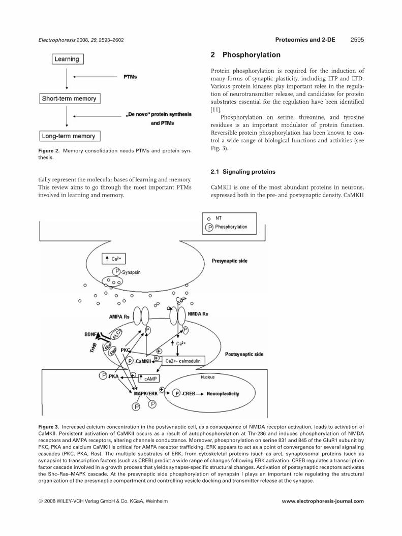
Figure 2. Memory consolidation needs PTMs and protein syn-thesis.
tially represent the molecular bases of learning and memory.This review aims to go through the most important PTMsinvolved in learning and memory.
2 Phosphorylation
Protein phosphorylation is required for the induction ofmany forms of synaptic plasticity, including LTP and LTD.Various protein kinases play important roles in the regula-tion of neurotransmitter release, and candidates for proteinsubstrates essential for the regulation have been identified[11].
Phosphorylation on serine, threonine, and tyrosineresidues is an important modulator of protein function.Reversible protein phosphorylation has been known to con-trol a wide range of biological functions and activities (seeFig. 3).
2.1 Signaling proteins
CaMKII is one of the most abundant proteins in neurons,expressed both in the pre- and postsynaptic density. CaMKII
Figure 3. Increased calcium concentration in the postsynaptic cell, as a consequence of NMDA receptor activation, leads to activation ofCaMKII. Persistent activation of CaMKII occurs as a result of autophosphorylation at Thr-286 and induces phosphorylation of NMDAreceptors and AMPA receptors, altering channels conductance. Moreover, phosphorylation on serine 831 and 845 of the GluR1 subunit byPKC, PKA and calcium CaMKII is critical for AMPA receptor trafficking. ERK appears to act as a point of convergence for several signalingcascades (PKC, PKA, Ras). The multiple substrates of ERK, from cytoskeletal proteins (such as arc), synaptosomal proteins (such assynapsin) to transcription factors (such as CREB) predict a wide range of changes following ERK activation. CREB regulates a transcriptionfactor cascade involved in a growth process that yields synapse-specific structural changes. Activation of postsynaptic receptors activatesthe Shc–Ras–MAPK cascade. At the presynaptic side phosphorylation of synapsin I plays an important role regulating the structuralorganization of the presynaptic compartment and controlling vesicle docking and transmitter release at the synapse.
© 2008 WILEY-VCH Verlag GmbH & Co. KGaA, Weinheim www.electrophoresis-journal.com

2596 B. Sunyer et al. Electrophoresis 2008, 29, 2593–2602
appears likely to be a mediator of primary importance inlinking transient calcium signals to neuronal plasticity[12].
The activity of CaMKII is regulated by autophos-phorylation on several sites. Autophosphorylation at T268allows constitutive activation of the enzyme and permits cal-cium-independent signaling, phosphorylation of NMDARsand AMPARs altering channels conductance. Indeed, it hasbeen shown that CaMKII has a pivotal role in hippocampalLTP [13, 14]. However, using autophosphorylation-deficientmutant mice in hippocampus- and amygdala-dependentlearning and memory tasks was found that autophos-phorylation of a-CaMKII is required for rapid learning but isnot essential for memory [15].
In addition to T268 autophosphorylation, CaMKII isregulated by two inhibitory autophosphorylation sites(threonines 305 and 306) located on the Ca21/calmodulinbinding site of the enzyme. These two inhibitory autophos-porylation sites are only exposed following autophos-phorylation at T286 which leads to dissociation of the Ca21/calmodulin complex [16, 17]. Inhibitory autophosphorylationdecreases CaMKII affinity toward Ca21/calmodulin prevent-ing another activation of the enzyme after subsequentincrease in intracellular Ca21. Moreover, inhibitory autophos-phorylation decreases the affinity of CaMKII to the post-synaptic density [16, 18]. These inhibitory phosphorylationsites are believed to be important regulatory sites in control-ling the threshold of LTP induction [16, 19].
A wealth of experimental data has highlighted that mito-gen-activated protein kinase/extracellular signal-regulatedkinase (MAPK/ERK) cascade contributes to synaptic plasti-city that underlies learning and memory. ERK appears to actas a point of convergence for several signaling cascades andit has been shown to be involved in both early- and late-phasehippocampal LTP [20].
Inhibition of MAPK phosphorylation was found to blockLTM consolidation following food-reward conditioning [21].Inhibition of p38MAPK in the CA1 region of the rat dorsalhippocampus in a one-trial inhibitory avoidance task blockedSTM and LTM formation [22]. Alvarez-Jaimes et al. showedincreased MAPK p42/p44 phosphorylation within thenucleus accumbens after training in a food-search spatiallearning task. Moreover, MAPK and PKC inhibition causeddeficits in the acquisition and retention of the spatial tasksuggesting that MAPK and PKC play important roles inspatial memory [23].
The multiple substrates of ERK, from cytoskeletal pro-teins (such as arc), synaptosomal proteins (such as synapsin)to transcription factors (such as CREB) predict a wide rangeof changes following ERK activation [12].
Synapsin I has been identified as a substrate for ERK [24,25] as well as for cAMP-dependent kinase (CaMKI) [26] andCaMKII [27], and its phosphorylation has been shown toreduce synapsin-actin bundling, an action which is con-sidered to induce vesicle movement to the active zone,increasing the likelihood of vesicle fusion [12, 27].
2.2 Transcription factors
Another key control point of LTM formation is CREB [28].CREB regulates a transcription factor cascade involved in agrowth process that yields synapse-specific structuralchanges [29]. Using immunohistochemistry and confocalimaging, the ratio between nonphosphorylated and phos-phorylated CREB at a single cell resolution were determinedin organotypic hippocampal slices from young adult ratsafter long-lasting LTP was induced. Induction of long-lastingLTP in CA1 was accompanied by a local increase in thepCREB/CREB ratio. CREB phosphorylation increment las-ted for 4 h, during LTP maintenance, indicating that CREBplays an important role during the late phases of LTP [30].
It is important to point out that, protein kinases andprotein phosphatases balance each other controlling synapticstrength and plasticity underlying LTP, learning and mem-ory [31]. An imbalanced regulation in protein kinases andprotein phosphatases in the affected neurons can cause dis-ease such as Alzheimer’s disease [32].
2.3 Neurotransmitter receptors/trafficking system
Activation of NMDARs is critical for the induction of LTP andLTD at diverse sets of synapses [33]. NMDARs are hetero-meric ligand-gated ion channels assembled from two famil-ies of subunits, NR1 and NR2. There are at least eight splic-ing variant isoforms of the NR1 subunit and four types ofNR2 subunits: NR2A, NR2B, NR2C, and NR2D. NMDARswith different subunit composition or different splice var-iants of NR1 subunit have different properties. The functionand localization of NMDARs is also modulated by PTMsincluding phosphorylation, glycosylation, and nitrosylation.NMDARs are phosphorylated in serines of both NR1 andNR2 subunits and in tyrosines of NR2 subunits [16].
Another factor modulating NMDARs function is thesynaptic localization: the trafficking and clustering ofNMDARs is modulated by phosphorylation and by interac-tion with other proteins [16, 34]. Exploration in spatialmemory formation in a radial arm maze has shown thatspatial learning induced tyrosine phosphorylation of NR2Bin the hippocampus [9].
AMPARs are ionotropic glutamate receptors composedof four subunits: GluR1-4. Phosphorylation is the mostimportant molecular mechanism to regulate the ligand-gated ion channels. It can regulate the physiological proper-ties of the channel as well as protein trafficking. All AMPARsubunits, with the exception of GluR3, have been reported tobe phosphorylated on several amino acid residues by a vari-ety of kinases [35]. Phosphorylation on serine 831 and 845 ofthe GluR1 subunit by PKC [35], PKA [36], and calciumCaMKII [37] is critical for AMPAR trafficking and for LTDand LTP. It has been shown that mice with knock-in muta-tions in the GluR1 phosphorylation sites show deficits inLTD and LTP and have memory defects in spatial learningtasks [16].
© 2008 WILEY-VCH Verlag GmbH & Co. KGaA, Weinheim www.electrophoresis-journal.com

Electrophoresis 2008, 29, 2593–2602 Proteomics and 2-DE 2597
GluR2 is a key subunit, which renders the AMPARchannel impermeable to Ca21 and confers specific biophysi-cal properties [16, 38]. There are several serine phosphoryla-tion sites (S863 and S880) on the intracellular carboxy-ter-minal of GluR2 that are phosphorylated by PKC. Phospho-rylation of S880 has been shown to be involved in synapticplasticity [16]. GluR2 is also phosphorylated on a tyrosineresidue (Y842) by Src playing a role in GluR2 internalization[16, 39].
Tyrosine phosphorylation also plays an important role inthe adult mammalian nervous system. Tyrosine kinase sig-naling cascades (the Trk receptor tyrosine kinases, the Srcfamily of nonreceptor tyrosine kinases and the Eph receptortyrosine kinases) have been implicated in both adult synapticplasticity and memory formation [40].
Postsynaptic density-95 (PSD-95) is an important scaf-fold protein that promotes the maturation and strengtheningof excitatory synapses. This key component of the post-synaptic density interacts with NMDARs meditating theirassociation with different downstream signaling molecules.PSD-95 has been also found to control AMPARs incorpora-tion during LTP and experience-driven synaptic plasticity [41,42]. Recent studies report that phosphorylation of ser-295enhances the synaptic accumulation of PSD-95 and the abil-ity of PSD-95 to recruit surface AMPARs and potentiateexcitatory postsynaptic currents [42–44].
Kim et al. [42] suggested that synaptic strength can beregulated by phosphorylation–dephosphorylation of ser-295of PSD-95 and that synaptic depression requires the dephos-phorylation of ser-295.
One-third of the proteins in the cell are phosphorylated,therefore a rapid and general method for the analysis of pro-tein phosphorylation in complex protein mixtures is needed.In recent years, the use of MS-based methods for analysis ofphosphopeptides that result from proteolytic digests ofphosphorylated proteins has become increasing popular [45–47] and several large-scale synaptic phosphoproteomicsmethods have been reported in the past 2–3 years [42]. Keysteps include enrichment of compounds of interest usingimmobilized metal affinity chromatography, detection ofphosphopeptides using mass mapping and precursor ionscans, localization of phosphorylation sites by peptide se-quencing. Using MALDI-TOF MS and nanospray Q-TOF,Ma et al. [48] have identified multiple autophosphorylationsites on the PKR like endoplasmic reticulum kinase.
3 Palmitoylation
Protein palmitoylation is a reversible post-translational lipidmodification. It increases the hydrophobicity of proteins andcontributes to their interactions with lipid bilayers. Besides,it participates in subcellular trafficking of proteins betweenorganelles and within microdomains of individual mem-brane compartments, as well as modulating certain protein–protein interaction.
Recent work shows that palmitate reversibly modifiesnumerous classes of neuronal proteins, including neuro-transmitter receptors, synaptic scaffolding proteins, andsecreted signaling molecules.
3.1 Transmitter release and neuronal trafficking
Targeting cytosolic proteins to cell membrane is a primaryfunction of protein palmitoylation. Palmitoylated proteinsoften concentrate on membranes enriched in cholesterol andsphingomyelin to form a detergent-insoluble, glycolipid-enriched complex (DIGs). DIGs in the brain are enriched insignaling molecules, and seem to help to organize efficientsignal transduction. Many proteins that are associated withDIG domains are sorted specifically to axons [49–51]. Forinstance, the axonal trafficking of GAP43 (growth associatedprotein 43) requires its palmitoylation-dependent incorpora-tion into functional DIGs [52].
Not all palmitoylated proteins are sorted to axons, andspecific palmitoylation motifs determine trafficking. Thedistinct palmitoylation of two cystein residues in the N-ter-minal region of PSD-95 is necessary for sorting to dendritesand participates in the postsynaptic clustering of PSD-95 inspines [53]. The level of PSD-95 palmitoylation is regulatedby synaptic activity, and inhibition of palmitoylation is asso-ciated with loss of PSD-95 from synapses [53, 42]. Changeson synaptic targeting or synaptic stability of PSD-95 couldplay an important role in the control of postsynapticmaturation and synaptic transmission [42].
Palmitoylation also controls axonal growth and trans-mitter release at nerve terminals. Palmitoylation of two cys-teine residues at the amino terminus of GAP43 directs it togrowth cone membranes and regulates its interaction with Gproteins [54]. Palmitoylation of GAP43 decreases as growthcones stabilize and mature. Besides, neural cell adhesionmolecule (NCAM140) is another growth cone molecule thatis palmitoylated. NCAM140 localizes to lipid rafts at thegrowth cone membrane. Disruption of NCAM140 palmitoy-lation sites prevents its association with lipid rafts and com-pletely blocks neurite outgrowth [55].
At presynaptic nerve terminals, palmitate modifies avariety of synaptic vesicle proteins to regulate neuro-transmitter release [56]. For instance, palmitoylation ofsynaptosome-associated protein of 25 kDa (SNAP25), whichresides at presynaptic axon terminals, is required for neuro-transmitter release and disassembly of the SNARE (SNAPreceptor) complex after this secretion [56].
3.2 Neurotransmitter receptors
On the postsynaptic side, the palmitoylation of ion channelsand neurotransmitter receptors, such as the a subunit of Na1
channels [57], b2a subunit of Ca21 channels [58, 59], sero-tonin 1B and 4A receptors [60, 61], dopamine D1 and D2Lreceptors [62, 63], a7 subunit of nicotinic receptors [64], and
© 2008 WILEY-VCH Verlag GmbH & Co. KGaA, Weinheim www.electrophoresis-journal.com

2598 B. Sunyer et al. Electrophoresis 2008, 29, 2593–2602
g2 subunit of GABAA receptors [65], regulates signal trans-duction [66, 67].
G protein-coupled receptors (GPCRs) are coupled to aneffector component by a G-protein and through secondmessengers trigger a biochemical cascade activating specificprotein kinases that phosphorylate a variety of the cell’s pro-teins or by mobilizing Ca21 ions from intracellular stores(see Fig. 1). This family contains the a- and b-adrenergicreceptors, the muscarinic acetylcholine receptors, theGABAB receptors, certain glutamate and serotonin receptorsand are known to be involved in learning and memory pro-cesses [6]. Palmitoylation of GPCRs has an important role inregulating GPCR-coupled pathways [68–71].
AMPARs trafficking and cellular localization is regulatedby palmitoylation. All AMPAR subunits are palmitoylated ontwo cysteine residues in their transmembrane domain 2 andin their C-terminal region. Palmitoylation on transmem-brane domain 2 leads to an accumulation of the receptor inthe Golgi and a reduction of receptor surface expression. C-terminal palmitoylation mediates agonist-induced AMPARsinternalization, while depalmitoylated receptors are stabi-lized at the cell surface [72].
Several synaptic scaffolding proteins which organizereceptors and their associated signaling elements, such asPSD-95, are palmitoylated and this regulates their mem-brane association and receptor-clustering function [73, 74].Postsynaptic density protein 95 (PSD95) binds NMDARs andAMPA receptor-associated protein stargazin (see Fig. 4).Please see review [52] for more information and precise sitesof palmitoylation.
4 Glycosylation
Glycosylation is the process of addition of saccharides toproteins and lipids. The process is one of four principal
cotranslational and PTM steps in the synthesis of membraneand secreted proteins.
Simple models for the study of learning processes haverevealed a number of molecules participating in memoryformation, despite of many forms of neural plasticityincluding growth cone guidance and synapse formationduring development.
4.1 Sialylation
Cell adhesion molecules are involved in synaptic changesunderlying memory formation, especially two members ofthe Ig superfamily of cell adhesion molecules, L1 and neuralcell adhesion molecule (NCAM). For instance, after one-trialpassive avoidance training in chicks, enhanced synthesis ofL1 and NCAM was observed suggesting that cell adhesionmolecules are important for memory consolidation [75].
Both, L1 and NCAM mediate cell–cell and cell–matrixinteractions, altering membrane properties and intracellularsignaling cascades, including the inositol phospholipids sig-nal pathway, intracellular Ca21 and nonreceptor tyrosinekinases of the Src family.
NCAM can be expressed in various isoforms and the coreprotein is further modified post-translationally through gly-cosylation with polysialic acid (PSA), a carbohydrate consist-ing of long homopolymers of sialic acid [76]. Adhesionproperties of L1 and NCAM can be efficiently regulatedthrough glycosylation.
Increased sialylation of NCAM has been observed 12 and24 h after on-trial passive avoidance task [77]. When rats wereintracranial injected endoneuraminidase-N (endo-N),removing PSA of NCAM, spatial learning was attenuatedand hippocampal LTP when endo-N was applied to brain sli-ces in vitro was blocked [78].
In addition, glycosylation can protect proteins from pro-teolytic degradation. Each AMPAR subunit can be N-glyco-
Figure 4. Palmitoylated pro-teins. On the presynaptic side,palmitoylated proteins, asSNAP25, regulate synaptic vesi-cle fusion and neurotransmittersynthesis and release. Palmitoy-lated presynaptic proteins alsoinclude the b2A-subunit of thevoltage-dependent calciumchannel, and the a-subunit ofsodium channels. On the post-synaptic side many receptorsare palmitoylated (GCPRs, iono-tropic, and metabotropic trans-membrane receptors) regulat-ing signal transduction. More-over, palmitoylation of PSD95 isessential for the modulation ofsynaptic AMPA and NMDAreceptors.
© 2008 WILEY-VCH Verlag GmbH & Co. KGaA, Weinheim www.electrophoresis-journal.com

Electrophoresis 2008, 29, 2593–2602 Proteomics and 2-DE 2599
sylated at 4–6 different sites located in the extracellulardomains of the protein facilitating its maturation, protectingthe receptor from proteolytic degradation and affectingreceptor current amplitudes. However, the lack of N-glycosy-lation does not affect AMPAR subunit synthesis, assembly,or trafficking [34].
4.2 Fucosylation
Increased modification of brain proteins by sugars termi-nating in Fuca (1–2)Gal residues by fucosylation has alsobeen correlated with improved learning and memory andwith increased synapse formation [79, 80]. Murrey et al.[81] used synaptic vesicle purification, immunoblots andgel electrophoresis, combined with MALDI-TOF MS, toidentify several Fuca (1–2)Gal-modified proteins during ratbrain development [81]. They showed that synapsins Iaand Ib are the main fucosylated proteins in adult brain.This fucosylation has profound effects on the expressionand turnover of synapsin in cells and protects synapsinfrom degradation by the calcium-activated protease cal-pain. As already mentioned above, synapsins are a familyof neuron cytoplasmatic proteins that reversibly bind toactin and to synaptic vesicles in a calcium-controlled,phosphorylation-dependent way to regulate the structuralorganization of the presynaptic compartment and to con-trol vesicle docking and transmitter release at the synapse(see Fig. 5) [82].
Fuca (1–2)Gal modification of synapsins seems to becritical to neuronal growth and morphology and to synapsefunction [83].
5 Acetylation
Protein acetylation occurs with short-chain and long-chainacyl CoAs as donor substrates to nucleophilic side chains inproteins. Acetylation of histones and transcription factorsthat affect selective gene transcription and chromatin struc-ture has become of great interest in neuroscience. As alreadymentioned previously gene transcription is necessary forlong-lasting forms of synaptic plasticity.
DNA is known to be packaged around octameric histonecores (H2A, H2B, H3, and H4) forming chromatin. Histoneacetylation alters chromatin structure [84] creating activechromatin complexes with DNA and increasing accessibilityfor RNA polymerases, thereby regulating gene transcription[85].
This enzymatic acetylation by histone acetyl transferasesis reversed by histone deacetylases (HDAC). Therefore,HDACs are potential targets for pharmacological treatmentof neurodegenerative disorders [86]. Vecsey et al. [87] showedthat enhancement of hippocampus-dependent memory bythricostatin A, a HDAC inhibitor, is mediated by the tran-scription factor CREB and recruitment of the transcriptionalcoactivator and histone acetyltransferase CREB-binding pro-tein (CBP) via the CREB-binding domain of CBP.
6 Ubiquitination
Ubiquitination is a rapid and reversible PTM that regulatesprotein stability, activity, and localization. Ubiquitinationconsists of the attachment of a single ubiquitin, a highly con-served 76-amino acid polypeptide, or a polymeric ubiquitin
Figure 5. Synapsin help tether the vesicles to the actin cytoskeleton. (1) Phosphorylation of synapsin by ERK, CaMKI, and CaMKII has beenshown to reduce synapsin-actin bundling, an action which is considered to induce vesicle movement to the active zone, increasing thelikelihood of vesicle fusion. (2) Dephosphorylation and prevention of fucosylation leads to synapsin degradation by the protease calpain.(3) Fucosylation prevents synapsin degradation. (4) Formation of O-glcNAc–Fuc complex with synapsin. (5) Before phosphorylation ofsynapsin O-glcNAc and perhaps fucosylation are removed.
© 2008 WILEY-VCH Verlag GmbH & Co. KGaA, Weinheim www.electrophoresis-journal.com

2600 B. Sunyer et al. Electrophoresis 2008, 29, 2593–2602
chain to a protein. The characteristic function of ubiquitina-tion is proteolysis through proteasomal pathway by irrever-sibly modifying proteins. However, several functions of ubi-quitin in regulating proteins by proteasome-independentprocesses have been reported [88].
An important role of the ubiquitin proteasome system insynaptic strength has been described in several speciesincluding Caenorhabditis elegans, Aplysia, Drosophila, andmammals [35, 89–95].
Moreover, the ubiquitin proteasome system has beenreported to regulate the abundance of both, presynaptic andpostsynaptic proteins influencing synaptic development,presynaptic function, and neurotransmitter release, as wellas alteration of postsynaptic density and plasticity, spinegrowth and stability [96, 97]. DiAntonio et al. [89] suggestedthat active ubiquitination negatively regulates synaptogen-ensis. Several studies indicate that ubiquitination of pre-synaptic proteins alters the efficacy of synaptic transmissionby transient cycles of monoubiquitination and deubiquitina-tion or by proteasome-dependent degradation of proteinsassociated with vesicle dynamics [89].
Ubiquitin seems to participate in the internalization ofmembrane proteins and in the control of gene transcription[91]. In the brain, ubiquitin signals the internalization ofpostsynaptic glycine receptors [98] and glutamate receptors[99].
Synaptic plasticity has also been reported to be influ-enced by ubiquitination. Ubiquitination-mediated degrada-tion of regulatory subunits of PKA makes PKA persistentlyactive, helping to convert the short-term synaptic facilitationto a long-lasting memory [91].
As mentioned above, long-term memory requires thegene expression cascade induced by CREB. Gene expressionby CREB is inhibited by repressors. Upadhya et al. [100]showed that Aplysia CREB repressor is a substrate for degra-dation by the ubiquitin-proteasome pathway and that PKCstimulates ubiquitination and degradation of the CREBrepressor [100].
Herein, we have taken together first information on thisrapidly growing area in neurobiology and indeed, determi-nation of PTMs and their function may form the key forunderstanding mechanisms in cognitive function. It is anenormous challenge to identify and determine the manypossible individual PTMs in the brain and to assign a func-tional role thus opening a new era in neuroscience.
PTMs as, e.g., phosphorylation are already known to becontrolled by drugs and the pharmacological regulation ofindividual PTMs may well lead to the design of new drugs tomodulate cognitive functions in health and cognitive-dementing disorders.
The authors have declared no conflict of interest.
7 References
[1] Braunewell, K. H., Manahan-Vaughan, D., Rev. Neurosci.2001, 12, 121–140.
[2] Miller, S., Mayford, M., Curr. Opin. Genet. Dev. 1999, 9, 333–337.
[3] Collingridge, G. L., Lester, R. A. J., Pharmacol. Rev. 1989, 40,143–210.
[4] Bleakman, D., Lodge, D., Neuropharmacology 1998, 37,1187–1204.
[5] Arai, A., Lynch, G., Brain Res. 1992, 598, 173–184.
[6] Kandel, E. R., Schwartz, J. H., Jessell, T. M., Principles ofNeural Science, McGraw-Hill Companies, Inc., New York2000, pp. 229–233, 1251–1257.
[7] Kandel, E. R., Science 2001, 294, 1030–1038.
[8] Routtenberg, A., Rekart, J. L., Trends Neurosci. 2005, 28, 12–19.
[9] Mizuno, M., Yamada, K., He, J., Nakajima, A., Nabeshima, T.,Learn. Mem. 2003, 10, 108–115.
[10] Lee, H. K., Takamiya, K., Han, J. S., Man, H. et al., Cell 2003,112, 631–643.
[11] Takahashi, M., Itakura, M., Kataoka, M., J. Pharmacol. Sci.2003, 93, 41–45.
[12] Lynch, M. A., Physiol. Rev. 2004, 84, 87–136.
[13] Malenka, R. C., Kauer, J. A., Perkel, D. J., Mauk, M. D. et al.,Nature 1989, 340, 554–557.
[14] Silva, A. J., Stevens, C. F., Tonegawa, S., Wang, Y., Science1992, 257, 201–206.
[15] Irvine, E. E., Vernon, J., Giese, K. P., Nat. Neurosci. 2005, 8,411–412.
[16] Lee, H. K., Pharmacol. Ther. 2006, 12, 810–832.
[17] Patton, B. L., Miller, S. G., Kennedy, M. B., J. Biol. Chem.1990, 26, 11204–11212.
[18] Elgersma, Y., Fedorov, N. B., Ikonen, S., Choi, E. S. et al.,Neuron 2002, 36, 493–505.
[19] Elgersma, Y., Sweatt, J. D., Giese, K. P., J. Neurosci. 2004, 24,8410–8415.
[20] McGahon, B., Maguire, C., Kelly, A., Lynch, M. A., Neu-roscience 1999, 90, 1167–1175.
[21] Ribeiro, M. J., Schofield, M. G., Kemenes, I., O’Shea, M. etal., Learn. Mem. 2005, 12, 538–545.
[22] Alonso, M., Bevilaqua, L. R., Izquierdo, I., Medina, J. H.,Cammarota, M., Neuroreport 2003, 14, 1989–1992.
[23] Alvarez-Jaimes, L., Feliciano-Rivera, M., Centeno-Gonzalez,M., Maldonado-Vlaar, C. S., J. Pharmacol. Exp. Ther. 2005,314, 1144–1157.
[24] Jovanovic, J. N., Benfenati, F., Siow, Y. L., Sihra, T. S. et al.,Proc. Natl. Acad. Sci. USA 1996, 93, 3679–3683.
[25] Matsubara, M., Kusubata, M., Ishiguro, K., Uchida, T. et al., J.Biol. Chem. 1996, 27, 21108–21113.
[26] John, J. P., Chen, W. Q., Pollak, A., Lubec, G., J. ProteomeRes. 2007, 6, 2695–2710.
[27] Greengard, P., Valtorta, F., Czernik, A., Benfenati, F., Science1993, 259, 780–785.
[28] Tully, T., Proc. Natl. Acad. Sci. USA 1997, 94, 4239–4241.
[29] Tully, T., Bourtchouladze, R., Scott, R., Tallman, J., Nat. Rev.Drug Discov. 2003, 2, 267–277.
© 2008 WILEY-VCH Verlag GmbH & Co. KGaA, Weinheim www.electrophoresis-journal.com

Electrophoresis 2008, 29, 2593–2602 Proteomics and 2-DE 2601
[30] Leutgeb, J. K., Frey, J. U., Behnisch, T., Neuroscience 2005,131, 601–610.
[31] Hédou, G., Mansuy, I. M., Philos. Trans. R. Soc. Lond. B Biol.Sci. 2003, 358, 797–804.
[32] Sun, L., Liu, S. Y., Zhou, X. W., Wang, X. C. et al., Neu-roscience 2003, 118, 1175–1182.
[33] Malenka, R. C., Bear, M. F., Neuron 2004, 44, 5–21.
[34] Llansola, M., Sanchez–Perez, A., Cauli, O., Felipo, V., Cere-bellum 2005, 4, 154–161.
[35] Jiang, J., Suppiramaniam, V., Wooten, M. W., Neurosignals2006–2007, 15, 266–282.
[36] Roche, K. W., O’Brien, R. J., Mammen, A. L., Bernhardt, J.,Huganir, R. L., Neuron 1996, 16, 1179–1188.
[37] Mammen, A. L., Kameyama, K., Roche, K. W., Huganir, R. L.,J. Biol. Chem. 1997, 272, 32528–32533.
[38] Tanaka, H., Grooms, S. Y., Bennett, M. V., Zukin, R. S., BrainRes. 2000, 886, 190–207.
[39] Hayashi, T., Huganir, R. L., J. Neurosci. 2004, 24, 6152–6160.
[40] Purcell, A. L., Carew, T. J., Trends Neurosci. 2003, 26, 625–630.
[41] Ehrlich, I., Malinow, R., J. Neurosci. 2004, 24, 916–927.
[42] Kim, M. J., Futai, K., Jo, J., Hayashi, Y. et al., Neuron. 2007,56, 488–502.
[43] Jaffe, H., Vinade, L., Dosemeci, A., Biochem. Biophys. Res.Commun. 2004, 321, 210–218.
[44] Trinidad, J. C., Specht, C. G., Thalhammer, A., Schoepfer, R.,Burlingame, A. L., Mol. Cell. Proteomics 2006, 5, 914–922.
[45] Quadroni, M., James, P., EXS 2000, 88, 199–213.
[46] Neubauer, G., Mann, M., Anal. Chem. 1999, 71, 235–242.
[47] Annan, R. S., Huddleston, M. J., Verma, R., Deshaies, R. J.,Carr, S. A., Anal. Chem. 2001, 73, 393–404.
[48] Ma, Y., Lu, Y., Zeng, H., Ron, D. et al., Rapid Commun. MassSpectrom. 2001, 15, 1693–1700.
[49] Dotti, C. G., Simons, K., Cell 1990, 62, 63–72.
[50] Dotti, C. G., Parton, R. G., Simons, K., Nature 1991, 349, 158–161.
[51] Ledesma, M. D., Brugger, B., Bunning, C., Wieland, F. T.,Dotti, C. G., EMBO J. 1999, 18, 1761–1771.
[52] El-Husseini, Ael-D, Craven, S. E., Broca, S. C., Bredt, D. S., J.Biol. Chem. 2001, 276, 44984–44992.
[53] El-Husseini, Ael-D, Bredt, D. S., Nat. Rev. Neurosci. 2002, 3,791–802.
[54] Sudo, Y., Valenzuela, D., Beck-Sickinger, A. G., Fishman, M.C., Strittmatter, S. M., EMBO J. 1992, 11, 2095–2102.
[55] Niethammer, P., Delling, M., Sytnyk, V., Dityatev, A. et al., J.Cell Biol. 2002, 157, 521–532.
[56] Washbourne, P., Cansino, V., Mathews, J. R., Graham, M. etal., Biochem. J. 2001, 357, 625–634.
[57] Schmidt, J. W., Catterall, W. A., J. Biol. Chem. 1987, 262,13713–13723.
[58] Chien, A. J., Carr, K. M., Shirokov, R. E., Rios, E., Hosey, M.M., J. Biol. Chem. 1996, 271, 26465–26468.
[59] Qin, N., Platano, D., Olcese, R., Costantin, J. L. et al., Proc.Natl. Acad. Sci. USA 1998, 95, 4690–4695.
[60] Ponimaskin, E. G., Schmidt, M. F., Heine, M., Bickmeyer, U.,Richter, D. W., Biochem. J. 2001, 353, 627–634.
[61] Ponimaskin, E. G., Heine, M., Joubert, L., Sebben, M., Bick-meyer, U. et al., J. Biol. Chem. 2002, 277, 2534–2546.
[62] Ng, G. Y., Mouillac, B., George, S. R., Caron, M. et al., Eur. J.Pharmacol. 1994, 267, 7–19.
[63] Ng, G. Y., O’Dowd, B. F., Caron, M., Dennis, M. et al., J. Neu-rochem. 1994, 63, 1589–1595.
[64] Drisdel, R. C., Manzana, E., Green, W. N., J. Neurosci. 2004,24, 10502–10510.
[65] Keller, C. A., Yuan, X., Panzanelli, P., Martin, M. L. et al., J.Neurosci. 2004, 24, 5881–5891.
[66] Dunphy, J. T., Linder, M. E., Biochim. Biophys. Acta 1998,1436, 245–261.
[67] Bizzozero, O. A., Neuropediatrics 1997, 28, 23–26.
[68] Iiri, T., Backlund, P. S., Jr., Jones, T. L., Wedegaertner, P. B.,Bourne, H. R., Proc. Natl. Acad. Sci. USA 1996, 93, 14592–14597.
[69] Moffett, S., Rousseau, G., Lagace, M., Bouvier, M., J. Neu-rochem. 2001, 76, 269–279.
[70] Tu, Y., Popov, S., Slaughter, C., Ross, E. M., J. Biol. Chem.1999, 274, 38260–38267.
[71] Stoffel, R. H., Inglese, J., Macrae, A. D., Lefkowitz, R. J., Pre-mont, R. T., Biochemistry 1998, 37, 16053–16059.
[72] Hayashi, T., Rumbaugh, G., Huganir, R. L., Neuron 2005, 47,709–723.
[73] Topinka, J. R., Bredt, D. S., Neuron 1998, 20, 125–134.
[74] DeSouza, S., Fu, J., States, B. A., Ziff, E. B., J. Neurosci. 2002,22, 3493–3503.
[75] Rose, S. P., Behav. Brain Res. 1995, 66, 73–78.
[76] Welzl, H., Stork, O., News Physiol. Sci. 2003, 18, 147–150.
[77] Doyle, E., Nolan, P. M., Bell, R., Regan, C. M., J. Neurosci.Res. 1992, 31, 513–523.
[78] Becker, C. G., Artola, A., Gerardy-Schahn, R., Becker, T. et al.,J. Neurosci. Res. 1996, 45, 143–152.
[79] Bullock, S., Potter, J., Rose, S. P., J. Neurochem. 1990, 54,135–142.
[80] Matthies, H., Staak, S., Krug, M., Brain Res. 1996, 725, 276–280.
[81] Murrey, H. E., Gama, C. I., Kalovidouris, S. A., Luo, W. I. et al.,Proc. Natl. Acad. Sci. USA 2006, 103, 21–26.
[82] De Camilli, P., Benfenati, F., Valtorta, F., Greengard, P., Annu.Rev. Cell Biol. 1990, 6, 433–460.
[83] Hart, G. W., Nat. Chem. Biol. 2006, 2, 67–68.
[84] Norton, V. G., Imai, B. S., Yau, P., Bradbury, E. M., Cell 1989,57, 449–457.
[85] Strahl, B. D., Allis, C. D., Nature 2000, 403, 41–45.
[86] Sweatt, J. D., Nature 2007, 447, 151–152.
[87] Vecsey, C. G., Hawk, J. D., Lattal, K. M., Stein, J. M. et al., J.Neurosci. 2007, 27, 6128–6140.
[88] Schnell, J. D., Hicke, L., J. Biol. Chem. 2003, 278, 35857–35860.
[89] DiAntonio, A., Haghighi, A. P., Portman, S. L., Lee, J. D.,Amaranto, A. M., Goodman, C. S., Nature 2001, 412, 449–452.
[90] Burbea, M., Dreier, L., Dittman, J. S., Grunwald, M. E.,Kaplan, J. M., Neuron 2002, 35, 107–120.
[91] Hegde, A. N., DiAntonio, A., Nat. Rev. Neurosci. 2002, 3, 854–861.
© 2008 WILEY-VCH Verlag GmbH & Co. KGaA, Weinheim www.electrophoresis-journal.com

2602 B. Sunyer et al. Electrophoresis 2008, 29, 2593–2602
[92] Murphey, R. K., Godenschwege, T. A., Neuron 2002, 36, 5–8.
[93] Speese, D. D., Trotta, N., Rodesch, C. K., Aravamudan, B.,Broadie, K., Curr. Biol. 2003, 13, 899–910.
[94] DiAntonio, A., Hicke, L., Annu. Rev. Neurosci. 2004, 2, 223–246.
[95] Kato, A., Rouach, N., Nicoll, R. A., Bredt, D. S., Proc. Natl.Acad. Sci. USA 2005, 102, 5600–5605.
[96] Yi, J. J., Ehlers, M. D., Neuron 2005, 47, 629–632.
[97] Yi, J. J., Ehlers, M. D., Pharmacol. Rev. 2007, 59, 14–39.
[98] Büttner, C., Sadtler, S., Leyendecker, A., Laube, B. et al., J.Biol. Chem. 2001, 276, 42978–42985.
[99] Burbea, M., Dreier, L., Dittman, J. S., Grunwald, M. E.,Kaplan, J. M., Neuron 2002, 35, 107–120.
[100] Upadhya, S. C., Smith, T. K., Hegde, A. N., J. Neurochem.2004, 91, 210–219.
© 2008 WILEY-VCH Verlag GmbH & Co. KGaA, Weinheim www.electrophoresis-journal.com
Copyright © 2022 FDOKUMEN