The Neutralization Breadth of HIV-1 Develops Incrementally over Four Years and Is Associated with...
13
JOURNAL OF VIROLOGY, May 2011, p. 4828–4840 Vol. 85, No. 10 0022-538X/11/$12.00 doi:10.1128/JVI.00198-11 Copyright © 2011, American Society for Microbiology. All Rights Reserved. The Neutralization Breadth of HIV-1 Develops Incrementally over Four Years and Is Associated with CD4 T Cell Decline and High Viral Load during Acute Infection Elin S. Gray, 1 Maphuti C. Madiga, 1 Tandile Hermanus, 1 Penny L. Moore, 1,2 Constantinos Kurt Wibmer, 1,2 Nancy L. Tumba, 1 Lise Werner, 3 Koleka Mlisana, 3 Sengeziwe Sibeko, 3 Carolyn Williamson, 4 Salim S. Abdool Karim, 3 Lynn Morris, 1,2 * and the CAPRISA 002 Study Team AIDS Virus Research Unit, National Institute for Communicable Diseases, Johannesburg, South Africa 1 ; University of Witwatersrand, Johannesburg, South Africa 2 ; Centre for the AIDS Programme of Research in South Africa (CAPRISA), University of KwaZulu-Natal, Durban, South Africa 3 ; and Institute of Infectious Disease and Molecular Medicine, University of Cape Town, Cape Town, South Africa 4 Received 28 January 2011/Accepted 24 February 2011 An understanding of how broadly neutralizing activity develops in HIV-1-infected individuals is needed to guide vaccine design and immunization strategies. Here we used a large panel of 44 HIV-1 envelope variants (subtypes A, B, and C) to evaluate the presence of broadly neutralizing antibodies in serum samples obtained 3 years after seroconversion from 40 women enrolled in the CAPRISA 002 acute infection cohort. Seven of 40 participants had serum antibodies that neutralized more than 40% of viruses tested and were considered to have neutralization breadth. Among the samples with breadth, CAP257 serum neutralized 82% (36/44 variants) of the panel, while CAP256 serum neutralized 77% (33/43 variants) of the panel. Analysis of longitudinal samples showed that breadth developed gradually starting from year 2, with the number of viruses neutralized as well as the antibody titer increasing over time. Interestingly, neutralization breadth peaked at 4 years postinfection, with no increase thereafter. The extent of cross-neutralizing activity correlated with CD4 T cell decline, viral load, and CD4 T cell count at 6 months postinfection but not at later time points, suggesting that early events set the stage for the development of breadth. However, in a multivariate analysis, CD4 decline was the major driver of this association, as viral load was not an independent predictor of breadth. Mapping of the epitopes targeted by cross-neutralizing antibodies revealed that in one individual these antibodies recognized the membrane-proximal external region (MPER), while in two other individuals, cross-neutralizing activity was adsorbed by monomeric gp120 and targeted epitopes that involved the N-linked glycan at position 332 in the C3 region. Serum antibodies from the other four participants targeted quaternary epitopes, at least 2 of which were PG9/16-like and depended on the N160 and/or L165 residue in the V2 region. These data indicate that fewer than 20% of HIV-1 subtype C-infected individuals develop antibodies with cross-neutral- izing activity after 3 years of infection and that these antibodies target different regions of the HIV-1 envelope, including as yet uncharacterized epitopes. Neutralizing antibodies are thought to be crucial in the pro- tective immune response against many viral infections, yet their role in HIV-1 infection remains controversial. During natural infection, they appear to have little impact on acute viremia, as they arise too late and the virus readily escapes type-specific neutralizing antibodies (35, 41, 42, 55). However, passive transfer of broadly neutralizing monoclonal antibodies (MAbs) has proven to be protective in nonhuman primate models (2, 11, 17, 18, 27, 28, 52), supporting the hypothesis that a vaccine capable of inducing this type of antibodies is likely to be effective. Despite rigorous efforts, designing an immunogen capable of inducing broadly neutralizing antibodies has so far not been feasible. Recently, researchers have turned their at- tention to understanding the factors associated with the pres- ence of broadly cross-neutralizing antibodies, which develop in a subset of chronically HIV-1-infected individuals. A number of reports from an assortment of different cohorts have found that the duration of infection, viral load, CD4 T cell count, and/or viral diversity is associated with the development of neutralization breadth (10, 37, 44). The B cell response to HIV-1 infection first appears within 8 days of detectable viremia and initially comprises antigen- antibody complexes (47). This is followed by the detection of circulating anti-gp41 antibodies 5 days later, with anti-gp120 antibodies delayed a further 14 days and targeting primarily the V3 loop. Autologous neutralizing antibodies develop months later (15) and target the variable regions via potent but extremely type-specific neutralizing antibodies (22, 33, 41, 55). Recent data from our laboratory suggest that during the first year of HIV-1 subtype C infection, within a single individual, a limited number of antibody specificities mediate autologous neutralization (34). These arise sequentially and show tempo- ral fluctuations as escape occurs. After years, antibodies with cross-neutralizing potential appear in as many as one-third of chronically infected individuals and target more conserved re- gions of the HIV-1 envelope (46). An increasing number of studies have focused on mapping * Corresponding author. Mailing address: National Institute for Communicable Diseases, Johannesburg, Private Bag X4, Sandringham 2131, Johannesburg, South Africa. Phone: 27-11-386-6332. Fax: 27-11- 386-6333. E-mail: [email protected]. Published ahead of print on 9 March 2011. 4828
-
Upload
independent -
Category
Documents
-
view
0 -
download
0
Transcript of The Neutralization Breadth of HIV-1 Develops Incrementally over Four Years and Is Associated with...
JOURNAL OF VIROLOGY, May 2011, p. 4828–4840 Vol. 85, No. 100022-538X/11/$12.00 doi:10.1128/JVI.00198-11Copyright © 2011, American Society for Microbiology. All Rights Reserved.
The Neutralization Breadth of HIV-1 Develops Incrementally overFour Years and Is Associated with CD4� T Cell Decline
and High Viral Load during Acute Infection�
Elin S. Gray,1 Maphuti C. Madiga,1 Tandile Hermanus,1 Penny L. Moore,1,2 Constantinos Kurt Wibmer,1,2
Nancy L. Tumba,1 Lise Werner,3 Koleka Mlisana,3 Sengeziwe Sibeko,3 Carolyn Williamson,4Salim S. Abdool Karim,3 Lynn Morris,1,2* and the CAPRISA 002 Study Team
AIDS Virus Research Unit, National Institute for Communicable Diseases, Johannesburg, South Africa1; University of Witwatersrand,Johannesburg, South Africa2; Centre for the AIDS Programme of Research in South Africa (CAPRISA), University of
KwaZulu-Natal, Durban, South Africa3; and Institute of Infectious Disease andMolecular Medicine, University of Cape Town, Cape Town, South Africa4
Received 28 January 2011/Accepted 24 February 2011
An understanding of how broadly neutralizing activity develops in HIV-1-infected individuals is needed toguide vaccine design and immunization strategies. Here we used a large panel of 44 HIV-1 envelope variants(subtypes A, B, and C) to evaluate the presence of broadly neutralizing antibodies in serum samples obtained3 years after seroconversion from 40 women enrolled in the CAPRISA 002 acute infection cohort. Seven of 40participants had serum antibodies that neutralized more than 40% of viruses tested and were considered tohave neutralization breadth. Among the samples with breadth, CAP257 serum neutralized 82% (36/44 variants)of the panel, while CAP256 serum neutralized 77% (33/43 variants) of the panel. Analysis of longitudinalsamples showed that breadth developed gradually starting from year 2, with the number of viruses neutralizedas well as the antibody titer increasing over time. Interestingly, neutralization breadth peaked at 4 yearspostinfection, with no increase thereafter. The extent of cross-neutralizing activity correlated with CD4� T celldecline, viral load, and CD4� T cell count at 6 months postinfection but not at later time points, suggestingthat early events set the stage for the development of breadth. However, in a multivariate analysis, CD4 declinewas the major driver of this association, as viral load was not an independent predictor of breadth. Mappingof the epitopes targeted by cross-neutralizing antibodies revealed that in one individual these antibodiesrecognized the membrane-proximal external region (MPER), while in two other individuals, cross-neutralizingactivity was adsorbed by monomeric gp120 and targeted epitopes that involved the N-linked glycan at position332 in the C3 region. Serum antibodies from the other four participants targeted quaternary epitopes, at least2 of which were PG9/16-like and depended on the N160 and/or L165 residue in the V2 region. These dataindicate that fewer than 20% of HIV-1 subtype C-infected individuals develop antibodies with cross-neutral-izing activity after 3 years of infection and that these antibodies target different regions of the HIV-1 envelope,including as yet uncharacterized epitopes.
Neutralizing antibodies are thought to be crucial in the pro-tective immune response against many viral infections, yettheir role in HIV-1 infection remains controversial. Duringnatural infection, they appear to have little impact on acuteviremia, as they arise too late and the virus readily escapestype-specific neutralizing antibodies (35, 41, 42, 55). However,passive transfer of broadly neutralizing monoclonal antibodies(MAbs) has proven to be protective in nonhuman primatemodels (2, 11, 17, 18, 27, 28, 52), supporting the hypothesis thata vaccine capable of inducing this type of antibodies is likely tobe effective. Despite rigorous efforts, designing an immunogencapable of inducing broadly neutralizing antibodies has so farnot been feasible. Recently, researchers have turned their at-tention to understanding the factors associated with the pres-ence of broadly cross-neutralizing antibodies, which develop ina subset of chronically HIV-1-infected individuals. A number
of reports from an assortment of different cohorts have foundthat the duration of infection, viral load, CD4� T cell count,and/or viral diversity is associated with the development ofneutralization breadth (10, 37, 44).
The B cell response to HIV-1 infection first appears within8 days of detectable viremia and initially comprises antigen-antibody complexes (47). This is followed by the detection ofcirculating anti-gp41 antibodies 5 days later, with anti-gp120antibodies delayed a further 14 days and targeting primarilythe V3 loop. Autologous neutralizing antibodies developmonths later (15) and target the variable regions via potent butextremely type-specific neutralizing antibodies (22, 33, 41, 55).Recent data from our laboratory suggest that during the firstyear of HIV-1 subtype C infection, within a single individual, alimited number of antibody specificities mediate autologousneutralization (34). These arise sequentially and show tempo-ral fluctuations as escape occurs. After years, antibodies withcross-neutralizing potential appear in as many as one-third ofchronically infected individuals and target more conserved re-gions of the HIV-1 envelope (46).
An increasing number of studies have focused on mapping
* Corresponding author. Mailing address: National Institute forCommunicable Diseases, Johannesburg, Private Bag X4, Sandringham2131, Johannesburg, South Africa. Phone: 27-11-386-6332. Fax: 27-11-386-6333. E-mail: [email protected].
� Published ahead of print on 9 March 2011.
4828

the antibody specificities responsible for the cross-neutralizingactivity found in selected HIV-1-positive plasmas (3, 16, 25, 44,45, 54). Using a variety of methodologies, it has been estab-lished that some of these neutralizing antibodies recognizeepitopes in the context of monomeric gp120, e.g., the CD4 andcoreceptor binding sites. In a few cases, the cross-neutralizingactivity could be attributed to antibodies recognizing linearepitopes in the membrane-proximal external region (MPER)of gp41 (14, 45). However, many of the antibody specificitiesresponsible for cross-neutralization could not be matched toknown epitopes in these studies. More recently, it has becomeapparent that a quaternary epitope at the tip of the trimericenvelope structure, involving the V2 and V3 loop, is frequentlythe target of cross-neutralizing antibodies (34, 53, 54). Anothercross-reactive specificity, involving the N332 residue in the C3region at the base of the V3 loop, was also described recently,but the epitope has yet to be defined. Taking these data to-gether, it appears that a limited number of neutralizing anti-body specificities are responsible for the broad neutralizingactivity in plasma samples (54). Isolation of MAbs from theseindividuals may delineate important targets on the HIV-1 en-velope glycoprotein and thus inform vaccine design. Indeed,naturally occurring broadly neutralizing MAbs, such as b12,2G12, 4E10, and 2F5 (6), have been crucial reagents for manyyears, and the more recently isolated MAbs VRC01 and PG9/PG16 (53, 56) are providing new insights into the native enve-lope structure. However, the mechanism by which these anti-bodies develop and why this occurs only in some individualsare not clear. Defining when cross-neutralizing antibodiesemerge might provide important clues as to their nature andhow to elicit them. For this purpose, suitable cohorts of HIV-infected individuals with antibodies able to cross-neutralizeheterologous viruses need to be identified and longitudinalsamples assessed.
Previously, we reported on the development of early type-specific neutralizing antibodies in the CAPRISA acute infec-tion cohort (15). While autologous neutralizing antibodieswere found in all individuals from 3 to 12 months postinfection,little heterologous neutralizing activity was detected in the 14participants analyzed at 12 months of infection. Here we ex-tended this analysis to 40 participants from the same cohortwho had reached at least 3 years postinfection to evaluate thekinetics of the development of cross-neutralizing antibodies.We also analyzed the association between neutralizationbreadth and clinical factors, such as viral load and CD4� T cellcount, and explored the epitopes targeted by these cross-reac-tive antibodies. Our results suggest that cross-neutralizing an-tibodies targeting different regions on the HIV-1 envelopedevelop over many years, with low CD4� T cell counts andhigh viral loads in early infection favoring their emergence.
MATERIALS AND METHODS
Study subjects. Plasma and serum samples were obtained from participants inthe CAPRISA acute infection cohort (CAPRISA 002), established in 2004 inDurban and Vulindlela, South Africa. Participants in Durban were part of aprospective study of 245 high-risk HIV-negative women who were followed upfor subsequent identification of HIV seroconversion (51). A total of 62 women(28 from the HIV-negative cohort and 34 from other seroincidence cohorts) whohad a reactive HIV antibody test within 5 months of a previously negative resultor detection of HIV-1 RNA by PCR (Roche Amplicor v1.5) in the absence ofHIV antibodies were enrolled. Thereafter, participants were monitored and
referred to the antiretroviral (ARV) therapy initiation program when theirCD4� T cell counts dropped below 350 cells/�l on 2 separate occasions. Theywere initiated on ARV therapy only once their CD4� T cell counts fell below 200cells/�l or other signs suggestive of WHO stage 4 disease were evident (2004National Antiretroviral Treatment Guidelines). After the implementation of the2010 South African Antiretroviral Treatment Guidelines, ARV treatment couldbe initiated with CD4� T cell counts of up to 350 cells/�l in Mycobacteriumtuberculosis-coinfected individuals. Samples were collected and stored at leastevery 3 months during chronic infection. This study received ethical approvalfrom the University of Witwatersrand, University of KwaZulu-Natal, and Uni-versity of Cape Town.
Envelope clones. Envelope genes used in this study were either cloned previ-ously in our laboratory (15) or obtained from the NIH AIDS Research andReference Reagent Program. The ConC plasmid, carrying an envelope generepresenting the consensus of all HIV-1 subtype C sequences deposited in theLos Alamos database by 2001, was obtained from Feng Gao (20). Mutationswere introduced into envelope clones by use of a QuikChange site-directedmutagenesis kit (Stratagene, La Jolla, CA). Plasmids encoding HIV-2/HIV-1MPER chimeras were obtained from George Shaw (15).
Neutralization assays. Neutralization was measured as a reduction in lucifer-ase gene expression after a single round of infection of JC53bl-13 cells, alsoknown as TZM-bl cells (NIH AIDS Research and Reference Reagent Program),with Env-pseudotyped viruses (32). Titer was calculated as the reciprocal plasma/serum dilution causing a 50% reduction of relative light units (ID50).
gp120 production and isolation. The gp120-encoding region of the ConCenvelope was inserted into the pPPI4 expression vector (Progenics Pharmaceu-ticals, Inc., Tarrytown, NY) (4). The D368R and I420R mutations were intro-duced by site-directed mutagenesis. The resulting constructs were transfectedinto 293T cells seeded in a HYPERFlask (Corning Inc., Lowell, MA) by usingFugene (Roche Applied Science, Indianapolis, IN). Cell supernatants were col-lected after 48 h and every second day thereafter for another three harvests.gp120 was isolated using Galanthus nivalis lectin agarose matrix (Sigma-Aldrich,St. Louis, MO) and eluted with 1 M methyl-�-D-manno-pyranoside (Sigma-Aldrich). Remaining protein contaminants were eliminated through ion-ex-change chromatography using FastFlow Q-Sepharose (GE Healthcare Life Sci-ence, Piscataway, NJ) equilibrated in phosphate-buffered saline (PBS) andreconstituted in 2 M NaCl–PBS. The pure protein was collected in the flow-through, washed in PBS, and concentrated to 5 mg/ml. The purity of the finalgp120 preparation was tested by subjecting 10 �g of protein to SDS-PAGE.Protein preparations with a purity of �99% were used in subsequent experi-ments.
Adsorption of anti-gp120 antibodies from plasma. Adsorption of plasma anti-gp120 antibodies, using gp120 covalently coupled to tosyl-activated magneticbeads, was done as previously described (16, 25). The depletion of anti-gp120antibodies was evaluated by enzyme-linked immunosorbent assay (ELISA) asdescribed elsewhere (25).
Statistical analysis. Statistical analysis was performed using GraphPad Prism5.0 and SAS software, version 9.1.3 (SAS Institute Inc., Cary, NC). Friedman’stest was performed for pairwise comparisons of the percentages of viruses neu-tralized for each subtype (A, B, and C). The same data were analyzed using theKruskal-Wallis rank sum unpaired test. A Wilcoxon matched-pair test was usedto compare the medians of viruses neutralized among CAPRISA viruses andZambian viruses.
The area under the curve (AUC) for viral loads between 6 months and 3 yearsof infection was calculated using 400 copies/ml as a baseline, which is the limit ofdetection of the viral load quantification assay used. Spearman rank tests wereused to determine the correlation between neutralization breadth and all factorsanalyzed.
Univariate and multivariate linear regression models were used to study thefactors associated with neutralization breadth. First, univariate models werefitted one at a time with each of the predictors, and then potential predictorswere analyzed in two multivariate logistic regression models. Model 1 evaluatedthe significance of each predictor among all the viral load measurements, whilemodel 2 included only the 6-month viral load and CD4� T cell decline (differ-ence between preinfection and 6-month CD4� T cell counts) as predictors.
A proportional hazard regression model and Kaplan-Meier survival analysiswere performed for the time between infection and a CD4� T cell count of �200cells/�l and/or antiretroviral therapy initiation (whichever came first). A log rankP value test was used to determine differences between participants who devel-oped cross-neutralizing antibodies and those who did not but had comparableviral loads. Those who did not initiate treatment were censored at their last visit.
VOL. 85, 2011 CROSS-NEUTRALIZING ANTIBODIES IN HIV-1 INFECTION 4829

RESULTS
Broadly cross-neutralizing antibodies develop in a smallproportion of HIV-1-infected individuals. Previous studieshave demonstrated that the development of cross-neutralizingantibodies during HIV-1 infection is greatly delayed, appear-ing in only a subset of individuals (45). To assess the extent ofheterologous neutralization in the CAPRISA 002 cohort, se-rum samples from participants who had documented HIV-1infection for 3 years were assayed. Of the 62 women enrolled,9 were referred to and initiated on antiretroviral treatment, 3were lost to follow-up, and 2 suffered HIV-unrelated deathsbefore reaching 3 years of infection. Of the remaining 48 in-dividuals, 40 reached 3 years postinfection by June 2010 with-out requiring antiretroviral treatment and were included in thisstudy. The median CD4� T cell count of the study group after3 years of infection was 442 cells/�l, and the median viral loadwas 15,700 copies/ml. Seven of the study participants wereclassified as rapid progressors and five as controllers, with therest being considered intermediate progressors (Fig. 1).
All serum samples were assayed using the TZM-bl neutral-ization assay against various standard virus reference panels.These included 6 subtype A (5), 12 subtype B (23), and 12subtype C (24) envelope-pseudotyped viruses. In addition, 12pseudoviruses isolated from some of the CAPRISA partici-pants during the acute phase of infection (15), as well as thereference strains ConC and Du151.12 (subtype C), were used.Samples were scored as positive if a virus was neutralized attiters above 1:45. Percent breadth was calculated based on thenumber of viruses in all panels that each serum sample neu-tralized (excluding autologous titers).
There was a wide range of activities, with most of the 3-yearsera lacking substantial heterologous activity against tier 2 vi-ruses (Fig. 1). However, samples from a few individualsshowed extensive cross-neutralization against viruses of differ-ent subtypes, while on the other extreme, some sera showed noactivity. Based on the percent neutralization, participants werecategorized into 3 groups. Samples from 7 individuals able toneutralize more than 40% of the panel were considered tohave neutralization breadth (group 1). Those with less than10% activity (n � 19 [48%]) were considered to have nobreadth (group 3), and those with activities between thesevalues (n � 14 [35%]) were considered to have intermediatebreadth (group 2). Among group 3 individuals, samples from 3participants (CAP45, CAP88, and CAP221) did not neutralizeany of the heterologous viruses tested, while those from 10others neutralized only 1 to 3 viruses, often at low titers.Among the individuals with breadth, CAP257 samples neutral-ized 36/44 (82%) panel members, while CAP256 samples neu-tralized 33/43 viruses (77%, excluding the autologous virus),the latter with exceptionally high titers against some viruses.Indeed, titers of heterologous neutralization varied greatly,possibly due to differences in the exposure or structural hetero-geneity of the epitopes targeted by these antibodies. Alterna-tively, these polyclonal serum samples contained mixtures ofantibodies, with each targeting a different virus. Autologoustiters were detected in all cases, as one might expect, given thatautologous viruses were isolated at earlier time points fromthese individuals.
All viruses in the 44-member panel were neutralized by at
least one serum sample. The most resistant viruses to thesesera were TRJO4551.58 (subtype B), REJO4541.67 (subtypeB), and Q461.e2 (subtype A), while the most sensitive viruseswere CAP85.9 (subtype C), ZM109F.PB4 (subtype C), andQ23.17 (subtype A). Interestingly, there was a strong correla-tion between the ability to neutralize the ConC virus (often athigh titers) and the development of neutralization breadth(P � 0.0001).
Analyzed all together, sera from CAPRISA participantsmore frequently neutralized viruses from the subtype C panels(23%) than subtype A (15%) or B (14%) viruses. This com-parison was statistically significant using a Friedman test, witha P value of 0.0003. However, this effect was clearly driven bya subset of the sera that had subtype-specific neutralization, asan unpaired comparison lacked statistical significance(Kruskal-Wallis test; P � 0.1213).
Given that many of the subtype C viruses used in this studywere from the CAPRISA cohort, we evaluated whether theseviruses were better neutralized by serum samples fromCAPRISA participants. Thus, we compared the percentage ofviruses neutralized and the geometric mean titer obtained witheach of the 40 serum samples against the 14 CAPRISA viruseswith those for the 7 Zambian viruses from the subtype C tier 2panel. No statistically significant differences were observed inWilcoxon matched-pair tests between the percentages of vi-ruses neutralized (P � 0.2462) or the geometric mean titers(P � 0.2862) for these two groups of viruses (data not shown),suggesting that CAPRISA sera did not preferentially neutral-ize epidemiologically related viruses obtained from within thesame cohort.
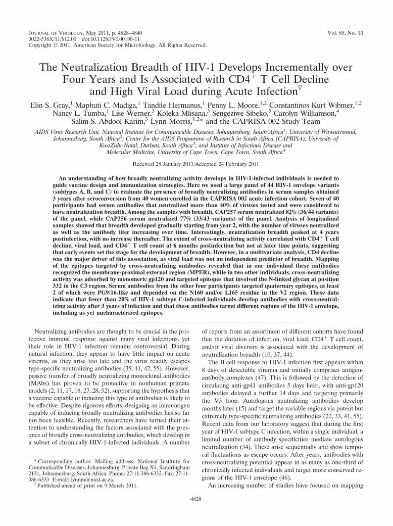
Viral load at set point and CD4� T cell decline stronglycorrelate with the development of breadth. Previous studieshave found an association between viral load and/or CD4� Tcell count and the presence of cross-neutralizing antibodies(10, 37, 44). Here we analyzed the correlation between thepercentage of viruses neutralized by each individual (as a mea-surement of neutralization breadth) and the viral load andCD4� T cell count at 6, 12, and 36 months postinfection (Fig.2). In addition, as a measurement of total antigenic exposure,we calculated the area under the curve for the viral loads overtime from 6 months to 3 years of infection (viral load AUC).These analyses revealed a significant correlation between theviral load at set point (6 months postinfection) and the per-centage of viruses neutralized at 3 years (Fig. 2A). Similarly, anegative correlation was found between the CD4� T cell countat 6 months postinfection and the development of breadth(Fig. 2D). However, no correlation was found between neu-tralization breadth and these two factors at later time points(Fig. 2B, C, E, and F). Furthermore, no significant correlationwas observed between neutralization breadth and the viral loadAUC (Fig. 2G), suggesting that the antigen load at a particulartime postinfection, not total antigenic exposure, predisposesindividuals toward the development of breadth. These corre-lations were confirmed using more robust statistical analyses(Table 1). Thus, while a univariate model analysis showed thatviral loads at 6 months and 12 months and the AUC weresignificantly associated with neutralization breadth, a multivar-iate analysis revealed that the 6-month viral load was the majordriver of this association. For every 1-log increase in viral load
4830 GRAY ET AL. J. VIROL.

FIG
.1.
Heterologous
neutralizingactivities
insera
fromthe
CA
PRISA
cohortat
3years
postinfection.The
neutralizationtiter
isshow
nas
thereciprocalof
theserum
dilutionrequired
toinhibit50%
ofinfectionfor
eachvirus-sam
plecom
bination.Titers
belowdetection,i.e.,those
of�
1:45,havebeen
omitted.T
hehighesttiters
areshow
nin
darkred
andthe
lowestin
lightyellow,
following
thedepicted
legend.Autologous
neutralizationtiters
arehighlighted
ingray
andw
erenotincluded
inthe
calculationsofpercentages
ofvirusesneutralized.Participants
were
rankedbased
oncross-neutralizing
activity.The
pseudovirusestested
were
fromfour
panels:CA
PRISA
subtypeC
(15),referencesubtype
C(24),reference
subtypeB
(23),andreference
subtypeA
(5).Viruses
areranked
fromleft
toright
within
eachpanelbased
onthe
number
ofsera
tow
hichthey
were
sensitive.The
referenceviruses
ConC
andD
u151.12are
depictedseparately
onthe
left.Clinicalprogression
isindicated
foreach
participant(* ,slow
progressors;†,rapidprogressors).
VOL. 85, 2011 CROSS-NEUTRALIZING ANTIBODIES IN HIV-1 INFECTION 4831
at 6 months, the percentage of cross-neutralization increasedby 12.4% (P � 0.002).
Interestingly, the correlation between CD4� T cell countprior to infection and neutralization breadth showed a positivetrend, although it was not statistically significant (Fig. 2H). Ofnote, preinfection CD4� T cell counts were available for only21 participants in the seronegative cohort, which included 3 ofthe 7 individuals with the broadest cross-neutralizing activities.These data are in stark contrast to the negative slope of theCD4� T cell count at 6 months postinfection (Fig. 2D). Fur-thermore, the decline in CD4� T cell number upon HIV-1infection, measured as the difference between CD4� T cellcounts preinfection and at 6 months postinfection, stronglycorrelated with the development of heterologous neutraliza-tion (r � 0.52; P � 0.0144) (Fig. 2I). This was confirmed in aunivariate model analysis (Table 1). Given the existing corre-
lation between viral load and CD4� T cell decline, both vari-ables were fitted into a second multivariate model, which re-vealed a significant association between the development ofbreadth and CD4� T cell decline (P � 0.0474) but not viralload (Table 1).
As shown above, the development of breadth was associatedwith factors such as high viral load and steep CD4� T celldecline in early infection, which are well known markers ofdisease progression (12, 13, 29). Of the 40 participants testedfor heterologous neutralization, 11 were initiated on treatmentduring the course of this study, including 5 of the 7 women ingroup 1. To investigate the potential relationship between thedevelopment of neutralization breadth and disease progres-sion, a Kaplan-Meier survival analysis was performed from thetime of infection until the first CD4� T cell count below 200cells/�l or therapy initiation as a marker of clinical AIDS.
FIG. 2. Factors correlated with the development of cross-neutralizing antibodies. The percentage of viruses neutralized by each serum wascorrelated with the 6-month (set point), 12-month, and 36-month (contemporaneous) viral loads (VL) (A, B, and C) and CD4� T cell counts (D,E, and F). Neutralization breadth was also correlated using the viral load AUC from 6 to 36 months postinfection (G), the preinfection CD4� Tcell count (H), and the decline in CD4� T cell count between preinfection and 6 months postinfection (I). Each correlation was analyzed usinga Spearman nonparametric test. The number of pairs (N) and P value for each correlation are shown. Statistically significant P values are markedwith asterisks.
4832 GRAY ET AL. J. VIROL.

Because of the link between viral load and disease progression,we excluded participants with low viral loads from this analysisand included only the 22 individuals for whom the viral loadAUC exceeded 5 million copies per ml. These were dividedinto two groups: 7 participants with cross-neutralizing antibod-ies (BCN) and 15 participants without breadth but with similarviremia (no BCN) at 3 years postinfection (Fig. 3). There wereno significant differences between the median viral loads andCD4� T cell counts of both groups at all time points. A Coxproportional hazard model showed that those who developedbreadth had a hazard ratio of 1.04 (95% confidence interval[95% CI], 0.32 to 3.34) and a log rank test P value of 0.9508.These results indicated that the development of cross-neutral-izing antibodies did not preclude or promote disease progres-sion.
Evolution of cross-neutralizing antibodies in individualswith the greatest breadth. Early serum samples from the 7individuals with the greatest neutralization breadth (group 1)
were assayed against all the viruses neutralized by the 3-yearsamples with titers above 1:100. On average, sera from 13 timepoints (range, 10 to 19 time points) over the first 3 years ofinfection were included. The longitudinal neutralization pro-files against both autologous and sensitive heterologous virusesfor each of the 7 participants are shown in Fig. 4. As describedpreviously (15), antibodies to the infecting virus appearedwithin months of infection and titers remained high over 3years. This suggested that despite multiple cycles of escape,sufficient neutralizing determinants remained on the infectingvirus to stimulate new autologous specificities. Some of thesenew specificities acquired the ability to neutralize heterologousviruses of different subtypes (Fig. 4 [viruses from differentsubtypes are color coded]).
Analysis of the individual neutralization patterns showedthat in one case (CAP206), the neutralizing activities againstthe majority of viruses occurred simultaneously (at 81 weekspostinfection), suggesting that a single dominant antibodyspecificity with cross-neutralizing activity emerged at this timepoint. Interestingly, this response waned at 107 weeks butreappeared later, targeting the same viruses (Fig. 4A). In par-ticipants CAP177 and CAP255, heterologous activity appearedmostly during the second year of infection (52 to 90 weekspostinfection), with titers reaching a plateau thereafter (Fig.4B and C). This might indicate affinity maturation of existingantibodies resulting in increased potency and breadth. Thesame can be said for the remaining four individuals, in whomthe neutralizing activity increased gradually over an extendedperiod (Fig. 4D, E, F, and G). Alternatively, the cross-neutral-izing activity in these four participants might involve the ap-pearance of new specificities which target different epitopes ondifferent viruses. Some of these new antibodies appeared to besubtype specific, as the early sera from participants CAP248,CAP255, CAP256, and CAP257 preferentially neutralized sub-type C viruses, with cross-subtype neutralization appearing atlater stages (Fig. 4).
Heterologous neutralization increases over time, peaking at4 years of infection. The above data from the 7 individuals ingroup 1 suggested that neutralization breadth continued toincrease over 3 years of infection. In order to more fully ex-plore the dynamics of the development of breadth, we tested
TABLE 1. Factors correlated with neutralization breadth
VariableUnivariate model Multivariate model 1 Multivariate model 2
Estimate (95% CI) P valuea Estimate (95% CI) P valuea Estimate (95% CI) P valuea
Viral load6 mo 0.11 (0.077, 0.143) 0.0020 0.124 (0.071, 0.178) 0.0259 0.032 (�0.008, 0.073) 0.430812 mo 0.077 (0.040, 0.114) 0.0437 �0.005 (�0.065, 0.055) 0.936636 mo 0.044 (0.008, 0.080) 0.2219 �0.045 (�0.096, 0.006) 0.3836
Viral load AUC 0.080 (0.043, 0.117) 0.0389 0.026 (�0.045, 0.098) 0.7145Preinfection CD4 countb 0.016 (0.008, 0.024) 0.0710CD4 declineb 0.024 (0.015, 0.032) 0.0094 0.021 (0.030, 0.011) 0.0474
CD4 count6 mo �0.029 (�0.046, �0.012) 0.099812 mo �0.024 (�0.041, �0.007) 0.156436 mo �0.021 (�0.037, �0.005) 0.1923
a Statistically significant P values are shown in bold.b Data are available for only 21 of the 40 study participants.
FIG. 3. Kaplan-Meier analysis of time from seroconversion untilARV initiation for CAPRISA participants with and without neutral-ization breadth. Study participants with similar viral loads were segre-gated based on their cross-neutralizing activity at 3 years postinfection,into BCN (group 1) (Fig. 1) and no BCN (groups 2 and 3) groups. ACox proportional hazard analysis was used to compare the two groupsbased on time of infection until the first CD4� T cell count below 200cells/�l or therapy initiation.
VOL. 85, 2011 CROSS-NEUTRALIZING ANTIBODIES IN HIV-1 INFECTION 4833

4834 GRAY ET AL. J. VIROL.
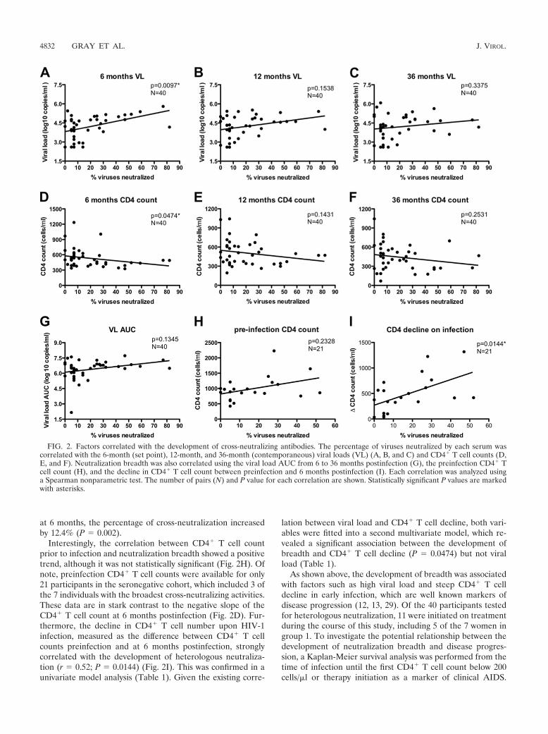
sera from all 40 individuals at 1, 2, and 3 years postinfection. Inaddition, samples were collected from 27 of the 40 participantsat 4 years of infection (7 participants, including CAP8 andCAP255, had initiated antiretroviral therapy, and 6 had not yetreached 4 years of infection). By 5 years of infection, samplesfrom 12 participants were available (3 participants, includingCAP256 and CAP257, started treatment before 5 years ofinfection, and 12, including CAP177, had not yet reached thistime point). Individual CAP206 was initiated on treatmentshortly after 5 years of infection, while subject CAP248 re-mained therapy naive over the course of the study. For thisanalysis, we used a smaller panel of 12 viruses, which included4 viruses of each subtype (A, B, and C). We found a goodcorrelation between the percentages of viruses neutralized us-ing the reduced panel and the full panel of 44 viruses with serafrom 3 years postinfection (R2 � 0.9012; P � 0.0001).
The heterologous neutralizing activity increased graduallyover time for the first 4 years (Fig. 5A), consistent with earlierreports that neutralization breadth was associated with dura-tion of infection (44). The titers at which viruses were neutral-ized also increased over this period. An analysis of the 27individuals at 4 years indicated that in addition to those pre-viously shown to have neutralization breadth (i.e., CAP177,CAP256, and CAP257), 2 other individuals (CAP37 andCAP267) showed a breadth of �40% of viruses neutralized atthis time point (Fig. 5B). CAP37 also showed more than 40%neutralization at 3 years of infection, reflecting minor differ-ences in the 2 viral panels used for Table 1 and Fig. 5. Most ofthe other individuals showed slight increases in breadth be-tween 3 and 4 years, but all remained below 40%. At 5 years ofinfection, no additional samples were found to have developedneutralization breadth. Only CAP206 and CAP248, previouslyshown to have breadth, had neutralization percentages above40% at this time (Fig. 5C). Indeed, there appeared to be noincrease in heterologous neutralization between 4 and 5 yearsof infection in this small subset, even among those withbreadth, suggesting that no new further maturation or newspecificities were generated after 4 years of infection.
Mapping the antibody specificities in broadly neutralizingplasmas. We previously reported the presence of anti-MPERantibodies at high titers (ID50s above 1:1,000) in participantCAP206 (15). Removal of these anti-MPER antibodies usingMPER peptide-coated beads resulted in a significant loss ofneutralizing activity against most heterologous viruses. Plas-mas from CAP248, CAP255, and CAP257 had undetectableneutralization titers against the HIV-2/HIV-1 MPER chimericvirus, while plasmas from CAP8, CAP177, and CAP256 hadlow anti-MPER neutralization titers, of 1:73, 1:280, and 1:274,respectively. The removal of these low-titer anti-MPER anti-bodies did not affect heterologous neutralization (data notshown).
FIG. 4. Kinetics of development of heterologous neutralization in individuals with breadth. Sequential serum samples from 0 to 3 years ofinfection (10 to 19 samples) from all 7 participants with breadth were tested against all the viruses previously shown to be sensitive to the 3-yearplasma samples. The ID50 titers versus weeks postinfection (p.i.) are represented for each virus, with the autologous viruses shown in solid black,subtype C viruses in red, subtype B viruses in blue, and subtype A viruses in green. The time points when detectable heterologous activity emergedare indicated using dashed vertical lines, with the number of additional viruses neutralized displayed above. The viral loads over time are shownas dashed black curves.
FIG. 5. Development of cross-neutralizing antibodies over 5 yearsof infection. The sera obtained at 1, 2, 3, 4, and 5 years of infectionwere tested for neutralization against a panel of 12 viruses of subtypesA, B, and C (4 of each subtype). The percentage of viruses neutralizedwas calculated for each sample. (A) Percentages of individuals capableof neutralizing more than 80%, 40 to 80%, 1 to 40%, and none of the12 viruses at 5 different time points. (B and C) Percentages of virusesneutralized over time for the 15 and 12 participants that reached 4 and5 years postinfection, respectively.
VOL. 85, 2011 CROSS-NEUTRALIZING ANTIBODIES IN HIV-1 INFECTION 4835

To further explore the specificities of the cross-neutralizingantibodies in the other 6 individuals, anti-gp120 antibodies inthe 3-year postinfection plasmas were removed using magneticbeads coated with recombinant ConC gp120. More than 97%of the gp120-binding antibodies were depleted in an ELISA(Fig. 6A). For CAP177 and CAP255, neutralizing activitiesagainst at least three viruses were significantly decreased by theremoval of anti-gp120 antibodies (Fig. 6B). Since no effect wasseen with the samples from the other four individuals, wepurified ConC trimers by gel exclusion chromatography andimmediately used these to coat magnetic beads for adsorptionexperiments. However, despite the removal of most gp145-
binding antibodies (Fig. 6A), this protein also failed to adsorbsignificant amounts of the neutralizing activity in plasmas fromCAP8, CAP248, CAP256, and CAP257 (Fig. 6C), suggestingthat neutralizing antibodies in these four plasmas recognizedepitopes only apparent on the quaternary structure of theenvelope glycoprotein.
Further experiments were performed to determine theepitope(s) within gp120 that was recognized by the CAP177and CAP255 neutralizing antibodies. We found that gp120mutated in the CD4 (D368R) or coreceptor (I420R) bindingsite effectively removed the heterologous activity (Fig. 7A),suggesting that the epitopes recognized by these plasmas do
FIG. 6. Adsorption of neutralizing antibodies using recombinant envelope proteins. Plasmas obtained at 3 years postinfection were adsorbedusing recombinant ConC monomeric gp120- or trimeric gp145-coated beads. Blank beads were used as a negative control. (A) Depleted plasmaswere tested for binding to ConC gp120 or ConC gp145 by ELISA. The percentage of antibody depleted was calculated using the following equation:[1 � (midpoint titer of plasma treated with gp120-coated beads/midpoint titer of plasma treated with blank beads)] � 100. (B) Plasmas adsorbedwith ConC gp120 were tested for neutralizing activity against various envelope-pseudotyped viruses. The percent depletion was calculated asfollows: [1 � (ID50 of plasma treated with gp120-coated beads/ID50 of plasma treated with blank beads)] � 100. Data represent the means for twoseparate neutralization experiments. (C) Plasmas adsorbed with ConC gp145 were tested for neutralizing activity against ConC and analyzed asdescribed for gp120.
FIG. 7. Epitope mapping of CAP177 and CAP255 anti-gp120 neutralizing antibodies. (A) The 3-year plasmas of these two participants wereadsorbed using gp120 mutated in the CD4 binding site (D368R) or coreceptor binding site (I420R) and gp120 with the V1V2 and V3 loops deleted(core gp120). Wild-type gp120-coated beads and blank beads were used as positive and negative controls, respectively. Depleted plasmas weretested for neutralization of ConC. (B) CAP177 plasma was adsorbed with V1V2- or V3-deleted gp120 prior to testing of ConC neutralization.(C) Both plasmas were tested for neutralization against wild-type ConC and three mutants in the core DMR epitope. Data represent the meansfor two separate neutralization experiments.
4836 GRAY ET AL. J. VIROL.
not overlap these regions or at least are not affected by thesetwo mutations. However, use of a core gp120 in which theV1V2 and V3 loops had been deleted affected the epitoperecognized by CAP177 but not that recognized by CAP255(Fig. 7A and B), suggesting that elements present in the vari-able loops were important for CAP177 neutralization. To de-termine if these two samples targeted an epitope in thegp120 core defined by the HJ16 MAb (7, 38), which alsooverlaps the CD4 binding site, plasmas were tested for neu-tralization of the DMR ConC mutants (D434A, M435A, orR436A). None of these mutants showed reduced sensitivityto the neutralizing antibodies in these 2 samples; in fact, theD434A mutant was more sensitive to neutralization than thewild-type virus (Fig. 7C).
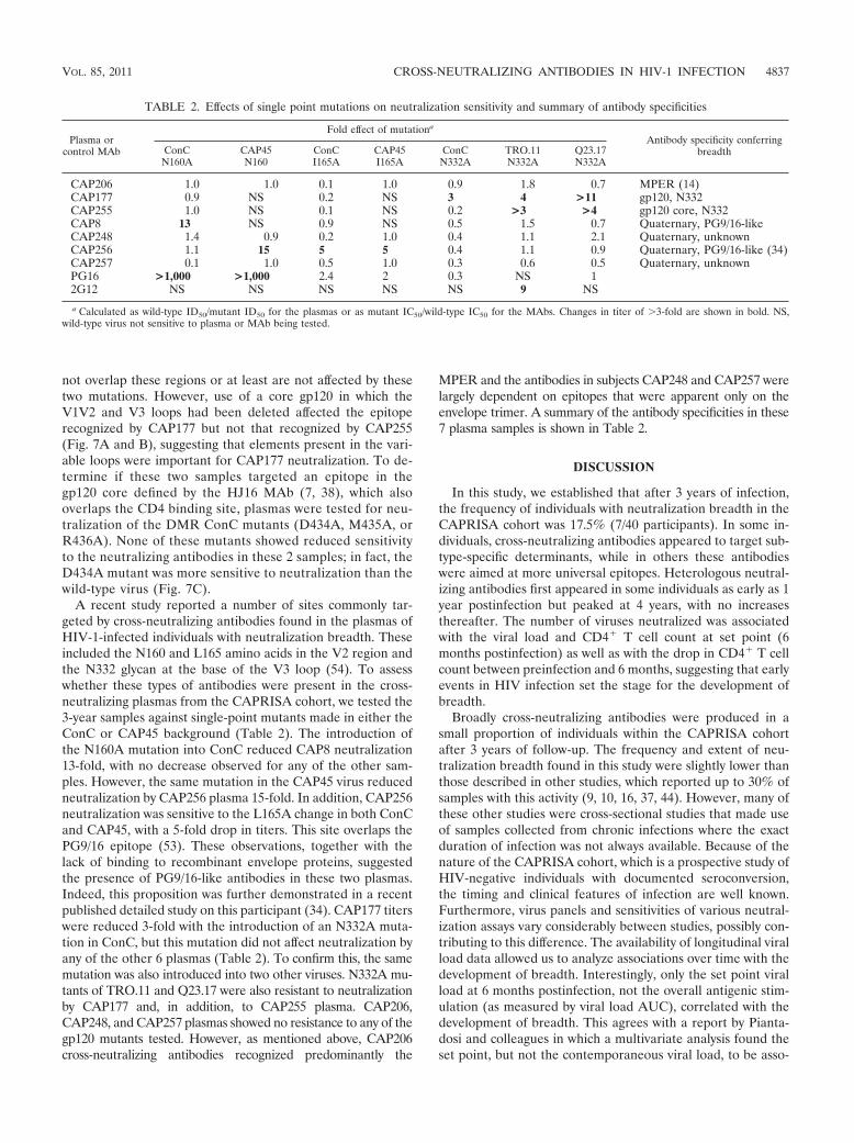
A recent study reported a number of sites commonly tar-geted by cross-neutralizing antibodies found in the plasmas ofHIV-1-infected individuals with neutralization breadth. Theseincluded the N160 and L165 amino acids in the V2 region andthe N332 glycan at the base of the V3 loop (54). To assesswhether these types of antibodies were present in the cross-neutralizing plasmas from the CAPRISA cohort, we tested the3-year samples against single-point mutants made in either theConC or CAP45 background (Table 2). The introduction ofthe N160A mutation into ConC reduced CAP8 neutralization13-fold, with no decrease observed for any of the other sam-ples. However, the same mutation in the CAP45 virus reducedneutralization by CAP256 plasma 15-fold. In addition, CAP256neutralization was sensitive to the L165A change in both ConCand CAP45, with a 5-fold drop in titers. This site overlaps thePG9/16 epitope (53). These observations, together with thelack of binding to recombinant envelope proteins, suggestedthe presence of PG9/16-like antibodies in these two plasmas.Indeed, this proposition was further demonstrated in a recentpublished detailed study on this participant (34). CAP177 titerswere reduced 3-fold with the introduction of an N332A muta-tion in ConC, but this mutation did not affect neutralization byany of the other 6 plasmas (Table 2). To confirm this, the samemutation was also introduced into two other viruses. N332A mu-tants of TRO.11 and Q23.17 were also resistant to neutralizationby CAP177 and, in addition, to CAP255 plasma. CAP206,CAP248, and CAP257 plasmas showed no resistance to any of thegp120 mutants tested. However, as mentioned above, CAP206cross-neutralizing antibodies recognized predominantly the
MPER and the antibodies in subjects CAP248 and CAP257 werelargely dependent on epitopes that were apparent only on theenvelope trimer. A summary of the antibody specificities in these7 plasma samples is shown in Table 2.
DISCUSSION
In this study, we established that after 3 years of infection,the frequency of individuals with neutralization breadth in theCAPRISA cohort was 17.5% (7/40 participants). In some in-dividuals, cross-neutralizing antibodies appeared to target sub-type-specific determinants, while in others these antibodieswere aimed at more universal epitopes. Heterologous neutral-izing antibodies first appeared in some individuals as early as 1year postinfection but peaked at 4 years, with no increasesthereafter. The number of viruses neutralized was associatedwith the viral load and CD4� T cell count at set point (6months postinfection) as well as with the drop in CD4� T cellcount between preinfection and 6 months, suggesting that earlyevents in HIV infection set the stage for the development ofbreadth.
Broadly cross-neutralizing antibodies were produced in asmall proportion of individuals within the CAPRISA cohortafter 3 years of follow-up. The frequency and extent of neu-tralization breadth found in this study were slightly lower thanthose described in other studies, which reported up to 30% ofsamples with this activity (9, 10, 16, 37, 44). However, many ofthese other studies were cross-sectional studies that made useof samples collected from chronic infections where the exactduration of infection was not always available. Because of thenature of the CAPRISA cohort, which is a prospective study ofHIV-negative individuals with documented seroconversion,the timing and clinical features of infection are well known.Furthermore, virus panels and sensitivities of various neutral-ization assays vary considerably between studies, possibly con-tributing to this difference. The availability of longitudinal viralload data allowed us to analyze associations over time with thedevelopment of breadth. Interestingly, only the set point viralload at 6 months postinfection, not the overall antigenic stim-ulation (as measured by viral load AUC), correlated with thedevelopment of breadth. This agrees with a report by Pianta-dosi and colleagues in which a multivariate analysis found theset point, but not the contemporaneous viral load, to be asso-
TABLE 2. Effects of single point mutations on neutralization sensitivity and summary of antibody specificities
Plasma orcontrol MAb
Fold effect of mutationa
Antibody specificity conferringbreadthConC
N160ACAP45N160
ConCI165A
CAP45I165A
ConCN332A
TRO.11N332A
Q23.17N332A
CAP206 1.0 1.0 0.1 1.0 0.9 1.8 0.7 MPER (14)CAP177 0.9 NS 0.2 NS 3 4 >11 gp120, N332CAP255 1.0 NS 0.1 NS 0.2 >3 >4 gp120 core, N332CAP8 13 NS 0.9 NS 0.5 1.5 0.7 Quaternary, PG9/16-likeCAP248 1.4 0.9 0.2 1.0 0.4 1.1 2.1 Quaternary, unknownCAP256 1.1 15 5 5 0.4 1.1 0.9 Quaternary, PG9/16-like (34)CAP257 0.1 1.0 0.5 1.0 0.3 0.6 0.5 Quaternary, unknownPG16 >1,000 >1,000 2.4 2 0.3 NS 12G12 NS NS NS NS NS 9 NS
a Calculated as wild-type ID50/mutant ID50 for the plasmas or as mutant IC50/wild-type IC50 for the MAbs. Changes in titer of �3-fold are shown in bold. NS,wild-type virus not sensitive to plasma or MAb being tested.
VOL. 85, 2011 CROSS-NEUTRALIZING ANTIBODIES IN HIV-1 INFECTION 4837

ciated with neutralization breadth (37). As they described, theassociation of contemporaneous viral load and breadth ob-served by them and others (37, 44) may have been driven bythe correlation of viral load within individuals at differentstages of infection. However, a high viral load is not an overallpredictor of neutralization breadth, as some highly viremicindividuals did not develop cross-neutralizing antibodies, evenafter 5 years of infection. This has also been noticed in previ-ous studies (10), suggesting that other factors in addition tohigh viremia determine the development of breadth.
A low CD4� T cell count at set point was also found tocorrelate with the development of cross-neutralizing antibod-ies. Moreover, the drop in CD4� T cell count from preinfec-tion to 6 months postinfection was a better predictor of theextent of heterologous neutralization than the set point viralload, despite the limited number of individuals in this analysis.This association was noted in another study in which the CD4�
T cell count at set point was also analyzed (10). It is intriguingthat a drop in CD4� T cell count was associated with theproduction of cross-neutralizing antibodies given the impor-tance of T cell help for B cell function. However, with thelymphocytic choriomeningitis virus (LCMV) model, it hasbeen reported that depletion of CD4� T cells improves virusneutralizing antibody production by reducing the CD4� T cell-dependent virus-nonspecific polyclonal hyperglobulinemia (21,40). HIV and hepatitis C virus infections, like LCMV infection,induce similar dysfunctions (8, 19, 43), suggesting that deple-tion of CD4� T cells in these infections might improve theneutralizing antibody response by reducing B cell polyclonalactivation. This is an interesting observation and raises fur-ther questions such as whether low CD4� T cell counts areassociated with a quicker upsurge and/or stronger autolo-gous neutralizing responses, whether hyperglobulinemia islower in participants who develop cross-neutralizing anti-bodies, and whether experimental depletion of CD4� T cellsin nonhuman primates may enhance the production of neu-tralizing antibodies.
Our results suggest that neutralization breadth is acquired at2 to 3 years postinfection. This agrees with a recent study donewith subtype B-infected individuals that showed that breadthdeveloped, on average, 2.5 years following infection (30). Thereason for the delay in the development of neutralizationbreadth is not understood. However, the association of breadthwith high viral loads suggests that antigen-driven selection iscrucial in the development of these antibodies. This coulddrive affinity maturation, as suggested in a study by Toran andcoworkers (48), who showed that all anti-gp120 neutralizingantibodies in a long-term nonprogressor were clonally related,with considerable somatic hypermutation. It has been pro-posed that HIV-1 has evolved to avoid germ line-like antibodyrecognition, possibly delaying the appearance of cross-neutral-izing antibodies (57). Furthermore, immunogenetic analysis ofexisting broadly neutralizing MAbs suggests that they haveundergone multiple rounds of affinity maturation to achievecross-neutralizing activity (36, 58). This is in contrast to othervirus infections, such as severe acute respiratory syndrome(SARS) coronavirus and henipavirus infections, where cross-neutralizing antibodies are induced early but carry very fewsomatic mutations (39).
Our observation that no further increase in neutralization
breadth occurred after 4 years is particularly intriguing andrather unexpected, although this was also recently reported forthe subtype B cohorts mentioned above (30). We hypothesizedthat cross-neutralizing capacity would increase steadily overtime even after 4 years of infection. If neutralization breadthoccurs stochastically, with relentless waves of escape and thedevelopment of new autologous neutralizing antibodies in thepresence of high antigenic loads resulting in the appearance ofantibodies that recognize a generally conserved epitope, thenthis should happen at any stage of disease. Alternatively, dys-regulation of the immune system over time (31) may result ina reduced ability to mount new antibody responses at laterstages of infection. This is suggested by preliminary data fromour laboratory (data not shown) and by reports from othersthat suggest that the capacity to mount new autologous re-sponses is impaired after 2 to 3 years of infection, independentof disease progression (49). However, this needs to be exploredfurther by measuring de novo autologous responses at varioustimes postinfection in individuals with a range of viral loads.
Interestingly, 5 of the 7 individuals identified here as havingcross-neutralizing activity were initiated on antiretroviral treat-ment because they had low CD4� T cell counts and/or othersigns of clinical AIDS. Only CAP177 and CAP248 were stillARV naive after 4 and 5 years of infection, respectively. Thisobservation is consistent with the report by Euler et al. thatcross-reactive antibodies do not protect against disease pro-gression (10), probably because the virus readily escapes evencross-neutralizing antibodies. Indeed, factors associated withthe development of neutralizing antibodies, such as high viralload and low CD4� T cell count, are strongly correlated withdisease progression (12, 13, 29). These factors herald a declineof immune function which, in the absence of successful anti-retroviral therapy, results in progression to AIDS. It is possiblethat the individuals with high viral loads and low CD4� T cellcounts who failed to mount a cross-neutralizing responsewithin this time frame “lost their opportunity” with the col-lapse of the immune system. On the other hand, participantswith low viral loads and high CD4� T cell counts did notdevelop breadth, probably due to a lack of sufficient antigenicstimulation and/or the overwhelming nonspecific hyperglobu-linemia. It would be interesting to explore whether these indi-viduals develop breadth at later stages of infection, either aswaves of autologous neutralization continue to develop againsta diversifying virus or through a sudden collapse in control anddrop in CD4� T cell count that result in the development ofcross-neutralizing antibodies.
A summary of what is currently known about the antibodyspecificities mediating breadth at 3 years postinfection in the 7individuals in group 1 is shown in Table 2. Only one participanthad neutralizing antibodies against the MPER of gp41. Theseplasma antibodies recognized an epitope centered on D674and were shown to be distinct from other antibodies withMPER specificities, such as 4E10 and Z13e1 (14). Confirma-tion of this comes from the recent isolation of a neutralizingMAb (CAP206-CH12), using circulating blood memory B cellsfrom CAP206, which showed similar epitope recognition (L.Morris et al., unpublished data). Two participants had anti-bodies that recognized epitopes in gp120 that involved theglycan at position 332. However, these two specificities arelikely to be different from one another given that the CAP255
4838 GRAY ET AL. J. VIROL.

epitope is present on the core gp120, while the CAP177 anti-body depends on variable loops. Walker and colleagues alsofound N332-dependent neutralizing antibodies in 5 of the 19plasmas that they analyzed. Only one of these plasmas wasshown to have 2G12-like activity, despite their epitope conver-gence on the N332 glycan (54). Antibodies targeting trimer-specific epitopes were the most common among the 7 individ-uals with neutralization breadth. Plasmas from two individuals,CAP248 and CAP257, could not be mapped using availablemethods. However, in two cases, CAP8 and CAP256, depen-dence on the N160 glycan suggested the presence of PG9/16-like antibodies. Although N160- and L165-dependent neutral-ization has been described recently as two distinct specificities(54), both affected neutralization by CAP256 plasma. The finemapping of the CAP256 quaternary specificity indicated that itdepended on various residues in the V2 loop which overlappedthe PG9/16 epitope (34).
Three of the four individuals whose antibodies recognizedquaternary neutralizing epitopes, i.e., CAP248, CAP256, andCAP257, neutralized subtype C viruses preferentially, and thisdiscrimination was more evident at early time points. However,CAP8 antibodies preferentially neutralized subtype B viruses.In contrast, the anti-MPER antibodies in CAP206 and theanti-gp120 antibodies conferring breadth in CAP177 andCAP255 neutralized viruses from different subtypes equally.Preferential intrasubtype neutralization was reported previ-ously by van Gils and colleagues (50). In their study of subtypeB-infected individuals, participants with intermediate breadthshowed more subtype B-restricted responses, which suggestedthat subtype specificity was related to low neutralization titers.In contrast, CAP256 and CAP257, the two participants withthe greatest breadth, had high titers against subtype C virusesdespite their subtype specificity. Therefore, we concluded thatsubtype-specific neutralization is not a general feature of in-termediate breadth but instead depends on the epitope recog-nized by the cross-neutralizing antibodies. Furthermore, ourdata suggest that antibodies targeting quaternary neutralizingepitopes are more likely to be subtype specific than those withother targets.
The V1V2 and C3 regions have previously been shown to becommon targets for autologous neutralizing antibodies in sub-type C infection (26, 33, 35, 42). It is interesting that mostheterologous activity found in this study overlapped with thesetwo regions. CAP8 and CAP256 plasmas contained cross-neu-tralizing antibodies that recognized epitopes within the V2loop, while CAP177 and CAP255 appeared to recognize anepitope in C3. This suggests that the V1V2 and C3 regions areparticularly immunodominant regions of the envelope and arethe focus of the neutralizing antibody response from earlytimes on, with these type-specific antibodies sometimes devel-oping into antibodies with cross-neutralizing specificities. On-going studies in our laboratory aim to determine the relation-ship between the evolution of autologous neutralizingantibodies and the development of broadly neutralizing activ-ity. Interestingly, the N332 glycan targeted by the heterologousneutralizing antibodies in CAP177 was not present in the trans-mitted founder virus isolated from this participant (1). Thisglycan appeared later, at 6 months postinfection, as an escapemutation driven by the primary autologous neutralizing anti-body response that targeted the alpha-2 helix of C3 (35). This
suggests that, at least in this case, later variants rather than theinfecting virus elicited antibodies with cross-neutralizing activ-ity. Elucidating the path between these two responses mayguide the design of immunization strategies that mimic thisprocess, focusing the response on the conserved motifs in theenvelope glycoprotein.
ACKNOWLEDGMENTS
We thank the participants in the CAPRISA 002 acute infectioncohort and the clinical and laboratory staff at CAPRISA for providingspecimens. We are grateful to Mary Phoswa and Sarah Cohen forsample and data management. We thank Progenics for supplying thevector pPPI4.
This work was funded by the Bill and Melinda Gates Collaborationfor AIDS Vaccine Discovery (CAVD), the Vaccine Immune Monitor-ing Consortium (VIMC) (grant 38619), the Center for HIV/AIDSVaccine Immunology (CHAVI) (grant AI64518), a HIVRAD grant(P01 AI088610-01), and the South African HIV/AIDS Research andInnovation Platform (SHARP) of the Department of Science andTechnology (DST). We thank the U.S. National Institutes of Health’sComprehensive International Program of Research on AIDS (CIPRAgrant AI51794) and the Columbia University-Southern African Fog-arty AIDS International Training and Research Programme (AITRPgrant D43TW00231) for the research infrastructure and training thatmade the CAPRISA 002 acute infection study possible. P.L.M. is aWellcome Trust Intermediate Fellow in Public Health and TropicalMedicine (grant 089933/Z/09/Z).
REFERENCES
1. Abrahams, M. R., et al. 2009. Quantitating the multiplicity of infection withHIV-1 subtype C reveals a non-Poisson distribution of transmitted variants.J. Virol. 83:3556–3567.
2. Baba, T. W., et al. 2000. Human neutralizing monoclonal antibodies of theIgG1 subtype protect against mucosal simian-human immunodeficiency virusinfection. Nat. Med. 6:200–206.
3. Binley, J. 2009. Specificity of broadly neutralizing antibodies in sera fromHIV-1 infected individuals. Curr. Opin. HIV AIDS 4:364–372.
4. Binley, J. M., et al. 2003. Redox-triggered infection by disulfide-shackledhuman immunodeficiency virus type 1 pseudovirions. J. Virol. 77:5678–5684.
5. Blish, C. A., R. Nedellec, K. Mandaliya, D. E. Mosier, and J. Overbaugh.2007. HIV-1 subtype A envelope variants from early in infection have vari-able sensitivity to neutralization and to inhibitors of viral entry. AIDS 21:693–702.
6. Burton, D. R., R. L. Stanfield, and I. A. Wilson. 2005. Antibody vs. HIV ina clash of evolutionary titans. Proc. Natl. Acad. Sci. U. S. A. 102:14943–14948.
7. Corti, D., et al. 2010. Analysis of memory B cell responses and isolation ofnovel monoclonal antibodies with neutralizing breadth from HIV-1-infectedindividuals. PLoS One 5:e8805.
8. De Milito, A., et al. 2004. Mechanisms of hypergammaglobulinemia andimpaired antigen-specific humoral immunity in HIV-1 infection. Blood 103:2180–2186.
9. Doria-Rose, N. A., et al. 2009. Frequency and phenotype of human immu-nodeficiency virus envelope-specific B cells from patients with broadly cross-neutralizing antibodies. J. Virol. 83:188–199.
10. Euler, Z., et al. 2010. Cross-reactive neutralizing humoral immunity does notprotect from HIV type 1 disease progression. J. Infect. Dis. 201:1045–1053.
11. Ferrantelli, F., et al. 2004. Complete protection of neonatal rhesus macaquesagainst oral exposure to pathogenic simian-human immunodeficiency virusby human anti-HIV monoclonal antibodies. J. Infect. Dis. 189:2167–2173.
12. Fraser, C., T. D. Hollingsworth, R. Chapman, F. de Wolf, and W. P. Hanage.2007. Variation in HIV-1 set-point viral load: epidemiological analysis andan evolutionary hypothesis. Proc. Natl. Acad. Sci. U. S. A. 104:17441–17446.
13. Goujard, C., et al. 2006. CD4 cell count and HIV DNA level are independentpredictors of disease progression after primary HIV type 1 infection inuntreated patients. Clin. Infect. Dis. 42:709–715.
14. Gray, E. S., et al. 2009. Broad neutralization of human immunodeficiencyvirus type 1 mediated by plasma antibodies against the gp41 membraneproximal external region. J. Virol. 83:11265–11274.
15. Gray, E. S., et al. 2007. Neutralizing antibody responses in acute humanimmunodeficiency virus type 1 subtype C infection. J. Virol. 81:6187–6196.
16. Gray, E. S., et al. 2009. Antibody specificities associated with neutralizationbreadth in plasma from human immunodeficiency virus type 1 subtype C-infected blood donors. J. Virol. 83:8925–8937.
17. Hessell, A. J., et al. 2009. Effective, low-titer antibody protection againstlow-dose repeated mucosal SHIV challenge in macaques. Nat. Med. 15:951–954.
VOL. 85, 2011 CROSS-NEUTRALIZING ANTIBODIES IN HIV-1 INFECTION 4839

18. Hessell, A. J., et al. 2009. Broadly neutralizing human anti-HIV antibody2G12 is effective in protection against mucosal SHIV challenge even at lowserum neutralizing titers. PLoS Pathog. 5:e1000433.
19. Hunziker, L., et al. 2003. Hypergammaglobulinemia and autoantibody in-duction mechanisms in viral infections. Nat. Immunol. 4:343–349.
20. Kothe, D. L., et al. 2006. Ancestral and consensus envelope immunogens forHIV-1 subtype C. Virology 352:438–449.
21. Lang, K. S., et al. 2007. “Negative vaccination” by specific CD4 T celltolerisation enhances virus-specific protective antibody responses. PLoS One2:e1162.
22. Li, B., et al. 2006. Evidence for potent autologous neutralizing antibody titersand compact envelopes in early infection with subtype C human immuno-deficiency virus type 1. J. Virol. 80:5211–5218.
23. Li, M., et al. 2005. Human immunodeficiency virus type 1 env clones fromacute and early subtype B infections for standardized assessments of vaccine-elicited neutralizing antibodies. J. Virol. 79:10200–10209.
24. Li, M., et al. 2006. Genetic and neutralization properties of subtype C humanimmunodeficiency virus type 1 molecular env clones from acute and earlyheterosexually acquired infections in Southern Africa. J. Virol. 80:11776–11790.
25. Li, Y., et al. 2009. Analysis of neutralization specificities in polyclonal seraderived from human immunodeficiency virus type 1-infected individuals.J. Virol. 83:1045–1059.
26. Lynch, R. M., et al. 2011. The B cell response is redundant and highlyfocused on V1V2 during early subtype C infection in a Zambian serocon-verter. J. Virol. 85:905–915.
27. Mascola, J. R., et al. 1999. Protection of macaques against pathogenic sim-ian/human immunodeficiency virus 89.6PD by passive transfer of neutraliz-ing antibodies. J. Virol. 73:4009–4018.
28. Mascola, J. R., et al. 2000. Protection of macaques against vaginal transmis-sion of a pathogenic HIV-1/SIV chimeric virus by passive infusion of neu-tralizing antibodies. Nat. Med. 6:207–210.
29. Mellors, J. W., et al. 1997. Plasma viral load and CD4� lymphocytes asprognostic markers of HIV-1 infection. Ann. Intern. Med. 126:946–954.
30. Mikell, I., et al. 2011. Characteristics of the earliest cross-neutralizing anti-body response to HIV-1. PLoS Pathog. 7:e1001251.
31. Moir, S., and A. S. Fauci. 2009. B cells in HIV infection and disease. Nat.Rev. Immunol. 9:235–245.
32. Montefiori, D. 2004. Evaluating neutralizing antibodies against HIV, SIVand SHIV in luciferase reporter gene assays, p. 12.11.1–12.11.15. In J. E.Coligan et al. (ed.), Current protocols in immunology. John Wiley & Sons,New York, NY.
33. Moore, P. L., et al. 2008. The c3-v4 region is a major target of autologousneutralizing antibodies in human immunodeficiency virus type 1 subtype Cinfection. J. Virol. 82:1860–1869.
34. Moore, P. L., et al. 2011. Potent and broad neutralization of HIV-1 subtypeC viruses by plasma antibodies targeting a quaternary epitope includingresidues in the V2 loop. J. Virol. 85:3128–3141.
35. Moore, P. L., et al. 2009. Limited neutralizing antibody specificities driveneutralization escape in early HIV-1 subtype C infection. PLoS Pathog.5:e1000598.
36. Pancera, M., et al. 2010. Crystal structure of PG16 and chimeric dissectionwith somatically related PG9: structure-function analysis of two quaternary-specific antibodies that effectively neutralize HIV-1. J. Virol. 84:8098–8110.
37. Piantadosi, A., et al. 2009. Breadth of neutralizing antibody response tohuman immunodeficiency virus type 1 is affected by factors early in infectionbut does not influence disease progression. J. Virol. 83:10269–10274.
38. Pietzsch, J., et al. 2010. Human anti-HIV-neutralizing antibodies frequentlytarget a conserved epitope essential for viral fitness. J. Exp. Med. 207:1995–2002.
39. Prabakaran, P., et al. 2009. Potent human monoclonal antibodies againstSARS CoV, Nipah and Hendra viruses. Expert Opin. Biol. Ther. 9:355–368.
40. Recher, M., et al. 2004. Deliberate removal of T cell help improves virus-neutralizing antibody production. Nat. Immunol. 5:934–942.
41. Richman, D. D., T. Wrin, S. J. Little, and C. J. Petropoulos. 2003. Rapidevolution of the neutralizing antibody response to HIV type 1 infection.Proc. Natl. Acad. Sci. U. S. A. 100:4144–4149.
42. Rong, R., et al. 2009. Escape from autologous neutralizing antibodies inacute/early subtype C HIV-1 infection requires multiple pathways. PLoSPathog. 5:e1000594.
43. Rosa, D., et al. 2005. Activation of naive B lymphocytes via CD81, a patho-genetic mechanism for hepatitis C virus-associated B lymphocyte disorders.Proc. Natl. Acad. Sci. U. S. A. 102:18544–18549.
44. Sather, D. N., et al. 2009. Factors associated with the development of cross-reactive neutralizing antibodies during human immunodeficiency virus type1 infection. J. Virol. 83:757–769.
45. Sather, D. N., and L. Stamatatos. 2010. Epitope specificities of broadlyneutralizing plasmas from HIV-1 infected subjects. Vaccine 28(Suppl. 2):B8–B12.
46. Stamatatos, L., L. Morris, D. R. Burton, and J. R. Mascola. 2009. Neutral-izing antibodies generated during natural HIV-1 infection: good news for anHIV-1 vaccine? Nat. Med. 15:866–870.
47. Tomaras, G. D., et al. 2008. Initial B-cell responses to transmitted humanimmunodeficiency virus type 1: virion-binding immunoglobulin M (IgM) andIgG antibodies followed by plasma anti-gp41 antibodies with ineffectivecontrol of initial viremia. J. Virol. 82:12449–12463.
48. Toran, J. L., et al. 1999. Molecular analysis of HIV-1 gp120 antibody re-sponse using isotype IgM and IgG phage display libraries from a long-termnon-progressor HIV-1-infected individual. Eur. J. Immunol. 29:2666–2675.
49. van Gils, M. J., et al. 2010. Rapid escape from preserved cross-reactiveneutralizing humoral immunity without loss of viral fitness in HIV-1-infectedprogressors and long-term nonprogressors. J. Virol. 84:3576–3585.
50. van Gils, M. J., D. Edo-Matas, B. Schweighardt, T. Wrin, and H. Schuite-maker. 2010. High prevalence of neutralizing activity against multiple unre-lated human immunodeficiency virus type 1 (HIV-1) subtype B variants insera from HIV-1 subtype B-infected individuals: evidence for subtype-spe-cific rather than strain-specific neutralizing activity. J. Gen. Virol. 91:250–258.
51. Van Loggerenberg, F., et al. 2008. Establishing a cohort at high risk of HIVinfection in South Africa: challenges and experiences of the CAPRISA 002acute infection study. PLoS One 3:e1954.
52. Veazey, R. S., et al. 2003. Prevention of virus transmission to macaquemonkeys by a vaginally applied monoclonal antibody to HIV-1 gp120. Nat.Med. 9:343–346.
53. Walker, L. M., et al. 2009. Broad and potent neutralizing antibodies from anAfrican donor reveal a new HIV-1 vaccine target. Science 326:285–289.
54. Walker, L. M., et al. 2010. A limited number of antibody specificities mediatebroad and potent serum neutralization in selected HIV-1 infected individu-als. PLoS Pathog. 6:e1001028.
55. Wei, X., et al. 2003. Antibody neutralization and escape by HIV-1. Nature422:307–312.
56. Wu, X., et al. 2010. Rational design of envelope identifies broadly neutral-izing human monoclonal antibodies to HIV-1. Science 329:856–861.
57. Xiao, X., et al. 2009. Germline-like predecessors of broadly neutralizingantibodies lack measurable binding to HIV-1 envelope glycoproteins: impli-cations for evasion of immune responses and design of vaccine immunogens.Biochem. Biophys. Res. Commun. 390:404–409.
58. Zhou, T., et al. 2010. Structural basis for broad and potent neutralization ofHIV-1 by antibody VRC01. Science 329:811–817.
4840 GRAY ET AL. J. VIROL.


![[ICPP'14] Yang You et al., Designing a Heuristic Cross-Architecture Combination for Breadth-First Search, 43rd International Conference on Parallel Processing](https://static.fdokumen.com/doc/165x107/631ade94d5372c006e03b0ab/icpp14-yang-you-et-al-designing-a-heuristic-cross-architecture-combination.jpg)