Role of SR protein modular domains in alternative splicing ...
Upload
independentCategory
view
1download
0
The multiple-specificity landscape of modular peptiderecognition domains
David Gfeller1,2,7, Frank Butty1,2, Marta Wierzbicka1,2, Erik Verschueren3, Peter Vanhee3, Haiming Huang1,2, Andreas Ernst1,2,Nisa Dar1,2,4, Igor Stagljar1,2,4, Luis Serrano3, Sachdev S Sidhu1,2,4, Gary D Bader1,2,4,5,6,* and Philip M Kim1,2,4,5,*
1 Banting and Best Department of Medical Research, The Donnelly Centre, University of Toronto, Toronto, Ontario, Canada, 2 Department of Biochemistry,University of Toronto, Toronto, Ontario, Canada, 3 EMBL-CRG Systems Biology Unit, CRG-Centre de Regulacio Genomica, Barcelona, Spain, 4 Department ofMolecular Genetics, University of Toronto, Toronto, Ontario, Canada, 5 Department of Computer Science, University of Toronto, Toronto, Ontario, Canada and6 Samuel Lunenfeld Research Institute, Mount Sinai Hospital, Toronto, Ontario, Canada7 Current address: Swiss Institute of Bioinformatics, Molecular Modeling, Genopode, CH-1015 Lausanne, Switzerland* Corresponding authors. GD Bader and PM Kim, Banting and Best Department of Medical Research, The Donnelly Centre for Cellular andBiomolecular Research, University of Toronto, 160 College Street, Toronto, Ontario M5S 3E1, Canada. Tel.: þ 1 416 946 3419; Fax: þ 1 416 978 8287;E-mail: [email protected] or [email protected]
Received 27.9.10; accepted 11.3.11
Modular protein interaction domains form the building blocks of eukaryotic signaling pathways.Many of them, known as peptide recognition domains, mediate protein interactions by recognizingshort, linear amino acid stretches on the surface of their cognate partners with high specificity.Residues in these stretches are usually assumed to contribute independently to binding, whichhas led to a simplified understanding of protein interactions. Conversely, we observe in largebinding peptide data sets that different residue positions display highly significant correlationsfor many domains in three distinct families (PDZ, SH3 and WW). These correlation patternsreveal a widespread occurrence of multiple binding specificities and give novel structuralinsights into protein interactions. For example, we predict a new binding mode of PDZdomains and structurally rationalize it for DLG1 PDZ1. We show that multiple specificity moreaccurately predicts protein interactions and experimentally validate some of the predictionsfor the human proteins DLG1 and SCRIB. Overall, our results reveal a rich specificity landscape inpeptide recognition domains, suggesting new ways of encoding specificity in protein interactionnetworks.Molecular Systems Biology 7: 484; published online 26 April 2011; doi:10.1038/msb.2011.18Subject Categories: bioinformatics; computational methodsKeywords: binding specificity; peptide recognition domains; PDZ; phage display; residue correlations
This is an open-access article distributed under the terms of the Creative Commons AttributionNoncommercial Share Alike 3.0 Unported License, which allows readers to alter, transform, or build uponthe article and thendistribute the resultingwork under the sameorsimilar license to thisone. Thework mustbe attributed back to the original author and commercial use is not permitted without specific permission.
Introduction
Modular peptide recognition domains are a widespread classof protein domains that mediate important protein interactionsin cell signaling pathways and are involved in the assemblyof many protein complexes (Pawson and Nash, 2003). Some ofthe largest families of these domains, including PDZ (Doyleet al, 1996; Harris and Lim, 2001), WW (Hu et al, 2004), SH3(Mayer, 2001) and kinases (Hutti et al, 2004; Miller et al, 2008),bind selectively to short linear motifs (Gould et al, 2010) oftenfound in disordered regions on the surface of proteins. Theseinteractions are usually sufficiently specific so that a detailedknowledge of the binding preferences of a given domain allowsfor accurate prediction of its interactions (Tonikian et al, 2009).Many high-throughput experimental techniques have been
developed to characterize the binding specificity of modularpeptide recognition domains. Microarrays (Stiffler et al, 2007)and synthetic peptide array technology (SPOT; Wiedemannet al, 2004) have been used to measure the binding of hundredsof selected peptides with different domains. Kinase specificityhas been studied using different methods, such as orientedpeptide libraries (Hutti et al, 2004) or quantitative phospho-proteomics (Olsen et al, 2010). Phage display provides anaccurate and unbiased way of studying in vitro the specificity ofmodular peptide recognition domains (Tong et al, 2002;Tonikian et al, 2008). This technology uses bacteriophage toexpress libraries of up to 10 billion random peptides as geneticfusions to phage coat proteins (Tonikian et al, 2007). Afterrepeatedly incubating the phage particles with a domain, andwashing away non-interacting phage, a specificity profile
Molecular Systems Biology 7; Article number 484; doi:10.1038/msb.2011.18Citation: Molecular Systems Biology 7:484& 2011 EMBO and Macmillan Publishers Limited All rights reserved 1744-4292/11www.molecularsystemsbiology.com
& 2011 EMBO and Macmillan Publishers Limited Molecular Systems Biology 2011 1
consisting of a set of strongly interacting peptides can beretrieved by sequencing the phage-encapsulated DNA.
For protein domains interacting with unstructured peptidesfound on the surface of proteins, it is often assumed that eachresidue contributes independently to the binding affinity.In other words, the presence of a given residue at someposition does not significantly influence the amino acidpreference at another position along the interacting peptide.This assumption of uncorrelated positions is underlyingseveral computational models of binding specificity (Chenet al, 2008; Tonikian et al, 2008). One such popular model isthe position weight matrix (PWM, sometimes called position-specific scoring matrix; Obenauer et al, 2003), which can bevisualized as a sequence logo. Disregarding correlations whenmodeling specificity implicitly assumes that domains arecharacterized by a single class of binding peptides, allfollowing the same binding mode.
Using large data sets derived from phage display experi-ments for human and worm PDZ domains (Tonikian et al,2008), yeast SH3 domains (Tonikian et al, 2009) and humanWW domains, we show that highly significant positionalcorrelations are found for almost half of the domains analyzedhere. Moreover, we observe that most correlation patterns canbe captured by clustering the peptides into a small number ofclusters. This result prompted us to represent domain-bindingspecificity with a mixture model that makes use of multiplePWMs, instead of a single one, and our results reveal awidespread occurrence of multiple specificity. Other machinelearning algorithms, such as hidden Markov models (HMMs;Noguchi et al, 2002), artificial neural networks (ANNs; Brusicet al, 1998; Blom et al, 1999; Nielsen et al, 1999; Emanuelssonet al, 2000; Miller et al, 2008) or support vector machines(Hui and Bader, 2010; Shao et al, 2010) have been usedpreviously in different contexts to account for positionalcorrelations. Our work suggests that the full complexity andnonlinearity of these models may not be required to accuratelymodel the specificity of protein domains binding to short linearpeptides. Moreover, thanks to simple visualization (which,mathematically speaking, can be related to linear approxi-mations of HMMs or ANNs) and direct interpretation, themultiple specificity model gives new structural insights intobinding modes of modular peptide recognition domains andpredicts new protein interactions within signaling pathwaysmediated by these domains.
Results
Positional correlations are widespread in knownspecificity profiles
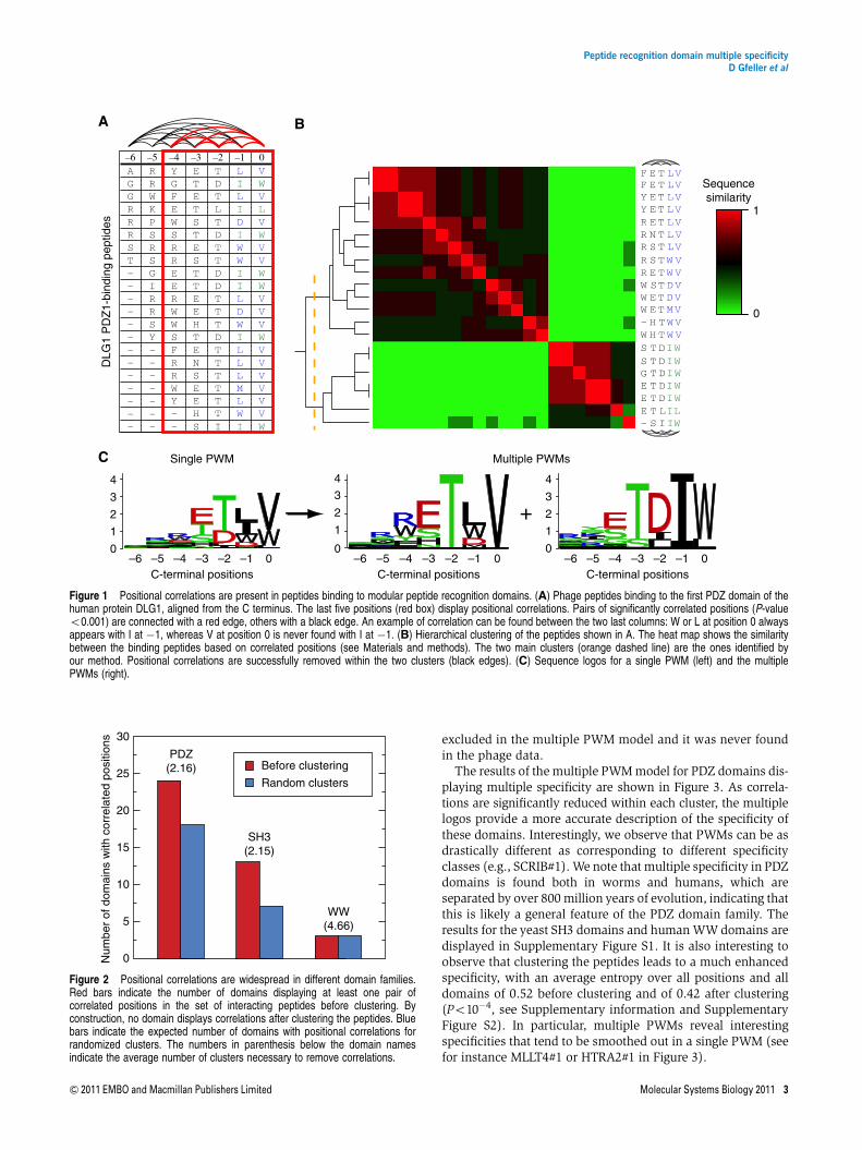
Positional correlations reflect the influence of an amino acid atone position in a set of interacting peptides over the amino acidpreferences at other positions. For instance, in peptides bind-ing to the first DLG1 PDZ domain, I and L are both observedseven times at position�1 (Figure 1A). However, Wat position0 is always found together with I at position�1 and never withL. To measure correlations among pairs of residue positions,we used mutual information. Taking a P-value threshold of0.001 to define significant correlations (see Materials andmethods), we observe that roughly a third of all tested
domains have at least one pair of significantly correlatedpositions in their specificity profile. Specifically, 24 out of 82PDZ domains, 13 out of 24 SH3 domains and the 3 WWdomains display positional correlations. For instance, peptidesinteracting with DLG1 PDZ1 display many correlated posi-tions, and significant P-values are observed among mostof the last five residues (red edges in Figure 1A). The profilesdisplaying strong positional correlations do not conformto the assumptions of positional independence of a singlePWM model and hence cannot be accurately modeled inthis way.
Positional correlations originate from multiplespecificity
To explore possible causes of these correlations and theirrelationship with the biophysical characteristics of proteininteractions, we first applied a clustering algorithm on each setof binding peptides using the percentage of sequence identityas a similarity measure (see Figure 1B and Materials andmethods). If correlations result from structural constraints,such as the presence of different binding modes, we expect tosee clusters of related peptides with much less positionalcorrelation within each cluster. Figure 1B shows how, in thecase of DLG1 PDZ1, two clusters are sufficient to removesignificant correlations for any position, suggesting twoclasses of specificity for this domain that can be accuratelymodeled with two PWMs (Figure 1C). Overall, we observe thata limited number of clusters (most often two or three, exceptfor some WW domains) are necessary to significantly removepositional correlations. Figure 2 summarizes the numberof domains with correlated positions before the clusteringprocedure (by construction, no domain displays correlatedpositions after clustering). The average number of clustersrequired to remove correlations is indicated in parenthesisfor each domain family. Randomly grouping the peptidesinto clusters of the same size, as the ones identified by ouralgorithm, clearly leaves several correlated positions (blue barin Figure 2, see Materials and methods), which highlights therelevance of the identified clusters. This observation led us tomodel the binding specificity of the domains with multiplePWMs rather than a single one. Toward this goal, we used themachine learning framework of mixture of PWMs (Bailey andElkan, 1994; Barash et al, 2003; Hannenhalli and Wang, 2005)that provides a general and computationally efficient wayof solving this problem (see Materials and methods andSupplementary information). The main idea of this approachis to fit K different PWMs to the aligned peptides, whereK is chosen here as the number of clusters found to removepositional correlations. The parameters of the multiple PWMs,as well as their weights, are directly learned from the datausing a maximum likelihood (ML) approach (Bailey andElkan, 1994; Bishop, 2006). Within this model, the specificityof each domain can be visualized as K different sequencelogos. For instance, Figure 1C shows both the single PWM andthe multiple PWM results for DLG1 PDZ1. Importantly, thisresult shows that predictions based on the single PWM can bemisleading; for instance, a peptide ending with ETIW appearsto match fairly well with a single PWM, whereas it is clearly
Peptide recognition domain multiple specificityD Gfeller et al
2 Molecular Systems Biology 2011 & 2011 EMBO and Macmillan Publishers Limited
excluded in the multiple PWM model and it was never foundin the phage data.
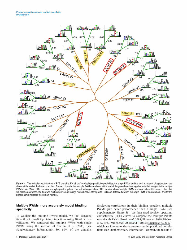
The results of the multiple PWM model for PDZ domains dis-playing multiple specificity are shown in Figure 3. As correla-tions are significantly reduced within each cluster, the multiplelogos provide a more accurate description of the specificity ofthese domains. Interestingly, we observe that PWMs can be asdrastically different as corresponding to different specificityclasses (e.g., SCRIB#1). We note that multiple specificity in PDZdomains is found both in worms and humans, which areseparated by over 800 million years of evolution, indicating thatthis is likely a general feature of the PDZ domain family. Theresults for the yeast SH3 domains and human WW domains aredisplayed in Supplementary Figure S1. It is also interesting toobserve that clustering the peptides leads to a much enhancedspecificity, with an average entropy over all positions and alldomains of 0.52 before clustering and of 0.42 after clustering(Po10�4, see Supplementary information and SupplementaryFigure S2). In particular, multiple PWMs reveal interestingspecificities that tend to be smoothed out in a single PWM (seefor instance MLLT4#1 or HTRA2#1 in Figure 3).
–6AGGRRRST–––––––––––––
–5RRWKPSRSGIRRSY–––––––
–4YGFEWSRREERWWSFRRWY––
–3ETETSTESTTEEHTENSEEHS
–2TDTLTDTTDDTTTDTTTTTTI
–1LILIDIWWIILDWILLLMLWI
0VWVLVWVVWWVVVWVVVVVVW S I IW
T LILT DIWT DIWT DIWT DIWT DIWH T W VH T W VE T MVE T D VST D VE T W VS T W VS T LVN T L VE T LVE T LVE T LV
–EEEGSSW–WWWRRRRRYYF E T LVF E T LV
1
0
Sequencesimilarity
A B
C
C-terminal positions
Single PWM Multiple PWMs
–6 –5 –4 –3 –2 –1 0–6 –5 –4 –3 –2 –1 00
1
2
3
4
0
1
2
3
4
0
1
2
3
4
–6 –5 –4 –3 –2 –1 0C-terminal positions C-terminal positions
DLG
1 P
DZ
1-bi
ndin
g pe
ptid
es
Figure 1 Positional correlations are present in peptides binding to modular peptide recognition domains. (A) Phage peptides binding to the first PDZ domain of thehuman protein DLG1, aligned from the C terminus. The last five positions (red box) display positional correlations. Pairs of significantly correlated positions (P-valueo0.001) are connected with a red edge, others with a black edge. An example of correlation can be found between the two last columns: W or L at position 0 alwaysappears with I at �1, whereas V at position 0 is never found with I at �1. (B) Hierarchical clustering of the peptides shown in A. The heat map shows the similaritybetween the binding peptides based on correlated positions (see Materials and methods). The two main clusters (orange dashed line) are the ones identified byour method. Positional correlations are successfully removed within the two clusters (black edges). (C) Sequence logos for a single PWM (left) and the multiplePWMs (right).
0
5
10
15
20
25
30
PDZ(2.16)
SH3(2.15)
WW(4.66)
Num
ber
of d
omai
ns w
ith c
orre
late
d po
sitio
ns
Before clustering
Random clusters
Figure 2 Positional correlations are widespread in different domain families.Red bars indicate the number of domains displaying at least one pair ofcorrelated positions in the set of interacting peptides before clustering. Byconstruction, no domain displays correlations after clustering the peptides. Bluebars indicate the expected number of domains with positional correlations forrandomized clusters. The numbers in parenthesis below the domain namesindicate the average number of clusters necessary to remove correlations.
Peptide recognition domain multiple specificityD Gfeller et al
& 2011 EMBO and Macmillan Publishers Limited Molecular Systems Biology 2011 3
Multiple PWMs more accurately model bindingspecificity
To validate the multiple PWMs model, we first assessedits ability to predict protein interactions using 10-fold cross-validation. We compared the multiple PWMs with singlePWMs using the method of Sharon et al (2008) (seeSupplementary information). For 80% of the domains
displaying correlations in their binding peptides, multiplePWMs give better performance than a single PWM (seeSupplementary Figure S3). We then used receiver operatingcharacteristic (ROC) curves to compare the multiple PWMsmodel with ANNs (Brusic et al, 1998; Blom et al, 1999; Nielsenet al, 1999; Miller et al, 2008) and HMMs (Noguchi et al, 2002),which are known to also accurately model positional correla-tions (see Supplementary information). Overall, the results of
Figure 3 The multiple specificity tree of PDZ domains. For all profiles displaying multiple specificities, the single PWMs and the total number of phage peptides areshown at the end of the brown branches. For each domain, the multiple PWMs are shown at the end of the green branches together with their weights in the multiplePWM model. Worm PDZ domains are highlighted in yellow. The red rectangles show PDZ domains whose multiple PWMs are most different from each other. Forvisualization purposes, the tree was built using average linkage hierarchical clustering with Euclidean distance between the single PWM of each domain. ‘#’ after theprotein name indicates the domain number.
Peptide recognition domain multiple specificityD Gfeller et al
4 Molecular Systems Biology 2011 & 2011 EMBO and Macmillan Publishers Limited
the different models are very good and quite similar, with anaverage area under ROC curve (AROC) of 0.98 for multiplePWMs and HMMs and 0.99 for ANNs (See SupplementaryTable S1 for the full list of AROC).
We then benchmarked the multiple PWM model on severalindependent data sets (see Supplementary information). Wefirst used a large interaction data set of 12 yeast SH3 domainsand 2 human PDZ domains generated by the SPOT technique,which provides a measure of affinity between domains andpeptides (Wiedemann et al, 2004; Tonikian et al, 2009). For allavailable domains, the correlation between the score of themultiple PWMs model and the SPOT signal is higher than thatwith a single PWM (see Supplementary Table S2). On average,multiple PWMs also give slightly better correlations thanHMMs, although the trend is not the same for all domains (asANNs are not probabilistic models, correlation values cannotbe directly compared). We carried out another validation usingyeast two-hybrid (Y2H) data (Tonikian et al, 2009). We againfound that better predictions are obtained using the multiplePWMs compared with the single PWMs, while performanceis similar with HMMs (see Supplementary Figure S4). In thiscase, ANNs did not perform as well (see Supplementaryinformation). We finally retrieved all experimentally deter-mined interactions from the PDZbase interaction database(Beuming et al, 2005) to build an independent benchmarkingdata set (see Supplementary Table S3). When tested on thisdata set, both multiple PWMs and HMMs give an averageAROC of 0.86, whereas ANNs give an average AROC of 0.85(see Supplementary Table S4 for the full list of AROC andP-values).
Taken together, the multiple PWMs outperform singlePWMs when used to predict domain–peptide interactions.Comparing with more complex machine learning frameworks,such as HMMs or ANNs, we observe similar performance,as expected, as different but mathematically related methodstrained on the same data, in general, provide similar results
(Nielsen et al, 1999). Having the added advantage of intuitiveinterpretation and visualization, multiple PWMs provide aparticularly suitable framework to study the specificity ofmodular peptide recognition domains.
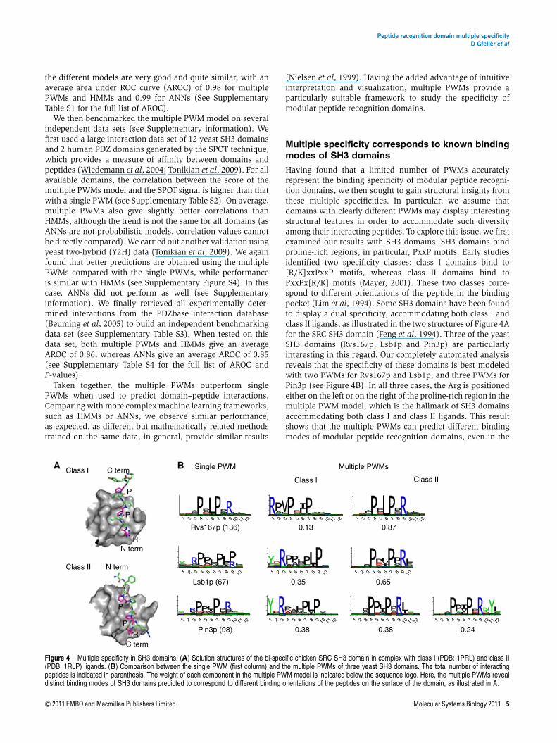
Multiple specificity corresponds to known bindingmodes of SH3 domains
Having found that a limited number of PWMs accuratelyrepresent the binding specificity of modular peptide recogni-tion domains, we then sought to gain structural insights fromthese multiple specificities. In particular, we assume thatdomains with clearly different PWMs may display interestingstructural features in order to accommodate such diversityamong their interacting peptides. To explore this issue, we firstexamined our results with SH3 domains. SH3 domains bindproline-rich regions, in particular, PxxP motifs. Early studiesidentified two specificity classes: class I domains bind to[R/K]xxPxxP motifs, whereas class II domains bind toPxxPx[R/K] motifs (Mayer, 2001). These two classes corre-spond to different orientations of the peptide in the bindingpocket (Lim et al, 1994). Some SH3 domains have been foundto display a dual specificity, accommodating both class I andclass II ligands, as illustrated in the two structures of Figure 4Afor the SRC SH3 domain (Feng et al, 1994). Three of the yeastSH3 domains (Rvs167p, Lsb1p and Pin3p) are particularlyinteresting in this regard. Our completely automated analysisreveals that the specificity of these domains is best modeledwith two PWMs for Rvs167p and Lsb1p, and three PWMs forPin3p (see Figure 4B). In all three cases, the Arg is positionedeither on the left or on the right of the proline-rich region in themultiple PWM model, which is the hallmark of SH3 domainsaccommodating both class I and class II ligands. This resultshows that the multiple PWMs can predict different bindingmodes of modular peptide recognition domains, even in the
Rvs167p (136)
Lsb1p (67)
Pin3p (98)
0.870.13
Class I Class II
0.35
0.38
0.65
0.38 0.24
Single PWM Multiple PWMs
1 12111098765432
1 12111098765432 1 12111098765432 1 12111098765432 1 12111098765432
1 1098765432 1 1098765432 1 1098765432
1 12111098765432 1 12111098765432
P
R
P
P
P
R
C term
N term
C term
N term
Class I
Class II
A B
Figure 4 Multiple specificity in SH3 domains. (A) Solution structures of the bi-specific chicken SRC SH3 domain in complex with class I (PDB: 1PRL) and class II(PDB: 1RLP) ligands. (B) Comparison between the single PWM (first column) and the multiple PWMs of three yeast SH3 domains. The total number of interactingpeptides is indicated in parenthesis. The weight of each component in the multiple PWM model is indicated below the sequence logo. Here, the multiple PWMs revealdistinct binding modes of SH3 domains predicted to correspond to different binding orientations of the peptides on the surface of the domain, as illustrated in A.
Peptide recognition domain multiple specificityD Gfeller et al
& 2011 EMBO and Macmillan Publishers Limited Molecular Systems Biology 2011 5
absence of crystal structures for a specific domain. For otherSH3 domains with correlated residues (see SupplementaryFigure S1), the interpretation of multiple specificity is not asclear as for the ones in Figure 4. Some of these cases maycorrespond to more detailed structural features of the mole-cular recognition events taking place at the SH3-binding site.
Multiple specificity predicts new binding modes ofPDZ domains
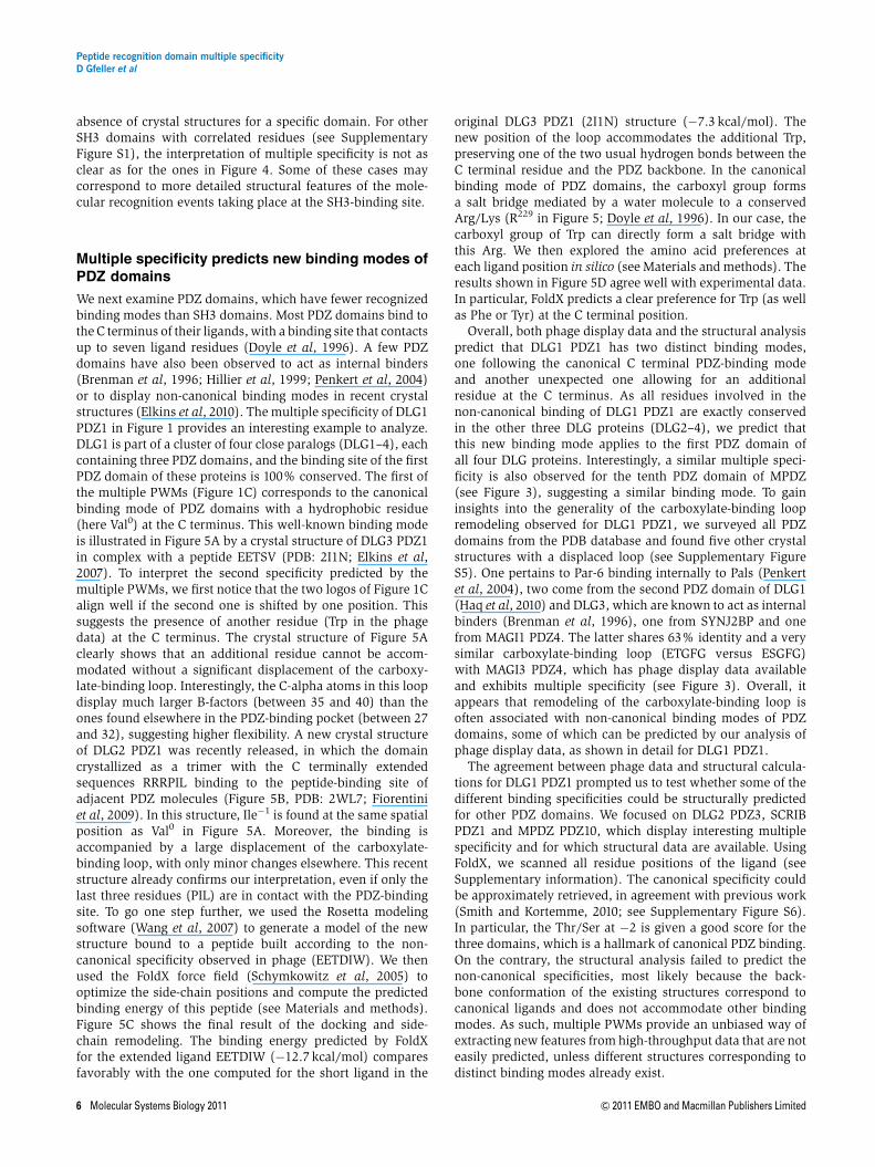
We next examine PDZ domains, which have fewer recognizedbinding modes than SH3 domains. Most PDZ domains bind tothe C terminus of their ligands, with a binding site that contactsup to seven ligand residues (Doyle et al, 1996). A few PDZdomains have also been observed to act as internal binders(Brenman et al, 1996; Hillier et al, 1999; Penkert et al, 2004)or to display non-canonical binding modes in recent crystalstructures (Elkins et al, 2010). The multiple specificity of DLG1PDZ1 in Figure 1 provides an interesting example to analyze.DLG1 is part of a cluster of four close paralogs (DLG1–4), eachcontaining three PDZ domains, and the binding site of the firstPDZ domain of these proteins is 100% conserved. The first ofthe multiple PWMs (Figure 1C) corresponds to the canonicalbinding mode of PDZ domains with a hydrophobic residue(here Val0) at the C terminus. This well-known binding modeis illustrated in Figure 5A by a crystal structure of DLG3 PDZ1in complex with a peptide EETSV (PDB: 2I1N; Elkins et al,2007). To interpret the second specificity predicted by themultiple PWMs, we first notice that the two logos of Figure 1Calign well if the second one is shifted by one position. Thissuggests the presence of another residue (Trp in the phagedata) at the C terminus. The crystal structure of Figure 5Aclearly shows that an additional residue cannot be accom-modated without a significant displacement of the carboxy-late-binding loop. Interestingly, the C-alpha atoms in this loopdisplay much larger B-factors (between 35 and 40) than theones found elsewhere in the PDZ-binding pocket (between 27and 32), suggesting higher flexibility. A new crystal structureof DLG2 PDZ1 was recently released, in which the domaincrystallized as a trimer with the C terminally extendedsequences RRRPIL binding to the peptide-binding site ofadjacent PDZ molecules (Figure 5B, PDB: 2WL7; Fiorentiniet al, 2009). In this structure, Ile�1 is found at the same spatialposition as Val0 in Figure 5A. Moreover, the binding isaccompanied by a large displacement of the carboxylate-binding loop, with only minor changes elsewhere. This recentstructure already confirms our interpretation, even if only thelast three residues (PIL) are in contact with the PDZ-bindingsite. To go one step further, we used the Rosetta modelingsoftware (Wang et al, 2007) to generate a model of the newstructure bound to a peptide built according to the non-canonical specificity observed in phage (EETDIW). We thenused the FoldX force field (Schymkowitz et al, 2005) tooptimize the side-chain positions and compute the predictedbinding energy of this peptide (see Materials and methods).Figure 5C shows the final result of the docking and side-chain remodeling. The binding energy predicted by FoldXfor the extended ligand EETDIW (�12.7 kcal/mol) comparesfavorably with the one computed for the short ligand in the
original DLG3 PDZ1 (2I1N) structure (�7.3 kcal/mol). Thenew position of the loop accommodates the additional Trp,preserving one of the two usual hydrogen bonds between theC terminal residue and the PDZ backbone. In the canonicalbinding mode of PDZ domains, the carboxyl group formsa salt bridge mediated by a water molecule to a conservedArg/Lys (R229 in Figure 5; Doyle et al, 1996). In our case, thecarboxyl group of Trp can directly form a salt bridge withthis Arg. We then explored the amino acid preferences ateach ligand position in silico (see Materials and methods). Theresults shown in Figure 5D agree well with experimental data.In particular, FoldX predicts a clear preference for Trp (as wellas Phe or Tyr) at the C terminal position.
Overall, both phage display data and the structural analysispredict that DLG1 PDZ1 has two distinct binding modes,one following the canonical C terminal PDZ-binding modeand another unexpected one allowing for an additionalresidue at the C terminus. As all residues involved in thenon-canonical binding of DLG1 PDZ1 are exactly conservedin the other three DLG proteins (DLG2–4), we predict thatthis new binding mode applies to the first PDZ domain ofall four DLG proteins. Interestingly, a similar multiple speci-ficity is also observed for the tenth PDZ domain of MPDZ(see Figure 3), suggesting a similar binding mode. To gaininsights into the generality of the carboxylate-binding loopremodeling observed for DLG1 PDZ1, we surveyed all PDZdomains from the PDB database and found five other crystalstructures with a displaced loop (see Supplementary FigureS5). One pertains to Par-6 binding internally to Pals (Penkertet al, 2004), two come from the second PDZ domain of DLG1(Haq et al, 2010) and DLG3, which are known to act as internalbinders (Brenman et al, 1996), one from SYNJ2BP and onefrom MAGI1 PDZ4. The latter shares 63% identity and a verysimilar carboxylate-binding loop (ETGFG versus ESGFG)with MAGI3 PDZ4, which has phage display data availableand exhibits multiple specificity (see Figure 3). Overall, itappears that remodeling of the carboxylate-binding loop isoften associated with non-canonical binding modes of PDZdomains, some of which can be predicted by our analysis ofphage display data, as shown in detail for DLG1 PDZ1.
The agreement between phage data and structural calcula-tions for DLG1 PDZ1 prompted us to test whether some of thedifferent binding specificities could be structurally predictedfor other PDZ domains. We focused on DLG2 PDZ3, SCRIBPDZ1 and MPDZ PDZ10, which display interesting multiplespecificity and for which structural data are available. UsingFoldX, we scanned all residue positions of the ligand (seeSupplementary information). The canonical specificity couldbe approximately retrieved, in agreement with previous work(Smith and Kortemme, 2010; see Supplementary Figure S6).In particular, the Thr/Ser at �2 is given a good score for thethree domains, which is a hallmark of canonical PDZ binding.On the contrary, the structural analysis failed to predict thenon-canonical specificities, most likely because the back-bone conformation of the existing structures correspond tocanonical ligands and does not accommodate other bindingmodes. As such, multiple PWMs provide an unbiased way ofextracting new features from high-throughput data that are noteasily predicted, unless different structures corresponding todistinct binding modes already exist.
Peptide recognition domain multiple specificityD Gfeller et al
6 Molecular Systems Biology 2011 & 2011 EMBO and Macmillan Publishers Limited

Protein interaction predictions
For human PDZ domains exhibiting multiple specificity, weused the multiple PWM model to scan the human proteomeand predict protein interactions. To test some of the predicted
interactions, we manually chose conservative thresholds onmultiple PWM scores, leading to a low false-positive rate. Theresulting network is displayed in Supplementary Figure S7.Out of 31 predicted interactions, 8 are known from previousstudies. To test whether some of the unknown interactions
G
F
G
LG
F
GL
G
F
G
L
W0
I–1
E–5
E–4
D–2
T–3
R229
Carboxylate-binding loop
β3
A
B
C
V0
S–1
T–2
E–3
E–4
R229
R229
I–1
P–2
L0
β2
α2
D
–5 –4 –3 –2 –1 0
Am
ino
acid
s
DecreasingΔΔG
IncreasingΔΔG
G
A
L
V
I
P
R
TS
C
M
K
E
Q
D
N
WY
F
H
C-terminal positions
Figure 5 Structural modeling of the predicted non-canonical binding mode of DLG1 PDZ1. (A) Crystal structure of DLG3 PDZ1 in complex with the ligand EETSV(PDB: 2I1N; Elkins et al, 2007). (B) Recent crystal structure of DLG2 PDZ1 (PDB: 2WL7; Fiorentini et al, 2009). The peptide PIL in the binding site corresponds to the Cterminus of the adjacent PDZ in the crystal structure. The carboxylate-binding loop displays a large conformational change on binding to this peptide. (C) Computationalmodeling of DLG1 PDZ1 in complex with an extended ligand EETDIW corresponding the new specificity observed in phage display-derived binding peptides. The orangecarboxylate-binding loop corresponds to the canonical binding mode of the PDZ domain in A and the green one corresponds to the new binding mode predicted by ourmethod and observed in the crystal structure shown in B. Dashed lines indicate important hydrogen bonds between the peptide and the domain. (D) Heat map viewof FoldX results for the amino acid preferences in the model of extended ligand binding to DLG1 PDZ1. The amino acids of the initial ligand EETDIW are marked withblack boxes.
Peptide recognition domain multiple specificityD Gfeller et al
& 2011 EMBO and Macmillan Publishers Limited Molecular Systems Biology 2011 7
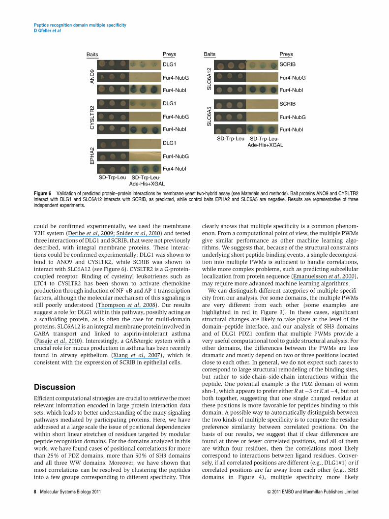
could be confirmed experimentally, we used the membraneY2H system (Deribe et al, 2009; Snider et al, 2010) and testedthree interactions of DLG1 and SCRIB, that were not previouslydescribed, with integral membrane proteins. These interac-tions could be confirmed experimentally: DLG1 was shown tobind to ANO9 and CYSLTR2, while SCRIB was shown tointeract with SLC6A12 (see Figure 6). CYSLTR2 is a G-protein-coupled receptor. Binding of cysteinyl leukotrienes such asLTC4 to CYSLTR2 has been shown to activate chemokineproduction through induction of NF-kB and AP-1 transcriptionfactors, although the molecular mechanism of this signaling isstill poorly understood (Thompson et al, 2008). Our resultssuggest a role for DLG1 within this pathway, possibly acting asa scaffolding protein, as is often the case for multi-domainproteins. SLC6A12 is an integral membrane protein involved inGABA transport and linked to aspirin-intolerant asthma(Pasaje et al, 2010). Interestingly, a GABAergic system with acrucial role for mucus production in asthma has been recentlyfound in airway epithelium (Xiang et al, 2007), which isconsistent with the expression of SCRIB in epithelial cells.
Discussion
Efficient computational strategies are crucial to retrieve the mostrelevant information encoded in large protein interaction datasets, which leads to better understanding of the many signalingpathways mediated by participating proteins. Here, we haveaddressed at a large scale the issue of positional dependencieswithin short linear stretches of residues targeted by modularpeptide recognition domains. For the domains analyzed in thiswork, we have found cases of positional correlations for morethan 25% of PDZ domains, more than 50% of SH3 domainsand all three WW domains. Moreover, we have shown thatmost correlations can be resolved by clustering the peptidesinto a few groups corresponding to different specificity. This
clearly shows that multiple specificity is a common phenom-enon. From a computational point of view, the multiple PWMsgive similar performance as other machine learning algo-rithms. We suggests that, because of the structural constraintsunderlying short peptide-binding events, a simple decomposi-tion into multiple PWMs is sufficient to handle correlations,while more complex problems, such as predicting subcellularlocalization from protein sequence (Emanuelsson et al, 2000),may require more advanced machine learning algorithms.
We can distinguish different categories of multiple specifi-city from our analysis. For some domains, the multiple PWMsare very different from each other (some examples arehighlighted in red in Figure 3). In these cases, significantstructural changes are likely to take place at the level of thedomain–peptide interface, and our analysis of SH3 domainsand of DLG1 PDZ1 confirm that multiple PWMs provide avery useful computational tool to guide structural analysis. Forother domains, the differences between the PWMs are lessdramatic and mostly depend on two or three positions locatedclose to each other. In general, we do not expect such cases tocorrespond to large structural remodeling of the binding sites,but rather to side-chain–side-chain interactions within thepeptide. One potential example is the PDZ domain of wormshn-1, which appears to prefer either R at�3 or K at�4, but notboth together, suggesting that one single charged residue atthese positions is more favorable for peptides binding to thisdomain. A possible way to automatically distinguish betweenthe two kinds of multiple specificity is to compute the residuepreference similarity between correlated positions. On thebasis of our results, we suggest that if clear differences arefound at three or fewer correlated positions, and all of themare within four residues, then the correlations most likelycorrespond to interactions between ligand residues. Conver-sely, if all correlated positions are different (e.g., DLG1#1) or ifcorrelated positions are far away from each other (e.g., SH3domains in Figure 4), multiple specificity more likely
AN
O9
CY
SLT
R2
SLC
6A12
SLC
6A5
EP
HA
2
Preys
DLG1
Fur4-NubG
Fur4-NubI
SD-Trp-Leu SD-Trp-Leu-Ade-His+XGAL
DLG1
Fur4-NubG
Fur4-NubI
DLG1
Fur4-NubG
Fur4-NubI
SCRIB
Fur4-NubG
Fur4-NubI
SCRIB
Fur4-NubG
Fur4-NubI
SD-Trp-Leu SD-Trp-Leu-Ade-His+XGAL
PreysBaits Baits
Figure 6 Validation of predicted protein–protein interactions by membrane yeast two-hybrid assay (see Materials and methods). Bait proteins ANO9 and CYSLTR2interact with DLG1 and SLC6A12 interacts with SCRIB, as predicted, while control baits EPHA2 and SLC6A5 are negative. Results are representative of threeindependent experiments.
Peptide recognition domain multiple specificityD Gfeller et al
8 Molecular Systems Biology 2011 & 2011 EMBO and Macmillan Publishers Limited
corresponds to distinct binding modes of the domain. Finally,not all domains appear to display positional correlations. Thismay be due to the availability of a limited number of bindingpeptides, which prevents us from observing slightly lessfavorable binding specificities, or some domains may beoptimized to accommodate only one specific kind of peptide.
An obvious question to ask is whether multiple specificitycan still be observed using other experimental data sets. Toobserve statistically significant correlated positions andautomatically detect multiple specificity, at least a dozeninteracting peptides are required (e.g., for two clusters of sixidentical peptides each, the mutual information P-value isE0.002), which partly explains why this feature has not oftenbeen observed in previous work. For instance, a recent study(Stiffler et al, 2007) used 217 peptides derived from Mousenatural C termini to probe the specificity of PDZ domains in aprotein-chip experiment. This screen yielded o10 interactingpeptides for most domains. Moreover, the set of initial peptidesis highly biased toward canonical PDZ-binding motifs. As aresult, we observed significant positional correlations foronly one domain in this dataset. However, with futuretechnological advances, this may change. High-throughputexperimental techniques such as phosphoproteomic methodsto study kinase specificity (Olsen et al, 2010) or ribosomedisplay (Hanes and Pluckthun, 1997), are increasinglybecoming available to generate large and unbiased sets ofinteracting peptides. Moreover, new sequencing technologiesare currently revolutionizing phage display experiments byallowing rapid sequencing of hundred of thousands of peptidesor proteins that have passed the selection runs (Ernst et al,2010; Fowler et al, 2010). Hence, such data are likely to becomeavailable in the near future for many natural domains, offeringnew opportunities to enhance both our biophysical under-standing of molecular recognition events and the accuracy ofcomputational protein interaction predictions. In the field ofprotein–DNA interactions, very large data sets are availableto map the specificity of transcription factors, and multiplespecificity has also been recently observed (Badis et al, 2009).It is likely that similar approaches, as the one presented inthis work, will yield new insights into modular peptide recog-nition domains or transcription factors studied with theseother experimental techniques. For other kinds of proteininteractions, such as those involving larger binding interfaceor non-peptide substrates, sequence-based approaches aremore difficult to apply. As such, the multiple PWM model isespecially suited for proteins interacting with small ligandsmade out of a limited number of building blocks (e.g., aminoacids or nucleotides) and adopting a few different bindingmodes on their targets.
At a system-wide level, the multiple specificity observed inmodular peptide recognition domains, such as PDZ, SH3 orWW, has several interesting consequences. First, it enablesadditional potential crosstalk in signaling pathways, wheredomains displaying multiple specificity could act as linkersbetween different pathways. Second, multiple specificityenables optimization of a domain to interact in a highlyspecific manner with a few very different ligands. Thisbinding-site plasticity yields interesting evolutionary advan-tages: it may provide a greater repertoire to build on thepathway topologies required to sustain cell activity using only
a limited number of components. In addition, it allows for theemergence of new specificities without necessarily altering theinitial one. As such, the evolution of specificity does notnecessarily need to follow a gradual process, but could ratherconsist in exploration of novel binding specificities that do notperturb the existing protein interactions and are retained onlywhen conferring an advantage to the organism. Such neutralevolutionary pathways are critical to enable innovation in asystem while preserving its robustness (Ciliberti et al, 2007),and our results suggest that multiple specificity may act as akey factor in this process.
Materials and methods
Phage display data
The experimental data sets used in this work come from large-scalephage display experiments (Tonikian et al, 2007). For PDZ domains, allphage display peptides found in Tonikian et al (2008) for wild-typedomains in humans and worms were used in our analysis. For eachdomain, the peptides were aligned from the C terminus. Owing to thepresence of STOP codons in the phage library, some peptides areshorter than seven amino acids (missing residues are labeled with X).For yeast SH3 domains, the phage peptides from Tonikian et al (2009)were automatically aligned with the MUSCLE alignment software(Edgar, 2004) using settings that prevented internal gaps. The new datafor the three WW domains come from a recent phage displayexperiment run with similar protocols as for the PDZ and SH3domains (see Supplementary Table S5 for the raw data). Because of theamplification step in phage display, the frequency of the peptidespulled out experimentally is difficult to interpret in terms of bindingstrength. For this reason, peptides retrieved multiple times weretreated as unique throughout our analysis.
Identifying correlated positions in peptidealignments
To identify correlated positions in peptide alignments, we used mutualinformation for all possible position pairs. Mutual information iscomputed as:
MI ¼X20
i¼1
X20
j¼1
Pði; jÞ logPði; jÞ
P1ðiÞP2ðjÞ
� �;
where P(i,j) stands for the probability of having amino acid iat one position together with amino acid j at the other positionin a peptide. P1(i) is the probability of having amino acid i atone position and P2(j) the probability of having amino acid j atthe other position. MI¼0 corresponds to independence of the twopositions, whereas larger values indicate that knowing which aminoacids are found at one position gives some information about whichones are expected at the other position. One limitation of mutualinformation is that non-zero values are often expected to be presentby chance, even for randomly generated peptides. We thereforeused the mutual information P-value as a filter (other statisticalmeasures such as the Z-scores could also be used). All P-values havebeen computed by randomly shuffling the amino acids within thealignment columns. A threshold of 0.001 has been used to definecorrelated positions.
Clustering domain-binding peptides
For each domain displaying correlated positions, the peptides wereclustered using the average linkage hierarchical clustering algorithmimplemented in R. The similarity measure between two peptides wascomputed as the ratio of identical amino acids, including onlypositions that are correlated with at least one other position. Althoughother measures of similarity, such as BLOSUM62 or biochemical
Peptide recognition domain multiple specificityD Gfeller et al
& 2011 EMBO and Macmillan Publishers Limited Molecular Systems Biology 2011 9
similarity, may be used, we did not observe any improvement whenusing them. The final clusters were defined by following the branchesof the dendrogram from its root and stopping whenever no morecorrelated positions are present within a cluster according to ourmutual information P-value cutoff (see Figure 1B). Although largerP-values are always expected for smaller peptide sets, the absenceof significant positional correlations within the clusters does notoriginate from this scaling effect, since random clusters of the samesize do not remove correlations, as shown in Figure 2. Clusterscontaining less than two peptides most often correspond to false-positives in phage and were filtered out in subsequent analyses.
Mixture of PWMs
A mixture of K PWMs is described by the model parameters ylik, which
correspond to the probability of having amino acid i and position laccording to the kth PWM, and the mixing coefficients pk quantifying
the weight of each PWM ðPK
k¼1 pk ¼ 1Þ (Bailey and Elkan, 1994;
Bishop, 2006). The score of a peptide X¼(x1yxL) of length L is thengiven by the equation:
PðXjy1; :::; yk;pÞ ¼XK
k¼1
pkPðXjykÞ ¼XK
k¼1
pkYLl¼1
yklxl
Given a data set of N interacting peptides, the different parameters ofthe mixture of PWMs are directly learned from the data using standardmaximum likelihood algorithms (see Supplementary information).Choosing the best value for K (i.e., number of PWMs) is a difficultmachine learning problem that does not have a general solution.In practice, one can either test different values and choose the mostmeaningful one or design heuristic strategies to estimate a reasonablevalue of K. Here, we chose K as the number of clusters found to removeall positional correlations. However, we stress that the mixture modelprovides a general framework that can be used with any other methodof choosing K. In particular, the method can also be used as a fastexploration tool by probing different values of K (i.e., different numberof PWMs) and manually identifying the most meaningful one, withoutperforming the initial clustering step.
Molecular modeling
The Rosetta software (Wang et al, 2007) was used to dock the ligandEETDIW to DLG1 PDZ1, using the recent PDB structure 2WL7 for thePDZ domain. Ligand position was optimized with Rosetta2.3 allowingfor backbone flexibility, and the highest scoring trajectory out of 100optimization runs was used in our model. Binding energies andresidue preferences in the peptides were analyzed with FoldX(Schymkowitz et al, 2005; see Supplementary information). Allstructures were visualized with Pymol (http://www.pymol.org).
Protein interaction predictions
The C-termini of all human proteins were scanned with the multiplePWM model and fairly stringent thresholds were used to generate thenetwork of Supplementary Figure S7. As DLG1 and DLG2 share480%sequence identity, the two proteins were merged in the network ofpredicted interactions. To experimentally test some of the predictedinteractions, we filtered away the ones already known from literatureand databases and the non-membrane proteins to comply with therequirements of the membrane Y2H system. We manually selected threeproteins (ANO9 and CYSLTR2 predicted to bind to DLG1 and SLC6A12predicted to bind to SCRIB) from the network in Supplementary Figure S7.All three proteins were tested with both DLG1 and SCRIB.
Experimental testing of protein interactions
Membrane Y2H constructsFull-length human ANO9, CYSLTR2 and SLC6A12 cDNAs, as well asthe controls EPHA2 and SLC6A5, were amplified by PCR andsubcloned by homologous recombination in yeast into bait vectors
pBT3-N and pTLB-1 (DualSystems Biotech) conferring the C terminalubiquitin (Cub) moiety and LexA-VP16 transcription factor at thebait N terminus (except for CYSLTR2 and EPHA2 in which theCub-LexA-VP16 tag was fused to the C terminus of the baitprotein). Similarly, full-length human DLG1 and SCRIB cDNA weresubcloned into prey vector pPR3N (DualSystems Biotech), whichconfers the N terminal ubiquitin (Nub) moiety to the N terminus of theprey protein.
Membrane Y2H assayYeast reporter strain THY.AP40 (MATa trp1 leu2 his3 LYS2::lexA-HIS3URA3::lexA-lacZ) was transformed with the indicated LexA-VP16-Cub-BAIT constructs by the lithium acetate protocol. Self-activation andmembrane localization were assessed by the Fur4-NubI/Fur4-NubGand Ost-NubI/Ost-NubG tests, as previously described (Deribe et al,2009; Snider et al, 2010). On passing the NubG/I test, pPR3N-DLG1/SCRIB preys were transformed into bait-containing yeast andtransformants were selected on SD-Trp-Leu. Three colonies for eachtransformation were spotted on selective media containing 5-bromo-4-chloro-3-indolyl-b-D-galactopyranoside (X-GAL), which turns blue inthe presence of b-galactosidase, indicating activation of the reportersystem. Figure 6 displays the results for these three independentexperiments for each interaction. The protein interactions from thispublication have been submitted to the IMEx (http://imex.sf.net)consortium through IntAct (Aranda et al, 2010) and assigned theidentifier IM-15347.
Supplementary information
Supplementary information is available at the Molecular SystemsBiology website (www.nature.com/msb).
AcknowledgementsDG acknowledges the financial support of the Swiss National ScienceFoundation (SNSF) and the European Molecular Biology Organization(EMBO). This work was supported by the Canadian Institutes ofHealth Research (Grant MOP-84324). The Stagljar lab is supported bygrants from the Canada Foundation for Innovation (CFI), the CanadianInstitutes of Health Research (CIHR), the Canadian Cancer SocietyResearch Institute (CCSRI), the Heart and Stroke Foundation, theCystic Fibrosis Foundation, the Ontario Genomics Institute andNovartis. The Kim lab is funded by grants from the Natural Sciencesand Engineering Research Council (NSERC), the Canada Foundationfor Innovation (CFI) and the Ontario Research Fund (ORF).
Author contributions: DG, GDB and PMK designed research; DG, FB,EV, PV, HH, ND performed research; DG, AE, LS, IS, SSS, GDB and PMKanalyzed data; DG, GDB and PMK wrote the paper.
Conflict of InterestThe authors declare that they have no conflict of interest.
References
Aranda B, Achuthan P, Alam-Faruque Y, Armean I, Bridge A, Derow C,Feuermann M, Ghanbarian AT, Kerrien S, Khadake J, KerssemakersJ, Leroy C, Menden M, Michaut M, Montecchi-Palazzi L, NeuhauserSN, Orchard S, Perreau V, Roechert B, van Eijk K et al (2010) TheIntAct molecular interaction database in 2010. Nucleic Acids Res 38:D525–D531
Badis G, Berger MF, Philippakis AA, Talukder S, Gehrke AR, Jaeger SA,Chan ET, Metzler G, Vedenko A, Chen X, Kuznetsov H, Wang CF,Coburn D, Newburger DE, Morris Q, Hughes TR, Bulyk ML (2009)Diversity and complexity in DNA recognition by transcriptionfactors. Science 324: 1720–1723
Peptide recognition domain multiple specificityD Gfeller et al
10 Molecular Systems Biology 2011 & 2011 EMBO and Macmillan Publishers Limited
Bailey TL, Elkan C (1994) Fitting a mixture model by expectationmaximization to discover motifs in biopolymers. Proc Int Conf IntellSyst Mol Biol 2: 28–36
Barash Y, Elidan G, Friedman N, Kaplan T (2003) Modelingdependencies in protein-DNA binding sites. In RECOMB ’03:Proceedings of the Seventh Annual International Conference onResearch In Computational Molecular Biology. Berlin, Germany:ACM
Beuming T, Skrabanek L, Niv MY, Mukherjee P, Weinstein H (2005)PDZBase: a protein-protein interaction database for PDZ-domains.Bioinformatics 21: 827–828
Bishop CM (2006) Pattern Recognition and Machine Learning, Vol. 1Singapore: Springer
Blom N, Gammeltoft S, Brunak S (1999) Sequence and structure-basedprediction of eukaryotic protein phosphorylation sites. J Mol Biol294: 1351–1362
Brenman JE, Chao DS, Gee SH, McGee AW, Craven SE, Santillano DR,Wu Z, Huang F, Xia H, Peters MF, Froehner SC, Bredt DS (1996)Interaction of nitric oxide synthase with the postsynaptic densityprotein PSD-95 and alpha1-syntrophin mediated by PDZ domains.Cell 84: 757–767
Brusic V, Rudy G, Honeyman G, Hammer J, Harrison L (1998)Prediction of MHC class II-binding peptides using an evolutionaryalgorithm and artificial neural network. Bioinformatics 14: 121–130
Chen JR, Chang BH, Allen JE, Stiffler MA, MacBeath G (2008)Predicting PDZ domain-peptide interactions from primarysequences. Nat Biotechnol 26: 1041–1045
Ciliberti S, Martin OC, Wagner A (2007) Innovation and robustness incomplex regulatory gene networks. Proc Natl Acad Sci USA 104:13591–13596
Deribe YL, Wild P, Chandrashaker A, Curak J, Schmidt MH,Kalaidzidis Y, Milutinovic N, Kratchmarova I, Buerkle L, FetchkoMJ, Schmidt P, Kittanakom S, Brown KR, Jurisica I, Blagoev B,Zerial M, Stagljar I, Dikic I (2009) Regulation of epidermal growthfactor receptor trafficking by lysine deacetylase HDAC6. Sci Signal2: ra84
Doyle DA, Lee A, Lewis J, Kim E, Sheng M, MacKinnon R (1996)Crystal structures of a complexed and peptide-free membraneprotein-binding domain: molecular basis of peptide recognitionby PDZ. Cell 85: 1067–1076
Edgar RC (2004) MUSCLE: multiple sequence alignment with highaccuracy and high throughput. Nucleic Acids Res 32: 1792–1797
Elkins JM, Gileadi C, Shrestha L, Phillips C, Wang J, Muniz JR,Doyle DA (2010) Unusual binding interactions in PDZ domaincrystal structures help explain binding mechanisms. Protein Sci 19:731–741
Elkins JM, Papagrigoriou E, Berridge G, Yang X, Phillips C, Gileadi C,Savitsky P, Doyle DA (2007) Structure of PICK1 and other PDZdomains obtained with the help of self-binding C-terminalextensions. Protein Sci 16: 683–694
Emanuelsson O, Nielsen H, Brunak S, von Heijne G (2000) Predictingsubcellular localization of proteins based on their N-terminalamino acid sequence. J Mol Biol 300: 1005–1016
Ernst A, Gfeller D, Kan Z, Seshagiri S, Kim PM, Bader GD, Sidhu SS(2010) Coevolution of PDZ domain-ligand interactions analyzed byhigh-throughput phage display and deep sequencing. Mol Biosyst 6:1782–1790
Feng S, Chen JK, Yu H, Simon JA, Schreiber SL (1994) Two bindingorientations for peptides to the Src SH3 domain: development of ageneral model for SH3-ligand interactions. Science 18: 1241–1247
Fiorentini M, Nielsen AK, Kristensen O, Kastrup JS, Gajhede M (2009)Structure of the first PDZ domain of human PSD-93. ActaCrystallogr Sect F Struct Biol Cryst Commun 65: 1254–1257
Fowler DM, Araya CL, Fleishman SJ, Kellogg EH, Stephany JJ, Baker D,Fields S (2010) High-resolution mapping of protein sequence-function relationships. Nat Methods 7: 741–746
Gould CM, Diella F, Via A, Puntervoll P, Gemund C, Chabanis-Davidson S, Michael S, Sayadi A, Bryne JC, Chica C, Seiler M,Davey NE, Haslam N, Weatheritt RJ, Budd A, Hughes T, Pas J,
Rychlewski L, Trave G, Aasland R et al (2010) ELM: the status ofthe 2010 eukaryotic linear motif resource. Nucleic Acids Res 38:D167–D180
Hanes J, Pluckthun A (1997) In vitro selection and evolution offunctional proteins by using ribosome display. Proc Natl Acad SciUSA 94: 4937–4942
Hannenhalli S, Wang LS (2005) Enhanced position weightmatrices using mixture models. Bioinformatics 21(Suppl 1):i204–i212
Haq SR, Jurgens MC, Chi CN, Koh CS, Elfstrom L, Selmer M,Gianni S, Jemth P (2010) The plastic energy landscape of proteinfolding: a triangular folding mechanism with an equili-brium intermediate for a small protein domain. J Biol Chem 285:18051–18059
Harris BZ, Lim WA (2001) Mechanism and role of PDZ domains insignaling complex assembly. J Cell Sci 114: 3219–3231
Hillier BJ, Christopherson KS, Prehoda KE, Bredt DS, Lim WA (1999)Unexpected modes of PDZ domain scaffolding revealed bystructure of nNOS-syntrophin complex. Science 284: 812–815
Hu H, Columbus J, Zhang Y, Wu D, Lian L, Yang S, Goodwin J, LuczakC, Carter M, Chen L, James M, Davis R, Sudol M, Rodwell J, HerreroJJ (2004) A map of WW domain family interactions. Proteomics 4:643–655
Hui S, Bader GD (2010) Proteome scanning to predict PDZ domaininteractions using support vector machines. BMC Bioinformatics11: 507
Hutti JE, Jarrell ET, Chang JD, Abbott DW, Storz P, Toker A, Cantley LC,Turk BE (2004) A rapid method for determining protein kinasephosphorylation specificity. Nat Methods 1: 27–29
Lim WA, Richards FM, Fox RO (1994) Structural determinants ofpeptide-binding orientation and of sequence specificity in SH3domains. Nature 372: 375–379
Mayer BJ (2001) SH3 domains: complexity in moderation. J Cell Sci114: 1253–1263
Miller ML, Jensen LJ, Diella F, Jorgensen C, Tinti M, Li L, Hsiung M,Parker SA, Bordeaux J, Sicheritz-Ponten T, Olhovsky M, PasculescuA, Alexander J, Knapp S, Blom N, Bork P, Li S, Cesareni G, PawsonT, Turk BE et al (2008) Linear motif atlas for phosphorylation-dependent signaling. Sci Signal 1: ra2
Nielsen H, Brunak S, von Heijne G (1999) Machine learningapproaches for the prediction of signal peptides and other proteinsorting signals. Protein Eng 12: 3–9
Noguchi H, Kato R, Hanai T, Matsubara Y, Honda H, Brusic V,Kobayashi T (2002) Hidden Markov model-based prediction ofantigenic peptides that interact with MHC class II molecules.J Biosci Bioeng 94: 264–270
Obenauer JC, Cantley LC, Yaffe MB (2003) Scansite 2.0: proteome-wideprediction of cell signaling interactions using short sequencemotifs. Nucleic Acids Res 31: 3635–3641
Olsen JV, Vermeulen M, Santamaria A, Kumar C, Miller ML, Jensen LJ,Gnad F, Cox J, Jensen TS, Nigg EA, Brunak S, Mann M (2010)Quantitative phosphoproteomics reveals widespread full phos-phorylation site occupancy during mitosis. Sci Signal 3: ra3
Pasaje CF, Kim JH, Park BL, Cheong HS, Chun JY, Park TJ, Lee JS,Kim Y, Bae JS, Park JS, Yoon SH, Uh ST, Choi JS, Kim YH, Kim MK,Choi IS, Cho SH, Choi BW, Park CS, Shin HD (2010) Association ofSLC6A12 variants with aspirin-intolerant asthma in a Koreanpopulation. Ann Hum Genet 74: 326–334
Pawson T, Nash P (2003) Assembly of cell regulatory systems throughprotein interaction domains. Science 300: 445–452
Penkert RR, DiVittorio HM, Prehoda KE (2004) Internal recognitionthrough PDZ domain plasticity in the Par-6-Pals1 complex. NatStruct Mol Biol 11: 1122–1127
Schymkowitz J, Borg J, Stricher F, Nys R, Rousseau F, Serrano L (2005)The FoldX web server: an online force field. Nucleic Acids Res 33:W382–W388
Shao X, Tan CS, Voss C, Li SS, Deng N, Bader GD (2010) A regressionframework incorporating quantitative and negative inter-action data improves quantitative prediction of PDZ domain-
Peptide recognition domain multiple specificityD Gfeller et al
& 2011 EMBO and Macmillan Publishers Limited Molecular Systems Biology 2011 11
peptide interaction from primary sequence. Bioinformatics 27:383–390
Sharon E, Lubliner S, Segal E (2008) A feature-based approach tomodeling protein-DNA interactions. PLoS Comput Biol 4: e1000154
Smith CA, Kortemme T (2010) Structure-based prediction of thepeptide sequence space recognized by natural and synthetic PDZdomains. J Mol Biol 402: 460–474
Snider J, Kittanakom S, Damjanovic D, Curak J, Wong V, Stagljar I(2010) Detecting interactions with membrane proteins using amembrane two-hybrid assay in yeast. Nat Protoc 5: 1281–1293
Stiffler MA, Chen JR, Grantcharova VP, Lei Y, Fuchs D, Allen JE,Zaslavskaia LA, MacBeath G (2007) PDZ domain binding selec-tivity is optimized across the mouse proteome. Science 317:364–369
Thompson C, Cloutier A, Bosse Y, Poisson C, Larivee P, McDonald PP,Stankova J, Rola-Pleszczynski M (2008) Signaling by the cysteinyl-leukotriene receptor 2. Involvement in chemokine gene transcrip-tion. J Biol Chem 283: 1974–1984
Tong AH, Drees B, Nardelli G, Bader GD, Brannetti B, Castagnoli L,Evangelista M, Ferracuti S, Nelson B, Paoluzi S, Quondam M,Zucconi A, Hogue CW, Fields S, Boone C, Cesareni G (2002) Acombined experimental and computational strategy to defineprotein interaction networks for peptide recognition modules.Science 295: 321–324
Tonikian R, Xin X, Toret CP, Gfeller D, Landgraf C, Panni S, Paoluzi S,Castagnoli L, Currell B, Seshagiri S, Yu H, Winsor B, Vidal M,Gerstein MB, Bader GD, Volkmer R, Cesareni G, Drubin DG,
Kim PM, Sidhu SS et al (2009) Bayesian modeling of the yeastSH3 domain interactome predicts spatiotemporal dynamics ofendocytosis proteins. PLoS Biol 7: e1000218
Tonikian R, Zhang Y, Boone C, Sidhu SS (2007) Identifying specificityprofiles for peptide recognition modules from phage-displayedpeptide libraries. Nat Protoc 2: 1368–1386
Tonikian R, Zhang Y, Sazinsky SL, Currell B, Yeh JH, Reva B, Held HA,Appleton BA, Evangelista M, Wu Y, Xin X, Chan AC, Seshagiri S,Lasky LA, Sander C, Boone C, Bader GD, Sidhu SS (2008) Aspecificity map for the PDZ domain family. PLoS Biol 6: e239
Wang C, Bradley P, Baker D (2007) Protein-protein docking withbackbone flexibility. J Mol Biol 373: 503–519
Wiedemann U, Boisguerin P, Leben R, Leitner D, Krause G, Moelling K,Volkmer-Engert R, Oschkinat H (2004) Quantification of PDZdomain specificity, prediction of ligand affinity and rational designof super-binding peptides. J Mol Biol 343: 703–718
Xiang YY, Wang S, Liu M, Hirota JA, Li J, Ju W, Fan Y, Kelly MM, Ye B,Orser B, O0Byrne PM, Inman MD, Yang X, Lu WY (2007) AGABAergic system in airway epithelium is essential for mucusoverproduction in asthma. Nat Med 13: 862–867
Molecular Systems Biology is an open-access journalpublished by European Molecular Biology Organiza-
tion and Nature Publishing Group. This work is licensed under aCreative Commons Attribution-Noncommercial-Share Alike 3.0Unported License.
Peptide recognition domain multiple specificityD Gfeller et al
12 Molecular Systems Biology 2011 & 2011 EMBO and Macmillan Publishers Limited
Copyright © 2022 FDOKUMEN