Contrast variation studies of clathrin coated vesicles by small-angle neutron scattering

Copyright C Blackwell Munksgaard 2002Traffic 2002; 3: 590–600
Blackwell Munksgaard ISSN 1398-9219
The m2 Subunit of the Clathrin Adaptor AP-2 Binds toFDNPVY and YppØ Sorting Signals at Distinct Sites
Werner Boll1,†, Iris Rapoport1,†,Christian Brunner1, Yorgo Modis2,Siegfried Prehn3 and Tomas Kirchhausen1,*
1 Department of Cell Biology and The Center for Blood
Research, Harvard Medical School, 200 Longwood Ave,
Boston, MA, 02115, USA2 Howard Hughes Medical Institute and Children’s
Hospital, Laboratory of Molecular Medicine Boston, MA
02115, USA3 Institut für Biochemie der Humboldt-Universität Berlin,
Hessische Str. 3–4, 10115 Berlin, Germany
* Corresponding author: Tomas Kirchhausen,
The endocytic sorting signal on the low-density lipo-protein receptor for clathrin-mediated internalization isthe sequence FDNPVY in the receptor’s cytosolic tail.We have used a combination of surface plasmon reson-ance and crosslinking with a photoactivated peptideprobe to demonstrate the interaction betweenFDNPVY-containing peptides and the m2 chain of puri-fied AP-2 clathrin adaptors (the complexes responsiblefor plasma membrane sorting). We show that recog-nition of the FDNPVY signal is mediated by a bindingsite in the m2-subunit that is distinct from the site forthe more general YppØ sorting signal, another tyro-sine-based sequence also recognized by m2-adaptin.These results suggest the possibility that low-densitylipoprotein receptor uptake may be modulatedspecifically and independently of other proteins in theclathrin pathway.
Key words: Alzheimer’s, AP-2 adaptor, clathrin, endo-cytosis, membrane traffic
Received 23 April 2002, revised and accepted for pub-lication 10 May 2002
Low-density lipoproteins (LDL) are a major carrier of chol-esterol in mammals. Accumulation of LDLs in blood vesselscauses the formation of atherosclerotic plaques, which canlead to heart failure or to stroke. LDL levels are regulated,in part, by receptor-mediated endocytosis; LDL particles arebound by the LDL receptor, actively internalized into cells,and transported to the lysosome for destruction (1). Since thepioneering work of Brown and Goldstein, it has been knownthat the LDL receptor enters by the clathrin-dependent path-way (2). This form of receptor-mediated endocytosis requires
† These authors contributed equally.
590
a short peptide sequence in the cytosolic tail of the receptor.It was originally proposed that this endocytic motif had thegeneral form NPXY (N denotes asparagine, P is proline, Xrefers to any amino acid, and Y is tyrosine) because otherproteins, known to be internalized through the clathrin path-way, including the Alzheimer’s b-amyloid precursor protein(b-APP), also have elements of this motif in their cytosolictails (3). Mutagenesis studies with the LDL receptor havesubsequently shown, however, that the hexapeptide,FDNPVY, is the sequence element recognized for its efficientendocytosis (4). Further studies have also demonstrated theexistence of a more widely used tyrosine-based sorting motif,of the form YppØ (Y denotes tyrosine, p is a polar residue,and Ø refers to amino acids with a large hydrophobic sidechain) [for recent review see (5)].
A large number of membrane proteins that traffic throughthe clathrin pathway are sorted into coated pits by clathrinadaptors, a group of proteins that recognize signals in thecytosolic portion of membrane proteins and also interact withclathrin [for recent reviews, see (5–7)]. The endocytic ad-aptor AP-2 is a heterotetrameric complex composed of a, b2,m2 and s2 subunits. The m2-adaptin recognizes tyrosine-based sorting signals of the form YppØ (8–11); the b2-adaptin contains binding sites for a second sorting signal, thedileucine-based motif, and also for clathrin (4,12–19). It wasinitially proposed that AP-2 would recognize the FDNPVY se-quence through the same interaction used for the canonicalYppØ motif (20). Unrecognized at the time, however, thisgroup used a recombinant tail that included a dileucine motifto show association with AP-2. Subsequent biochemicalstudies showed that peptides bearing the FDNPVY motif ofthe LDL receptor do not compete with YppØ-containing pep-tides for binding to m2 (9). Indeed, in cells overexpressing thetransferrin receptor, which carries a canonical YppØ endocyt-ic signal (YTRF), there is a decrease in the rate of internaliz-ation of other YppØ-containing proteins, but no inhibition ofLDL receptor internalization (21). Thus, the endocytic sortingmotif found in the cytosolic tail of the LDL receptor, FDNPVY,is distinct from the more common YppØ motif and is uniqueto members of the LDL receptor family (4,18,19). More re-cently, from results obtained using nuclear magnetic reson-ance, it was proposed that sorting of the LDL receptor intocoated pits does not require association with clathrin ad-aptors but instead results from a direct contact of the hexa-peptide FDNPVY on the LDL receptor with the N-terminaldomain of the clathrin heavy chain (22). Since clathrin iscommon to the endocytic and exocytic pathways, however,this suggestion would not explain the fact that the FDNPVYmotif in the LDL receptor is not used for sorting by clathrin-

AP-2 Binds to the LDL FDNPVY Sorting Signal
Figure1: Interaction between AP-2 adaptors and the internalization motif FDNPVY of the LDL receptor. (A) Schematic represen-tation of the synthetic peptides used for SPR and UV-crosslinking experiments containing FDNPVY, YppØ and LL sorting signals used in thestudy. The indicated motifs were defined in previous studies by analyzing the effect of point mutations on the traffic of the LDL receptor(LDLR) (5,20) and TGN38 (37,38) transmembrane proteins, and on Nef (45,47), a myristoylated protein encoded by the HIV-1 retrovirus.The sequences represent portions of the corresponding cytoplasmic portions of these proteins that surround these motifs. The solubility ofthe LDL-*FDNPVY peptide increased considerably by inclusion of benzoylphenylalanine (bpa) at serine 799, upstream of the FDNPVY motif,and allowed its use as a competitor for specificity determination in the crosslinking experiments. TGN38-YQRL (23) contains arginine insteadof lysine residues at positions π4 and π8, thus minimizing chemical crosslinking of internal portions of the peptide with the chip. (B)Real-time surface plasmon resonance (SPR) analysis of the binding of clathrin coat components to a sensor chip containing immobilizedpeptidesbearing the FDNPVY endocytic motif of the LDL receptor (LDLR-FDNPVY). Samples containing 200nM AP-1, AP-2, clathrin, or arecombinant fragment corresponding to the N-terminal domain of clathrin (TD40) were tested for their ability to bind to the surface of thechip. The chip contained 500 resonance units (RU) of the immobilized 18-mer LDLR-FDNPVY. The protein samples were injected at 20ml/min for 120s followed by a 120-s washout period; in order to compare the tracings from proteins of different sizes to each other, they werenormalized to the molecular weight of AP-2. (C) Effect of varying the sequence of the LDL receptor endocytic motif on its ability to bind AP-2 adaptors. 200nM AP-2 (the ligand) were injected at 20 ml/min for 120s and used to evaluate its binding capacity to different chipscontaining the peptides LDLR-FDNPVY (490 RU immobilized), LDLR-ADNPVY (560 RU immobilized), or LDLR-FDNPVA (470 RU immobil-ized).
591Traffic 2002; 3: 590–600
Boll et al.
based traffic at the trans-Golgi network (in which sorting ofproteins containing certain YppØ and dileucine-signals is me-diated by the exocytic clathrin adaptor AP-1). Moreover, theFDNPVY motif in all members of the LDL receptor family issituated close to the membrane, usually 12 amino acids fromthe transmembrane domain (4). A 12-residue segment couldspan no more that 30–40Å, even if completely extended,while the N-terminal domain of clathrin within the coat liesout of reach, about 100Å from the membrane (23,24). Thus,the geometry of the proposed interaction seems unlikely.
We therefore decided to reinvestigate whether a peptide cor-responding to part of the cytosolic tail of the LDL receptorand containing the FDNPVY motif could interact with eitherAP-2 or clathrin. We used two independent methods: onebased on surface plasmon resonance (SPR) spectroscopy(8,9,25–29) and the other on photoactivated crosslinkingtechniques (9,15,30) that have been used successfully to de-tect relatively weak associations between sorting signals andadaptors. In particular, SPR is suitable for detecting short-lived interactions that are hard to see with more conventional‘pull-down’ or chromatographic methods. We demonstratehere that AP-2 interacts through the m2 subunit with pep-tides, derived from the cytosolic tail of the LDL receptor, thatcontain the FDNPVY endocytic sorting signal. This interactioninvolves a site in m2-adaptin distinct from the one that recog-nizes the more common YppØ tyrosine-based sorting signal.
Results
The clathrin adaptor AP-2 interacts with the FDNPVY
sorting signal
We immobilized on a SPR chip an 18-residue peptide (LDLR-FDNPVY) containing the FDNPVY endocytic motif of the LDLreceptor, and asked whether we could detect an interactionwith isolated protein components from coated vesicles. Thesequence of the LDLR-FDNPVY peptide (Figure 1A) corre-sponds to residues 6–23 of the LDL receptor cytoplasmictail, that includes the sequence required to support endo-cytosis of the LDL receptor (4). The SPR tracings in Figure1(B) indicate that AP-2 binds specifically to the LDLR-FDNPVY chip, whereas equal amounts of AP-1 bind substan-tially less. In contrast, the LDLR-FDNPVY peptide barelybinds clathrin purified from coated vesicles, and it does notbind the clathrin N-terminal domain [TD40, a fragmentknown to be correctly folded in a b-propeller conformation(17,31)] or bovine serum albumin (used as a negative control),even at concentrations of up to 5mM (not shown). To examinethe specificity of the interaction between the LDL receptorpeptide and AP-2, we used two modified LDL receptor pep-tides in which single amino acids in the FDNPVY motif werereplaced by alanine (Figure 1A). It is known that LDL receptormutants containing FDNPVA or ADNPVY, instead ofFDNPVY, have lower rates of receptor-mediated endocytosisand significantly impaired lysosomal degradation of the inter-nalized LDL particles (9% or 37% of the control value, re-
592 Traffic 2002; 3: 590–600
spectively) (4). Using chips with the altered sequences alsocaused a significant reduction in AP-2 binding (Figure 1C).We therefore believe we can rule out the proposed role ofclathrin N-terminal domain as a significant element in theendocytic machinery responsible for LDL receptor sorting byrecognition of the FDNPVY motif (22). Attempts to immobil-ize peptides with the sequence FDNAVY or FDAPVY corre-sponding to other LDL mutants also impaired in endocytosis(4) were not successful because of their tendency to aggre-gate.
The binding site for the FDNPVY sorting signal is
located in the AP-2 core
The binding sites in clathrin adaptors that recognize the YppØor the dileucine sorting signals are located within the ‘AP-core’ (8,15), the part of the complex that remains after enzy-matic proteolysis consisting of the N-terminal fragments of a
and b2 and the intact m2 and s2 subunits (32,33). To testwhether the AP-2 core is also involved in the recognition ofthe FDNPVY motif, we subjected clathrin adaptors to limitedtrypsin digestion (Figure 2A) and asked whether AP-corescan still recognize the LDL receptor motif. As shown by theSPR tracing in Figure 2(B), AP-2 cores bind to the LDLR-FDNPVY chip, to about the same extent as intact AP-2s(compare the amplitude in the SPR tracings in Figure 1B,Cwith Figure 2B). As expected, the association of AP-2 coresto the LDLR-FDNPVA or LDLR-ADNPVY chips was signifi-cantly reduced (Figure 2B), confirming the specificity of theinteraction.
The m2 subunit of AP-2 interacts with the FDNPVY
sorting signal
Because m2-adaptin binds directly to peptides bearing YppØsorting signals, and because both the YppØ and the FDNPVYsignals are tyrosine based, we asked whether the free m2subunit of AP-2 could also interact with the immobilizedLDLR-FDNPVY peptide. Two constructs of m2 were used forSPR experiments, His-Dm2 (hexa histidine-tag fused to resi-dues 122–435 of rat brain m2 (34)) and His-DDm2 (hexa histi-dine-tag fused to residues 158–435). His-Dm2 and His-DDm2contain the binding site for YppØ (8), fold correctly and haveatomic structures that are essentially identical as determinedby X-ray crystallography (11,35).
Both constructs, His-Dm2 and His-DDm2, bind to TGN38-YQRL, a 16-residue peptide corresponding to the well-studied endocytic motif at the C-terminus of the trans-GolgiTGN38 protein (36,37), as expected from published data(8,9,11,38) and from data shown here (Figure 3A). Likewise,each one of these fragments bound equally well to the LDLR-FDNPVY chip, although with a lower binding capacity thanthe TGN38-YQRL chip (Figure 3B). The calculated associ-ation constant (ka) for the interaction of His-Dm2 with theLDLR-FDNPVY peptide was 7¿105 (Mª1 ¿sª1) and the dis-sociation constant (kDa) was 1.2¿10ª2 (sª1). Likewise, forAP-2 the association and dissociation constants were

AP-2 Binds to the LDL FDNPVY Sorting Signal
Figure2: AP-2 cores bind to the LDLR-FDNPVY peptide. (A)Generation of AP-2 cores. The protein bands of AP-2 before (lane1) or after (lane 2) limited digestion with trypsin were visualizedby 10% SDS-PAGE fractionation and Coomassie Blue staining. Thebands for intact a, b1 and m2-adaptins in AP-2 and the remainingN-terminal (trunk) domains of a and b together with the m2 con-tained within the AP-2 core are shown. (B) Binding of AP-2 coresto the LDLR-FDNPVY peptide is specific. The SPR tracings corre-spond to the recruitment of 200nM AP-2 cores to three differentchips containing either wild-type peptide LDLR-FDNPVY or mutantpeptides LDLR-ADNPVY and LDLR-FDNPVA.
7.4¿103 (Mª1 ¿sª1) and 2¿10ª3 (sª1). Due to the poorsolubility of the LDL-based peptides, we could not reach suf-ficient concentrations to include them as competitors duringthe dissociation phase, and thus prevent rebinding of the re-leased proteins back to the chip. Therefore, these valuesshould only be considered as approximate, since the meas-
593Traffic 2002; 3: 590–600
ured dissociation constants are likely to be significantly lowerthan the real values. His-DDm2 also binds to the LDL-FDNPVYchip, suggesting that the m2 binding site for the LDLR-FDNPVY peptide lies in the 158–435 fragment and not in thesegment between residues 122 and 157 that is disordered inthe X-ray crystal structure of His-Dm2 and AP-2 cores (11,39).Further confirmation that m2 is the subunit of AP-2 respon-sible for selective recognition of the FDNPVY motif is pre-sented in Figure 3(C). It shows that, like intact or cleaved AP-2, the His-Dm2 construct binds less efficiently to chips withthe LDLR-ADNPVY and LDLR-FDNPVA peptides.
As an alternative to the SPR experiments, we used an inde-pendent approach involving peptides bearing a photo-activ-atable crosslinker to verify that the m2 subunit interacts withthe LDL-based peptide in a specific way. This method wassuccessfully used before to investigate the regulation in-volved in the recognition of YppØ sorting signals by m2 inclathrin adaptors and to establish that their b subunits recog-nize dileucine-based sorting signals (15,30). The results pre-sented in Figure 4(A) (lanes 1–3) are completely consistentwith the SPR data. His-Dm2 crosslinked with the LDL-*FDNPVY photo-reactive peptide (lane 1), and formation ofthe crosslinked species was prevented by the nonbiotinylatedform of the LDL-probe (lane 2). As a control for specificity,we used excess amounts of the TGN38-YQRL peptide bear-ing the canonical YppØ motif and found that it does not re-duce formation of the crosslinked product with LDL-*FDNPVY (lane 3); the same concentration of TGN38-YQRL,however, completely blocked the generation of crosslinkedspecies of His-Dm2 and the photo-reactive peptide TGN38-*YQRL (lane 7). Use of AP-2 with LDL-*FDNPVY led to thegeneration of a major crosslinked species involving m2, to-gether with a weaker labeling of a and b-adaptin, whosestaining intensity varied between experiments.
The binding sites in m2 for FDNPVY and YppØ are not
the same
One indication that m2 has two distinct binding sites, one forthe FDNPVY motif and the other for the YppØ motif, comesfrom the observation that soluble TGN38-YQRL peptide iseffective in blocking the recruitment of His-Dm2 to theTGN38-YQRL chip (Figure 4B), whereas it has no effect onthe recruitment of His-Dm2 to the LDL-FDNPVY chip (Figure4C). A second line of evidence comes from using a mutatedform of m2 that cannot interact with peptides bearing YppØmotifs (11,40). Peptides bearing the endocytic motifs YQRL(from TGN38) or YRAL (from the epidermal growth factorreceptor) bind to m2 in a site with contributions from residuesalong the b-1 and b-16 strands in subdomain A of m2 (11). Akey residue in m2 required to support this interaction is W421,which forms hydrophobic contacts with the arginine at Yπ2of the YppØ motif, leading to the preferential association withm2. Indeed, cells expressing m2 in which W421 has beenchanged to alanine assemble AP-2 adaptors carrying the mu-tated m2 subunit but are deficient in receptor-mediated endo-cytosis of transferrin (40). Therefore, we constructed the mu-

Boll et al.
594 Traffic 2002; 3: 590–600
tated His-Dm2 (W421A) and asked whether this proteinwould be hindered in its capacity to bind not only to theTGN38-YQRL peptide but also to the LDL-FDNPVY peptide.As expected, His-Dm2 (W421A) did not bind to the TGN38-YQRL chip (Figure 4B). We found, however, that His-Dm2(W421A) still bound to the LDL-FDNPVY chip, to a similarextent as His-Dm2 (Figure 4C).
These results were confirmed in two ways with the cross-linking approach (Figure 4A). Using a concentration ofTGN38-YQRL peptide that blocks the crosslinking reactionwith TGN-*YQRL (Figure 4A, lane 7), we were not able toprevent the crosslinking reaction between LDL-*FDNPVY andHis-Dm2 (compare lanes 3 and 1), suggesting that the interac-tion occurs at different sites in m2. In the second approach,we used His-Dm2 (W241A) and found that, as expected, ittotally failed to react with the TGN38-*YQRL photoactivepeptide (lane 8); in contrast, His-Dm2 (W241A) interactedperfectly well with the LDL-*FDNPVY photo-active peptide(lane 4). Taking the results from the SPR and crosslinkingexperiments together, we conclude that m2 contains distinctbinding sites for the FDNPVY and YppØ motifs.
The interaction of the FDNPVY motif with AP-2 is
enhanced by the presence of soluble peptides
containing the YQRL motif
Recently it was found that a 14-mer TGN38-based peptidecontaining its YQRL motif elicits an enhancement of the inter-action between AP-2 and synaptotagmin, a transmembraneprotein found in synaptic vesicles, thereby facilitating its in-corporation into coated vesicles (41). It is possible that inter-action of peptides bearing YppØ motifs with the m2 subunitin AP-2 has a general ‘activation’ effect on the adaptor tofacilitate cargo loading during the formation of coated pits(41). There is no significant sequence similarity between thecytosolic portions of the LDL receptor and synaptotagmin,and it is therefore unlikely that both proteins contact the samebinding site in AP-2. Although the TGN38-YQRL peptidedoes not inhibit the binding of AP-2 to the LDLR-FDNPVYchip, to our surprise, addition of TGN38-YQRL peptide actu-ally stimulated recruitment of AP-2 to the LDLR-FDNPVY
Figure3: The m2-adaptin recognizes the FDNPVY internaliz-ation motif of the LDL receptor. (A) The SPR tracings show therecruitment of 200nM of the recombinant forms of m2 His-Dm2 orHis-DDm2 to a chip containing 600 RUs of immobilized TGN38-YQRL peptide. (B) His-Dm2 and His-DDm2 bind equally well to thepeptide containing the FDNPVY internalization motif. The SPR trac-ings illustrate the relative association of 200nM His-Dm2 or His-DDm2to a chip containing 500 RU of immobilized LDLR-FDNPVY. (C)Binding of His-Dm2 to the LDLR-FDNPVY peptide is specific. TheSPR tracings illustrate the recruitment of 200nM His-Dm2 to a chipcontaining wild-type peptide LDLR-FDNPVY and the minimal as-sociation to two different chips containing the mutant peptidesLDLR-ADNPVY or LDLR-FDNPVA.
AP-2 Binds to the LDL FDNPVY Sorting Signal
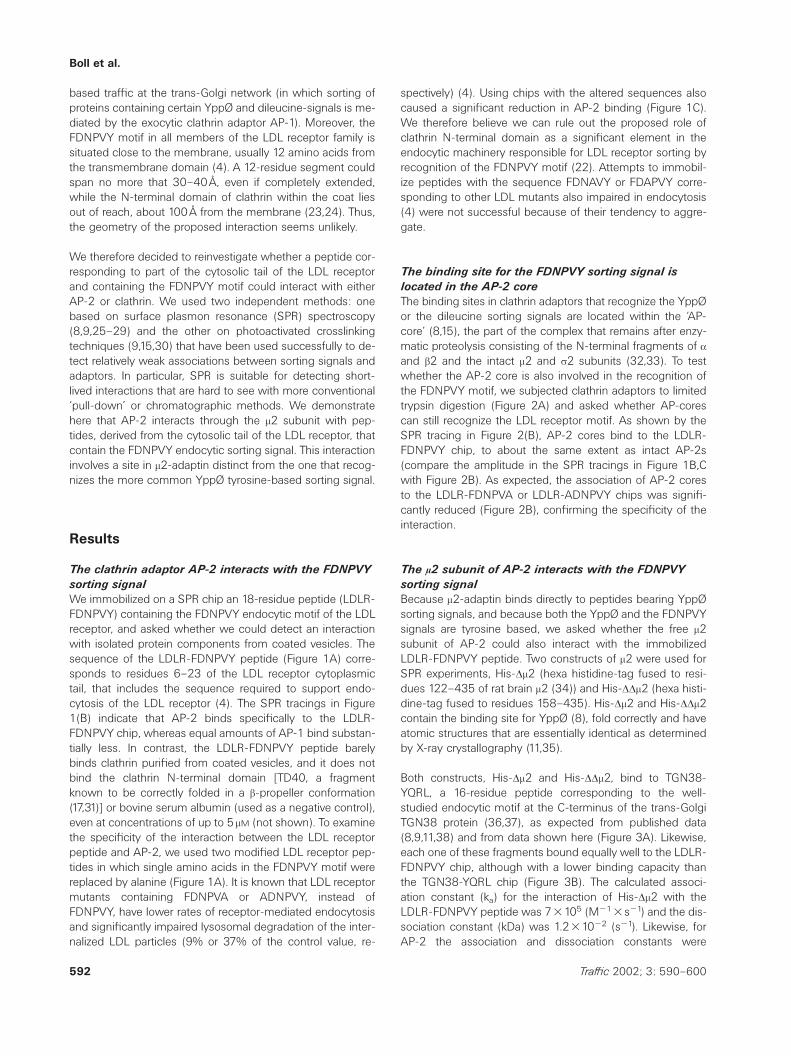
Figure4: Distinct sites in m2-adaptin recognize the FDNPVY internalization motif or the canonical YppØ endocytic signal. (A)His-Dm2 has two different binding sites, one for FDNPVY and the other for YQRL sorting signals. The blot shows the appearance of crosslinkedproducts between His-Dm2 (lanes 1–3, 5–7) or His-Dm2 (W421A) (lanes 4, 8) and the photo-reactive peptide probes LDL-*FDNPVY (lanes1–4) and TGN38-*YQRL (lanes 5–8). (B) Interaction of His-Dm2 with TGN38-YQRL is specific and depends on the specific contact betweenthe YQRL sequence and its binding site in m2. Two SPR tracings correspond to the recruitment of His-Dm2 to immobilized TGN38-YQRLpeptide carried in the absence (thick tracing) or presence (thin tracing) of 200 mM TGN38-YQRL peptide (added as a competitor). The thirdSPR tracing (thin) shows that the mutated m2-adaptin His-Dm2 (W421A) does not bind to the chip with TGN38-YQRL because it is impairedin its recognition of YppØ endocytic motifs. (C) Association of His-Dm2 to the LDLR-FDNPVY peptide does not require the YppØ bindingsite in m2. The SPR tracings illustrate that His-Dm2 and His-Dm2 (W421A) bind similarly to the LDLR-FDNPVY chip. The presence of 200mM
TGN-YQRL peptide does not influence binding of His-Dm2 to the LDL-FDNPVY chip.
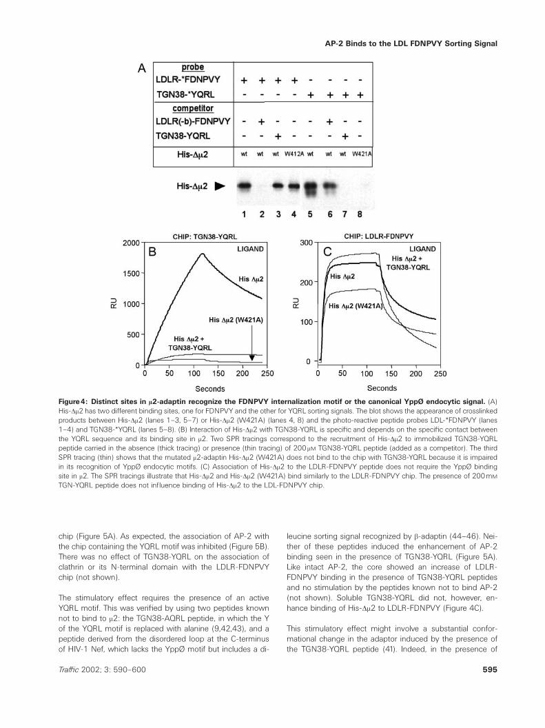
chip (Figure 5A). As expected, the association of AP-2 withthe chip containing the YQRL motif was inhibited (Figure 5B).There was no effect of TGN38-YQRL on the association ofclathrin or its N-terminal domain with the LDLR-FDNPVYchip (not shown).
The stimulatory effect requires the presence of an activeYQRL motif. This was verified by using two peptides knownnot to bind to m2: the TGN38-AQRL peptide, in which the Yof the YQRL motif is replaced with alanine (9,42,43), and apeptide derived from the disordered loop at the C-terminusof HIV-1 Nef, which lacks the YppØ motif but includes a di-
595Traffic 2002; 3: 590–600
leucine sorting signal recognized by b-adaptin (44–46). Nei-ther of these peptides induced the enhancement of AP-2binding seen in the presence of TGN38-YQRL (Figure 5A).Like intact AP-2, the core showed an increase of LDLR-FDNPVY binding in the presence of TGN38-YQRL peptidesand no stimulation by the peptides known not to bind AP-2(not shown). Soluble TGN38-YQRL did not, however, en-hance binding of His-Dm2 to LDLR-FDNPVY (Figure 4C).
This stimulatory effect might involve a substantial confor-mational change in the adaptor induced by the presence ofthe TGN38-YQRL peptide (41). Indeed, in the presence of
Boll et al.
Figure5: Peptide bearing the YQRL endocytic signal stimulates the interaction between AP-2 adaptors or AP-2 cores and theLDLR-FDNPVY chip. (A) Stimulatory effect of TGN38-YQRL peptide on the recruitment of AP-2 to a chip containing the FDNPVY motif.LDLR-(C)FDNPVY peptides were immobilized to the chip by a thiol-coupling reaction. The tracings reflect the relative association of 0.2 mM
AP-2 to the chip in the presence of 200 mM TGN38-YQRL, TGN38-AQRL or HIV Nef-LL peptides. (B) TGN38-YQRL peptide does notstimulate the recruitment of AP-2 to a chip containing the YQRL motif. The SPR tracings show the binding of AP-2 to a chip containingimmobilized TGN38-YQRL (23) peptide carried out in the absence or presence of 200 mM of free TGN38-YQRL. This experiment wasperformed with the slightly longer TGN38-YQRL (23) peptide immobilized to the chip, instead of TGN38-YQRL, because it improved bindingof AP-2 to the chip. Similar binding results were obtained with a chip with immobilized TGN38-YQRL (not shown).
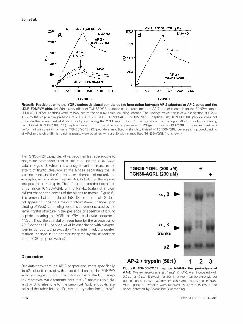
the TGN38-YQRL peptide, AP-2 becomes less susceptible toenzymatic proteolysis. This is illustrated by the SDS-PAGEdata in Figure 6, which show a significant decrease in theextent of tryptic cleavage at the hinges separating the N-terminal trunk and the C-terminal ear domains of not only thea-adaptin, as was shown earlier (41), but also at the equiva-lent position in b-adaptin. This effect requires the interactionof m2, since TGN38-AQRL or HIV Nef-LL (data not shown)did not change the access of the hinges to trypsin (Figure 6).It is known that the isolated 158–435 segment of m2 doesnot appear to undergo a major conformational change uponbinding of YppØ-containing peptides as demonstrated by thesame crystal structure in the presence or absence of boundpeptides bearing the YQRL or YRAL endocytic sequences(11,35). Thus, the stimulation seen here for the association ofAP-2 with the LDL-peptide, or of its association with synapto-tagmin as reported previously (41), might involve a confor-mational change in the adaptor triggered by the associationof the YQRL-peptide with m2.
Discussion
Our data show that the AP-2 adaptor and, more specifically,its m2 subunit interact with a peptide bearing the FDNPVYendocytic signal found in the cytosolic tail of the LDL recep-tor. Moreover, we document here that m2 contains two dis-tinct binding sites: one for the canonical YppØ endocytic sig-nal and the other for the LDL receptor tyrosine-based motif.
596 Traffic 2002; 3: 590–600
Figure6: TGN38-YQRL peptide inhibits the proteolysis ofAP-2. Twenty micrograms (at 1mg/ml) AP-2 was incubated with0.5mg (at 10mg/ml) trypsin for 30min at room temperature withoutpeptide (lane 1), with 0.2mM TGN38-YQRL (lane 2) or TGN38-AQRL (lane 3). Proteins were resolved by 12% SDS-PAGE andbands detected by Coomassie Blue staining.

AP-2 Binds to the LDL FDNPVY Sorting Signal
Beyond the original observation by Pearse that a very smallfraction of iodinated AP-2 is retained in an affinity supportcontaining an immobilized portion of the LDL receptor cyto-solic tail (20), it has been hard to detect interaction usingmethods such as coimmunoprecipation from cells, pull-downs with recombinant proteins or yeast-two-hybrid ap-proaches. Here we detect and study the interaction betweenthe LDL receptor internalization motif and m2 of AP-2 withtwo independent methods.
Since the binding site on m2 for FDNPVY is distinct from thatfor YppØ [this work and also (9)], it is possible to imagineindependent regulation in the internalization of the LDL-re-ceptor class of molecules from the much more commonclass of proteins that use YppØ for sorting. Recruitment andassembly of the coat components at the plasma membraneis a dynamic process that undoubtedly involves a large num-ber of regulatory steps (5–7). The interaction of AP-2 withclathrin or with phosphoinositides, especially those phos-phorylated in the inositol ring at the D-3 position, also leadsto a considerable increase in the affinity of interaction be-tween AP-2 and YppØ endocytic motifs (30). AP-2 recruit-ment to plasma membranes of neuronal and non-neuronalmembranes is also enhanced by peptides bearing the YppØmotif (41). This recruitment is probably mediated by synapto-tagmin, an AP-2 binding protein proposed to function as adocking site for AP-2 in synaptic membranes, which bindsbetter to AP-2 in the presence of peptides with the YppØ-tyrosine-based motif. Thus, the binding of YppØ endocyticmotifs to the m2 chain of AP-2 appears to increase AP-2 af-finity for at least two types of molecules: synaptotagmin and,as reported now here, to a peptide corresponding to part ofcytosolic tail of the LDL receptor. Therefore, there is signifi-cant cross-talk among a number of interacting molecules,orchestrated by the adaptor complexes.
These observations are consistent with the general modelproposed by us and by others linking cargo loading withclathrin coat formation (30,41). These interactions might helpto cluster the cargo, or might help to ensure that a vesicledoes not form unless it can capture a sufficient number ofcargo molecules. Indeed, other observations suggest thatcoat formation is linked to cargo capture: electron micro-scopy of cells in tissue culture shows that about 60–70% oftheir clathrin-coated profiles are labeled with markers for theLDL and transferrin and epidermal growth factor receptors(47–49).
The intracellular traffic of the LDL receptor is far from simple,and is clearly under the control of a number of steps throughthe endocytic as well as the secretory pathway. The stimu-latory results documented here on the association of the LDLpeptide containing the FDPNPY sequence and AP-2 are con-sistent with the possibility that the uptake of the LDL receptorcan be subjected to specific regulation within the clathrinpathway. This regulatory step could complement the well-established modulation of the number of LDL receptors avail-able for endocytosis that is exerted by transcriptionally based
597Traffic 2002; 3: 590–600
long-term control in response to available cellular cholesterol(18,50).
During biosynthesis, LDL receptors that transit the TGN arenot sorted by the ubiquitously expressed AP-1A adaptors(51). Our in vitro data are consistent with these observationsin that the ubiquitous AP-1A adaptor (equivalent to the neur-onal AP-1 used here) binds with less affinity than AP-2 tothe LDL receptor internalization motif. In fact, the polarizedsecretory traffic of the receptor to the basolateral plasmamembrane involves a second sorting signal located else-where in the cytosolic tail of the receptor, which differs fromthe FDNPVY internalization motif (52,53). Basolateral localiz-ation is mediated by a specialized, less abundant m1b speciesfound in AP-1B, an adaptor characteristic of polarized epi-thelial cells, through a mechanism of interaction that stillneeds to be resolved (51).
Another way to control the traffic of the LDL receptor mightinvolve recognition of its FDNPVY motif by proteins contain-ing phosphotyrosine binding (PTB)-domains, since these do-mains can recognize an NPXY motif whose tyrosine is phos-phorylated. Since mutations in a PTB-domain containing pro-tein lead to autosomal recessive hypercholesterolemia (ARH)protein, it was suggested that the ARH protein interacts withthe LDL receptor through the NPVY motif, thereby regulatingthe intracellular traffic of the receptor (54).
In conclusion, we demonstrate that m2 contains two distinctbinding sites: one for peptides containing the FDNPVY inter-nalization motif of the LDL receptor, and a second one thatrecognizes the more common YppØ tyrosine-based sortingsignal. Moreover, the interaction of AP-2 with peptides bear-ing the FDNPVY motif seems to be modulated by a confor-mational change in AP-2, involving its association with YQRL-containing peptides.
Materials and Methods
Proteins
Clathrin, AP-1, and AP-2 were purified from bovine-brain-coated vesiclesas described (15,55). Clathrin was kept at 4 æC in 500mM TRIS, 1mM
EDTA, 0.02% NaN3. APs were kept at 4 æC in 100mM MES pH7.0, 150mM
NaCl, 1mM EDTA, 0.02% NaN3, 0.5mM DTT (AP buffer). Limited trypticproteolysis of AP-2 results in the release of the carboxyl-terminal domains(the ear and hinge) of the large b1/b2 and a subunits of AP-2 from the AP-core (containing the N-terminal domains of a and b-adaptins, together withm2 and s2). AP-2 cores were generated upon incubation of AP-2 (0.4mg/ml) with 5mg/ml (final concentration) of trypsin at 25 æC for 30min(12,32,33). Proteolysis was ended by addition of 75ml benzamidine-agar-ose beads (Pharmacia) and end-over-end rotation for 10min at 4 æC. Thebeads were separated from the protein solution using a spin column (Bio-Spin; Bio-Rad, Hercules, CA, USA). The extent of proteolysis was verifiedby 10% SDS-PAGE and staining with Coomassie Blue. The AP-2 coreswere stored in AP buffer at 4 æC in the presence of 2mM (final concen-tration) of benzamidine.

Boll et al.
Recombinant TD40 corresponds to the N-terminal domain of rat brainclathrin (residues 1–393) (17), and contains six histidines added to the N-terminus. TD40 was expressed in Escherichia coli and purified by Ni-NTAchromatography and gel filtration (Superdex 75) in 20mM MES, pH8.0,150mM NaCl, 6mM MgCl2, and 0.5mM DTT. His-Dm2 and His-DDm2 weregenerated by polymerase chain reaction from a plasmid template contain-ing the complete rat brain m2 open reading frame (34). His-Dm2 and His-DDm2 span residues 122–435 and 158–435, respectively, and were in-serted between the NcoI and EcoRI sites of the pProEXTMHTA expressionvector (Life Technologies, Grand Island, NY, USA). The proteins were ex-pressed in E. coli and purified by Ni-NTA chromatography. The recombin-ant proteins were stored at ª80 æC in 50mM TRIS-HCl pH8.0, 500mM
LiCl, 3mM b-mercaptoethanol, 200mM imidazole and 20% glycerol (His-Dm2 or His-DDm2). Prior to SPR experiments, the glycerol in the buffer ofthe protein solutions was removed by gel filtration (Sephadex G-25, NAP5columns, Pharmacia, Piscataway, NJ, USA), followed by 50-fold dilution ofthe protein sample in AP buffer. Aggregates were eliminated by high-speed centrifugation at 100000r.p.m. for 20min and 4 æC (TLA-100.4 ro-tor, Optima TLX centrifuge, Beckman, Fullerton, CA, USA).
Surface plasmon resonance
Most of the results shown were obtained at 25 æC with a BIAcore 3000instrument (Pharmacia Biosensor), although a BIAcore 2000 was usedwith similar results to follow the interaction of the sample proteins withsensor chips containing immobilized peptides. The SPR experiments werecarried in AP buffer supplemented with 1mg/ml carboxymethyl-dextranand with 0.02% Triton X-100. The peptides were synthesized by standardM-MOC procedures, dissolved in water and kept at ª80 æC. The peptideswere immobilized to a final amount of 400–600 resonance units (RUs)onto the surface of CM5 sensor chips (research grade; Pharmacia Biosens-or) using EDC/NHS. In some cases we immobilized the LDLR-FDNPVYpeptide with a cysteine at its N-terminal end (LDLR-(C)FDNPVY) by coup-ling it to a PDEA-derivatized chip. Use of relatively low amounts of im-mobilized peptides (400–600 RUs), in conjunction with a flow rate of20ml/ml and a relatively low concentration (200nM) of soluble protein (in-jected as 40-ml samples), consistent with a reasonable and reproduciblebinding signal, allowed us to minimize possible kinetic artifacts due tosteric hindrance, crowding and aggregation. Similar conditions have beenused to study the interaction between APs and peptides containing sortingsignals (26,28,29). Similarly to their results, we observed that a fraction ofAPs bound irreversibly to the chip, perhaps reflecting a small portion ofAPs that forms dimers or higher aggregates. All experiments were carriedout in the presence of 1mg/ml carboxy-methylated dextran. No differ-ences in the binding profiles were detected in the absence of carboxy-methylated dextran. None of the proteins bound to any significant level tochips derivatized in the absence of peptide, ruling out the possibility thatthe observed associations were nonspecific and charge dependent. As-sociation and dissociation rate constants were calculated using BIAevalu-ation 3.1 software from injections into flow cells immobilized with 150–200 RU of peptide. The sensorgrams represent the binding of ligand aftersubtraction of changes in refractive index caused by bulk flow of proteinand/or peptides through the flow cells.
When the protein samples were co-injected with soluble peptide (dis-solved in AP buffer), they were preincubated for 15min at room tempera-ture before injection and were present during the dissociation phase. Aftereach injection, the surfaces of the chips were regenerated by injection of8–10-s pulses (80ml/min) of 10mM NaOH and 0.25% SDS until the pre-injection baseline was restored. The results presented in the figures arerepresentative of at least three independent experiments performed usingdifferent sensor chips and different protein preparations. The stimulationeffect of TGN38-YQRL on the binding of AP-2 to the FDNPVY motif de-noted in Figure 5 represents the results of 20 different experiments.
598 Traffic 2002; 3: 590–600
UV-induced crosslinking reaction and detection of crosslinked
products
The UV-induced crosslinking reaction and detection of products was car-ried out as previously described (15,30). Sixteen microliters of a solutioncontaining HisDm2 or HisDm2 (W421A) (2mM final concentration) weremixed with 2ml of the photoreactive peptides (1mM LDLR-*FDNPVY or0.2mM TGN38-*YQRL final concentrations). Competition experiments weredone by including 2ml of the nonbiotinylated peptides LDL(–b)-FDNPVY(60mM final concentration) or TGN38-YQRL (200mM final concentration).Crosslinking was triggered by exposure to UV-illumination for 5min in wetice. The UV illumination was also carried out with samples frozen in dryice, with identical results (not shown).
Acknowledgments
We thank Ian Levesque for his help in the preparation of the manuscript.This work was supported by a grant from the National Institutes of Health(GM36548) to T.K.
References
1. Goldstein JL, Brown MS. Binding and degradation of low density lipo-proteins by cultured human fibroblasts. J Biol Chem 1974;16:5153–5162.
2. Goldstein JL, Anderson RG, Brown MS. Coated pits, coated vesicles,and receptor-mediated endocytosis. Nature 1979;279:679–685.
3. Lai A, Sisodia SS, Trowbridge IS. Characterization of sorting signals inthe beta-amyloid precursor protein cytoplasmic domain. J Biol Chem1995;270:3565–3573.
4. Chen WJ, Goldstein JL, Brown MS. NPXY, a sequence often found incytoplasmic tails, is required for coated pit-mediated internalizationof the low density lipoprotein receptor. J Biol Chem 1990;265:3116–3123.
5. Kirchhausen T. Adaptors for clathrin mediated traffic. Annu Rev CellDev Biol 1999;15:705–732.
6. Schmid SL. Clathrin-coated vesicle formation and protein sorting: Anintegrated process. Annu Rev Biochem 1997;66:511–548.
7. Hirst J, Robinson MS. Clathrin and adaptors. Biochem Biophys Acta1998;1404:173–193.
8. Ohno H, Stewart J, Fournier MC, Bosshart H, Rhee I, Miyatake S, SaitoT, Gallusser A, Kirchhausen T, Bonifacino JS. Interaction of tyrosine-based sorting signals with clathrin-associated proteins. Science1995;269:1872–1875.
9. Boll W, Ohno H, Songyang Z, Rapoport I, Cantley LC, Bonifacino JS,Kirchhausen T. Sequence requirements for the recognition of tyrosine-based endocytic signals by clathrin AP-2 complexes. EMBO J1996;15:5789–5795.
10. Ohno H, Aguilar RC, Yeh D, Taura D, Saito T, Bonifacino JS. The me-dium subunits of adaptor complexes recognize distinct but overlap-ping sets of tyrosine-based sorting signals. J Biol Chem1998;273:25915–25921.
11. Owen DJ, Evans PR. A structural explanation for the recognition oftyrosine-based endocytotic signals. Science 1998;282:1327–1332.
12. Ahle S, Ungewickell E. Identification of a clathrin binding subunit inthe HA2 adaptor protein complex. J Biol Chem 1989;264:20089–20093.
13. Gallusser A, Kirchhausen T. The b1 and b2 subunits of the AP com-plexes are the clathrin coat assembly components. EMBO J1993;12:5237–5244.
14. Shih W, Gallusser A, Kirchhausen T. A clathrin-binding site in the hinge

AP-2 Binds to the LDL FDNPVY Sorting Signal
of the b-2 chain of mammalian AP-2 complexes. J Biol Chem1995;270:31083–31090.
15. Rapoport I, Chen YC, Cupers P, Shoelson SE, Kirchhausen T. Dileucine-based sorting signals bind to the beta chain of AP-1 at a site distinctand regulated differently from the tyrosine-based motif-binding site.EMBO J 1998;17:2148–2155.
16. Dell’Angelica EC, Klumperman J, Stoorvogel W, Bonifacino JS. As-sociation of the AP-3 adaptor complex with clathrin. Science1998;280:431–434.
17. Ter Haar E, Harrison SC, Kirchhausen T. Peptide-in–groove interactionslink target proteins to the b-propeller of clathrin. Proc Natl Acad SciUSA 2000;97:1096–1100.
18. Goldstein JL, Brown MS, Anderson RG, Russell DW, Schneider WJ.Receptor-mediated endocytosis: Concepts emerging from the LDL re-ceptor system. Annu Rev Cell Biol 1985;1:1–39.
19. Davis CG, van Driel IR, Russell DW, Brown MS, Goldstein JL. The lowdensity lipoprotein receptor. J Biol Chem 1987;262:4075–4082.
20. Pearse BMF. Receptors compete for adaptors found in plasma mem-brane coated pits. EMBO J 1988;7:3331–3336.
21. Warren RA, Green FA, Stenberg PE, Enns CA. Distinct saturable path-ways for the endocytosis of different tyrosine motifs. J Biol Chem1998;273:17056–17063.
22. Kibbey RG, Rizo J, Gierasch LM, Anderson RGW. The LDL receptorclustering motif interacts with the clathrin terminal domain in a reverseturn conformation. J Cell Biol 1998;142:59–67.
23. Smith CJ, Grigorieff N, Pearse BM. Clathrin coats at 21 Å resolution:a cellular assembly designed to recycle multiple membrane receptors.EMBO J 1998;17:4943–4953.
24. Musacchio A, Smith CJ, Roseman AM, Harrison SC, Kirchhausen T,Pearse BMF. Functional organization of clathrin in coats: Combiningelectron cryomicroscopy and x-ray crystallography. Mol Cell1999;3:761–770.
25. Boll W, Gallusser A, Kirchhausen T. Role of the regulatory domain ofthe EGF-receptor cytoplasmic tail in selective binding of the clathrin-associated complex AP-2. Curr Biol 1995;5:1168–1178.
26. Pitcher C, Honing S, Fingerhut A, Bowers K, Marsh M. Cluster of differ-entiation antigen 4 (CD4) endocytosis and adaptor complex bindingrequire activation of the CD4 endocytosis signal by serine phos-phorylation. Mol Biol Cell 1999;10:677–691.
27. Nesterov A, Kurten RC, Gill GN. Association of epidermal growth factorreceptors with coated pit adaptins via a tyrosine-phosphorylation-regulated mechanism. J Biol Chem 1995;270:1–8.
28. Honing S, Sandoval IV, von Figura K. A di-leucine-based motif in thecytoplasmic tail of LIMP-II and tyrosinase mediates selective bindingof AP-3. EMBO J 1998;17:1304–1314.
29. Honing S, Griffith J, Geuze HJ, Hunziker W. The-tyrosine based lys-osomal targeting signal in LAMP-1 mediates sorting into Golgi-de-rived clathrin-coated vesicles. EMBO J 1996;15:5230–5239.
30. Rapoport I, Miyazaki M, Boll W, Duckworth B, Cantley LC, Shoelson S,Kirchhausen T. Regulatory interactions in the recognition of endocyticsorting signals by AP-2 complexes. EMBO J 1997;16:2240–2250.
31. Ter Haar E, Musacchio A, Harrison SC, Kirchhausen T. Atomic structureof clathrin – a b propeller terminal domain joins an a zigzag linker. Cell1998;95:563–573.
32. Zaremba S, Keen JH. Limited proteolytic digestion of coated vesicleassembly polypeptides abolishes reassembly activity. J Cell Biochem1985;28:47–58.
33. Kirchhausen T, Nathanson KL, Matsui W, Vaisberg A, Chow EP, BurneC, Burne C, Keen JH, Davis AE. Structural and functional division intotwo domains of the large (100- to 115-kDa) chains of the clathrin-associated protein complex AP-2. Proc Natl Acad Sci USA1989;86:2612–2616.
34. Thurieau C, Brosius J, Burne C, Jolles P, Keen JH, Mattaliano RJ, ChowEP, Ramachandran KL, Kirchhausen T. Molecular cloning and com-
599Traffic 2002; 3: 590–600
plete amino acid sequence of AP50, an assembly protein associatedwith clathrin-coated vesicles. DNA 1988;7:663–669.
35. Modis Y, Boll W, Rapoport I, Kirchhausen T. m2-adaptin subunit (AP50)of AP-2 clathrin adaptor, complexed with EGFR internalization peptideFYRALM at 2.5 Å resolution. PDB entry 1I31 2001.
36. Humphrey JS, Peters PJ, Yuan LC, Bonifacino JS. Localization ofTGN38 to the trans-Golgi network: Involvement of a cytoplasmic tyro-sine-containing sequence. J Cell Biol 1993;120:1123–1135.
37. Bos K, Wraight C, Stanley KK. TGN38 is maintained in the trans-Golginetwork by a tyrosine-containing motif in the cytoplasmic domain.EMBO J 1993;12:2219–2228.
38. Stephens DJ, Crump CM, Clarke AR, Banting G. Serine 331 and tyro-sine 333 are both involved in the interaction between the cytosolicdomain of TGN38 and the m-2 subunit of the AP2 clathrin adaptorcomplex. J Biol Chem 1997;272:14104–14109.
39. Collins BM, McCoy AJ, Kent HM, Evans PR, Owen DJ. Moleculararchitecture and functional model of the endocytic AP-2 complex. Cell2002; in press.
40. Nesterov A, Carter RE, Sorkina T, Gill GN, Sorkin A. Inhibition of thereceptor-binding function of clathrin adaptor protein AP-2 by domi-nant-negative mutant m2 subunit and its effects on endocytosis.EMBO J 1999;18:2489–2499.
41. Haucke V, De Camilli P. AP-2 recruitment to synaptotagmin stimulatedby tyrosine-based endocytic motifs. Science 1999;285:1268–1271.
42. Ohno H, Fournier M-C, Poy G, Bonifacino JS. Structural determinantsof interaction of tyrosine-based sorting signals with the adaptor me-dium chains. J Biol Chem 1996;271:29009–29015.
43. Shiratori T, Miyatake S, Ohno H, Nakaseko C, Isono K, Bonifacino JS,Saito T. Tyrosine phosphorylation controls internalization of CTLA-4 byregulating its interaction with clathrin-associated adaptor complex AP-2. Immunity 1997;6:583–589.
44. Craig HM, Pandori MW, Guatelli JC. Interaction of HIV-1 Nef with thecellular dileucine-based sorting pathway is required for CD4 down-regulation and optimal viral infectivity. Proc Natl Acad Sci USA1998;95:11229–11234.
45. Bresnahan PA, Yonemoto W, Ferrell S, Williamsherman D, GeleziunasR, Greene WC. A dileucine motif in HIV-1 Nef acts as an internalizationsignal for CD4 downregulation and binds the AP-1 clathrin adaptor.Curr Biol 1998;8:1235–1238.
46. Greenberg M, Detulleo L, Rapoport I, Skowronski J, Kirchhausen T. Adileucine motif in HIV-1 Nef is essential for sorting into clathrin-coatedpits and for downregulation of CD4. Curr Biol 1998;8:1239–1242.
47. Carpentier JL, Gorden P, Anderson RG, Goldstein JL, Brown MS, Co-hen S, Orci L. Co-localization of 125I-epidermal growth factor and ferri-tin-low density lipoprotein in coated pits: a quantitative electron micro-scopic study in normal and mutant human fibroblasts. J Cell Biol1982;95:73–77.
48. Schlessinger J, Shechter Y, Willingham M, Pastan I. Direct visualizationof binding, aggregation, and internalization of insulin and epidermalgrowth factor on living fibroblastic cells. Proc Natl Acad Sci1978;75:2659–2663.
49. Hanover JA, Willingham MC, Pastan I. Kinetics of transit of transferrinand epidermal growth factor through clathrin-coated membranes. Cell1984;39:283–293.
50. Brown MS, Goldstein JL. The SREBP pathway – regulation of chol-esterol metabolism by proteolysis of a membrane-bound transcriptionfactor. Cell 1997;89:331–340.
51. Folsch H, Pypaert M, Schu P, Mellman I. Distribution and function ofAP-1 clathrin adaptor complexes in polarized epithelial cells. J CellBiol 2001;152:595–606.
52. Matter K, Yamamoto EM, Mellman I. Structural requirements and se-quence motifs for polarized sorting and endocytosis of LDL and Fcreceptors in MDCK cells. J Cell Biol 1994;126:991–1004.

Boll et al.
53. Matter K, Hunziker W, Mellman I. Basolateral sorting of LDL receptorin MDCK cells: The cytoplasmic domain contains two tyrosine-de-pendent targeting determinants. Cell 1992;71:741–753.
54. Garcia CK, Wilund K, Arca M, Zuliani G, Fellin R, Maioli M, CalandraS, Bertolini S, Cossu F, Grishin N, Barnes R, Cohen JC, Hobbs HH.Autosomal recessive hypercholesterolemia caused by mutations in a
600 Traffic 2002; 3: 590–600
putative LDL receptor adaptor protein. Science 2001;292:1394–1398.
55. Matsui W, Kirchhausen T. Stabilization of clathrin coats by the core ofthe clathrin-associated protein complex AP-2. Biochemistry1990;29:10791–10798.
Copyright © 2022 FDOKUMEN