Role of the Clathrin Terminal Domain in Regulating Coated Pit Dynamics Revealed by Small Molecule...
14
Resource Role of the Clathrin Terminal Domain in Regulating Coated Pit Dynamics Revealed by Small Molecule Inhibition Lisa von Kleist, 1 Wiebke Stahlschmidt, 1 Haydar Bulut, 1,8 Kira Gromova, 1,8 Dmytro Puchkov, 1,8 Mark J. Robertson, 2 Kylie A. MacGregor, 2 Nikolay Tomlin, 3 Arndt Pechstein, 1,3 Ngoc Chau, 4 Megan Chircop, 4 Jennette Sakoff, 5 Jens Peter von Kries, 6 Wolfram Saenger, 1 Hans-Georg Kra ¨ usslich, 7 Oleg Shupliakov, 3 Phillip J. Robinson, 4 Adam McCluskey, 2 and Volker Haucke 1,6, * 1 Institute of Chemistry and Biochemistry & Neurocure Cluster of Excellence, Freie Universita ¨ t Berlin, 14195 Berlin, Germany 2 Centre for Chemical Biology, Chemistry, The University of Newcastle, Callaghan NSW 2308, Australia 3 Department of Neuroscience, DBRM, Karolinska Institutet, 17117 Stockholm, Sweden 4 Cell Signalling Unit, Children’s Medical Research Institute, The University of Sydney, Sydney NSW 2145, Australia 5 Department of Medical Oncology, Calvary Mater Newcastle Hospital, Waratah NSW 2298, Australia 6 Leibniz-Institut fu ¨ r Molekulare Pharmakologie (FMP), 13125 Berlin-Buch, Germany 7 Department of Infectious Diseases, Virology, University of Heidelberg, 69120 Heidelberg, Germany 8 These authors contributed equally to this work *Correspondence: [email protected] DOI 10.1016/j.cell.2011.06.025 SUMMARY Clathrin-mediated endocytosis (CME) regulates many cell physiological processes such as the inter- nalization of growth factors and receptors, entry of pathogens, and synaptic transmission. Within the endocytic network, clathrin functions as a central organizing platform for coated pit assembly and dissociation via its terminal domain (TD). We report the design and synthesis of two compounds named pitstops that selectively block endocytic ligand asso- ciation with the clathrin TD as confirmed by X-ray crystallography. Pitstop-induced inhibition of cla- thrin TD function acutely interferes with receptor- mediated endocytosis, entry of HIV, and synaptic vesicle recycling. Endocytosis inhibition is caused by a dramatic increase in the lifetimes of clathrin coat components, including FCHo, clathrin, and dy- namin, suggesting that the clathrin TD regulates coated pit dynamics. Pitstops provide new tools to address clathrin function in cell physiology with potential applications as inhibitors of virus and path- ogen entry and as modulators of cell signaling. INTRODUCTION Clathrin-mediated endocytosis (CME) regulates the cell surface levels and endocytic uptake of important plasma membrane proteins (Brodsky et al., 2001; Conner and Schmid, 2003). These include nutrient and growth factor receptors, ion channels, adhe- sion proteins, and synaptic vesicle (SV) proteins in the brain. The clathrin-based endocytic machinery is also hijacked by patho- gens such as bacteria and viruses to get access to the cell inte- rior (Conner and Schmid, 2003; Miyauchi et al., 2009; Veiga et al., 2007). Assembly of clathrin-coated pits (CCPs) likely is initiated by the recruitment of early-acting endocytic proteins such as FCHo 1/2, intersectins, and Eps15/Eps15R (Henne et al., 2010; Pechstein et al., 2010). These factors serve as scaffolds for the coassembly of AP-2, which coordinates recognition of trans- membrane cargo (Edeling et al., 2006) with recruitment of acces- sory proteins. Early endocytic structures are stabilized by an assembling clathrin coat built from soluble clathrin triskelia comprising three heavy and three light chains (Brodsky et al., 2001; Conner and Schmid, 2003; Edeling et al., 2006). The central building block of triskelia is the 190 kDa clathrin heavy chain, which forms an extended three-legged structure. The N-terminal b-propeller domain (TD) at the distal end of the leg adopts a WD40-like fold (ter Haar et al., 2000), while the C terminus is near the vertex of the triskelion (Brodsky et al., 2001). The clathrin cage has been postulated to serve at least two major functions in endocytosis: (1) stabilizing deformed mem- brane domains (Hinrichsen et al., 2006) and (2) providing an inter- action hub for the recruitment of accessory factors that regulate progression of endocytosis (Schmid and McMahon, 2007). Structural and proteomic studies have revealed a surprisingly simple architecture of accessory protein-clathrin interactions (Schmid and McMahon, 2007). Most of these factors, including amphiphysins, AP180, and SNX9, harbor so-called clathrin box motifs (or variants thereof), simple degenerate peptides that bind to a structurally well-defined site on the TD of clathrin heavy chain (ter Haar et al., 2000). Consistent with the proposed func- tion of the TD as a central protein-protein interaction hub (Schmid and McMahon, 2007) overexpression of clathrin- binding fragments of endocytic proteins causes the sequestra- tion of clathrin within the cytoplasm (Slepnev et al., 2000), sug- gesting that multiple redundant interactions of accessory proteins with the TD serve to recruit clathrin to membranes. However, overexpression experiments need to be interpreted Cell 146, 471–484, August 5, 2011 ª2011 Elsevier Inc. 471
Transcript of Role of the Clathrin Terminal Domain in Regulating Coated Pit Dynamics Revealed by Small Molecule...
Resource
Role of the Clathrin Terminal Domainin Regulating Coated Pit DynamicsRevealed by Small Molecule InhibitionLisa von Kleist,1 Wiebke Stahlschmidt,1 Haydar Bulut,1,8 Kira Gromova,1,8 Dmytro Puchkov,1,8 Mark J. Robertson,2
Kylie A. MacGregor,2 Nikolay Tomlin,3 Arndt Pechstein,1,3 Ngoc Chau,4 Megan Chircop,4 Jennette Sakoff,5
Jens Peter von Kries,6 Wolfram Saenger,1 Hans-Georg Krausslich,7 Oleg Shupliakov,3 Phillip J. Robinson,4
Adam McCluskey,2 and Volker Haucke1,6,*1Institute of Chemistry and Biochemistry & Neurocure Cluster of Excellence, Freie Universitat Berlin, 14195 Berlin, Germany2Centre for Chemical Biology, Chemistry, The University of Newcastle, Callaghan NSW 2308, Australia3Department of Neuroscience, DBRM, Karolinska Institutet, 17117 Stockholm, Sweden4Cell Signalling Unit, Children’s Medical Research Institute, The University of Sydney, Sydney NSW 2145, Australia5Department of Medical Oncology, Calvary Mater Newcastle Hospital, Waratah NSW 2298, Australia6Leibniz-Institut fur Molekulare Pharmakologie (FMP), 13125 Berlin-Buch, Germany7Department of Infectious Diseases, Virology, University of Heidelberg, 69120 Heidelberg, Germany8These authors contributed equally to this work*Correspondence: [email protected]
DOI 10.1016/j.cell.2011.06.025
SUMMARY
Clathrin-mediated endocytosis (CME) regulatesmany cell physiological processes such as the inter-nalization of growth factors and receptors, entry ofpathogens, and synaptic transmission. Within theendocytic network, clathrin functions as a centralorganizing platform for coated pit assembly anddissociation via its terminal domain (TD). We reportthe design and synthesis of two compounds namedpitstops that selectively block endocytic ligand asso-ciation with the clathrin TD as confirmed by X-raycrystallography. Pitstop-induced inhibition of cla-thrin TD function acutely interferes with receptor-mediated endocytosis, entry of HIV, and synapticvesicle recycling. Endocytosis inhibition is causedby a dramatic increase in the lifetimes of clathrincoat components, including FCHo, clathrin, and dy-namin, suggesting that the clathrin TD regulatescoated pit dynamics. Pitstops provide new tools toaddress clathrin function in cell physiology withpotential applications as inhibitors of virus and path-ogen entry and as modulators of cell signaling.
INTRODUCTION
Clathrin-mediated endocytosis (CME) regulates the cell surface
levels and endocytic uptake of important plasma membrane
proteins (Brodsky et al., 2001; Conner and Schmid, 2003). These
include nutrient and growth factor receptors, ion channels, adhe-
sion proteins, and synaptic vesicle (SV) proteins in the brain. The
clathrin-based endocytic machinery is also hijacked by patho-
gens such as bacteria and viruses to get access to the cell inte-
rior (Conner and Schmid, 2003;Miyauchi et al., 2009; Veiga et al.,
2007). Assembly of clathrin-coated pits (CCPs) likely is initiated
by the recruitment of early-acting endocytic proteins such as
FCHo 1/2, intersectins, and Eps15/Eps15R (Henne et al., 2010;
Pechstein et al., 2010). These factors serve as scaffolds for the
coassembly of AP-2, which coordinates recognition of trans-
membrane cargo (Edeling et al., 2006) with recruitment of acces-
sory proteins. Early endocytic structures are stabilized by an
assembling clathrin coat built from soluble clathrin triskelia
comprising three heavy and three light chains (Brodsky et al.,
2001; Conner and Schmid, 2003; Edeling et al., 2006). The
central building block of triskelia is the 190 kDa clathrin heavy
chain, which forms an extended three-legged structure. The
N-terminal b-propeller domain (TD) at the distal end of the leg
adopts a WD40-like fold (ter Haar et al., 2000), while the C
terminus is near the vertex of the triskelion (Brodsky et al., 2001).
The clathrin cage has been postulated to serve at least two
major functions in endocytosis: (1) stabilizing deformed mem-
brane domains (Hinrichsen et al., 2006) and (2) providing an inter-
action hub for the recruitment of accessory factors that regulate
progression of endocytosis (Schmid and McMahon, 2007).
Structural and proteomic studies have revealed a surprisingly
simple architecture of accessory protein-clathrin interactions
(Schmid and McMahon, 2007). Most of these factors, including
amphiphysins, AP180, and SNX9, harbor so-called clathrin box
motifs (or variants thereof), simple degenerate peptides that
bind to a structurally well-defined site on the TD of clathrin heavy
chain (ter Haar et al., 2000). Consistent with the proposed func-
tion of the TD as a central protein-protein interaction hub
(Schmid and McMahon, 2007) overexpression of clathrin-
binding fragments of endocytic proteins causes the sequestra-
tion of clathrin within the cytoplasm (Slepnev et al., 2000), sug-
gesting that multiple redundant interactions of accessory
proteins with the TD serve to recruit clathrin to membranes.
However, overexpression experiments need to be interpreted
Cell 146, 471–484, August 5, 2011 ª2011 Elsevier Inc. 471

Figure 1. Pitstops Selectively Inhibit Ligand Association with the Clathrin Terminal Domain
(A) Hypothetical models depicting the potential roles of clathrin TD-ligand association in endocytosis. Multiple redundant interactions of the clathrin TD with its
endocytic ligands (blue) may serve to stably recruit clathrin to membranes, thereby facilitating assembly through local enrichment (left). Alternatively, clathrin
assembled at endocytic sites could serve as an organizing scaffold that regulates CCP dynamics by providing spatially defined binding sites on its TD for
accessory proteins that drive CCP maturation and disassembly (right).
(B) Schematic diagram outlining the compound screening strategy.
(C) Chemical structures of pitstops 1 and 2.
(D) Dose dependence of pitstop-mediated interference with clathrin TD-amphiphysin B/C complex formation. Values from ELISA were normalized to solvent
controls (set to 100%). Data represent SEM (n = 3 independent experiments).
(E) IC50 values of pitstop-mediated interference with clathrin TD complex formation determined by ELISA. AP180, C-terminal domain of AP180; Synaptojanin,
C-terminal domain of synaptojanin 1-p170; OCRL, PH domain of OCRL. Data represent SEM (n = 3 independent experiments).
472 Cell 146, 471–484, August 5, 2011 ª2011 Elsevier Inc.

with caution as evidenced by the comparablymild phenotypes of
clathrin-binding accessory protein knockdowns or knockouts in
mice (Dittman and Ryan, 2009; Mettlen et al., 2009). Moreover,
dominant-negative and genetic approaches bear the inherent
disadvantage that indirect effects caused by sustained expres-
sion or loss-of-function cannot be distinguished from direct
phenotypes. It is thus essential to identify molecular reagents
that can selectively and acutely interfere with CME. No specific
compounds targeting clathrin are available so far.
Here, we describe the identification and characterization of
two small molecule inhibitors of clathrin TD function, named pit-
stops for their ability to stall clathrin-coated pit (CCP) dynamics,
which acutely interfere with receptor-mediated endocytosis,
entry of HIV, and SV reformation. Pitstops provide tools to
address clathrin function in cell physiology with potential appli-
cations as inhibitors of virus and pathogen entry and as modula-
tors of cell signaling.
RESULTS
Pitstops Are Nontoxic Specific Inhibitors of EndocyticLigand Association with the Clathrin Terminal DomainClathrin function in mammalian cells has been addressed mainly
via knockdown (Motley et al., 2003) or dominant-negative
approaches (Dittman and Ryan, 2009; Slepnev et al., 2000). At
invertebrate neuromuscular junctions using transgenic flies cla-
thrin has also been targeted by photoinactivation (Heerssen
et al., 2008). While these strategies have shown that clathrin is
essential for CCP formation or stability, no information exists
regarding the physiological role of endocytic ligand association
of its TD. This is a surprising knowledge gap given the fact that
clathrin box-TD interactions form a major hub within the endo-
cytic network (Schmid and McMahon, 2007). Based on previous
cell biological and biochemical studies, it was suggested that
multiple redundant interactions of the clathrin TD with its endo-
cytic ligands may serve to stably recruit clathrin to membranes,
thereby facilitating clathrin assembly through local enrichment
(Figure 1A, left). Alternatively, clathrin assembled at nascent en-
docytic sites could serve as an organizing scaffold that regulates
CCP dynamics by providing spatially defined binding sites on its
TD for accessory proteins that drive CCP maturation and disas-
sembly (Figure 1A, right).
To distinguish between these models, we used a chemical
biology approach designed to identify inhibitors of complex
formation between the TD and its endocytic ligands. We carried
out an ELISA-based screen for small molecules that can interfere
with clathrin TD association of the B/C domain of amphiphysin,
an endocytic protein harboring a clathrin box motif (Slepnev
et al., 2000). Screening a library of about 17,000 small molecules
from the central open access technology platform of the Chem-
BioNet, we identified and validated (see Extended Experimental
Procedures available online) two lead compounds that exhibited
selective inhibition of clathrin TD-amphiphysin association (Fig-
ure 1B). Retrosynthetic analysis identified simple approaches
(F) Binding of amphiphysin 1 B/C (250-578) or a truncation lacking the clathrin box
(G) Pitstops do not affect other peptide-in-groove interactions in membrane traffi
See also Figure S1 and Table S1.
to in-house synthesis of two focused compound libraries based
on the original leads. This led to the identification of two mole-
cules of distinct chemical scaffolds, which specifically disrupt
ligand association with the clathrin TD. Based on subsequent
studies and their ability to impair CCP function (see below) we
named these compounds pitstops 1 (from the naphthalimide
library) and 2 (from the rhodanine library, Figure 1C). Pitstops 1
and 2 selectively inhibited amphiphysin association of clathrin
TD with IC50 values of 18 and 12 mM, respectively (Figure 1D).
Consistent with a crucial role for the clathrin box within amphi-
physin for clathrin TD association a truncation mutant lacking
the clathrin box sequence displayed a near complete loss of its
clathrin TD binding ability (Figure 1F), similar to that seen in pit-
stop-treated samples. Clathrin TD binding of other endocytic
proteins such as the C-terminal domains of AP180 and synapto-
janin 1 (p170), or the PH domain of OCRL was also inhibited with
IC50s between 24 and 40 mM (Figure 1E). By contrast, neither
compound affected complex formation between the ear domain
of AP-2a and amphiphysin (a top-site ligand) or stonin 2 (a side-
site ligand), or between the AP-1 g-ear and its accessory binding
partner gadkin (Figure 1G). Pitstops also did not influence the
in vitro GTPase activity of full-length native sheep brain dynamin
1 (data not shown). Pitstops did not exhibit generalized cytotox-
icity after 8 hr exposure in HeLa cells assayed by LDH activity
(Figure S1A) or TUNEL staining to detect apoptotic cells (Fig-
ure S1B). Prolonged exposure (20 hr) to pitstops did not affect
cell membrane integrity (Figures S1C and S1D) or induce apo-
ptotic cell death (Figure S1E). Even longer exposure (72 hr) did
not affect cell viability and proliferation in a variety of cell lines
(Table S1). We conclude that pitstops act as selective inhibitors
of clathrin TD function that do not adversely affect cell viability.
Pitstop Association with the Clathrin Terminal DomainObstructs Binding of Clathrin Box LigandsTo understand the molecular basis of clathrin TD inhibition by
pitstops, we determined the structure of the clathrin TD in com-
plex with pitstops by protein X-ray crystallography. Clathrin TD-
containing crystals diffracted up to a resolution of 1.7 A and the
structure was solved by molecular replacement using the cla-
thrin TD as a search model (Table S2). Following refinement,
inhibitor molecules in complex with clathrin TD could be identi-
fied and modeled into the electron densities (Figures S2A and
S2B). The overall structures of clathrin TD in complex with either
pitstops 1 or 2 are shown in Figures 2A and 2C. The clathrin TD
adopts a WD40-like structure comprised of a seven bladed
b-propeller each with four antiparallel strands (ter Haar et al.,
2000). Both inhibitors bind the interface between the first and
second blades (Figures 2B and 2D) at a site largely overlapping
with that used by clathrin box-containing accessory proteins
(Figures 2E and 2F).
Pitstop 1 lies in a hydrophobic cavity formed by four isoleu-
cines (52, 62, 80, 93), Leu82, and Phe91. Its conformation is
stabilized by five hydrogen bonds at both ends of pitstop 1.
Only one direct hydrogen bond is formed between the sulfonate
(365-578) to clathrin TD. Data represent SEM (n = 2) independent experiments.
c. Data represent SEM (n = 3 independent experiments).
Cell 146, 471–484, August 5, 2011 ª2011 Elsevier Inc. 473

Figure 2. Pitstops Compete with Clathrin Box Ligands for a Common Site on the Clathrin TD
(A and C) Ribbon representations of the clathrin TD (top view). Blades of the TD-b-propeller are numbered from 1–7. Both inhibitors bind specifically to the
clathrin-box binding site (between blades 1 and 2 of the clathrin TD).
(B) Close-up view of the binding site for pitstop 1. The inhibitor and amino acids of the binding groove are shown in ball-and-stick mode. Hydrogen bonds formed
at both ends of pitstop 1 are indicated by broken lines. The nonpolar portion of pitstop 1 is sandwiched between Phe91 and the Arg64 side chains. On one side of
the nonpolar portion of pitstop 1 parallel p-p stacking interactions take place with the aromatic ring of Phe91. On the other end polar p-stacking interactions take
place between the guanidinium group of Arg64 (d+ dipole) and the p-electrons of the aromatic rings (d- dipole) of the inhibitor.
(D) Close-up view of the binding site for pitstop 2. The inhibitor and amino acids are shown in ball-and-stick mode. Bidentate hydrogen bonds at 3.0 A distance are
formed between the guanidinium group of Arg64 and O and N atoms of the central portion of pitstop 2. The aromatic ring of Phe91 is located in the same position
as in the inhibitor-free form of the clathrin TD and stacks against the edge of the bromobenzene of pitstop 2.
(E and F) For comparison pitstops are superimposed with the clathrin box-containing AP-3 b3-hinge peptide (sequence AVSLLDLDA) highlighted with yellow
sticks. Pitstop 1 is shown in green (left), pitstop 2 in blue (right), respectively.
See also Figure S2 and Table S2.
474 Cell 146, 471–484, August 5, 2011 ª2011 Elsevier Inc.

group of pitstop 1 and the 3-amino group (N32 atom) of Gln 89. All
other hydrogen bonds are mediated by water (Figure 2B).
Comparison of ligand-free clathrin TD with the pitstop 1-bound
form shows that two residues in the clathrin box-binding site
undergo major conformational changes upon ligand binding.
While the phenyl ring of Phe 91 rotates by 60� around the
Ca-Cb bond and stacks against the nonpolar portion of the
compound on one side, the guanidinium group of the Arg64
side-chain stacks on pitstop 1 on the opposite side (Figure S2C).
Pitstop 2 occupies the identical hydrophobic cavity used by
pitstop 1. However, we also note significant differences in the
binding modes of both molecules (Figure 2D). The side chain
of Arg64 is positioned along the central part of pitstop 2 and
forms bidentate hydrogen bonds with O and N atoms of the thia-
zol-4(5H)-one ring. The conformational change of Arg 64 allows
enough space for the nonpolar portion of pitstop 2 to insert
deeply into the hydrophobic cavity, toward the first blade. The
phenyl ring of Phe 91 stacks against the nonpolar edge of
the bromobenzene group of pitstop 2 (Figure 2D). Since the
solvent-exposed bromobenzene moiety lies on the second
blade of the clathrin TD, the first and the second blades are
blocked by pitstop 2.
Our analysis reveals that two chemically unrelated pitstops fit
into the targeted TD groove in two different poses and induce
distinct structural changes in clathrin TD (Figure S2C). This
provides a molecular basis for the selective displacement of cla-
thrin box ligands from the TD and accounts for the in vitro and
in vivo activities of pitstops.
Pitstop 2 Selectively Inhibits Clathrin-MediatedEndocytosis and HIV EntryTo examine whether pitstop 2 can act in living cells, we analyzed
its effect on the internalization of transferrin (Tf) and epidermal
growth factor (EGF), known ligands for CME (Conner and
Schmid, 2003; Goh et al., 2010). When exposed to Alexa568-Tf
HeLa cells efficiently internalized Tf into perinuclear recycling en-
dosomes. Preincubation of HeLa cells with pitstop 2 led to a
dose-dependent inhibition of Tf uptake with an IC50 value
(12–15 mM) very similar to that measured for blocking clathrin
TD function in vitro (Figures 3A and 3B). Application of 30 mM
pitstop 2 completely blocked Tf endocytosis, similar to what
has been observed in clathrin knockdown cells (Motley et al.,
2003). Pitstop 2-induced block of Tf endocytosis in HeLa cells
was completely reversed within 1–3 hr of drug washout. In
another cell line, U2OS, the IC50 for Tf uptake was 9.7 ±
1.5 mM (n = 5 independent experiments, three distinct synthetic
batches). Treatment with similar concentrations of pitstop 2 also
blocked Tf uptake in other cell types including Cos7 and BSC1
cells, astrocytes, or primary neurons (compare Figure S6). Pit-
stop 2 also caused a potent inhibition of EGF uptake (Figure 3C),
consistent with the notion that the majority of EGF is endocy-
tosed via a clathrin-mediated entry route in most cell types
(Goh et al., 2010). Pitstop 1, which displays comparably low
cell membrane penetration, exhibited qualitatively similar
effects, albeit at much higher doses (data not shown).
To better understand the mechanism by which pitstop 2
inhibits receptor-mediated endocytosis, we incubated Cos7
cells with Alexa488-EGF at 4�C to accumulate ligand-receptor
complexes in CCPs. A similar, albeit slightly enhanced accumu-
lation of EGF was observed in pitstop 2-treated cells (Figures 3D
and 3E), suggesting that pitstop 2 does not interfere with cargo
recognition or sequestration into CCPs, a step mediated by en-
docytic adaptors such as AP-2. These data argue against
nonspecific effects of pitstop 2 on plasma membrane organiza-
tion and suggests that pitstop 2 blocks CME at a step subse-
quent to cargo sequestration.
Shiga toxin is known to enter cells via a clathrin-independent
glycosphingolipid-dependent route (Johannes and Romer,
2010). HeLa cells rapidly internalized Shiga toxin, which accu-
mulated in the cis-Golgi area, irrespective of the presence of
pitstop 2 (Figures 3F and 3G). We noticed a slight, though statis-
tically insignificant delay, in Shiga toxin delivery to the cis-Golgi
in pitstop 2-treated cells (Figure 3H), indicating that clathrin TD-
ligand interactions are not required for retrograde transport of
Shiga toxin, although they may facilitate its endosomal sorting
(Saint-Pol et al., 2004). This supports the specificity of pitstop
2 for the CME pathway.
Many viruses and pathogens exploit the clathrin-dependent
endocytic machinery for cell entry. HIV-1 entry into cells has
recently been shown to occur predominantly or exclusively via
CME in studies using dominant-negative dynamin or Eps15
(Daecke et al., 2005), or the small molecule dynamin inhibitor
dynasore (Miyauchi et al., 2009). This suggests an obligatory
role for clathrin in HIV-1 entry. To address this, we infected
HeLa reporter cell lines for 2 hr with HIV-1 in the presence or
absence of pitstop 2. Subsequent fusion events were blocked
with an HIV entry inhibitor and infection was scored by luciferase
readout. Pitstop 2 potently and specifically reduced HIV-1 infec-
tivity by > 90% (Figure 3I and Figure S3F). A similar inhibition was
seen if cytosolic HIV-1 entry was measured by quantifying virus-
associated b-lactamase activity (Figure 3J) or by visualizing
synthesis of the HIV structural protein p24 (Figure S3G). These
data confirm that CME is the main route of productive HIV entry
in this cell line.
Our results demonstrate that pitstop 2 is an efficient and selec-
tive inhibitor of CME that acts via blocking ligand access to the
clathrin TD.
The Clathrin TD Plays a Crucial Role in RegulatingCoated Pit DynamicsGiven the lack of knowledge regarding the physiological role of
ligand binding to the clathrin TD in endocytosis, we chose to
study clathrin dynamics in more detail. Live Cos7 cells stably
expressing eGFP-clathrin light chains (LC) were monitored by
total internal reflection microscopy (TIRFM). Consistent with
previous data (Ehrlich et al., 2004) clathrin LC-containing CCPs
formed and disappeared at the plasma membrane with most
events exhibiting lifetimes between 26 and 89 s. A smaller frac-
tion of long-lived clathrin-coated structures were detected with
lifetimes of more than 90 s (Saffarian et al., 2009). Treatment of
cells with pitstop 2 led to a dramatic shift toward long-lived cla-
thrin-coated structures, many of which exhibiting lifetimes well
beyond our usual imaging time (limited by the eventual bleaching
of clathrin LC-eGFP) (Figures 4A and 4B). This is obvious from
kymographs (Figure 4A) and from cumulative plots of CCP life-
times (Figure 4C). Increased CCP lifetimes were paired with
Cell 146, 471–484, August 5, 2011 ª2011 Elsevier Inc. 475
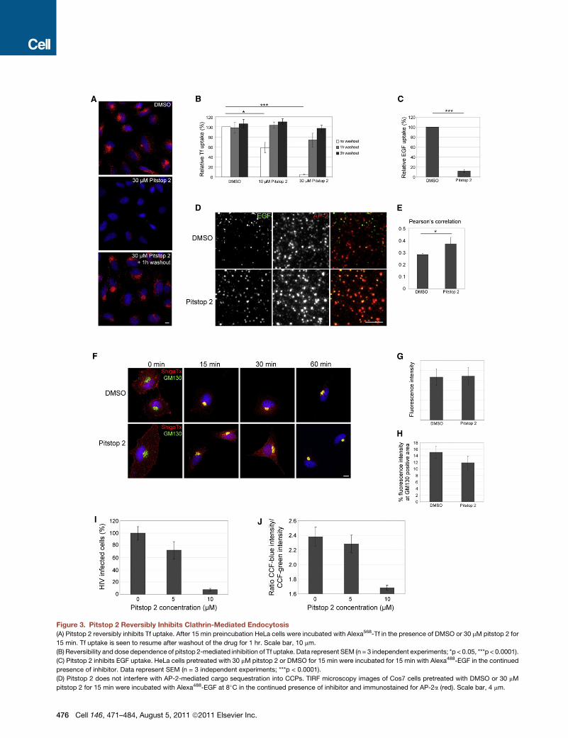
Figure 3. Pitstop 2 Reversibly Inhibits Clathrin-Mediated Endocytosis
(A) Pitstop 2 reversibly inhibits Tf uptake. After 15 min preincubation HeLa cells were incubated with Alexa568-Tf in the presence of DMSO or 30 mM pitstop 2 for
15 min. Tf uptake is seen to resume after washout of the drug for 1 hr. Scale bar, 10 mm.
(B) Reversibility and dose dependence of pitstop 2-mediated inhibition of Tf uptake. Data represent SEM (n = 3 independent experiments; *p < 0.05, ***p < 0.0001).
(C) Pitstop 2 inhibits EGF uptake. HeLa cells pretreated with 30 mM pitstop 2 or DMSO for 15 min were incubated for 15 min with Alexa488-EGF in the continued
presence of inhibitor. Data represent SEM (n = 3 independent experiments; ***p < 0.0001).
(D) Pitstop 2 does not interfere with AP-2-mediated cargo sequestration into CCPs. TIRF microscopy images of Cos7 cells pretreated with DMSO or 30 mM
pitstop 2 for 15 min were incubated with Alexa488-EGF at 8�C in the continued presence of inhibitor and immunostained for AP-2a (red). Scale bar, 4 mm.
476 Cell 146, 471–484, August 5, 2011 ª2011 Elsevier Inc.

a minor decrease in the mean clathrin LC-eGFP intensity per
object in TIRFM (Figure 4D). We noted a slightly increased
dimming of CCPs in confocal when compared to TIRF imaging,
presumably due to increased bleaching of stalled clathrin
puncta. Thus, surprisingly, pitstop 2-mediated interference
with clathrin TD-ligand association does not have a strong effect
on the stability of membrane-associated clathrin, but dramati-
cally perturbs CCP dynamics.
To corroborate the role of clathrin TD function in CCP
dynamics fluorescence recovery after photobleaching (FRAP)
was paired with live cell spinning disc confocal microscopy
imaging. Following the bleaching laser pulse clathrin LC-eGFP
fluorescence rapidly recovered to about 80% of its initial value
with a t of approximately 30 s in control cells, consistent with
the mean CCP lifetimes measured by TIRFM-based particle
tracking (compare Figures 4B and 4C). Pitstop 2-treated cells
did not show recovery of clathrin LC-eGFP fluorescence within
120 s, confirming that clathrin TD function indeed regulates
CCP dynamics. Recovery was so slow that a reliable t value
could not be determined under the imaging conditions. These
data indicate that pitstop 2 stalls the maturation and/or
consumption of pre-existing CCPs.
We next asked whether clathrin TD function may be required
for the de novo assembly of clathrin-coated structures. We
made use of 1-butanol, an alcohol which depletes cellular
PI(4,5)P2 levels causing CCP disassembly and loss of clathrin
from the plasma membrane (Boucrot et al., 2006; Henne et al.,
2010). Clathrin LC-eGFP containing pits rapidly reformed after
washout of 1-butanol in control and pitstop 2-treated cells (Fig-
ure S3A). To corroborate that TD-ligand interactions are not
required for the de novo clathrin recruitment, we re-expressed
siRNA-resistant clathrin HC mutants in cells depleted of endog-
enous clathrin HC. Mutation of R64A/Q89M/F91A within the cla-
thrin TD nearly abolished its ability to associate with amphiphysin
to an extent similar to that seen for an amphiphysin mutant lack-
ing its clathrin box (compare Figure 1F) but did not impair recruit-
ment of full-length clathrin HC (R64A/Q89M/F91A) to the plasma
membrane or to the TGN (Figure S3C). A clathrin HC mutant
lacking the TD altogether (DTD) was also recruited to mem-
branes (Figure S3C). We conclude that TD-ligand interactions
are dispensable for clathrin recruitment to membranes.
If indeed pitstop 2 mainly interferes with CCP dynamics, one
would expect relatively subtle effects on the distribution of
endogenous endocytic proteins. Indeed, no significant changes
were observed in the colocalization of clathrin, FCHo 1/2, inter-
sectin, AP-2, or dynamin 2 (Figures S4A and S4B). We also did
not detect overt changes with respect to the steady-state local-
ization of HIP1, Dab2, GAK/auxilin2, or CALM (data not shown).
(E) Pearson’s correlation between Alexa488-EGF and AP-2. Data represent SEM
(F) Pitstop 2 does not affect clathrin-independent endocytosis of Shiga toxin. HeL
toxin (red) and the cis-Golgi marker GM130 (green). Scale bar, 10 mm.
(G and H) Quantitative analysis of total and Golgi localized fractions of internalized
Scale bar, 10 mm.
(I–J) Pitstop 2 blocks HIV infection. (I) Shown is the HIV Tat-driven luciferase activ
HIV Tat-driven luciferase activity in mock-treated cells was set as 100% infectio
tiometric measurements (CCF-blue/CCF-green) of substrate cleavage by b-lactam
See also Figure S3.
This is not surprising as many endocytic proteins likely are re-
tained at the plasma membrane by multiple interactions, i.e.,
by binding to AP-2 and/or membrane PI(4,5)P2 as well as to
each other (Schmid and McMahon, 2007).
Among the endocytic proteins analyzed, two clathrin acces-
sory proteins displayed altered localizations. Endogenous am-
phiphysin 1 was partially lost from the plasma membrane of pit-
stop 2-treated cells (Figures S4C and S4D). Conversely, the
mean intensity of SNX9-containing puncta was increased in pit-
stop 2-treated cells, often resulting in the formation of large clus-
ters (Figures S3E–S3G). These findings correlate with the recent
observation that amphiphysin and SNX9 operate at consecutive
steps during CME (Taylor et al., 2011) and suggest that clathrin
TD interactions may coordinate turnover of endocytic proteins
at nascent CCPs.
We also determined the distribution of various TGNor endoso-
mal proteins including AP-1, EEA1, Gadkin, CD63, TGN46, or the
mannose 6-phosphate receptor MPR46. None of these factors
showed a significant change in localization within 15 min of pit-
stop 2 application (Figures S5A and S5B). We did, however,
note a partial shift of transferrin receptor (TfR) from perinuclear
endosomes to the cell surface of pitstop 2-treated cells (Fig-
ure S4H), consistent with defective Tf endocytosis (Figures 3A
and 3B).
Dynamics of FCHo2, AP-2, and Dynamin in Pitstop2-Treated CellsSince pitstop 2 impaired clathrin dynamics we next examined
the behavior of other key endocytic proteins, which act either
prior to or concomitant with clathrin assembly. FCHo proteins
are early-acting factors that link PI(4,5)P2-rich sites to AP-2
binding scaffolds such as intersectin and Eps15/Eps15R (Henne
et al., 2010). TIRFM-based imaging of eGFP-FCHo2 dynamics in
Cos7 cells revealed that FCHo2-containing structures were rela-
tively long-lived, with lifetimes between 26 s and over 120 s. The
mean lifetimes of FCHo2-eGFP-containing puncta were dramat-
ically increased following application of pitstop 2with puncta ap-
pearing nearly immobile (Figures 5A and 5B). Moreover, FCHo2-
positive spots failed to recover in FRAP experiments (Figures 5C
and 5D). Hence, clathrin TD function is required for the produc-
tive consumption of nascent CCPs containing FCHo2. Consis-
tent with this and with the observation that pitstop 2 displaces
the PI(4,5)P2-consuming 5-phosphatases OCRL and synaptoja-
nin 1-p170 from the clathrin TD (Figure 1E) pitstop 2 treatment
also stabilized clathrin coats on membranes in vitro (Figures
S3D and S3E).
Surprisingly, pitstop 2 induced a comparatively minor shift in
the mean life span of AP-2s-eGFP puncta (Figures 5E and 5F).
(n = 3 independent experiments; *p < 0.05).
a cells pretreated with DMSO or 30 mM pitstop 2 stained for internalizing Shiga
Shiga toxin after 30 min of internalization. (SEM; differences are insignificant).
ity in cells treated with increasing concentrations of pitstop 2 during infection.
n. (J) Cytosolic entry of HIV-1-associated b-lactamase activity. Shown are ra-
ase in cells treated with increasing concentrations of pitstop 2 during infection.
Cell 146, 471–484, August 5, 2011 ª2011 Elsevier Inc. 477

Figure 4. Pitstop 2 Affects the Dynamics of Clathrin-Coated Pits
Effects of pitstop 2 on clathrin dynamics were analyzed by live-cell TIRF or spinning disc confocal microscopy in Cos7 cells stably expressing eGFP-clathrin LC
at 37�C.(A) View of the clathrin distribution at the bottom surface of a cell pretreated for 5 min with 0.1% DMSO or 30 mM pitstop 2. Kymographs (right) display the
dynamics of clathrin during the time-lapse series (2 min, 0.5 Hz). Scale bar, 10 mm.
(B) Pooled lifetime distribution of CCPs depicted in (A). Values were determined from the time between the appearance and disappearance of clathrin puncta
(SEM; n = 2 independent experiments; **p < 0.01).
(C) Cumulative plot of clathrin lifetime distributions.
(D) Intensity of clathrin puncta in pitstop 2-treated cells. Mean fluorescence intensities of clathrin puncta in living cells were measured following addition of 0.1%
DMSO or 30 mM pitstop 2 (SEM; n = 3 independent experiments; **p < 0.01).
478 Cell 146, 471–484, August 5, 2011 ª2011 Elsevier Inc.
These results are consistent with our observation that pitstop
2-treated cells remain able to sequester cargo into CCPs,
a process largely mediated by AP-2 (compare Figures 3D
and 3E).
Finally, we investigated the life span of the fissioning enzyme
dynamin 2. Dynamin 2-eGFP displayed a lifetime distribution
similar to that observed for AP-2 or clathrin. Pitstop 2 shifted
this distribution to longer life spans with a behavior intermediary
between the effects seen for clathrin and AP-2 (Figures 5G and
5H). Prolonged lifetimes of dynamin puncta at CCPs also fit
well with the altered distribution of the dynamin-binding clathrin
accessory proteins amphiphysin and SNX9 observed in pitstop-
treated cells (Figures S3C–S3G).
Morphological Analysis of Pitstop 2-Treated CellsOur live imaging data collectively argue that pitstop 2-mediated
inhibition of CME results from defects in endocytic protein
dynamics. As CME is a constitutive process in nonneuronal cells
with CCPs forming and disassembling continuously, one would
expect that acute perturbation of endocytic protein dynamics
by pitstop 2-mediated inhibition of clathrin TD function results
in acute ‘‘trapping’’ of clathrin-coated intermediates represent-
ing multiple stages of the pathway. To this aim, control or pitstop
2-treated Cos7 cells were subjected to electronmicroscopic and
morphometric analyses. Clathrin-coated endocytic intermedi-
ates were classified according to their morphological profiles
as shallow CCPs, nonconstricted u-shaped CCPs, constricted
U-shaped CCPs corresponding to late fission stages, and struc-
tures containing a complete clathrin coat (Figure 6A). These latter
intermediatesmay comprise free clathrin-coated vesicles aswell
as late intermediates that remain attached to the plasma
membrane. Endocytic profiles of control cells and those incu-
bated with pitstop 2 appeared very similar. No major changes
in the total number of CCPs (Figure 6B) or in the representation
of various endocytic stages were observed (Figure 6C). We
conclude that pitstop-mediated inhibition of clathrin TD function
acutely arrests CME at multiple stages.
Pitstops Inhibit Endocytic Recycling of SynapticVesicles in Neurons and In SituOne of themost prominent functions of CME in vertebrates is the
exo-endocytic recycling of synaptic vesicle (SV) membranes at
nerve terminals (Dittman and Ryan, 2009). Unlike CME in non-
neuronal cells endocytic reformation of SVs in neurons is strictly
coupled to stimulation-dependent exocytosis resulting in a wave
of endocytic activity (Dittman and Ryan, 2009). We tested the
effects of pitstop 2-mediated clathrin inhibition on SV reforma-
tion in primary hippocampal neurons. Chemical stimulation of
SV exocytosis resulted in a pronounced depletion of SVs from
nerve terminals of pitstop 2-treated neurons, suggesting that
SV reformation by endocytosis was inhibited. No significant
(E) Pitstop 2 stalls clathrin dynamics. Fluorescence recovery after photobleachin
imaged 5 min after the addition of 30 mM pitstop 2 or DMSO (0.1%) using spinnin
FRAP, followed by imaging for another 2 min after photobleaching. The panels rep
pitstop 2-treated cells acquired before (�6 s), right after (0 s), 1 min, and 2 min a
(F) Corrected relative fluorescence intensities of clathrin puncta in the FRAP regi
See also Figure S4.
changes in SV numbers were seen in resting control neurons
(Figures S6A–S6C). Loss of SV membranes in pitstop 2-treated
neurons appeared to be counterbalanced partly by presynaptic
membrane expansion (Figure S6D).
The poor membrane penetration of pitstop 1 prevented anal-
ysis of its effects on endocytosis in living cells. However, its
high solubility in aqueous media provided ideal conditions for
its intracellular application by microinjection. We therefore as-
sessed the effect of pitstops 1 and 2 on SV endocytosis in the
lamprey reticulospinal synapse (Pechstein et al., 2010). We mi-
croinjected pitstop 1 into lamprey reticulospinal axons and stim-
ulated at 5 Hz for 20min to induce SV recycling at a physiological
rate. Analysis of ultrathin sections revealed a profound loss of
SVs at active zones in synapses from these axons (Figures 7A,
7B, and 7G), compared to control noninjected axons (Figure 7C).
No morphological alterations were observed in axons microin-
jected with pitstop 1 and kept at rest (Figure 7E). Loss of SVs
in stimulated axons was accompanied by a dramatic expansion
of the plasma membrane, which due to anatomical constraints
formed membrane pockets around the active zone (Figures 7A,
7B, and 7H). A significant accumulation of CCPs was observed
within the membrane expansions compared with control syn-
apses (Figures 7A–7C and 7I). Consistent with our observations
in Cos7 cells, no dramatic alterations in stage representation of
CCPs were observed (Figure 7J). No detectable morphological
differences of CCPs were detected at high-magnification
(Figures 7D and 7E). A similar phenotype including a pronounced
loss of SVs and accumulation of multistage CCPs was observed
in pitstop 2-injected reticulospinal synapses (Figure S7). We
conclude that intra-axonal application of pitstops strongly
inhibits SV recycling in situ by inhibiting CME at multiple stages.
DISCUSSION
We have developed chemically distinct small molecule inhibitors
of clathrin TD function termed pitstops that competitively inter-
fere with the association of endocytic clathrin box ligands.
In vitro experiments in conjunction with structural data based
on protein X-ray crystallography as well as experiments in living
cells clearly demonstrated that these compounds selectively
perturb clathrin TD function. Effects of pitstop 2 on CME are
evident within a few minutes of cell treatment, presumably only
being limited by the rate of drug diffusion across the cell
membrane. We observe reversible inhibition of CME in a broad
variety of cells at concentrations closely matching the IC50
values of the in vitro binding experiments. While pitstop 2 readily
crosses cell membranes, pitstop 1 has greatly reduced efficacy,
limiting its use to live cell applications based on intracellular
application. The similarity of phenotypes exhibited by pitstops
1 and 2 in lamprey reticulospinal synapses in situ and in Cos 7
cells in vitro suggests that both compounds share a common
g (FRAP) of clathrin LC-labeled CCPs in an area of 15 3 15 mm2. Cells were
g disc confocal microscopy. Frames were taken at 0.5 Hz, starting 10 s before
resent images of the FRAP region and an unbleached control area in DMSO- or
fter photobleaching. Scale bar, 2 mm.
on over time (SEM; n = 2 independent experiments; **p < 0.01).
Cell 146, 471–484, August 5, 2011 ª2011 Elsevier Inc. 479

Figure 5. Differential Effects of Pitstop 2 on the Dynamics of FCHo2, AP-2, and Dynamin
Effects of pitstop 2 on endocytic protein dynamics were analyzed by live-cell TIRF or spinning disc confocal microscopy in Cos7 cells transiently expressing low
levels of eGFP-tagged FCHo2, AP-2s, or dynamin 2.
(A, E, and G) Pooled lifetime distributions of eGFP-FCHo2 (A), AP-2s-eGFP (E), and Dyn2-eGFP (G) puncta in cells pretreated for 5min with 0.1%DMSO or 30 mM
pitstop 2. Plots represent the time between the appearance and disappearance of the fusion protein-containing puncta in a time lapse-series acquired for 2min at
0.5 Hz (SEM; n = 2 independent experiments; *p < 0.05, **p < 0.01, ***p < 0.001).
(C) Effect of pitstop 2 on FRAP of FCHo2-containing puncta in an area of 153 15 mm2. Samples were analyzed as described in the legend to Figure 4E (SEM; n = 2
independent experiments).
(D) Images of the FRAP region in DMSO- or pitstop 2-treated cells acquired before (�6 s), right after (0 s), and 2 min after photobleaching. Scale bar, 2 mm.
(B, F, and H) Cumulative plots of the lifetime distributions of FCHo2 (B), AP-2 (F), and Dynamin 2 (H).
See also Figure S5.
480 Cell 146, 471–484, August 5, 2011 ª2011 Elsevier Inc.

Figure 6. Ultrastructural Analysis of Intermediates Observed in Pitstop 2-Treated Cells
(A and B) Representative examples of clathrin-coated structures observed at the plasma membrane of Cos7 cells pretreated for 5 min with 0.1% DMSO (A) or
30 mM pitstop 2 (B). Scale bar, 200 nm. Morphological groups were shallow CCPs (stage 1), nonconstricted u-shaped CCPs (stage 2), constricted U-shaped
CCPs (stage 3), or structures containing complete clathrin coats (stage 4).
(C) Bar diagramdisplaying the numbers of clathrin-coated endocytic intermediates observed along the perimeter of Cos7 cells treated with 0.1%DMSO or 30 mM
pitstop 2.
(D) Bar diagram showing the relative abundance of different stages of coated endocytic intermediates observed along the perimeter of Cos7 cells treated with
0.1% DMSO or 30 mM pitstop 2.
See also Figure S6.
clathrin TD-based molecular mechanism of action. In the pres-
ence of pitstop 2 clathrin-independent internalization pathways
and secretory traffic (data not shown) remain unperturbed.
Therefore, pitstops represent new cellular tools to selectively
inhibit CME.
Pitstops Block CME by Interfering with CCP DynamicsThe use of pitstops in living cells revealed an unexpected role for
the clathrin TD and its ligands in endocytic pit dynamics while
clathrin recruitment was unperturbed. Together with the obser-
vation that clathrin lacking its TD is targeted to CCPs, it is likely
that other interactions such as the association of the b-ear
domain of AP-2 with the clathrin leg (Knuehl et al., 2006) mediate
clathrin recruitment to membranes.
Our data are explained by a model where pitstops interfere
with the progression of CCPs at multiple stages, consistent
with the functional diversity of clathrin TD ligands. Clathrin is
one of a few molecules within the endocytic network with the
ability to spatiotemporally regulate vesicle formation and disas-
sembly after fission. Most endocytic proteins partially lack stable
tertiary structure or contain extensive segments of natively
unfolded polypeptide chains, but clathrin triskelia are stable enti-
ties with a fixed arrangement, distance, and geometry between
their TDs (Brodsky et al., 2001). We, thus, favor a model whereby
clathrin assembled at nascent endocytic sites serves as an orga-
nizing scaffold that regulates CCP dynamics by providing
spatially defined binding sites on its TD for accessory proteins
that drive CCP maturation and disassembly (Figure 1A, right).
The specific mechanisms that regulate such transitions within
an assembling or disassembling CCP are unknown, but pitstops
together with dynamin inhibitors (Joshi et al., 2010; Macia et al.,
2006) may provide powerful tools to tackle these questions.
Unraveling the Roles of Clathrin in Cell PhysiologyEvidence from morphological, genetic, and knockdown studies
has suggested that CME may be a key mechanism for SV recy-
cling (Dittman andRyan, 2009). The fact that synapses can adapt
to chronic loss of endocytic proteins is a substantial drawback
for RNAi and genetic approaches. Our data using pitstops
provide a direct demonstration for an important role of clathrin
Cell 146, 471–484, August 5, 2011 ª2011 Elsevier Inc. 481

Figure 7. Pitstop 1 Inhibits Activity-Induced SV Recycling in Lamprey Reticulospinal Synapses
(A and B) Electron micrographs of reticulospinal synapses microinjected with pitstop 1 and stimulated at 5 Hz for 20 min. SVs (sv) are depleted from sites of
release. Instead, large membrane expansions and pockets (asterisks) are seen, which in some sections (e.g., in B) surrounded the active zone (marked with thick
arrows). Boxed area containing clathrin-coated pits (CCPs) is shown as inset at higher magnification in (A).
(C) EM image of a control noninjected synapse from the same spinal cord preparation.
(D and E) Constricted CCPs from a control noninjected axon and a synapse microinjected with pitstop 1. Note the similar shape of CCPs and the presence of
clathrin coats in both cases.
(F) Electron micrograph of a synapse microinjected with pitstop 1 but kept at rest.
(G–I) Bar diagrams displaying numbers of SVs (Nsv), curvature index (CI), and the number of clathrin-coated intermediates (Nccp) between control synapses
stimulated at 5 Hz (gray bars) and pitstop 1-microinjected stimulated synapses (black bars), respectively. ax, axoplasmic matrix; d, dendrite.
(J) Bar diagram showing the relative abundance of different stages of coated endocytic intermediates (see legend to Figure 6A) in control synapses stimulated at
5 Hz (gray bars) and stimulated synapses microinjected with pitstop 1 (black bars).
Scale bars for (A–C) and (F), 0.5 mm and for (D) and (E), 100 nm.
See also Figure S7.
in SV recycling in vertebrate neurons under conditions of high
activity. The development of pitstops will allow further molecular
dissection of clathrin function under different stimulation para-
digms and in different preparations.
Our data also lend strong support to the hypothesis that HIV
enters cells largely via CME (Daecke et al., 2005; Miyauchi
et al., 2009). Hence, we predict that the inhibitors developed
here or derivatives thereof may serve as lead compounds for
the development of drugs blocking the entry of viruses and path-
ogens, which hijack the clathrin machinery, including HIV, hepa-
titis C virus (Blanchard et al., 2006), ebola virus (Bhattacharyya
et al., 2010), and Listeria monocytogenes (Veiga et al., 2007).
It is to be expected that acute interference with clathrin TD
function will affect other clathrin-dependent pathways. Clathrin
plays roles in bidirectional traffic between the trans-Golgi net-
work and endosomes, a pathway mediated by AP-1 and GGA
adaptors (Bonifacino and Traub, 2003; Hirst et al., 2009). Clathrin
482 Cell 146, 471–484, August 5, 2011 ª2011 Elsevier Inc.
coats have also been observed on endosomes, where clathrin
may fulfill a structural role in the segregation of endosomal
membrane domains involved in targeting of growth factor recep-
tors (Goh et al., 2010) to the multivesicular body pathway or in
directing retrograde transport (Saint-Pol et al., 2004). Finally,
clathrin has been detected at the mitotic spindle where it has
been proposed to stabilize microtubules (Royle et al., 2005).
Whether and to which degree these functions depend on the
association of conventional clathrin box ligands with the TD can
now be addressed using the small molecule inhibitor approach.
Pitstops will serve as an important tool to address these ques-
tions as well as more generally the role of clathrin function in cell
physiology including cell signaling. The availability of crystal
structures of pitstops in complex with the clathrin TD will pave
the way for the development of second- and third-generation
compounds with increased affinity and improved molecular
properties that could serve as prodrugs for the treatment of

multiple diseases ranging from neurological disorders to micro-
bial infections.
EXPERIMENTAL PROCEDURES
Information regarding compound synthesis, antibodies, DNA constructs, cell
viability assays, protein purification, crystallography, fluorescence micros-
copy, Shiga toxin uptake, and HIV infection are available as supplementary
material online.
Screening Procedure
Seventeen thousand small molecules (100 mM) from the central open access
technology platform of the ChemBioNet hosted by FMP (http://fmp-berlin.
info/screening_unit.html) were screened using an automated platform for their
ability to perturb clathrin TD-amphiphysin association using an ELISA-based
assay in a 384-well format (see below for details). Forty-eight initial hits
(>80% inhibition) were selected. Ten of these hits could be validated by
dose-response analysis (at 3–300 mM) and structural hit clustering. Hits were
verified using compounds re-ordered from the ChemDiv collection. Two lead
compounds were selected based on the absence of off-target effects and inhi-
bition of transferrin endocytosis. Focused libraries synthesized based on these
leads were re-screened as above resulting in the identification of pitstops 1
and 2. Pitstop, pitstop 1, and pitstop 2, are trademarks of Freie Universitat Ber-
lin, Children’s Medical Research Institute and Newcastle Innovation Ltd. Pit-
stop 1 and pitstop 2 are available from Ascent Scientific Ltd.
ELISA-Based Binding Assay
Purified His6-tagged protein was diluted into screening buffer (20 mM HEPES
[pH 7.4], 50 mM NaCl, 1 mM DTT, 1 mM PMSF), added to a 384-well ELISA
plate (high-binding PS Microplate, Greiner Bio-One) and bound for 1 hr at
RT. Nonspecific binding was blocked by incubation in 50 ml blocking buffer
(20 mM HEPES [pH 7.4], 50 mM NaCl, 1 mM DTT, 1 mM PMSF, 2% BSA,
2.5% milk) overnight at 4�C. Following extensive washes with 20 mM HEPES
(pH 7.4), 50 mM NaCl, 0.05% Tween 20, chemical compounds diluted in
DMSO (10 ml) were added and incubated together with GST-tagged protein
for 1 hr at RT in screening buffer. After three washes, HRP-coupled anti-
GST antibodies were added in screening buffer and the plate was incubated
for 15 min at RT. Following additional washes, bound protein was determined
by photometric measurement in a plate reader at 450 nm using TMB as
a substrate (Pierce Biotechnology).
Confocal and TIRF-Based Live Imaging
For live cell microscopy Cos7 cells stably expressing eGFP-clathrin LC (Gai-
darov et al., 1999), or transiently expressing FCHo2-eGFP, AP-2s-eGFP (Gai-
darov et al., 1999), and dynamin2-eGFP were used. CCP dynamics were
imaged by TIRFM (Visitron) under the control of Slidebook 5 (3i Inc). Time
series of 2 min (at 0.5 Hz) were acquired 5 min after addition of 30 mMpitstop 2
or DMSO (0.1%) at 37�C. Fluorescence recovery after photobleaching (FRAP)
experiments were performed using a spinning disc confocal microscope (Per-
kin Elmer) controlled by Volocity (Improvision). Time series were acquired
5 min after addition of 30 mM pitstop 2 or DMSO (0.1%) at 37�C. Cells were
imaged for 12 s, then bleached in a region of 15 3 15 mm2, and imaged for
an additional 120 s at 0.5 Hz. Fluorescence recovery was analyzed by defining
CCPs in the FRAP region before bleaching, followed by measuring the fluores-
cence intensity in the FRAP region over time. Intensity values were corrected
for photobleaching in a nonbleached control area.
Electron Microscopy
Electron microscopic analyses were essentially done as described in Fergu-
son et al. (2009). See Extended Experimental Procedures for details.
Microinjection of Compounds into Lamprey Reticulospinal Axons
and Analysis by Electron Microscopy
Microinjection experiments and electronmicroscopic analysis were essentially
done as previously described (Pechstein et al., 2010). See supplement for
details.
ACCESSION NUMBERS
Structures are accessible under PDB codes 2xzg and 2xzh.
SUPPLEMENTAL INFORMATION
Supplemental Information includes Extended Experimental Procedures, two
tables, and seven figures and can be found with this article online at doi:10.
1016/j.cell.2011.06.025.
ACKNOWLEDGMENTS
We thank Drs. Gilbert Di Paolo, Ludger Johannes, and Oliver Daumke for crit-
ical comments and Drs. Harvey Mc Mahon, Ludger Johannes, Pietro De
Camilli, Tom Sudhof, and Sven Carlsson for reagents. Cytotoxicity assays
were conducted with help from Aimee Novelle, Swetha Perera, and Dr. Jayne
Gilbert. This work was supported by grants from the Deutsche Forschungs-
gemeinschaft (FOR 806-HA2686/3-1,3-2; SFB765/B4 to V.H.; Exc-257), the
European Science Foundation (Euromembrane-HA2686/6-1), and the
Swedish Research Council (grants 13473 and 20587 to O.S.).
Received: December 21, 2010
Revised: May 6, 2011
Accepted: June 14, 2011
Published: August 4, 2011
REFERENCES
Bhattacharyya, S., Warfield, K.L., Ruthel, G., Bavari, S., Aman, M.J., and
Hope, T.J. (2010). Ebola virus uses clathrin-mediated endocytosis as an entry
pathway. Virology 401, 18–28.
Blanchard, E., Belouzard, S., Goueslain, L., Wakita, T., Dubuisson, J.,
Wychowski, C., and Rouille, Y. (2006). Hepatitis C virus entry depends on
clathrin-mediated endocytosis. J. Virol. 80, 6964–6972.
Bonifacino, J.S., and Traub, L.M. (2003). Signals for sorting of transmembrane
proteins to endosomes and lysosomes. Annu. Rev. Biochem. 72, 395–447.
Boucrot, E., Saffarian, S., Massol, R., Kirchhausen, T., and Ehrlich, M. (2006).
Role of lipids and actin in the formation of clathrin-coated pits. Exp. Cell Res.
312, 4036–4048.
Brodsky, F.M., Chen, C.Y., Knuehl, C., Towler, M.C., and Wakeham, D.E.
(2001). Biological basket weaving: formation and function of clathrin-coated
vesicles. Annu. Rev. Cell Dev. Biol. 17, 517–568.
Conner, S.D., and Schmid, S.L. (2003). Regulated portals of entry into the cell.
Nature 422, 37–44.
Daecke, J., Fackler, O.T., Dittmar, M.T., and Krausslich, H.G. (2005). Involve-
ment of clathrin-mediated endocytosis in human immunodeficiency virus type
1 entry. J. Virol. 79, 1581–1594.
Dittman, J., and Ryan, T.A. (2009). Molecular circuitry of endocytosis at nerve
terminals. Annu. Rev. Cell Dev. Biol. 25, 133–160.
Edeling, M.A., Smith, C., and Owen, D. (2006). Life of a clathrin coat: insights
from clathrin and AP structures. Nat. Rev. Mol. Cell Biol. 7, 32–44.
Ehrlich, M., Boll, W., Van Oijen, A., Hariharan, R., Chandran, K., Nibert, M.L.,
and Kirchhausen, T. (2004). Endocytosis by random initiation and stabilization
of clathrin-coated pits. Cell 118, 591–605.
Ferguson, S.M., Raimondi, A., Paradise, S., Shen, H.,Mesaki, K., Ferguson, A.,
Destaing, O., Ko, G., Takasaki, J., Cremona, O., et al. (2009). Coordinated
actions of actin and BAR proteins upstream of dynamin at endocytic cla-
thrin-coated pits. Dev. Cell 17, 811–822.
Gaidarov, I., Santini, F., Warren, R.A., and Keen, J.H. (1999). Spatial control of
coated-pit dynamics in living cells. Nat. Cell Biol. 1, 1–7.
Goh, L.K., Huang, F., Kim, W., Gygi, S., and Sorkin, A. (2010). Multiple mech-
anisms collectively regulate clathrin-mediated endocytosis of the epidermal
growth factor receptor. J. Cell Biol. 189, 871–883.
Cell 146, 471–484, August 5, 2011 ª2011 Elsevier Inc. 483

Heerssen, H., Fetter, R.D., and Davis, G.W. (2008). Clathrin dependence of
synaptic-vesicle formation at the Drosophila neuromuscular junction. Curr.
Biol. 18, 401–409.
Henne,W.M., Boucrot, E., Meinecke, M., Evergren, E., Vallis, Y., Mittal, R., and
McMahon, H.T. (2010). FCHo proteins are nucleators of clathrin-mediated
endocytosis. Science 328, 1281–1284.
Hinrichsen, L., Meyerholz, A., Groos, S., and Ungewickell, E.J. (2006). Bending
a membrane: how clathrin affects budding. Proc. Natl. Acad. Sci. USA 103,
8715–8720.
Hirst, J., Sahlender, D.A., Choma, M., Sinka, R., Harbour, M.E., Parkinson, M.,
and Robinson, M.S. (2009). Spatial and functional relationship of GGAs and
AP-1 in Drosophila and HeLa cells. Traffic 10, 1696–1710.
Johannes, L., and Romer, W. (2010). Shiga toxins—from cell biology to
biomedical applications. Nat. Rev. Microbiol. 8, 105–116.
Joshi, S., Perera, S., Gilbert, J., Smith, C.M., Mariana, A., Gordon, C.P., Sakoff,
J.A., McCluskey, A., Robinson, P.J., Braithwaite, A.W., and Chircop,M. (2010).
The dynamin inhibitors MiTMAB and OcTMAB induce cytokinesis failure and
inhibit cell proliferation in human cancer cells. Mol. Cancer Ther. 9, 1995–2006.
Knuehl, C., Chen, C.Y., Manalo, V., Hwang, P.K., Ota, N., and Brodsky, F.M.
(2006). Novel binding sites on clathrin and adaptors regulate distinct aspects
of coat assembly. Traffic 7, 1688–1700.
Macia, E., Ehrlich, M., Massol, R., Boucrot, E., Brunner, C., and Kirchhausen,
T. (2006). Dynasore, a cell-permeable inhibitor of dynamin. Dev. Cell 10,
839–850.
Mettlen, M., Stoeber, M., Loerke, D., Antonescu, C.N., Danuser, G., and
Schmid, S.L. (2009). Endocytic accessory proteins are functionally distin-
guished by their differential effects on the maturation of clathrin-coated pits.
Mol. Biol. Cell 20, 3251–3260.
Miyauchi, K., Kim, Y., Latinovic, O., Morozov, V., and Melikyan, G.B. (2009).
HIV enters cells via endocytosis and dynamin-dependent fusion with endo-
somes. Cell 137, 433–444.
484 Cell 146, 471–484, August 5, 2011 ª2011 Elsevier Inc.
Motley, A., Bright, N.A., Seaman, M.N., and Robinson, M.S. (2003). Clathrin-
mediated endocytosis in AP-2-depleted cells. J. Cell Biol. 162, 909–918.
Pechstein, A., Bacetic, J., Vahedi-Faridi, A., Gromova, K., Sundborger, A.,
Tomlin, N., Krainer, G., Vorontsova, O., Schafer, J.G., Owe, S.G., et al.
(2010). Regulation of synaptic vesicle recycling by complex formation between
intersectin 1 and the clathrin adaptor complex AP2. Proc. Natl. Acad. Sci. USA
107, 4206–4211.
Royle, S.J., Bright, N.A., and Lagnado, L. (2005). Clathrin is required for the
function of the mitotic spindle. Nature 434, 1152–1157.
Saffarian, S., Cocucci, E., and Kirchhausen, T. (2009). Distinct dynamics of en-
docytic clathrin-coated pits and coated plaques. PLoS Biol. 7, e1000191.
Saint-Pol, A., Yelamos, B., Amessou, M., Mills, I.G., Dugast, M., Tenza, D.,
Schu, P., Antony, C., McMahon, H.T., Lamaze, C., and Johannes, L. (2004).
Clathrin adaptor epsinR is required for retrograde sorting on early endosomal
membranes. Dev. Cell 6, 525–538.
Schmid, E.M., and McMahon, H.T. (2007). Integrating molecular and network
biology to decode endocytosis. Nature 448, 883–888.
Slepnev, V.I., Ochoa, G.C., Butler, M.H., and De Camilli, P. (2000). Tandem
arrangement of the clathrin and AP-2 binding domains in amphiphysin 1 and
disruption of clathrin coat function by amphiphysin fragments comprising
these sites. J. Biol. Chem. 275, 17583–17589.
Taylor, M.J., Perrais, D., and Merrifield, C.J. (2011). A high precision survey of
the molecular dynamics of mammalian clathrin-mediated endocytosis. PLoS
Biol. 9, e1000604.
ter Haar, E., Harrison, S.C., and Kirchhausen, T. (2000). Peptide-in-groove
interactions link target proteins to the beta-propeller of clathrin. Proc. Natl.
Acad. Sci. USA 97, 1096–1100.
Veiga, E., Guttman, J.A., Bonazzi, M., Boucrot, E., Toledo-Arana, A., Lin, A.E.,
Enninga, J., Pizarro-Cerda, J., Finlay, B.B., Kirchhausen, T., and Cossart, P.
(2007). Invasive and adherent bacterial pathogens co-Opt host clathrin for
infection. Cell Host Microbe 2, 340–351.