The Intracellular Functions of Ot6~ 4 Integrin Are Regulated by EGF
13
The Intracellular Functions of Ot6~ 4 Integrin Are Regulated by EGF Fabrizio Mainiero, Angela Pepe, Mitchell Yeon, Yunling Ren, and Filippo G. Giancotti Department of Pathology, Kaplan Cancer Center, New York University School of Medicine, New York 10016 Abstract. Upon ligand binding, the 0/.6[3 4 integrin be- comes phosphorylated on tyrosine residues and com- bines sequentially with the adaptor molecules Shc and Grb2, linking to the ras pathway, and with cytoskeletal elements of hemidesmosomes. Since et6[34is expressed in a variety of tissues regulated by the EGF receptor (EGFR), we have examined the effects of EGF on the cytoskeletal and signaling functions of O~6[~ 4. Experi- ments of immunoblotting with anti-phosphotyrosine antibodies and immunoprecipitation followed by phos- phoamino acid analysis and phosphopeptide mapping showed that activation of the EGFR causes phosphory- lation of the 1~4 subunit at multiple tyrosine residues, and this event requires ligation of the integrin by lami- nins or specific antibodies. Immunoprecipitation exper- iments indicated that stimulation with EGF does not result in association of 0t6134 with Shc. In contrast, EGF can partially suppress the recruitment of Shc to ligated Ot6[~ 4. Immunofluorescent analysis revealed that EGF treatment does not induce increased assembly of hemidesmosomes, but instead causes a deterioration of these adhesive structures. Finally, Boyden chamber as- says indicated that exposure to EGF results in upregu- lation of a6134-mediated cell migration toward laminins. We conclude that EGF-dependent signals suppress the association of activated tX6~ 4 with both signaling and cy- toskeletal molecules, but upregulate et6134-dependent cell migration. The changes in 0t6134 function induced by EGF may play a role during wound healing and tumor- igenesis. T o fully understand embryonic development, tissue repair, and tumor invasion, it is important to eluci- date the mechanisms by which growth factor- and integrin-dependent signals are integrated inside cells. It is known that integrins transmit positional cues from the ex- tracellular matrix to the cell interior, and the mechanisms by which these signals affect cellular responses to growth and differentiation factors are being actively investigated (Juliano and Haskill, 1993; Giancotti and Mainiero, 1994; Schwartz et al., 1995). Conversely, growth factors and cy- tokines can modulate a number of integrin-dependent functions, including cell adhesion (Serve et al., 1995; Ki- nashi et al., 1995), cell migration (Chen et al., 1993; Mat- thay et al., 1993; Klemke et al., 1994), and cytoskeletal or- ganization (Ridley and Hall, 1992; Ridley et al., 1992), but the mechanisms underlying these phenomena are less clear. The interaction between growth factor receptors and in- tegrins has been largely examined in fibroblasts and plate- lets. Most of the studies have focused on the focal adhe- sion kinase p125FAK (Schaller et al., 1992). In addition to being activated and undergoing autophosphorylation in response to ligation of 131and 133 integrins (Guan and Shal- Address all correspondence to Filippo G. Giancotti, Department of Pa- thology, MSB 548, New York University School of Medicine, 550 First Avenue, New York, NY 10016. Tel.: (212) 263-5343. Fax: (212) 263-8211. e-mail: giancF01 @mcrcr6.med.nyu.edu. Y. Ren's present address is 21-17 36th Street, Astoria, NY 11105. loway, 1992; Hanks et al., 1992; Lipfert et al., 1992), p125FAKis the target of signals originating from a number of growth factors and mitogenic neuropeptides (Zachary and Rozengurt, 1992). The activation of p125FAK has been linked to changes potentially important for the regulation of actin cytoskeleton, such as the phosphorylation of paxil- lin and tensin (Burridge et al., 1992; Bockholt and Bur- ridge, 1993) and the activation of Rho (McNamee et al., 1992; Chong et al., 1994) and PI-3 kinase (Chen and Guan, 1994). In addition, activated p125FAK can combine with the Grb2/mSOS complex potentially leading to stimulation of the ras-MAP (mitogen-activated protein) kinase pathway (Schlaepfer et al., 1994), and insulin stimulation promotes association of the etv133integrin with the Insulin Receptor Substrate 1 and the Grb2/mSOS complex (Vuori and Ruo- slahti, 1994). These observations suggest that integrin- and growth factor-dependent signals may converge on p125FAK and Insulin Receptor Substrate 1 to regulate gene expres- sion and the actin cytoskeleton. Much less is known about the integration of growth fac- tor- and integrin-dependent signals in epithelial and other cells that are in contact with the basement membrane. The ~6134 integrin is expressed in epithelial, endothelial, and Schwann cells and binds to various isoforms of the base- ment membrane component laminin (Lee et al., 1992; Niessen et al., 1994; Spinardi et al., 1995). Our previous studies have focused on the mechanisms by which this in- tegrin interacts with the cytoskeleton and with signaling molecules. In contrast to other integrins that localize to fo- © The Rockefeller University Press, 0021-9525/96/07/241/13 $2.00 The Journal of Cell Biology, Volume 134, Number 1, July 1996 241-253 241 on June 12, 2015 jcb.rupress.org Downloaded from Published July 1, 1996
Transcript of The Intracellular Functions of Ot6~ 4 Integrin Are Regulated by EGF

The Intracellular Functions of Ot6~ 4 Integrin Are Regulated by EGF Fabrizio Mainiero, Angela Pepe, Mitchell Yeon, Yunling Ren, and Filippo G. Giancotti
Department of Pathology, Kaplan Cancer Center, New York University School of Medicine, New York 10016
A b s t r a c t . Upon ligand binding, the 0/.6[3 4 integrin be- comes phosphorylated on tyrosine residues and com- bines sequentially with the adaptor molecules Shc and Grb2, linking to the ras pathway, and with cytoskeletal elements of hemidesmosomes. Since et6[34 is expressed in a variety of tissues regulated by the EGF receptor (EGFR), we have examined the effects of EGF on the cytoskeletal and signaling functions of O~6[~ 4. Experi- ments of immunoblotting with anti-phosphotyrosine antibodies and immunoprecipitation followed by phos- phoamino acid analysis and phosphopeptide mapping showed that activation of the EGFR causes phosphory- lation of the 1~4 subunit at multiple tyrosine residues, and this event requires ligation of the integrin by lami- nins or specific antibodies. Immunoprecipitation exper-
iments indicated that stimulation with EGF does not result in association of 0t6134 with Shc. In contrast, EGF can partially suppress the recruitment of Shc to ligated Ot6[~ 4. Immunofluorescent analysis revealed that EGF treatment does not induce increased assembly of hemidesmosomes, but instead causes a deterioration of these adhesive structures. Finally, Boyden chamber as- says indicated that exposure to EGF results in upregu- lation of a6134-mediated cell migration toward laminins. We conclude that EGF-dependent signals suppress the association of activated tX6~ 4 with both signaling and cy- toskeletal molecules, but upregulate et6134-dependent cell migration. The changes in 0t6134 function induced by EGF may play a role during wound healing and tumor- igenesis.
T o fully understand embryonic development, tissue repair, and tumor invasion, it is important to eluci- date the mechanisms by which growth factor- and
integrin-dependent signals are integrated inside cells. It is known that integrins transmit positional cues from the ex- tracellular matrix to the cell interior, and the mechanisms by which these signals affect cellular responses to growth and differentiation factors are being actively investigated (Juliano and Haskill, 1993; Giancotti and Mainiero, 1994; Schwartz et al., 1995). Conversely, growth factors and cy- tokines can modulate a number of integrin-dependent functions, including cell adhesion (Serve et al., 1995; Ki- nashi et al., 1995), cell migration (Chen et al., 1993; Mat- thay et al., 1993; Klemke et al., 1994), and cytoskeletal or- ganization (Ridley and Hall, 1992; Ridley et al., 1992), but the mechanisms underlying these phenomena are less clear.
The interaction between growth factor receptors and in- tegrins has been largely examined in fibroblasts and plate- lets. Most of the studies have focused on the focal adhe- sion kinase p125 FAK (Schaller et al., 1992). In addition to being activated and undergoing autophosphorylation in response to ligation of 131 and 133 integrins (Guan and Shal-
Address all correspondence to Filippo G. Giancotti, Department of Pa- thology, MSB 548, New York University School of Medicine, 550 First Avenue, New York, NY 10016. Tel.: (212) 263-5343. Fax: (212) 263-8211. e-mail: giancF01 @mcrcr6.med.nyu.edu.
Y. Ren's present address is 21-17 36 th Street, Astoria, NY 11105.
loway, 1992; Hanks et al., 1992; Lipfert et al., 1992), p125 FAK is the target of signals originating from a number of growth factors and mitogenic neuropeptides (Zachary and Rozengurt, 1992). The activation of p125 FAK has been linked to changes potentially important for the regulation of actin cytoskeleton, such as the phosphorylation of paxil- lin and tensin (Burridge et al., 1992; Bockholt and Bur- ridge, 1993) and the activation of Rho (McNamee et al., 1992; Chong et al., 1994) and PI-3 kinase (Chen and Guan, 1994). In addition, activated p125 FAK can combine with the Grb2/mSOS complex potentially leading to stimulation of the ras -MAP (mitogen-activated protein) kinase pathway (Schlaepfer et al., 1994), and insulin stimulation promotes association of the etv133 integrin with the Insulin Receptor Substrate 1 and the Grb2/mSOS complex (Vuori and Ruo- slahti, 1994). These observations suggest that integrin- and growth factor-dependent signals may converge on p125 FAK and Insulin Receptor Substrate 1 to regulate gene expres- sion and the actin cytoskeleton.
Much less is known about the integration of growth fac- tor- and integrin-dependent signals in epithelial and other cells that are in contact with the basement membrane. The ~6134 integrin is expressed in epithelial, endothelial, and Schwann cells and binds to various isoforms of the base- ment membrane component laminin (Lee et al., 1992; Niessen et al., 1994; Spinardi et al., 1995). Our previous studies have focused on the mechanisms by which this in- tegrin interacts with the cytoskeleton and with signaling molecules. In contrast to other integrins that localize to fo-
© The Rockefeller University Press, 0021-9525/96/07/241/13 $2.00 The Journal of Cell Biology, Volume 134, Number 1, July 1996 241-253 241
on June 12, 2015jcb.rupress.org
Dow
nloaded from
Published July 1, 1996

cal adhesions or otherwise interact with the actin filament sys tem, 1R6134 is found in hemidesmosomes in close proxim- ity to molecules linking to the keratin filament system (Carter et al., 1990; Stepp et al., 1990). There is evidence indicating that the association of 0L6134 with the hemides- mosomal cytoskeleton requires the uniquely large cyto- plasmic domain of [~4 and specifically a ~300-amino acid region, which includes the first two type III fibronectin- like modules and the connecting segment (Spinardi et al., 1993). The ability of a tail-less mutant 134 subunit to pro- duce a dominant negative effect on the assembly of hemides- mosomes without suppressing cell adhesion to laminins in- dicates that Ot6134 plays an essential role in organizing the hemidesmosomal cytoskeleton (Spinardi et al., 1995). Taken together, these observations suggest that laminin binding to 0~6134 promotes the nucleation of hemidesmo- somal cytoskeleton, and this activity is mediated by the 134 cytoplasmic domain.
Recent studies have indicated that ligation of the extra- cellular portion of 0t6134 c a u s e s tyrosine phosphorylation of the 134 subunit, and this event is mediated by protein ki- nase(s) physically associated with the integrin. Coimmu- noprecipitation experiments have shown that, upon liga- tion of the extracellular portion of 0t6134 , the adaptor protein Shc forms a complex with the tyrosine-phosphory- lated 134 subunit. Shc is then phosphorylated on tyrosine residues and recruits the adaptor protein Grb2, thereby potentially linking 0t6134 to the ras pathway. The 134 subunit is phosphorylated on multiple tyrosine residues in vivo, in- cluding a tyrosine-based activation motif (TAM) 1 resem- bling those found in the T cell and B cell receptors. Since phenylalanine substitutions at the 134 TAM disrupt the as- sociation of 0t6134 with hemidesmosomes, but do not inter- fere with tyrosine phosphorylation of Shc and recruitment of Grb2, distinct sites in 0~6134 mediate assembly of the hemidesmosomal cytoskeleton and linkage to the ras path- way (Mainiero et al., 1995).
The 0/.6[34 integrin is expressed in a variety of epithelial tissues that are regulated by the EGF (Sonnenberg et al., 1990). In this study, we have examined the effects of EGF on the cytoskeletal and signaling functions of Ot6134. Our re- sults indicate that activation of the EGF receptor (EGFR) causes tyrosine phosphorylation of the 134 subunit, but this event is not followed by association of the integrin with Shc or by increased assembly of hemidesmosomes. In con- trast, EGF-dependent signals interfere with the ability of activated Ot6134 to associate with both signaling and cyto- skeletal molecules. Exposure to EGF causes deterioration of hemidesmosomes and leads to increased a6134-mediated cell migration toward laminins.
Materials and Methods
Cell Lines, Transfections, Antibodies, and ExtraceUular Matrix Molecules Human epidermoid carcinoma A431 cells were cultured in DME with 5% bovine FCS. Mouse mammary RAC-11P/SD cells (Sonnenberg et al., 1993) and rat bladder 804G cells (Izumi et al., 1981) were cultured in
1. Abbreviat ions used in this paper: BPAG 2, bullous pemphigoid antigen 2; EGFR, EGF receptor; MHC, major histocompatibility complex; TAM, tyrosine-based activation motif.
DME with 10% FCS or bovine calf serum, respectively. Human primary keratinocytes were cultured in keratinocyte growth medium (GIBCO BRL, Gaithersburg, MD). The 804G cells were cotransfected with the ex- pression vector pRK5-hEGF-R, encoding a full-length human EGFR (Ull- rich et al., 1984), and the hygromycin resistance plasmid pHBO by the cal- cium coprecipitation method (Giancotti et al., 1994). Stable cell lines expressing moderate levels of recombinant EGFR (25-35 times lower than the endogenous EGFR in A431 cells) were selected by fluorescence activated cell sorting analysis and cultured with medium supplemented with 200 ixg/ml hygromycin (Calbiochem-Novabiochem Corp., La Jolla, CA). NIH-3T3 cells overexpressing a recombinant human EGFR (clone HER 14) (Honegger et al., 1987) were cultured in DME supplemented with 10% bovine calf serum and geneticin (GIBCO BRL).
The mAb 3E1 reacting with the extracellular portion of human 134 and the rabbit polyclonal antiserum to the COOH-terminal peptide of 134 were described previously (Giancotti et al., 1992). The mAbs BV7 and TS2/16 bind to the extracellular portion of the human [31 subunit (Martin-Padura et al., 1994; Arroyo et al., 1992). The rabbit antiserum to the cytoplasmic domain of av was previously described (Vogel et al., 1993). The anti- major histocompatibility complex (MHC) mAb W6.32 reacts with human and cultured rat cells (Kahn-Perles et al., 1987). The rabbit polyclonal anti-P-Tyr serum #72 was produced according to published procedures (Kamps and Sefton, 1988). The monoclonal anti-P-Tyr antibody 4G10 was obtained from UBI (Lake Placid, NY). The monoclonal anti-P-Tyr anti- body PY20 and the monoclonal anti-She antibody were from Transduc- tion Laboratories (Lexington, KY). The polyclonal anti-She serum #554 was obtained by immunizing a rabbit with a glutathione-S-transferase fu- sion protein comprising the SH2 domain of the protein. The bullous Pem- phigoid antigen 2 (BPAG 2)-specific rabbit polyclonal antiserum was raised by immunization with a glutathione-S-transferase fusion protein comprising the major antigenic determinant of the mouse protein in the laboratory of Jouni Uitto (Thomas Jefferson University, Philadelphia, PA). The antiserum against the a5131 integrin purified from human pla- centa was generated in the laboratory of Erkki Ruoslahti (La Jolla Cancer Research Foundation, La Jolla, CA) as previously described (Argraves et al., 1986). This antiserum cross-reacts with rodent 131 integrins and blocks their function. The agarose-coupled 1G2 mAb was purchased from Onco- gene Science (Uniondale, NY).
Human plasma fibronectin and human placental laminin 4 were pur- chased from GIBCO BRL. Laminin 5 matrices were prepared as de- scribed previously (Sonnenberg et al., 1993; Spinardi et al., 1995).
Biochemica l M e t h o d s
To test the effect of EGF on a6134, subconfluent A431 cells were serum starved and then treated with human recombinant EGF (Intergen Co., Purchase, NY). When indicated, the cells were detached by 10 mM EDTA and either kept in suspension or plated on dishes coated with extracellular matrix proteins before EGF stimulation. To examine the effect of selec- tive ligation of a6134, the cells were plated on fibronectin-eoated dishes, and then incubated with sulfate polystyrene latex beads coated with the 3El or control mAbs TS2/16 and W6.32. Stimulation of suspended cells with antibody-coated beads was performed as previously described (Mainiero et al., 1995). At the end of incubation, the cells were extracted for 30 rain at 0°C with RIPA buffer (50 mM Tris, pH 7.5, 150 mM NaCI, 0.5% Triton X-100, 0.5 % sodium deoxycolate, 0.1% sodium dodecyl sul- fate) or lysis buffer (50 mM Hepes, pH 7.5, 150 mM NaC1, 1% Triton X-100) containing I mM sodium orthovanadate, 50 mM sodium pyrophos- phate, 100 mM sodium fluoride, 0.01% aprotinin, 4 p.g/rnl pepstatin A, 10 ~g/ml leupeptin, 1 mM PMSF, 1 mM EDTA, and 1 mM EGTA (all from Sigma Chemical Co., St. Louis, MO).
Immunopreeipitation and immunoblotting were performed as previ- ously described (Giancotti and Ruoslahti, 1990; Giancotti et al., 1992). Ni- trocellulose-bound antibodies were detected by chemiluminescence with ECL (Amersham Life Sciences, Little Chalfont, UK).
Phosphopeptide mapping was performed essentially as described by Boyle et al. (1991). Serum-starved cells were labeled metabolically with [32p]orthophosphate (3 mCi/ml; ICN Biochemicals, Inc., Irvine, CA) for 3 h and then either treated with 250 ng/ml EGF for 5 min at 37°C or with 500 I~M sodium orthovanadate and 3 mM H202 for 10 min at 37°C. After im- munoprecipitation with the 3E1 antibody, the samples were transferred to nitrocellulose. The nitrocellulose fragments containing 134 were soaked in 0.5% polyvinylpyrrolidone (PVP-360; Sigma Chemical Co.), 100 mM ace- tic acid at 37°C for 30 min. Complete digestion was achieved by incubating the bands in 200 ~1 of 50 mM phosphate buffer, pH 7.8, containing 25 Ixg
The Journal of Cell Biology, Volume 134, 1996 242
on June 12, 2015jcb.rupress.org
Dow
nloaded from
Published July 1, 1996

of Staphylococcus aureus V8 protease (Worthington Biochemical Corp., Freehold, NJ) for 48 h at 37°C. The samples were separated by two- dimensional TLC. Separation in the first dimension was achieved by elec- trophoresis in pH 1.9 buffer (2.5% formic acid, 7.8% acetic acid) (1.5 kV, 50 min) and in the second, by ascending chromatography in phospho chro- matography buffer (37.5% n-butanol, 25 % pyridin, 7.5 % acetic acid).
Phosphoamino acid analysis was performed as described by Boyle et al. (1991). 32p-labeled 134 was eluted from fixed polyacrylamide gels and pre- cipitated with 20% TCA. 32p-labeled peptides were scraped off TLC plates, eluted in 20% acetonitrile and 0.08% trifluoroacetic acid, and lyo- philized. Both types of sample were subjected to acid hydrolysis in 6 N HCI at l l0°C for 1 h. Phosphoamino acids were separated by two-dimen- sional TLC: electrophoresis in pH 1.9 buffer for the first dimension (1.5 kV, 40 min) and in pH 3.5 buffer (5% acetic acid, 0.5% pyridine) for the second dimension (1.5 kV, 30 min). Nonradioactive standards were de- tected by ninhydrin staining.
Adhesion and Migration Assays Adhesion assays were performed essentially as previously described (Gian- cotti et al., 1985). Before the assay, the cells were serum starved and either treated with 100 ng/ml EGF for 5 min or left untreated. After detachment by incubation in 10 mM EDTA, they were washed and plated on extracel- lular matrix-coated plates in the presence of anti-131 serum at 1:50. The re- sults were quantitated as previously described (Giancotti et al., 1986).
Cell migration assays were performed by using modified Boyden cham- bers containing porous (8-1xm) polycarbonate membranes (Nunc, Ros- kilde, Denmark). To measure migration toward fibronectin and laminin 4, the lower aspect of the membrane was coated with 10 i~g/ml of each extra- cellular matrix protein. To measure migration toward laminin 5, RAC- l lP/SD cells were cultured on the lower aspect of the filter, and their laminin 5-containing matrix was prepared as described previously (Son- nenberg et al., 1993; Spinardi et al., 1995). Cells (50,000) were added to the upper chamber in 200 Ixl of serum-free DME supplemented with 1% ITS+ (Collaborative Research, New Bedford, MA). EGF (50 ng/ml) and PDGF (5 ng/ml) were placed in the lower migration chamber in 500 ~1 of the same medium. When indicated, the inhibitory anti-131 serum was added at 1"50 final dilution. After 12 or 48 h of incubation at 37°C, the cells that had migrated across the membrane were fixed with 3% paraformaldehyde, stained with crystal violet, and counted.
I m m u n o f l u o r e s c e n c e
The 804G transfectants and primary human keratinocytes were cultured on glass coverslips, starved for ~24 h and then treated with EGF, PDGF, or left untreated. After extraction with PBS containing 0.2% Triton X-100 for 5 min on ice, the cells were fixed with methanol and stained for 45 min with the various antibodies. The anti-134 cytoplasmic peptide rabbit serum was diluted 1:200. The anti-BPAG 2 IgGs were used at 25 p~g/ml, and the 3E1 mAb was used at 5 p~g/ml. After extensive washing, the cells were in- cubated for 45 min with 0.5-1 I~g/ml affinity-purified FITC-conjugated goat anti-rabbit or anti-mouse IgGs (Molecular Probes, Inc., Eugene, OR). The coverslips were mounted in Citi-Fluor (Chemical Laboratory of the University of Kent, Canterbury, UK). Samples were examined with a fluorescent microscope (Axiophot; Carl Zeiss, Inc., Thornwood, NY).
Results
EGF-mediated Tyrosine Phosphorylation of the [34 Subunit
To test the hypothesis of a potential link between the in- tracellular responses elicited by EGF and the function of Ot6~ 4 integrin, we examined if treatment with EGF could induce tyrosine phosphorylation of Ot6~ 4 in cultured epi- thelial cells. The human epidermoid carcinoma A431 cells, which express high levels of the EGFR, were serum starved and either left untreated or exposed to EGF. Im- munoprecipitation with the anti-134 mAb 3E1 followed by immunoblotting with anti-phosphotyrosine (anti-P-Tyr) antibodies indicated that treatment with EGF causes sig- nificant tyrosine phosphorylation of the [34 subunit, sug-
gesting that ~6134 is a direct or indirect target of the EGFR (Fig. 1 A). To explore the selectivity of the effect of EGF on [34 phosphorylation, we asked if exposure to the growth factor also caused tyrosine phosphorylation of 131 or et v in- tegrins. We reasoned that this experiment would have pro- vided for a good control, as the cytoplasmic domains of 131, 133, 135, and 136 contain a conserved sequence motif resem- bling a major tyrosine autophosphorylation site in the EGFR (Hynes, 1992). As shown in Fig. 1 A, immunopre- cipitation with the anti-131 mAb BV7 or an anti-tXv cyto- plasmic domain serum followed by immunoblotting with anti-P-Tyr antibodies showed that exposure to EGF does not induce tyrosine phosphorylation of ~31 or av containing integrins (Fig. 1 A). This result suggests that the effect of EGF on 134 phosphorylation is selective. Experiments of [32p]orthophosphate labeling and phosphoamino acid anal- ysis were performed to confirm the ability of EGF to in- duce tyrosine phosphorylation of 134. The results indicated that the 134 subunit is constitutively phosphorylated on serine, and it becomes phosphorylated on tyrosine resi- dues in response to EGF treatment (Fig. 1 B). Taken to- gether, these results demonstrate that the 134 subunit is phosphorylated on tyrosine residues in cells exposed to EGF.
Immunoblotting with anti-phosphotyrosine antibodies indicated that the phosphorylation of 134 induced by EGF is dose dependent. In A431 cells, we detected a significant level of 134 phosphorylation in response to as little as 10 ng/ ml EGF, and maximal phosphorylation in response to 250 ng/ml EGF (Fig. 2 A). The results of time course experi- ments indicated that the phosphorylation of 134 induced by EGF in A431 cells follows a biphasic kinetics character- ized by a first rapid peak occurring at 2 min and a second one at ~120 min from the initial challenge (Fig. 2 B). The decline in 134 phosphorylation observed at 4 and 8 min af- ter the initial stimulus may be related to the internaliza- tion of EGF receptor, a phenomenon that occurs rapidly after ligand binding (Beguinot et al., 1984). This interpre- tation is supported by the observation that the second peak of 134 phosphorylation induced by EGF occurs at a time when the downregulation of the EGF receptor has al- ready subsided (Teslenko et al., 1987). The stoichiometry of EGF-induced 134 phosphorylation was estimated in A431 cells treated for 20 min with 50 ng/ml EGF. After ex- traction, the tyrosine-phosphorylated integrin was sepa- rated from the nonphosphorylated one by affinity chroma- tography on the anti-P-Tyr mAb 1G2, and both fractions were subjected to immunoblotting with anti-134 antibodies (not shown). Densitometric analysis of the results indi- cated that 83% of the total [34 subunit had bound to the anti-phosphotyrosine affinity column. From these experi- ments, we concluded that the tyrosine phosphorylation of [34 induced by EGF in A431 cells is rapid, dose dependent, and characterized by a high stoichiometry.
We next wondered if treatment with EGF caused ty- rosine phosphorylation of [34 also in normal epithelial cells, which express lower levels of the EGFR than A431 cells. As shown in Fig. 2 C (left), treatment of primary human keratinocytes with 10 ng/ml EGF caused significant ty- rosine phosphorylation of the [34 subunit. A similar result was obtained with rat epithelial 804G cells expressing moderate levels of recombinant human EGFR (35 times lower than the endogenous EGFR in A431 cells) (Fig. 2 C,
Mainiero et al. Regulation ofa6134 lntegrin by EGF 243
on June 12, 2015jcb.rupress.org
Dow
nloaded from
Published July 1, 1996
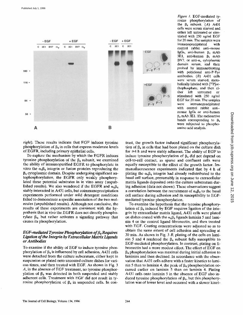
Figure 1. EGF-mediated ty- rosine phosphorylation of the [~4 subunit. (A) A431 cells were serum starved and either left untreated or stim- ulated with 250 ng/ml EGF for 20 min. The samples were immunoprecipitated with control rabbit anti-mouse IgGs, anti-human 134 mAb 3El, anti-human 131 mAb BV7, or anti-et v cytoplasmic domain serum, and then probed by immunoblotting with polyclonal anti-P-Tyr antibodies. (B) A431 cells were serum starved, meta- bolically labeled with [32p]or- thophosphate, and then ei- ther left untreated or stimulated with 250 ng/ml EGF for 20 min. The samples were immunoprecipitated with control rabbit anti- mouse IgGs or anti-human 134 mAb 3El. The radioactive bands corresponding to 134 were subjected to phospho- amino acid analysis.
right). These results indicate that EGF induces tyrosine phosphorylation of 134 in cells that express moderate levels of EGFR, including primary epithelial cells.
To explore the mechanism by which the EGFR induces tyrosine phosphorylation of the 134 subunit, we examined the ability of immunopurified EGFR to phosphorylate in vitro the Ot6~ 4 integrin or fusion proteins reproducing the 134 cytoplasmic domain. Despite undergoing significant au- tophosphorylation, the EGFR only weakly phosphory- lated these potential substrates in in vitro assay (unpub- lished results). We also wondered if the EGFR and 0t6134 stably interacted in A431 cells, but coimmunoprecipitation experiments performed under mild detergent conditions failed to demonstrate a specific association of the two mol- ecules (unpublished results). Although not conclusive, the results of these experiments are consistent with the hy- pothesis that in vivo the EGFR does not directly phospho- rylate 134, but rather activates a signaling pathway that causes its phosphorylation.
E G F-mediated Tyrosine Phosphorylation o f fl 4 Requires Ligation of the Integrin by ExtraceUular Matrix Ligands or Antibodies
To examine if the ability of EGF to induce tyrosine phos- phorylation of 134 is influenced by cell adhesion, A431 cells were detached from the culture substratum, either kept in suspension or plated onto uncoated culture dishes for vari- ous times, and then treated with EGF. As shown in Fig. 3 A, in the absence of EGF treatment, no tyrosine phosphor- ylation of 134 was detected in both suspended and stably adherent cells. Treatment with EGF did not result in ty- rosine phosphorylation of 134 in suspended cells. In con-
trast, the growth factor induced significant phosphoryla- tion of 134 in cells that had been plated on the culture dish for t>4 h and were stably adherent. The ability of EGF to induce tyrosine phosphorylation of 134 did not depend on cell-to-cell contact, as sparse and confluent cells were equally susceptible to the effect of the growth factor. Im- munofluorescence experiments indicated that by 4 h of plating the 0t6134 integrin had already redistributed to the basal cell surface, presumably in response to extracellular matrix ligands deposited onto the culture substratum dur- ing adhesion (data not shown). These observations suggest a correlation between the recruitment of 0t6134 to the basal cell surface during adhesion and its susceptibility to EGF- mediated tyrosine phosphorylation.
To examine the hypothesis that the tyrosine phosphory- lation of 134 induced by EGF requires ligation of the inte- grin by extracellular matrix ligand, A431 cells were plated on dishes coated with the 0t6134 ligands laminin 5 and lami- nin 4 or the control ligand fibronectin, and then treated with EGF. Coating concentrations were adjusted so as to obtain the same extent of cell adhesion and spreading at 30 min. As shown in Fig. 3 B, plating of the cells on lami- nin 5 and 4 rendered the 134 subunit fully susceptible to EGF-mediated phosphorylation. In contrast, plating on fi- bronectin had a more modest effect. The effect of EGF on 134 phosphorylation was maximal during initial adhesion to laminins and then declined. In accordance with the obser- vation that A431 cells adhere with a faster kinetics to lami- nin 5 than to laminin 4, the peak of 134 phosphorylation oc- curred earlier on laminin 5 than on laminin 4. Plating A431 cells onto laminin 5 in the absence of EGF also in- duced tyrosine phosphorylation of 134, but this phosphory- lation was of lower level and occurred with a slower kinet-
The Journal of Cell Biology, Volume 134, 1996 244
on June 12, 2015jcb.rupress.org
Dow
nloaded from
Published July 1, 1996

Figure 2. Dose dependence and kinetics of EGF-mediated ty- rosine phosphorylation of 134. Serum-starved A431 cells were treated for 20 min with the indicated concentrations of EGF (A), or treated with 50 ng/ml EGF for the indicated times (B). After immunoprecipitation with the 3El mAb, the samples were probed by immunoblotting with polyclonal anti-P-Tyr or anti-134 cytoplasmic domain antibodies. Growth factor-starved primary human keratinocytes (C, left) and 804G cells expressing a recom- binant EGFR (C, right) were treated with the indicated concen- trations of EGF for 15 min. Immunoprecipitation was with anti-134 cytoplasmic domain antibodies and immunoblotting with poly- clonal anti-P-Tyr antibodies.
ics than in the presence of E G F (Mainiero et al., 1995). Control experiments revealed that the ability of E G F R to undergo autophosphorylation, as well as to induce ty- rosine phosphorylation of several cellular substrates, was similar in cells freshly plated on each one of the extracellu- lar matrix proteins tested, including fibronectin (Fig. 3 C). These results are consistent with the notion that ligand binding to 0t684 is required for optimal tyrosine phosphor- ylation of 84 in response to E G F stimulation.
Since the A431 cells express at least another integrin, ot381, capable of binding to laminin 5 and possibly to lami- nin 4, we wished to obtain direct evidence that ligation of Ot6134 at the cell surface is required for optimal phosphory- lation of 134. A431 cells were plated for 60 min on the con- trol ligand fibronectin, and then incubated for different times with polystyrene beads coated with the anti-84 m A b 3El, the anti-81 mAb TS2/16, or the control anti-MHC mAb W6.32. As shown in Fig. 4, t reatment with E G F caused sig- nificant tyrosine phosphorylation of 84 in cells exposed for 10 min to the anti-84 beads, but not in cells treated with anti-81 or anti-MHC beads. Incubation with soluble 3El m A b produced a very modest effect. Control experiments indicated that treatment of A431 cells with anti-131 beads in the absence of E G F induces, as expected, a significant tyrosine phosphorylation of p125 FAK (data not shown). These results suggest that Ot6134 must be oligomerized at the cell surface to be susceptible to EGF-media ted phos- phorylation.
Interestingly, while E G F could consistently induce sig- nificant tyrosine phosphorylation of 134 in cells that had been plated onto a plastic culture substratum for 4 h or more (Fig. 3 A), maximal phosphorylation of 84 occurred only transiently in cells plated on laminin 5 and 4 (Fig. 3 B) or incubated with anti-134 beads (Fig. 4). The transient na- ture of the effect induced by initial ligation of 0/.684 o n ty- rosine phosphorylation of 84 may be explained by the pre-
Figure 3. EGF-mediated ty- rosine phosphorylation of 134 requires extracellular matrix ligand binding. (A) A431 cells were detached, resus- pended in complete me- dium, and either kept in sus- pension or replated at low density onto uncoated dishes for 4 or 12 h. Where indi- cated by the asterisk, the cells were plated at high den- sity and reached confluence by 12 h. The cells were either left untreated or stimulated with 250 ng/ml EGF for 20 min, and then immunopre- cipitated with the 3El mAb. Samples were probed by im-
munoblotting with polyclonal anti-P-Tyr antibodies. (B) A431 cells were serum starved, detached, and then replated in serum-free me- dium on dishes coated with laminin 5 matrix, 10 i~g/ml laminin 4, or 10 ~g/ml fibronectin for the indicated times. In all cases, the cells were treated with 50 ng/ml EGF for 5 min, and then immunoprecipitated with the 3El mAb. Samples were probed by immunoblotting with polyclonal anti-P-Tyr antibodies. (C) A431 cells were serum starved, detached, and then either kept in suspension or replated in serum-free medium on dishes coated with laminin 5 matrix, 10 ~g/ml laminin 4, or 10 Ixg/ml fibronectin for 30 min. In all cases, the cells were treated with 50 ng/ml EGF for 5 min. Total proteins were probed by immunoblotting with polyclonal anti-P-Tyr antibodies.
Mainiero et al. Regulation of %~4 lntegrin by EGF 245
on June 12, 2015jcb.rupress.org
Dow
nloaded from
Published July 1, 1996

Figure 4. EGF-mediated tyrosine phosphorylation of ~4 requires ligation of the integrin. A431 cells were serum starved, detached, replated in serum-free medium on dishes coated with 10 txg/ml fi- bronectin for 60 min, and then incubated for the indicated times with beads coated with the anti-J34 mAb 3El, the anti-131 mAb TS2/16, or the control anti-MHC mAb W6.32. In all cases, the cells were treated with 50 ng/ml EGF for 5 min, and then immu- noprecipitated with the 3E1 mAb. The samples were probed by immunoblotting with polyclonal anti-P-Tyr antibodies.
vious observation that tyrosine phosphorylation of [34 is negatively regulated by tyrosine phosphatases (Mainiero et al., 1995). Thus, although ligation of O~6134 can activate a tyrosine kinase responsible for [34 phosphorylation and synergize with the effect of EGF, the subsequent activa- tion of tyrosine phosphatases able to reverse the phosphor- ylation of [34 is likely to antagonize the effect of EGF. Taken together, these results suggest that clustering of 0t6134 induced by extracellular matrix ligands is required for optimal phosphorylation of the [34 subunit in response to EGF stimulation. They further suggest that, depending on the timing, ligand binding to 0t6134 can either synergize or antagonize with a signal from the EGFR to induce ty- rosine phosphorylation of [34.
EGF Induces Phosphorylation of Multiple [34 Tyrosine Residues
Phosphopeptide mapping experiments were performed to analyze the [34 sites phosphorylated in response to EGF treatment. Since cross-linking of 0t6134 by antibodies or plating on laminin 5 did not induce a level of tyrosine phosphorylation of [34 sufficient for high resolution map- ping, the sites phosphorylated in response to EGF were compared to those phosphorylated in response to pervan- adate. Previous results have shown that treatment with pervanadate results in phosphorylation of multiple [34 resi- dues, including the [34 TAM and presumably also the Shc binding sites, since pervanadate can induce association of 56[34 with Shc (Mainiero et al., 1995). A431 cells were met- abolically labeled with [32p]orthophosphate, and then ei- ther left untreated or stimulated with EGF or pervanadate. After immunoprecipitation, the [34 subunit was digested with Staphylococcus V8 protease, and the resulting peptides were separated by bidimensional TLC. As shown in Fig. 5 A, the [34 subunit from unstimulated cells was resolved in a number of phosphopeptides (S1-S10). In accordance with the observation that [34 is phosphorylated constitutively on
serine residues (Fig. 5 B), phosphoamino acid analysis in- dicated that these peptides contained only phosphoserine. Treatment with EGF resulted in the appearance of a num- ber of additional phosphopeptides (Y1-Y8) (Fig. 5 B), and phosphoamino acid analysis of several of them (Y1-Y6) confirmed that they contained exclusively phosphoty- rosine. These results indicate that exposure to EGF results in phosphorylation of multiple tyrosine residues in the ~4 cytoplasmic domain. The phosphopeptide map of [34 from pervanadate-treated cells was similar, but not identical, to that of [34 from EGF-stimulated cells. It contained the pep- tides S1-S10 and Y1-Y8, but also an additional phospho- tyrosine-containing peptide, Y9. Furthermore, the inten- sity of the spot corresponding to phosphopeptide Y1 was much larger in pervanadate than in EGF-treated cells, and conversely, phosphopeptide Y6 was more intensely labeled in EGF than in pervanadate-treated cells (Fig. 5 C). Previ- ous experiments of site-directed mutagenesis and phos- phopeptide mapping of [34 from pervanadate-treated cells have indicated that peptide Y5 contains tyrosine 1440, the COOH-terminal element of the TAM, and have provided circumstantial evidence that peptide Y2 contains tyrosine 1422, the NH2-terminal element of the TAM (Mainiero et al., 1995). Since exposure to EGF resulted in the appear- ance of phosphopeptides Y5 and Y2, we concluded that EGF induces the phosphorylation of multiple tyrosine res- idues in [34 and that these include the COOH-terminal, and possibly the NH2-terminal, element of the TAM.
EGF-mediated Tyrosine Phosphorylation of [34 Does Not Result in Recruitment of the Adaptor Proteins Shc and Grb2
To examine if EGF-mediated tyrosine phosphorylation of [34 results in association of the adaptor protein Shc to 0t6134 , A431 cells were either incubated with anti-[34 beads in sus- pension or treated with EGF while adherent. The resulting extracts were immunoprecipitated with anti-[34 antibodies and probed by immunoblotting with anti-[34 and anti-Shc antibodies. As shown in Fig. 6 A, ligation of Ct6134 led to re- cruitment of Shc. In contrast, treatment with EGF did not result in association of this adaptor molecule to 0t6134. Im- munoprecipitation with anti-Shc antibodies followed by immunoblotting with anti-[34 antibodies confirmed that EGF stimulation does not result in recruitment of She to Ot6~ 4 (Fig. 6 B). Control experiments indicated that a certain amount of the adaptor molecule remained available in the cytoplasm of EGF-treated cells (not shown; see also Fig. 6 C). The results of these experiments suggest that EGF does not induce phosphorylation of the Shc binding sites in [34 .
The inability of EGF to induce association of Shc with the Ot6134 integrin raises the possibility that the EGFR and ot6134 may, when simultaneously ligated, compete for this adaptor molecule in vivo. To explore this possibility, we examined the effect of EGF on the recruitment of Shc to ac t iva ted a6134, As shown in Fig. 6 C, the amount of Shc coimmunoprecipitated with 56[34 was lower in cells stimu- lated with anti-[34 beads and EGF than in cells treated only with anti-[34 beads. The inhibitory effect of EGF was espe- cially evident in cells that had been incubated with anti-[34 beads for 5 or 10 min, irrespective of whether the EGF was
The Journal of Cell Biology, Volume 134, 1996 246
on June 12, 2015jcb.rupress.org
Dow
nloaded from
Published July 1, 1996

Figure 5. Phosphorylation of multiple [~4 tyrosine residues in response to EGF. A431 cells were metabolically labeled with [32p]ortho- phosphate and left untreated (.4), stimulated with 250 ng/ml EGF for 5 rain (B), or with 500 p~M pervanadate for 10 rain (C). After im- munoprecipitation with the 3E1 mAb and separation by SDS-PAGE, the radioactive bands corresponding to [3 4 w e r e subjected to V8 protease digestion, and the resulting phosphopeptides were separated by bidimensional TLC.
applied together with the beads or before the beads. In ad- dition to indicating that a certain amount of Shc remains available for binding to Ot6~ 4 in EGF-t rea ted cells, these results indicate that the E G F R and 0t613 4 integrin compete
Figure 6. EGF interferes with the recruitment of Shc to ¢3t6[~ 4. (A) A431 cells were serum starved and either incubated for 10 rain in suspension with polystyrene beads coated with the anti-134 mAb 3E1 or treated for 5 min while adherent with 200 ng/ml EGF. Control cells consisted of suspended cells left untreated. Equal amounts of total proteins were immunoprecipitated with anti-134 cytoplasmic peptide serum and probed by immunoblotting with the same antiserum (top) or anti-Shc mAb (bottom). (B) A431 cells were treated as above, but immunoprecipitated with anti- Shc polyclonal antibodies and probed with either anti-134 cyto- plasmic peptide serum (top) or the anti-P-Tyr mAb PY20 (bot- tom). (C) A431 cells were serum starved, detached and replated in serum-free medium on dishes coated with 10 p.g/ml fibronectin for 60 min. They were then incubated for the indicated times with 3E1 mAb-coated beads, 50 ng/ml EGF, or 3E1 mAb-coated beads followed or preceded by a 5-min exposure to 50 ng/ml EGF. The extracts were immunoprecipitated with anti-134 cyto- plasmic peptide antibody and probed by immunoblotting with anti-Shc mAb.
for this adaptor molecule in cultured cells, raising the pos- sibility that the ability of ~6~4 to link to the ras pathway may be suppressed by EGF-dependent signals in vivo.
Disruption of Hemidesmosomes by EGF
To examine the effect of E G F on the ability of ct6134 to as- sociate with the hemidesmosomal cytoskeleton, we elected to use the rat 804G bladder epithelial cells, which form hemidesmosomes in vitro. Since these cells express very low levels of the E G F R , we used cell lines expressing mod- erate levels of human E G F R from cDNA. Immunoblot- ting analysis of total proteins with anti-P-Tyr antibodies indicated that exposure of the EGFR-transfected cells to E G F resulted in tyrosine phosphorylation of the recombi- nant E G F R and of several of its cellular substrates, includ- ing the [3 4 subunit (data not shown and Fig. 2 C, right).
The EGFR-transfected 804G cells were starved, and then either left untreated or treated for various times with 100 ng/ml EGF. Immunofluorescent analysis revealed that while in control cells, ~6134 and the B P A G 2 were concen- trated at the basal cell surface within Triton X-100-resistant, "Swiss cheese"-like structures corresponding to hemides- mosomes (Fig. 7, a and b); in cells treated with EGF, these molecules had undergone a profound redistribution and were no longer detected in association with these struc- tures (Fig. 7, d and e). To confirm the physiological signifi- cance of these observations, we examined the effect of E G F on the hemidesmosome-like structures formed by normal human primary keratinocytes in culture. As shown in Fig. 7 (c and f), t reatment with E G F resulted in loss of hemidesmosomal staining also in these cells, suggesting that disassembly of hemidesmosomes may be one of the physiological consequences of activation of the E G F R in primary epithelial cells. Immunofluorescent analysis of E G F R transfected 804G cells treated for various times with E G F indicated that the effect of the growth factor on hemidesmosomes was already significant after 1 h and
Mainiero et al. Regulation of ct6fl 4 Integrin by EGF 247
on June 12, 2015jcb.rupress.org
Dow
nloaded from
Published July 1, 1996

Figure 7. Disruption of hemidesmosomes by EGF. EGFR-transfected 804G cells (a, b, d, and e) and primary human keratinocytes (c and f) were cultured on glass coverslips for 48 h, serum starved, and either left untreated (a--c) or treated with 100 ng/ml EGF for 12 h (d-f). After extraction with 0.2% Triton X-100, the cells were fixed and stained with anti-BPAG 2 antibodies (a and d), anti-J34 cytoplas- mic peptide serum (b and e), or the antiq34 mAb 3El (c and f) followed by FITC-labeled afffinity-purified secondary antibodies.
complete by 12 h of treatment (Fig. 8, a-e). Dose- dependency experiments indicated that 25 ng/ml of EGF were sufficient to induce a significant effect on hemides- mosomes (data not shown). The effect of EGF was specific as PDGF did not cause any change in hemidesmosome staining (Fig. 8 f). Similar results were obtained with three independent clones of EGFR-transfected 804G cells. These observations indicate that EGF treatment causes disrup- tion of hemidesmosomes.
The ability of EGF to induce hemidesmosome disassem- bly was unexpected because phosphopeptide mapping had indicated that the [34 TAM, which mediates a signaling event required for the association of Ot6[~ 4 with hemides- mosomes (Mainiero et al., 1995), is phosphorylated in re- sponse to EGF. We wondered if the effect of EGF on hemidesmosomes was caused by its ability to downregu- late ligand binding to Ot6[~ 4 by a mechanism of inside- to-outside signaling. The effect of EGF treatment on the adhesion of EGFR-transfected 804G cells to laminin 4 and 5 was therefore examined. To block [31-dependent adhe- sion, the cells were plated on the two extracellular matrix
proteins in the presence of inhibitory anti-J31 antibodies. As shown in Fig. 9 , the extent to which the EGFR-trans- fected 804G cells adhered to laminin 4 and 5 was not sig- nificantly changed after treatment with EGF. A similar re- sult was obtained with A431 cells (not shown). These results indicate that exposure to EGF does not cause a sig- nificant change in ligand binding to Ot6[~4, thus suggesting that the deterioration of hemidesmosomes observed in EGF-treated cells is not caused by a downregulation of ligand binding. Together with the observation that EGF induces phosphorylation of the [~4 TAM, these data sug- gest the hypothesis that EGF-dependent signals suppress the association of OL6[~ 4 with the hemidesmosomal cyto6kel- eton by interfering with the functioning of signaling and cytoskeletal molecules downstream of the [~4 TAM.
Increased a6~4-dependent Cell Migration in Response to EGF
To determine if the apparent disruption of hemidesmo- somes caused by EGF correlates with a change in ot6134-
The Joufnal of Cell Biology, Volume 134, 1996 248
on June 12, 2015jcb.rupress.org
Dow
nloaded from
Published July 1, 1996

Figure 8. Kinetics and specificity of EGF-induced hemidesmosome disruption. EGFR-transfected 804G cells were cultured on glass coverslips for 48 h, serum starved, and either left untreated (a) or treated with 100 ng/ml EGF for 30 min (b), 1 h (c), 3 h (d), or 12 h (e). As a control, cells were exposed to 5 ng/ml PDGF for 12 h (f). After extraction with 0.2% Triton X-100, the cells were fixed and stained with anti-BPAG 2 antibodies followed by FITC-labeled affinity-purified secondary antibodies.
dependent cell migration, we measured the ability of con- trol and EGFR-transfected 804G ceils to migrate toward various extracellular matrix components by using a Boy- den chamber system. As shown in Fig. 10, treatment with EGF resulted in increased migration of the EGFR-trans- fected 804G cells toward the two et6134 ligands laminin 4 and 5, but not the control ligand fibronectin, suggesting that EGF-dependent signals can increase cell migration toward Ot6[~ 4 ligands. In addition, the basal migration of EGFR-transfected 804G cells toward laminin 4 and 5 was greater than that of control 804G cells. This result suggests that the recombinant EGFR may be partially active in the absence of exogenous ligand in 804G ceils, perhaps be- cause these cells secrete EGF or TGF-cx. In accordance with this hypothesis, we found that the medium condi- tioned by 804G cells is capable of stimulating the auto- phosphorylation of recombinant EGFR expressed in transfected 804G cells (data not shown). Inhibitory anti-131 antibodies were able to suppress the migration of unstimu- lated cells toward laminin 5 by 91 +_ 6%, but only inhibited the migration of EGF-treated cells by 11 ___ 3%, indicating
that the EGF-stimulated migration toward laminin 5 was largely dependent o n ot6~ 4 function. This conclusion was also supported by the observation that EGFR-transfected NIH-3T3 fibroblasts, which do not express ot6134, did not respond to EGF with increased migration toward laminin 4 (Fig. 10, top). Finally, the effect of EGF was specific, since it was not observed in response to PDGF or with the control 804G cells in response to EGF. Taken together, these results indicate that EGF specifically upregulates et6~a-dependent migration toward laminins.
Discussion
Several observations suggest that ct6~ 4- and growth factor- dependent signals may cooperate to control epidermal cell proliferation and migration. In stratified epithelia, such as the epidermis, 0~6134 mediates the interaction of basal kera- tinocytes with the basement membrane (Kajiji et al., 1989), and there is evidence indicating that these cells have to remain in contact with this extracellular matrix to main- tain their proliferative potential (Green, 1977; Hall and
Mainiero et al. Regulation of a6134 lntegrin by EGF 249
on June 12, 2015jcb.rupress.org
Dow
nloaded from
Published July 1, 1996

0,2
0.7
0.6
0.5
0.4'
0.3,
~ + E G F
I m ! "m m 0 2 4 6 8 1 Laminin 4 (pglml)
0.8
12
o81 0 . 6 .
v 0.4'
0.2.
0.0
- - i
---- + E G F
' ' '" ' ' '5 3 ' 0 " 5 10 15 20 2 3 5
Laminin 5 (min) Figure 9. EGF does not affect ot6134-dependent cell adhesion. EGFR-transfected 804G cells were starved and either left un- treated or stimulated with 100 ng/ml EGF. The cells were plated in the presence of inhibitory anti-J31 antibodies on dishes coated with the indicated amounts of laminin 4 for 60 min (top) or on laminin 5 matrix-coated dishes for the indicated times (bottom).
Watt, 1989). Furthermore, the coincident expression of 0t6134 and laminins by keratinocytes migrating into corneal wounds suggests a role for a6134-mediated migration dur- ing the reepithelialion of wounds (Kurpakus et al., 1991). Prompted by the prominent role of EGF and transforming growth factor a in controlling keratinocyte growth and mi- gration (Rheinwald and Green, 1977; Barrandon and Green, 1987), and by the coincident expression of a6134 and EGFR in basal keratinocytes in vivo (Green et al., 1987; Kajiji et al., 1989), we have examined the effect of EGFR activation on the intracellular functions of Ot-61~ 4. Our re- suits indicate that EGF-dependent signals have a complex effect on 0t.6134 function: they cause tyrosine phosphoryla- tion of 134 without promoting the association of Shc, induce
/ soo-I Laminin 4
soot I~ m~ • ~ 400 -,
'i lOO
o ~ 3T3 804G 804G EGFR EGFR
300 '
"o
o
" i 200'
~ 100 '
Laminin 5 'o
,r,,,I
1000
~ 0
804G EGFR
Flbronectln
804G EGFR Figure 10. EGF stimulates et6134-dependent cell migration. The indicated cell lines were allowed to migrate toward laminin 4 for 48 h, laminin 5 for 12 h, and fibronectin for 48 h in the presence of control medium (open bars), 50 ng/ml EGF (closed bars), or 5 ng/ ml PDGF (hatched bars).
disassembly of hemidesmosomes, and upregulate cell mi- gration on laminins.
In this study, we provide direct evidence that activation of the EGFR causes tyrosine phosphorylation of the 134 subunit. This phosphorylation is characterized by a rapid kinetics and, at least in A431 cells, by a high stoichiometry. Since we have been unable to obtain evidence that the EGFR efficiently phosphorylate 134 in vitro, it is our hy- pothesis that the E G F R does not directly phosphorylate ~4 in vivo, but rather it activates a signaling pathway that results in its phosphorylation. The observation that EGF- mediated phosphorylation of 134 requires ligation of the in- tegrin by extracellular ligand or antibodies suggests that this phosphorylation event is mediated by an integrin-as- sociated kinase acting in trans. Future studies will be re- quired to determine if Ot6134 is indeed an indirect target of
The Journal of Cell Biology, Volume 134, 1996 250
on June 12, 2015jcb.rupress.org
Dow
nloaded from
Published July 1, 1996

the EGFR and if it is associated with two distinct tyrosine kinases, one activated by EGF and the other by extracellu- lar matrix binding, or with a single tyrosine kinase acti- vated by both stimuli.
The results of phosphopeptide mapping indicate that EGF causes phosphorylation of several distinct [34 tyrosine residues. Although the majority of the tyrosine phospho- rylation sites in [34 remain to be identified and their func- tion assessed, the complexity of the tyrosine phosphoryla- tion pattern induced by EGF suggests that many O/.6~ 4 functions may be regulated by the growth factor. One ma- jor intracellular function of ~6[~4 that is negatively regu- lated by EGF is the recruitment of the adaptor molecule Shc. Treatment with EGF does not result in the associa- tion of 0t6134 with Shc and presumably Grb2. In fact, expo- sure to EGF partially suppresses the recruitment of Shc to the ligated integrin. Although it is possible that EGF causes a conformational change or another posttransla- tional modification of 0t6134 that prevents it from binding to Shc, the most likely explanation of these results is that the growth factor does not induce phosphorylation of the Shc binding motifs in 134. The observation that the EGFR can compete with 0~6134 for the recruitment of Shc is in accor- dance with the recognized ability of activated EGFR to as- sociate with this adaptor molecule (Pellicci et al., 1992) and suggests that a significant activation of the EGFR may interfere with the ability of ligand-occupied Ot6134 to acti- vate signaling in vivo. In contrast, when suboptimally li- gated, the EGFR and et6134 are likely to cooperate with each other to activate the r a s pathway. This latter predic- tion may be relevant to understanding anchorage-depen- dent cell growth in epithelial cells.
The results of our immunofiuorescent analysis indicate that treatment with EGF causes disruption of hemidesmo- somes in both EGFR-transfected 804G ceils and primary human keratinocytes. What is the mechanism by which EGF interferes with the assembly of hemidesmosomes? Our previous studies suggest that the nucleation of hemidesmosomes requires a signal mediated by the [34 TAM (Mainiero et al., 1995). It is, however, unlikely that the phosphorylation of the TAM is the only Ot6~ 4 function necessary for the assembly of hemidesmosomes. Deletion mutagenesis experiments have indicated that the associa- tion of Ot6134 with the hemidesmosomal cytoskeleton not only requires the connecting segment, which includes the TAM, but also sequences within the two type III fibronec- tin-like modules upstream of the connecting segment (Spinardi, L., and F.G. Giancotti, unpublished results). This observation is consistent with the hypothesis that a TAM-dependent signal renders one or more cytoskeletal elements of hemidesmosomes competent for binding to sequences within the first two type III fibronectin-like modules of 134. Further assembly of hemidesmosomes may then be driven by the cooperative binding of additional cy- toskeletal elements. Based on this model, EGF-dependent signals may interfere with the assembly of hemidesmo- somes at one or more of several steps. Since EGF does not affect et6134-mediated adhesion to laminins and does not suppress phosphorylation of the [34 TAM, the growth fac- tor may interfere with the functioning of one or more sig- naling or cytoskeletal molecules located downstream of the TAM in the pathway that controls the association of
0t6134 with the cytoskeleton. Furthermore, it is possible that EGF induces the phosphorylation of tyrosine residues lo- cated within the first two type III fibronectin-like modules of the [34 tail, thus directly interfering with the association of cytoskeletal molecules. Finally, as the process of hemidesmosome formation is likely to be complex and to require the function of many components in addition to Ot6[~ 4 and the molecules to which it binds, EGF may disrupt hemidesmosomes by acting on one or more of these addi- tional components.
Most of the previous studies on the regulation of the cy- toskeleton by growth factors have focused on the effects of EGF and PDGF on the actin filament system. It has been known for long that these growth factors can induce pro- found changes in the architecture of the actin cytoskeleton (Bockus and Stiles, 1984; Herman and Pledger, 1985). Re- cent studies have indicated that they can induce the se- quential formation of filopodia, lamellipodia, and focal ad- hesions, and that these cytoskeletal changes are mediated by a GTPase cascade involving Cdc 42, Rac, and Rho (Nobes and Hall, 1995). Our current observations clearly indicate that EGF can also profoundly affect the keratin filament system, thereby providing evidence for a novel mechanism of cytoskeletal regulation by EGF.
The changes in the association of 0t6134 with the cytoskel- eton induced by activated EGFR are likely to be signifi- cant in both physiological and pathological situations. Several lines of evidence support the notion that hemides- mosomes mediate stable adhesion to the basement mem- brane (Uitto and Christiano, 1992; Guo et al., 1995; Spinardi et al., 1995). Their disruption may therefore result in a more dynamic interaction with the extracellular matrix. In accordance with this hypothesis, we have observed that the disassembly of hemidesmosomes caused by EGF cor- relates with an increase in ot6[34-dependent cell migration. This observation suggests that the ability of Ot6134 to medi- ate cell migration on laminins can be upregulated by fac- tors that interfere with its association with the hemidesmo- somal cytoskeleton. It is well known that EGF and TGF-et can promote the reepithelialization of wounds (Schultz et al., 1991), and it has recently been observed that kerati- nocytes lose their hemidesmosomes as they migrate into corneal wounds (Gipson et al., 1993). Thus, the ability of activated EGFR to coordinately disassemble hemidesmo- somes and increase cell migration on laminins is likely to be important during wound healing. In addition, there is evidence indicating that keratinocytes of patients affected by the skin disease psoriasis overproduce TGF-ot (Elder et al., 1989) and that squamous carcinoma cells overexpress the EGFR (Yamamoto et al., 1986; Ozanne et al., 1986). In both pathological situations, the expression of 0t6134 is no longer restricted to the basal surface of those cells that abut the basement membrane, but extends suprabasally (Kimmel and Carey, 1986; Pellegrini et al., 1992). Our cur- rent results suggest that the loss of e%[34 polarity observed in these diseases may result from the ability of activated EGFR to disrupt the association of the integrin to the hemidesmosomal cytoskeleton. They further suggest that the ability of EGFR to affect the association of ~6134 with the cytoskeleton may contribute to the invasive ability of squamous carcinoma cells.
F. Mainiero and A. Pepe contributed equally to this work. We thank Elis-
Mainiero et al. Regulation of a6fl 4 lntegrin by EGF 251
on June 12, 2015jcb.rupress.org
Dow
nloaded from
Published July 1, 1996

abetta Dejana, Eva Engvall, Erkki Ruoslahti, and Jouni Uitto for antibod- ies, Jossi Schlessinger for the EGFR expression construct and antibodies, members of our laboratory for helpful suggestions, Miki Blurnemberg for human primary keratinocytes, and Jan Sap for critical comments on the manuscript.
This work was supported by Public Health Service (PHS) grant R01- CA58976, grant DAMD 17-94-J4306 from the U.S. Army Medical Re- search and Material Command, and PHS core support grant P30-CA16087. F. Mainiero is supported by a fellowship from the American Italian Foun- dation for Cancer Research. F.G. Giancotti is a recipient of awards from the Lucille P. Markey and Irma T. Hirschl Charitable Trusts.
Received for publication 28 November 1995 and in revised form 16 March 1996.
References
Argraves, W.S., R. Pytela, S. Suzuki, J.L. Millan, M.D. Pierschbacher, and E. Ruoslahti. 1986. cDNA sequences from the ct subunit of the fibronectin re- ceptor predict a transmembrane domain and a short cytoplasmic peptide. J. Biol. Chem. 261:12922-12924.
Arroyo, A.G., P. S~lnchez-Mateos, M.R. Campanero, I. Martfn-Padura, E. De- jana, and F. Sanchez-Madrid. 1992. Regulation of VLA integrin-ligand in- teractions through the 131 subunit. J. Cell Biol. 117:659~70.
Barrandon, Y., and H. Green. 1987. Cell migration is essential for substained growth of keratinocyte colonies: the roles of transforming growth factor-ct and epidermal growth factor. Cell 50:1131-1137.
Beguinot, L., R.M. Lyall, M.C. Willingham, and I. Pastan. 1984. Down-regula- tion of the epidermal growth factor receptor in KB ceils is due to receptor in- ternalization and subsequent degradation in lysosomes. Proc. Natl. Acad. Sci. USA. 81:2384-2388.
Burridge, K., C.E. Turner, and L.H. Romer. 1992. Tyrosine phosphorylation of paxillin and pp125 FAR accompanies cell adhesion to extracellular matrix: a role in cytoskeletal assembly. J. Cell Biol. 119:893-903.
Bockholt, S.M., and K. Burridge. 1993. Cell spreading on extracellular matrix proteins induces tyrosine phosphorylation of tensin. J. Biol. Chem. 268: 14565-14567.
Bockus, B.J., and C.D. Stiles. 1984. Regulation of cytoskeletal architecture by platelet-derived factor, insulin and epidermal growth factor. Exp. Cell Res. 153:186-197.
Boyle, W.J., P. Van Der Geer, and T. Hunter. 1991. Phosphopeptide mapping and phosphoaminoacid analysis by two-dimensional separation on thin layer cellulose plates. Methods Enzymol. 201:110-149.
Carter, W.G., P. Kaur, S.G. Gil, P.J. Gahr, and E.A. Wayner. 1990. Distinct functions for integrins a3131 in focal adhesions and ot6134/bullous pemphigoid antigen in a new stable anchoring contact (SAC) of keratinocytes: relation to hemidesmosomes. J. Cell Biol. 111:3141-3154.
Chen, H.-C., and J.-L. Guan. 1994. Stimulation of phosphatidylinositol 3'-kinase association with focal adhesion kinase by platelet-derived growth factor. J. Biol. Chem. 269:31229-31233.
Chen, J., J. Kim, K. Zhang, Y. Sarret, K. Wynn, R. Kramer, and D. Woodley. 1993. Epidermal growth factor (EGF) promotes human keratinocytes loco- motion on collagen by increasing the % integrin subunit. Exp. Cell Res. 209: 216-223.
Chong, L.D., A. Traynor-Kaplan, G.M. Bokoch, and M.A. Schwartz. 1994. The small GTP-binding protein Rho regulates a phosphatidylinositol 4-phos- phate 5-kinase in mammalian cells. Cell. 79:507-513.
Elder, J.T., G.J. Fisher, P.B. Lindquist, G.L. Bennett, M.R. Pittelkow, R.J. Cof- fey, L. Ellingsworth, R. Derynck, and J.J. Voorhees. 1989. Overexpression of transforming growth factor alpha in psoriatic epidermis. Science (Wash. DC). 243:811-814.
Giancotti, F.G., and E. Ruoslahti. 1990. Elevated levels of the ~s131 fibronectin receptor suppress the transformed phenotype of Chinese hamster ovary cells. Cell. 60:849-859.
Giancotti, F.G., and F. Mainiero. 1994. Integrin-mediated adhesion and signal- ing in tumorigenesis. Biochim. Biophys. Acta. 1198:47-64.
Giancotti, F.G., G. Tarone, K. Knudsen, C. Damsky, and P.M. Comoglio. 1985. Cleavage of a 135 kD cell surface glycoprotein correlates with loss of fibro- blast adhesion to fibronectin. Exp. Cell Res. 156:182-190.
Giancotti, F.G., P.M. Comoglio, and G. Tarone. 1986. Fibronectin-plasma membrane interaction in the adhesion of hemopoietic cells. Z Cell Biol. 103: 429-437.
Giancotti, F.G., M.A. Stepp, S. Suzuki, E. Engvall, and E. Ruoslahti. 1992. Pro- teolytic processing of endogenous and recombinant 134 integrin subunit. J. Cell Biol. 118:951-959.
Giancotti, F.G., L. Spinardi, F. Mainiero, and R. Sanders. 1994. Expression of heterologons integrin genes in cultured eukaryotic cells. Methods Enzymol. 245:297-316.
Gipson, 1.K., S. Spurr-Michand, A. Tisdale, J. Elwell, and M.A. Stepp. 1993. Redistribution of the hemidesmosome components alpha 6 beta 4 integrin and bullous pemphigoid antigens during epithelial wound healing. Exp. Cell
Res. 207:86-98. Green, H. 1977. Terminal differentation of cultured human epidermal cells.
Cell. 11:405-416. Green, M.R., C. Mycock, C.G. Smith, and J.R. Couchman. 1987. Biochemical
and ultrastructural processing of [1251]-epidermal growth factor in rat epider- mis and hair follicles: accumulation of nuclear label. J. Invest. Dermatol. 88: 25%265.
Guan, J.-L., and D. Shalloway. 1992. Regulation of focal adhesion-associated protein tyrosine kinase by both cellular adhesion and oncogenic transforma- tion. Nature (Lond.). 358:690-692.
Guo, L., L. Degestein, J. Dowling, Q-C. Yu, R. Wollmam, B. Perman, and E. Fuchs. 1995, Gene targeting of BPAGI: abnormalities in mechanical strength and cell migration in stratified epithelia and neurologic degenera- tion. Cell. 81:233-243.
Hall, P.A., and F.M. Watt. 1989. Stem ceils: the generation and maintenance of cellular diversity. Development 106:619-633.
Hanks, S.K., M.B. Calalb, M.C. Harper, and S.K. Patel. 1992. Focal adhesion protein-tyrosine kinase phosphorylated in response to cell attachment to fi- bronectin. Proc. Natl. Acad. ScL USA. 89:8487-8491.
Herman., B., and W.J. Pledger. 1985. Platelet-derived growth factor-induced al- terations in vinculin and actin distribution in BALB/c-3T3 cells. Z Cell Biol. 100:1031-1040.
Honegger, A.M., T.J. Dull, S. Felder, E. Van Obberghen, F. Bellot, D. Szapary, A. Schmidt, A. Ullrich, and J. Schlessinger. 1987. Point mutation at the ATP binding site of EGF receptor abolishes protein-tyrosine kinase activity and alters cellular routing. Cell. 50:19%209.
Hynes, R.O. 1992. Integrins: versatility, modulation and signaling in cell adhe- sion. Cell. 69:11-25.
Izumi, K., Y. Hirao, L. Hopp, and R. Oyasu. 1981. In vitro induction of orni- thine decarboxylase in urinary bladder carcinoma cells. Cancer Res. 41:405- 409.
Juliano, R.L., and S. Haskill. 1993. Signal transduction from the extracellular matrix. J. Cell Biol. 120:577-585.
Kahn-Perles, B., C. Boyer, B. Arnold, A.R. Sanderson, P. Ferrier, and F. Le- monnier. 1987. Acquisition of HLA class I W6/32 defined antigenic determi- nant by heavy chains from different species following association with bovine 132-microglobulin. J. ImmunoL 138:2190-2196.
Kajiji, S., R.N. Tamura, and V. Quaranta. 1989. A novel integrin (az134) from human epithelial cells suggests a fourth family of integrin adhesion recep- tors. EMBO (Eur Mol. Biol. Organ.) J. 8:673-680.
Kamps, M.P., and B.M. Sefton. 1988.' Identification of novel polypeptide sub- strates of the v-src, v-yes, v-fps, v-ros, v-erb-B oncogenic tyrosine protein ki- nases utilizing antisera against phosphotyrosine. Oncogene. 2:305-315.
Kimmel, K.A., and T.E. Carey. 1986. Altered expression in squamous carci- noma cells of an orientation restricted epithelial antigen detected by mono- clonal antibody A9. Cancer Res. 46:3614--2623.
Kinashi, T., J.A. Escobedo, L.T. Williams, K. Takatsu, and T.A. Springer. 1995. Receptor tyrosine kinase stimulates cell-matrix adhesion by phosphatidyl- inositol 3 kinase and phospholipase C-yl pathways. Blood. 86:2086-2090.
Klemke, R.L., M. Yebra, E.M. Bayna, and D.A. Cheresh. 1994. Receptor ty- rosine kinase signaling required for integrin ad35-directed cell motility but not adhesion on vitronectin. J. Cell Biol. 127:859-866.
Kurpakus, M.A., V. Quaranta, and J.C.R. Jones. 1991. Surface relocation of alpha6 beta4 integrins and assembly of hemidesmosomes in an in vitro model of wound healing. J. Cell Biol. 115:1737-1750.
Lee, E.C., M.M. Lotz, G.D. Steele, and A.M. Mercurio. 1992. The integrin ot6134 is a laminin receptor. J. Cell Biol. 117: 671~78.
Lipfert, L., B. Haimovich, B.M. Shaller, B.S. Cobb, J.T. Parsons, and J.S. Brugge. 1992. lntegrin-dependent phosphorylation and activation of the pro- tein tyrosine kinase pp125 FAR in platelets. J. Cell Biol. 119:905--912.
Malniero, F., A. Pepe, K.K. Wary, L. Spinardi, M. Mohammadi, J. Schlessinger, and F.G. Giancotti. 1995. Signal transduction by the a6134 integrin: distinct 134 subunit sites mediate recruitment of Shc/Grb2 and association with the cy- toskeleton of hemidesmosomes. EMBO (Eur. Mol. Biol. Organ.) J. 14:4470- 4481.
Mart/n-Padura, I., G. Bazzoni, A. Zanetti, S. Bernasconi, M.J. Elices, A. Man- tovani, and E. Dejana. 1994. A novel mechanism of colon carcinoma cell ad- hesion to the endothelium triggered by 131 integrin chain. J. Biol. Chem. 269: 6124-6132.
Matthay, M., J. Thiery, F. Lafont, M. Stampfer, and B. Boyer. 1993. Transient effect of epidermal growth factor on the motility of an immmortalized mam- mary epithelial cell line. Z Cell Sci. 106:869-878.
McNamee, H.M., D.E. Ingber, and M.A. Schwartz. 1992. Adhesion to fibronec- tin stimulates inositol lipid synthesis and enhances PDGF-inositol lipid breakdown. J. Cell Biol. 121:673-678.
Niessen, C.M., F. Hogervorst, L.H. Jaspars, A.A. De Melker, G.O. Delwel, E.H.M. Hulsman, I. Kuikman, and A. Sonnenberg. 1994. The ct6134 integrin is a receptor for both laminin and kalinin. Exp. Cell Res. 2111:360-367.
Nobes, C.D., and A. Hall. 1995. Rho, rac, and Cdc42 GTPases regulate the as- sembly of multimolecular focal complexes associated with actin stress fibers, lamellipodia, and filopodia. Cell. 81:53-62.
Ozanne. B., C.S. Richards, F. Hendler, D. Burns, and B. Gusterson. 1986. Over- expression of the EGF receptor is a hallmark of squamous cell carcinoma. J. PathoL 149:%14.
Pelicci, G., L. Lanfrancone, F. Grignani, J. McGlade, F. Cavallo, G. Forni, I.
The Journal of Cell Biology, Volume 134, 1996 252
on June 12, 2015jcb.rupress.org
Dow
nloaded from
Published July 1, 1996

Nicoletti, F. Grignani, T. Pawson, and P.G. Pelicci. 1992. A novel transform- ing protein (Shc) with an SH2 domain is implicated in mitogenic signal trans- duction. Cell. 70:93-104.
Pellegrini, G., M. De Luca, G. Orecchia, F. Balzac, O. Cremona, P. Savoia, R. Cancedda, and P.C. Marchisio. 1992. Expression, topography, and function of integrin receptors are severely altered in keratinocytes from involved and uninvolved psoriatic skin. J. Clin. Invest. 89:1783-1795.
Rheinwald, J.G,, and H. Green. 1977. Epidermal growth factor and the multi- plication of cultured human epidermal keratinocytes. Nature (Lond.). 265: 421-424.
Ridley, A.J., and A. Hall. 1992. The small GTP-binding protein rho regulates the assembly of focal adhesions and actin stress fibers in response to growth factors. Cell. 70:389-399.
Ridley, A.J., H.F. Paterson, C.L. Johnston, D. Diekmann, and A. Hall. 1992. The small GTP-binding protein rac regulates growth factor-induced mem- brane ruffling. Cell. 70:401-410.
Schlaepfer, D.D., S.K. Hanks, T. Hunter, and P. van der Geer. 1994. Integrin- mediated signal transduction linked to Ras pathway by GRB2 binding to fo- cal adhesion kinase. Nature (Lond.). 372:786-791.
Schultz, G., D.S. Rotatori, and W. Clark. 1991. EGF and TGF-alpha in wound healing and repair. J. Cell, Biochem. 45:346-352.
Schwartz, M.A., M.D. Shaller, and M.H. Ginsberg. 1995. Integrins: emerging paradigms of signal transduction. Annu. Rev. Cell Dev. BioL 11:549-599.
Serve, H., N.S. Yee, G. Stella, L. Sepp-Lorenzino, J.C. Tan, and P. Besmer. 1995. Differential roles of PI3-kinase and Kit tyrosine 821 in Kit receptor- mediated proliferation, survival and cell adhesion in mast cell. EMBO (Eur. Mol, Biol. Organ.) J. 14:473-483.
Schaller, M.D., C.A. Borgman, B.S. Cobb, R.R. Vines, A.B. Reynolds, and J.T. Parsons. 1992. pp125 FAK, a structurally distinctive protein-tyrosine kinase as- sociated with focal adhesions. Proc. Natl. Acad. Sci. USA. 89:5192-5196.
Sonnenberg, A., C.J.T. Linders, J.H. Daams, and S.J. Kennel. 1990. The ct6131 (VLA-6) and ct6134 protein complexes: tissue distribution and biochemical properties. J. Cell Sci. 96:207-217.
Sonnenberg, A., A.A. de Melker, A.M. Martinez de Velasco, H. Janssen, J. Calafat, and C.M. Niessen. 1993. Formation of hemidesmosomes in cells of a
transformed murine cell line and mechanisms involved in adherence of these cells to laminin and kalinin. Z Cell Sci. 106:1083-1102.
Spinardi, L., Y.-L. Ren, R. Sanders, and F.G. Giancotti. 1993. The 134 subunit cytoplasmic domain mediates the interaction of o.6134 integrin with the cy- toskeleton of hemidesmosomes. Mol, Biol. Cell 4:871-884.
Spinardi, L , S. Einheber, T. Cullen., T.A. Milner, and F.G. Giancotti. 1995. A recombinant tail-less integrin 134 subunit disrupts hemidesmosomes, but does not suppress ct6134-mediated cell adhesion to laminins. J. Cell Biol. 129:473- 487.
Stepp, M.A., S. Spurr-Michaud, A. Tisdale, J. Elwell, and I.K. Gipson. 1990. Al- pha 6 beta 4 integrin heterodimer is a component of hemidesmosomes. Proc. Natl. Acad. Sci. USA. 87:8970-8974.
Teslenko, L.V., E.S. Kornilova, A.D. Sorkin, and N.N. Nikolsky. 1987. Recy- cling of epidermal growth factor in A431 cells. FEBS Lett. 221:105-109.
Uitto, J., and A. Christiano. 1992. Molecular genetics of the cutaneous base- ment membrane zone. J. Clin. Invest. 90:687-692.
Ullrich, A., L. Coussens, J.S. Hayflick, T.J. Dull, A. Gray, A.W. Tam, J. Lee, Y. Yarden, T.A. Libermann, J. Schlessinger et al. 1984. Human epidermal growth factor receptor cDNA sequence and aberrant expression of the am- plified gene in A431 epidermoid carcinoma cells. Nature (Lond.). 309:418- 425.
Vogel, B.E., S.J. Lee, A. Hildebrand, W. Craig, M. Pierschbacher, F. Wong- Staal, and E. Ruoslathi. 1993. A novel integrin specificity exemplified by binding of the av135 integrin to the basic domain of the HIV tat protein and vitronectin. J. Cell Biol. 121:461-468.
Vuori, K., and E. Ruoslahti. 1994. Association of insulin receptor substrate-1 with integrins. Science (Wash. DC). 266:1576-1578.
Yamamoto, T., N. Kamata, H. Kawano, S. Shimizu, T. Kuroki, K. Toyoshima, K. Rikimaru, N. Nomura, R. Ishizaki, I. Pastan et al. 1986. High incidence of amplification of the epidermal growth factor gene in human squamous cell lines. Cancer Res. 46:414--416.
Zachary, I., and E. Rozengurt. 1992. Focal adhesion kinase (p125FAK): a point of convergence in the action of neuropeptides, integrins, and oncogenes. Cell. 71:891-894.
Mainiero et al. Regulation o f ct6~ 4 lntegrin by EGF 253
on June 12, 2015jcb.rupress.org
Dow
nloaded from
Published July 1, 1996