The influence of feedback valence in associative learning
18
The Influence of Feedback Valence in Associative Learning Amanda Bischoff-Grethe 1 , Eliot Hazeltine 2 , Lindsey Bergren 3 , Richard B. Ivry 4 , and Scott T. Grafton 3 1Psychology Service, VA San Diego Healthcare System, and Department of Psychiatry, University of California, San Diego, La Jolla, CA 92093, USA 2Department of Psychology, University of Iowa, Iowa City, IA 52242-1407, USA 3Center for Cognitive Neuroscience, Dartmouth College, Hanover, NH 03755, USA 4Dept. of Psychology, University of California, Berkeley, Berkeley, CA 94720-1650, USA Abstract The neural systems engaged by intrinsic positive or negative feedback were defined in an associative learning task. Through trial and error, participants learned the arbitrary assignments of a set of stimuli to one of two response categories. Informative feedback was provided on less than 25% of the trials. During positive feedback blocks, half of the trials were eligible for informative feedback; of these, informative feedback was only provided when the response was correct. A similar procedure was used on negative feedback blocks, but here informative feedback was only provided when the response was incorrect. In this manner, we sought to identify regions that were differentially responsive to positive and negative feedback as well as areas that were responsive to both types of informative feedback. Several regions of interest, including the bilateral nucleus accumbens, caudate nucleus, anterior insula, right cerebellar lobule VI, and left putamen, were sensitive to informative feedback regardless of valence. In contrast, several regions were more selective to positive feedback compared to negative feedback. These included the insula, amygdala, putamen, and supplementary motor area. No regions were more strongly activated by negative feedback compared to positive feedback. These results indicate that the neural areas supporting associative learning vary as a function of how that information is learned. In addition, areas linked to intrinsic reinforcement showed considerable overlap with those identified in studies using extrinsic reinforcers. Keywords fMRI; nucleus accumbens; punishment; reward; stimulus-response mapping; striatum Reinforcement is a fundamental mechanism for shaping behavior. A basic distinction can be made between positive and negative reinforcement. Positive reinforcers, or rewards, serve a number of basic functions. They promote selected behaviors, induce subjective feelings of pleasure and other positive emotions, and maintain stimulus-response associations (Thut et al., 1997). Negative reinforcement also plays an essential role in shaping behavior. Error signals generated during movement can be used to make rapid on-line adjustments (Ito, 2000). Correspondence should be addressed to: Scott T. Grafton, M.D., Present address: Sage Center for the Study of Mind, Department of Psychology, 3837 Psychology East (Bldg 251), UC Santa Barbara, Santa Barbara, CA 93106, Phone: (805) 893-6046, Fax: (805) 893-4303, Email: [email protected]. Publisher's Disclaimer: This is a PDF file of an unedited manuscript that has been accepted for publication. As a service to our customers we are providing this early version of the manuscript. The manuscript will undergo copyediting, typesetting, and review of the resulting proof before it is published in its final citable form. Please note that during the production process errors may be discovered which could affect the content, and all legal disclaimers that apply to the journal pertain. NIH Public Access Author Manuscript Neuroimage. Author manuscript; available in PMC 2010 January 1. Published in final edited form as: Neuroimage. 2009 January 1; 44(1): 243–251. doi:10.1016/j.neuroimage.2008.08.038. NIH-PA Author Manuscript NIH-PA Author Manuscript NIH-PA Author Manuscript
Transcript of The influence of feedback valence in associative learning

The Influence of Feedback Valence in Associative Learning
Amanda Bischoff-Grethe1, Eliot Hazeltine2, Lindsey Bergren3, Richard B. Ivry4, and Scott T.Grafton3
1Psychology Service, VA San Diego Healthcare System, and Department of Psychiatry, University ofCalifornia, San Diego, La Jolla, CA 92093, USA
2Department of Psychology, University of Iowa, Iowa City, IA 52242-1407, USA
3Center for Cognitive Neuroscience, Dartmouth College, Hanover, NH 03755, USA
4Dept. of Psychology, University of California, Berkeley, Berkeley, CA 94720-1650, USA
AbstractThe neural systems engaged by intrinsic positive or negative feedback were defined in an associativelearning task. Through trial and error, participants learned the arbitrary assignments of a set of stimulito one of two response categories. Informative feedback was provided on less than 25% of the trials.During positive feedback blocks, half of the trials were eligible for informative feedback; of these,informative feedback was only provided when the response was correct. A similar procedure wasused on negative feedback blocks, but here informative feedback was only provided when theresponse was incorrect. In this manner, we sought to identify regions that were differentiallyresponsive to positive and negative feedback as well as areas that were responsive to both types ofinformative feedback. Several regions of interest, including the bilateral nucleus accumbens, caudatenucleus, anterior insula, right cerebellar lobule VI, and left putamen, were sensitive to informativefeedback regardless of valence. In contrast, several regions were more selective to positive feedbackcompared to negative feedback. These included the insula, amygdala, putamen, and supplementarymotor area. No regions were more strongly activated by negative feedback compared to positivefeedback. These results indicate that the neural areas supporting associative learning vary as afunction of how that information is learned. In addition, areas linked to intrinsic reinforcementshowed considerable overlap with those identified in studies using extrinsic reinforcers.
KeywordsfMRI; nucleus accumbens; punishment; reward; stimulus-response mapping; striatum
Reinforcement is a fundamental mechanism for shaping behavior. A basic distinction can bemade between positive and negative reinforcement. Positive reinforcers, or rewards, serve anumber of basic functions. They promote selected behaviors, induce subjective feelings ofpleasure and other positive emotions, and maintain stimulus-response associations (Thut et al.,1997). Negative reinforcement also plays an essential role in shaping behavior. Error signalsgenerated during movement can be used to make rapid on-line adjustments (Ito, 2000).
Correspondence should be addressed to: Scott T. Grafton, M.D., Present address: Sage Center for the Study of Mind, Department ofPsychology, 3837 Psychology East (Bldg 251), UC Santa Barbara, Santa Barbara, CA 93106, Phone: (805) 893-6046, Fax: (805)893-4303, Email: [email protected]'s Disclaimer: This is a PDF file of an unedited manuscript that has been accepted for publication. As a service to our customerswe are providing this early version of the manuscript. The manuscript will undergo copyediting, typesetting, and review of the resultingproof before it is published in its final citable form. Please note that during the production process errors may be discovered which couldaffect the content, and all legal disclaimers that apply to the journal pertain.
NIH Public AccessAuthor ManuscriptNeuroimage. Author manuscript; available in PMC 2010 January 1.
Published in final edited form as:Neuroimage. 2009 January 1; 44(1): 243–251. doi:10.1016/j.neuroimage.2008.08.038.
NIH
-PA Author Manuscript
NIH
-PA Author Manuscript
NIH
-PA Author Manuscript

Negative reinforcers such as the reprimand of a parent or the loss of money are intended tohelp change behavior in the future by promoting more acceptable actions or wiser decisions.
An important question for investigations of learning is to establish similarities and differencesbetween neural networks that process positive and negative reinforcement signals. Overlappingsystems allow the control of behavior in a bidirectional manner: a positive reward promotesthe reinforced behavior, whereas a negative reward attenuates that behavior. On the other hand,the computations required for using positive and negative feedback in terms of modifyingsynaptic efficiency can be quite different (Albus, 1971; Schultz and Dickinson, 2000), andthese might be performed by distinct neural systems. For example, computational models havesuggested different learning methodologies for the basal ganglia, cerebellum, and cerebralcortex (Doya, 1999). The basal ganglia have long been considered to employ reinforcementlearning. Neurophysiological recordings have demonstrated that midbrain dopamine neuronsinitially respond to rewards, but that eventually this response shifts to the conditioned stimulus,suggesting dopamine neurons encode both present and future rewards (Schultz, 1998).Dopaminergic projections terminate upon corticostriatal synaptic spines, thereby modulatingsynaptic plasticity and enabling learning to take place. In comparison, motor learning with thecerebellum depends upon the error signal generated by climbing fiber input to the Purkinje cellsynapses (Thompson et al., 1997). This enables both the on-line correction of individualbehaviors as well as long-term adjustments for improved performance. The cerebral cortex,however, may engage in unsupervised learning, based upon the concepts of Hebbian plasticityand the reciprocal connections both within and between regions of the cortex (Sanger, 1989;von der Malsburg, 1973). The integration of these different methodologies may account for awide variety of motor and cognitive behaviors.
It has been well established through animal and human studies that the basal ganglia play arole in reinforcement and motivation. In humans, neuroimaging has shown, for example, thatthe ventral striatum responds to numerous rewarding stimuli, including cocaine (Breiter et al.,1997), money (Breiter et al., 2001; Delgado et al., 2000; Elliott et al., 2003; Thut et al.,1997), and pleasurable tastes (Berns et al., 2001; McClure et al., 2003a). It has also frequentlybeen shown to be engaged during conditional motor learning (Toni and Passingham, 1999).Other regions involved in reward processing include the amygdala and medial frontal areas,which process both appetitive and aversive stimuli (Baxter and Murray, 2002; Everitt et al.,2003; Seymour et al., 2004), and the insula, which assesses sensory input (Mesulam andMufson, 1982) and is engaged during uncertainty in decision-making (Casey et al., 2000;Huettel et al., 2005; Paulus et al., 2005; Paulus et al., 2003).
In much of this work, the direct manipulation of one type of reinforcer may also, albeitindirectly, provide information about the state of another reinforcer (Breiter et al., 2001;Delgado et al., 2000). Consider the parent who chooses to use only positive feedback to promotegood behavior in her child at a restaurant. Whenever the child uses her utensils properly ormakes a polite request, the parent offers praise. If the child reaches over the table or eats withher fingers, the parent becomes silent. The child may readily come to recognize the silence asa negative reinforcer; that is, the absence of positive reinforcement serves as a negativereinforcer. The presence of multiple reinforcers, whether explicit or not, makes it difficult todetermine if a neural system is preferentially engaged by a particular class of reinforcementsignals.
The preceding example also makes clear that much of human behavior is shaped and guidedby intrinsic rewards and motivation as well as extrinsic rewards. It is difficult to tease the twoapart, as often an intrinsically rewarding event, such as the subjective pleasure from workingon a jigsaw puzzle, might be modulated by the outcome of the event, such as solving the puzzle.Both behavioral and neuroimaging reward paradigms have long focused upon external rewards;
Bischoff-Grethe et al. Page 2
Neuroimage. Author manuscript; available in PMC 2010 January 1.
NIH
-PA Author Manuscript
NIH
-PA Author Manuscript
NIH
-PA Author Manuscript

few studies have relied on intrinsic reward in studying the reward circuitry (Breiter et al.,2001; Elliott et al., 1997).
In this pilot study, we examined the neural systems engaged by positive or negative intrinsicreinforcement signals in an associative learning task. Through trial and error, participantslearned the arbitrary assignments of a set of stimuli to one of two response categories. Animportant feature of the study was that the task was designed to assess how neural responseswere influenced by feedback valence under conditions in which the absence of one type ofreinforcement (e.g., positive) would not indirectly provide the opposite type of reinforcement(e.g., negative). To this end, informative reinforcement was provided on some trials, whereason other trials neutral, uninformative feedback was given. The type of reinforcing (informative)feedback, positive or negative, was manipulated between scanning runs. Within a scan, theresponse to a stimulus might be followed by informative feedback or it might be followed byuninformative feedback. Thus, in the positive reinforcement condition, the absence of positivefeedback did not provide information that the selected response was incorrect (i.e., serve asnegative reinforcement). Similarly, the absence of negative feedback in the negativereinforcement condition did not serve as an indirect positive reinforcer. This design allowedus to isolate neural regions associated with positive or negative reinforcement, uncontaminatedby the indirect engagement of systems associated with the other form of reinforcement. Thebasic question in this study focused upon the degree of overlap and difference between neuralregions involved in processing positive and negative intrinsic reinforcement signals.
We employed a region of interest (ROI) analysis to establish further evidence that theamygdala, nucleus accumbens, caudate nucleus, and putamen would play a role in intrinsicfeedback and would be sensitive to feedback valence, whereas the insula, a region associatedwith punishment and negative emotionality, would respond to negative feedback. Because weused an association learning paradigm, we hypothesized that two areas linked to stimulus-response learning – the cerebellum and supplementary motor area (SMA) – would bedifferentially influenced by feedback valence, due to their established involvement in eithererror learning and stimulus-response remapping (cerebellum) (Bischoff-Grethe et al., 2002;Rushworth et al., 2002), response execution (SMA-proper) or response inhibition (pre-SMA)(Garavan et al., 1999).
MATERIALS AND METHODSExperimental Design
Twelve right-handed participants (five male, seven female) aged 18 to 27 years (average age20 years) gave written informed consent in accordance with the Dartmouth College humansubjects committee. Subjects were told that the study would examine their ability to learnsimple response associations through trial and error.
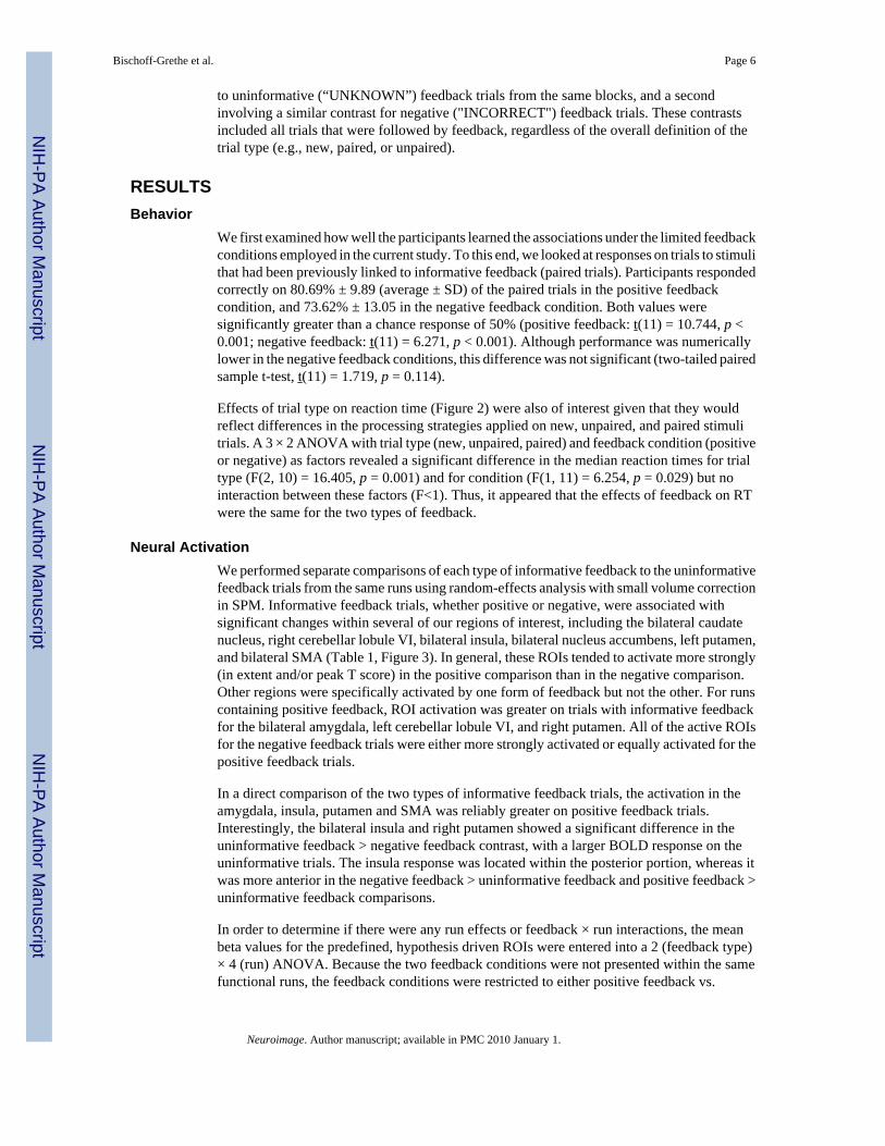
On each trial, the subject saw a single letter stimulus and pressed one of two response keyswith either the index or middle finger of the right hand. The stimulus set consisted of all 26letters, with each letter arbitrarily assigned to one of two response categories (see below). Thestimulus was presented for 1.0 s and then replaced by a fixation cross for 0.8 s (Figure 1).Subjects were instructed that they had to respond during the time of the stimulus presentation.If the response was made within 1.8 s of stimulus onset, feedback was provided (see below).The feedback screen was presented for 1.0 s and then replaced by a fixation cross for 1.0 s untilthe start of the next trial. The entire trial duration was 3.8 s. In addition to these stimulus-presenttrials, we also included 23% fixation trials in which no stimulus was presented, and the “+”fixation point remained on the screen for the entire 3.8 s duration. The inclusion of these fixationtrials allowed for the characterization of event-specific responses in a pseudorandomlydesigned rapid presentation of all trial types (Friston et al., 1999b).
Bischoff-Grethe et al. Page 3
Neuroimage. Author manuscript; available in PMC 2010 January 1.
NIH
-PA Author Manuscript
NIH
-PA Author Manuscript
NIH
-PA Author Manuscript

Two types of runs were used that differed in terms of feedback. On positive feedback runs, theword "CORRECT" was displayed when a) the trial was a candidate for informative feedbackand b) the response was correct. These trials allowed the subject to learn the correct responseassociation for that stimulus. On average, positive feedback was provided on 27% of thestimulus-present trials. Of the remaining stimulus-present trials, the word "UNKNOWN" waspresented as feedback. Similarly, on negative feedback runs, the word "INCORRECT" wasdisplayed when a) the trial was a candidate for informative feedback and b) the response wasincorrect; otherwise the word "UNKNOWN" was presented after the response. On average,negative feedback was provided on 18% of the stimulus-present trials on negative feedbackruns. The differential occurrence of negative and positive feedback was due to the fact that, asparticipants learned, the probability for both informative feedback conditions being met wasless in the negative feedback condition than in the positive feedback condition. By making thepresentation of informative feedback conditional in terms of overall probability as well as thespecific response for that trial, subjects were not able to infer the correct response on most ofthe trials. For example, the absence of positive feedback following a response in a positive rundid not imply that the response was incorrect. This design allowed us to compare associativelearning on the basis of either positive feedback or negative feedback.
A new subset of sixteen stimuli was selected for each scanning run. During each run, a stimuluswas considered “new” if it were being presented for the first time. Subsequent viewings of thesame stimulus were either “paired” or “unpaired.” Paired stimuli were those in which a priortrial gave informative feedback (i.e., the stimulus was associated with a specific response);unpaired stimuli were those in which no prior trials had provided informative feedback (i.e.,all prior trials were uninformative, meaning the stimulus had yet to be associated with aresponse). Because it was possible within a single run to view the same stimulus multiple times,an adaptive procedure (described below) was used so that new, paired, and unpaired stimuliwere equally likely to receive reinforcement and were distributed evenly throughout the scan.This enabled us to look at behavioral differences based upon levels of knowledge with respectto the stimulus-response (S-R) pairs.
A run consisted of 80 trials. Of these, 24 were fixation trials, randomly distributed among thebehavioral trials. The 5 behavioral trial types were defined by the type of stimulus (new,unpaired, or paired) and whether it received informative or uninformative feedback. New trials(whether receiving informative or uninformative feedback) were those in which the stimulushad not been viewed previously. The number of “new” stimuli was distributed across the runby equating them to the number of stimuli previously viewed regardless of prior informativefeedback. Unpaired, uninformative trials were those in which only uninformative feedback hadbeen given previously to that stimulus, and no feedback was presented on the current trial.Unpaired, informative trials were those in which only uninformative feedback had been givenpreviously, and informative feedback was presented on the current trial. Because eitherresponse could, in theory, be correct on these trials, an on-line algorithm was used to categorizethe stimuli after the response to ensure that the desired distribution of trial types wasmaintained. Thus, the response category for each stimulus was not fixed a priori, but determinedafter the first response to that stimulus on a trial with informative feedback. Paired,uninformative trials were those in which informative feedback had been given previously, andno feedback was presented on the current trial. Paired, informative trials were those in whichinformative feedback had been given previously. On these trials, feedback was given only ifthe participant made the appropriate response (the category assignment for a stimulus was fixedonce informative feedback had been presented). Each of these 5 trial types was evenlydistributed, so that on any given trial, the 5 trial types were equally likely. The first 10 trialsin the run consisted of 6 trials with new stimuli and 4 fixation trials. For the remaining 50 non-fixation trials, each trial type occurred 10 times.
Bischoff-Grethe et al. Page 4
Neuroimage. Author manuscript; available in PMC 2010 January 1.
NIH
-PA Author Manuscript
NIH
-PA Author Manuscript
NIH
-PA Author Manuscript

The order of the two feedback conditions was counterbalanced: half of the participants startedwith four blocks of positive feedback followed by four blocks of negative feedback; the orderwas reversed for the other participants. Participants were given one practice block prior to theirfirst positive scan and first negative feedback scan. At the end of each run, a learning scorewas presented to motivate the participants. Perfect performance led to a score of 100, chancewas 0, and a score of −100 indicated that all of the responses were incorrect. The program onlyscored trials on which the subject should have known the correct stimulus-response associationbased upon feedback from a previous trial.
MRIEight fMRI runs of 152 scans each were obtained. Functional MRI was performed withgradient-recalled echoplanar imaging (reaction time, 2000 msec; echo time, 35 msec; flipangle, 90°; 64 × 64 matrix; 27 5.5 mm contiguous axial slices) on a GE 1.5 T scanner (Kwonget al., 1992; Ogawa et al., 1992). A coplanar T1-weighted structural and a high resolution MRIwere obtained for each individual for subsequent spatial normalization.
Statistical analysisThe data were analyzed using Statistical Parametric Mapping (SPM2; Wellcome Departmentof Cognitive Neurology, London UK)(Friston et al., 1995). Motion correction to the firstfunctional scan was performed within subject using a six-parametric rigid-body transformation.The mean of the motion-corrected images was first coregistered to the individual’s high-resolution MRI using mutual information, followed by coregistration of the structural MRI.The images were then spatially normalized to the Montreal Neurologic Institute (MNI)template (Talairach and Tournoux, 1988) by applying a 12-parameter affine transformationfollowed by a nonlinear warping using basis functions (Ashburner and Friston, 1999). Thespatially normalized scans were then smoothed with a 6 mm isotropic Gaussian kernel toaccommodate anatomical differences across participants. Six subjects had one volume in whichthe slices were improperly reconstructed. These volumes were discarded and replaced withaverage volumes based upon the volumes directly preceding and following them. Due totechnical difficulties, two subjects each lost one positive feedback run, and one subject losttwo negative feedback runs. Their remaining data was included in the analysis.
The data were first analyzed with the general linear model on an individual subject basis withan event-related design and convolved with the SPM canonical hemodynamic responsefunction with temporal and dispersion derivative terms. Next, a random effects model wasperformed to make group statistical inferences (Friston et al., 1999a), and contrasts (describedbelow) were applied using a false detection rate correction (Genovese et al., 2002) at p < 0.05.We then employed a region of interest (ROI) analysis with small volume correction.Anatomically defined ROIs from an automated atlas (Tzourio-Mazoyer et al., 2002) were usedto restrict analyses to the putamen, caudate nucleus, amygdala, insula, supplementary motorarea, and cerebellar lobule VI. Because no anatomical definition of the nucleus accumbens wasavailable, a sphere centered at x, y, z = ±10, 8, −4 with radius = 8 mm was defined. Thesecoordinates have been used in other ROI analyses and are near the ventral striatal foci as definedin previous studies (Breiter et al., 2001; Cools et al., 2002; Delgado et al., 2000). This ROIpartially overlapped the MNI ROI for the putamen (left: 21.2% overlap; right: 2.4% overlap);however, given the variability of the nucleus accumbens location we felt this partial overlapwas acceptable.
To determine the extent the ROIs were engaged by informative feedback, we implemented acontrast that compared informative feedback trials to uninformative feedback trials: the runscontaining positive feedback were treated separately from those containing negative feedback.That is, we conducted two contrasts, one comparing positive (“CORRECT”) feedback trials
Bischoff-Grethe et al. Page 5
Neuroimage. Author manuscript; available in PMC 2010 January 1.
NIH
-PA Author Manuscript
NIH
-PA Author Manuscript
NIH
-PA Author Manuscript
to uninformative (“UNKNOWN”) feedback trials from the same blocks, and a secondinvolving a similar contrast for negative ("INCORRECT") feedback trials. These contrastsincluded all trials that were followed by feedback, regardless of the overall definition of thetrial type (e.g., new, paired, or unpaired).
RESULTSBehavior
We first examined how well the participants learned the associations under the limited feedbackconditions employed in the current study. To this end, we looked at responses on trials to stimulithat had been previously linked to informative feedback (paired trials). Participants respondedcorrectly on 80.69% ± 9.89 (average ± SD) of the paired trials in the positive feedbackcondition, and 73.62% ± 13.05 in the negative feedback condition. Both values weresignificantly greater than a chance response of 50% (positive feedback: t(11) = 10.744, p <0.001; negative feedback: t(11) = 6.271, p < 0.001). Although performance was numericallylower in the negative feedback conditions, this difference was not significant (two-tailed pairedsample t-test, t(11) = 1.719, p = 0.114).
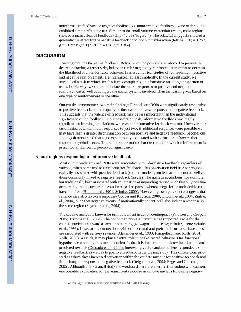
Effects of trial type on reaction time (Figure 2) were also of interest given that they wouldreflect differences in the processing strategies applied on new, unpaired, and paired stimulitrials. A 3 × 2 ANOVA with trial type (new, unpaired, paired) and feedback condition (positiveor negative) as factors revealed a significant difference in the median reaction times for trialtype (F(2, 10) = 16.405, p = 0.001) and for condition (F(1, 11) = 6.254, p = 0.029) but nointeraction between these factors (F<1). Thus, it appeared that the effects of feedback on RTwere the same for the two types of feedback.
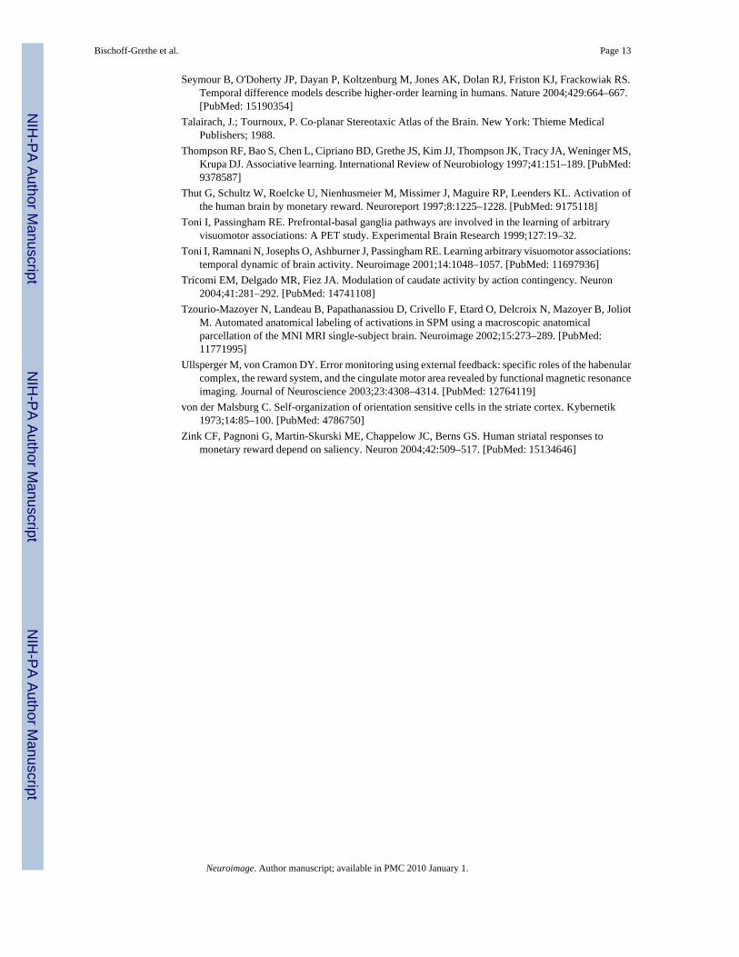
Neural ActivationWe performed separate comparisons of each type of informative feedback to the uninformativefeedback trials from the same runs using random-effects analysis with small volume correctionin SPM. Informative feedback trials, whether positive or negative, were associated withsignificant changes within several of our regions of interest, including the bilateral caudatenucleus, right cerebellar lobule VI, bilateral insula, bilateral nucleus accumbens, left putamen,and bilateral SMA (Table 1, Figure 3). In general, these ROIs tended to activate more strongly(in extent and/or peak T score) in the positive comparison than in the negative comparison.Other regions were specifically activated by one form of feedback but not the other. For runscontaining positive feedback, ROI activation was greater on trials with informative feedbackfor the bilateral amygdala, left cerebellar lobule VI, and right putamen. All of the active ROIsfor the negative feedback trials were either more strongly activated or equally activated for thepositive feedback trials.
In a direct comparison of the two types of informative feedback trials, the activation in theamygdala, insula, putamen and SMA was reliably greater on positive feedback trials.Interestingly, the bilateral insula and right putamen showed a significant difference in theuninformative feedback > negative feedback contrast, with a larger BOLD response on theuninformative trials. The insula response was located within the posterior portion, whereas itwas more anterior in the negative feedback > uninformative feedback and positive feedback >uninformative feedback comparisons.
In order to determine if there were any run effects or feedback × run interactions, the meanbeta values for the predefined, hypothesis driven ROIs were entered into a 2 (feedback type)× 4 (run) ANOVA. Because the two feedback conditions were not presented within the samefunctional runs, the feedback conditions were restricted to either positive feedback vs.
Bischoff-Grethe et al. Page 6
Neuroimage. Author manuscript; available in PMC 2010 January 1.
NIH
-PA Author Manuscript
NIH
-PA Author Manuscript
NIH
-PA Author Manuscript
uninformative feedback or negative feedback vs. uninformative feedback. None of the ROIsexhibited a main effect for run. Similar to the small volume correction results, most regionsshowed a main effect of feedback (all p < 0.05) (Figure 4). The bilateral amygdala showed aquadratic run effect for the negative feedback condition × run interaction (left: F(3, 30) = 3.257,p = 0.035; right: F(3, 30) = 4.154, p = 0.014).
DISCUSSIONLearning requires the use of feedback. Behavior can be positively reinforced to promote adesired behavior; alternatively, behavior can be negatively reinforced in an effort to decreasethe likelihood of an undesirable behavior. In most empirical studies of reinforcement, positiveand negative reinforcements are intermixed, at least implicitly. In the current study, weintroduced a task in which feedback was completely uninformative on a large proportion oftrials. In this way, we sought to isolate the neural responses to positive and negativereinforcement as well as compare the neural systems involved when the learning was based onone type of reinforcement or the other.
Our results demonstrated two main findings: First, all our ROIs were significantly responsiveto positive feedback, and a majority of them were likewise responsive to negative feedback.This suggests that the valence of feedback may be less important than the motivationalsignificance of the feedback. In our association task, informative feedback was highlysignificant to learning associations, whereas noninformative feedback was not. However, ourtask limited potential motor responses to just two; if additional responses were possible wemay have seen a greater discrimination between positive and negative feedback. Second, ourfindings demonstrated that regions commonly associated with extrinsic reinforcers alsorespond to symbolic cues. This supports the notion that the context in which reinforcement ispresented influences its perceived significance.
Neural regions responding to informative feedbackMost of our predetermined ROIs were associated with informative feedback, regardless ofvalence, when compared to uninformative feedback. This observation held true for regionstypically associated with positive feedback (caudate nucleus, nucleus accumbens) as well asthose commonly linked to negative feedback (insula). The nucleus accumbens, for example,has traditionally been associated with anticipation of impending reward, such that only positiveor more favorable cues produce an increased response, whereas negative or undesirable cueshave no effect (Breiter et al., 2001; Schultz, 2000). However, growing evidence suggests thatsalience may also invoke a response (Cooper and Knutson, 2008; Tricomi et al., 2004; Zink etal., 2004), such that negative events, if motivationally salient, will also induce a response inthe same region (Seymour et al., 2004).
The caudate nucleus is known for its involvement in action contingency (Knutson and Cooper,2005; Tricomi et al., 2004). The nonhuman primate literature has supported a role for thecaudate nucleus in reward association learning (Kawagoe et al., 1998; Schultz, 1998; Schultzet al., 1998). It has strong connections with orbitofrontal and prefrontal cortices; these areasare associated with sensory rewards (Alexander et al., 1990; Kringelbach and Rolls, 2004;Rolls, 2000). As such, it may play a central role in goal-directed behavior. One functionalhypothesis concerning the caudate nucleus is that it is involved in the detection of actual andpredicted rewards (Delgado et al., 2004). Interestingly, the caudate nucleus responded tonegative feedback as well as to positive feedback in the present study. This differs from priorstudies which show increased activation within the caudate nucleus for positive feedback andlittle change in response to negative feedback (Delgado et al., 2004; Seger and Cincotta,2005). Although this is a small study and we should therefore interpret this finding with caution,one possible explanation for the significant response in caudate nucleus following negative
Bischoff-Grethe et al. Page 7
Neuroimage. Author manuscript; available in PMC 2010 January 1.
NIH
-PA Author Manuscript
NIH
-PA Author Manuscript
NIH
-PA Author Manuscript

reinforcement may be related to the context of our task compared to others studies providinginformative feedback. In studies showing transient changes within the caudate nucleus(Delgado et al., 2000; Elliott et al., 2000), a positive reward of some kind was always possiblewithin each trial. In our study, however, positive reinforcement was never provided in thenegative feedback runs and, correspondingly, there should never have been an expectation ofpositive reinforcement. Moreover, the absence of feedback in this condition was ambiguous.Thus, these results suggest that the caudate nucleus response may have been modulated by thecognitive aspect of the task. There is some support for this in the literature, where a responsehas been associated with either salience or contingency detection (Tricomi et al., 2004; Zinket al., 2004). An intriguing corollary here is that negative reinforcement is not equivalent tothe absence of positive reinforcement, at least when the context contains considerableuncertainty (e.g., the high percentage of uninformative trials).
In contrast to the activation in the caudate, the activation within the putamen varied as a functionof the type of feedback. While the left ventral putamen responded to all kinds of informativefeedback, the right ventral putamen responded only to positive feedback when compared touninformative feedback. Unlike the caudate nucleus, which receives prefrontal inputs and isassociated with high level cognition, the putamen receives a mixture of prefrontal, premotorand motor inputs and is more closely associated with the motoric aspect of S-R learning(Middleton and Strick, 2000, 2001). Overall, these results are consistent with responsereinforcement.
The anterior insula responded to informative feedback regardless of valence. While activationin the insula has been associated with negative events including pain (Craig, 2003), rejectionwithin a social context (Eisenberger et al., 2003), and punishment in reward paradigms (Ableret al., 2005; O'Doherty et al., 2003; Ullsperger and von Cramon, 2003), it has also beenimplicated in decision-making and behavioral monitoring (Casey et al., 2000; Huettel et al.,2005; Paulus et al., 2005; Paulus et al., 2003). While these processes would be engagedfollowing informative reinforcement, the current results would indicate that the engagementof anterior insula here may be greater when the reinforcement is negative. It is possible thatsubjects experienced emotional arousal in relation to informative feedback, and that this drovethe anterior insula activation.
The right cerebellar lobule VI was also activated following both positive and negative feedback.Both the cerebellum and the putamen have long been associated with motor learning. However,computational models of these two systems typically emphasize different feedbackmethodologies. Basal ganglia learning models focus upon the reward signaling properties ofdopamine (e.g., McClure et al., 2003b), whereas cerebellar learning models focus upon errorsignals generated by climbing fiber input (e.g., Albus, 1971; Marr, 1969). Neuropsychologicaland neuroimaging studies have suggested that the cerebellum plays a critical role in highercognition rather than being strictly related to motor learning and motor control. One functionalaccount of these effects is that the cerebellum may be involved in the planning and rehearsalof actions as part of a process that anticipates potential outcomes (Bischoff-Grethe et al.,2002). It may be that informative feedback of either valence engages these operations as theindividual attempts to associate a response to a stimulus that just received informative feedback.
Learning with intrinsic cuesAn important feature of this task is that even symbolic cues, associated with intrinsicreinforcement, activated regions commonly associated with more extrinsic feedback. Much ofthe reward literature has used some form of extrinsic reinforcement. Neuroimaging studies inhumans have demonstrated responses within the limbic circuit for both primary (e.g., appetitivestimuli) (Berns et al., 2001; McClure et al., 2007) and secondary (e.g., monetary gain or loss)reinforcers (Breiter et al., 2001; Delgado et al., 2004). However, the valence of these reinforcers
Bischoff-Grethe et al. Page 8
Neuroimage. Author manuscript; available in PMC 2010 January 1.
NIH
-PA Author Manuscript
NIH
-PA Author Manuscript
NIH
-PA Author Manuscript

may also influence neural response due to the context in which they are presented. Rewardprocessing systems determine whether an outcome is favorable based upon the range ofpossible outcomes (Breiter et al., 2001; Nieuwenhuis et al., 2005); an outcome of $0 isundesirable when other outcomes provide a monetary reward, but is desirable when onlynegative outcomes are possible. This implies that, within the right framework, symbolic cuescould indeed drive the reward system. Our results support this hypothesis by demonstrating astrong limbic response to informative feedback trials.
The receipt of informative feedback was based upon action-contingencies: in the positivefeedback condition, a correct stimulus-response association was necessary in order to allowthe possibility of positive feedback. In the negative condition, the inverse was true. Action-contingencies may also have played a role in the involvement of the reward circuitry. Withoutthe association learning aspect, it is likely that our symbolic cues are meaningless, and thereforewould fail to induce reward-related responses. This is in contrast to primary and secondaryreinforcement paradigms, in which simply attending to the stimuli activated the reward system(Breiter et al., 2001).
Successful performance of an associative task requires forming and maintaining the correctvisuomotor association. Overall, participants responded more slowly for unpaired and pairedtrials than they did for new trials. The effect of trial type likely reflects response retrieval:unpaired and paired trials may have been slower because they involved retrieval processes thatdetermined whether the stimulus had been associated with a particular response on a previouspresentation. Recognition that a stimulus is novel would have precluded or aborted suchretrieval processes. For all trial types, participants were slower to respond in the negativefeedback condition, consistent with the finding that learning tended to be more difficult withnegative feedback.
The right dorsoposterior putamen and bilateral posterior insula both demonstrated an increasedresponse for uninformative feedback when compared to negative feedback. This peakactivation was distinct from that seen for informative feedback when compared touninformative feedback. The posterior insula is associated with motor sensation and otherhomeostatic information (Craig, 2003). This region responds to predictable aversive events, incontrast to the anterior insula, which responds to unpredictable aversive events (Carlsson etal., 2006). The insula has bidirectional connections to both the amygdala and to the nucleusaccumbens (Reynolds and Zahm, 2005) as well as the orbitofrontal cortex (Ongur and Price,2000). In the amygdala, neurons can rapidly adjust their activity to reflect both positive andnegative value of an external stimulus, which is predictive of how fast monkeys learn to respondto a stimulus (Paton et al., 2006). Therefore, the insula is centrally placed to receive informationabout the salience (both appetitive and aversive) and relative value of the stimulus environmentand integrate this information with the effect that these stimuli may have on the body state. Asanxiety can induce homeostatic changes in an individual, we suggest that the receipt ofuninformative feedback in the negative condition induced more anxiety due to its context.Unless an S-R pairing had previously received negative feedback, subjects did not know thecorrect response in this condition. This uncertainty may have increased anxiety levels regardingtask performance.
It is possible that, despite efforts for an unbiased, neutral stimulus, the uninformative feedbacktrials might be viewed negatively and be a source of frustration to the participant. Ifuninformative feedback trials had negative connotations, we would expect a reduced likelihoodof activation in comparison of negative informative feedback to uninformative feedback.However, both positive and negative feedback, when compared to uninformative feedback,produced responses that 1) support the proposal that positive informative feedback isrewarding, and 2) show salience is also critical, regardless of feedback valence. It is therefore
Bischoff-Grethe et al. Page 9
Neuroimage. Author manuscript; available in PMC 2010 January 1.
NIH
-PA Author Manuscript
NIH
-PA Author Manuscript
NIH
-PA Author Manuscript

likely that uninformative feedback produced little negative interference that would confoundour results.
ConclusionAn important form of associative learning requires the arbitrary linkage of an action to astimulus. Most studies of associative learning use a trial and error approach (Toni andPassingham, 1999; Toni et al., 2001) in which feedback is provided on every trial. Thisprocedure makes it difficult to dissociate the effects of feedback unless a large temporal gapseparates the stimulus, response, and feedback signals. By separating blocks that used positiveand negative feedback in combination with a partial reinforcement schedule, we were able toidentify areas associated with specific types of feedback. While some neural regions wereassociated with these processes independent of whether the feedback was positive or negative,we also observed regions that were sensitive to the valence of feedback. This experiment alsodemonstrates that regions commonly associated with extrinsic rewards and punishments canalso respond to more intrinsic forms of feedback.
ACKNOWLEDGEMENTSSupported by PHS grants NS33504 and NS40813. Corresponding author: Scott T. Grafton, M.D., Sage Center for theStudy of Mind, Department of Psychology, Psychology East, UC Santa Barbara, Santa Barbara CA 93106.
REFERENCESAbler B, Walter H, Erk S. Neural correlates of frustration. Neuroreport 2005;16:669–672. [PubMed:
15858403]Albus JS. The theory of cerebellar function. Mathematical Biosciences 1971;10:25–61.Alexander, GE.; Crutcher, MD.; DeLong, MR. Basal ganglia-thalamocortical circuits: Parallel substrates
for motor, oculomotor, "prefrontal" and "limbic" functions. In: Uylings, HBM.; Eden, CGV.; Bruin,JPCD.; Corner, MA.; Feenstra, MGP., editors. Progress in Brain Research. New York: Elsevier SciencePublishers B. V.; 1990. p. 119-146.
Ashburner J, Friston KJ. Nonlinear spatial normalization using basis functions. Human Brain Mapping1999;7:254–266. [PubMed: 10408769]
Baxter MG, Murray EA. The amygdala and reward. Nat Rev Neurosci 2002;3:563–573. [PubMed:12094212]
Berns GS, McClure SM, Pagnoni G, Montague PR. Predictability modulates human brain response toreward. Journal of Neuroscience 2001;21:2793–2798. [PubMed: 11306631]
Bischoff-Grethe A, Ivry RB, Grafton ST. Cerebellar Involvement in Response Reassignment Rather ThanAttention. Journal of Neuroscience 2002;22:536–553. [PubMed: 11784800]
Breiter HC, Aharon I, Kahneman D, Dale A, Shizgal P. Functional imaging of neural responses toexpectancy and experience of monetary gains and losses. Neuron 2001;30:619–639. [PubMed:11395019]
Breiter HC, Gollub RL, Weisskoff RM, Kennedy DN, Makris N, Berke JD, Goodman JM, Kantor HL,Gastfriend DR, Riorden JP, Mathew RT, Rosen BR, Hyman SE. Acute effects of cocaine on humanbrain activity and emotion. Neuron 1997;19:591–611. [PubMed: 9331351]
Carlsson K, Andersson J, Petrovic P, Petersson KM, Ohman A, Ingvar M. Predictability modulates theaffective and sensory-discriminative neural processing of pain. Neuroimage 2006;32:1804–1814.[PubMed: 16861005]
Casey BJ, Thomas KM, Welsh TF, Badgaiyan RD, Eccard CH, Jennings JR, Crone EA. Dissociation ofresponse conflict, attentional selection, and expectancy with functional magnetic resonance imaging.Proceedings of the National Academy of Sciences of the United States of America 2000;97:8728–8733. [PubMed: 10900023]
Bischoff-Grethe et al. Page 10
Neuroimage. Author manuscript; available in PMC 2010 January 1.
NIH
-PA Author Manuscript
NIH
-PA Author Manuscript
NIH
-PA Author Manuscript

Cools R, Clark L, Owen AM, Robbins TW. Defining the neural mechanisms of probabilistic reversallearning using event-related functional magnetic resonance imaging. Journal of Neuroscience2002;22:4563–4567. [PubMed: 12040063]
Cooper JC, Knutson B. Valence and salience contribute to nucleus accumbens activation. Neuroimage2008;39:538–547. [PubMed: 17904386]
Craig AD. Interoception: the sense of the physiological condition of the body. Current Opinion inNeurobiology 2003;13:500–505. [PubMed: 12965300]
Delgado MR, Nystrom LE, Fissell C, Noll DC, Fiez JA. Tracking the hemodynamic responses to rewardand punishment in the striatum. Journal of Neurophysiology 2000;84:3072–3077. [PubMed:11110834]
Delgado MR, Stenger VA, Fiez JA. Motivation-dependent Responses in the Human Caudate Nucleus.Cerebral Cortex 2004;14:1022–1030. [PubMed: 15115748]
Doya K. What are the computations of the cerebellum, the basal ganglia and the cerebral cortex? NeuralNetworks 1999;12:961–974. [PubMed: 12662639]
Eisenberger NI, Lieberman MD, Williams KD. Does rejection hurt? An FMRI study of social exclusion.Science 2003;302:290–292. [PubMed: 14551436]
Elliott R, Friston KJ, Dolan RJ. Dissociable Neural Responses in Human Reward Systems. Journal ofNeuroscience 2000;20:6159–6165. [PubMed: 10934265]
Elliott R, Frith CD, Dolan RJ. Differential neural response to positive and negative feedback in planningand guessing tasks. Neuropsychologia 1997;35:1395–1404. [PubMed: 9347486]
Elliott R, Newman JL, Longe OA, Deakin JF. Differential response patterns in the striatum andorbitofrontal cortex to financial reward in humans: a parametric functional magnetic resonanceimaging study. Journal of Neuroscience 2003;23:303–307. [PubMed: 12514228]
Everitt BJ, Cardinal RN, Parkinson JA, Robbins TW. Appetitive behavior: impact of amygdala-dependentmechanisms of emotional learning. Ann N Y Acad Sci 2003;985:233–250. [PubMed: 12724162]
Friston KJ, Holmes AP, Worsley KJ. How many subjects constitute a study? Neuroimage 1999a;10:1–5. [PubMed: 10385576]
Friston KJ, Holmes AP, Worsley KJ, Poline J-B, Frith CD, Frackowiak RSJ. Statistical parametric mapsin functional imaging: A general linear approach. Human Brain Mapping 1995;2:189–210.
Friston KJ, Zarahn E, Josephs O, Henson RN, Dale AM. Stochastic designs in event-related fMRI.Neuroimage 1999b;10:607–619. [PubMed: 10547338]
Garavan H, Ross TJ, Stein EA. Right hemispheric dominance of inhibitory control: an event-relatedfunctional MRI study. Proceedings of the National Academy of Sciences of the United States ofAmerica 1999;96:8301–8306. [PubMed: 10393989]
Genovese CR, Lazar NA, Nichols T. Thresholding of statistical maps in functional neuroimaging usingthe false discovery rate. Neuroimage 2002;15:870–878. [PubMed: 11906227]
Huettel SA, Song AW, McCarthy G. Decisions under uncertainty: probabilistic context influencesactivation of prefrontal and parietal cortices. Journal of Neuroscience 2005;25:3304–3311. [PubMed:15800185]
Ito M. Mechanisms of motor learning in the cerebellum. Brain Res 2000;886:237–245. [PubMed:11119699]
Kawagoe R, Takikawa Y, Hikosaka O. Expectation of reward modulates cognitive signals in the basalganglia. Nature Neuroscience 1998;1:411–416.
Knutson B, Cooper JC. Functional magnetic resonance imaging of reward prediction. Curr Opin Neurol2005;18:411–417. [PubMed: 16003117]
Kringelbach ML, Rolls ET. The functional neuroanatomy of the human orbitofrontal cortex: evidencefrom neuroimaging and neuropsychology. Prog Neurobiol 2004;72:341–372. [PubMed: 15157726]
Kwong KK, Belliveau JW, Chesler DA, Goldberg IE, Weisskoff RM, Poncelet BP, Kennedy DN, HoppelBE, Cohen MS, Turner R, et al. Dynamic magnetic resonance imaging of human brain activity duringprimary sensory stimulation. Proceedings of the National Academy of Sciences of the United Statesof America 1992;89:5675–5679. [PubMed: 1608978]
Marr D. A theory of cerebellar cortex. J Physiol 1969;202:437–470. [PubMed: 5784296]
Bischoff-Grethe et al. Page 11
Neuroimage. Author manuscript; available in PMC 2010 January 1.
NIH
-PA Author Manuscript
NIH
-PA Author Manuscript
NIH
-PA Author Manuscript

McClure SM, Berns GS, Montague PR. Temporal prediction errors in a passive learning task activatehuman striatum. Neuron 2003a;38:339–346. [PubMed: 12718866]
McClure SM, Daw ND, Montague PR. A computational substrate for incentive salience. Trends Neurosci2003b;26:423–428. [PubMed: 12900173]
McClure SM, Ericson KM, Laibson DI, Loewenstein G, Cohen JD. Time discounting for primaryrewards. Journal of Neuroscience 2007;27:5796–5804. [PubMed: 17522323]
Mesulam MM, Mufson EJ. Insula of the old world monkey. III: Efferent cortical output and commentson function. Journal of Comparative Neurology 1982;212:38–52. [PubMed: 7174907]
Middleton FA, Strick PL. Basal ganglia and cerebellar loops: Motor and cognitive circuits. BrainResearch Reviews 2000;31:236–250. [PubMed: 10719151]
Middleton, FA.; Strick, PL. A revised neuroanatomy of frontal-subcortical circuits. In: Lichter, DG.;Cummings, JL., editors. Frontal-subcortical circuits in psychiatric and neurological disorders. NewYork: Guilford Press; 2001. p. 44-58.
Nieuwenhuis S, Heslenfeld DJ, Alting von Geusau NJ, Mars RB, Holroyd CB, Yeung N. Activity inhuman reward-sensitive brain areas is strongly context dependent. Neuroimage 2005;25:1302–1309.[PubMed: 15945130]
O'Doherty J, Critchley H, Deichmann R, Dolan RJ. Dissociating valence of outcome from behavioralcontrol in human orbital and ventral prefrontal cortices. Journal of Neuroscience 2003;23:7931–7939.[PubMed: 12944524]
Ogawa S, Tank DW, Menon R, Ellermann JM, Kim SG, Merkle H, Ugurbil K. Intrinsic signal changesaccompanying sensory stimulation: Functional brain mapping with magnetic resonance imaging.Proceedings of the National Academy of Sciences of the United States of America 1992;89:5951–5955. [PubMed: 1631079]
Ongur D, Price JL. The organization of networks within the orbital and medial prefrontal cortex of rats,monkeys and humans. Cerebral Cortex 2000;10:206–219. [PubMed: 10731217]
Paton JJ, Belova MA, Morrison SE, Salzman CD. The primate amygdale represents the positive andnegative value of visual stimuli during learning. Nature 2006;439:865–870. [PubMed: 16482160]
Paulus MP, Feinstein JS, Leland D, Simmons AN. Superior temporal gyrus and insula provide responseand outcome-dependent information during assessment and action selection in a decision-makingsituation. Neuroimage 2005;25:607–615. [PubMed: 15784440]
Paulus MP, Hozack N, Frank L, Brown GG, Schuckit MA. Decision making by methamphetamine-dependent subjects is associated with error-rate-independent decrease in prefrontal and parietalactivation. Biol Psychiatry 2003;53:65–74. [PubMed: 12513946]
Reynolds SM, Zahm DS. Specificity in the projections of prefrontal and insular cortex to ventralstriatopallidum and the extended amygdala. Journal of Neuroscience 2005;25:11757–11767.[PubMed: 16354934]
Rolls ET. The orbitofrontal cortex and reward. Cerebral Cortex 2000;10:284–294. [PubMed: 10731223]Rushworth MF, Hadland KA, Paus T, Sipila PK. Role of the human medial frontal cortex in task
switching: a combined fMRI and TMS study. Journal of Neurophysiology 2002;87:2577–2592.[PubMed: 11976394]
Sanger T. Optimal unsupervised learning in a single-layer linear feedforward neural network. NeuralNetworks 1989;2:459–473.
Schultz W. Predictive reward signal of dopamine neurons. Journal of Neurophysiology 1998;80:1–27.[PubMed: 9658025]
Schultz W. Multiple reward signals in the brain. Nat Rev Neurosci 2000;1:199–207. [PubMed: 11257908]Schultz W, Dickinson A. Neuronal coding of prediction errors. Annual Review of Neuroscience
2000;23:473–500.Schultz W, Tremblay L, Hollerman JR. Reward prediction in primate basal ganglia and frontal cortex.
Neuropharmacology 1998;37:421–429. [PubMed: 9704983]Seger CA, Cincotta CM. The roles of the caudate nucleus in human classification learning. Journal of
Neuroscience 2005;25:2941–2951. [PubMed: 15772354]
Bischoff-Grethe et al. Page 12
Neuroimage. Author manuscript; available in PMC 2010 January 1.
NIH
-PA Author Manuscript
NIH
-PA Author Manuscript
NIH
-PA Author Manuscript
Seymour B, O'Doherty JP, Dayan P, Koltzenburg M, Jones AK, Dolan RJ, Friston KJ, Frackowiak RS.Temporal difference models describe higher-order learning in humans. Nature 2004;429:664–667.[PubMed: 15190354]
Talairach, J.; Tournoux, P. Co-planar Stereotaxic Atlas of the Brain. New York: Thieme MedicalPublishers; 1988.
Thompson RF, Bao S, Chen L, Cipriano BD, Grethe JS, Kim JJ, Thompson JK, Tracy JA, Weninger MS,Krupa DJ. Associative learning. International Review of Neurobiology 1997;41:151–189. [PubMed:9378587]
Thut G, Schultz W, Roelcke U, Nienhusmeier M, Missimer J, Maguire RP, Leenders KL. Activation ofthe human brain by monetary reward. Neuroreport 1997;8:1225–1228. [PubMed: 9175118]
Toni I, Passingham RE. Prefrontal-basal ganglia pathways are involved in the learning of arbitraryvisuomotor associations: A PET study. Experimental Brain Research 1999;127:19–32.
Toni I, Ramnani N, Josephs O, Ashburner J, Passingham RE. Learning arbitrary visuomotor associations:temporal dynamic of brain activity. Neuroimage 2001;14:1048–1057. [PubMed: 11697936]
Tricomi EM, Delgado MR, Fiez JA. Modulation of caudate activity by action contingency. Neuron2004;41:281–292. [PubMed: 14741108]
Tzourio-Mazoyer N, Landeau B, Papathanassiou D, Crivello F, Etard O, Delcroix N, Mazoyer B, JoliotM. Automated anatomical labeling of activations in SPM using a macroscopic anatomicalparcellation of the MNI MRI single-subject brain. Neuroimage 2002;15:273–289. [PubMed:11771995]
Ullsperger M, von Cramon DY. Error monitoring using external feedback: specific roles of the habenularcomplex, the reward system, and the cingulate motor area revealed by functional magnetic resonanceimaging. Journal of Neuroscience 2003;23:4308–4314. [PubMed: 12764119]
von der Malsburg C. Self-organization of orientation sensitive cells in the striate cortex. Kybernetik1973;14:85–100. [PubMed: 4786750]
Zink CF, Pagnoni G, Martin-Skurski ME, Chappelow JC, Berns GS. Human striatal responses tomonetary reward depend on saliency. Neuron 2004;42:509–517. [PubMed: 15134646]
Bischoff-Grethe et al. Page 13
Neuroimage. Author manuscript; available in PMC 2010 January 1.
NIH
-PA Author Manuscript
NIH
-PA Author Manuscript
NIH
-PA Author Manuscript

Figure 1.Examples of A) positive, B) unknown, and C) negative feedback trials. A single trial lasted3.8 s, and subjects had 1.8 s to respond once the stimulus (e.g., the letter ‘A’) was displayed.Subjects received one of three forms of feedback; positive feedback (occurring in the positivefeedback blocks), negative feedback (occurring in the negative feedback blocks), or unknown(occurring in both positive and negative feedback blocks).
Bischoff-Grethe et al. Page 14
Neuroimage. Author manuscript; available in PMC 2010 January 1.
NIH
-PA Author Manuscript
NIH
-PA Author Manuscript
NIH
-PA Author Manuscript
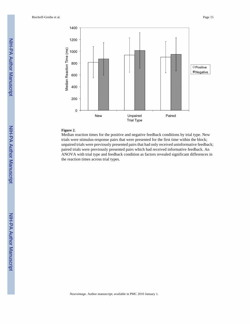
Figure 2.Median reaction times for the positive and negative feedback conditions by trial type. Newtrials were stimulus-response pairs that were presented for the first time within the block;unpaired trials were previously presented pairs that had only received uninformative feedback;paired trials were previously presented pairs which had received informative feedback. AnANOVA with trial type and feedback condition as factors revealed significant differences inthe reaction times across trial types.
Bischoff-Grethe et al. Page 15
Neuroimage. Author manuscript; available in PMC 2010 January 1.
NIH
-PA Author Manuscript
NIH
-PA Author Manuscript
NIH
-PA Author Manuscript

Figure 3.Statistical parametric maps of the functional localization within the coronal plane (p <0.05,FDR-corrected). a) Regions responding to positive feedback > uninformative feedbackincluded the bilateral cerebellum (z = −50), the bilateral amygdala (z =− 4), and the nucleusaccumbens, caudate nucleus, putamen, insula, and SMA bilaterally (z = +12). b) For thenegative feedback > uninformative feedback comparison, activated regions included the rightcerebellum (z = −74) and the bilateral nucleus accumbens, caudate nucleus, putamen, insula,and SMA (z = +16).
Bischoff-Grethe et al. Page 16
Neuroimage. Author manuscript; available in PMC 2010 January 1.
NIH
-PA Author Manuscript
NIH
-PA Author Manuscript
NIH
-PA Author Manuscript

Figure 4.Region of interest analysis comparing mean beta values defined at each location for thecontrasts Positive Feedback – Uninformative Feedback and Negative Feedback –Uninformative Feedback. Beta values were determined by the general linear model in SPMfor each individual. Beta weights were averaged across an anatomical region of interest, andthen averaged across subjects. Anatomical regions were as defined in an automated atlas(Tzourio-Mazoyer et al., 2002), except for the nucleus accumbens, which was defined as aspherical region of interest (x, y, z = ±10, 8, −4; radius = 8 mm). Regions with statisticallysignificant between mean beta value and baseline are indicated as follows: *p < 0.05; **p <0.01; ***p<0.005.
Bischoff-Grethe et al. Page 17
Neuroimage. Author manuscript; available in PMC 2010 January 1.
NIH
-PA Author Manuscript
NIH
-PA Author Manuscript
NIH
-PA Author Manuscript

NIH
-PA Author Manuscript
NIH
-PA Author Manuscript
NIH
-PA Author Manuscript
Bischoff-Grethe et al. Page 18Ta
ble
1A
ctiv
atio
n pe
aks w
ithin
the
RO
Is fo
r the
con
trast
s (Po
sitiv
e Fe
edba
ck >
Uni
nfor
mat
ive
Feed
back
; Neg
ativ
e Fe
edba
ck >
Uni
nfor
mat
ive
Feed
back
; Uni
nfor
mat
ive
Feed
back
> N
egat
ive
Feed
back
; and
(Pos
itive
Fee
dbac
k –
Uni
nfor
mat
ive
Feed
back
) > (N
egat
ive
Feed
back
–U
ninf
orm
ativ
e Fe
edba
ck))
. Sig
nific
ance
was
set
at p
< 0
.05
with
sm
all v
olum
e co
rrec
tion
for t
he re
gion
s of
inte
rest
. Coo
rdin
ates
are
base
d on
the
Mon
treal
Neu
rolo
gica
l Ins
titut
e br
ain
tem
plat
e.
Lef
t Hem
isph
ere
Rig
ht H
emis
pher
e
Clu
ster
Siz
ep
(FD
R-c
orr)
T S
core
MN
I Coo
rdin
ates
(mm
)C
lust
er S
ize
p (F
DR
-cor
r)T
Sco
reM
NI C
oord
inat
es (m
m)
POSI
TIV
E F
EE
DB
AC
K >
UN
INFO
RM
AT
IVE
FE
ED
BA
CK
Am
ygda
la11
1<
0.00
17.
68−2
2−4
−12
165
< 0.
001
8.28
20−2
−12
Cau
date
nuc
leus
816
< 0.
001
12.5
6−1
214
−879
2<
0.00
112
.80
1414
−12
Cer
ebel
lar l
obul
e V
I70
10.
021
6.03
−36
−50
−24
943
0.01
35.
6532
−46
−22
Insu
la96
80.
002
7.51
−28
16−8
968
0.03
05.
7450
8−6
180.
035
2.61
−32
−18
22
Nuc
leus
acc
umbe
ns12
2<
0.00
110
.43
−12
12−8
123
< 0.
001
9.21
1212
−8
Puta
men
972
< 0.
001
10.4
3−1
212
−886
7<
0.00
19.
8216
14−1
0
SMA
664
0.00
27.
270
1054
831
0.00
16.
996
1446
460.
032
2.67
−14
−14
6412
20.
009
3.52
14−2
254
NE
GA
TIV
E F
EE
DB
AC
K >
UN
INFO
RM
AT
IVE
FE
ED
BA
CK
Cau
date
nuc
leus
258
0.04
45.
05−1
416
053
00.
017
5.15
1018
−8
Cer
ebel
lar l
obul
e V
I74
40.
011
6.07
32−7
4−2
4
Insu
la62
80.
004
6.74
−28
24−8
600
0.01
15.
8348
14−2
Nuc
leus
acc
umbe
ns89
0.01
24.
46−1
412
−298
0.01
14.
3014
4−2
Puta
men
296
0.00
96.
09−2
020
−6
SMA
821
0.00
36.
23−2
254
721
0.00
66.
212
052
UN
INFO
RM
AT
IVE
FE
ED
BA
CK
> N
EG
AT
IVE
FE
ED
BA
CK
Insu
la55
90.
024
5.91
−38
−14
239
80.
043
5.76
38−1
04
Puta
men
177
0.04
85.
5436
−10
2
(PO
SIT
IVE
FE
ED
BA
CK
– U
NIN
FOR
MA
TIV
E F
EE
DB
AC
K) >
(NE
GA
TIV
E F
EE
DB
AC
K –
UN
INFO
RM
AT
IVE
FE
ED
BA
CK
)
Am
ygda
la96
0.02
24.
74−2
40
−12
134
0.00
36.
0022
−2−1
4
Insu
la68
0<
0.00
111
.61
−38
−48
437
0.00
97.
3338
−88
Puta
men
734
< 0.
001
10.6
4−2
8−1
08
637
0.00
19.
9534
−22
SMA
269
0.01
06.
82−6
−20
5227
80.
014
6.28
12−2
254
Neuroimage. Author manuscript; available in PMC 2010 January 1.