On Unilateral Constraints, Friction and Plasticity - Archive ...
Upload
independentCategory
view
1download
0
Hearing Research 203 (2005) 134–143
www.elsevier.com/locate/heares
The inXuence of a sensitive period on central auditory development inchildren with unilateral and bilateral cochlear implants
Anu Sharma a,¤,1, Michael F. Dorman b, Andrej Kral a,2
a Behavioral and Brain Sciences, University of Texas at Dallas, Dallas, TX, USAb Speech and Hearing Science, Arizona State University, Tempe, AZ 85287, USA
Received 3 September 2004; accepted 22 December 2004Available online 23 January 2005
Abstract
We examined the longitudinal development of the cortical auditory evoked potential (CAEP) in 21 children who were Wtted withunilateral cochlear implants and in two children who were Wtted with bilateral cochlear implants either before age 3.5 years or afterage 7 years. The age cut-oVs (<3.5 years for early-implanted and >7 years for late-implanted) were based on the sensitive period forcentral auditory development described in [Ear Hear. 23 (6), 532.] Our results showed a fundamentally diVerent pattern of develop-ment of CAEP morphology and P1 cortical response latency for early- and late-implanted children. Early-implanted children andone child who received bilateral implants by age 3.5 years showed rapid development in CAEP waveform morphology and P1latency. Late-implanted children showed aberrant waveform morphology and signiWcantly slower decreases in P1 latency postim-plantation. In the case of a child who received his Wrst implant by age 3.5 years and his second implant after age 7 years, CAEPresponses elicited by the second implant were similar to late-implanted children. Our results are consistent with animal models ofcentral auditory development after implantation and conWrm the presence of a relatively brief sensitive period for central auditorydevelopment in young children. 2005 Elsevier B.V. All rights reserved.
Keywords: Central auditory development; Sensitive period; P1 cortical auditory evoked potential; Cochlear implant; Bilateral implants
1. Introduction
The human cochlea is functional at birth (Granier-Deferre et al., 1985; Rubel, 1985), but the auditorysystem is still immature and undergoes many develop-mental changes postnatally. This postnatal develop-ment is reXected in age-related changes in cortical
Abbreviation: CAEP, cortical auditory evoked potential¤ Corresponding author. Tel.: +214 905 3185; fax: +214 905 3146.E-mail address: [email protected] (A. Sharma).
1 Present address: Callier Center for Communication Disorders,University of Texas at Dallas, 1966 Inwood Road, Dallas, TX 75206,USA.
2 Institut für Neurophysiologie und Pathophysiologie, Universi-tätsklinikum Hamburg-Eppendorf, Hamburg, Germany.
0378-5955/$ - see front matter 2005 Elsevier B.V. All rights reserved.doi:10.1016/j.heares.2004.12.010
evoked potentials. For example, the cortical P1response, which is generated by auditory thalamic andcortical sources, systematically decreases in latency withincreasing age (Sharma et al., 1997; Cunningham et al.,2000; Ponton et al., 2000).
Because P1 latency changes with age, P1 latency hasbeen used as an index of maturation of the auditorypathway in populations with abnormal auditory experi-ence (Ponton et al., 1996a,b, 1999; Ponton and Egger-mont, 2001; Sharma et al., 2002a,b,c). Sharma et al.(2002c) compared the P1 latencies of 97 congenitallydeaf children Wtted with cochlear implants with the 95%conWdence intervals for P1 latencies derived from age-matched, normal-hearing children. Prelingually deafchildren implanted under 3.5 years of age had age-appropriate P1 latencies within 6 months following the
A. Sharma et al. / Hearing Research 203 (2005) 134–143 135
onset of stimulation. In contrast, the P1 latencies of chil-dren implanted after age 7 were delayed relative to nor-mal-hearing controls even after years of stimulation.Sharma et al. (2002c) concluded that the central auditorysystem is highly plastic for a sensitive period of 3.5 yearsin early childhood.
While it is likely that there is more than one sensitiveperiod in the central auditory system (Lee et al., 2001;Eggermont and Ponton, 2003; Mostafapour et al., 2002;Gordon et al., 2003) the duration of the sensitive perioddescribed by Sharma et al. (2002c) coincides with sev-eral developmental changes at the structural level of theauditory cortex. During the Wrst 2–4 postnatal years amassive reorganization of dendritic trees in the cerebralcortex occurs (Conel, 1939; Moore, 2004). Over the Wrsttwo years, there is a steep increase in the complexity ofthe branching pattern of the dendritic trees. The com-plexity decreases in subsequent years. Studies of synap-tic densities support this concept by showing anincrease in densities during the Wrst year of life (‘syna-ptogenesis’), a peak in synaptic densities between 2 and4 years (“synaptic overshoot”), and a subsequent slowdecrease in synaptic densities to approximately 50% ofthe peak numbers in adolescent brains (Huttenlocherand Dabholkar, 1997). From the temporal correspon-dence of these synaptic changes with the sensitiveperiod demonstrated by Sharma et al. (2002c) we canassume that these changes are a signiWcant factor in theetiology of the sensitive period. Other developmentalchanges continue beyond the duration of the sensitiveperiod, e.g., myelination (Yakovlev, 1967; Paus et al.,1999) and development of neuroWlament structure(Moore and Guan, 2001; Moore, 2002), and are lesslikely to play a major role in the etiology of this sensi-tive period.
If there is a brief sensitive period for central audi-tory system development, it is reasonable to supposethat brain activity in the auditory cortex would diVersubstantially in deaf children deprived of sound for along period of time and in children deprived of soundfor a short period. This is, indeed, the case. Severalgroups have reported that the higher-order corticescan be cross-modally reorganized in deaf and blindsubjects who have experienced long periods of sensorydeprivation (Nishimura et al., 1999, 2000; Petitto et al.,2000; Finney et al., 2001; Lee et al., 2001; Neville andBavelier, 2002; Roder et al., 2002). Some of these stud-ies suggest that cross-modal reorganization in theauditory cortex limits the plastic adaptation of the cor-tex to aVerent inputs from the auditory system, i.e.,cochlear implants. On this view, the onset of cross-modal reorganization, perhaps at around age 7 years,signals the end of the sensitive period (e.g., Lee et al.,2001).
If there is diVerent activity in the auditory cortices ofchildren implanted within and beyond the sensitive
period, then central auditory development following theonset of electrical stimulation should unfold in a diVer-ent manner in early and late implanted, congenitally deafchildren. To assess whether this is the case, we examinedlongitudinal changes in the P1 cortical evoked potentialof congenitally deaf children who received unilateral orbilateral cochlear implants either before age 3.5 years orafter age 7 years. The issue was whether, over time, themorphology and latency of cortical evoked responsesfrom the two groups would exhibit similar patterns ofdevelopment.
2. Materials and methods
2.1. Subjects
Subjects were 21 children with unilateral cochlearimplants and two children with bilateral cochlearimplants. All subjects were congenitally deafened, i.e.,diagnosed with profound hearing loss by age 1 year. In12 cases the etiology of the deafness was unknown, andin the other cases the etiology of deafness was due tocytomegalovirus (two cases), meningitis (two cases),maternal rubella (two cases), Waardenburg syndrome(two cases) and a cochlear malformation. The childrenwere divided into two groups based on age of implanta-tion. Twelve children who were implanted with a singlemulti-channel cochlear implant before age 3.5 years con-stituted the ‘early-implanted’ group and eight childrenwho were implanted with a single multi-channel cochlearimplant after age 7 years formed the ‘late-implanted’group. For the early-implanted group, the mean age atimplant activation was 1.77 years (range 1.01–3.33years). For the late-implanted group, the mean age atimplant activation was 11.7 years (range 7.95–17.53years). The children were tested at the time of implantactivation and then at approximately the following timeperiods: 1–3 weeks later, 1 month later, 3–4 months later,6–9 months later and 12–24 months later. Not everychild was able to attend every test session.
Two subjects had bilateral cochlear implants. Onesubject received her Wrst implant at age 1.07 years andher second implant at age 2.07 years. The second subjectreceived her Wrst implant at age 2.08 years and was Wttedwith her second implant at age 10.10 years.
2.2. Stimuli
Cortical auditory evoked responses were recorded inresponse to a synthesized speech syllable /ba/. The dura-tion of the speech sound was 90 ms. This stimulus wasidentical to the one used in Sharma et al. (1997,2002a,b,c). The Wve formant CV stimulus was generatedusing the Klatt speech synthesizer. The starting frequen-cies of F1 and F2 were 234 and 616 Hz, respectively. The
136 A. Sharma et al. / Hearing Research 203 (2005) 134–143
center frequencies for the formants of the vowel /a/ were769, 1232, 2862, 3600 and 4500 Hz for F1, F2, F3, F4 andF5, respectively. F3, F4 and F5 were steady-state for-mants. The amplitude of voicing was constant for 80 msand fell linearly to 0 in the last 10 ms of the stimuli. Thefundamental frequency began at 103 Hz, increased line-arly to 125 Hz over 35 ms and then decreased to 80 Hzover 55 ms.
The stimulus was presented at an oVset-to-onset inter-stimulus interval of 610 ms. The stimulus was deliveredvia a loudspeaker placed at an angle of 45° on the side ofthe implant. Subjects who wore bilateral implants weretested with each implant individually (while the secondimplant was turned oV). Subjects implant processorswere set at their usual settings and the older subjects typ-ically informed us that they could hear the stimulus at acomfortable loudness level.
2.3. Evoked response recording procedures
Subjects were seated comfortably in a recliningchair placed in a sound booth. Younger children wereseated on their parent’s laps. Subjects watched a video-tape movie or cartoon of their choice on a TV monitorplaced in front of them in the sound booth. We havefound this to be an eVective way of engaging youngsubjects (see also Kraus et al., 1995). Evoked potentialswere collected using a Compumedics® evoked poten-tials system. Silver/silver chloride cup electrodes wereused for the recordings. The active electrode wasplaced at Cz. The reference electrode was placed on thenon-implanted ear (or right ear in the case of bilateralimplants) and ground on the forehead. Eye movementswere monitored using a bipolar electrode montage(lateral outer canthus-superior outer canthus). Theeye-blink monitoring electrode was placed on the non-implanted side (or the right side in the case of bilateralimplants).
In a few children, the P1 response was obscured bythe presence of a stimulus artifact in the Wrst 100 ms ofthe recording. In these cases, the reference electrode wasmoved along the isopotential Weld of the artifact (typi-cally around the forehead) to a point of null polarity,where the amplitude of the artifact was minimal and theP1 response was easily visualized (Finley, personal com-munication).
Averaging was automatically suspended by therecording computer when eye blinks were detected.The recording window included a 100 ms pre-stimulusand 600 ms poststimulus time. Responses were sam-pled at 1.0 kHz. Incoming evoked responses were ana-log Wltered from 0.1 to 100 Hz. Approximately, tworuns of 300 response sweeps were collected for eachsubject. The typical test session including electrodeapplication and evoked response recording lasted forabout 30 min.
2.4. Data analysis
Sweeps greater than §100 �V were rejected oZine,after that the remaining sweeps were averaged to com-pute an averaged waveform. Individual subjects had atleast two averaged AEP waveforms of 300 sweeps each.If the waveforms were judged replicable based onvisual inspection then the waveforms were averagedtogether to create a grand average waveform for indi-vidual subjects. P1 was deWned as the Wrst robust posi-tivity in the waveform. Latency values were determinedfor P1 without regard to the age of implantation ofsubjects.
3. Results
3.1. Unilaterally implanted children
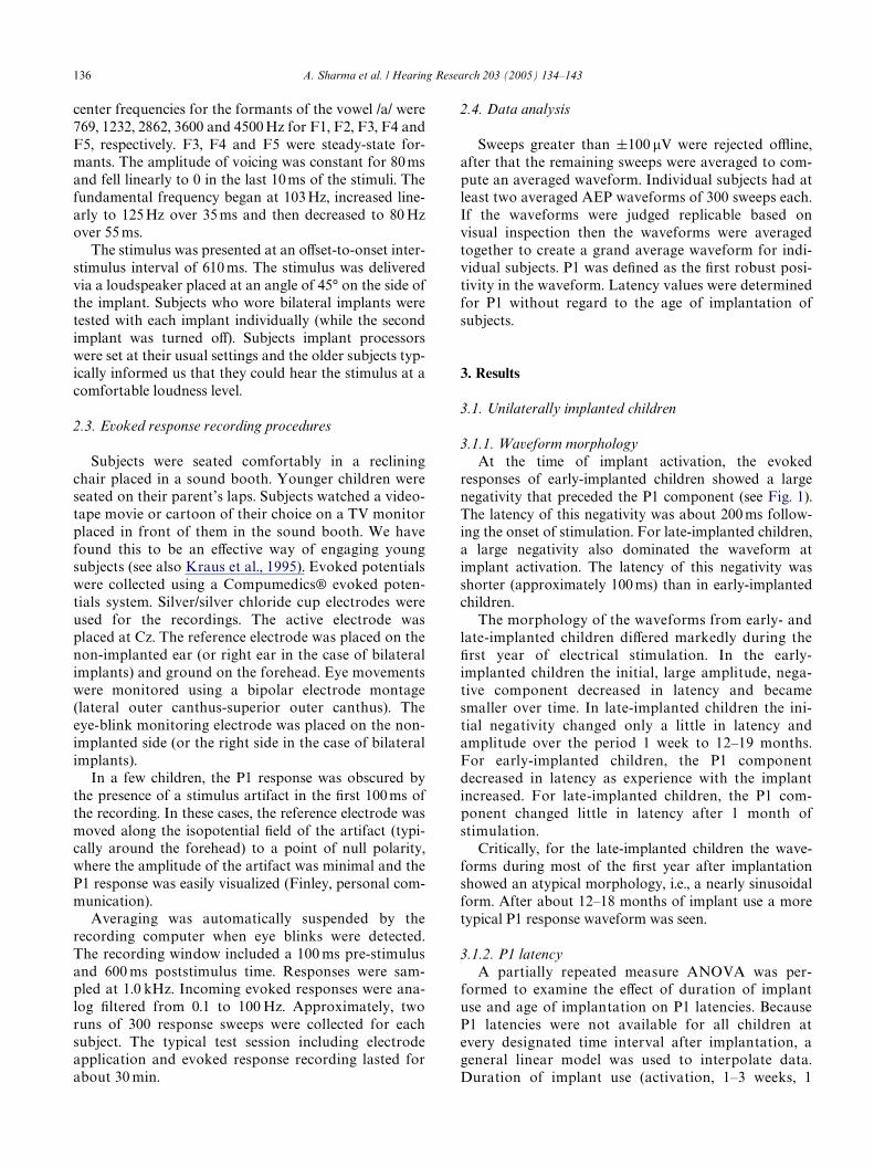
3.1.1. Waveform morphologyAt the time of implant activation, the evoked
responses of early-implanted children showed a largenegativity that preceded the P1 component (see Fig. 1).The latency of this negativity was about 200 ms follow-ing the onset of stimulation. For late-implanted children,a large negativity also dominated the waveform atimplant activation. The latency of this negativity wasshorter (approximately 100 ms) than in early-implantedchildren.
The morphology of the waveforms from early- andlate-implanted children diVered markedly during theWrst year of electrical stimulation. In the early-implanted children the initial, large amplitude, nega-tive component decreased in latency and becamesmaller over time. In late-implanted children the ini-tial negativity changed only a little in latency andamplitude over the period 1 week to 12–19 months.For early-implanted children, the P1 componentdecreased in latency as experience with the implantincreased. For late-implanted children, the P1 com-ponent changed little in latency after 1 month ofstimulation.
Critically, for the late-implanted children the wave-forms during most of the Wrst year after implantationshowed an atypical morphology, i.e., a nearly sinusoidalform. After about 12–18 months of implant use a moretypical P1 response waveform was seen.
3.1.2. P1 latencyA partially repeated measure ANOVA was per-
formed to examine the eVect of duration of implantuse and age of implantation on P1 latencies. BecauseP1 latencies were not available for all children atevery designated time interval after implantation, ageneral linear model was used to interpolate data.Duration of implant use (activation, 1–3 weeks, 1
A. Sharma et al. / Hearing Research 203 (2005) 134–143 137
month, 3–4 months, 6–9 months, and 12–18 months)was treated as the within subjects variable and age ofimplantation (early vs. late) was treated as thebetween subjects variable. Duration of implant usecan also be interpreted as ‘time in sound’ (Pontonet al., 1996a,b).
Statistical analyses revealed a signiWcant main eVectof age of implantation (F D 12.57; p D 0.002) and asigniWcant interaction between duration of implant useand age of implantation (F D 15.24; p < 0.001). Post hoctests were used to assess the interaction between dura-tion of implant use and age of implantation. A post hocanalysis of pairwise comparisons (Bonferroni correctionfor multiple comparisons) was performed in order to
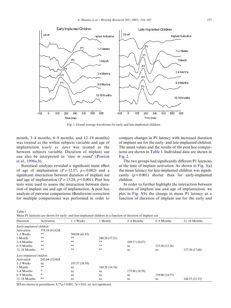
compare changes in P1 latency with increased durationof implant use for the early- and late-implanted children.The mean values and the results of the post hoc compar-isons are shown in Table 1. Individual data are shown inFig. 2.
The two groups had signiWcantly diVerent P1 latenciesat the time of implant activation. As shown in Fig. 3(a)the mean latency for late-implanted children was signiW-
cantly (p < 0.001) shorter than for early-implantedchildren.
In order to further highlight the interaction betweenduration of implant use and age of implantation, weplot in Fig. 3(b) the change in mean P1 latency as afunction of duration of implant use for the early and
Fig. 1. Grand average waveforms for early and late-implanted children.
Table 1Mean P1 latencies are shown for early- and late-implanted children as a function of duration of implant use
SD are shown in parentheses. $,**p < 0.001; *p < 0.01; ns: not signiWcant.
Duration Activation 1–3 Weeks 1 Month 3–4 Months 6–9 Months 12–18 Months
Early-implanted childrenActivation 378.18 (61.62)$1–3 Weeks ** 300.89 (41.03)1 Month ** ** 240.20 (37.21)3–4 Months ** ** ** 169.17 (10.87)6–9 Months ** ** ** ns 133.20 (13.26)12–18 Months ** ** ** ns ns 137.50 (17.68)
Late-implanted childrenActivation 245.84 (52.08)$1–3 Weeks ns 195.57 (38.38)1 Month ** ns 160.28 (14.74)3–4 Months * ns ns 175.98 (18.59)6–9 Months * ns ns ns 159.00 (14.57)12–18 Months ** ns ns ns ns 148.55 (21.52)

138 A. Sharma et al. / Hearing Research 203 (2005) 134–143
late groups. In this Wgure latencies at hookup havebeen normalized. The values on the x-axis are the midpoints of the time intervals shown in Table 1. Inspec-tion of Fig. 3(b) indicates (i) that changes in latencyfollowing hookup are larger for the early-implantedchildren than for the late-implanted children and (ii)that changes in latency extend further in time for theearly-implanted children than for the late-implantedchildren.
Both early- and late-implanted children showed a 35%decrease in P1 latency in the initial month after implanta-tion. After that time, P1 latency for early-implanted chil-dren continued to decrease (mean decrease of 64% overthe 12–18 month period following implantation). In con-trast, for the late-implanted children latencies decreasedsigniWcantly (35%) only over the Wrst month of stimula-tion and then reached a plateau (mean decrease of 39% in12–18 month period following implantation).
Fig. 2. Individual trajectories for P1 latency changes for the early- and late-implanted groups. In all subjects (except those indicated by asterisks) theinitial data point was obtained at the time of implant activation. The solid lines represent the 95% conWdence intervals for normal development of P1latencies.
Fig. 3. (a) Mean P1 latency for the early- and late-implanted groups at the time of initial stimulation with the cochlear implant. (b) Changes in meanP1 latency following stimulation for the early- and late-implanted children are shown following normalization of starting latencies. The values on thex-axis are the mid points of the time intervals shown in Table 1.

A. Sharma et al. / Hearing Research 203 (2005) 134–143 139
3.2. Bilaterally implanted children
Longitudinal P1 latency data from two sequentiallybilaterally implanted children are shown in Figs. 4and 5. Subject 1 (Fig. 4) received her Wrst implant atage 1.07 years and her second implant at age 2.07years. As expected, after several months experiencewith the Wrst implant, P1 latencies were within normallimits. The P1 latency elicited by the second implantwas within normal limits after 1 month of experiencewith bilateral implants. The data from both ears Wt theproWle of a patient implanted within the sensitiveperiod.
Subject 2 (Fig. 5) received her Wrst implant at age2.08 years and was Wtted with her second implant at
Fig. 4. Trajectories for P1 latency changes following sequential bilat-eral implantation for Subject 1. The solid lines represent the 95% conW-dence intervals for normal development of P1 latencies.
Fig. 5. Trajectories for P1 latency changes following sequential bilat-eral implantation for Subject 2. The solid lines represent the 95% conW-dence intervals for normal development of P1 latencies.
age 10.10 years. As expected, when tested with the Wrstimplant after 7 years of stimulation, the P1 latency waswithin normal limits. In contrast, the P1 latenciesrecorded in response to stimulation via the secondimplant were delayed even after 9 months of bilateralimplant use.
4. Discussion
We have examined the longitudinal development ofthe P1 cortical response in groups of early- and late-implanted children. The age cut-oVs for the two groups(<3.5 years for early-implanted and >7 years for late-implanted) were based on the sensitive period for centralauditory development described by Sharma et al.(2002c). Our results show a markedly diVerent pattern ofP1 development for early and late implanted children.
4.1. Early-implanted children
For early-implanted children, at implant activationthe waveform was dominated by a large negativity pre-ceding the P1 response. We have seen this negativityconsistently in congenitally deaf children at the time ofimplantation and in profoundly hearing-impaired chil-dren at the time of initial Wtting with a hearing aid(Sharma et al., 2004). This early negativity is strikinglysimilar to the ‘long-latency negative potential’ reportedin studies on preterm infants before 25 weeks postcon-ception (Salamy et al., 1984; Weitzman et al., 1967).The similarity suggests that CAEP morphology andlatency at the time of implantation can be interpretedas a sign of a naive (i.e., unstimulated) auditory system.Alternately, the early negativity may reXect involve-ment of the non-primary auditory areas in the genera-tion of the cortical auditory evoked potential in theabsence of sensory input to the primary auditory path-ways (Kraus and McGee, 1995; Møller and Rollins,2002).
For early-implanted children, there was a large andrapid decrease in P1 latency (approximately 100 ms)within a week of cochlear implant usage. The rapidchange in the state of the auditory pathway is consistentwith rapid changes in the visual pathways following theonset of patterned visual stimulation after the removalof congenital cataracts. Maurer et al. (1999) report animprovement in acuity with as little as an hour of pat-terned visual input.
One week after implant use P1 latencies were similarto those of normal-hearing newborns. The early nega-tivity preceding P1 seen in children with little auditoryexperience diminished in amplitude and latency withmore auditory experience. Within 6–8 months of age P1latencies reached normal limits. This outcome is consis-tent with our previous Wndings (Sharma et al., 2002a).
140 A. Sharma et al. / Hearing Research 203 (2005) 134–143
The neurophysiologic mechanisms underpinning thesechanges in P1 latency are not clear. However, Kral et al.(2000) have shown that congenitally deaf cats (i) show arestricted (atypical) pattern of activation within the lay-ers of the primary auditory cortex compared to normal-hearing cats and (ii) show signs of desynchronization ofactivity among diVerent cortical layers. As a conse-quence, there are changes in morphology of local Weldpotentials recorded at the cortical surface. If similarprocesses are at work in young deaf children, then wecan hypothesize that a change in interaction betweendiVerent cortical layers, driven by the onset of electricalstimulation, results in a rearrangement in the generatorsof the P1 response (for cats, see Klinke et al., 1999).Such a process, which would be maximized during theperiod of synaptic overshoot (Kral et al., in press), islikely to be the major factor in the rapid changes inwaveform morphology and the latency that we havefound.
4.2. Late-implanted children
Late-implanted children show a diVerent pattern ofcentral auditory development than that shown by earlyimplanted children. The early negativity linked withauditory deprivation dominates the waveform at thetime of implant activation. In late-implanted childrenthe initial negativity occurs at a shorter latency than theinitial negativity in early-implanted children. It is alsothe case that the P1 latency at implant activation isshorter in late-implanted children than in early-implanted children. Both outcomes suggest that there issome degree of intrinsic development of the central audi-tory pathways in the absence of stimulation. This out-come is consistent with the Wndings of Kral et al. (inpress) who showed that middle and long-latencyresponses appeared early in development in naive con-genitally deaf cats even in absence of any auditoryinputs, although the further development diVered signiW-
cantly from development in hearing controls. Corre-spondingly, in late-implanted children, the polyphaiscmorphology of the cortical evoked waveform response isatypical and remains atypical for several months follow-ing the onset of stimulation. Gradually between the 12thand 18th months after implantation, the morphology ofthe waveform becomes more typical for late-implantedchildren.
4.3. Two-processes in development
The relative change in P1 latency between hookupand one month was the same (35%) for early and late-implanted children (see Fig. 3(b)). Thus an early,input-dependent maturation process in P1 latency iscomparable between early- and late-implanted children.However, after approximately 4 weeks of stimulation,
the maturation process becomes arrested in late-implanted children, but continues in early-implantedchildren. The initial, rapid change in latency for bothgroups could be related to synaptic processes like long-term potentiation. The continuing change in latencyfor the early-implanted children could be related tostructural rearrangement of synaptic contacts (synapticformation and elimination). Only the latter processdemonstrates a sensitive period.
Given the diVerence in the morphology of the CAEPwaveform between the early- and late-implanted chil-dren, it is possible that the peak that we have labeled asP1 does not have the same generator for the two groups.Future studies should examine this possibility usingmulti-channel recordings. In the present study it is usefulto label the Wrst, large, positive component as P1 for thetwo groups as this allows us to quantitatively comparethe development of cortical activity across the groupsfollowing the onset of electrical stimulation.
Duration of implant use is obviously critical fordevelopment of the central auditory pathways followingimplantation (Ponton et al., 1996a,b, 2001). The presentresults emphasize the interaction of duration of implantuse with age of implantation and speak to the impor-tance of an early sensitive period. Together these factorsshape cortical development (or responsiveness) afterimplantation.
Our results suggest that both the latency and mor-phology of the P1 response serve as markers for thedevelopmental status of central auditory pathways. Wecurrently use the P1 response as a clinical tool for assess-ment of central auditory development in hearing-impaired and cochlear-implanted children.
4.4. Bilateral implantation
The present study describes, for the Wrst time, corticaldevelopment in children with bilateral cochlear implants.Because only two children have been investigated theseresults have to be treated as case reports.
One child received both her Wrst and second implantswithin the sensitive period. The second child received herWst implant within the sensitive period and received hersecond implant after the end of the sensitive period. Ourinterest in these cases was in the eVects on central audi-tory development of stimulation contralateral and ipsi-lateral to the early-implanted ear.
Figs. 4 and 5 show longitudinal P1 latency datafrom the two children. The subject shown in Fig. 4received her Wrst implant at age 1.07 years and her sec-ond implant at age 2.07 years. As expected, after sev-eral months experience with the Wrst implant, P1latencies were within normal limits. Based on animalstudies (Kral et al., 2002) we assume that the P1response reXected activity mainly from the cortexcontralateral to the implanted ear. At the time of
A. Sharma et al. / Hearing Research 203 (2005) 134–143 141
activation of the second implant, the P1 latency wasshorter than the P1 latency recorded from the Wrstimplant at activation. This suggests either a beneWcialeVect of stimulation ipsilateral to the Wrst implantedear, age-related intrinsic development, or both. How-ever, the response was delayed relative to normal sug-gesting a ‘weak’ activation of the ipsilateral pathway.This speculation is consistent with Wndings fromimplanted kittens (Kral et al., 2002). The P1 latencyelicited by the second implant was within normal lim-its within a very short period (1 month) of implant usesuggesting rapid development of the central auditorypathways when both implants are Wtted within thesensitive period.
The subject in Fig. 5 received her Wrst implant at age2.08 years. As expected, after 7 years of stimulation theP1 latency was within normal limits. The child was Wttedwith her second implant at age 10.10 years. At hookupthe P1 latency was similar to that of children whoreceived their Wrst implant at age 10. This demonstratesthat long-term stimulation from the Wrst implant hadonly a limited inXuence on the development of thecentral auditory system ipsilateral to the Wrst implant.This outcome might arise if the early (primary) auditorycortical areas ipsilateral to the Wrst implant are stimu-lated by both ears, but the higher-order areas generatingthe P1 waves evoked by the Wrst implant are connectedonly to the cortex contralateral to the Wrst implant. Alter-natively, the entire pathways activated by the two earsmight remain (or become) functionally segregated in thecongenitally deaf. More research with multi-electroderecordings is necessary to account for our outcome.
The trajectory of P1 latency change for stimulationthrough the second implant was similar to the trajectoriesshown in Fig. 2 for late-implanted children. This, onceagain, suggests limited beneWt to central auditory devel-opment from stimulation ipsilateral to the implantedear. We note also that the morphology of the waveformselicited via the two implants diVered markedly. Thewaveform elicited in response to stimulation from thesecond implant displayed a polyphasic form similar tothat seen in late-implanted, congenitally deaf children.Taken together, these outcomes suggest that long-termstimulation of auditory pathways ipsilateral to animplant is not suYcient to preserve the plasticity of thosepathways. If this is the case, then the stimulation from asecond late implant will be to a cortex which does nothave normal connections within cortical layers andwhich does not have normal connections to higher-orderauditory and language cortex. The beneWt of such animplant may be very limited.
4.5. Cortical mechanisms underlying the sensitive period
In the introduction we speculated that auditorydevelopment would proceed diVerently in deaf children
deprived of sound for a short period – less than 3.5years – and in deaf children deprived of sound for along period – greater than 7 years – because of a briefsensitive period for normal development of centralauditory pathways. In the present study, using the mor-phology and latency of the P1 cortical evoked potentialas measures of development, we Wnd that developmentdoes indeed proceed in a diVerent manner in the twosamples of deaf children. Our Wnding of diVerent devel-opmental histories for early- and late-implantedchildren is bolstered by results from another measure –the N1 component of the cortical evoked response. Pre-liminary results suggest that children implanted withinthe sensitive period show an age-appropriate N1response while those implanted after age 7 years do not(Gilley et al., 2004). These data are in contrast to otherstudies which report the absence of an N1 in cochlearimplanted children (Ponton and Eggermont, 2001;Singh et al., 2004).
Given the diVerences in development in early- andlate-implanted children we should ask, “What is diVer-ent in terms of cortical activity during and after theend of the sensitive period?” Congenitally deaf cats area model system for cortical activity after the end of thesensitive period. In kittens, the sensitive period fordevelopment of central auditory pathways lasts up to5 months of age (Kral et al., 2002). When electricalstimulation is started after 4 months of deafness thereis a delay in the activation of supragranular layers anda near absence of activity at longer latencies and ininfragranular layers (layers V and VI) (Kral et al., inpress). The additional, near-absence of outward cur-rents in layers IV and III of congenitally deaf cats sug-gests incomplete development of inhibitory synapsesand an alteration of information Xow from layer IV tosupragranular layers. This abnormal pattern of activ-ity within the auditory cortex is likely to be the basisfor diVerences in evoked potential morphology andlatency we Wnd in children implanted during the sensi-tive period and after the end of the sensitive period.Because the higher-order auditory cortex projectsback to A1 (primary auditory cortex) mainly to theinfragranular layers, the absence of activity in infra-granular layers suggests a decoupling of primary cor-tex from higher order auditory cortex. Such adecoupling would allow other sensory input to pre-dominate in the higher-order auditory cortex in chil-dren deprived of sound for a long period. Thissupposition is consistent with the functional imagingdata from deaf children reported by Lee et al. (2001).The hypothesis of a decoupling of primary cortex fromhigher order auditory and language cortex in childrendeprived of sound for a long period provides anaccount for the language learning diYculties of chil-dren who receive an implant after the end of the sensi-tive period.
142 A. Sharma et al. / Hearing Research 203 (2005) 134–143
Acknowledgements
We gratefully acknowledge the assistance of PhillipGilley in statistical analysis and compilation of theWgures. We are grateful to Kathryn Martin, M.S, CCC-Awho collected the data. This work was supported byNIH R01 DC 004552 and NIH R01 DC 006257.
References
Conel, J.L., 1939. The Postnatal Development of Human Cerebral Cor-tex, vols. I–VIII. Harvard University Press, Cambridge, MA.
Cunningham, J., Nicol, T., Zecker, S., Kraus, N., 2000. Speech-evokedneurophysiologic responses in children with learning in problems:development and behavioral correlates of perception. Ear Hear. 21(6), 554–568.
Eggermont, J.J., Ponton, C.W., 2003. Auditory-evoked potential stud-ies of cortical maturation in normal hearing and implanted chil-dren: correlations with changes in structure and speech perception.Acta Otolaryngol. 123, 249–252.
Finney, E.M., Fine, I., Dobkins, K.R., 2001. Visual stimuli activateauditory cortex in the deaf. Nat. Neurosci. 4, 1171–1173.
Granier-Deferre, C., Lecanuet, J.P., Cohen, H., Busnel, M.C., 1985.Feasibility of prenatal hearing test. Acta Otolaryngol. (Stockh.)Suppl. 421, 93–101.
Gilley, P., Sharma, A., Dorman, M., Todd, N.W., Martin, K., Fainberg,J., 2004. Assessing development of the auditory system in normal-hearing children and children with cochlear implants by use of astimulus train with decreasing interstimulus intervals. In: Associa-tion for Research in Otolaryngology, Midwinter Meeting 2004,Daytona Beach, FL.
Gordon, K., Papsin, B., Harrison, R., 2003. Activity-dependent devel-opmental plasticity of the auditory brainstem in children who usecochlear implants. Ear Hear. 24 (6), 485–500.
Huttenlocher, P.R., Dabholkar, A.S., 1997. Regional diVerences in syn-aptogenesis in human cerebral cortex. J. Comp. Neurol. 387, 167–178.
Klinke, R., Kral, A., Heid, S., Tillein, J., Hartmann, R., 1999. Recruit-ment of the auditory cortex in congenitally deaf cats by long-termcochlear electrostimulation. Science 285, 1729–1733.
Kral, A., Hartmann, R., Tillein, J., Heid, S., Klinke, R., 2000. Congeni-tal auditory deprivation reduces synaptic activity within the audi-tory cortex in a layer-speciWc manner. Cereb. Cortex 10 (7), 714–726.
Kral, A., Hartmann, R., Tillein, J., Heid, S., Klinke, R., 2002. Hearingafter congenital deafness: central auditory plasticity and sensorydeprivation. Cereb. Cortex 12, 797–807.
Kral, A., Tillein, J., Heid, S., Hartmann, R., Klinke, R., in press. Postna-tal cortical development in congenital auditory deprivation. Cereb.Cortex.
Kraus, N., McGee, T., 1995. The middle latency response generatingsystem. Clin. Neurophysiol. 44, 93–101.
Kraus, N., McGee, T., Carrell T, Sharma, A., 1995. Neurophysiologicbases of speech discrimination. Neurophysiologic bases of speechdiscrimination. Ear Hear. 16 (1), 19–37.
Lee, D.S., Lee, J.S., Oh, S.H., Kim, S.K., Kim, J.W., Chung, J.K., Lee,M.C., Kim, C.S., 2001. Cross-modal plasticity and cochlearimplants. Nature 409 (6817), 149–150.
Maurer, D., Lewis, T.L., Brent, H.P., Levin, A.V., 1999. Rapid improve-ment in the acuity of infants after visual input. Science 286 (5437),108–110.
Møller, A.R., Rollins, P.R., 2002. The non-classical auditory pathwaysare involved in hearing in children but not in adults. Neurosci. Lett.319, 41–44.
Moore, J.K., 2004. The Human Central Auditory System: A Timelineof Development, NHS Conference, Lake Como, Italy.
Moore, J.K., 2002. Maturation of human auditory cortex: implicationsfor speech perception. Ann. Otol. Rhinol. Laryngol. Suppl. 189, 7–10.
Moore, J.K., Guan, Y.L., 2001. Cytoarchitectural and axonal matura-tion in human auditory cortex. J. Assoc. Res. Otolaryngol. 2, 297–311.
Mostafapour, S.P., Del Puerto, N.N., Rubel, E.W., 2002. bcl-2 Overex-pression eliminates deprivation-induced cell death of brainstemauditory neurons. J. Neurosci. 22 (11), 4670–4674.
Neville, H., Bavelier, D., 2002. Human brain plasitcity: evidence fromsensory deprivation and altered language experience. Prog. BrainRes. 138, 177–188.
Nishimura, H., Doi, K., Iwaki, T., Hashikawa, K., Oku, N., Teratani,T., Hasegawa, T., Watanabe, A., Nishimura, T., Kubo, T., 2000.Neural plasticity detected in shortand long-term cochlear implantusers using PET. Neuroreport 11, 811–815.
Nishimura, H., Hashikawa, K., Doi, K., Iwaki, T., Watanabe, Y.,Kusuoka, H., Nishimura, T., Kubo, T., 1999. Sign language ‘heard’in the auditory cortex. Nature 397, 116.
Paus, T., Zijdenbos, A., Worsley, K., Collins, D.L., Blumenthal, J.,Giedd, J.N., Rapoport, J.L., Evans, A.C., 1999. Structural matura-tion of neural pathways in children and adolescents: in vivo study.Science 283, 1908–1911.
Petitto, L.A., Zatorre, R.J., Gauna, K., Nikelski, E.J., Dostie, D., Evans,A.C., 2000. Speech-like cerebral activity in profoundly deaf peopleprocessing signed languages: implications for the neural basis ofhuman language. Proc Natl. Acad. Sci. USA 97, 13961–13966.
Ponton, C.W., Don, M., Eggermont, J.J., Waring, M.D., Kwong, B.,Masuda, A., 1996a. Auditory system plasticity in children after longperiods of complete deafness. Neuroreport 8 (1), 61–65.
Ponton, C.W., Don, M., Eggermont, J.J., Waring, M.D., Masuda, A.,1996b. Maturation of human cortical auditory function: diVerencesbetween normal-hearing children and children with cochlearimplants. Ear Hear. 17 (5), 430–437.
Ponton, C.W., Moore, J., Eggermont, J., 1999. Prolonged deafness lim-its auditory system developmental plasticity: evidence from anevoked potential study in children with cochlear implants. Scand.Audiol., Suppl. 51, 13–22.
Ponton, C.W., Eggermont, J.J., Kwong, B., Don, M., 2000. Maturationof human central auditory system activity: evidence from multi-channel evoked potentials. Clin. Neurophysiol. 111, 220–236.
Ponton, C.W., Eggermont, J., 2001. Of kittens and kids: altered corticalmaturation following profound deafness and cochlear implant use.Audiol. Neurotol. 6, 363–380.
Roder, B., Stock, O., Bien, S., Neville, H., Rosler, F., 2002. Speech pro-cessing activates visual cortex in congenitally blind humans. Eur. J.Neurosci. 16, 930–936.
Rubel, E.W., 1985. Strategies and problems for future studies of audi-tory development. Acta Otolaryngol. (Stockh.) 421, 114–128.
Salamy, A., Eggermont, J.J., Eldredge, L., 1984. Neurodevelopemntand Auditory Function in Preterm Infants. In: Jacobson, J.T. (Ed.),Principles and Apllication in Auditory Evoked Potentials. Allyn &Bacon, Needham Heights, pp. 287–312.
Sharma, A., Kraus, N., McGee, T., Nicol, T., 1997. Developmentalchanges in P1 & N1 auditory responses elicited by consonant-vowelsyllables. Clin. Neurophysiol. 104, 540–545.
Sharma, A., Dorman, M.F., Spahr, A.J., 2002a. Rapid development ofcortical auditory evoked potentials after early cochlear implanta-tion. Neuroreport 13 (10), 1365–1368.
Sharma, A., Dorman, M.F., Spahr, A.J., Todd, N.W., 2002b. Early cochlearimplantation in children allows normal development of central audi-tory pathways. Ann. Otol. Rhinol. Laryngol. Suppl. 189, 38–41.
Sharma, A., Dorman, M., Spahr, T., 2002c. A sensitive period for thedevelopment of the central auditory system in children withcochlear implants. Ear Hear. 23 (6), 532–539.

A. Sharma et al. / Hearing Research 203 (2005) 134–143 143
Sharma, A., Tobey, E., Dorman, M., Martin, K., Gilley, P., Kunkel, F.,2004. Central auditory maturation and babbling development ininfants with cochlear implants. Arch. Otolaryngol.-Head NeckSurg. 130 (5), 511–610.
Singh, S., Alki, L., Kaubab, R., Towell, A., Luxon, L., 2004. Event-related potentials in pediatric cochlear implanted patients. EarHear. 25 (6), 598.
Weitzman, L., Graziani, L., Duhamel, L., 1967. Maturation and topog-raphy of the auditory evoked response of the prematurely borninfant. Clin. Neurophysiol. 23 (1), 82–83.
Yakovlev, P.J., 1967. The myelogenetic cycles of regional matura-tion of the brain. In: Minkowski, A. (Ed.), Regional Develop-ment of the Brain in Early Life, Wrst ed. Blackwell, Oxford,pp. 1–3.
Copyright © 2022 FDOKUMEN