The human survivin promoter: a novel transcriptional targeting strategy for treatment of glioma
10
ALIGNANT gliomas are the most common tumor in the central nervous system. Despite recent ad- vances in treatment, their prognosis remains poor. The 5-year survival rate in patients harboring a GBM, the most common and malignant glioma subtype, is less than 3%, whereas the 5-year survival rate for patients with a lower grade astrocytoma is 30%. 3,6,7,38 Therefore, the identi- fication of novel therapeutic strategies for malignant gliomas merits a high priority. In this regard, gene therapy with adenoviral vectors is a promising new modality for the treatment of glioma. 9,10,23,26 Adenoviral vectors may be genetically designed to express therapeutic genes in cancer cells. To this end, tumor-selective transgene expression is critical and can be obtained by placing the transgene under the control of a TSP. 12,13,29,31,35 Specifically, to achieve a high therapeutic index with adenoviral vectors in vivo, repres- sion of TSPs in normal tissues, primarily those of the liver, emerges as the most critical predictor of a high tumor/nor- mal tissue ratio. 12 Current TSP candidates for glioma target- J. Neurosurg. / Volume 104 / April, 2006 J Neurosurg 104:583–592, 2006 The human survivin promoter: a novel transcriptional targeting strategy for treatment of glioma WINAN J. VAN HOUDT , M.D., YOSEF S. HAVIV , M.D., BAOGEN LU, M.D., MINGHUI W ANG, M.D., ANGEL A. RIVERA,PH.D., ILYA V. ULASOV ,PH.D., MARTINE L. M. LAMFERS,PH.D., DANIEL REIN, M.D., MACIEJ S. LESNIAK, M.D., GENE P. SIEGAL, M.D., PH.D., CLEMENS M. F. DIRVEN, M.D., PH.D., DAVID T. CURIEL, M.D., PH.D., AND ZENG B. ZHU, M.D. Department of Neurosurgery, VU Universiteit Medische Center, Amsterdam, The Netherlands; Department of Medicine and the Gene Therapy Institute, Hadassah–Hebrew University Medical Center, Jerusalem, Israel; Division of Human Gene Therapy, Departments of Medicine, Pathology, Surgery, and Cell Biology, and The Gene Therapy Center, University of Alabama at Birmingham, Alabama; Division of Neurosurgery, The University of Chicago, Illinois; Department of Obstetrics and Gynecology, University of Düsseldorf Medical Center, Düsseldorf, Germany Object. Malignant brain tumors have been proved to be resistant to standard treatments and therefore require new therapeutic strategies. Survivin, a recently described member of the inhibitor of apoptosis protein family, is overex- pressed in several human brain tumors, primarily gliomas, but is downregulated in normal tissues. The authors hypoth- esized that the expression of tumor-specific survivin could be exploited for treatment of gliomas by targeting the tumors with gene therapy vectors. Methods. Following confirmation of survivin expression in glioma cell lines, an adenoviral vector containing the survivin promoter and the reporter gene luciferase was tested in established and primary glioma cells, normal astro- cytic cells, and normal human brain tissues. High levels of reporter gene expression were observed in established tumor and primary tumor cell lines and low levels of expression in astrocytes and normal human brain tissue. To test oncolyt- ic potency, the authors constructed survivin promoter–based conditionally replicative adenoviruses (CRAds), com- posed of survivin promoter–regulated E1 gene expression and an RGD-4C capsid modification. These CRAds could efficiently replicate within and kill a variety of established glioma tumor cells, but were inactive in a normal human liver organ culture. Finally, survivin promoter–based CRAds significantly inhibited the growth of glioma xenografts in vivo. Conclusions. Together these data indicate that the survivin promoter is a promising tumor-specific promoter for transcriptional targeting of adenovirus-based vectors and CRAds for malignant gliomas. The strategy of using sur- vivin–CRAds may thus translate into an experimental therapeutic approach that can be used in human clinical trials. KEY WORDS • survivin gene • tumor-specific promoter • transcriptional targeting • adenoviral vector • conditionally replicative adenovirus M 583 Abbreviations used in this paper: Ad5 = adenovirus type 5; CMV = cytomegalovirus; CRAd = conditionally replicative adeno- virus; DMEM = Dulbecco modified Eagle medium; FBS = fetal bo- vine serum; GAPDH = glyceraldehyde-3-phosphate dehydrogenase; GBM = glioblastoma multiforme; hTERT = human telomerase re- verse transcriptase; IAP = inhibitor of apoptosis; MOI = multiplicity of infection; PBS = phosphate-buffered saline; PCR = polymerase chain reaction; vp = viral particles; TSP = tumor-specific promoter.
-
Upload
independent -
Category
Documents
-
view
0 -
download
0
Transcript of The human survivin promoter: a novel transcriptional targeting strategy for treatment of glioma
ALIGNANT gliomas are the most common tumor inthe central nervous system. Despite recent ad-vances in treatment, their prognosis remains poor.
The 5-year survival rate in patients harboring a GBM, themost common and malignant glioma subtype, is less than
3%, whereas the 5-year survival rate for patients with alower grade astrocytoma is 30%.3,6,7,38 Therefore, the identi-fication of novel therapeutic strategies for malignantgliomas merits a high priority. In this regard, gene therapywith adenoviral vectors is a promising new modality for thetreatment of glioma.9,10,23,26 Adenoviral vectors may begenetically designed to express therapeutic genes in cancercells. To this end, tumor-selective transgene expression iscritical and can be obtained by placing the transgene underthe control of a TSP.12,13,29,31,35 Specifically, to achieve a hightherapeutic index with adenoviral vectors in vivo, repres-sion of TSPs in normal tissues, primarily those of the liver,emerges as the most critical predictor of a high tumor/nor-mal tissue ratio.12 Current TSP candidates for glioma target-
J. Neurosurg. / Volume 104 / April, 2006
J Neurosurg 104:583–592, 2006
The human survivin promoter: a novel transcriptionaltargeting strategy for treatment of glioma
WINAN J. VAN HOUDT, M.D., YOSEF S. HAVIV, M.D., BAOGEN LU, M.D., MINGHUI WANG, M.D.,ANGEL A. RIVERA, PH.D., ILYA V. ULASOV, PH.D., MARTINE L. M. LAMFERS, PH.D., DANIEL REIN, M.D., MACIEJ S. LESNIAK, M.D., GENE P. SIEGAL, M.D., PH.D., CLEMENS M. F. DIRVEN, M.D., PH.D., DAVID T. CURIEL, M.D., PH.D., AND ZENG B. ZHU, M.D.
Department of Neurosurgery, VU Universiteit Medische Center, Amsterdam, The Netherlands;Department of Medicine and the Gene Therapy Institute, Hadassah–Hebrew University MedicalCenter, Jerusalem, Israel; Division of Human Gene Therapy, Departments of Medicine, Pathology,Surgery, and Cell Biology, and The Gene Therapy Center, University of Alabama at Birmingham,Alabama; Division of Neurosurgery, The University of Chicago, Illinois; Department of Obstetricsand Gynecology, University of Düsseldorf Medical Center, Düsseldorf, Germany
Object. Malignant brain tumors have been proved to be resistant to standard treatments and therefore require newtherapeutic strategies. Survivin, a recently described member of the inhibitor of apoptosis protein family, is overex-pressed in several human brain tumors, primarily gliomas, but is downregulated in normal tissues. The authors hypoth-esized that the expression of tumor-specific survivin could be exploited for treatment of gliomas by targeting thetumors with gene therapy vectors.
Methods. Following confirmation of survivin expression in glioma cell lines, an adenoviral vector containing thesurvivin promoter and the reporter gene luciferase was tested in established and primary glioma cells, normal astro-cytic cells, and normal human brain tissues. High levels of reporter gene expression were observed in established tumorand primary tumor cell lines and low levels of expression in astrocytes and normal human brain tissue. To test oncolyt-ic potency, the authors constructed survivin promoter–based conditionally replicative adenoviruses (CRAds), com-posed of survivin promoter–regulated E1 gene expression and an RGD-4C capsid modification. These CRAds couldefficiently replicate within and kill a variety of established glioma tumor cells, but were inactive in a normal humanliver organ culture. Finally, survivin promoter–based CRAds significantly inhibited the growth of glioma xenograftsin vivo.
Conclusions. Together these data indicate that the survivin promoter is a promising tumor-specific promoter fortranscriptional targeting of adenovirus-based vectors and CRAds for malignant gliomas. The strategy of using sur-vivin–CRAds may thus translate into an experimental therapeutic approach that can be used in human clinical trials.
KEY WORDS • survivin gene • tumor-specific promoter • transcriptional targeting •adenoviral vector • conditionally replicative adenovirus
M
583
Abbreviations used in this paper: Ad5 = adenovirus type 5; CMV = cytomegalovirus; CRAd = conditionally replicative adeno-virus; DMEM = Dulbecco modified Eagle medium; FBS = fetal bo-vine serum; GAPDH = glyceraldehyde-3-phosphate dehydrogenase;GBM = glioblastoma multiforme; hTERT = human telomerase re-verse transcriptase; IAP = inhibitor of apoptosis; MOI = multiplicityof infection; PBS = phosphate-buffered saline; PCR = polymerasechain reaction; vp = viral particles; TSP = tumor-specific promoter.
ing do not fully meet these criteria,21 and therefore novelTSPs are required for transcriptional targeting of gliomas.
The recently discovered survivin gene encodes the sur-vivin protein, a member of the IAP protein family, whichplays an important role in the survival of cancer cells andthe progression of malignancy. Members of the IAP familydirectly inhibit terminal effector caspases through bac-ulovirus IAP repeat–dependent recognition, thereby pre-venting apoptosis.4,24 Survivin is normally expressed duringembryogenesis and is undetectable in fully differentiatedadult tissues.6 In contrast, survivin is repeatedly expressedin a broad spectrum of human cancers including brain,breast, pancreas, esophageal, and ovarian tumors.
Survivin-mediated suppression of apoptosis and growthfactor–independent cell survival is implicated in the resis-tance of the tumor to standard therapy. In this regard, 80%of GBM cells demonstrate abundant survivin expression,6
and a clear correlation is seen between the histologicalgrade of a glioma and the fraction of survivin-positive tu-mor samples. In glioma tumors, survivin expression is alsocorrelated with a resistance to chemo- and radiotherapy andwith poor prognosis.6,15,34
Given this promising role for survivin, we developed asurvivin promoter–based adenoviral vector for the purposeof transcriptional targeting of gliomas. We hypothesizedthat this strategy of tumor-specific regulation of gene ex-pression could provide a novel transcriptional targeting ap-proach for human gliomas. Furthermore, we used this sur-vivin TSP approach in the context of CRAds use fortumor-selective viral replication and oncolysis. To achievethis goal, a TSP is required to control E1 gene expression inthe adenovirus. Although other TSPs have been previouslysuggested in the context of glioma, such as hTERT,19,20 on-costatin-M promoter (a hematopoietic cytokine), the vascu-lar endothelial growth factor promoter,30 the c-Myc promot-er activated in medulloblastoma,37 and gas1,44 in this studywe identified the survivin promoter as a uniquely transcrip-tional targeting strategy for human glioma.
Materials and Methods
Cells, Tissues, and Animals
Samples of the human glioma tumor cell lines U251MG, U87MG,U373MG, D54MG, and D65MG were kind gifts from Dr. G. Y. Gil-lespie (Department of Neurosurgery, University of Alabama at Bir-mingham). Samples of the M59K and U118MG cell lines were ob-tained from the American Type Culture Collection (Manassas, VA).Cells of the U87MG line were cultured in minimal essential medium(Mediatech, Herndon, VA) containing 15% FBS (HyClone, Logan,UT), 2 mM L-glutamine, penicillin (100 IU/ml), and streptomycin(100 mg/ml). The other cell lines—U251MG, D54MG, U118MG,M59K, and D65MG—were cultured in DMEM–Ham F12 50:50(Mediatech) containing 10% FBS and supplemented with L-gluta-mine, penicillin, and streptomycin as previously described. In addi-tion, 911 cells (a kind gift from Dr. Van Der Eb, Leiden University,The Netherlands) were maintained in DMEM. Each medium was al-so supplemented with fetal calf serum, penicillin, and streptomycin.Astrocyte cells, a kind gift from Dr. Charles Bonus, were cultured inDMEM and supplemented with 10% FBS, penicillin, and streptomy-cin. Three primary glioma tumor cells, VU84, VU119, and VU78,23
were cultured in DMEM with 10 to 15% FBS, L-glutamine, and an-tibiotic agents. The cells were incubated at 37˚C in a 95% air/5%CO2 environment under humidified conditions.
Human liver samples were separated from donor hepatectomy
remnants that had been obtained during liver transplantation; thestudy was approved by the local internal review board. To generatehuman liver organ cultures, the tissue was serially dissected into 0.5-mm-thick slices by using a Krumdieck tissue slicer (Alabama Re-search Development, Munford, AL). Next, the liver tissue was cul-tured in 24-well plates in RPMI medium supplemented with 10%FBS, 100 U/ml penicillin, 100 mg/ml streptomycin, and 5 mg/ml in-sulin. The tissue cultures were maintained at 37˚C in a humidified at-mosphere of 95% air/5% CO2. Three tissue slices were examined pergroup.
Female C57BL/6 mice and female BALB/c nude mice (6–8weeks of age; Charles River, Wilmington, MA) were used in the invivo experiments. All animals received humane care based on guide-lines established by the American Veterinary Association. All exper-imental protocols involving live animals were reviewed and ap-proved by the Institutional Animal Care and Use Committee of theUniversity of Alabama at Birmingham.
Adenoviral Vectors
The following recombinant adenoviral vectors and CRAds were constructed in our lab. The recombinant adenoviral vectorsreAdGL3BCox2 (or Ad-Cox2),42 reAdGL3Midkine (or Ad-Mk),1
reAdGL3BSurvivin (or Ad-S),45 and reAdGL3BCMV (or Ad-CMV), each of which was used in this experiment, are all isogen-ic in that they are composed of the pGL3B plasmid adenoviralbackbone, which carries the same luciferase cassette but under thecontrol of different promoters, that is, Cox-2, midkine, survivin,or CMV, respectively. Two CRAds—CRAd-S-S and CRAd-S-L—were generated by our group and have been described elsewhere.47 Inthese CRAds, the adenoviral native E1 promoter was completelydeleted and the AD5 E1 gene was regulated by a short survivin pro-moter, S-S (nucleotides 2230 to 130), or a longer segment, S-L(nucleotides 21430 to 130). To increase the killing of glioma can-cer cells, these CRAd genomes also contain a capsid modification,RGD-4C, which was inserted genetically into the viral capsid. As areplicative control for the survivin–CRAds, we used a wild-type ade-novirus (Adwt). The viral particle/plate forming unit ratio for eachvirus was 34 for CRAd-S-S and 16 for CRAd-S-L.
Analysis of Survivin Promoter Activity in Glioma Cells
A recombinant adenoviral vector, AdSurvivin, was used in thisstudy as reported previously.45 To determine the transcriptional ac-tivity of the survivin promoter, 5 3 104 cells of each established glio-ma cancer cell line (U87MG, M59K, U251MG, U118MG, D65MG,and D54MG), primary glioma cells, and normal controls (primarymesothelial cells, derived in our department from ascites fluid, andkeratinocytes), were plated into 24-well tissue plates and infectedwith Ad-CMV, Ad-Cox2, Ad-Mk, or Ad-S at an MOI of 100 (100vp/cell) in 200 ml of growth medium containing 2% FBS (infec-tion medium). The infection medium was replaced by fresh mediumcontaining 10% FBS after 2 hours. Twenty-four hours postinfection,luciferase activity was determined using the Reporter Lysis Bufferand Luciferase Assay System (Promega, Madison, WI) followingthe manufacturer’s protocol. Experiments were performed in tripli-cate, and luciferase activities are presented as relative light units nor-malized to the CMV promoter activity.
Quantitative Real-Time PCR for Detection of the HumanSurvivin Gene and Ad5 E4 Gene Expression
Total cellular RNA or DNA was extracted from cell cultures in6-well plates by using the RNeasy mini RNA extraction kit or theblood DNA kit (Qiagen, Valencia, CA). Both RNA and DNA sam-ples were treated with RNase-free DNase and DNase-free RNase,respectively, to remove possible contamination. The survivin genetranscripts were detected in RNA samples by using an oligo pair(forward primer 59TGGAAGGCTGGGAGCCA and reverse primer59GAAAGCGCAACCGGACG) and the probe (ORF6TGACGACCCCATAGAGGAACATAAAAAGCAT); the Ad5 E4 gene wasdetected in DNA samples by using an oligo pair (forward primer59GGAGTGCGCCGAGACAAC and reverse primer 59ACTACGTCCGGCGTTCCAT) and the probe (ORF6TGGCATGACAC
W. J. van Houdt, et al.
584 J. Neurosurg. / Volume 104 / April, 2006

TACGACCAACACGATCT). The oligos were designed using Pri-mer Express (version 1.0; Perkin-Elmer, Foster City, CA) and syn-thesized by Applied Biosystems. Specifics of the real-time PCRreaction have been described by us elsewhere.45,46 Negative controlswithout templates were included in each reaction series, and an inter-nal control (human GAPDH or b-actin) was used to normalize thecopy number for the survivin and E4 genes.
Survivin Promoter Activity in Normal Human Brain Tissue
Specimens of normal human brain were obtained following ap-proval from our internal review board and consisted of 200-mmslices of gray matter from the temporal lobe, which were acquiredduring surgery for epilepsy. The brain slices were prepared as previ-ously described by Verwer, et al.40 Experiments were performed onnormal brain slices excised from two patients with epilepsy. On Day2, the brain specimens were infected in quadruplicate with 2 3 108
vp of Ad-S or Ad-CMV. On Day 5, the brain specimens were har-vested and the luciferase assays were performed.
Analysis of CRAd Replication in Tumor Cell Lines
Glioma cells (105/well) were cultured in the manner described ear-lier, infected with 100 vp/cell of CRAd-S-S, CRAd-S-L, Adwt, orAd-S in infection medium containing 2% FBS, and incubated at37˚C in a 95% air/5% CO2 environment. After 2 hours in incubation,the infection medium was removed, the cells were washed threetimes with PBS, and the cells were placed in fresh culture mediumwith 10% FBS. Media from triplicate wells were collected 1, 3, and9 days postinfection; DNA was extracted from 200 ml of media byusing the DNeasy Tissue Kit (Qiagen), and quantitative real-timePCR was performed in the manner described earlier. The adenovirusE4 gene copy numbers were detected.
Replication of CRAd in the Human Liver
Human liver organ cultures, described earlier, were infected withCRAd-S-S, CRAd-S-L, Adwt, or Ad-S (108 vp/slice, ~ 500 vp/cell)and incubated at 37˚C in a humidified atmosphere of 95% air/5%CO2. Three tissue slices were included per group. After a 2-day incu-bation, total DNA was extracted from the human liver organ culturesby using the DNeasy Tissue Kit (Qiagen). The DNA samples weretreated with DNase-free RNase to remove possible RNA contamina-tion and were stored at 280˚C until use. The Ad5 E4 gene copynumbers were detected and normalized by referral to humanGAPDH.
Analysis of In Vitro Glioma Cell Killing
The in vitro cytocidal effects of CRAd-S-S and CRAd-S-L wereanalyzed by performing crystal violet staining. Briefly, 25,000 cells(including D65MG, U251MG, U373MG, U87MG, U118MG, orOV4, an ovarian cancer cell line used as a control) per well wereplated onto a 24-well plate, and the cells were infected at 100, 10, 1,and 0 vp/cell with CRAd-S-S, CRAd-S-L, or Ad-S in infectionmedium. Two hours later, the infection medium was replaced withcomplete medium. After 10 more days of culture, the cells werefixed with 10% buffered formalin for 10 minutes and stained with1% crystal violet in 70% ethanol for 20 minutes, followed by wash-ing three times with tap water and air drying. Trypan blue exclusionexperiments also were performed.
Analysis of CRAd Replication in Glioma Xenografts
The U118MG glioma cells were injected into the flanks of nudemice (5 3 106 cells in 200 ml PBS per tumor, five animals/group).When the resulting tumors reached 5 mm in diameter, 5 3 108 vp ofCRAd-S-S or Adwt diluted in 50 ml PBS were injected into eachtumor. On Days 1 and 7 postinjection, the tumors were collected andsnap-frozen with dry ice/ethanol. The DNA was extracted, and therelative E4 copy numbers were determined.
Antitumor Effect of CRAd on Mouse Xenografts
Five BALB/c nude mice in each group were inoculated in theflanks with 5 3 106 U118MG cells. The tumor cells were verified
to have 95% viability by Trypan blue exclusion. When the tumorreached 5 mm in its widest diameter, 109 vp of viral vector (CRAd-S-S, CRAd-S-L, or Ad-S) or PBS was injected intratumorally. Thevector Ad-S was used as a nonreplicative control virus with an iden-tical viral backbone. The same dose was repeated after 1 week.Tumor volumes were monitored twice a week, and the volumes werecalculated by using the formula 1/2 xy2, where x is the longest diam-eter and y the shortest diameter of the tumor. Based on the deter-mined volume, the following equations were applied.
Tumor volume ratio = tumor volume at Day 30/tumor volume at Day1 3 100%.
Tumor growth inhibition rate = 100% 2 (tumor volume at Day 30/tumor volume at Day 1 3 100%).
Statistical Analysis
The luciferase activities measured in this study were analyzed byperforming the Student t-test using commercial software (StatisticsAnalysis Software version 8.2; SAS Inc., Cary, NC). A probabilityvalue less than 0.05 was considered statistically significant.
Results
Survivin Promoter is Active in Both Established andPrimary Glioma Cells
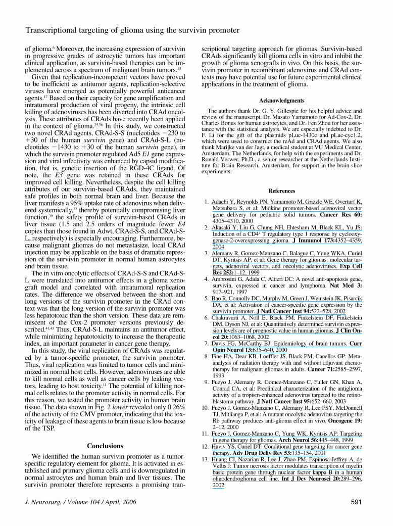
Four recombinant adenoviral vectors, Ad-Cox2, Ad-Mk,Ad-S, and Ad-CMV, were used in these experiments. Allrecombinant adenoviruses are isogenic, thereby allowingthe determination of promoter activity via reporter geneexpression. The CMV promoter in Ad-CMV is constitu-tively active, thereby serving as a positive control to allownormalization for each given cell line. The data shown inFig. 1 represent promoter activity in established and prima-ry low-passage glioma cells. Of all three TSPs, the survivinpromoter exhibited the highest activity in the establishedglioma cell lines, with a mean of 11.2% of the activity of theCMV promoter. In contrast, both the Cox-2 and midkinepromoters were relatively inactive, showing less than 4% ofthe activity of the CMV promoter. The differences were sig-nificant (p , 0.01). All three promoters were inactive inboth normal primary mesothelial cells and the normal ker-atinocyte cell line. Because established cancer cell linesmay not reflect the true nature of cancer cells, we repeatedthese experiments in primary low-passage glioma cells.Again, we observed that the survivin promoter was the mostactive of all three promoters in three primary glioma cells.The mean survivin promoter activity was 20% of the activi-ty of the CMV promoter in three primary cells, but with widevariation among the three cell types (5, 12.5, and 42.5% ofthe activity of the CMV promoter in VU84, VU119, andVU78 cells, respectively). The mean Cox-2 and midkinepromoter activities were less than 5% of the CMV promoter.Thus, the survivin promoter is repressed in normal cells andis active in both established and primary glioma cells.
Survivin Promoter is Less Active in Normal Astrocytes andNormal Human Brain
A critical determinant of any candidate transcriptionaltargeting approach for glioma is tumor specificity. In theprevious section, we described our study of survivin pro-moter activity in glioma cells. Next, we tested whether thispromoter may be applicable to glioma gene therapy, that is,whether it is inactive in the normal brain. To this end, we
J. Neurosurg. / Volume 104 / April, 2006
Transcriptional targeting of glioma using the survivin promoter
585
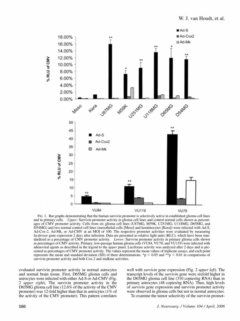
evaluated survivin promoter activity in normal astrocytesand normal brain tissue. First, D65MG glioma cells andastrocytes were infected with either Ad-S or Ad-CMV (Fig.2 upper right). The survivin promoter activity in theD65MG glioma cell line (12.6% of the activity of the CMVpromoter) was 12-fold higher than that in astrocytes (1% ofthe activity of the CMV promoter). This pattern correlates
well with survivin gene expression (Fig. 2 upper left). Thetranscript levels of the survivin gene were sixfold higher inthe D65MG glioma cell line (310 copies/ng RNA) than inprimary astrocytes (48 copies/ng RNA). Thus, high levelsof survivin gene expression and survivin promoter activitywere observed in glioma cells but not in normal astrocytes.
To examine the tumor selectivity of the survivin promot-
W. J. van Houdt, et al.
586 J. Neurosurg. / Volume 104 / April, 2006
FIG. 1. Bar graphs demonstrating that the human survivin promoter is selectively active in established glioma cell linesand in primary cells. Upper: Survivin promoter activity in glioma cell lines and control normal cells shown as percent-ages of CMV promoter activity. Cells from six glioma cell lines (U87MG, M59K, U251MG, U118MG, D65MG, andD54MG) and two normal control cell lines (mesothelial cells [Meso] and keratinocytes [Kera]) were infected with Ad-S,Ad-Cox-2, Ad-Mk, or Ad-CMV at an MOI of 100. The respective promoter activities were evaluated by measuringluciferase gene expression 2 days after infection. Data are presented as relative light units (RLU), which have been stan-dardized as a percentage of CMV promoter activity. Lower: Survivin promoter activity in primary glioma cells shownas percentages of CMV activity. Primary, low-passage human glioma cells (VU84, VU78, and VU119) were infected withadenoviral agents as described in the legend to the upper panel. Luciferase activity was analyzed after 2 days and is pre-sented as percentages of CMV promoter activity. The values represent the mean values of triplicate assays, and each pointrepresents the mean and standard deviation (SD) of three determinations. *p , 0.05 and **p , 0.01 in comparisons ofsurvivin promoter activity and both Cox-2 and midkine activities.
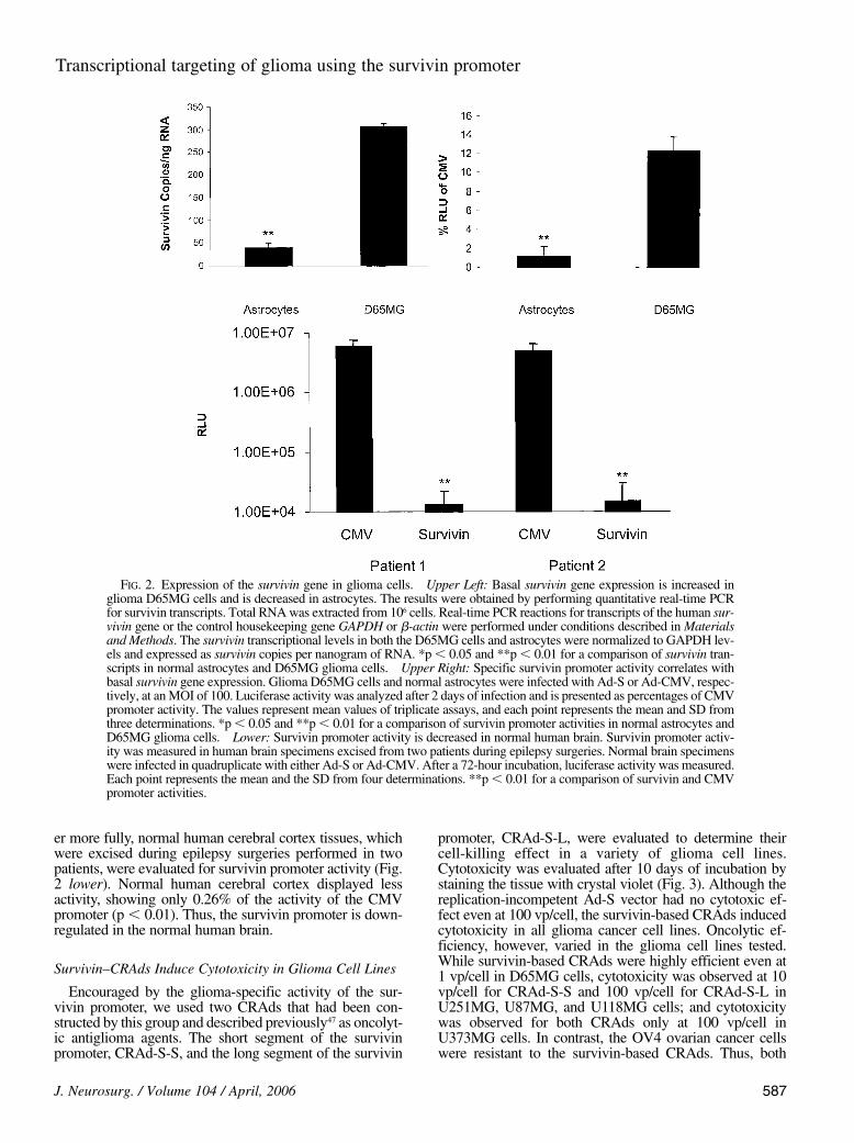
er more fully, normal human cerebral cortex tissues, whichwere excised during epilepsy surgeries performed in twopatients, were evaluated for survivin promoter activity (Fig.2 lower). Normal human cerebral cortex displayed lessactivity, showing only 0.26% of the activity of the CMVpromoter (p , 0.01). Thus, the survivin promoter is down-regulated in the normal human brain.
Survivin–CRAds Induce Cytotoxicity in Glioma Cell Lines
Encouraged by the glioma-specific activity of the sur-vivin promoter, we used two CRAds that had been con-structed by this group and described previously47 as oncolyt-ic antiglioma agents. The short segment of the survivinpromoter, CRAd-S-S, and the long segment of the survivin
promoter, CRAd-S-L, were evaluated to determine theircell-killing effect in a variety of glioma cell lines.Cytotoxicity was evaluated after 10 days of incubation bystaining the tissue with crystal violet (Fig. 3). Although thereplication-incompetent Ad-S vector had no cytotoxic ef-fect even at 100 vp/cell, the survivin-based CRAds inducedcytotoxicity in all glioma cancer cell lines. Oncolytic ef-ficiency, however, varied in the glioma cell lines tested.While survivin-based CRAds were highly efficient even at1 vp/cell in D65MG cells, cytotoxicity was observed at 10vp/cell for CRAd-S-S and 100 vp/cell for CRAd-S-L inU251MG, U87MG, and U118MG cells; and cytotoxicitywas observed for both CRAds only at 100 vp/cell inU373MG cells. In contrast, the OV4 ovarian cancer cellswere resistant to the survivin-based CRAds. Thus, both
J. Neurosurg. / Volume 104 / April, 2006
Transcriptional targeting of glioma using the survivin promoter
587
FIG. 2. Expression of the survivin gene in glioma cells. Upper Left: Basal survivin gene expression is increased inglioma D65MG cells and is decreased in astrocytes. The results were obtained by performing quantitative real-time PCRfor survivin transcripts. Total RNA was extracted from 106 cells. Real-time PCR reactions for transcripts of the human sur-vivin gene or the control housekeeping gene GAPDH or b-actin were performed under conditions described in Materialsand Methods. The survivin transcriptional levels in both the D65MG cells and astrocytes were normalized to GAPDH lev-els and expressed as survivin copies per nanogram of RNA. *p , 0.05 and **p , 0.01 for a comparison of survivin tran-scripts in normal astrocytes and D65MG glioma cells. Upper Right: Specific survivin promoter activity correlates withbasal survivin gene expression. Glioma D65MG cells and normal astrocytes were infected with Ad-S or Ad-CMV, respec-tively, at an MOI of 100. Luciferase activity was analyzed after 2 days of infection and is presented as percentages of CMVpromoter activity. The values represent mean values of triplicate assays, and each point represents the mean and SD fromthree determinations. *p , 0.05 and **p , 0.01 for a comparison of survivin promoter activities in normal astrocytes andD65MG glioma cells. Lower: Survivin promoter activity is decreased in normal human brain. Survivin promoter activ-ity was measured in human brain specimens excised from two patients during epilepsy surgeries. Normal brain specimenswere infected in quadruplicate with either Ad-S or Ad-CMV. After a 72-hour incubation, luciferase activity was measured.Each point represents the mean and the SD from four determinations. **p , 0.01 for a comparison of survivin and CMVpromoter activities.
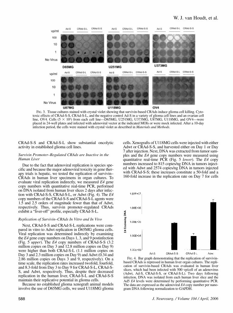
CRAd-S-S and CRAd-S-L show substantial oncolyticactivity in established glioma cell lines.
Survivin Promoter–Regulated CRAds are Inactive in theHuman Liver
Due to the fact that adenoviral replication is species spe-cific and because the major adenoviral toxicity in gene ther-apy trials is hepatic, we tested the replication of survivin–CRAds in human liver specimens in organ cultures. Toevaluate viral replication indirectly, we measured E4 genecopy numbers with quantitative real-time PCR, performedon DNA isolated from human liver slices 2 days after infec-tion with CRAd-S-S, CRAd-S-L, or Adwt (Fig. 4). The E4copy numbers of the CRAd-S-S and CRAd-S-L agents were1.5 and 2.5 orders of magnitude fewer than that of Adwt,respectively. Thus, survivin promoter–regulated CRAdsexhibit a “liver-off” profile, especially CRAd-S-L.
Replication of Survivin–CRAds In Vitro and In Vivo
Next, CRAd-S-S and CRAd-S-L replications were com-pared in vitro to Adwt replication in D65MG glioma cells.Viral replication was determined indirectly by examiningthe E4 gene copy numbers on Days 1, 3, and 9 postinfection(Fig. 5 upper). The E4 copy numbers of CRAd-S-S (3.2million copies on Day 3 and 12.8 million copies on Day 9)were higher than both CRAd-S-L (1.1 million copies onDay 3 and 2.3 million copies on Day 9) and Adwt (0.34 and2.86 million copies on Days 3 and 9, respectively). On atime scale, the replication rates increased twofold, fourfold,and 8.3-fold from Day 3 to Day 9 for CRAd-S-L, CRAd-S-S, and Adwt, respectively. Thus, despite their decreasedreplication in the human liver, CRAd-S-L and CRAd-S-Smaintain their replicative potential in glioma cells.
Because no established glioma xenograft animal modelsinvolve the use of D65MG cells, we used U118MG glioma
cells. Xenografts of U118MG cells were injected with eitherAdwt or CRAd-S-S, and harvested either on Day 1 or Day7 after injection. Next, DNA was extracted from tumor sam-ples and the E4 gene copy numbers were measured usingquantitative real-time PCR (Fig. 5 lower). The E4 copynumbers increased to 815 copies/ng DNA in tumors inject-ed with Adwt and 2574 copies/ng DNA in tumors injectedwith CRAd-S-S; these increases constitute a 50-fold and a160-fold increase in the replication rate on Day 7 for cells
W. J. van Houdt, et al.
588 J. Neurosurg. / Volume 104 / April, 2006
FIG. 3. Tissue cultures stained with crystal violet showing that survivin-based CRAds induce glioma cell killing. Cyto-toxic effects of CRAd-S-S, CRAd-S-L, and the negative control Ad-S in a variety of glioma cell lines and an ovarian cellline, OV4. Cells (5 3 104) from each cell line—D65MG, U251MG, U373MG, U87MG, U118MG, and OV4—wereplaced in 24-well plates and infected with adenoviral vector at the indicated MOIs or were mock infected. After a 10-dayinfection period, the cells were stained with crystal violet as described in Materials and Methods.
FIG. 4. Bar graph demonstrating that the replication of survivin-based CRAds is repressed in human liver organ cultures. The repli-cation of survivin-based CRAds was evaluated in human liverslices, which had been infected with 500 vp/cell of an adenovirus(Adwt, Ad-S, CRAd-S-S, or CRAd-S-L). Two days followinginfection, DNA was isolated from each human liver slice and theAd5 E4 levels were determined by performing quantitative PCR.The data are expressed as the adenoviral E4 copy number per nano-gram DNA following normalization to GAPDH.
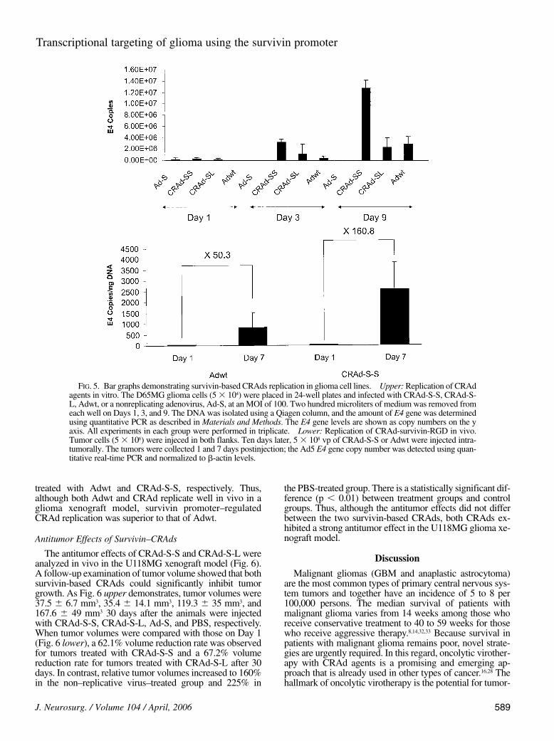
treated with Adwt and CRAd-S-S, respectively. Thus,although both Adwt and CRAd replicate well in vivo in aglioma xenograft model, survivin promoter–regulatedCRAd replication was superior to that of Adwt.
Antitumor Effects of Survivin–CRAds
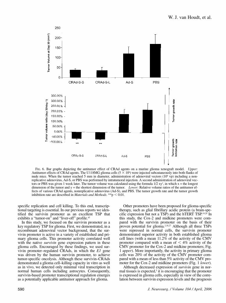
The antitumor effects of CRAd-S-S and CRAd-S-L wereanalyzed in vivo in the U118MG xenograft model (Fig. 6).A follow-up examination of tumor volume showed that bothsurvivin-based CRAds could significantly inhibit tumorgrowth. As Fig. 6 upper demonstrates, tumor volumes were37.5 6 6.7 mm3, 35.4 6 14.1 mm3, 119.3 6 35 mm3, and167.6 6 49 mm3 30 days after the animals were injectedwith CRAd-S-S, CRAd-S-L, Ad-S, and PBS, respectively.When tumor volumes were compared with those on Day 1(Fig. 6 lower), a 62.1% volume reduction rate was observedfor tumors treated with CRAd-S-S and a 67.2% volumereduction rate for tumors treated with CRAd-S-L after 30days. In contrast, relative tumor volumes increased to 160%in the non–replicative virus–treated group and 225% in
the PBS-treated group. There is a statistically significant dif-ference (p , 0.01) between treatment groups and controlgroups. Thus, although the antitumor effects did not differbetween the two survivin-based CRAds, both CRAds ex-hibited a strong antitumor effect in the U118MG glioma xe-nograft model.
Discussion
Malignant gliomas (GBM and anaplastic astrocytoma)are the most common types of primary central nervous sys-tem tumors and together have an incidence of 5 to 8 per100,000 persons. The median survival of patients withmalignant glioma varies from 14 weeks among those whoreceive conservative treatment to 40 to 59 weeks for thosewho receive aggressive therapy.8,14,32,33 Because survival inpatients with malignant glioma remains poor, novel strate-gies are urgently required. In this regard, oncolytic virother-apy with CRAd agents is a promising and emerging ap-proach that is already used in other types of cancer.16,28 Thehallmark of oncolytic virotherapy is the potential for tumor-
J. Neurosurg. / Volume 104 / April, 2006
Transcriptional targeting of glioma using the survivin promoter
589
FIG. 5. Bar graphs demonstrating survivin-based CRAds replication in glioma cell lines. Upper: Replication of CRAdagents in vitro. The D65MG glioma cells (5 3 104) were placed in 24-well plates and infected with CRAd-S-S, CRAd-S-L, Adwt, or a nonreplicating adenovirus, Ad-S, at an MOI of 100. Two hundred microliters of medium was removed fromeach well on Days 1, 3, and 9. The DNA was isolated using a Qiagen column, and the amount of E4 gene was determinedusing quantitative PCR as described in Materials and Methods. The E4 gene levels are shown as copy numbers on the yaxis. All experiments in each group were performed in triplicate. Lower: Replication of CRAd-survivin-RGD in vivo.Tumor cells (5 3 106) were injeced in both flanks. Ten days later, 5 3 108 vp of CRAd-S-S or Adwt were injected intra-tumorally. The tumors were collected 1 and 7 days postinjection; the Ad5 E4 gene copy number was detected using quan-titative real-time PCR and normalized to b-actin levels.
specific replication and cell killing. To this end, transcrip-tional targeting is essential. In our previous reports we iden-tified the survivin promoter as an excellent TSP thatexhibits a “tumor-on” and “liver-off” profile.45
In this study, we focused on the survivin promoter as akey regulatory TSP for glioma. First, we demonstrated, in arecombinant adenoviral vector background, that the sur-vivin promoter is active in a variety of established and pri-mary glioma cells. This promoter activity correlated wellwith the native survivin gene expression pattern in theseglioma cells. Encouraged by these findings, we used sur-vivin promoter–regulated CRAds, in which the E1 genewas driven by the human survivin promoter, to achievetumor-specific oncolysis. Although these survivin–CRAdsdemonstrated a glioma cell–killing capacity in vitro as wellas in vivo, we detected only minimal viral transcription innormal human cells including astrocytes. Consequently,survivin-based promoter transcriptional regulation emergesas a potentially applicable antitumor approach for glioma.
Other promoters have been proposed for glioma-specifictherapy, such as glial fibrillary acidic protein (a brain-spe-cific expression but not a TSP) and the hTERT TSP.19,20 Inthis study, the Cox-2 and midkine promoters were com-pared with the survivin promoter on the basis of theirproven potential for glioma.2,18,27 Although all three TSPswere repressed in normal cells, the survivin promoterdemonstrated superior activity in both established gliomacell lines (with a mean 11.2% of the activity of the CMVpromoter compared with a mean of , 4% activity of theCMV promoter for the Cox-2 and midkine promoters; Fig.1 upper). More importantly, the activity in primary gliomacells was 20% of the activity of the CMV promoter com-pared with a mean of less than 5% activity of the CMV pro-moter for the Cox-2 and midkine promoters (Fig. 1 lower).
Although decreased expression of survivin gene in nor-mal tissues is expected,5 it is encouraging that the promoteris expressed in glioma cells, especially in view of the corre-lation between survivin expression levels and the prognosis
W. J. van Houdt, et al.
590 J. Neurosurg. / Volume 104 / April, 2006
FIG. 6. Bar graphs depicting the antitumor effect of CRAd agents on a murine glioma xenograft model. Upper:Antitumor effects of CRAd agents. The U118MG glioma cells (5 3 106) were injected subcutaneously into both flanks ofnude mice. When the tumor reached 5 mm in diameter, administration of adenoviral vectors (109 vp) including a non-replicative adenovirus, Ad-S, or PBS was performed by intratumoral injection. A second administration of adenoviral vec-tors or PBS was given 1 week later. The tumor volume was calculated using the formula 1/2 xy2, in which x = the longestdimension of the tumor and y = the shortest dimension of the tumor. Lower: Relative volume ratios of the antitumor ef-fects of various CRAd agents, nonreplicative adenovirus (Ad-S), and PBS. The tumor growth rate and the tumor growthinhibition rate are described in Materials and Methods. **p , 0.01.
of glioma.6 Moreover, the increasing expression of survivinin progressive grades of astrocytic tumors has importantclinical application, as survivin-based therapies can be im-plemented across a spectrum of malignant brain tumors.15
Given that replication-incompetent vectors have provedto be inefficient as antitumor agents, replication-selectiveviruses have emerged as potentially powerful anticanceragents.17 Based on their capacity for gene amplification andintratumoral production of viral progeny, the intrinsic cellkilling of adenoviruses has been diverted into CRAd oncol-ysis. These attributes of CRAds have recently been appliedin the context of glioma.25,36 In this study, we constructedtwo novel CRAd agents, CRAd-S-S (nucleotides 2230 to130 of the human survivin gene) and CRAd-S-L (nu-cleotides 21430 to 130 of the human survivin gene), inwhich the survivin promoter regulated Ad5 E1 gene expres-sion and viral infectivity was enhanced by capsid modifica-tion, that is, genetic insertion of the RGD-4C ligand. Ofnote, the E3 gene was retained in these CRAds forimproved cell killing. Nevertheless, despite the cell killingattributes of our survivin-based CRAds, they maintainedsafe profiles in both normal brain and liver. Because theliver manifests a 95% uptake rate of adenovirus when deliv-ered systemically,22 thereby potentially compromising liverfunction,39 the safety profile of survivin-based CRAds inliver tissue (1.5 and 2.5 orders of magnitude fewer E4copies than those found in Adwt, CRAd-S-S, and CRAd-S-L, respectively) is especially encouraging. Furthermore, be-cause malignant gliomas do not metastasize, local CRAdinjection may be applicable on the basis of dramatic repres-sion of the survivin promoter in normal human astrocytesand brain tissue.
The in vitro oncolytic effects of CRAd-S-S and CRAd-S-L were translated into antitumor effects in a glioma xeno-graft model and correlated with intratumoral replicationrates. The difference we observed between the short andlong versions of the survivin promoter in the CRAd con-text was that the long version of the survivin promoter wasless hepatotoxic than the short version. These data are rem-iniscent of the Cox-2 promoter versions previously de-scribed.41,43 Thus, CRAd-S-L maintains an antitumor effect,while minimizing hepatotoxicity to increase the therapeuticindex, an important parameter in cancer gene therapy.
In this study, the viral replication of CRAds was regulat-ed by a tumor-specific promoter, the survivin promoter.Thus, viral replication was limited to tumor cells and mini-mized in normal host cells. However, adenoviruses are ableto kill normal cells as well as cancer cells by leaking vec-tors, leading to host toxicity.11 The potential of killing nor-mal cells relates to the promoter activity in normal cells. Forthis reason, we tested the promoter activity in human braintissue. The data shown in Fig. 2 lower revealed only 0.26%of the activity of the CMV promoter, indicating that the tox-icity of leakage of these agents to brain tissue is low becauseof the TSP.
Conclusions
We identified the human survivin promoter as a tumor-specific regulatory element for glioma. It is activated in es-tablished and primary glioma cells and is downregulated innormal astrocytes and human brain and liver tissues. Thesurvivin promoter therefore represents a promising tran-
scriptional targeting approach for gliomas. Survivin-basedCRAds significantly kill glioma cells in vitro and inhibit thegrowth of glioma xenografts in vivo. On this basis, the sur-vivin promoter in recombinant adenovirus and CRAd con-texts may have potential use for future experimental clinicalapplications in the treatment of glioma.
Acknowledgments
The authors thank Dr. G. Y. Gillespie for his helpful advice andreview of the manuscript, Dr. Masato Yamamoto for Ad-Cox-2, Dr.Charles Bonus for human astrocytes, and Dr. Fen Zhou for her assis-tance with the statistical analysis. We are especially indebted to Dr.F. Li for the gift of the plasmids pLuc-1430c and pLuc-cyc1.2,which were used to construct the reAd and CRAd agents. We alsothank Marijke van der Jagt, a medical student at VU Medical Center,Amsterdam, The Netherlands, for help with the experiments and Dr.Ronald Verwer, Ph.D., a senior researcher at the Netherlands Insti-tute for Brain Research, Amsterdam, for support in the brain-sliceexperiments.
References
1. Adachi Y, Reynolds PN, Yamamoto M, Grizzle WE, Overturf K,Matsubara S, et al: Midkine promoter-based adenoviral vectorgene delivery for pediatric solid tumors. Cancer Res 60:4305–4310, 2000
2. Akasaki Y, Liu G, Chung NH, Ehtesham M, Black KL, Yu JS:Induction of a CD4+ T regulatory type 1 response by cyclooxy-genase-2-overexpressing glioma. J Immunol 173:4352–4359,2004
3. Alemany R, Gomez-Manzano C, Balague C, Yung WKA, CurielDT, Kyritsis AP, et al: Gene therapy for gliomas: molecular tar-gets, adenoviral vectors, and oncolytic adenoviruses. Exp CellRes 252:1–12, 1999
4. Ambrosini G, Adida C, Altieri DC: A novel anti-apoptosis gene,survivin, expressed in cancer and lymphoma. Nat Med 3:917–921, 1997
5. Bao R, Connolly DC, Murphy M, Green J, Weinstein JK, PisarcikDA, et al: Activation of cancer-specific gene expression by thesurvivin promoter. J Natl Cancer Inst 94:522–528, 2002
6. Chakravarti A, Noll E, Black PM, Finkelstein DF, FinkelsteinDM, Dyson NJ, et al: Quantitatively determined survivin expres-sion levels are of prognostic value in human gliomas. J Clin On-col 20:1063–1068, 2002
7. Davis FG, McCarthy BJ: Epidemiology of brain tumors. CurrOpin Neurol 13:635–640, 2000
8. Fine HA, Dear KB, Loeffler JS, Black PM, Canellos GP: Meta-analysis of radiation therapy with and without adjuvant chemo-therapy for malignant gliomas in adults. Cancer 71:2585–2597,1993
9. Fueyo J, Alemany R, Gomez-Manzano C, Fuller GN, Khan A,Conrad CA, et al: Preclinical characterization of the antigliomaactivity of a tropism-enhanced adenovirus targeted to the retino-blastoma pathway. J Natl Cancer Inst 95:652–660, 2003
10. Fueyo J, Gomez-Manzano C, Alemany R, Lee PSY, McDonnellTJ, Mitlianga P, et al: A mutant oncolytic adenovirus targeting theRb pathway produces anti-glioma effect in vivo. Oncogene 19:2–12, 2000
11. Fueyo J, Gomez-Manzano C, Yung WK, Kyritsis AP: Targetingin gene therapy for gliomas. Arch Neurol 56:445–448, 1999
12. Haviv YS, Curiel DT: Conditional gene targeting for cancer genetherapy. Adv Drug Deliv Rev 53:135–154, 2001
13. Huang CJ, Nazarian R, Lee J, Zhao PM, Espinosa-Jeffrey A, deVellis J: Tumor necrosis factor modulates transcription of myelinbasic protein gene through nuclear factor kappa B in a humanoligodendroglioma cell line. Int J Dev Neurosci 20:289–296,2002
J. Neurosurg. / Volume 104 / April, 2006
Transcriptional targeting of glioma using the survivin promoter
591
14. Huncharek M, Muscat J: Treatment of recurrent high grade astro-cytoma; results of a systematic review of 1,415 patients. Anti-cancer Res 18:1303–1311, 1998
15. Kajiwara Y, Yamasaki F, Hama S, Yahara K, Yoshioka H, Sugi-yama K, et al: Expression of survivin in astrocytic tumors: correla-tion with malignant grade and prognosis. Cancer 97:1077–1083,2003
16. Khuri FR, Nemunaitis J, Ganly I, Arseneau J, Tannock IF, RomelL, et al: A controlled trial of intratumoral ONYX-015, a selec-tively-replicating adenovirus, in combination with cisplatin and 5-fluorouracil in patients with recurrent head and neck cancer. NatMed 6:879–885, 2000
17. Kirn D, Martuza RL, Zwiebel J: Replication-selective virotherapyfor cancer: biological principles, risk management and future di-rections. Nat Med 7:781–787, 2001
18. Kohno S, Nakagawa K, Hamada K, Harada H, Yamasaki, K, Ha-shimoto K, et al: Midkine promoter-based conditionally replica-tive adenovirus for malignant glioma therapy. Oncol Rep 12:73–78, 2004
19. Komata T, Koga S, Hirohata S, Takakura M, Germano IM, InoueM, et al: A novel treatment of human malignant gliomas in vitroand in vivo: FADD gene transfer under the control of the humantelomerase reverse transcriptase gene promoter. Int J Oncol 19:1015–1020, 2001
20. Komata T, Kondo Y, Kanzawa T, Hirohata S, Koga S, SumiyoshiH, et al: Treatment of malignant glioma cells with the transfer ofconstitutively active caspase-6 using the human telomerase cat-alytic subunit (human telomerase reverse transcriptase) gene pro-moter. Cancer Res 61:5796–5802, 2001
21. Komata T, Kondo Y, Kanzawa T, Ito H, Hirohata S, Koga S, et al:Caspase-8 gene therapy using the human telomerase reverse tran-scriptase promoter for malignant glioma cells. Hum Gene Ther13:1015–1025, 2002
22. Kruyt FA, Curiel DT: Toward a new generation of conditionallyreplicating adenoviruses: pairing tumor selectivity with maximaloncolysis. Hum Gene Ther 13:485–495, 2002
23. Lamfers MLM, Grill J, Dirven CMF, van Beusechem VW,Geoerger B, Van Den Berg J, et al: Potential of the conditionallyreplicative adenovirus Ad5-delta24RGD in the treatment of ma-lignant gliomas and its enhanced effect with radiotherapy. CancerRes 62:5736–5742, 2002
24. Li F, Ambrosini G, Chu EY, Plescia J, Tognin S, Marchisio PC, etal: Control of apoptosis and mitotic spindle checkpoint by sur-vivin. Nature 396:580–584, 1998
25. Lou E: Oncolytic viral therapy and immunotherapy of malignantbrain tumors: two potential new approaches of translational re-search. Ann Med 36:2–8, 2004
26. Manome Y, Kunieda T, Wen PY, Koga T, Kufe DW, Ohno T:Transgene expression in malignant glioma using a replication-defective adenoviral vector containing the Egr-1 promoter: acti-vation by ionizing radiation or uptake of radioactive iododeoxy-uridine. Hum Gene Ther 9:1409–1417, 1998
27. Mishima K, Asai A, Kadomatsu K, Ino Y, Nomura K, Narita Y,et al: Increased expression of midkine during the progression ofhuman astrocytomas. Neurosci Lett 233:29–32, 1997
28. Morris JC, Wildner O: Therapy of head and neck squamous cellcarcinoma with an oncolytic adenovirus expressing HSV-tk. MolTher 1:56–62, 2000
29. Nanda D, Vogels R, Havenga M, Avezaat CJ, Bout A, Smitt PS:Treatment of malignant gliomas with a replicating adenoviral vec-tor expressing herpes simplex virus-thymidine kinase. CancerRes 61:8743–8750, 2001
30. Repovic P, Fears CY, Gladson CL, Benveniste EN: Oncostatin-Minduction of vascular endothelial growth factor expression in as-troglioma cells. Oncogene 22:8117–8124, 2003
31. Ruan H, Su H, Hu L, Lamborn KR, Kan YW, Deen DF: A hypox-ia-regulated adeno-associated virus vector for cancer-specificgene therapy. Neoplasia 3:255–263, 2001
32. Salcman M: Survival in glioblastoma: historical perspective. Neu-rosurgery 7:435–439, 1980
33. Salcman M, Scholtz H, Kaplan RS, Kulik S: Long-term survival inpatients with malignant astrocytoma. Neurosurgery 34:213–220,1994
34. Sasaki T, Lopes MBS, Hankins GR, Helm GA: Expression of sur-vivin, an inhibitor of apoptosis protein, in tumors of the nervoussystem. Acta Neuropathol 104:105–109, 2002
35. Segovia J, Vergara P, Brenner M: Differentiation-dependent ex-pression of transgenes in engineered astrocyte cell lines. NeurosciLett 242:172–176, 1998
36. Shah AC, Benos D, Gillespie GY, Markert JM: Oncolytic viruses:clinical applications as vectors for the treatment of malignantgliomas. J Neurooncol 65:203–226, 2003
37. Siu IM, Lal A, Blankenship JR, Aldosari N, Riggins GJ: c-Myc pro-moter activation in medulloblastoma. Cancer Res 63:4773–4776,2003
38. Surawicz TS, Davis F, Freels S, Laws ER Jr, Menck HR: Braintumor survival: results from the National Cancer Data Base. JNeurooncol 40:151–160, 1998
39. van der Eb MM, Cramer SJ, Vergouwe Y, Schagen FH, van Krie-ken JH, van der Eb AJ, et al: Severe hepatic dysfunction after ade-novirus-mediated transfer of the herpes simplex virus thymidinekinase gene and ganciclovir administration. Gene Ther 5:451–458,1998
40. Verwer RW, Hermens WT, Dijkhuizen P, Ter Brake O, Baker RE,Salehi A, et al: Cells in human postmortem brain tissue slices re-main alive for several weeks in culture. FASEB J 16:54–60, 2002
41. Wesseling JG, Yamamoto M, Adachi Y, Bosma PJ, van WijlandM, Blackwell JL, et al: Midkine and cyclooxygenase-2 promotersare promising for adenoviral vector gene delivery of pancreaticcarcinoma. Cancer Gene Ther 8:990–996, 2001
42. Yamamoto M, Alemany R, Adachi Y, Grizzle WE, Curiel DT:Characterization of the cyclooxygenase-2 promoter in an adenovi-ral vector and its application for the mitigation of toxicity in suicidegene therapy of gastrointestinal cancers. Mol Ther 3:385–394,2001
43. Yamamoto M, Davydova J, Wang M, Siegal GP, Krasnykh V,Vickers SM, et al: Infectivity enhanced, cyclooxygenase-2 pro-moter-based conditionally replicative adenovirus for pancreaticcancer. Gastroenterology 125:1203–1218, 2003
44. Zamorano A, Lamas M, Vergara P, Naranjo JR, Segovia J: Tran-scriptionally mediated gene targeting of gas 1 to glioma cells elic-its growth arrest and apoptosis. J Neurosci Res 71:256–263, 2003
45. Zhu ZB, Makhija SK, Lu B, Wang M, Kaliberova L, Liu B, et al:Transcriptional targeting of adenoviral vector through the CXCR4tumor-specific promoter. Gene Ther 11:645–648, 2004
46. Zhu ZB, Makhija SK, Lu B, Wang M, Rivera AA, Kim-Park S, etal: Incorporating the survivin promoter in an infectivity enhancedCRAd-analysis of oncolysis and anti-tumor effects in vitro and invivo. Int J Oncol 27:237–246, 2005
47. Zhu ZB, Makhija SK, Lu BG, Wang M, Kaliberova L, Liu B, etal: Transcriptional targeting of tumors with a novel tumor-specif-ic survivin promoter. Cancer Gene Ther 11:256–262, 2004
Manuscript received April 19, 2005.Accepted in final form December 6, 2005.This study was supported by National Institutes of Health Grant
Nos. R01 CA83821, R01 CA94084, R01 CA93796, R01 CA93796,K12 HD01261-02, and R01 DK063615-01, and by Dutch CancerSociety Grant No. VU2002-2594 to Drs. Dirven and Lamfers. Dr.van Houdt was supported by the Saal van Zwananberg Foundation,the Dutch Cancer Society, and the Dutch Brain Foundation.
Address reprint requests to: Zeng B. Zhu, M.D., Division of Hu-man Gene Therapy, Gene Therapy Center, BMR II 534, 2172, 90119th Street South, University of Alabama at Birmingham, Birming-ham, Alabama 35291. email: [email protected].
W. J. van Houdt, et al.
592 J. Neurosurg. / Volume 104 / April, 2006