The Role of a CA Repeat Polymorphism in the Promoter ...
130
The Role of a CA Repeat Polymorphism in the Promoter Region of the Insulin like Growth Factor-I gene in Physiology and the Pathophysiology of Diabetes Mellitus Ingrid Rietveld IGF-I in Physiology and Pathophysiology of Diabetes Mellitus Ingrid Rietveld
-
Upload
khangminh22 -
Category
Documents
-
view
1 -
download
0
Transcript of The Role of a CA Repeat Polymorphism in the Promoter ...

The Role of a CA Repeat Polymorphism in the
Promoter Region of the Insulin like
Growth Factor-I gene in Physiology and the
Pathophysiology of Diabetes Mellitus
Ingrid Rietveld
IGF-I in Physiology and Pathophysiology of D
iabetes Mellitus
Ingrid Rietveld


The Role of a CA Repeat Polymorphism in the Promoter Region of the Insulin like
Growth Factor-I gene in Physiology and the Pathophysiology of Diabetes Mellitus
Ingrid Rietveld
ACKNOWLEDGEMENT
The work presented in this thesis was conducted at the Department of Epidemiology & Bio-
statistics and Internal Medicine of the Erasmus Medical Center of Rotterdam. The Rotterdam
Study is supported by the Erasmus Medical Center and Erasmus University Rotterdam, The
Netherlands Organization for Scientific Research (NMO), the Netherlands Organization for
Health Research and Development (ZonMw), the Research Institute for Diseases in the Elderly
(RIDE), the Ministry of Education, Culture and Science, the Ministry of Health, Welfare and
Sports, the European Commission (DG XII), and the municipality of Rotterdam. The contribu-
tions of the general practitioners and pharmacists of the Ommoord district to the Rotterdam
Study are gratefully acknowledged.
Financial support for this work by the Dutch Diabetes Foundation is gratefully acknowledged.
The printing of this thesis was supported by Novartis Farma BV, Eli Lilly Nederland B.V., Servier
Nederland Farma B.V, Novo Nordisk B.V., Boehringer Ingelheim B.V.
Layout and print by Optima Grafische Communicatie, Rotterdam
Cover: Mountains in Switzerland
ISBN: 978-90-8559-600-4
©Ingrid Rietveld, 2009.
No part of this thesis may be reproduced, stored in a retrieval system or transmitted in any
form or by any means without permission of the author or, when appropriate, of the publish-
ers of the publications.

The Role of a CA Repeat Polymorphism in the Promoter Region of the Insulin like
Growth Factor-I Gene in Physiology and the Pathophysiology of Diabetes Mellitus
De rol van een CA repeat polymorfisme in de promoter regio van het Insuline-achtige Groei
factor-I gen in de fysiologie en in de pathofysiologie van diabetes mellitus
Proefschrift
Ter verkrijging van de graad van doctor aan de
Erasmus Universiteit Rotterdam
op gezag van de
rector magnificus
Prof.dr. H.G. Schmidt
en volgens besluit van het College voor Promoties
De openbare verdediging zal plaatsvinden op
Woensdag16 december 2009 om 15.30 uur
door
Ingrid Rietveld
geboren te Udenhout

PROMOTIECOMMISSIE
Promotoren:
Prof.dr. S.W.J. Lamberts
Prof.dr.ir. C.M. van Duijn
Overige leden:
Prof.dr. T.J. Visser
Prof.dr. A.G. Uitterlinden
Prof.dr. A.J. van der Lelij
Co-promotor:
Dr. J.A.M.J.L. Janssen

CONTENTS
Chapter 1 General introduction 7
Chapter 2 A polymorphism in the IGF-I gene influences the age-related
decline in circulating total IGF-I levels
27
European Journal of Endocrinology Feb;148(2):171-5; 2003
Chapter 3 A polymorphic CA repeat in the IGF-I gene is associated with
gender-specific difference in body height, but has no effect on
the secular trend in body height
37
Clinical Endocrinology Aug 61(2):195-203; 2004
Chapter 4 Insulin-like growth factor-I gene influences beta-cell function in
normal glucose tolerance
53
Submitted
Chapter 5a An IGF-I gene polymorphism modifies the risk of diabetic
retinopathy
67
Diabetes 55(8):2387-2391; 2006
Chapter 5b An insulin-like Growth Factor-I gene polymorphism modifies
the risk of micro-albuminuria in subjects with abnormal glucose
tolerance
79
European Journal of Endocrinology 154:715-721; 2006
Chapter 6 An insulin-like Growth Factor-I promoter polymorphism is
associated with increased mortality in subjects with myocardial
infarction in an elderly Caucasian population
93
The American Journal of Cardiology 97:1274-1276;2006
Chapter 7 General discussion 99
Summary 111
Samenvatting (summary in Dutch) 115
Dankwoord 119
About the author 121
List of publications 123
PhD portfolio 125


1General introduction


9
General introduction
1 PHYSIOLOGY IGF-I
Insulin like growth factor-1 (IGF-I) is a ubiquitous » 7.5 KDa polypeptide, which influences
cell proliferation, differentiation and survival in many tissues [1, 2]. IGF-I is synthesized by
most organs and may act as an endocrine, paracrine and/or autocrine growth factor [3]. The
most important function of IGF-I is mediating physiological growth [4]. Historically, it was
suggested that the growth promoting effects of GH were mediated by a liver derived factor
initially termed “sulphation factor” [5]. Simultaneously, a fraction of non-suppressible insulin-
like activity from human serum was discovered, which they called “NSILA” [5]. These factors
turned out to be identical and were subsequently called “somatomedin”. Finally, the protein
was sequenced and hereafter termed insulin like growth factor I (IGF-I) [5].
Human postnatal growth and development is largely under the influence of the growth
hormone (GH) – insulin like growth factor (IGF) axis [6]. GH is not essential for intra-uterine
growth and development, as demonstrated by the existence of normal-sized infants with
either congenital absence of the pituitary or deletions in the genes encoding GH or the
GH receptor [3, 7]. Mice carrying null mutations of the IGF-I gene however, are born small
in size and grow poorly in the postnatal stages of development [3]. Also in patients with
intra uterine growth retardation and postnatal growth deficit, impairment of the IGF-I gene
and IGF-I receptor gene has been found [7-10]. Postnatal growth is dependent on GH as GH
deficiency or deletions in the GH receptor result in growth retardation [3]. The effects of GH
on growth are mediated via intermediate factors of which IGF-I is the most important [3]. Tall
children have higher IGF-I and IGFBP-3 levels compared to short children [11].
IGF-I levels increase with age and pubertal development and decline after puberty through-
out adulthood [12, 13]. Serum IGF-I levels peak at puberty and occurs earlier in girls than
in boys [12, 13]. The relation of age with IGF-I was the same for IGFBP-3 and GH [13, 14].
However, GH response to a combination of GHRH and GHRP-6 or GHRH ± arginine remains
the same throughout adult life, suggesting that the pituitary GH reserve is preserved even
in older adults [6]. Factors that contribute to the age-related decline of GH and IGF-I include
sex steroids, malnutrition, higher body mass index, changes in sleep pattern and physically
inactivity [6].
IGF-I has structural and functional homology with insulin, although action takes place by its
own receptor activation [1, 2]. IGF-I can interact with the insulin receptor although with a
lower affinity than that of insulin [15]. High concentrations of IGF-I are likely to cross-activate
with the insulin receptor [16]. The role of IGF-I in connection with glucose metabolism was
first postulated by Froesch, who described that NSILA has a hypoglycemic action and is
more active on muscle than in adipose tissue when compared to insulin [16]. In a study with
healthy volunteers, recombinant human IGF-I administration had a different effect on counter
regulatory response compared with insulin [17]. IGF-I infusion reduced glucagon levels and
attenuated GH release [17]. Under euglycemic conditions, recombinant IGF-I infusion resulted

Chapter 1
10
in a reduction of plasma insulin, C-peptide and glucagon levels comparable to insulin infusion
[18]. Moreover, it lowered plasma glucose levels by stimulation of peripheral glucose uptake
and inhibition of hepatic glucose production, even though the liver has almost no IGF-I
receptors [18]. Possible mechanisms for this inhibitory effects include IGF-I mediated effects
via the insulin receptor, via IGF-I/insulin hybrid receptors or via IGF-I mediated changes in
substrate delivery for hepatic gluconeogenesis [18].
2 REGULATION OF IGF-I
Like stated before, IGF-I is one of the factors that are involved in the regulation of somatic
growth and cellular proliferation [4, 19]. The liver is the main source of plasma IGF-I as proven
by gene targeting studies [20]. Its action is determined by the availability of free IGF-I to in-
teract with the IGF-I receptor (IGF-IR) [19]. The rate of IGF-I production, clearance and degree
of binding to the IGFBPs, modulate the levels of free IGF-I [2, 19].
Twin studies demonstrated that circulating IGF-I levels have a marked genetic component,
which may account for large variation between persons [5]. Most circulating IGF-I is under
GH control [4]. Its concentration is also influenced by nutritional status, sex steroids, insulin
and glucocorticoids [1, 4]. Effects of IGF-I are regulated by the IGF-I receptor, but are also
modulated by IGF-I binding proteins [4].
The most important regulators of plasma concentration of IGF-I are separately described
below.
3'5'
1 2 3 4 5 6
a b
Figure 1: Schematic presentation of the IGF-I gene. The IGF-I gene contains six exons (boxes), separated by five introns (black lines). The gene contains two independent promoters, promoter 1 (a) and promoter 2 (b). The (CA)n polymorphism is located near promoter 1.
a IGF-I geneThe human IGF-I gene, located on the long arm of chromosome 12, is a complex, multi-
component gene with six exons, with the mature peptide being encoded by exons 3 and
4 [21]. Alternative leader exons are exons 1 and 2 [21, 22]. Several forms of IGF-I mRNA are
transcribed, including the 6 kb form that is regulated by GH [22, 23]. Specific residues of the
A chain are critical for recognition by four of the six IGF binding proteins. Specific amino
acids in the B chain are required for binding to any of the six forms of IGF binding proteins.
Tyrosines 24, 60 and to some extent 31 are critical for IGF-I receptor recognition. The IGF-I
gene contains two independent promoters, which are regulated in a cell-type dependent
way [22, 24].

11
General introduction
Over the years, several polymorphisms in the IGF-I gene have been studied in relation to IGF-I
serum levels. In children small for gestational age (SGA), polymorphisms of the IGF-I gene
studied are: a cytosine-adenine repeat near the promoter region, a microsatellite marker
(CA repeat) lying at the 3’ part of the gene, an intronic cytosine-thymidine (CT) repeat lying
between exon 2 and 3 [25, 26] and two single nucleotide polymorphism (SNP) markers [26].
In these children, only significant differences in IGF-I serum levels were related with the CT
repeat polymorphism. The wild type allele (in this study an allele with a PCR product of 189-
bp), was transmitted most often from parent to child [25, 26]. Children homozygous for the
189-bp allele had significantly lower levels of IGF-I [25].
Since Rosen found an association between a CA repeat variation near the promoter region
of the IGF-I gene and IGF-I serum levels, more studies were performed to examine the
functionality of this polymorphism. The length of this CA repeat sequence ranges from 10 to
24 CA repeats [27, 28]. In the Caucasian population, the most frequent allele contains 19 CA
repeats [27-31]. In African-Americans the most frequent allele is shorter and contains 18 CA
repeats [32-34]. The (CA)n polymorphism was associated with serum IGF-I levels in several
study cohorts [27-29, 31, 35, 36], although in different directions. In other studies, no relation
was found [30, 33, 37, 38]. Furthermore, this polymorphism was also related to body height,
type 2 diabetes, myocardial infarction [28] and birth weight [39] in the Rotterdam study.
Also a significant relation with BMD [40] and risk for fragility at old age in women and with
bone structure in both genders was observed [41]. Other studies also found a relation with
cardiovascular disease [42] or stroke [43].
b IGF binding proteinsIn all extracellular tissues, most of the IGF-I is bound to a family of insulin-like growth fac-
tor binding proteins (IGFBPs), which prolong the half life in the circulation; the half life of
unbound IGF-I is less than 10 minutes, but of bound IGF-I approximately 12 hours [1, 19].
There are six IGFBPs, which are produced in a variety of biological tissues and are found
in various biologic fluids like plasma [44]. The majority of the IGF-I is bound in a trimeric
complex, composed of IGFBP3 or IGFBP5 and a liver derived glycoprotein known as acid lable
subunit (ALS) [1, 19]. Approximately 90% of IGFBP3 and 55% of IGFBP5 circulates in these
complexes in healthy individuals [19, 45]. All three components of the trimeric complex are
induced by GH and therefore are affected by GH excess or deficiency [19]. It is assumed that
complex formation occurs in the liver as IGF-I and ALS are derived from hepatocytes, whereas
IGFBP3 is produced in the Kuppfer cells. The ternary complex with IGFBP5 is considered an
IGF reservoir [4]. About 25% of circulating IGFs are bound in binary complexes, which can
leave the circulation [4], whereas less than 1% of IGF-I circulates in the free form [19].
IGFBPs have several effects on IGF-I and different functional properties:
1. prolongation of IGFs half life in the circulation
2. prevention of IGF induced hypoglycemia

Chapter 1
12
3. regulation of the passage of IGFs from the intravascular to the extra vascular space
4. limitation of the bioavailability of free IGFs to interact with the IGF receptors
5. enhancement of IGF actions by the formation of a pool of slow release IGFs
6. direct cellular actions mediated through their own receptors, acting independently of
IGFs
IGFBP3 is the predominant circulating IGFBP [4]. IGFBP3 forms, as stated before, a ternary
complex with IGF-I [19]. In the last few years, it has become apparent that especially IGFBP3
has IGF independent effects [45].
IGFBP1 has a RGD sequence in its structure, a recognition sequence for membrane integrin
receptors, suggesting the possibility of an IGF independent action via these receptors [19].
IGFBP1 is metabolically regulated and its expression is inhibited by insulin [4]. IGFBP3 levels
in the circulation do not change acutely while IGFBP1 and IGFBP2 levels depend on the meta-
bolic state and the insulin level [19]. IGFBP2 is a major BP in cerebrospinal fluid [19]. IGFBP2
and IGFBP4 have mainly inhibitory effects on IGF mediated functions by competitively
binding to IGF-I [19]. IGFBP4 is regulated by vitamin D and PTH [19]. IGFBP5 inhibits growth
since it is expressed inversely with renal tissue growth status but stimulates actions of IGF-I
particularly in bone [19]. In some circumstances some IGFBPs, especially IGFBP3 and IGFBP5,
may potentiate IGF action [4]. IGFBP6 which is mainly found in cerebrospinal fluid and serum
preferentially binds to IGF-II by over two orders of magnitude better than to IGF-I [19].
IGFBP proteases are proteolytic enzymes that can catalyze the limited hydrolysis of IGFBPs,
causing release of free IGFs to interact with its receptor [19, 45]. There are three classes of
IGFBP proteases: kallikreins, cathepsins and matrix metalloproteinases proteases [19].
c IGF-I receptorIGF-I preferably binds to the IGF-I receptor, but under certain conditions also binds to the
IGF-II receptor, the insulin receptor and hybrid receptors. Actions of IGF-I as a potent mi-
togen, anti-apoptotic factor and modulator of differentiation are mediated mainly through
the IGF-IR [46]. IGF-I receptors are found on most tissues except, notably, liver and adipose
tissue [15, 16]. The number of IGF-I receptors is regulated by GH and thyroxin and is tightly
controlled within a range of 20,000 to 35,000 receptors per cell. The IGF-I receptor (IGF-IR)
and insulin receptor are tyrosine kinase receptors and are composed of two extracellular
alpha subunits and two intracellular beta subunits [4, 19, 46, 47]. The alpha subunits contains
the IGF-binding domain with an affinity constant of 10(-9) M for IGF-I; the affinity constant
is sixfold lower for IGF-II and 200- to 300-fold lower for insulin [15, 46]. The beta subunits
have intrinsic tyrosine kinase activity [19, 46, 47]. Binding of IGF-I to the receptor results in
phosphorylation of the insulin receptor substrates 1 and -2 (IRS-1 and -2) [4, 46]. Eventually
the mitogen activated protein (MAP) kinase pathway is activated, which is important for
stimulation of cell growth by IGF-I [4]. Activation of PI-3 kinase is important for stimulation of
protein synthesis and glucose transport [4]. This pathway is also important for IGF-I stimula-

13
General introduction
tion of cell motility and inhibition of apoptosis [2, 46]. The presence of IGF-I prevents cells in
culture to undergo apoptosis [46].
Targeted disruption of the IGF-IR results in cell refractoriness to viral and cellular oncogenes,
increasing the probability of apoptotic cell death and can inhibit neoplastic proliferation [46].
Deletions of IGF-IR result in decreased IGF-I binding and intrauterine growth retardation [2].
d Growth hormoneThe regulation of the GH/IGF-I system is dependent on the integrity of the hypothalamus,
the pituitary and the liver. GH is released in pulses under the influence of growth hormone
releasing hormone (GHRH) and somatostatin. GH release is inhibited by antibodies to GHRH,
by GH itself and by IGF-I via a direct effect of IGF-I on the IGF-I receptor in the pituitary [6]. GH
action is mediated by the binding of GH to the GH receptor, resulting in its dimerization and
the auto-phosphorylation of the tyrosine kinase, Janus kinase 2 (JAK2). This kinase stimulates
phosphorylation of signaling proteins, STAT proteins STAT1, STAT3 and STAT5. An intact JAK2-
STAT5b signaling pathway is essential for GH stimulation of IGF-I gene expression [48]. The
effects of GH on plasma IGF-I concentrations are complex [3]. Although it stimulates IGF-I
gene transcription and secretion by the liver, part of the GH-mediated increase in circulating
IGF-I levels is controlled by concurrent stimulation of IGFBP-3 and ALS [49]. Together, these
three proteins form a stable ternary complex; any factor that attenuates the relative increase
in any of the three components will produce a major reduction in serum IGF-I [2].
Postnatal growth is dependent on normal pulsatile secretion of GH from pituitary somato-
trophs [50]. Most growth promoting effects of GH are believed to be mediated by IGF-I [3]. In
states of GH deficiency, levels of IGF-I are also reduced [3, 51]. Similarly, excess GH results in
acromegaly with concomitant increase in circulating IGF-I levels [3, 50, 52].
e InsulinIGF-I has 48% structural homology with pro-insulin; the A and B domains have 60-70% homol-
ogy, but there is no homology with the C domain [16]. Delivery of insulin by the portal system
influences hepatic IGF-I production and inversely regulates IGFBP-1 hepatic production [5].
Therefore, normal hepatic insulin action is required for normal rates of IGF-I synthesis [53].
During hyperinsulinaemic clamping, free IGF-I increased while total IGF-I was unaltered [5].
Furthermore, 60 hours of fasting decreased free IGF-I levels [54]. IGF-I can bind to the insulin
receptor, but with an affinity of only 1-5% of that of insulin [44].
f Nutritional statusNutritional status is an important determinant of plasma IGF-I [5]. A minimum intake of 20
kcal/kg per day of energy and 0.6 g/kg of protein are necessary to maintain normal plasma
values. IGF-I levels are low in patients with protein-calorie malnutrition, but can be normal-
ized by adequate nutritional rehabilitation [55]. Serum GH concentrations are often high in
Chapter 1
14
malnourished patients, suggesting that they are resistant to the action of GH, whereas basal
insulin concentrations are normal or low [55]. A decrease in levels is also seen in diseases
associated with malnutrition such as hepatic failure, inflammatory bowel disease and renal
failure [55]. Overfeeding causes serum IGF-I to increase by 19% in a few days [55].
3 DIABETES MELLITUS
a Epidemiology and classification of Diabetes mellitusDiabetes mellitus is the result of less or insufficient insulin production or action. Type 1 diabe-
tes is characterized by absolute insulin deficiency due to beta cell destruction caused by an
auto immune process and acute onset, usually before 25 years of age [56, 57]. Type 2 diabetes
is caused by disturbances in insulin action and beta cell dysfunction [56, 58] and increases
with age [59]. In the Rotterdam Study, prevalence of diabetes increased from 5.9 percent at
ages < 60 years to 19.8 percent at ages >85 years in males and from 3.8 at ages < 60 years to
18.9 percent at ages > 85 years in women. Impaired glucose tolerance increased from 8.8 and
11.0 percent to 24.3 in men and women aged < 60 years to 34.7 and 24.3 percent in men and
women aged > 85 years [59].
The number of people with type 2 diabetes mellitus is increasing due to population growth,
aging, urbanization and increasing prevalence of obesity [60]. Most patients with type 2 dia-
betes are obese and obesity itself causes some degree of insulin resistance [61, 62]. Intensive
glucose control has delayed the development and progression of retinopathy, nephropathy
and neuropathy in patients with type 1 [63, 64] and those with type 2 diabetes [65, 66], and
decreased diabetes –related mortality or myocardial infarction in a group of newly diagnosed
type 2 diabetes [66, 67]. Impaired glucose tolerance has been associated with cardiovascular
disease risk factors and events, whereas impaired fasting glucose was much less strongly
associated with CVD events and mortality [68].
In 1997 recommendations for the classification and diagnosis of diabetes were released by a
collaboration between members of the American Diabetes Association (ADA) and the World
Health Organization (WHO) [56]. Diabetes is defined as a random glucose ≥ 11.1 mmol/l or
fasting plasma glucose (FPG) ≥ 7.0 mmol/L or 2-hour post-load glucose ≥ 11.1 mmol/L after
a 75-g glucose tolerance test [56]. FPG is the preferred diagnostic test because it predicts
adverse outcomes and is more reproducible. When FPG is above 5.6 mmol/L, risk of retinopa-
thy greatly increases [69]. The ability of baseline levels of FPG to predict diabetes have been
recently analyzed by the Expert committee in four populations and it was concluded that
lowering the IFG cut point to 5.6 mmol/L optimizes its sensitivity and specificity [68]. The
lower limit for IFG was then set to 5.6 mmol/L [61] and it still is [62]. Impaired glucose homeo-
stasis is defined as a FPG from 5.6 to <7.0 mmol/L (impaired fasting glucose, IFG) and/or 2
hour post-load glucose from 7.8 to <11.1 mmol/L (impaired glucose tolerance, IGT) [62]. IFG

15
General introduction
and IGT are referred as having pre-diabetes and are associated with the metabolic syndrome
[62].
b Pathophysiology diabetes mellitusPeripheral insulin resistance is often considered the first step in the development of type 2
diabetes [58, 70]. Insulin resistance is found in the majority of patients with type 2 diabetes
and it is present in the early prediabetic stage of impaired glucose tolerance [70]. People with
impaired glucose tolerance have approximately 50% of normal beta cell function; people
with diabetes have less than 15% of normal beta cell function [71].
Insulin resistance results in insufficient insulin action and as a consequence hyperinsulinemia,
eventually leading to glucose intolerance and hyperglycemia because of defective adaptation
of beta cells [56, 58]. Hyperinsulinemia is thought to contribute to the development of
atherosclerosis and hypertension [70]. Deficient action of insulin also results in abnormalities
in carbohydrate, fat and protein metabolism [72]. Toxicities from chronic hyperglycemia and
elevated free fatty acids (FFA) may directly affect beta cells and aggravate insulin resistance,
further worsening hyperglycemia. Glucotoxicity begins to impair beta cell function when
fasting blood glucose exceeds 8.9 mmol/l [73]. Even in patients with normal beta cell
function, hyperinsulinemia may fail to compensate for insulin resistance if FFA levels are high.
Reducing FFA supply through weight loss is then recommended [73].
c Role of IGF- I in the development of diabetesIGF-I levels are decreased in children with newly diagnosed insulin-dependent diabetes
[5]. In both adult type 1 and type 2 diabetes, significantly lower IGF-I levels are observed
compared to controls [74-76]. Also IGFBP3 levels are significantly lower as IGFBP1 levels are
significantly higher [77]. Currently it is believed that the low IGF-I concentrations are caused
by a combination of decreased hepatic release of IGF-I and hepatic hypersecretion of IGFBP-I,
an inhibitor of IGF action, due to low portal insulin concentrations [78]. Reduced IGF-I levels
lead to elevated pulsatile secretion of GH, secondary increased lipolysis and elevated free
fatty acid levels, which results in insulin resistance [50].
In recent years, it has become clear that GH and IGF-I not only control somatic growth, but are
also significantly involved in carbohydrate, lipid and protein metabolism [50]. In GH receptor
knock out mice, IGF-I restored islet cell mass and improved insulin secretion and glucose
tolerance [50]. In humans, GH deficiency is associated with hypoglycemia in infants [50]. GH
excess in adults causes insulin resistance and hyperglycemia because of GH’s anti-insulin
actions [79]. Reduction of the elevated GH levels by pegvisomant (a GH-receptor antagonist)
results in a marked improvement in carbohydrate metabolism and the diabetic state [80].
IGF-I has different effects on carbohydrate metabolism [50]. In healthy volunteers, rhIGF-I im-
proved whole body insulin sensitivity as demonstrated with a hyperinsulinemic-euglycemic
clamp [81]. In other studies, infusion of rhIGF-I increased stimulated glucose uptake and

Chapter 1
16
glucose disposal [82]. IGF-I causes hypoglycemia despite a significant reduction in circulating
insulin levels, which suggests that IGF-I works through the IGF-IR in muscle and liver [50].
IGF-I is expressed by beta cells throughout life [83]. It stimulates growth, maturation and
functions of the islet beta cells and plays a dominant role in increasing the population of islet
beta cells in the developing and regenerating pancreas. IGF-I is only effective in regenerating
the pancreatic beta cells in the physiologically relevant glucose concentration range between
6 and 18 mM [84, 85]. In addition, at glucose concentrations > 18 mM, beta cell proliferation
by IGF-I was reduced [85]. Prolonged hyperglycemia contributes to reducing cell mass by
inhibiting beta cell growth [85].
Studies reported a much higher IGF-IR expression on muscle, hypothesizing that under nor-
mal circumstances IGF-I might influence glucose homeostasis largely through its insulin like
effects on muscle [86]. In the presence of insulin resistance, there is upregulation of insulin/
IGF-I hybrid receptor expression in both muscle and fat tissue [86].
A significant positive correlation between insulin sensitivity and circulating IGF-I concen-
tration among patients with varying degrees of glucose tolerance have been reported
[87]. Individuals with IGF-I concentration above the mean in the Ely study (>152 ug/l) had
a significantly lower risk of developing impaired glucose tolerance or type 2 diabetes [88].
Individuals in the highest tertile of baseline IGF-I had significantly lower follow-up 2h glucose
concentrations than those in the lowest tertile [88].
In type 2 diabetes, first phase insulin secretion is lost, resulting in excessive rise of postprandial
glucose and as a result to a hyperinsulinemic second-phase insulin response [73]. In animal
models, ablation of the IGF-I receptor from pancreatic beta cells results in the absence of the
first phase of glucose stimulated insulin secretion and a strong reduction in second phase
insulin secretion [89]. Beta cell specific deletion of the IGF-IR leads to hyperinsulinemia and
absent first phase insulin response after glucose stimulation and a blunted second-phase
insulin secretion [90]. In these mice, there was a normal islet beta cell mass [90].
In type 1 diabetes, rhIGF-I reduced insulin requirements by 50% and a decrease in blood
glucose levels, which suggested an increase in insulin sensitivity [91]. Treatment with re-
combinant IGF-I led to significant reductions in HbA1c [92]. Furthermore, patients who had
increased IGF-I levels had the greatest reductions in HbA1c [92]. In type 2 diabetes, adminis-
tration of rhIGF-I resulted in significant improvement in insulin sensitivity and a decrease in
blood glucose levels [93]. Insulin treatment also normalized the reduced free IGF-I and total
IGF-I, but it showed a slower pattern of normalization [5].

17
General introduction
d IGF-I in relation to micro-angiopathic complications of diabetes mellitus
Retinopathy
Pituitary ablation by hypophysectomy or yttrium-90 implantation prevented and improved
diabetic retinopathy, suggesting that GH might play a central role in the pathogenesis of
diabetic complications [94]. Likewise, GH deficient dwarfs with glucose intolerance rarely
developed severe proliferative retinopathy [94]. Intravitreal IGF-I concentrations have been
found to be higher in diabetes patients, where it promotes chemotaxis of retinal endothelial
cells, an essential step in vascular neogenesis [95]. In retinopathy of prematurity, IGF-I is criti-
cal for normal retinal vascular growth in knockout mice [96] and infants [97].
The existing literature on the role of IGF-I in the development of diabetic retinopathy is
conflicting [98]. Proliferative diabetic retinopathy has been associated with low, normal and
high total IGF-I levels. Also free IGF-I levels were not exclusively associated with retinopathy.
No significant differences between total IGF-I serum levels were found in diabetics with
varying degrees of diabetic retinopathy, although patients without retinopathy had signifi-
cantly higher levels compared to controls and those with retinopathy [99]. In a prospective
study, however, total IGF-I levels were raised significantly in patients with active proliferative
retinopathy, while IGF-I levels did not differ in patients with preproliferative and active pro-
liferative retinopathy [100]. Another study observed a positive relation between IGF-I levels
and diabetic retinopathy [74]. However, lower IGF-I levels were observed in the group with
minimal retinopathy [74].
Free IGF-I levels were significantly lower in patients with diabetic retinopathy in both type 1
and type 2 diabetics [94]. In addition, free IGF-I levels were significantly negative associated
with HbA1c levels [94]. Other studies found no relation between free IGF-I levels and diabetic
retinopathy [75, 76].
Nephropathy
Micro albuminuria is an early marker of renal disease in type 1 and type 2 diabetes, but is also
related with cardiovascular disease in the diabetic and general population. Macroalbumin-
uria is a marker for progressive diabetic kidney disease [101]. Insufficient diabetic control in
diabetes is an important contributor to the overall risk of developing diabetic nephropathy
[102]. Interaction between haemodynamic and metabolic factors leads to development of
diabetic nephropathy. Hyperglycemia leads to increased oxidative stress, renal polyol for-
mation and accumulation of advanced glycation end products (AGEs). Formation of AGEs
results from the reaction between the reduction of sugars and amino residues on proteins,
lipids and nucleic acids. Binding of AGEs to its receptor leads to activation of a number of
pathways, implicated in the development of diabetic complications, especially diabetic renal
disease, because of accumulation of glycosylation products on proteins such as vessel wall
collagen and crystallins. Inhibition of AGEs reduced albuminuria in a type 1 diabetic rodent

Chapter 1
18
model. Other factors involved in the development of diabetic nephropathy are protein kinase
C, epithelial mesenchymal transition (leading to invasion into tubulointerstitium through
disrupted basement membranes and contribute to increased matrix synthesis), transforming
growth factor β1 and connective tissue growth factor [103].
IGF-I has also been implicated in the development of diabetic nephropathy. Most of the IGF-I
is released from the liver, but the kidneys also synthesize relatively large amounts of the pep-
tide [104]. IGF-I is synthesized by the glomeruli and considerable concentrations are present
in the collecting ducts [104]. In the nephron, IGF-I receptors are expressed at anatomic sites
different from where IGF-I is synthesized [104]. IGF-I enhances renal plasma flow, glomerular
filtration rate (GFR) and creatinine clearance [104]. In the kidney, IGF-I has been associated
with renal hypertrophy and compensatory renal growth in a number of conditions, includ-
ing diabetes mellitus [104]. In animal models, IGF-I stimulates proliferation and migration of
mesangial cells by increasing IGF-I sensitivity during hyperglycemia. Also migration and sur-
vival of podocytes is altered by inhibiting apoptosis of the podocytes under hyperglycemic
conditions. Progressive renal failure in diabetic nephropathy results from chronic interstitial
fibrosis due to increased activity of connective tissue growth factor by IGF-I [105].
Chronic kidney disease (CKD) in children results in growth retardation due to amongst others
resistance to hormones mediating growth [48, 106]. In children and adults with CKD, the half
life of GH is prolonged, likely due to attenuated IGF-I feedback [48, 106]. In uremia a defect in
post receptor GH activated JAK2 signal transducer and STAT transduction is described as one
of the mechanisms of GH resistance [48]. Raised renal concentrations of IGF-I are thought to
protect diabetic kidney cells from ischemic injury and to accelerate tissue repair and recovery
of renal function [1, 107]. In a study by Zandbergen et all, free IGF-I levels increased insignifi-
cantly by 6% after ten weeks of treatment with the angiotensin receptor antagonist losartan
[108]. There was a significant reduction of the urinary albumin excretion rate by 43% and a
significant reduction of blood pressure [108]. Recombinant human IGF-I therapy improved
GFR in patients with end-stage renal failure [92, 109, 110]. Unfortunately, this effect could not
be replicated by investigators in a recent study [111].
4 SCOPE OF THIS THESIS
The aim of this thesis was to investigate the influence of a variation in the promoter region
of the IGF-I gene on physiologic endpoints like the age-related decline of IGF-I and body
height. Second, we studied the association between this IGF-I polymorphism and the beta
cell function and the development of micro- and macrovascular complications in patients
with type 2 diabetes.
In chapter 2 we examined the influence of this IGF-I polymorphism on the age-related decline
of IGF-I.

19
General introduction
In chapter 3 we investigated the relation between the length of the IGF-I alleles and body
height. Furthermore, we investigated whether the IGF-I gene influenced the secular trend in
body height.
In chapter 4, the role of the IGF-I gene on the beta cell function, stratified by glucose level,
was determined.
In chapter 5, we studied the association between the IGF-I polymorphism and microvascular
complications of type 2 diabetes; retinopathy and development of micro-albuminuria in
patients with normal as well as impaired glucose tolerance.
In chapter 6 the role of the IGF-I polymorphism and mortality in type 2 diabetes patients was
investigated.
Finally, conclusions and the general discussion are presented in chapter 7.

Chapter 1
20
REFERENCES
1. Froesch, E. R., Hussain, M. A., Schmid, C., and Zapf, J., Insulin-like growth factor I: physiology, meta-bolic effects and clinical uses. Diabetes/metabolism reviews, 1996. 12(3): p. 195-215.
2. Rosen, C. J. and Pollak, M., Circulating IGF-I: New Perspectives for a New Century. Trends in endocri-nology and metabolism, 1999. 10(4): p. 136-141.
3. LeRoith, D., Scavo, L., and Butler, A., What is the role of circulating IGF-I? Trends in endocrinology and metabolism, 2001. 12(2): p. 48-52.
4. Bach, L. A., The Insulin-like Growth Factor System: Towards Clinical Applications. The Clinical bio-chemist. Reviews, 2004. 25(3): p. 155-64.
5. Juul, A., Serum levels of insulin-like growth factor I and its binding proteins in health and disease. Growth hormone & IGF research, 2003. 13(4): p. 113-70.
6. Sherlock, M. and Toogood, A. A., Aging and the growth hormone/insulin like growth factor-I axis. Pituitary, 2007. 10(2): p. 189-203.
7. Kiess, W., Kratzsch, J., Keller, E., Schneider, A., Raile, K., Klammt, J., Seidel, B., Garten, A., Schmidt, H., and Pfaffle, R., Clinical examples of disturbed IGF signaling: intrauterine and postnatal growth retardation due to mutations of the insulin-like growth factor I receptor (IGF-IR) gene. Reviews in endocrine & metabolic disorders, 2005. 6(3): p. 183-7.
8. Woods, K. A., Camacho-Hubner, C., Savage, M. O., and Clark, A. J., Intrauterine growth retardation and postnatal growth failure associated with deletion of the insulin-like growth factor I gene. The New England Journal of Medicine, 1996. 335(18): p. 1363-1367.
9. Walenkamp, M. J. and Wit, J. M., Genetic disorders in the growth hormone - insulin-like growth factor-I axis. Hormone research, 2006. 66(5): p. 221-30.
10. Klammt, J., Pfaffle, R., Werner, H., and Kiess, W., IGF signaling defects as causes of growth failure and IUGR. Trends in endocrinology and metabolism, 2008. 19(6): p. 197-205.
11. Blum, W. F., Albertsson-Wikland, K., Rosberg, S., and Ranke, M. B., Serum levels of insulin-like growth factor I (IGF-I) and IGF binding protein 3 reflect spontaneous growth hormone secretion. The Journal of Clinical Endocrinology and Metabolism, 1993. 76(6): p. 1610-6.
12. Juul, A., Bang, P., Hertel, N. T., Main, K., Dalgaard, P., Jorgensen, K., Muller, J., Hall, K., and Skak-kebaek, N. E., Serum insulin-like growth factor-I in 1030 healthy children, adolescents, and adults: relation to age, sex, stage of puberty, testicular size, and body mass index. The Journal of Clinical Endocrinology and Metabolism, 1994. 78(3): p. 744-52.
13. Ho, K. K. and Hoffman, D. M., Aging and growth hormone. Hormone research, 1993. 40(1-3): p. 80-6. 14. Juul, A., Dalgaard, P., Blum, W. F., Bang, P., Hall, K., Michaelsen, K. F., Muller, J., and Skakkebaek,
N. E., Serum levels of insulin-like growth factor (IGF)-binding protein-3 (IGFBP-3) in healthy infants, children, and adolescents: the relation to IGF-I, IGF-II, IGFBP-1, IGFBP-2, age, sex, body mass index, and pubertal maturation. The Journal of Clinical Endocrinology and Metabolism, 1995. 80(8): p. 2534-42.
15. De Meyts, P. and Whittaker, J., Structural biology of insulin and IGF1 receptors: implications for drug design. Nature reviews. Drug discovery., 2002. 1(10): p. 769-83.
16. Simpson, H. L., Umpleby, A. M., and Russell-Jones, D. L., Insulin-like growth factor-I and diabetes. A review. Growth hormone & IGF research, 1998. 8(2): p. 83-95.
17. Sherwin, R. S., Borg, W. P., and Boulware, S. D., Metabolic effects of insulin-like growth factor I in normal humans. Hormone research, 1994. 41 Suppl 2: p. 97-101; discussion 102.
18. Boulware, S. D., Tamborlane, W. V., Rennert, N. J., Gesundheit, N., and Sherwin, R. S., Comparison of the metabolic effects of recombinant human insulin-like growth factor-I and insulin. Dose-response

21
General introduction
relationships in healthy young and middle-aged adults. The Journal of clinical investigation, 1994. 93(3): p. 1131-9.
19. Collett-Solberg, P. F. and Cohen, P., Genetics, chemistry, and function of the IGF/IGFBP system. Endo-crine, 2000. 12(2): p. 121-36.
20. Yakar, S., Liu, J. L., Stannard, B., Butler, A., Accili, D., Sauer, B., and LeRoith, D., Normal growth and development in the absence of hepatic insulin-like growth factor I. Proceedings of the National Academy of Sciences of the United States of America, 1999. 96(13): p. 7324-9.
21. Mittanck, D. W., Kim, S. W., and Rotwein, P., Essential promoter elements are located within the 5’ untranslated region of human insulin-like growth factor-I exon I. Molecular and cellular endocrinol-ogy, 1997. 126(2): p. 153-63.
22. Jansen, E., Steenbergh, P. H., van Schaik, F. M., and Sussenbach, J. S., The human IGF-I gene contains two cell type-specifically regulated promoters. Biochemical and biophysical research communica-tions, 1992. 187(3): p. 1219-26.
23. Kim, S. W., Lajara, R., and Rotwein, P., Structure and function of a human insulin-like growth factor-I gene promoter. Molecular endocrinology, 1991. 5(12): p. 1964-72.
24. Jansen, E., Steenbergh, P. H., LeRoith, D., Roberts, C. T., Jr., and Sussenbach, J. S., Identification of multiple transcription start sites in the human insulin-like growth factor-I gene. Molecular and cellular endocrinology, 1991. 78(1-2): p. 115-25.
25. Arends, N., Johnston, L., Hokken-Koelega, A., van Duijn, C., de Ridder, M., Savage, M., and Clark, A., Polymorphism in the IGF-I gene: clinical relevance for short children born small for gestational age (SGA). The Journal of Clinical Endocrinology and Metabolism, 2002. 87(6): p. 2720-2724.
26. Johnston, L. B., Dahlgren, J., Leger, J., Gelander, L., Savage, M. O., Czernichow, P., Wikland, K. A., and Clark, A. J., Association between insulin-like growth factor I (IGF-I) polymorphisms, circulating IGF-I, and pre- and postnatal growth in two European small for gestational age populations. The Journal of Clinical Endocrinology and Metabolism, 2003. 88(10): p. 4805-10.
27. Rosen, C. J., Kurland, E. S., Vereault, D., Adler, R. A., Rackoff, P. J., Craig, W. Y., Witte, S., Rogers, J., and Bilezikian, J. P., Association between serum insulin growth factor-I (IGF-I) and a simple sequence repeat in IGF-I gene: implications for genetic studies of bone mineral density. The Journal of Clinical Endocrinology and Metabolism, 1998. 83(7): p. 2286-90.
28. Vaessen, N., Heutink, P., Janssen, J. A., Witteman, J. C., Testers, L., Hofman, A., Lamberts, S. W., Oos-tra, B. A., Pols, H. A., and van Duijn, C. M., A polymorphism in the gene for IGF-I: functional properties and risk for type 2 diabetes and myocardial infarction. Diabetes, 2001. 50(3): p. 637-42.
29. Fehringer, G., Ozcelik, H., Knight, J. A., Paterson, A. D., and Boyd, N. F., Association between IGF1 CA microsatellites and mammographic density, anthropometric measures, and circulating IGF-I levels in premenopausal Caucasian women. Breast cancer research and treatment, 2008.
30. Allen, N. E., Davey, G. K., Key, T. J., Zhang, S., and Narod, S. A., Serum insulin-like growth factor I (IGF-I) concentration in men is not associated with the cytosine-adenosine repeat polymorphism of the IGF-I gene. Cancer Epidemiology Biomarkers & Prevention, 2002. 11(3): p. 319-20.
31. Frayling, T. M., Hattersley, A. T., McCarthy, A., Holly, J., Mitchell, S. M., Gloyn, A. L., Owen, K., Davies, D., Smith, G. D., and Ben-Shlomo, Y., A putative functional polymorphism in the IGF-I gene: associa-tion studies with type 2 diabetes, adult height, glucose tolerance, and fetal growth in U.K. populations. Diabetes, 2002. 51(7): p. 2313-2316.
32. Takacs, I., Koller, D. L., Peacock, M., Christian, J. C., Hui, S. L., Conneally, P. M., Johnston, C. C., Jr., Foroud, T., and Econs, M. J., Sibling pair linkage and association studies between bone mineral density and the insulin-like growth factor I gene locus. The Journal of Clinical Endocrinology and Metabolism, 1999. 84(12): p. 4467-4471.

Chapter 1
22
33. Kato, I., Eastham, J., Li, B., Smith, M., and Yu, H., Genotype-phenotype analysis for the polymorphic CA repeat in the insulin-like growth factor-I (IGF-I) gene. European Journal of Epidemiology, 2003. 18(3): p. 203-9.
34. DeLellis, K., Ingles, S., Kolonel, L., McKean-Cowdin, R., Henderson, B., Stanczyk, F., and Probst-Hensch, N. M., IGF1 genotype, mean plasma level and breast cancer risk in the Hawaii/Los Angeles multiethnic cohort. Breast cancer research and treatment, 2003. 88(2): p. 277-82.
35. Missmer, S. A., Haiman, C. A., Hunter, D. J., Willett, W. C., Colditz, G. A., Speizer, F. E., Pollak, M. N., and Hankinson, S. E., A sequence repeat in the insulin-like growth factor-1 gene and risk of breast cancer. International Journal of Cancer, 2002. 100(3): p. 332-336.
36. Hernandez, W., Grenade, C., Santos, E. R., Bonilla, C., Ahaghotu, C., and Kittles, R. A., IGF-1 and IGFBP-3 gene variants influence on serum levels and prostate cancer risk in African-Americans. Carci-nogenesis, 2007. 28(10): p. 2154-9.
37. Kim, J. G., Roh, K. R., and Lee, J. Y., The relationship among serum insulin-like growth factor-I, insulin-like growth factor-I gene polymorphism, and bone mineral density in postmenopausal women in Korea. American journal of obstetrics and gynecology, 2002. 186(3): p. 345-50.
38. Jernstrom, H., Deal, C., Wilkin, F., Chu, W., Tao, Y., Majeed, N., Hudson, T., Narod, S. A., and Pollak, M., Genetic and nongenetic factors associated with variation of plasma levels of insulin-like growth factor-I and insulin-like growth factor- binding protein-3 in healthy premenopausal women. Cancer epidemiology, biomarkers & prevention, 2001. 10(4): p. 377-84.
39. Vaessen, N., Janssen, J. A., Heutink, P., Hofman, A., Lamberts, S. W., Oostra, B. A., Pols, H. A., and van Duijn, C. M., Association between genetic variation in the gene for insulin-like growth factor-I and low birthweight. Lancet, 2002. 359(9311): p. 1036-7.
40. Rivadeneira, F., Houwing-Duistermaat, J. J., Vaessen, N., Vergeer-Drop, J. M., Hofman, A., Pols, H. A., Van Duijn, C. M., and Uitterlinden, A. G., Association between an insulin-like growth factor I gene promoter polymorphism and bone mineral density in the elderly: the Rotterdam Study. The Journal of Clinical Endocrinology and Metabolism, 2003. 88(8): p. 3878-84.
41. Rivadeneira, F., Houwing-Duistermaat, J. J., Beck, T. J., Janssen, J. A., Hofman, A., Pols, H. A., Van Duijn, C. M., and Uitterlinden, A. G., The influence of an insulin-like growth factor I gene promoter polymorphism on hip bone geometry and the risk of nonvertebral fracture in the elderly: the Rot-terdam Study. Journal of bone and mineral research, 2004. 19(8): p. 1280-90.
42. Bleumink, G. S., Rietveld, I., Janssen, J. A., van Rossum, E. F., Deckers, J. W., Hofman, A., Witteman, J. C., van Duijn, C. M., and Stricker, B. H., Insulin-like growth factor-I gene polymorphism and risk of heart failure (the Rotterdam Study). The American journal of cardiology, 2004. 94(3): p. 384-6.
43. van Rijn, M. J., Slooter, A. J., Bos, M. J., Catarino, C. F., Koudstaal, P. J., Hofman, A., Breteler, M. M., and van Duijn, C. M., Insulin-like growth factor I promoter polymorphism, risk of stroke, and survival after stroke: the Rotterdam study. Journal of neurology, neurosurgery, and psychiatry, 2006. 77(1): p. 24-7.
44. Cusi, K. and Defronzo, R., Treatment of NIDDM, IDDM, and other insulin-resistant states with IGF-I. Diabetes Reviews, 1995. 3(2): p. 206-236.
45. Firth, S. M. and Baxter, R. C., Cellular actions of the insulin-like growth factor binding proteins. Endo-crine reviews, 2002. 23(6): p. 824-54.
46. Vincent, A. M. and Feldman, E. L., Control of cell survival by IGF signaling pathways. Growth hor-mone & IGF research, 2002. 12(4): p. 193-7.
47. Raile, K., Klammt, J., Schneider, A., Keller, A., Laue, S., Smith, R., Pfaffle, R., Kratzsch, J., Keller, E., and Kiess, W., Clinical and functional characteristics of the human Arg59Ter insulin-like growth factor i

23
General introduction
receptor (IGF1R) mutation: implications for a gene dosage effect of the human IGF1R. The Journal of Clinical Endocrinology and Metabolism, 2006. 91(6): p. 2264-71.
48. Mahesh, S. and Kaskel, F., Growth hormone axis in chronic kidney disease. Pediatric nephrology, 2008. 23(1): p. 41-8.
49. Binoux, M., GH, IGFs, IGF-binding protein-3 and acid-labile subunit: what is the pecking order? Euro-pean journal of endocrinology, 1997. 137(6): p. 605-9.
50. LeRoith, D. and Yakar, S., Mechanisms of disease: metabolic effects of growth hormone and insulin-like growth factor 1. Nature clinical practice. Endocrinology & metabolism, 2007. 3(3): p. 302-10.
51. de Boer, H., Blok, G. J., and Van der Veen, E. A., Clinical aspects of growth hormone deficiency in adults. Endocrine reviews, 1995. 16(1): p. 63-86.
52. Skjaerbaek, C., Frystyk, J., Kaal, A., Laursen, T., Moller, J., Weeke, J., Jorgensen, J. O., Sandahl Christiansen, J., and Orskov, H., Circadian variation in serum free and total insulin-like growth fac-tor (IGF)-I and IGF-II in untreated and treated acromegaly and growth hormone deficiency. Clinical endocrinology, 2000. 52(1): p. 25-33.
53. Clemmons, D. R., Role of insulin-like growth factor iin maintaining normal glucose homeostasis. Hormone research, 2004. 62 Suppl 1: p. 77-82.
54. Frystyk, J., Skjaerbaek, C., Dinesen, B., and Orskov, H., Free insulin-like growth factors (IGF-I and IGF-II) in human serum. FEBS letters, 1994. 348(2): p. 185-91.
55. Thissen, J. P., Ketelslegers, J. M., and Underwood, L. E., Nutritional regulation of the insulin-like growth factors. Endocrine reviews, 1994. 15(1): p. 80-101.
56. Mayfield, J., Diagnosis and classification of diabetes mellitus: new criteria. American family physi-cian, 1998. 58(6): p. 1355-62, 1369-70.
57. Swenne, I., Pancreatic beta-cell growth and diabetes mellitus. Diabetologia, 1992. 35(3): p. 193-201. 58. van Haeften, T. W. and Twickler, T. B., Insulin-like growth factors and pancreas beta cells. European
journal of clinical investigation, 2004. 34(4): p. 249-55. 59. Stolk, R. P., Pols, H. A., Lamberts, S. W., de Jong, P. T., Hofman, A., and Grobbee, D. E., Diabetes
mellitus, impaired glucose tolerance, and hyperinsulinemia in an elderly population. The Rotterdam Study. American Journal of Epidemiology, 1997. 145(1): p. 24-32.
60. Wild, S., Roglic, G., Green, A., Sicree, R., and King, H., Global prevalence of diabetes: estimates for the year 2000 and projections for 2030. Diabetes Care, 2004. 27(5): p. 1047-53.
61. Diagnosis and classification of diabetes mellitus. Diabetes Care, 2004. 27 Suppl 1: p. S5-S10. 62. Diagnosis and classification of diabetes mellitus. Diabetes Care, 2008. 31 Suppl 1: p. S55-60. 63. Reichard, P., Berglund, B., Britz, A., Cars, I., Nilsson, B. Y., and Rosenqvist, U., Intensified conventional
insulin treatment retards the microvascular complications of insulin-dependent diabetes mellitus (IDDM): the Stockholm Diabetes Intervention Study (SDIS) after 5 years. Journal of Internal Medicine, 1991. 230(2): p. 101-8.
64. The effect of intensive treatment of diabetes on the development and progression of long-term complications in insulin-dependent diabetes mellitus. The Diabetes Control and Complications Trial Research Group. The New England Journal of Medicine, 1993. 329(14): p. 977-86.
65. Ohkubo, Y., Kishikawa, H., Araki, E., Miyata, T., Isami, S., Motoyoshi, S., Kojima, Y., Furuyoshi, N., and Shichiri, M., Intensive insulin therapy prevents the progression of diabetic microvascular complica-tions in Japanese patients with non-insulin-dependent diabetes mellitus: a randomized prospective 6-year study. Diabetes research and clinical practice, 1995. 28(2): p. 103-17.
66. Intensive blood-glucose control with sulphonylureas or insulin compared with conventional treat-ment and risk of complications in patients with type 2 diabetes (UKPDS 33). UK Prospective Diabetes Study (UKPDS) Group. Lancet, 1998. 352(9131): p. 837-53.

Chapter 1
24
67. Holman, R. R., Paul, S. K., Bethel, M. A., Matthews, D. R., and Neil, H. A., 10-year follow-up of intensive glucose control in type 2 diabetes. The New England Journal of Medicine, 2008. 359(15): p. 1577-89.
68. Genuth, S., Alberti, K. G., Bennett, P., Buse, J., Defronzo, R., Kahn, R., Kitzmiller, J., Knowler, W. C., Lebovitz, H., Lernmark, A., Nathan, D., Palmer, J., Rizza, R., Saudek, C., Shaw, J., Steffes, M., Stern, M., Tuomilehto, J., and Zimmet, P., Follow-up report on the diagnosis of diabetes mellitus. Diabetes Care, 2003. 26(11): p. 3160-7.
69. Report of the Expert Committee on the Diagnosis and Classification of Diabetes Mellitus. Diabetes Care, 1997. 20(7): p. 1183-97.
70. Goldstein, B. J., Insulin resistance as the core defect in type 2 diabetes mellitus. The American journal of cardiology, 2002. 90(5A): p. 3G-10G.
71. Buchanan, T. A., Pancreatic beta-cell loss and preservation in type 2 diabetes. Clinical therapeutics, 2003. 25 (Suppl B): p. B32-46.
72. van Haeften, T. W., Early disturbances in insulin secretion in the development of type 2 diabetes mel-litus. Molecular and cellular endocrinology, 2002. 197(1-2): p. 197-204.
73. LeRoith, D., Beta-cell dysfunction and insulin resistance in type 2 diabetes: role of metabolic and genetic abnormalities. The American journal of medicine, 2002. 113 Suppl 6A: p. 3S-11S.
74. Dills, D. G., Moss, S. E., Klein, R., and Klein, B. E., Association of elevated IGF-I levels with increased retinopathy in late- onset diabetes. Diabetes, 1991. 40(12): p. 1725-30.
75. Janssen, J. A., Jacobs, M. L., Derkx, F. H., Weber, R. F., van der Lely, A. J., and Lamberts, S. W., Free and total insulin-like growth factor I (IGF-I), IGF-binding protein-1 (IGFBP-1), and IGFBP-3 and their rela-tionships to the presence of diabetic retinopathy and glomerular hyperfiltration in insulin-dependent diabetes mellitus. The Journal of Clinical Endocrinology and Metabolism, 1997. 82(9): p. 2809-15.
76. Frystyk, J., Bek, T., Flyvbjerg, A., Skjaerbaek, C., and Orskov, H., The relationship between the cir-culating IGF system and the presence of retinopathy in Type 1 diabetic patients. Diabetic Medicine, 2003. 20(4): p. 269-76.
77. Frystyk, J., Nyholm, B., Skjaerbaek, C., Baxter, R. C., Schmitz, O., and Orskov, H., The circulating IGF system and its relationship with 24-h glucose regulation and insulin sensitivity in healthy subjects. Clinical endocrinology, 2003. 58(6): p. 777-84.
78. Moller, N. and Orskov, H., Does IGF-I therapy in insulin-dependent diabetes mellitus limit complica-tions? Lancet, 1997. 350(9086): p. 1188-9.
79. Mauras, N., O’Brien, K. O., Welch, S., Rini, A., Helgeson, K., Vieira, N. E., and Yergey, A. L., Insulin-like growth factor I and growth hormone (GH) treatment in GH-deficient humans: differential effects on protein, glucose, lipid, and calcium metabolism. The Journal of Clinical Endocrinology and Metabo-lism, 2000. 85(4): p. 1686-94.
80. Rose, D. R. and Clemmons, D. R., Growth hormone receptor antagonist improves insulin resistance in acromegaly. Growth hormone & IGF research, 2002. 12(6): p. 418-24.
81. Hussain, M. A., Schmitz, O., Mengel, A., Keller, A., Christiansen, J. S., Zapf, J., and Froesch, E. R., Insulin-like growth factor I stimulates lipid oxidation, reduces protein oxidation, and enhances insulin sensitivity in humans. The Journal of clinical investigation, 1993. 92(5): p. 2249-56.
82. Rennert, N. J., Boulware, S. D., Kerr, D., Caprio, S., Tamborlane, W. V., and Sherwin, R. S., Metabolic ef-fects of rhIGF-1 in normal human subjects. Advances in experimental medicine and biology, 1993. 343: p. 311-8.
83. Hill, D. J. and Duvillie, B., Pancreatic development and adult diabetes. Pediatric Research, 2000. 48(3): p. 269-74.
84. Hugl, S. R., White, M. F., and Rhodes, C. J., Insulin-like growth factor I (IGF-I)-stimulated pancreatic beta-cell growth is glucose-dependent. Synergistic activation of insulin receptor substrate-mediated

25
General introduction
signal transduction pathways by glucose and IGF-I in INS-1 cells. The Journal of biological chemistry, 1998. 273(28): p. 17771-9.
85. Rhodes, C. J., IGF-I and GH post-receptor signaling mechanisms for pancreatic beta-cell replication. Journal of molecular endocrinology, 2000. 24(3): p. 303-11.
86. Rajpathak, S. N., Gunter, M. J., Wylie-Rosett, J., Ho, G. Y., Kaplan, R. C., Muzumdar, R., Rohan, T. E., and Strickler, H. D., The role of insulin-like growth factor-I and its binding proteins in glucose homeo-stasis and type 2 diabetes. Diabetes/metabolism research and reviews, 2009. 25(1): p. 3-12.
87. Sesti, G., Sciacqua, A., Cardellini, M., Marini, M. A., Maio, R., Vatrano, M., Succurro, E., Lauro, R., Federici, M., and Perticone, F., Plasma concentration of IGF-I is independently associated with insulin sensitivity in subjects with different degrees of glucose tolerance. Diabetes Care, 2005. 28(1): p. 120-5.
88. Sandhu, M. S., Heald, A. H., Gibson, J. M., Cruickshank, J. K., Dunger, D. B., and Wareham, N. J., Circulating concentrations of insulin-like growth factor-I and development of glucose intolerance: a prospective observational study. Lancet, 2002. 359(9319): p. 1740-5.
89. t Hart, L. M., Fritsche, A., Rietveld, I., Dekker, J. M., Nijpels, G., Machicao, F., Stumvoll, M., van Duijn, C. M., Haring, H. U., Heine, R. J., Maassen, J. A., and van Haeften, T. W., Genetic factors and insulin secretion: gene variants in the IGF genes. Diabetes, 2004. 53 (Suppl 1): p. S26-30.
90. Kulkarni, R. N., Holzenberger, M., Shih, D. Q., Ozcan, U., Stoffel, M., Magnuson, M. A., and Kahn, C. R., beta-cell-specific deletion of the Igf1 receptor leads to hyperinsulinemia and glucose intolerance but does not alter beta-cell mass. Nature Genetics, 2002. 31(1): p. 111-5.
91. Clemmons, D. R., Moses, A. C., McKay, M. J., Sommer, A., Rosen, D. M., and Ruckle, J., The combina-tion of insulin-like growth factor I and insulin-like growth factor-binding protein-3 reduces insulin requirements in insulin-dependent type 1 diabetes: evidence for in vivo biological activity. The Jour-nal of Clinical Endocrinology and Metabolism, 2000. 85(4): p. 1518-24.
92. Acerini, C. L., Patton, C. M., Savage, M. O., Kernell, A., Westphal, O., and Dunger, D. B., Randomised placebo-controlled trial of human recombinant insulin-like growth factor I plus intensive insulin therapy in adolescents with insulin-dependent diabetes mellitus. Lancet, 1997. 350(9086): p. 1199-204.
93. Moses, A. C., Young, S. C., Morrow, L. A., O’Brien, M., and Clemmons, D. R., Recombinant human insulin-like growth factor I increases insulin sensitivity and improves glycemic control in type II diabe-tes. Diabetes, 1996. 45(1): p. 91-100.
94. Feldmann, B., Jehle, P. M., Mohan, S., Lang, G. E., Lang, G. K., Brueckel, J., and Boehm, B. O., Diabetic retinopathy is associated with decreased serum levels of free IGF-I and changes of IGF-binding pro-teins. Growth hormone & IGF research, 2000. 10(1): p. 53-9.
95. Merimee, T., The interface between diabetic retinopathy, diabetes management, and insulin-like growth factors. The Journal of Clinical Endocrinology and Metabolism, 1997. 82(9): p. 2806-8.
96. Hellstrom, A., Perruzzi, C., Ju, M., Engstrom, E., Hard, A. L., Liu, J. L., Albertsson-Wikland, K., Carls-son, B., Niklasson, A., Sjodell, L., LeRoith, D., Senger, D. R., and Smith, L. E., Low IGF-I suppresses VEGF-survival signaling in retinal endothelial cells: direct correlation with clinical retinopathy of prematurity. Proceedings of the National Academy of Sciences of the United States of America, 2001. 98(10): p. 5804-8.
97. Hellstrom, A., Engstrom, E., Hard, A. L., Albertsson-Wikland, K., Carlsson, B., Niklasson, A., Lofqvist, C., Svensson, E., Holm, S., Ewald, U., Holmstrom, G., and Smith, L. E., Postnatal serum insulin-like growth factor I deficiency is associated with retinopathy of prematurity and other complications of premature birth. Pediatrics, 2003. 112(5): p. 1016-20.

Chapter 1
26
98. Warpeha, K. M. and Chakravarthy, U., Molecular genetics of microvascular disease in diabetic reti-nopathy. Eye, 2003. 17(3): p. 305-11.
99. Hyer, S. L., Sharp, P. S., Brooks, R. A., Burrin, J. M., and Kohner, E. M., Serum IGF-1 concentration in diabetic retinopathy. Diabetic Medicine, 1988. 5(4): p. 356-60.
100. Hyer, S. L., Sharp, P. S., Brooks, R. A., Burrin, J. M., and Kohner, E. M., A two-year follow-up study of serum insulinlike growth factor-I in diabetics with retinopathy. Metabolism, 1989. 38(6): p. 586-9.
101. Mogensen, C. E., Microalbuminuria and hypertension with focus on type 1 and type 2 diabetes. Journal of Internal Medicine, 2003. 254(1): p. 45-66.
102. Schernthaner, G., Kidney disease in diabetology: lessons from 2008. Nephrology Dialysis Transplan-tation, 2009. 24(2): p. 396-9.
103. Soldatos, G. and Cooper, M. E., Diabetic nephropathy: important pathophysiologic mechanisms. Diabetes research and clinical practice, 2008. 82 Suppl 1: p. S75-9.
104. Hirschberg, R. and Adler, S., Insulin-like growth factor system and the kidney: physiology, patho-physiology, and therapeutic implications. American journal of kidney diseases, 1998. 31(6): p. 901-19.
105. Vasylyeva, T. L. and Ferry, R. J., Jr., Novel roles of the IGF-IGFBP axis in etiopathophysiology of diabetic nephropathy. Diabetes research and clinical practice, 2007. 76(2): p. 177-86.
106. Tonshoff, B., Kiepe, D., and Ciarmatori, S., Growth hormone/insulin-like growth factor system in children with chronic renal failure. Pediatric nephrology, 2005. 20(3): p. 279-89.
107. Janssen, J. A. and Lamberts, S. W., Circulating IGF-I and its protective role in the pathogenesis of diabetic angiopathy. Clinical endocrinology, 2000. 52(1): p. 1-9.
108. Zandbergen, A. A., Lamberts, S. W., Baggen, M. G., Janssen, J. A., Boersma, E., and Bootsma, A. H., The IGF-I system and the renal and haemodynamic effects of losartan in normotensive patients with type 2 diabetes mellitus: a randomized clinical trial. Clinical endocrinology, 2006. 64(2): p. 203-8.
109. Miller, S. B., Moulton, M., O’Shea, M., and Hammerman, M. R., Effects of IGF-I on renal function in end-stage chronic renal failure. Kidney International, 1994. 46(1): p. 201-7.
110. Vijayan, A., Franklin, S. C., Behrend, T., Hammerman, M. R., and Miller, S. B., Insulin-like growth factor I improves renal function in patients with end-stage chronic renal failure. The American journal of physiology, 1999. 276(4 Pt 2): p. R929-34.
111. Kuan, Y., Surman, J., Frystyk, J., El Nahas, A. M., Flyvbjerg, A., and Haylor, J. L., Lack of effect of IGF-I on the glomerular filtration rate in non-diabetic patients with advanced chronic kidney disease. Growth hormone & IGF research, 2008.

2A polymorphism in the IGF-I gene influences
the age-related decline in circulating total IGF-I levels

Chapter 2
28
Recent studies have demonstrated an association between a 192-bp polymorphism of the
insulin-like growth factor-I (IGF-I) gene and total IGF-I serum levels, birth weight, body height
and the risk to develop diabetes and cardiovascular diseases later on in life. This IGF-I gene
polymorphism in the promoter region of the IGF-I gene may directly influence the expression
of IGF-I. In the present study we evaluated the role of this polymorphism in the age-related
decline in serum IGF-I levels.
All subjects were participants of the Rotterdam Study, a population based cohort study
of diseases in the elderly. We studied a total group of 346 subjects, which comprised two
subgroups; a randomly selected population-based sample of 196 subjects, and a group of
150 subjects selected on IGF-I genotype. In the total group of 346 individuals the relation-
ship between this 192-bp polymorphism and the age-related decline in circulating total IGF-I
levels was studied.
Homozygous carriers of the 192-bp allele demonstrated significant decline in serum IGF-I
with age (r = -0.29, p = 0.002). This decline is similar to that seen in the general population.
An age-related decline in serum total IGF-I was not observed in heterozygotes (r = -0.06, p
= 0.48) and non-carriers (r = -0.12, p = 0.32). Interestingly, the relationship between age and
serum IGFBP-3 levels showed the same pattern.
We observed only in homozygous carriers of the 192-bp alleles of the IGF-I gene an age-
related decline in circulating total IGF-I levels, but not in heterozygotes and non-carriers of
the 192-bp allele. We hypothesize that this IGF-I gene polymorphism directly or indirectly
influences GH-mediated regulation of IGF-I secretion.

29
Age related decline
INTRODUCTION
Insulin-like growth factor-I (IGF-I) is a peptide, which stimulates skeletal growth, cell differen-
tiation and metabolism and influences body composition. Its secretion is regulated amongst
others by growth hormone (GH), the nutritional status, liver function and insulin, whereby
circulating IGF-I is mainly synthesized by the liver [1]. Most of the IGF-I is bound to one of
the six IGF binding proteins, of which IGFBP-3 is the most abundant. This binding protein
forms a ternary complex with IGF-I and acid lable subunit (ALS), which inhibits the functional
properties of IGF-I [1, 2]. Most of the effects of GH on linear growth are mediated by IGF-I [1].
Deficiency of GH leads to a change in body length and body composition. [1, 3, 4]. The degree
of impaired growth and body size composition depends on age of onset of GH deficiency [5].
GH deficiency in children is characterized by lowered serum IGF-I levels, short stature and
low body weight [6, 7]. In patients who become GH-deficient later in life, circulating IGF-I
concentrations are often normal in relation with age- and sex-related normal values [5, 8].
This suggests that circulating IGF-I levels later in life become less and less GH-dependent.
Furthermore, IGF-I levels are regulated by several variables other than growth hormone
[9]. Elderly men and women secrete GH less frequently and at lower amplitude than young
individuals [10]. GH secretion declines to approximately 20% of that in puberty and serum
IGF-I and IGFBP-3 levels decline in parallel [11, 12].
Recently, a polymorphism in the promoter region of the IGF-I gene has been identified, which
was associated with IGF-I serum levels, birth weight and body height [13, 14]. Non-carriers
of the most frequent allele (length 192-bp) were demonstrated to have low total serum IGF-I
levels and lower height [13] as well as lower birth weight [14]. In the present study we inves-
tigated the relationship between this polymorphism in the IGF-I gene and age-dependent
decline of circulating IGF-I levels.
SUBJECTS AND METHODS
Study populationAll subjects included for the present study were participants from the Rotterdam Study, a
population-based cohort study of diseases in the elderly. The Rotterdam Study is a single-
Center prospective follow-up study in which all residents aged 55 years and over of the
Rotterdam suburb Ommoord were invited to take part. The study was approved by the
Medical Ethics Committee of Erasmus Medical Center Rotterdam and written informed
consent was obtained from all participants. The aim of the Rotterdam study is to investigate
determinants of chronic and disabling cardiovascular, neurodegenerative, locomotor and
ophthalmologic diseases. The design of the study has been described previously [15]. The

Chapter 2
30
baseline examination of the Rotterdam Study was conducted between 1990 and 1993. A
total of 7,983 participants (response rate 78 percent) were examined.
A group of 346 subjects aged between 55 and 75 years was studied for the present study,
which comprised two different subsamples from the Rotterdam Study: a) The first study
group consisted of 196 subjects which was randomly selected. b) The second study group
of 150 healthy subjects had been selected based on their IGF-I genotype (50 homozygous
carriers of the 192-bp allele, 50 heterozygotes and 50 non-carriers) as described earlier by
Vaessen et al. [13].
Twenty-two participants who had diabetes, diagnosed on a history of medication, a fasting
glucose level of 7.8 mmol/L or above and / or a random glucose of 11.1 mmol/L or above,
were excluded since diabetes mellitus and its treatment may affect the GH-IGF-I axis. Also
three subjects using estrogen replacement therapy medication were excluded, because this
type of medication is known to influence IGF-I concentrations [16]. In three individuals, no
IGF-I genotype could be determined. After exclusion we studied the remaining group of 318
subjects which comprised a population based sample of 168 subjects and a sample selected
on genotype consisting of 150 subjects.
MeasurementsBlood sampling and storage have been described elsewhere [17]. Serum was separated by
centrifugation and quickly frozen in liquid nitrogen. Blood measurements were performed
on fasting blood samples, unless otherwise specified. Total IGF-I was determined by a
commercially available radioimmunoassay (Medgenix Diagnostics, with intra-assay and
inter-assay variation of 6.1% and 9.9%). Because of financial restrictions measurements of
IGFBP-1, IGFBP-3, glucose and insulin were only done in the first study group consisting of
168 subjects. Commercially available immunoradiometric assays were used for the measure-
ment of IGFBP-1 and IGFBP-3 (Diagnostic System Laboratories Inc, intra-assay and inter-assay
C.V. for IGFBP-1: 4.0% and 6.0%, respectively; and for IGFBP-3: 1.8% and 1.9% respectively).
Serum glucose levels were determined using a standard glucose hexokinase method. Insulin
was measured by a commercially available assay (IRMA, Medgenix Diagnostics, intra-assay
and inter-assay variation of 3-6% and 5-12%).
Genotypes of the 192-bp IGF-I promoter polymorphism were determined as described earlier
[13]. This resulted in three possible genotypes: carriers homozygous for the 192-bp allele, car-
riers heterozygous for the 192-bp allele and non-carriers of the 192-bp allele. At the baseline
examination height and weight were measured wearing indoor clothes and without shoes.
Body mass index was defined as weight (kilograms) divided by the square of height (meters).
Statistical analysisMean age, serum total IGF-I, BMI and body height between the study groups were compared
using an independent samples t-test. Distribution of sex between the two study groups was

31
Age related decline
compared using a chi-square test. Body height and serum IGF-I were compared between
genotypes using analyses of variance. Serum IGF-I values were logarithmically transformed
for the analyses to achieve normal distribution and adjusted for the possible confounders
age, sex and BMI. Because of ease of interpretation non-transformed IGF-I data are presented.
Data given on IGF-I levels and height are presented as mean ± SEM.
A linear regression was used to study the correlation between serum IGF-I and age and par-
tial correlation coefficients are given. In all these analyses we adjusted for the same possible
confounders, except age. All analyses were performed using the SPSS for Windows software
package, version 10.0.5 (SPSS Inc., USA).
RESULTS
We studied the differences in the two study groups with regard to mean age, serum total
IGF-I, BMI, body height and the distribution of sex. The subgroup of 150 individuals were
significantly younger (60.7 years ± 0.3 vs. 67.0 years ± 0.4; p £ 0.001) and comprised less
men (36.8% vs. 47.6%; p = 0.03) than the subgroup of 168 subjects. There were no significant
differences in mean serum total IGF-I, BMI and body height.
The total study group comprised 73 non-carriers of the 192-bp allele (23%), 126 heterozy-
gous carriers of the 192-bp allele (39.6%) and 119 homozygous carriers of the 192-bp allele
(37.4%). In the group as a whole, non-carriers had lower levels of IGF-I (16.5 ± 0.6 nmol/L
vs. 18.7 ± 0.6 nmol/L; p = 0.01) and shorter height compared to homozygous carriers (167.0
± 0.9 cm vs. 169.7 ± 0.8 cm; p = 0.03). With regard to fasting IGFBP-3, IGFBP-1, insulin and
glucose no significant differences were observed between the three IGF-I genotypes. (data
not shown).
Total study group
50 60 70 80
0
10
20
30
40
50r = -0.13p = 0.002N = 318
Age (Years)
IGF-
I (nm
ol/L
)
IGF-I gene.pzm:Total - Mon Oct 05 14:11:58 2009
Figure 1. Relation between serum total IGF-I and age in the whole study group. Regression line is presented and partial correlation coefficient is calculated from the data after adjustment for gender and BMI.

Chapter 2
32
Figure 1 shows the age-related decline of IGF-I in the total study group of 318 subjects. A sig-
nificant inverse relation between IGF-I and age was observed (N = 318, R = -0.14, p = 0.002).
This relation remains after adjustment for gender and BMI. Stratification of the relationship
between serum IGF-I level and age according to genotype showed that there was only a
2a
Homozygous carriers of the192-bp allele
50 60 70 80
0
10
20
30
40
50r = -0.29p = 0.002N = 119
Age (Years)
IGF-
I (nm
ol/L
)
IGF-I gene.pzm:Homozygotes - Mon Oct 05 14:12:29 2009
2b
Heterozygous carriers of the192-bp allele
50 60 70 80
0
10
20
30
40
50r = -0.06p = 0.48N = 126
Age (Years)
IGF-
I (nm
ol/L
)
IGF-I gene.pzm:Heterozygotes - Mon Oct 05 14:12:51 2009
2c
Non-carriers
50 60 70 80
0
10
20
30
40
50r = - 0.12p = 0.32N = 73
Age (Years)
IGF-
I (nm
ol/L
)
IGF-I gene.pzm:Non-carriers - Mon Oct 05 14:13:08 2009
Figure 2. Relation between serum total IGF-I and age according to the IGF-I genotype in (a) homozygous carriers of the 192-bp allele, (b) heterozygous carriers of the 192-bp allele and (c) non-carriers of the 192-bp allele. Regression lines are presented and partial correlation coefficients are calculated from the data after adjustment for gender and BMI.

33
Age related decline
highly significant relation between IGF-I and age in homozygous carriers of the 192-bp allele.
This was not significant in heterozygous carriers and in non-carriers. Figure 2 shows the rela-
tion between IGF-I levels and age in the three IGF-I genotype strata. Adjustment for gender
and BMI did not change these relationships.
IGFBP-3 levels were only measured in a sample of 168 subjects. In this sample, IGFBP-3 levels
decreased with age (r = -0.18, p = 0.02) and stratification per genotype showed again only a
highly significant correlation in homozygous carriers (N = 72; r = - 0.35; p = 0.002), which was
not observed in heterozygous carriers (N = 79; r = - 0.07; p = 0.57) and in non-carriers (N 24; r
= 0.06; p = 0.77) (data not shown). These correlation coefficients remained after adjustment
for gender and BMI. Fasting insulin, glucose and IGFBP-1 were not related to the presence of
the 192-bp allele of the IGF-I gene.
DISCUSSION
GH secretion, circulating IGF-I and IGFBP-3 levels in normal individuals demonstrate a gradual
continuous decline during aging [12]. In the present study we observed that this well-known
relationship with regard to IGF-I and IGFBP-3 was highly influenced by the presence of two
192-bp alleles in the IGF-I gene.
GH is secreted in a pulsatile way, while serum IGF-I and IGFBP-3 levels are constant over
the day. The principal factor enhancing the production and secretion of IGF-I and IGFBP-3
is growth hormone [18] and IGF-I and IGFBP-3 levels correlate in children and adolescents
well with the 24 hours secretion of GH [18]. In adults, a significant relation between 24-hours
GH levels and IGF-I levels has been reported in normal and GH deficient (GHD) subjects [19].
However, in adults there is also an important influence of non-GH factors on the IGF-I levels
[9]. In accordance with this observation, in previous studies of GH deficient adults, it was
demonstrated that determination of serum IGF-I and IGFBP-3 levels become increasingly less
discriminative as a diagnostic measurement for GHD as patients get older [5, 8]. This suggests
that the actual level of circulating IGF-I and IGFBP-3 becomes less and less GH dependent with
aging, ultimately leading to a situation in which circulating IGF-I concentrations becomes
more and more influenced by nutrition, liver function, sex steroid levels and insulin [1].
In all clinical situations a close correlation between IGF-I and IGFBP-3 levels has been ob-
served [20]. GH production and secretion decreases with increasing age.
In our study a genotype-specific pattern of age-related decrease in circulating IGF-I
and IGFBP-3 levels was only observed in homozygous carriers of the 192-bp allele. For
heterozygotes and non-carriers no relationship between circulating IGF-I and IGFBP-3 levels
with age was observed. This suggests that only in the presence of two 192-bp alleles of the
IGF-I gene the circulating IGF-I levels are influenced by GH secretion, but in the presence
of only one or none of these alleles this relationship in elderly subjects is absent. In

Chapter 2
34
heterozygotes and non-carriers, circulating IGF-I levels seems less GH-dependent and more
influenced by other factors such as nutrition, liver function, sex steroids and insulin levels. To
test our hypothesis, a study should be performed to investigate whether there is a difference
in IGF-I response to recombinant human GH (rhGH) administration between homozygotes
compared to heterozygote and non-carriers of the 192-bp allele. In this way, the effect of
other non-GH factors on the IGF-I level can be minimized.
A shortcoming of our study is that we did not measure GH secretion and that we did not in-
vestigate a population-based sample. In fact, the sample was oversampled with a higher than
expected number of non-carriers of the 192-bp allele. This was done in order to maximize
the statistical power in the rare subgroup of non-carriers. Genotyping a population-based
sample would represent homozygous carriers of the 192-bp allele as the most common allele.
In conclusion, in healthy elderly individuals aged between 55 and 75 years, we observed
that the age-related decline in circulating IGF-I and IGFBP-3 levels was only present in ho-
mozygous carriers of the 192-bp allele of the IGF-I gene. This suggests that this particular
polymorphism, which is located 1 kb upstream of the promoter region of the IGF-I gene
might, directly or indirectly, be responsible for the GH-driven regulation of IGF-I levels.
ACKNOWLEDGEMENTS
This study is supported by a grant from the Dutch Diabetes Fund. The Rotterdam study was
supported by a grant for the Research Institute for Diseases in the Elderly (RIDE) from the
Netherlands Organization for Scientific Research (NWO). We would like to thank J. Vergeer, A.
Bertoli, T. Rademaker, L. Testers, R. Oskamp and B. de Graaf for their help in genotyping and
data management.

35
Age related decline
REFERENCES
1. Froesch, E. R., Hussain, M. A., Schmid, C., and Zapf, J., Insulin-like growth factor I: physiology, meta-bolic effects and clinical uses. Diabetes/metabolism reviews, 1996. 12(3): p. 195-215.
2. Binoux, M., GH, IGFs, IGF-binding protein-3 and acid-labile subunit: what is the pecking order? Euro-pean journal of endocrinology, 1997. 137(6): p. 605-9.
3. de Boer, H., Blok, G. J., and Van der Veen, E. A., Clinical aspects of growth hormone deficiency in adults. Endocrine reviews, 1995. 16(1): p. 63-86.
4. Binnerts, A., Deurenberg, P., Swart, G. R., Wilson, J. H., and Lamberts, S. W., Body composition in growth hormone-deficient adults. The American Journal of Clinical Nutrition, 1992. 55(5): p. 918-23.
5. Attanasio, A. F., Lamberts, S. W., Matranga, A. M., Birkett, M. A., Bates, P. C., Valk, N. K., Hilsted, J., Bengtsson, B. A., and Strasburger, C. J., Adult growth hormone (GH)-deficient patients demonstrate heterogeneity between childhood onset and adult onset before and during human GH treatment. Adult Growth Hormone Deficiency Study Group. The Journal of Clinical Endocrinology and Metabo-lism, 1997. 82(1): p. 82-8.
6. Skjaerbaek, C., Frystyk, J., Kaal, A., Laursen, T., Moller, J., Weeke, J., Jorgensen, J. O., Sandahl Christiansen, J., and Orskov, H., Circadian variation in serum free and total insulin-like growth fac-tor (IGF)-I and IGF-II in untreated and treated acromegaly and growth hormone deficiency. Clinical endocrinology, 2000. 52(1): p. 25-33.
7. Collett-Solberg, P. F. and Cohen, P., Genetics, chemistry, and function of the IGF/IGFBP system. Endo-crine, 2000. 12(2): p. 121-36.
8. Ghigo, E., Aimaretti, G., Gianotti, L., Bellone, J., Arvat, E., and Camanni, F., New approach to the diagnosis of growth hormone deficiency in adults. European journal of endocrinology, 1996. 134(3): p. 352-6.
9. Jorgensen, J. O., Vahl, N., Hansen, T. B., Skjaerbaek, C., Fisker, S., Orskov, H., Hagen, C., and Chris-tiansen, J. S., Determinants of serum insulin-like growth factor I in growth hormone deficient adults as compared to healthy subjects. Clinical endocrinology, 1998. 48(4): p. 479-86.
10. Juul, A., Dalgaard, P., Blum, W. F., Bang, P., Hall, K., Michaelsen, K. F., Muller, J., and Skakkebaek, N. E., Serum levels of insulin-like growth factor (IGF)-binding protein-3 (IGFBP-3) in healthy infants, children, and adolescents: the relation to IGF-I, IGF-II, IGFBP-1, IGFBP-2, age, sex, body mass index, and pubertal maturation. The Journal of Clinical Endocrinology and Metabolism, 1995. 80(8): p. 2534-42.
11. Ho, K. K. and Hoffman, D. M., Aging and growth hormone. Hormone research, 1993. 40(1-3): p. 80-6. 12. Copeland, K. C., Colletti, R. B., Devlin, J. T., and McAuliffe, T. L., The relationship between insulin-like
growth factor-I, adiposity, and aging. Metabolism, 1990. 39(6): p. 584-7. 13. Vaessen, N., Heutink, P., Janssen, J. A., Witteman, J. C., Testers, L., Hofman, A., Lamberts, S. W., Oos-
tra, B. A., Pols, H. A., and van Duijn, C. M., A polymorphism in the gene for IGF-I: functional properties and risk for type 2 diabetes and myocardial infarction. Diabetes, 2001. 50(3): p. 637-42.
14. Vaessen, N., Janssen, J. A., Heutink, P., Hofman, A., Lamberts, S. W., Oostra, B. A., Pols, H. A., and van Duijn, C. M., Association between genetic variation in the gene for insulin-like growth factor-I and low birthweight. Lancet, 2002. 359(9311): p. 1036-7.
15. Hofman, A., Grobbee, D. E., de Jong, P. T., and van den Ouweland, F. A., Determinants of disease and disability in the elderly: the Rotterdam Elderly Study. European Journal of Epidemiology, 1991. 7(4): p. 403-22.

Chapter 2
36
16. Weissberger, A. J., Ho, K. K., and Lazarus, L., Contrasting effects of oral and transdermal routes of estrogen replacement therapy on 24-hour growth hormone (GH) secretion, insulin- like growth factor I, and GH-binding protein in postmenopausal women. The Journal of Clinical Endocrinology and Metabolism, 1991. 72(2): p. 374-81.
17. Stolk, R. P., Pols, H. A., Lamberts, S. W., de Jong, P. T., Hofman, A., and Grobbee, D. E., Diabetes mellitus, impaired glucose tolerance, and hyperinsulinemia in an elderly population. The Rotterdam Study. American Journal of Epidemiology, 1997. 145(1): p. 24-32.
18. Blum, W. F., Albertsson-Wikland, K., Rosberg, S., and Ranke, M. B., Serum levels of insulin-like growth factor I (IGF-I) and IGF binding protein 3 reflect spontaneous growth hormone secretion. The Journal of Clinical Endocrinology and Metabolism, 1993. 76(6): p. 1610-6.
19. Reutens, A. T., Hoffman, D. M., Leung, K. C., and Ho, K. K., Evaluation and application of a highly sensitive assay for serum growth hormone (GH) in the study of adult GH deficiency. The Journal of Clinical Endocrinology and Metabolism, 1995. 80(2): p. 480-5.
20. Blum, W., Insulin-like growth factors and IGF-binding proteins: their use for diagnosis of growth hormone deficiency. 1 ed. Growth hormone in adults., ed. A.J.J. Jorgensen. 1996, New York: Cam-bridge University Press. 48-74.

3A polymorphic CA repeat in the IGF-I gene is
associated with gender-specific difference in body height, but has no effect on the secular
trend in body height

Chapter 3
38
A polymorphism near the promoter region of the IGF-I gene has been associated with serum
IGF-I levels, age related decline of serum IGF-I levels, body height, birth weight and intima
media thickness in hypertensive subjects.
We investigated the association between the length of the IGF-I alleles of this promoter
polymorphism and IGF-I levels and body height. Furthermore, we investigated the potential
influence of this polymorphism on final height in relation to the secular trend of individuals
born between 1917 and 1945. All subjects were participants of the Rotterdam Study.
We observed, in analyses including only homozygous carriers, the highest IGF-I levels in
homozygous carriers of the 192-bp allele (18.7 nmol/L ± 0.6) and homozygous carriers of
the 194-bp allele (17.7 nmol/L ± 1.4). IGF-I levels were significantly lower in individuals with
homozygous longer alleles (>194-bp (12.0 nmol/L ± 1.2; p < 0.001)) and homozygous shorter
alleles (<192-bp (15.6 nmol/L ± 1.4; p < 0.05)) compared to homozygous carriers of the
192-bp and the 194-bp allele. In males and females separately, an optimum for serum IGF-I
was also observed in homozygous carriers of the 192-bp and 194-bp allele. Only in males,
homozygous carriers of the 192-bp allele were significantly taller than homozygous carriers
of the shorter alleles (174.9 cm ± 0.2 vs. 171.5 cm ± 1.4; p = 0.01). When all subjects genotyped
for the IGF-I promoter polymorphism were included in the analysis, a clear optimum for IGF-I
levels and body height was observed in carriers of the 192-bp and/or 194-bp allele in the
total population.
Between 1917 and 1945, a secular trend in body height was observed in our Dutch popula-
tion. Mean final body height was significantly higher in carriers of the most frequent alleles
(192-bp and/or the 194-bp), than carriers of the remaining shorter and longer genotypes
(p-trend < 0.01).
In conclusion, we observed an optimum in IGF-I levels and final body height for the 192-bp
and 194-bp allele of the IGF-I gene. A gender-specific effect of the IGF-I alleles on body height
was observed. The secular trend in body height observed in our elderly Dutch population was
similar for the different genotypes; carriers of the 192-bp and/or the 194-bp allele remained
significantly taller throughout time.

39
Broader optimum in IGF-I gene
INTRODUCTION
A polymorphism in the Insulin-like Growth Factor-I (IGF-I) gene has been identified near the
promoter region, comprising a variable length of a CA-repeat sequence. The number of CA-
repeats ranges between 10 and 24 with the most common allele containing 19 CA-repeats
in the Caucasian population [1-5]. This 19-CA repeat allele equals the 192-bp allele described
in previous studies, where we observed an association between this polymorphism and
circulating serum total IGF-I levels, body height and birth weight [2, 6], as well as with
age related decline of serum IGF-I levels [7] and intima media thickness in hypertensive
individuals [8]. However, other investigators have reported different associations between
this polymorphism and circulating IGF-I levels: homozygous carriers of the 192-bp allele were
reported to have higher [2], lower [3] or similar IGF-I levels [1] compared to non-carriers of
this allele. An inverse correlation was observed between the number of CA repeats in the
IGF-I gene and circulating IGF-I levels in two previous studies [4, 9].
Also for other genes an inverse relation between transcriptional activity and length of alleles
has been suggested e.g. between the number of CAG repeats in the androgen receptor
(AR) gene and functionality of the AR protein [10, 11]. Furthermore, severity of disease has
been associated with the deletion of two octapeptide repeats of the prion protein gene in
Creutzfeldt-Jakob disease [12] as well with increasing length of the CGG repeat in the fragile
X syndrome [13].
IGF-I is not only a candidate for influencing body height, but determines also the response
to famine in animals [14, 15]. Secular trends in body composition have been observed for
body height, body weight and BMI and seem primarily related to lifestyle, environmental and
socio-economic factors [16-18]. Genetic factors might also play a role, but so far no major
genes have been found to be related to secular trends in body composition.
The aim of the present study was to investigate the relationship between the number of CA
repeats in the IGF-I gene and total circulating IGF-I serum levels and body height. We also
studied the influence of this (CA)n polymorphism on the secular trend in body height in a
sample of the Dutch population.
SUBJECTS AND METHODS
Study populationAll subjects included in the present study were participants of the Rotterdam Study. The
Rotterdam Study is a single-center prospective follow-up study in which all residents aged 55
years and over of the Rotterdam suburb Ommoord were invited to participate. The study was
approved by the Medical Ethics Committee of the Erasmus Medical Center Rotterdam and
written informed consent was obtained from all participants. The aim of the Rotterdam study

Chapter 3
40
is to investigate determinants of chronic and disabling cardiovascular, neurodegenerative,
locomotor and ophthalmologic diseases. The design of the study has been described
previously [19]. Baseline examination of the Rotterdam Study was conducted between 1990
and 1993. A total of 7983 participants (response rate 78 percent) were examined. Between
2000 and 2001, an additional baseline population within the Rotterdam Study of 2679
residents (response rate 67 percent), who were mainly between 55 and 65 years of age,
participated (see flow-chart below at A). In total, the study population consisted of 10662
individuals, mean age 69.1 ± 9.7, 40.4% males, height 167.0 ± 9.5 cm, weight 74.1 ± 12.6
kg and BMI 26.6 ± 3.9 kg/m2. Mean body height in males was 174.8 ± 6.9 cm and in females
161.4 ± 6.7 cm. Overall, the individuals in the second baseline survey were younger (64.7 ±
7.8 years; p < 0.001). The flow-chart below represents which study populations were used to
investigate our study objectives.
Measurement of IGF-I levelsWe measured circulating total IGF-I levels in blood samples of a subsample from the first
baseline cohort consisting of 396 subjects aged between 55 and 75 years (see flowchart be-
low at B). The samples were derived from the following study groups: a) 196 healthy subjects,
selected at random [20]; b) 150 healthy subjects were selected on the basis of their IGF-I
genotype (50 homozygous carriers of the 192-bp allele, 50 heterozygotes and 50 individu-
als with both alleles shorter or longer than 192-bp) [2]; c) finally, for the present study we
selected an additional 50 subjects on the basis of the following IGF-I genotypes: homozygous
Chapter 3: Broader optimum in the IGF-I gene
Study populations of the Rotterdam Study
Study population on anthropometricsN = 9278
After exclusion: Additional baseline population (2)
N = 2575
After exclusion: Baseline population (1)
N = 6703
Additional baseline population (2)2000 – 2001
N = 2679
Baseline population (1)1990 – 1993
N = 7983
Measurement of IGF-I serum levels
N = 396
Study population for secular trend
N = 2575
A
After exclusion Study population for IGF-I serum levels
N = 359
Total populationN = 10662
B
C D
Figure 1: Study populations of the Rotterdam Study

41
Broader optimum in IGF-I gene
carriers with less than 192-bp and homozygous carriers with more than 194-bp. This group
of 50 subjects included all individuals in the age range between 55 and 75 years with these
particular genotypes found among the 7983 participants of the first baseline examination of
the Rotterdam study. Measurements of IGFBP’s were not performed in all 396 participants
and therefore were not used for the analyses.
Thirty-one participants who had diabetes, were excluded from the analyses since diabetes
mellitus and its treatment may affect the GH-IGF-I axis [21]. Also three subjects using estrogen
replacement therapy were excluded, because this type of medication is known to influence
IGF-I concentrations [22]. In three individuals, no IGF-I genotype could be determined. After
exclusion we studied the remaining group consisting of 359 individuals. Since subjects
participating in the second survey were examined approximately 10 years later, we didn’t
measure circulating IGF-I levels in this cohort, because variations in the characteristics of the
immuno-assays used for the determination of the IGF-I levels had to be excluded.
Anthropometric studiesThe relation between the IGF-I genotypes and body height was studied in the pooled popu-
lation of the first and second baseline examination together. Of the 10662 subjects, 9587
subjects of them were successfully genotyped for the promoter polymorphism in the IGF-I
gene. Data on anthropometrics were missing for 309 subjects and were excluded, leaving
9278 subjects for the analyses (see flowchart on previous page at C). The influence of this
polymorphism on the secular trend on final body height in relation to the secular trend was
after exclusion only studied in the second baseline cohort of 2575 individuals (see flowchart
on previous page at D). To study the influence on final body height, we divided the birth
cohorts in quartiles. For this part of the study we selected individuals born between 1917 and
1945. The year 1917 was chosen, because the number of participants born before this year
was very small (N=37).
Determination of the IGF-I genotypesIGF-I genotypes were determined as described previously [2]. The most common allele
contains 19 CA-repeats, which equals a length of 192-bp as described in previous studies [2,
6]. Four different divisions for the IGF-I genotypes were made to investigate the role of the
different length of alleles of the IGF-I gene and functional parameters.
a) Analyses were first performed on the genotypes based on the presence of the most
frequent allele (192-bp) as described by Vaessen et al. [2], resulting in homozygous, hetero-
zygous and non-carriers of the 192-bp allele.
b) Secondly, to analyze the relation between the length of the alleles and IGF-I serum levels
and body height, we selected only subjects homozygous for one of the most frequent alleles
(192-bp or 194-bp) or who were homozygous for alleles shorter than 192 bp or longer than
194 bp. This resulted in homozygous carriers of the 192-bp allele, homozygous carriers of the

Chapter 3
42
194-bp allele, homozygous carriers of alleles <192 bp (homozygous short) and homozygous
carriers of alleles >194 bp (homozygous long). We analyzed the relation between the length
of the IGF-I alleles and IGF-I serum levels in 189 homozygous carriers and for body height in
4486 homozygous carriers.
c) The relation between length of the alleles and IGF-I serum levels and body height was
also analyzed including all subjects genotyped for the IGF-I polymorphism. The approach
used was based on our findings from the homozygous (non-) carriers of the 192-bp and 194-
bp allele (see method b) and comprised the following six groups of the IGF-I genotypes: 1)
<192-bp/<192-bp; 2) <192-bp/192-bp and <192-bp/194-bp; 3) 192-bp/192-bp, 194-bp/194-
bp and 192-bp/194-bp; 4) >194-bp/192-bp; 5) >194-bp/192-bp and >194-bp/194-bp; and 6)
>194-bp/>194-bp
d) The last approach comprised only two IGF-I genotype groups: 1) homozygous 192-bp,
homozygous 194-bp and heterozygous 192-bp/194-bp, 2) all other genotype groups (192/-,
194/-, -/- where (–) represents non-carriers of the 192-bp and 194-bp allele). This approach
was used to analyze the relation between the IGF-I gene and the secular trend in body height.
Table 1: Allele distribution of the IGF-I promoter polymorphism in 9278 subjects
Baseline 1 & 2 Total Females Males
Length PCR product (CA)n N = 9278 N = 5386 N = 3892
198 bp 22 1,5% 1,5% 1,5%
196 bp 21 6,9% 6,9% 6,9%
194 bp 20 19,3% 19,4% 19,2%
192 bp 19 65,5% 65,3% 65,8%
190 bp 18 4,5% 4,6% 4,3%
188 bp 17 1,8% 1,9% 1,8%
Other rare allelesç - 0,5% 0,4% 0,5%
All Rotterdam Study participants genotyped for the IGF-I promoter polymorphism ç174-bp CA10, 176-bp CA11, 186-bp CA16 and 200-bp CA23
N indicates number of people; percentages are based on allele frequencies of the pooled populations.
MeasurementsAt baseline examinations height and weight were measured wearing indoor clothes and
without shoes. Blood sampling and storage have been described elsewhere [23]. Serum was
separated by centrifugation and quickly frozen in liquid nitrogen and kept at -80º C. Blood
measurements were performed on non-fasting blood samples. Total IGF-I was determined
by a commercially available radio immunoassay (Medgenix Diagnostics, with intra-assay and
inter-assay variation of 6.1% and 9.9%).
Statistical analysisAge between genotypes was compared using analyses of variance and distribution of sex
by a chi-square test. Body height and body weight were compared between genotypes by

43
Broader optimum in IGF-I gene
general linear model and were adjusted for age, sex and cohort. Body weight was addition-
ally adjusted for body height, since weight correlates with height. After stratification by
gender, analyses were adjusted for age and cohort. Serum IGF-I values were compared by
a general linear model after logarithmically transformation to achieve normal distribution
and adjusted for age, sex and BMI. Because of ease of interpretation non-transformed IGF-I
data are presented. In all analyses we checked the use of pooled variance. Adjusted values
are presented and data given on all above-mentioned parameters are presented as mean ±
standard error of mean (SEM) or number with percentages. Analyses regarding body height
in different birth cohorts were performed using a linear regression. Data are presented as
mean ± SEM. The secular trend in body height was analyzed by comparing mean body height
between the IGF-I genotypes in the birth cohort quartiles using a general linear model. In-
teraction between year of birth and body height was also tested in the general linear model.
All analyses were performed using the SPSS for Windows software package, version 10.0.5
(SPSS Inc., USA).
RESULTS
Table 1 shows the allele frequencies of the (CA)n polymorphism in the promoter region of the
IGF-I gene for all the finally included subjects (N = 9278). Allele frequencies were similar for
the first and second baseline cohort.
First we analyzed in this population the relation between IGF-I genotypes, based on the
division made by Vaessen et al., and body height [2]. As previously observed, homozygous
carriers of the 192-bp allele were significantly taller (167.3 cm ± 0.1; p < 0.01) compared to
non-carriers of the 192-bp allele (166.6 cm ± 0.2). In the subsample form the first baseline
cohort, in which total circulating IGF-I levels were measured (N = 359), homozygous carriers
of the 192-bp allele also had significantly higher IGF-I levels (18.7 nmol/L ± 0.6; p < 0.001)
compared to non-carriers of the 192-bp allele.
Next, we selected only subjects homozygous for one of the most frequent alleles (192-bp
or 194-bp) or who were homozygous for alleles shorter than 192-bp or longer than 194-bp
and whose circulating IGF-I levels had been measured. In this group we analyzed the relation
between the length of the alleles and serum IGF-I levels. Figure 2 shows mean serum total
IGF-I levels based on the length of the allele in all 189 homozygous carriers, as well as in men
and women separately. We observed the highest IGF-I levels for homozygous carriers of the
192-bp allele (N = 119, 18.7 nmol/L ± 0.6) and homozygous carriers of the 194-bp allele (N =
20, 17.7 nmol/L ± 1.4) in the total group. Both the short alleles (both alleles <192-bp) and the
long alleles (both alleles >194-bp) were associated with significantly lower serum total IGF-I
levels (N = 21, 15.6 nmol/L ± 1.4; p < 0.05 respectively N = 29, 12.0 nmol/L ± 1.2; p < 0.001),
compared to the 192-bp allele and the 194-bp allele. Homozygous long alleles (>194-bp) had

Chapter 3
44
significant lower IGF-I levels than homozygous short alleles (<192-bp) (p ≤ 0.05). In males and
females separately, IGF-I serum levels were comparable between homozygous carriers of the
192-bp allele and the 194-bp allele (p = NS). In females, the highest IGF-I levels were observed
in homozygous carriers of the 192-bp allele (N = 56) and in males in homozygous carriers of
the 194-bp allele (N = 3). In both males and females, the lowest IGF-I levels were observed
in homozygous short and long alleles. In males, homozygous long alleles had significantly
lower IGF-I levels than homozygous short alleles (p≤ 0.05). After adjustment for age, BMI and
sex (only in the total group) the differences remained statistically significant.
Subsequently, we studied the relation between the length of alleles and some anthropometric
data in subjects homozygous for one of the most frequent alleles (192-bp or 194-bp) or
who were homozygous for alleles shorter than 192-bp or longer than 194-bp. Of the total
population of 9178 participants, 4486 subjects were homozygous carriers. Table 2 shows the
results of the relation between the length of alleles and body height and body weight in
all 4486 homozygous carriers of the first and second survey, as well as in men and women
separately. Overall, homozygous carriers of the 192-bp allele were taller (167.3 cm ± 0.1)
than homozygous shorter alleles (<192-bp (165.7 cm ± 0.9)) and homozygous longer alleles
(>194-bp (166.0 cm ± 0.7)), but the difference didn’t reach statistical significance (p = 0.1 and
p = 0.1, respectively). Body height was significantly higher in homozygous carriers of the
0
5
10
15
20
25
30
Overall (N = 189) Males (N = 83) Females (N = 106)
Seru
m t
ota
l IG
F-I
level (n
mo
l/L
)
Homozygous < 192-bp
Homozygous 192-bp
Homozygous 194-bp
Homozygous > 194-bp
* **
*
**
*
**
*
21 119 20 29 9 56 3 15 12 63 17 14
**
*
**
Figure 2: Mean IGF-I levels in nmol/L ± SEM in 189 individuals (left; overall), 83 males (middle) and 106 females (right). This selection was based on homozygosity for the 192-bp, 194-bp, <192-bp and >194-bp alleles. Number of subjects per IGF-I genotype is indicated in the bars.* p £ 0.05, ** p < 0.01, *** p < 0.001

45
Broader optimum in IGF-I gene
192-bp alleles pooled with homozygous carriers of the 194-bp alleles than homozygous
carriers <192-bp alleles pooled with homozygous >194-bp alleles (p=0.02). In males,
homozygous carriers of the 192-bp allele were significantly taller (174.9 cm ± 0.2) than
homozygous < 192-bp allele (171.5 cm ± 1.4; p = 0.01). In females, no significant differences
in body height were observed between the genotypes. Analyses in all subjects showed a
significant higher body weight for homozygous carriers of the 192-bp allele compared to
homozygous >194-bp allele (p = 0.03). This relation was not observed in males and females
separately.
Figure 3A shows the relation between the IGF-I genotype and IGF-I serum levels in the sample
of 359 subjects after adjustment for age, sex and BMI. Figure 3B shows the relation between
the IGF-I genotypes and body height in all 9278 participants after adjustment for age, sex
and cohort. All IGF-I genotypes are included and groups were made based on our previous
findings. An optimum for the group consisting of the 192-bp/192-bp, 194-bp/194-bp and
192-bp/194-bp genotypes is observed for both serum total IGF-I levels and body height. IGF-I
serum levels and body height increased with increasing length of the IGF-I alleles shorter
than 192-bp and decreased with increasing length of the IGF-I alleles longer than 194-bp.
The same optimum for the 192-bp allele and the 194-bp allele in IGF-I serum level was also
observed in males and females separately. For body height, this optimum was only observed
in males (data not shown).
Between 1917 and 1945, a significant increase in body height with increasing year of birth is
observed (b = 0.23 ± 0.03 cm/year; p < 0.001). Mean body height increased approximately 5
cm over this period. After stratification per IGF-I genotype, we observed a significantly higher
Table 2: Relation length alleles IGF-I gene and functional parameters in pooled populations of homozygous carriers
Homozygous <192-bp allele
Homozygous 192-bp allele
Homozygous 194-bp allele
Homozygous >194-bp allele
p* p** p*** p****
Overall Number 53 4011 343 79
(N=4486) Height (cm) 165.7 ± 0.9 167.3 ± 0.1 167.0 ± 0.3 166.0 ± 0.7 0.1 0.1 0.8 0.02
Weight (kg) 71.8 ± 1.5 74.3 ± 0.2 74.0 ± 0.6 70.9 ± 1.3 0.1 0.01 0.7 0.002
Males Number 22 1692 144 37
(N=1896) Height (cm) 171.5 ± 1.4 174.9 ± 0.2 175.2 ± 0.5 173.9 ± 1.1 0.01 0.4 0.2 0.02
Weight (kg) 77.3 ± 2.2 79.8 ± 0.3 79.9 ± 0.9 76.6 ± 1.7 0.3 0.07 0.8 0.04
Females Number 31 2318 199 42
(N=2590) Height (cm) 161.5 ± 1.1 161.7 ± 0.1 160.9 ± 0.4 160.2 ± 1.0 0.9 0.1 0.3 0.2
Weight (kg) 67.8 ± 2.1 70.3 ± 0.2 69.8 ± 0.8 66.7 ± 1.8 0.2 0.05 0.7 0.03
IGF-I divided in homozygous carriers of alleles shorter than 192-bp, homozygous carriers of 192-bp, homozygous carriers of 194-bp allele and homozygous carriers of alleles longer than 194-bp. Overall adjusted for age, sex and cohort; males and females adjusted for age and cohort. Body weight was additionally adjusted for body height. All data are presented as mean ± SEM. p* between homozygous 192-bp allele and homozygous < 192-bp allele; p** between homozygous 192-bp allele and homozygous > 194-bp allele; p*** between homozygous <192-bp allele and homozygous > 194-bp allele, p**** between homozygous 192-bp and 194-bp versus homozygous <192-bp and >194-bp

Chapter 3
46
body height in the group consisting of 192-bp/192-bp, 194-bp/194-bp and 192-bp/194-bp
compared to the group consisting of the remaining genotypes as shown in figure 4 (p-trend
< 0.01). In all groups the difference in body height was approximately 1.5 cm between the
two genotype groups, but reached only statistical significance in the period between 1938
and 1945. Interaction between year of birth and body height was not significant (p = 0.9).
When the data were examined by gender, the trend was seen in both males and females.
3a
Relation between IGF-I genotypes andserum total IGF-I levels
1 2 3 4 5 6
5
10
15
201 <192-bp / <192-bp
2 <192-bp / 192-bp <192-bp / 194-bp
3 192-bp / 192-bp 192-bp / 194-bp 194-bp / 194-bp
4 >194-bp / <192-bp
5 >194-bp / 192-bp >194-bp / 194-bp
6 >194-bp / >194-bp21 43 197 13 56 29
IGF-I genotypes
IGF-
I lev
el (n
mol
/L)
Figure 2 paper functionality by Rietveld et al.pzm:IGF-I levels - Mon Oct 05 14:02:11 2009
3b
Relation between IGF-I genotypes andbody height
1 2 3 4 5 6
162
164
166
168 1 <192-bp/<192-bp
2 <192-bp / 192-bp <192-bp / 194-bp
3 192-bp / 192-bp 192-bp 194-bp 194-bp / 194-bp
5 >194-bp / 192-bp >194-bp / 194-bp
4 >194-bp / <192-bp
6 >194-bp / >194-bp53 1027 6714 122 1283 79
IGF-I genotypes
Bod
y he
ight
(cm
)
Figure 2 paper functionality by Rietveld et al.pzm:Body height - Mon Oct 05 14:02:30 2009
Figure 3: (a) Relation between IGF-I genotypes and IGF-I levels in 359 subjects after adjustment for age, sex and BMI . (b) Relationship between IGF-I genotypes and body height in 9278 persons after adjustment for age, sex and cohort .All IGF-I genotypes are included and 6 genotype groups are made: 1) <192-bp/<192-bp; 2) <192-bp/192-bp and <192-bp/194-bp; 3) 192-bp/192-bp, 192-bp/194-bp and 194-bp/194-bp; 4) >194-bp/<192-bp; 5) >194-bp/192-bp and >194-bp/194-bp; 6) >194-bp/>194-bp. Number of subjects is indicated in italic below the bars.

47
Broader optimum in IGF-I gene
DISCUSSION
In transfected cells, promoter elements in the 5’ untranslated region of the IGF-I gene may
influence basal gene transcription leading to a higher level of transcriptional activity, while
truncation, deletions and mutations in the gene result in significantly lower transcriptional
activity [24]. No studies so far have been performed on the transcriptional activity of alleles
longer than the wildtype allele. Previous studies on the association of the different length
of the alleles of the IGF-I gene promoter showed comparable frequencies in alleles, but sug-
gested an inverse relation between the CA repeat and IGF-I levels [4, 9]. In this study we
demonstrate that there seems to be an optimum in circulating IGF-I levels for the 192-bp and
162
164
166
168
170
172
Year of birth in quartiles
Bo
dy h
eig
ht
(cm
)
192/192 bp, 192/194 bp, 194/194 bp
All other genotypes
*
N 65 234 90 195 196 491 377 890
1917-1924 1924-1931 1931-1938 1938-1945
p-trend for effect of IGF-I genotypes adjusted for birth cohort < 0,01
162
164
166
168
170
172
Year of birth in quartiles
Bo
dy h
eig
ht
(cm
)
192/192 bp, 192/194 bp, 194/194 bp
All other genotypes
*
N 65 234 90 195 196 491 377 890
1917-1924 1924-1931 1931-1938 1938-1945
p-trend for effect of IGF-I genotypes adjusted for birth cohort < 0,01
Figure 4: Relation between mean body height and year of birth after stratification by IGF-I genotype in second baseline cohort comprising 2538 individuals after excluding 37 individuals, who were born before 1917. IGF-I genotypes made in two groups, one group consisted of homozygous 192-bp, homozygous 194-bp and 192-bp/194-bp alleles. Second group consisted of the remaining genotypes (192-bp/-, 194-bp/-, -/-). Data are presented as mean ± SEM.

Chapter 3
48
the 194-bp allele, while both alleles shorter than 192-bp and longer than 194-bp seem to
have lower circulating IGF-I levels.
A gender-specific association of the IGF-I alleles was found for body height. In males with
homozygous short alleles, height was significantly less than in homozygous carriers of the
192-bp allele and the 194-bp allele. In women no such relation was found. In girls pubertal
growth spurt stops earlier after the appearance of estradiol, whereas in boys IGF-I regulates
for a longer period the effect of GH on body height. This may lead to genetically determined
differences in body height between the sexes.
In males, a relation between IGF-I serum levels and body height for the different IGF-I geno-
types was observed. This relation between IGF-I serum levels and body height was not ob-
served in females. This suggests that IGF-I has less growth promoting effects in females than
in males. Previously, it has been suggested that there is a prepubertal gender difference in
GH sensitivity [25]. Our study suggests that the effect of IGF-I on linear growth is also gender-
specific and opens the possibility that there is also (prepubertal) gender difference in IGF-I
sensitivity. Unfortunately, in our cohort, we do not have information on prepubertal growth
of these subjects to determine if the sex-specific effect of the alleles is indeed related to
pubertal IGF-I gene expression. The number of males with the homozygous 194-bp genotype
appears under-represented in the cohort selected to measure IGF-I levels. This is probably
due to the selection on the basis of the IGF-I genotypes. Therefore, we did not investigate
a population-based sample, but selection was made in order to maximize statistical power.
We observed comparable effects of the 192-bp and the 194-bp alleles on IGF-I serum levels
and body height, suggesting a broader optimum for IGF-I gene regulated transcriptional
activity. This might shed new light on the conflicting data presented previously in several
populations studied by Rosen [5], Frayling [3] and Allen [1]. By recalculating the association
between the genotypes as suggested in our study, we anticipate that the conflicting reports
so far might be harmonized, since frequencies of the IGF-I genotypes differed between the
different populations studied.
The combination of the short (< 192-bp) and long alleles (> 194-bp) appear to have an IGF-I
serum level and body height more comparable with homozygous carriers of the 192-bp
and 194-bp alleles, than with homozygous carriers of shorter and longer alleles. This was
unexpected and is probably due to a smaller number of subjects in this group.
As expected from previous studies, we observed a secular trend in body height over a 28-year
period between 1917 and 1945. Mean body height increased approximately 5 cm over this
period. The secular trend in the Netherlands has been observed since 1858 and is based on
data from conscripts, with temporary interruptions or even decreases of the shift, during
agrarian crises, during the world crisis around 1930 and during World War II [26]. From the
middle of the 19th century onwards, a steep increase in body height has been reported which
still continues [26, 27]. Anthropometry provides information on body composition in a non-
invasive manner and reflects the interaction between genes, health and nutritional status [28,

49
Broader optimum in IGF-I gene
29]. The magnitude of this shift is supposed to be influenced by genetic, environmental and
lifestyle factors [16]. In all quartiles studied, the group of individuals with the 192-bp/192-
bp, 192-bp/194-bp and 194-bp/194-bp alleles were approximately 1.5 cm taller compared
to the group consisting of the remaining shorter and longer genotypes. The difference in
body height reached only statistical significance in the period between 1938 and 1945 (time
period after the recession and during World War II in the Netherlands). Since the difference
in body height between the IGF-I genotype groups didn’t increase over time, we believe that
there is no specific effect of the IGF-I gene on the secular trend, nor in times of malnutrition.
In conclusion, we observed an optimum in IGF-I levels for the 192-bp and 194-bp alleles of
the promoter polymorphism in the IGF-I gene. For body height, the same optimum was only
observed in males, suggesting that the effect of IGF-I on linear growth is gender-specific.
A secular trend in body height was observed in our elderly Dutch population, which was
similar for the different genotypes; carriers of the 192-bp and/or 194-bp allele remained
significantly taller throughout time compared to the remaining IGF-I genotypes.
ACKNOWLEDGEMENT
This study is supported by a grant from the Dutch Diabetes Fund. The Rotterdam study was
supported by a grant for the Research Institute for Diseases in the Elderly (RIDE) from the
Netherlands Organization for Scientific Research (NWO). We would like to thank J. Vergeer,
A. Bertoli, T. Rademaker, F. de Rooij, L. Testers, R. Oskamp and B. de Graaf for their help in
genotyping and data management.

Chapter 3
50
REFERENCES
1. Allen, N. E., Davey, G. K., Key, T. J., Zhang, S., and Narod, S. A., Serum insulin-like growth factor I (IGF-I) concentration in men is not associated with the cytosine-adenosine repeat polymorphism of the IGF-I gene. Cancer Epidemiology Biomarkers & Prevention, 2002. 11(3): p. 319-20.
2. Vaessen, N., Heutink, P., Janssen, J. A., Witteman, J. C., Testers, L., Hofman, A., Lamberts, S. W., Oos-tra, B. A., Pols, H. A., and van Duijn, C. M., A polymorphism in the gene for IGF-I: functional properties and risk for type 2 diabetes and myocardial infarction. Diabetes, 2001. 50(3): p. 637-42.
3. Frayling, T. M., Hattersley, A. T., McCarthy, A., Holly, J., Mitchell, S. M., Gloyn, A. L., Owen, K., Davies, D., Smith, G. D., and Ben-Shlomo, Y., A putative functional polymorphism in the IGF-I gene: associa-tion studies with type 2 diabetes, adult height, glucose tolerance, and fetal growth in U.K. populations. Diabetes, 2002. 51(7): p. 2313-2316.
4. Missmer, S. A., Haiman, C. A., Hunter, D. J., Willett, W. C., Colditz, G. A., Speizer, F. E., Pollak, M. N., and Hankinson, S. E., A sequence repeat in the insulin-like growth factor-1 gene and risk of breast cancer. International Journal of Cancer, 2002. 100(3): p. 332-336.
5. Rosen, C. J., Kurland, E. S., Vereault, D., Adler, R. A., Rackoff, P. J., Craig, W. Y., Witte, S., Rogers, J., and Bilezikian, J. P., Association between serum insulin growth factor-I (IGF-I) and a simple sequence repeat in IGF-I gene: implications for genetic studies of bone mineral density. Journal of Clinical Endocrinology and Metabolism, 1998. 83(7): p. 2286-90.
6. Vaessen, N., Janssen, J. A., Heutink, P., Hofman, A., Lamberts, S. W., Oostra, B. A., Pols, H. A., and van Duijn, C. M., Association between genetic variation in the gene for insulin-like growth factor-I and low birthweight. Lancet, 2002. 359(9311): p. 1036-7.
7. Rietveld, I., Janssen, J. A., Hofman, A., Pols, H. A., van Duijn, C. M., and Lamberts, S. W., A polymor-phism in the IGF-I gene influences the age-related decline in circulating total IGF-I levels. European Journal of Endocrinology, 2003. 148(2): p. 171-5.
8. Schut, A. F., Janssen, J. A., Deinum, J., Vergeer, J. M., Hofman, A., Lamberts, S. W., Oostra, B. A., Pols, H. A., Witteman, J. C., and van Duijn, C. M., Polymorphism in the promoter region of the insulin-like growth factor I gene is related to carotid intima-media thickness and aortic pulse wave velocity in subjects with hypertension. Stroke, 2003. 34(7): p. 1623-7.
9. Yu, H., Li, B. D., Smith, M., Shi, R., Berkel, H. J., and Kato, I., Polymorphic CA repeats in the IGF-I gene and breast cancer. Breast Cancer Research and Treatment, 2001. 70(2): p. 117-122.
10. Nelson, K. A. and Witte, J. S., Androgen receptor CAG repeats and prostate cancer. American Journal of Epidemiology, 2002. 155(10): p. 883-90.
11. Zitzmann, M. and Nieschlag, E., The CAG repeat polymorphism within the androgen receptor gene and maleness. International Journal of Andrology, 2003. 26(2): p. 76-83.
12. Beck, J. A., Mead, S., Campbell, T. A., Dickinson, A., Wientjens, D. P., Croes, E. A., Van Duijn, C. M., and Collinge, J., Two-octapeptide repeat deletion of prion protein associated with rapidly progressive dementia. Neurology, 2001. 57(2): p. 354-6.
13. Oostra, B. A. and Chiurazzi, P., The fragile X gene and its function. Clinical Genetics, 2001. 60(6): p. 399-408.
14. Kenyon, C., A conserved regulatory system for aging. Cell, 2001. 105(2): p. 165-8. 15. Bartke, A., Mutations prolong life in flies; implications for aging in mammals. Trends in Endocrinol-
ogy and Metabolism, 2001. 12(6): p. 233-4. 16. Dey, D. K., Rothenberg, E., Sundh, V., Bosaeus, I., and Steen, B., Height and body weight in elderly
adults: a 21-year population study on secular trends and related factors in 70-year-olds. Journal of Gerontology And Biological Sciences and Medical Sciences, 2001. 56(12): p. M780-4.

51
Broader optimum in IGF-I gene
17. Lahti-Koski, M., Jousilahti, P., and Pietinen, P., Secular trends in body mass index by birth cohort in eastern Finland from 1972 to 1997. International Journal of Obesity, 2001. 25(5): p. 727-34.
18. Seidell, J. C., Verschuren, W. M., and Kromhout, D., Prevalence and trends of obesity in The Nether-lands 1987-1991. International Journal of Obesity, 1995. 19(12): p. 924-7.
19. Hofman, A., Grobbee, D. E., de Jong, P. T., and van den Ouweland, F. A., Determinants of disease and disability in the elderly: the Rotterdam Elderly Study. European Journal of Epidemiology, 1991. 7(4): p. 403-22.
20. Stolk, R. P., Lamberts, S. W., de Jong, F. H., Pols, H. A., and Grobbee, D. E., Gender differences in the associations between cortisol and insulin in healthy subjects. Journal of Endocrinology, 1996. 149(2): p. 313-8.
21. Janssen, J. A. and Lamberts, S. W., The role of IGF-I in the development of cardiovascular disease in type 2 diabetes mellitus: is prevention possible? European Journal of Endocrinology, 2002. 146(4): p. 467-77.
22. Weissberger, A. J., Ho, K. K., and Lazarus, L., Contrasting effects of oral and transdermal routes of estrogen replacement therapy on 24-hour growth hormone (GH) secretion, insulin- like growth fac-tor I, and GH-binding protein in postmenopausal women. Journal of Clinical Endocrinology and Metabolism, 1991. 72(2): p. 374-81.
23. Stolk, R. P., Pols, H. A., Lamberts, S. W., de Jong, P. T., Hofman, A., and Grobbee, D. E., Diabetes mellitus, impaired glucose tolerance, and hyperinsulinemia in an elderly population. The Rotterdam Study. American Journal of Epidemiology, 1997. 145(1): p. 24-32.
24. Mittanck, D. W., Kim, S. W., and Rotwein, P., Essential promoter elements are located within the 5’ untranslated region of human insulin-like growth factor-I exon I. Molecular and Cellular Endocrinol-ogy, 1997. 126(2): p. 153-63.
25. Cohen, P., Bright, G. M., Rogol, A. D., Kappelgaard, A. M., and Rosenfeld, R. G., Effects of dose and gender on the growth and growth factor response to GH in GH-deficient children: implications for efficacy and safety. J Clin Endocrinol Metab, 2002. 87(1): p. 90-8.
26. Roede, M. J. and van Wieringen, J. C., Growth diagrams 1980. Tijdschrift voor Sociale Gezond-heidszorg, 1985. 63(suppl): p. 1-34.
27. Fredriks, A. M., van Buuren, S., Burgmeijer, R. J., Meulmeester, J. F., Beuker, R. J., Brugman, E., Roede, M. J., Verloove-Vanhorick, S. P., and Wit, J. M., Continuing positive secular growth change in The Netherlands 1955-1997. Pediatric Research, 2000. 47(3): p. 316-23.
28. WHO, Physical status: the use and interpretation of anthropometry. Report of a WHO Expert Commit-tee. World Health Organ Technical Report Series, 1995. 854: p. 1-452.
29. Chumlea, W. C. and Baumgartner, R. N., Status of anthropometry and body composition data in elderly subjects. American Journal of Clinical Nutrition, 1989. 50(5 Suppl): p. 1158-66; discussion 1231-5.


4Insulin-like growth factor-I gene influences
beta-cell function in normal glucose tolerance

Chapter 4
54
Insulin resistance and beta cell dysfunction are both prerequisites for the development of
type 2 diabetes. Recent studies indicate that insulin-like growth factor-I (IGF-I) is an important
regulator of pancreatic beta-cell growth and maturation. In this study we examined whether
a genetically determined low expression of IGF-I has an effect on pancreatic beta cell function
and insulin sensitivity.
We studied a polymorphism in the regulatory region of the IGF-I gene. Previously we found
that variant carriers of this IGF-I gene promoter polymorphism have lower circulating IGF-I
levels and lower body height than carriers of the wild type genotype. The relation between
the IGF-I polymorphism, beta cell function and insulin sensitivity was assessed in a case-
control study of 1069 subjects of the Rotterdam Study, selected on the basis of an oral
glucose tolerance test. This selection comprised 595 persons with normal glucose tolerance,
254 individuals with pre-diabetes and 220 subjects with type 2 diabetes. Beta cell function
was assessed by HOMA-B and first- and second phase insulin secretion. Furthermore we
calculated HOMA-IR, the insulin sensitivity index and the disposition index (DI).
In the study population 787 subjects were carriers of the wild type IGF-I genotype (73.6%) and
282 subjects were variant carriers (26.4%).We observed in normal glucose tolerant individu-
als with genetically low IGF-I levels (variant carriers) decreased first- and second phase insulin
secretion and HOMA-B but normal insulin sensitivity. In individuals with pre-diabetes and in
persons with type 2 diabetes there were no differences in parameters of insulin sensitivity
and beta cell function between variant carriers and carriers of the wild type of this IGF-I gene
promoter polymorphism.
In conclusion, we observed in individuals with normal glucose tolerance, that genetically
determined low IGF-I expression is associated with a decreased beta cell function and insulin
secretion after an oral glucose load. This suggests that an IGF-I promoter polymorphism may
in part determine a genetically programmed pancreatic beta cell dysfunction that leads to
diabetes in persons with insulin resistance or obesity.

55
IGF-I and beta cell function
INTRODUCTION
Insulin resistance is strongly related with obesity, which is growing in major proportions
within the developed world [1]. Despite the fact that insulin resistance is one of the earliest
features of type 2 diabetes mellitus, only 20 percent of persons with insulin resistance
will ultimately develop diabetes mellitus [2]. This is largely explained by the ability of the
beta cells to compensate insulin secretion in order to maintain normal glucose levels. The
functional pancreatic beta cell mass is the major factor determining the amount of insulin
that can be secreted. Beta cell mass is reduced in subjects with type 2 diabetes and in those
with impaired glucose tolerance [3]. Changes in insulin sensitivity are compensated by
inverse changes in beta cell responsiveness. Beta cell dysfunction in the presence of insulin
resistance leads to type 2 diabetes.
Insulin-like growth factor-I (IGF-I) is a hormone that regulates proliferation and differentiation
of numerous cell types. IGF-I has been shown to be an important regulator of the pancreatic
beta cell mass [4-9]. Recent studies indicate that IGF-I is involved in pancreatic islet develop-
ment during embryogenesis and also promotes compensatory beta cell proliferation and
survival in situations of increasing insulin demands [4, 7, 8, 10, 11] Patients with diabetes
mellitus are characterized by low serum levels of IGF-I [12, 13]. In addition, the IGF-I/IGFBP3
complex protects NOD mice from developing type 1 diabetes [14].
We recently demonstrated that the absence of the wild type allele of a polymorphism in the
promoter region of the IGF-I gene is associated with low circulating IGF-I concentrations,
low body height, reduced birth weight and an increased risk for type 2 diabetes mellitus
[15, 16]. Furthermore, non-carriers of the wild type allele of this polymorphism in the IGF-I
gene had an increased intima media thickness when hypertensive [17] and did not show the
age-related decline with circulating total IGF-I levels [18].
In this study we tested the hypothesis that a genetically determined reduction in IGF-I
expression results in reduced pancreatic beta cell function and insulin sensitivity in normal
glucose tolerant people and individuals with pre-diabetes. Furthermore, we investigated if
pancreatic beta cell dysfunction and diminished insulin sensitivity is influenced by BMI and/
or WHR as indicators of obesity.
SUBJECTS AND METHODS
Study populationAll persons included in the present study were participants of the Rotterdam Study. The
Rotterdam Study is a single-center prospective follow-up study in which all residents aged 55
years and over of the Rotterdam suburb Ommoord were invited to participate. The study was
approved by the Medical Ethics Committee of the Erasmus Medical Center Rotterdam and

Chapter 4
56
written informed consent was obtained from all participants. The aim of the Rotterdam study
is to investigate determinants of chronic and disabling cardiovascular, neurodegenerative,
locomotor and ophthalmologic diseases. The design of the study has been described
previously [19]. The baseline examination of the Rotterdam Study was conducted between
1990 and 1993. A total of 7983 participants (response rate 78 percent) were examined.
During the first follow-up examination in 1993-1994, 4830 participants aged 55 – 75 years
participated. A sample was invited to participate in the present study [20]. Subjects with
probable dementia were not invited. The subjects were divided into diabetics (using anti-
diabetic medication or had a random or postload glucose level of 11.1 mmol/L or above),
hyperinsulinemic (upper quintile of the sex-specific postload insulin distribution in subjects
without impaired glucose tolerance or diabetes mellitus) or normal glucose tolerant (random
glucose < 7.8, according to criteria used in 1994, when participants were selected). A random
sample was taken from each glucose tolerance group, which resulted in 200 subjects with
diabetes, 400 with hyperinsulinemia and 600 with normal glucose tolerance. In total, 1069
persons consented to participate. Based on a fasting OGTT a definitive classification of the
glucose tolerance status was made. Normal glucose tolerance was defined as fasting glucose
below 6.1 mmol/l and 2 hours post-load glucose below 7.8 mmol/l. Subjects with high glucose
levels not meeting criteria for diabetes but too high to be considered normal are referred to
as having pre-diabetes. Pre-diabetes includes persons with impaired fasting glucose (IFG)
and/or impaired glucose tolerance (IGT). IFG was defined as fasting glucose between 6.1 and
7.0 mmol/l and IGT as a 2 hours postload glucose between 7.8 and 11.1 mmol/l. A diagnosis
of diabetes mellitus was made if patients were treated for diabetes or had a fasting glucose
level of 7.0 mmol/l or above and/or a 2 hours post-load glucose of 11.1 mmol/l or above [21].
Data CollectionInformation concerning health status, smoking and drug use was obtained with a
computerized questionnaire. Height and weight were measured and body mass index (BMI
in kg/m2) was calculated. Body fat distribution was assessed by the ratio of waist and hip
circumferences (WHR).
Blood sampling and storage have been described elsewhere [22]. Serum was separated by
centrifugation and quickly frozen in liquid nitrogen. Glucose and insulin levels were measured
in fasting serum and postload serum samples. Persons that were treated for diabetes did not
undergo a glucose tolerance test. Therefore, postload glucose and insulin measurements,
HOMA-B, HOMA-IR, first- and second phase insulin secretion, insulin sensitivity index and
disposition index were only available for 990 persons not using anti-diabetic medication
(595 subjects with normal glucose tolerance, 254 subjects with pre-diabetes and 141 newly
diagnosed diabetic subjects). Glucose was measured by the glucose hexokinase method
[23]. Insulin was measured by a commercially available assay (IRMA, Medgenix Diagnostics,
Brussels, Belgium; intra-assay and inter-assay variation of 3-6 percent and 5-12 percent,

57
IGF-I and beta cell function
respectively). HbA1c was measured by HPLC (normal values: 5.0 – 6.3%) (Variant HPLC-Biorad,
Veenendaal, The Netherlands).
To determine the relation between the IGF-I gene and beta cell function and insulin sensitivity
we used the Homeostasis Model Assessment (HOMA), and the measurements proposed by
Stumvoll et al., since they correlate well with parameters derived from the euglycemic clamp
technique [24-29].
Beta cell function was assessed by HOMA-B [25] and first and second phase insulin secretion
[29]:
First phase insulin secretion: 1283 + 1.829 * ins30 – 138.7 * glu30 + 3.722 * ins0
Second phase insulin secretion: 286 + 0.416 * ins30 – 25.94 * glu30 + 0.926 * ins0
As indicator of insulin sensitivity we used HOMA-IR [26] and the index (ISI) suggested by
Stumvoll et al. [29]: 0.213 – 0.00305 * BMI – 0.000308 * ins0 – 0.000640 * age
The disposition index (DI) was calculated as the product of first-phase insulin secretion and
insulin sensitivity index (ISI) according to Bergman et al. [30].
Genotyping of the IGF-I promoter polymorphism The human IGF-I gene contains a polymorphic cytosine-adenine repeat 1 Kb upstream of
the promoter region [31]. IGF-I genotypes were determined as described previously [15]. The
most common allele contains 19 CA-repeats, which equals a length of 192-bp as described
in previous studies [15, 16]. Since carriers of the 194-bp-allele have similar IGF-I levels and
body height as carriers of the 192-bp allele, two different IGF-I genotype groups were made
as previously described [32]: 1) homozygous carriers of the 192-bp, homozygous carriers of
the 194-bp and heterozygous carriers of 192-bp/194-bp alleles; “the wild type genotype” 2)
All other combinations of alleles, i.e. heterozygous carriers of the 192-bp or 194-bp allele and
non-carriers of the 192-bp and 194-bp alleles “the variant genotype”.
Data analysesDifferences in gender were compared using a chi-square test. Differences in age were com-
pared using analysis of variance. BMI, WHR, fasting glucose and insulin concentration, HbA1c,
postload glucose and insulin concentration, first and second phase insulin secretion, HOMA-
B, HOMA-IR, ISI and DI were compared using analyses of variance and presented as mean
values ± standard error of mean (SEM) after adjustment for age and sex. First and second
phase insulin secretion, HOMA-B, HOMA-IR, ISI and DI were additionally adjusted for BMI.
The relation between the IGF-I genotype and the risk to develop type 2 diabetes was re-
examined with a new division of the IGF-I genotypes [32] using a logistic regression. Odds
ratios are presented after adjustment for age and sex. The relation between the IGF-I gene
and parameters of beta-cell function and insulin sensitivity was studied using a general
linear model. Data are presented as mean values ± SEM after adjustment for age and sex.
A stratified analysis was conducted, based on a BMI lower (<) or equal or higher (≥) than

Chapter 4
58
27 kg/m2, since it has been found that insulin sensitivity for glucose disposal is impaired
in human subjects with normal glucose tolerance with a BMI of 27 kg/m2 or above [33]. To
evaluate the role of BMI as an intermediate factor in the relation between the IGF-I gene and
beta cell function, we assessed the relation between the IGF-I gene and BMI using analyses of
variance. All analyses were performed using the SPSS for Windows software package, version
11.0 (SPSS Inc., USA).
RESULTS
Table 1 shows the characteristics of the study population. Diabetes patients and subjects with
pre-diabetes were more frequently male (p < 0.05). Mean age, BMI, WHR, fasting glucose and
insulin levels, HbA1c, postload glucose and insulin concentrations, were significantly higher
in persons with pre-diabetes and/or diabetes mellitus (p < 0.005). First and second phase
insulin secretion, ISI and DI were significantly lower in subjects with pre-diabetes or type 2
diabetes. HOMA-B and HOMA-IR were only significantly lower in subjects with diabetes.
Table 1: Characteristics of the study population
Normal Glucose Tolerance Pre-diabetes Diabetes MellitusNumber of subjects 595 254 220
Men (%) 45.9 51.2† 56.4†
Age - yr 65.9 ± 0.2 66.6 ± 0.3* 67.9 ± 0.4*
Body Mass Index - kg/m2 25.9 ± 0.1 27.3 ± 0.2* 27.6 ± 0.2*
Waist-to-hip ratio 0.90 ± 0.003 0.93 ± 0.005* 0.93 ± 0.005*
Fasting glucose - mmol/l 5.5 ± 0.1 6.2 ± 0.1* 8.5 ± 0.1*
Fasting insulin - mU/l 11.7 ± 0.5 15.5 ± 0.7* 19.1 ± 0.8*
Hemoglobin A1c - % 5.9 ± 0.03 6.0 ± 0.04* 6.9 ± 0.05*
Number of subjects 595 254 141**
Postload glucose 30’ – mmol/l 8.5 ± 0.1 9.9 ± 0.1* 12.2 ± 0.1*
Postload insulin 30’ – mU/l 86.6 ± 2.5 87.1 ± 3.9 73.3 ± 5.2†
First phase insulin secretion – pmol/l 1589.5 ± 37.7 1402.1 ± 59.0† 930.1 ± 76.8*
Second phase insulin secretion – pmol/l
410.8 ± 8.6 376.8 ± 13.5† 283.9 ± 17.6*
HOMA-B (%) 97.9 ± 1.5 96.6 ± 2.4 92.4 ± 2.5
HOMA-IR 1.95 ± 0.05 1.84 ± 0.08 1.71 ± 0.09†
ISI – umol * kg-1 * min-1 * pmol/l 0.062 ± 0.001 0.056 ± 0.002* 0.049 ± 0.002*
DI – umol * kg-1 * min-1 90.4 ± 2.0 63.7 ± 3.1* 24.1 ± 4.1*
Data are expressed as percentage or mean ± standard error of mean (SEM). Postload glucose and insulin concentration were measured at t = 30’. HOMA-B = homeostasis model assessment of steady state beta cell function; HOMA-IR = homeostasis model assessment of insulin resistance; ISI = insulin sensitivity index, DI = disposition index. Mean Body Mass Index, waist-to-hip ratio, fasting glucose and insulin concentration, hemoglobin A1c, postload glucose and insulin at t = 30’ are corrected for differences in age and gender between the groups and presented after adjustment. First and second phase insulin secretion, HOMA-B, HOMA-IR, ISI and DI are additionally adjusted for BMI. Overall difference between groups † p < 0.05 versus persons with normal glucose tolerance. * p < 0.005 versus persons with normal glucose tolerance. ** Only available for subjects not using anti-diabetic medication

59
IGF-I and beta cell function
In the whole study population 787 subjects were carriers of the wild type IGF-I genotype
(73.6%) and 282 subjects were variant carriers (26.4%).
Table 2 shows the relation between the IGF-I genotypes and fasting glucose and insulin,
HbA1c and postload glucose and insulin in subjects with normal glucose tolerance. No sig-
nificant relation was observed between the IGF-I genotypes and fasting glucose and insulin,
HbA1c and postload glucose and insulin. After stratification by BMI (< or ≥ 27 kg/m2) and
WHR (< or ≥ 0.92), no significant differences were observed between carriers and non-carriers
of the 192-bp and 194-bp alleles (data not shown).
Total IGF-I levels were significantly lower in variant carriers than in carriers of the wild type
of this IGF-I gene polymorphism (variant carriers: 15.3 ± 1.2 nmol/L (N=30) versus carriers of
the wild type: 19.0 ± 0.8 nmol/L (N=60); p=0.03 after adjustment for age and sex). In subjects
with normal glucose tolerance total IGF-I levels were lower in variant carriers than carriers of
the wild type (14.4 ± 1.8 nmol/L (N=15) versus 19.6 nmol/L (N=35), p=0.06). Risk of diabetes
was 1.3 times higher in variant carriers compared to wild type carries. This was not significant
(data not shown).
Table 3 shows the relation between IGF-I genotype and quantitative parameters of insulin
sensitivity and beta cell function in subjects with normal glucose tolerance. Variant carriers of
this IGF-I gene promoter polymorphism had a lower first and second phase insulin secretion
and beta cell function (as assessed by HOMA) than wild type carriers. Insulin sensitivity (as
Table 2 Relation IGF-I genotype and fasting and postload glucose and insulin in normal glucose tolerance
Wild type carriers Variant type carriersNumber (N = 582) 444 141Fasting glucose - mmol/l 5.5 ± 0.03 5.5 ± 0.02
Fasting insulin - mU/l 11.7 ± 0.3 11.2 ± 0.5
Postload glucose 30’ - mmol/l 8.5 ± 0.1 8.6 ± 0.1
Postload insulin 30’ - mU/l 88.0 ± 2.6 81.4 ± 4.5
Hemoglobin A1c - % 5.8 ± 0.02 5.9 ± 0.04
Data are expressed as mean ± standard error of mean (SEM) adjusted for age and sex. Postload glucose and insulin concentration were measured at t = 30’. No significant differences were observed between the IGF-I genotypes
Table 3 Relation between IGF-I genotype and quantative parameters of insulin sensitivity, beta cell function and IGF-I levels in subjects with normal glucose tolerance
Wild type carriers Variant type carriers p-valueNumber (N = 591) 441 150Mean glucose OGTT (mmol/L) 6.4 ± 0.3 6.4 ± 0.06 0.73
First phase insulin secretion (ρmol/L) 1573.7 ± 37.8 1452.0 ± 65.0 0.02
Second phase insulin secretion (ρmol/L) 406.5 ± 8.7 379.2 ± 14.9 0.07
HOMA-B (%) 99.6 ± 1.8 92.5 ± 3.1 0.04
HOMA-IR 1.91 ± 0.07 2.05 ± 0.12 0.36
Data are expressed as percentages or mean ± standard error of mean (SEM). First and second phase insulin secretion, HOMA-B and HOMA-IR are additionally adjusted for BMI

Chapter 4
60
assessed by ISI and HOMA-IR) and DI did not differ between variant carriers and carriers of the
wild type of this IGF-I gene promoter polymorphism.
The role of BMI as an intermediate factor in the relation between the IGF-I gene and beta
cell function was assessed by determining the relation between the IGF-I gene and BMI. In
addition to a lower first and second phase insulin secretion and beta cell function, we ob-
served in non-obese variant carriers of this IGF-I gene promoter polymorphism a statistically
significant lower DI than in carriers of the wild type (Table 4). In obese subjects no differences
in parameters of insulin sensitivity and beta cell function were observed between variant
carriers and carriers of the wild type of this IGF-I gene promoter polymorphism.
In individuals with pre-diabetes and in persons with type 2 diabetes, also no relation between
the IGF-I genotype and parameters of insulin sensitivity and beta cell function was observed
(data not shown).
DISCUSSION
IGF-I is an important regulator of the beta cell mass, possibly by regulating differentiation,
proliferation and survival of pancreatic beta cells [7, 8, 11]. Diminished IGF-I expression may
result in a physiologically relevant shortage of functional beta cells. Furthermore, it is re-
ported that low plasma IGF-I levels predict a decline in whole body glucose uptake indepen-
dent of body fat, free fatty acids, waist to hip ratio and basal respiratory quotient [34]. Within
normal glucose tolerant people, a genetic predisposition for type 2 diabetes exists, which
Table 4: Relation between IGF-I genotype and some quantitative parameters of insulin sensitivity and beta cell function in subjects with normal glucose tolerance after stratification by BMI
Wild type carriers Variant type carriers p-valueNon-obese subjects (N = 400) 298 102First phase insulin secretion 1472.9 ± 42.0 1293.4 ± 71.8 0.01
Second phase insulin secretion 382.4 ± 9.6 341.3 ± 16.5 0.02
HOMA-B 99.8 ± 2.3 91.1 ± 4.0 0.06
HOMA-IR 1.98 ± 0.10 2.19 ± 0.17 0.29
IS (μmol *kg-1 * min-1 *ρmol/L 0.074 ± 0.001 0.075 ± 0.001 0.67
DI (μmol *kg-1 * min-1) 102.7 ± 2.4 92.7 ± 4.1 0.03
Obese subjects (N = 179) 134 45First phase insulin secretion 1197.9 ± 76.9 1784.8 ± 132.7 0.96
Second phase insulin secretion 459.8 ± 17.6 459.1 ± 30.4 0.93
HOMA-B (%) 99.5 ± 2.9 94.6 ± 5.0 0.40
HOMA-IR 1.80 ± 0.08 1.73 ± 0.13 0.62
IS (μmol *kg-1 * min-1 *ρmol/L 0.049 ± 0.002 0.049 ± 0.003 0.82
DI (μmol *kg-1 * min-1) 78.5 ± 3.6 80.5 ± 6.1 0.74
Data are expressed as mean ± standard error of mean (SEM)after adjustment for age and sex. First and second phase insulin secretion are expressed in ρmol/L

61
IGF-I and beta cell function
may become evident with increasing BMI and decreasing insulin sensitivity. In our study, we
observed in individuals with a normal glucose tolerance and who were variant carriers of an
IGF-I gene polymorphism, a decreased beta cell function and a lower first and second phase
insulin secretion and a normal insulin sensitivity. The relation between beta cell function
and insulin sensitivity in healthy individuals is hyperbolic. Changes in insulin sensitivity are
normally compensated by inverse changes in beta cell responsiveness, such that the product
of insulin sensitivity and insulin secretion (the disposition index) remains constant. Hence
the DI indicates the metabolic status of an individual, i.e. the relative contributions of insulin
sensitivity and beta cell function to their degree of glucose tolerance [9].
Our study suggests, that in healthy subjects with genetically low IGF-I levels, beta cell
responds insufficient to an oral glucose load. Our results are supported by a recent study
showing a strong relation between low circulating IGF-I levels and subsequent development
of glucose intolerance in an elderly population [35]. Furthermore, ‘t Hart et al observed the
same trend in persons with normal glucose tolerance, but no significant association was
observed, probably due to low numbers in their study population [36]. BMI could be a me-
diating factor in the causal chain of the IGF-I gene polymorphism leading to alterations in
glucose metabolism. However, in our study no relation between the IGF-I gene and BMI was
observed.
In patients with overt diabetes mellitus, it is not possible to distinguish defects in insulin
secretion and sensitivity that are pathogenetically involved in the development of diabetes
mellitus from defects that are secondary to hyperglycemia [37] In addition, application of
these OGTT based methods to individuals with an impaired beta cell insulin secretion capac-
ity (for example those with IGT and frank diabetes) are confounded by differences in gastric
emptying, beta cell function and glucose effectiveness and therefore might underestimate
insulin resistance [38]. This may explain why we did not observe a relation between the IGF-I
gene and HOMA parameters, ISI and DI in persons with pre-diabetes and diabetes.
In a previous publication we found that subjects with genetically determined low serum
IGF-I levels had an increased risk to develop type 2 diabetes [15]. In the present study with a
different approach this relation was again observed, although no statistical significance was
reached. However, in contrast to our findings, Frayling et al. found that non-carriers of the
wildtype alleles had higher IGF-I levels and reduced risk of type 2 diabetes [39]. It has been
suggested that these inconsistent and opposite findings may have resulted from a variety of
factors: e.g. linkage disequilibrium with another functional variant, differences in environ-
mental factors and selection of participants [40]. In addition, the performed studies to date
have been relatively small and it has been argued that all these studies have a reduced power
to detect small genetic effects [41].
Obesity or weight gain plays a pivotal role in beta cell function and insulin sensitivity. Children
with rapid postnatal weight gain had higher BMI and waist circumference and lower insulin
sensitivity at 8 years of age [42]. IGF-I levels at 5 years of age predicted gain in height SDS

Chapter 4
62
between 5 and 8 years of age and were also positively related to insulin secretion at 8 years
of age [42]. Kloppel et al showed in 1985 that in diabetic as well as in non-diabetic patients,
the beta-cell mass was about 40% higher in obese subjects compared with lean subjects,
suggesting there is compensatory growth of beta-cell mass if insulin resistance increases in
obesity [43] In our study, we observed a significant association between the IGF-I gene and
insulin secretion in the total study population and non-obese persons. This association was
not found in obese individuals. An explanation for this latter observation is that our study
lacked power to detect an effect of the IGF-I gene on insulin secretion in obese persons.
We estimated beta cell function from fasting and OGTT measurements using established,
validated techniques [24-28] Although estimates derived from these techniques correlate
highly with more invasive and time-consuming gold-standard methods, i.e., the hypergly-
cemic and euglycemic clamp, some measurement error may have occurred. Since all clinical
measurements were performed without knowledge of the genotype, such misclassification
is most likely not related to the genotype and will therefore have resulted in an underestima-
tion of the true effect.
In conclusion, we observed in individuals with normal glucose tolerance, that variant carriers
of an IGF-I gene promoter polymorphism had a decreased beta cell function and insulin
secretion after an oral glucose load. This finding supports the view that this polymorphism
in the regulatory region of the IGF-I gene plays a role in the regulation of beta cell function
and thus may contribute to the susceptibility for diabetes. When our findings could be
reconfirmed in other studies, further studies could be started to investigate whether IGF-I
increasing therapies may help to prevent the development of IGT and/or type 2 diabetes in
normoglycaemic variant carriers of this IGF-I gene promoter polymorphism.
ACKNOWLEDGMENTS
I. Rietveld is supported by a grant of the Dutch Diabetes Foundation. The Rotterdam Study
is supported by the Erasmus Medical Center and Erasmus University Rotterdam, the Nether-
lands Organization for Scientific Research (NWO), the Netherlands Organization for Health
Research and Development (ZonMw), the Research Institute for Diseases in the Elderly (RIDE),
the Ministry of Education, Culture and Science, the Ministry of Health, Welfare and Sports, the
European Commission (DG XII), and the Municipality of Rotterdam. The contributions of the
general practitioners and pharmacists of the Ommoord district to the Rotterdam Study are
greatly acknowledged.
We would like to thank L Testers and J Vergeer, R. Oskamp, B de Graaf, F de Rooij and T Rade-
maker for their help in genotyping and data management.

63
IGF-I and beta cell function
REFERENCES
1. Goldstein, B. J., Insulin resistance as the core defect in type 2 diabetes mellitus. The American journal of cardiology, 2002. 90(5A): p. 3G-10G.
2. Bonner-Weir, S., Islet growth and development in the adult. Journal of molecular endocrinology, 2000. 24(3): p. 297-302.
3. Buchanan, T. A., Pancreatic beta-cell loss and preservation in type 2 diabetes. Clinical therapeutics, 2003. 25 (Suppl B): p. B32-46.
4. Bonner-Weir, S., Life and death of the pancreatic beta cells. Trends in endocrinology and metabo-lism, 2000. 11(9): p. 375-8.
5. Bonner-Weir, S., Perspective: Postnatal pancreatic beta cell growth. Endocrinology, 2000. 141(6): p. 1926-9.
6. Lingohr, M. K., Buettner, R., and Rhodes, C. J., Pancreatic beta-cell growth and survival--a role in obesity-linked type 2 diabetes? Trends in molecular medicine, 2002. 8(8): p. 375-84.
7. Rhodes, C. J., IGF-I and GH post-receptor signaling mechanisms for pancreatic beta-cell replication. Journal of molecular endocrinology, 2000. 24(3): p. 303-11.
8. Withers, D. J., Burks, D. J., Towery, H. H., Altamuro, S. L., Flint, C. L., and White, M. F., Irs-2 coordinates Igf-1 receptor-mediated beta-cell development and peripheral insulin signalling. Nature Genetics, 1999. 23(1): p. 32-40.
9. Bergman, R. N., Finegood, D. T., and Kahn, S. E., The evolution of beta-cell dysfunction and insulin resistance in type 2 diabetes. European journal of clinical investigation, 2002. 32 (Suppl 3): p. 35-45.
10. Hill, D. J., Petrik, J., Arany, E., McDonald, T. J., and Delovitch, T. L., Insulin-like growth factors prevent cytokine-mediated cell death in isolated islets of Langerhans from pre-diabetic non-obese diabetic mice. The Journal of Endocrinology, 1999. 161(1): p. 153-65.
11. Hill, D. J. and Duvillie, B., Pancreatic development and adult diabetes. Pediatric Research, 2000. 48(3): p. 269-74.
12. Janssen, J. A., Jacobs, M. L., Derkx, F. H., Weber, R. F., van der Lely, A. J., and Lamberts, S. W., Free and total insulin-like growth factor I (IGF-I), IGF-binding protein-1 (IGFBP-1), and IGFBP-3 and their rela-tionships to the presence of diabetic retinopathy and glomerular hyperfiltration in insulin-dependent diabetes mellitus. The Journal of Clinical Endocrinology and Metabolism, 1997. 82(9): p. 2809-15.
13. Tan, K. and Baxter, R. C., Serum insulin-like growth factor I levels in adult diabetic patients: the effect of age. The Journal of Clinical Endocrinology and Metabolism, 1986. 63(3): p. 651-5.
14. Chen, W., Salojin, K. V., Mi, Q. S., Grattan, M., Meagher, T. C., Zucker, P., and Delovitch, T. L., Insulin-like growth factor (IGF)-I/IGF-binding protein-3 complex: therapeutic efficacy and mechanism of protection against type 1 diabetes. Endocrinology, 2004. 145(2): p. 627-38.
15. Vaessen, N., Heutink, P., Janssen, J. A., Witteman, J. C., Testers, L., Hofman, A., Lamberts, S. W., Oos-tra, B. A., Pols, H. A., and van Duijn, C. M., A polymorphism in the gene for IGF-I: functional properties and risk for type 2 diabetes and myocardial infarction. Diabetes, 2001. 50(3): p. 637-42.
16. Vaessen, N., Janssen, J. A., Heutink, P., Hofman, A., Lamberts, S. W., Oostra, B. A., Pols, H. A., and van Duijn, C. M., Association between genetic variation in the gene for insulin-like growth factor-I and low birthweight. Lancet, 2002. 359(9311): p. 1036-7.
17. Schut, A. F., Janssen, J. A., Deinum, J., Vergeer, J. M., Hofman, A., Lamberts, S. W., Oostra, B. A., Pols, H. A., Witteman, J. C., and van Duijn, C. M., Polymorphism in the promoter region of the insulin-like growth factor I gene is related to carotid intima-media thickness and aortic pulse wave velocity in subjects with hypertension. Stroke, 2003. 34(7): p. 1623-7.

Chapter 4
64
18. Rietveld, I., Janssen, J. A., Hofman, A., Pols, H. A., van Duijn, C. M., and Lamberts, S. W., A polymor-phism in the IGF-I gene influences the age-related decline in circulating total IGF-I levels. European journal of endocrinology, 2003. 148(2): p. 171-5.
19. Hofman, A., Grobbee, D. E., de Jong, P. T., and van den Ouweland, F. A., Determinants of disease and disability in the elderly: the Rotterdam Elderly Study. European Journal of Epidemiology, 1991. 7(4): p. 403-22.
20. Baan, C. A., Stolk, R. P., Grobbee, D. E., Witteman, J. C., and Feskens, E. J., Physical activity in elderly subjects with impaired glucose tolerance and newly diagnosed diabetes mellitus. American Journal of Epidemiology, 1999. 149(3): p. 219-27.
21. Report of the expert committee on the diagnosis and classification of diabetes mellitus. Diabetes Care, 2003. 26 (Suppl 1): p. S5-20.
22. Stolk, R. P., Pols, H. A., Lamberts, S. W., de Jong, P. T., Hofman, A., and Grobbee, D. E., Diabetes mellitus, impaired glucose tolerance, and hyperinsulinemia in an elderly population. The Rotterdam Study. American Journal of Epidemiology, 1997. 145(1): p. 24-32.
23. Neeley, W. E., Simple automated determination of serum or plasma glucose by a hexokinase-glucose-6 -phosphate dehydrogenase method. Clinical Chemistry, 1972. 18(6): p. 509-15.
24. Albareda, M., Rodriguez-Espinosa, J., Murugo, M., de Leiva, A., and Corcoy, R., Assessment of insulin sensitivity and beta-cell function from measurements in the fasting state and during an oral glucose tolerance test. Diabetologia, 2000. 43(12): p. 1507-11.
25. Hermans, M. P., Levy, J. C., Morris, R. J., and Turner, R. C., Comparison of tests of beta-cell function across a range of glucose tolerance from normal to diabetes. Diabetes, 1999. 48(9): p. 1779-86.
26. Hermans, M. P., Levy, J. C., Morris, R. J., and Turner, R. C., Comparison of insulin sensitivity tests across a range of glucose tolerance from normal to diabetes. Diabetologia, 1999. 42(6): p. 678-87.
27. Kanauchi, M., Akai, Y., and Hashimoto, T., Validation of simple indices to assess insulin sensitivity and pancreatic Beta-cell function in patients with renal dysfunction. Nephron, 2002. 92(3): p. 713-5.
28. Stumvoll, M., Mitrakou, A., Pimenta, W., Jenssen, T., Yki-Jarvinen, H., Van Haeften, T., Renn, W., and Gerich, J., Use of the oral glucose tolerance test to assess insulin release and insulin sensitivity. Diabetes Care, 2000. 23(3): p. 295-301.
29. Stumvoll, M., Van Haeften, T., Fritsche, A., and Gerich, J., Oral glucose tolerance test indexes for in-sulin sensitivity and secretion based on various availabilities of sampling times. Diabetes Care, 2001. 24(4): p. 796-7.
30. Bergman, R. N., Ader, M., Huecking, K., and Van Citters, G., Accurate assessment of beta-cell func-tion: the hyperbolic correction. Diabetes, 2002. 51 (Suppl 1): p. S212-20.
31. Weber, J. L. and May, P. E., Abundant class of human DNA polymorphisms which can be typed using the polymerase chain reaction. American journal of human genetics, 1989. 44(3): p. 388-96.
32. Rietveld, I., Janssen, J. A., Van Rossum, E. F., Houwing-Duistermaat, J. J., Rivadeneira, F., Hofman, A., Pols, H. A., Van Duijn, C. M., and Lamberts, S. W., A polymorphic CA repeat in the IGF-I gene is associated with gender-specific differences in body height, but has no effect on the secular trend in body height. Clinical endocrinology, 2004. 61(2): p. 195-203.
33. Campbell, P. J. and Gerich, J. E., Impact of obesity on insulin action in volunteers with normal glucose tolerance: demonstration of a threshold for the adverse effect of obesity. The Journal of Clinical Endocrinology and Metabolism, 1990. 70(4): p. 1114-8.
34. Paolisso, G., Tagliamonte, M. R., Rizzo, M. R., Carella, C., Gambardella, A., Barbieri, M., and Varricchio, M., Low plasma insulin-like growth factor-1 concentrations predict worsening of insulin-mediated glucose uptake in older people. Journal of the American Geriatrics Society, 1999. 47(11): p. 1312-8.

65
IGF-I and beta cell function
35. Sandhu, M. S., Heald, A. H., Gibson, J. M., Cruickshank, J. K., Dunger, D. B., and Wareham, N. J., Circulating concentrations of insulin-like growth factor-I and development of glucose intolerance: a prospective observational study. Lancet, 2002. 359(9319): p. 1740-5.
36. t Hart, L. M., Fritsche, A., Rietveld, I., Dekker, J. M., Nijpels, G., Machicao, F., Stumvoll, M., van Duijn, C. M., Haring, H. U., Heine, R. J., Maassen, J. A., and van Haeften, T. W., Genetic factors and insulin secretion: gene variants in the IGF genes. Diabetes, 2004. 53 (Suppl 1): p. S26-30.
37. Polonsky, K. S., Sturis, J., and Bell, G. I., Seminars in Medicine of the Beth Israel Hospital, Boston. Non-insulin-dependent diabetes mellitus - a genetically programmed failure of the beta cell to compensate for insulin resistance. The New England Journal of Medicine, 1996. 334(12): p. 777-83.
38. Janssen, J. A., van der Lely, A. J., and Lamberts, S. W., Is there a role of ghrelin in preventing catabo-lism? Journal of Endocrinological Investigation, 2004. 27(4): p. 400-3.
39. Frayling, T. M., Hattersley, A. T., McCarthy, A., Holly, J., Mitchell, S. M., Gloyn, A. L., Owen, K., Davies, D., Smith, G. D., and Ben-Shlomo, Y., A putative functional polymorphism in the IGF-I gene: associa-tion studies with type 2 diabetes, adult height, glucose tolerance, and fetal growth in U.K. populations. Diabetes, 2002. 51(7): p. 2313-2316.
40. Frayling, T. M., Hattersley, A. T., Smith, G. D., and Ben-Shlomo, Y., Conflicting results on variation in the IGFI gene highlight methodological considerations in the design of genetic association studies. Diabetologia, 2002. 45(11): p. 1605-6.
41. Dunger, D., Yuen, K., and Ong, K., Insulin-like growth factor I and impaired glucose tolerance. Hor-mone research, 2004. 62 Suppl 1: p. 101-7.
42. Ong, K. K., Petry, C. J., Emmett, P. M., Sandhu, M. S., Kiess, W., Hales, C. N., Ness, A. R., and Dunger, D. B., Insulin sensitivity and secretion in normal children related to size at birth, postnatal growth, and plasma insulin-like growth factor-I levels. Diabetologia, 2004.
43. Kloppel, G., Lohr, M., Habich, K., Oberholzer, M., and Heitz, P. U., Islet pathology and the pathogen-esis of type 1 and type 2 diabetes mellitus revisited. Survey and synthesis of pathology research, 1985. 4(2): p. 110-25.


5aAn IGF-I gene polymorphism modifies the
risk of diabetic retinopathy

Chapter 5a
68
The role of IGF-I in the pathogenesis of diabetic retinopathy is unclear. We studied, prospec-
tively, the relationship between an IGF-I gene polymorphism, retinal vessel diameters, and
incident diabetic retinopathy in subjects with impaired glucose tolerance (IGT) or type 2
diabetes. In all 5505 participants of the population-based Rotterdam Study (775 with IGT,
394 with type 2 diabetes and 4336 control subjects), fundus color transparencies were taken
at baseline (between 1990 and 1993) and at follow-up (from 1997 to 1999). The wild-type
genotype (i.e. carriers of the 192- or 194-bp alleles) was present in 72.7% of the participants,
while 27.3% were variant carriers Variant carriers with IGT or diabetes appeared to have
larger retinal arteriolar and venular diameters at baseline than individuals with the wild-type
genotype, but these differences did not reach statistical significance. This trend was espe-
cially observed in subjects who developed retinopathy at follow-up. In variant carriers with
IGT/diabetes, an increase (odds ratio 1.8 [95% CI 1.0 – 3.2]; p = 0.04) in the risk of retinopathy
was observed compared with participants with the wild-type genotype. In conclusion, our
findings suggest that this IGF-I gene polymorphism is associated with an increased risk of
diabetic retinopathy.

69
IGF-I and diabetic retinopathy
INTRODUCTION
The role of IGF-I in the pathogenesis of diabetic retinopathy (hereafter referred to simply
as retinopathy) in both type 1 and type 2 diabetes mellitus is unclear and controversial. It
has been suggested that hereditary factors are involved in the pathogenesis to develop
retinopathy [1].
In humans, a CAn polymorphism in the promoter region of the IGF-I gene has been identified
[2]. We previously observed that this CAn polymorphism indeed was associated with low
normal serum total IGF-I levels, a lower body height, and a higher risk for developing type 2
diabetes and myocardial infarction [2]. Although these findings have not been consistently
confirmed in other studies, they suggest that this IGF-I gene polymorphism may be used as a
proxy for the genetically determined IGF-I expression in the body.
The purpose of our study was to examine the association between this CAn IGF-I gene
promoter polymorphism and retinal vascular diameters, as well as diabetic retinopathy, in
participants with type 2 diabetes and impaired glucose tolerance (IGT).
RESEARCH DESIGN AND METHODS
The Rotterdam Study is a single-center prospective follow-up study in which all residents
aged ≥ 55 years of the Rotterdam suburb Ommoord were invited to participate [3]. The
appropriate medical ethics committee of the Erasmus University approved the study, and
written informed consent was obtained from all participants. In total, 7,983 participants
(response rate 78 percent) were examined. The ophthalmologic part of the study became
operational after the screening of the participants had started, leading to ophthalmologic
examinations of 6780 individuals. Of them, 6436 had transparencies taken. From these, 762
were excluded because they had ungradable fundus transparencies on either eye, resulting
in 5674 eligible individuals [4]
Glucose was measured in 5505 of the 5674 individuals of the ophthalmologic cohort. Geno-
typing for the IGF-I gene was successful for 5393 of the 5505 individuals whose information
on glucose tolerance was also available. The present study was based on these 5393 individu-
als (4247 control subjects, 759 subjects with IGT, and 387 type 2 diabetic subjects).
Ophthalmologic examination. Baseline examinations were conducted between 1990 and 1993, and for the prospective
study, we used data from the follow-up period of 1997-1999 (n = 3296 participants [554 in-
dividuals with IGT/diabetes] and mean follow-up time 6.5 years [range 5.1 - 8.5]). At baseline
and follow-up, 35° color fundus transparencies, centered on the fovea (field 2), were taken
after pharmacological mydriasis [4]

Chapter 5a
70
To measure the sum of the retinal vessel diameters, 20° field color transparencies taken at
baseline and centered on the optic disc were digitized [4]. Per person, the image with the
best quality (left or right eye) was analyzed with a semiautomated system (Retinal Analysis;
Optimate, Madison, WI) by four trained graders masked for any endpoint [5-7]. Summary
retinal arteriolar and venular diameters (in micrometers) were calculated with the improves
Parr-Hubbard formula, adjusted for magnification differences due to possible errors of the
eye [5, 8]. In a random subsample of 100 participants, we found no differences between the
right and left eyes for the arteriolar and venular diameters; therefore, only one eye of each
individual was used.
For the prospective study, the level of retinopathy was graded in each eye according the
Early Treatment Diabetic Retinopathy Study scale [9, 10]. Grading was performed at baseline
and at follow-up. Incident retinopathy was defined as absent retinopathy at baseline and
presence of retinopathy in any eye at follow-up.
Other measurements.Information concerning health and smoking status was obtained during a home interview
at baseline. Blood pressure was measured with a random-zero sphygmomanometer.
Hypertension was defined as a diastolic blood pressure ≥ 90 mm Hg and/or a systolic blood
pressure ≥ 140 mm Hg and/or the use of antihypertensive medication [11]. Blood sampling
and storage haven been describes elsewhere. Glucose levels were measured by the glucose
hexokinase method and were available due to logistic reasons in 97% of the ophthalmologic
cohort. Diabetes mellitus was defined as a non-fasting glucose ≥ 11.1 mmol/L and/or use
of anti-diabetic medication and, similarly, IGT as glucose between ≥ 7.8 and 11.1 mmol/l.
Normal glucose tolerance was defined as a nonfasting glucose <7.8 mmol/L without use of
antidiabetic medication [12].
Genotyping of the IGF-I promoter polymorphism. IGF-I genotypes were determined as described previously [2]. In a previous study, we exam-
ined the role of the various lengths of the alleles of the IGF-I promoter polymorphism and
observed for carriers of the 194-bp-allele similar IGF-I levels and body height as carriers of
the 192-bp allele [13].
Statistical analyses. General and ophthalmologic characteristics were compared using ANOVA. Data are pre-
sented as means ± SE after adjustment for age and sex. Hypertension, current smoking, and
prevalent or incident retinopathies were compared between the groups using χ2 statistics.
Data are presented as numbers and percentages.
Logistic regression was used to calculate the odds ratios (ORs) and 95% CIs for incident
retinopathy in diabetic and IGT case and control subjects stratified by the IGF-I genotypes,

71
IGF-I and diabetic retinopathy
after adjustment for age and sex. All analyses were performed using the SPSS for Windows
software package, version 11.0.
RESULTS
At baseline, participants with IGT or type 2 diabetes were younger, were more frequently
male, had a higher systolic blood pressure, were more often diagnosed with hypertension
and were more frequently current smokers than the control subjects (Table 1). They also
had significantly higher mean retinal arteriolar and venular diameters than control subjects
(Table 1).
Mean arteriolar and venular diameters at baseline were not different between participants
with the wild type (n = 3922) and variant carriers (n = 1471) (arteriolar diameter [means ± SE]
in participants with the wild type 146.7 ± 0.2 μm, p = 0.3; venular diameters in individuals
with the wild type 221.7 ± 0.3 μm vs. variant carriers 222.5 ± 0.5 μm, p = 0.2).
Variant carriers with IGT or type 2 diabetes appeared to have larger retinal arteriolar and
venular diameters at baseline than participants with the wild type, but these differences did
not reach statistical significance (Table 2).
When further separated in those with and without incident retinopathy during follow-up,
mean retinal arteriolar and venular diameters at baseline tended to be larger in variant carriers
with incident retinopathy during follow-up than in those without (Table 3), but probably due
to low numbers, these differences did not reach statistical significance (Table 3).
Retinal arteriolar and venular diameters at baseline tended to be largest in variant carriers
with IGT or type 2 diabetes, who developed incident retinopathy during follow-up, compared
with control subjects and variant carriers with IGT or type 2 diabetes, who did not develop
Table 1: General characteristics and ophthalmologic parameters of the study population at baseline.
Control subjects Participants with diabetes or IGT p
N 4247 1146
Age (years) 67.5 ± 0.1 59.5 ± 0.2 <0.001
Males 1684 (39.7%) 549 (47.9%) <0.001
Systolic blood pressure (mm Hg) 137.7 ± 0.3 141.8 ± 0.6 <0.001*
Diastolic blood pressure (mm Hg) 73.7 ± 0.2 73.8 ± 0.3 0.8*
Hypertension 1282 (30.2) 495 (43.2) <0.001
Current smoker 961 (22.6) 298 (26.0) <0.01
Arteriolar diameter (μm) 146.6 ± 0.2 147.8 ± 0.4 0.01*
Venular diameter (μm) 221.4 ± 0.3 223.7 ± 0.6 0.001*
Prevalent retinopathy 147 (13.0)
Data are presented as means ± SE or n (%). *Adjusted for age and sex.
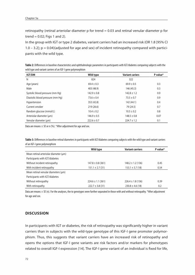
Chapter 5a
72
retinopathy (retinal arteriolar diameter p for trend = 0.03 and retinal venular diameter p for
trend = 0.02; Figs 1 and 2).
In the group with IGT or type 2 diabetes, variant carriers had an increased risk (OR 1.8 [95% CI
1.0 – 3.2]; p = 0.04)(adjusted for age and sex) of incident retinopathy compared with partici-
pants with the wild type.
DISCUSSION
In participants with IGT or diabetes, the risk of retinopathy was significantly higher in variant
carriers than in subjects with the wild-type genotype of this IGF-I gene promoter polymor-
phism. Thus, this suggests that variant carriers have an increased risk of retinopathy and
opens the options that IGF-I gene variants are risk factors and/or markers for phenotypes
related to overall IGF-I expression [14]. The IGF-I gene variant of an individual is fixed for life,
Table 2: Differences in baseline characteristics and ophthalmologic parameters in participants with IGT/diabetes comparing subjects with the wild type and variant carriers of an IGF-I gene polymorphism
IGT/DM Wild type Variant cariers P value*N 824 322
Age (years) 69.4 ± 0.3 69.9 ± 0.5 0.3
Male 403 (48.9) 146 (45.3) 0.3
Systolic blood pressure (mm Hg) 142.9 ± 0.8 142.8 ± 1.2 0.9
Diastolic blood pressure (mm Hg) 73.6 ± 0.4 73.5 ± 0.7 0.9
Hypertension 353 (42.8) 142 (44.1) 0.4
Current smoker 219 (26.6) 79 (24.5) 0.7
Random glucose (mmol/L) 10.4 ± 0.2 10.5 ± 0.2 0.8
Arteriolar diameter (μm) 146.9 ± 0.5 148.5 ± 0.8 0.07
Venular diameter (μm) 222.6 ± 0.7 224.7 ± 1.2 0.1
Data are means ± SE or n (%). *After adjustment for age and sex.
Table 3: Differences in baseline retinal diameters in participants with IGT/diabetes comparing subjects with the wild type and variant carriers of an IGF-I gene polymorphism
Wild type Variant carriers P value*Mean retinal arteriolar diameter (μm)
Participants with IGT/diabetes
Without incident retinopathy 147.8 ± 0.8 (361) 148.2 ± 1.2 (136) 0.45
With incident retinopathy 151.1 ± 2.7 (31) 153.1 ± 3.7 (18) 0.54
Mean retinal venular diameters (μm)
Participants with IGT/diabetes
Without retinopathy 224.6 ± 1.1 (361) 226.4 ± 1.8 (136) 0.39
With retinopathy 222.7 ± 3.8 (31) 230.8 ± 4.6 (18) 0.2
Data are means ± SE (n). For the analyses, the to genotypes were further separated in those with and without retinopathy. *After adjustment for age and sex.

73
IGF-I and diabetic retinopathy
and the relationship between an IGF-I variant and a phenotype is not susceptible to poten-
tial confounding factors such as age, insulin deficiency, hyperglycemia and nutrition state.
However, it is at present unknown whether this IGF-I gene polymorphism directly affects
expression of IGF-I in vivo and mediates its effects through circulating IGF-I levels. No in vivo
studies have been carried out demonstrating that this IGF-I gene promoter polymorphism is
indeed associated with modified IGF-I gene expression. Variations in gene variants are tradi-
tionally difficult to study; subtle differences between gene variants may be present that are
not detected by the presently available in vitro assays [15, 16]. Another unanswered question
at the moment is whether this IGF-I gene promoter polymorphism is related to paracrine/
autocrine IGF-I production in the body. This latter aspect could be very important because it
is not only thought that the IGF-I system has important paracrine/autocrine actions but also
that locally produced IGF-I may produce effects other than circulating IGF-I [17].
Although these differences did not reach statistical significance, variant carriers with IGT or
diabetes who developed retinopathy at follow-up tended to have the largest retinal arteriolar
and venular diameters (Figs 1B and 2B). In contrast, such a trend was absent in participants
with the wild type with IGT or diabetes who developed retinopathy at follow-up (Figs 1A and
2A). Vascular dilatation of the retinal vessels, especially the retinal venules, is observed in the
early stages of diabetic retinopathy [18]. Vascular dilatation is thought to indicate increased
Figure 1 Mean (± SE) baseline retinal arteriolar diameters comparing control subjects, participants with IGT/diabetes without retinopathy (IGT/
DM RP -), and participants with IGF/diabetes with retinopathy (IGT/DM RP +) in subjects with the wildtype (A) and variant carriers (B). *After adjustment for age and sex.

Chapter 5a
74
retinal blood flow and is probably related to hyperglycemic hypoxia and reduced vascular
tone [18]. In addition, larger arteriolar and venular diameters, independent of retinopathy
severity level, have been associated with increased 4-year progression of retinopathy in
type 1 diabetes [19, 20]. Moreover, larger venular diameters were positively associated with
4-year incidence of proliferative retinopathy [19]. Our study observed larger retinal diameters
at baseline in variant carriers with IGT or diabetes, who developed incident retinopathy at
follow-up and the absence of this trend in participants with the wild type may thus already
point out at baseline an increased risk for progression of retinopathy in variant carriers than
in participants with the wild type.
The low observed incidence of retinopathy in our study is probably related to the fact that
we studied only field 2 of the standard diabetic fundus photographs. By studying only this
field, we had no information on ~ 50% of the retina and this will have contributed to an
underestimation of the incidence of retinopathy (R. Klein, personal communication).
In conclusion, in individuals with IGT or diabetes, variant carriers of this IGF-I gene polymor-
phism had an increased risk of retinopathy compared with carriers of the wild-type genotype
during a mean follow-up of 6.5 years. Variant carriers with IGT/diabetes, who developed
retinopathy during follow-up, had a significant trend for larger retinal arteriolar and venular
Figure 2. Mean (± SE) baseline retinal venualr diameters comparing control subjects, participants with IGT/diabetes without retinopathy (IGT/DM RP -), and participants with IGF/diabetes with retinopathy (IGT/DM RP +) in subjects with the wildtype (A) and variant carriers (B). *After adjustment for age and sex.

75
IGF-I and diabetic retinopathy
diameters at baseline, while such trend was absent for subjects with the wild-type genotype
with IGT or diabetes, who developed retinopathy at follow-up. Since arteriolar and venular
diameters have been associated with an increased progression of retinopathy, our findings
suggest that this IGF-I gene polymorphism may modulate the susceptibility and/or the
progression of retinopathy.
ACKNOWLEDGMENTS
I. R. was supported by a grant of the Dutch Diabetes Foundation.
The Rotterdam Study is supported by the Erasmus Medical Center and Erasmus University
Rotterdam; the Netherlands Organization for Scientific Research (NOW; grant 904-61-155);
the Netherlands Organization for Health Research and Development; the Research Institute
for Diseases in the Elderly; the Ministry of Education, Culture and Science; the Ministry of
Health, Welfare and Sports; the European Commission (DG XII); and the Municipality of
Rotterdam. Support also came from the following foundations: Optimix, Amsterdam; NOW,
The Hague; Physico Therapeutic Institue, Rotterdam; Blindenpenning, Amsterdam; Sint
Laurentius Institue, Rotterdam; Bevordering van Volkskracht, Rotterdam; Blindenhulp, The
Hague; Rotterdamse Blindenbelangen Association, Rotterdam; OOG, The Hague; kfHein,
Utrecht; Ooglijders, Rotterdam; Prins Bernhard Cultuurfonds, Amsterdam; Van Leeuwen Van
Lignac, Rotterdam; Verhagen, Rotterdam; Netherlands Society for the Prevention of Blind-
ness, Doorn; LSBS (Landelijke Stichting voor Blinden en Slechtzienden), Utrecht; and Elise
Mathilde, Maarn. Unrestricted grants were obtained from Topcon Europe, Capelle aan de
Ijssel; Lameris Ootech, Nieuwegein; Carl Zeiss Nederland, Sliedrecht (all in the Netherlands);
and from Heidelberg Engineering, Dossenheim, Germany.
The contributions of the general practitioners and pharmacists of the Ommoord district to
the Rotterdam Study are greatly acknowledged. We also thank L Testers, J Vergeer, R. Oskamp,
B de Graaf, F de Rooij and T Rademaker for their help in genotyping and data management.

Chapter 5a
76
REFERENCES
1. Warpeha, K. M. and Chakravarthy, U., Molecular genetics of microvascular disease in diabetic reti-nopathy. Eye, 2003. 17(3): p. 305-11.
2. Vaessen, N., Heutink, P., Janssen, J. A., Witteman, J. C., Testers, L., Hofman, A., Lamberts, S. W., Oos-tra, B. A., Pols, H. A., and van Duijn, C. M., A polymorphism in the gene for IGF-I: functional properties and risk for type 2 diabetes and myocardial infarction. Diabetes, 2001. 50(3): p. 637-42.
3. Hofman, A., Grobbee, D. E., de Jong, P. T., and van den Ouweland, F. A., Determinants of disease and disability in the elderly: the Rotterdam Elderly Study. European Journal of Epidemiology, 1991. 7(4): p. 403-22.
4. Ikram, M. K., de Jong, F. J., Vingerling, J. R., Witteman, J. C., Hofman, A., Breteler, M. M., and de Jong, P. T., Are retinal arteriolar or venular diameters associated with markers for cardiovascular disorders? The Rotterdam Study. Investigative ophthalmology & visual science, 2004. 45(7): p. 2129-34.
5. Hubbard, L. D., Brothers, R. J., King, W. N., Clegg, L. X., Klein, R., Cooper, L. S., Sharrett, A. R., Davis, M. D., and Cai, J., Methods for evaluation of retinal microvascular abnormalities associated with hyper-tension/sclerosis in the Atherosclerosis Risk in Communities Study. Ophthalmology, 1999. 106(12): p. 2269-80.
6. Parr, J. C. and Spears, G. F., Mathematic relationships between the width of a retinal artery and the widths of its branches. American Journal of Ophthalmology, 1974. 77(4): p. 478-83.
7. Parr, J. C. and Spears, G. F., General caliber of the retinal arteries expressed as the equivalent width of the central retinal artery. American Journal of Ophthalmology, 1974. 77(4): p. 472-7.
8. Littmann, H., [Determining the true size of an object on the fundus of the living eye] Zur Bestimmung der wahren Grosse eines Objektes auf dem Hintergrund eines lebenden Auges.
Klinische Monatsblätter für Augenheilkunde, 1988. 192(1): p. 66-7. 9. ETDRS, Grading diabetic retinopathy from stereoscopic color fundus photographs--an extension of
the modified Airlie House classification. ETDRS report number 10. Early Treatment Diabetic Retinopa-thy Study Research Group. Ophthalmology, 1991. 98(5 Suppl): p. 786-806.
10. Wolfs, R. C., Borger, P. H., Ramrattan, R. S., Klaver, C. C., Hulsman, C. A., Hofman, A., Vingerling, J. R., Hitchings, R. A., and de Jong, P. T., Changing views on open-angle glaucoma: definitions and prevalences--The Rotterdam Study. Investigative ophthalmology & visual science, 2000. 41(11): p. 3309-21.
11. 1999 World Health Organization-International Society of Hypertension Guidelines for the Manage-ment of Hypertension. Guidelines Subcommittee. The Journal of Hypertension, 1999. 17(2): p. 151-83.
12. Stolk, R. P., Pols, H. A., Lamberts, S. W., de Jong, P. T., Hofman, A., and Grobbee, D. E., Diabetes mellitus, impaired glucose tolerance, and hyperinsulinemia in an elderly population. The Rotterdam Study. American Journal of Epidemiology, 1997. 145(1): p. 24-32.
13. Rietveld, I., Janssen, J. A., Van Rossum, E. F., Houwing-Duistermaat, J. J., Rivadeneira, F., Hofman, A., Pols, H. A., Van Duijn, C. M., and Lamberts, S. W., A polymorphic CA repeat in the IGF-I gene is associated with gender-specific differences in body height, but has no effect on the secular trend in body height. Clinical endocrinology, 2004. 61(2): p. 195-203.
14. Kostek, M. C., Delmonico, M. J., Reichel, J. B., Roth, S. M., Douglass, L., Ferrell, R. E., and Hurley, B. F., Muscle strength response to strength training is influenced by insulin-like growth factor 1 genotype in older adults. Journal of applied physiology, 2005. 98(6): p. 2147-54.

77
IGF-I and diabetic retinopathy
15. Kenneson, A., Zhang, F., Hagedorn, C. H., and Warren, S. T., Reduced FMRP and increased FMR1 transcription is proportionally associated with CGG repeat number in intermediate-length and pre-mutation carriers. Human molecular genetics, 2001. 10(14): p. 1449-54.
16. Rao, A., Chang, B. L., Hawkins, G., Hu, J. J., Rosser, C. J., Hall, M. C., Meyers, D. A., Xu, J., and Cramer, S. D., Analysis of G/A polymorphism in the androgen response element I of the PSA gene and its interac-tions with the androgen receptor polymorphisms. Urology, 2003. 61(4): p. 864-9.
17. Hambrecht, R., Schulze, P. C., Gielen, S., Linke, A., Mobius-Winkler, S., Erbs, S., Kratzsch, J., Schubert, A., Adams, V., and Schuler, G., Effects of exercise training on insulin-like growth factor-I expression in the skeletal muscle of non-cachectic patients with chronic heart failure. European journal of cardiovascular prevention and rehabilitation, 2005. 12(4): p. 401-6.
18. Ditzel, J., Affinity hypoxia as a pathogenetic factor of microangiopathy with particular reference to diabetic retinopathy. Acta Endocrinologica. Supplementum, 1980. 238: p. 39-55.
19. Klein, R., Klein, B. E., Moss, S. E., Wong, T. Y., Hubbard, L., Cruickshanks, K. J., and Palta, M., The relation of retinal vessel caliber to the incidence and progression of diabetic retinopathy: XIX: the Wisconsin Epidemiologic Study of Diabetic Retinopathy. Archives of ophthalmology, 2004. 122(1): p. 76-83.
20. Wong, T. Y., Shankar, A., Klein, R., and Klein, B. E., Retinal vessel diameters and the incidence of gross proteinuria and renal insufficiency in people with type 1 diabetes. Diabetes, 2004. 53(1): p. 179-84.


5bAn insulin-like Growth Factor-I gene
polymorphism modifies the risk of micro-albuminuria in subjects with abnormal
glucose tolerance

Chapter 5b
80
Microalbuminuria (MA) is related to cardiovascular disease both in diabetic patients and non-
diabetic subjects.
We investigated whether a polymorphism near the promoter region of the IGF-I gene was
related to the development of MA.
For this study, 1069 participants of the Rotterdam study were selected (440 participants with
an abnormal glucose tolerance (AGT); 220 participants with type 2 diabetes and 254 subjects
with pre-diabetes, and 595 subjects with a normal glucose tolerance (NGT).
787 subjects were carriers of the wild type IGF-I genotype (73.6%) and 282 subjects were
variant carriers (26.4%) of this IGF-I gene polymorphism. Compared to subjects with NGT
the risk for microalbuminuria was higher (Odds Ratio (OR): 3.1 (95% CI: 1.2–7.7); P = 0.02) in
variant carriers with AGT than in carriers of the wild type of this IGF-I gene polymorphism (OR:
2.2 (95% CI: 1.2–4.0); P = 0.009). Compared with wild type carriers with AGT, the relative risk
for MA was unadjusted and non-significantly increased in variant carriers with AGT (1.6; 95%
CI: 0.8 –2.9). However, after adjustment for possible confounding factors (age, gender, mean
blood pressure, fasting insulin, fasting glucose and smoking) this risk became significant (OR:
RR 2.1; 95% CI: 1.1 – 4.4; P = 0.04).
In subjects with AGT, a higher risk for MA was observed in variant carriers than in carriers of
the wild type genotype of this IGF-I gene polymorphism. Since MA is primarily associated
with cardiovascular disease in subjects with AGT, our study suggests that variant carriers
have a higher risk for cardiovascular disease than carriers of the wild type when they develop
an AGT.

81
IGF-I and micro-albuminuria
INTRODUCTION
The presence of microalbuminuria (MA) may be a marker of generalized vascular endothelial
dysfunction reflecting generalized vascular disease [1, 2]. Development of MA is associated
with glycaemic control, blood pressure, smoking, and male gender [3, 4]. MA may precede
type 2 diabetes, occurring in parallel with the metabolic syndrome and its components,
obesity and hypertension [1]. MA is also considered to be a marker for diabetic nephropathy
in type 2 diabetes [5-7].
The insulin-like growth factor-I (IGF-I) system exerts multiple physiologic effects on the vas-
culature through both endocrine and autocrine/paracrine mechanisms [8]. Both macrovessel
and microvessel endothelial cells express IGF-I receptor (IGF-IR; [9]). The expression of IGF-I in
endothelial cells is low and might reflect mainly IGF-I sequestered from serum [10]. IGF-I has
also been implicated in the development of cardiovascular disease [8, 11-13].
IGF-I has been further implicated in the development of diabetic nephropathy [14, 15]. IGF-I
bioactivity is regulated by genetic and non-genetic factors like growth hormone, nutrition
and insulin [16]. In humans, a cytosine–adenine (CA)n polymorphism in the promoter region
of the IGF-I gene has been identified [17]. Studies of other genes have suggested that poly-
morphic CA repeats in the promoter region of a gene, affects transcription activity of a gene
[18]. This polymorphism has thus the potential to influence directly the expression of IGF-I in
the body [19].
Circulating IGF-I levels are often used as a substitute for tissue IGF-I bioactivity due to the lack
of in vivo methods to measure the latter. However, determinations of circulating total IGF-I
levels by radio-immunoassays may be especially problematic in pathological conditions,
due to interferences of IGF-binding proteins. As a consequence, circulating IGF-I levels
may not adequately reflect the IGF-I bioactivity. An alternative approach may be genetic
association studies. In these studies, genetic variants can be treated as risk factors that
cannot be influenced by secondary factors such as hypertension or hyperglycemia, unless
these phenotypes are in the causal pathway. This opens the opportunity to characterize
on a genetic basis, individuals who are at risk for MA. The aim of the present study was to
investigate whether a polymorphism near the promoter region of the IGF-I gene was related
to the development of MA.
SUBJECTS AND METHODS
Study population All subjects included in the present study were participants of the Rotterdam Study. The Rot-
terdam Study is a single-center prospective follow-up study in which all residents aged 55
years and over of the Rotterdam suburb Ommoord were invited to participate. The Medical

Chapter 5b
82
Ethics Committee of Erasmus Medical Center Rotterdam approved the study and written
informed consent was obtained from all participants. The aim of the Rotterdam study and the
design of the study has been described previously [20]. The baseline examination of the Rot-
terdam Study was conducted between 1990 and 1993. A total of 7983 participants (response
rate 78 percent) were examined.
Because of practical and financial reasons, only a proportion of the Rotterdam Study (n =
1069) underwent a fasting glucose tolerance test. From this group we selected our cases
and controls for a case-control study in which we assessed the relation between both IGF-I
genotypes, glucose tolerance and albuminuria [21]. Criteria for the present study were age
younger than 75 years, no use of anti-epileptics, corticosteroids, hormonal replacement
therapy, cytostatics and people with dementia. After blood was drawn from the participants
after an overnight fast, participants underwent an oral glucose tolerance test with 75 g
glucose. Diabetic patients who were treated with anti-diabetic medication did not undergo
a glucose tolerance test. Therefore, post load glucose measurements were only available for
persons not using anti-diabetic medication. For the oral glucose tolerance test blood was
drawn after 2 hours. Normal glucose tolerance (NGT) was defined as fasting glucose below
6.1 mmol/l and 2 hour post load below 7.8 mmol/l. Individuals with high glucose levels not
meeting criteria for diabetes, but were too high to be considered normal were diagnosed as
having prediabetes. Prediabetes includes persons with impaired fasting glucose (IFG) and/
or impaired glucose tolerance (IGT). IFG was defined as fasting glucose between 6.1 and 7.0
mmol/l and IGT as a 2 hour post load glucose between 7.8 and 11.1 mmol/l. A diagnosis of
diabetes mellitus was made if subjects were treated for diabetes or had a fasting glucose
level of 7.0 mmol/l or above and/or a 2 hour post load glucose of 11.1 mmol/l or above [22].
All individuals meeting criteria for diabetes or prediabetes were to be considered as subjects
with an abnormal glucose tolerance (AGT). To determine insulin sensitivity we used the
homeostasis model assessment (HOMA) [23].
MeasurementsA questionnaire was used to collect data on glucose status, smoking status and use of
anti-hypertensive medication. Blood pressure was measured in a sitting position at the right
upper arm with a random-zero sphygmomanometer and the average obtained of two mea-
surements at one occasion was used.
Blood sampling and storage have been described elsewhere [24]. Serum was separated by
centrifugation and quickly frozen in liquid nitrogen. For the present study, we used blood
measurements performed on fasting blood samples. Glucose levels were measured by the
glucose hexokinase method (Medgenix Diagnostics, Brussels, Belgium) with an intra-assay
variation of less than 2.5 and 6.0% respectively. Fasting insulin was measured by a commer-
cially available assay (IRMA, Medgenix Diagnostics) with an intra-and inter-assay variation
of 3 – 6 and 5 – 12 percent, respectively. Serum creatinine levels were assessed using an

83
IGF-I and micro-albuminuria
automated enzymatic procedure (Roche, Mannheim, Germany). Creatinine clearance was
determined by the formula of Cockcroft and Gault [25]. Serum cholesterol and triglycerides
were determined by a standard laboratory method.
Participants collected timed overnight urine one day before the examination, albumin and
creatinine were measured. Albumin and creatinine in urine were determined by a turbidimetric
method and measured by a Hitachi 917 analyzer (Roche/Hitachi Diagnostics, Mannheim,
Germany). Detection limit ranged from 2 to 400 mg/l and therefore all measurements
below 2 mg/l were set at 1 mg/l. MA was defined as an albumin-to-creatinine ratio (ACR) of
.2.5 mg/mmol in males and 3.5 mg/mmol in females [26]. From 34 subjects, no data of MA
were available. Fourteen subjects (NGT: four subjects, AGT: ten subjects) met the criteria for
macroalbuminuria, they were included in the analysis.
Genotyping of the IGF-I promoter polymorphism The human IGF-I gene contains a polymorphic CA repeat 1 Kb upstream of the promoter re-
gion [27]. IGF-I genotypes were determined as described previously [17]. The most common
allele contains 19 CA-repeats, which equals a length of 192-bp [28]. In a previous study, we
examined the role of the various lengths of the alleles of the IGF-I promoter polymorphism
and observed an optimum for IGF-I levels and body height [29]. Mean IGF-I levels and body
height for homozygous carriers of the 192-bp allele, and homozygous carriers of the 194-bp-
allele of this IGF-I gene promoter polymorphism were equal and significantly higher than
the values measured in subjects homozygous for alleles smaller than 192-bp and longer
than 194-bp, respectively, suggesting an optimum for IGF-I expression by these two vari-
ants. Based on carrier ship of the 192-bp and 194-bp alleles, we therefore distinguished two
different IGF-I genotypes in the present study: As wild type genotype we considered carriers
homozygous for the 192-bp or for the 194-bp allele, and carriers heterozygote for these two
alleles, participants with this genotype are further in the text denoted as carriers of the wild
type. Participants with all other combinations of alleles were considered carriers of the vari-
ant genotype, which is further in the text denoted as variant carriers. 787 persons (73.6%)
were carriers of the wild type and 282 subjects were variant carriers (26.4%).
Carotid ultrasonography Carotid atherosclerosis was assessed by duplex scan ultrasonography of the carotid arteries,
using a 7.5 MHz linear array transducer (ATL, Ultramark IV). Measurements of intima media
thickness (IMT) were performed offline from the still images recorded on videotape. Details
about this measurement have been published previously [30]. Briefly, the interfaces of the far
and near walls of the distal common carotid artery are marked over a length of 10 mm. We
used the average of the measurements of three still images of both the left and right arteries.
Common carotid IMT was determined as the mean IMT of near and far wall measurements
of both the left and right arteries. Results from a reproducibility study of IMT measurements

Chapter 5b
84
have been published elsewhere [31]. The mean differences±S.D. in common carotid IMT be-
tween paired measurements of sonographers, readers, and visits were -004±0.10, 0.066±0.07,
and -0.013±0.13 mm, respectively.
Data analysesExcept when otherwise mentioned, data are presented as mean±S.D. Diabetic and non-
diabetic groups were compared with the Mann – Whitney test. Means of parameters were
compared between genotypes after stratification for glucose tolerance using analyses of vari-
ance. Insulin, albuminuria and ACR did not met the criteria for normality and were logarithmic
transformed before analysis in order to obtain approximate normal distribution. Therefore
results of these parameters are presented as geometric means with 95% confidence intervals
(CI). Distribution of sex, use of antihypertensive drugs, lipid lowering drugs and frequency of
the metabolic syndrome between genotypes was compared using a chi-square test.
Risk for MA was calculated by a binary logistic regression analysis and all results are presented
unadjusted, and when explicitly mentioned, after adjustment for age, sex, mean blood pres-
sure, fasting glucose and insulin levels and smoking. All analyses were performed using the
SPSS for Windows software package, version 10.0.5 (SPSS Inc., Chicago, IL, USA).
Table 1: General characteristics of the study population, comparing subjects with normal glucose tolerance (NGT) and subjects with abnormal glucose tolerance (AGT) after stratification by IGF-I genotype
NGT AGTWild type Variant type P2 Wild type Variant type P2 P1
Number of subjects 444 151 343 131
Age 66.2 ± 5.6 66.0 ± 4.8 0.67 67.7 ± 5.8 66.8 ± 5.8 <0.001 <0.001
Sex (M/F) 202/242 71/80 0.75 184/159 70/61 0.01 0.01
Creatinine (μmol//L) 89.3 ± 15.0 89.4 ± 13.9 0.93 92.2 ± 18.0 91.0 ± 17.9 0.61 0.27
Fasting glucose (mmol/L) 5.5 ± 0.4 5.4 ± 0.3 0.85 7.2 ± 2.0 7.3 ± 2.2 0.74 <0.001
2-h glucose post-load (mmol/L)
5.3 ± 1.2 5.3 ± 1.2 0.76 9.4 ± 3.9 9.2 ± 4.0 0.67 <0.001
Mean systolic bp (mmHg) 136 ± 21 135 ± 19 0.56 143 ± 20 142 ± 22 0.87 <0.001
Mean diastolic bp (mmHg) 77 ± 77 75 ± 11 0.52 78 ± 11 78 ± 12 0.67 0.002
Total cholesterol (mmol/L) 6.63 ± 1.06 6.54 ± 0.98 0.37 6.53 ± 1.13 6.46 ± 1.09 0.47 0.35
HDL-cholesterol (mmol/L) 1.33 ± 0.32 1.33 ± 0.32 0.88 1.22 ± 0.30 1.20 ± 0.30 0.48 <0.001
Triglycerides (mmol/L) 1.50 ± 0.66 1.44 ± 0.50 0.31 1.95 ± 1.10 1.96 ± 1.00 0.96 <0.001
BMI (kg/m2) 25.8 ± 3.1 26.0 ± 3.3 0.69 27.5 ± 3.6 27.4 ± 3.7 0.73 <0.001
Current smoking (%) 10.0 14.1 0.21 14.6 8.9 0.39 0.53
Antihypertensive drugs (%) 22.7 18.5 0.30 34.1 38.9 0.18 <0.001
Lipid lowering drugs (%) 5.0 2.6 0.24 7.3 6.1 0.67 0.08
IMT (mm) 0.75 ± 0.13 0.75 ± 0.12 0.69 0.78 ± 0.15 0.79 ± 0.11 0.51 <0.01
HOMA-IR 1.92 ± 0.06 2.05 ± 0.10 0.36 1.78 ± 0.07 1.78 ± 0.11 0.98 0.04
P1, p-value comparing subjects with NGT and subjects with AGT after adjustment for age and sex; P2, p-value comparing wild type with variant type adjusted for age and sex; HOMA-IR, homeostasis model assessment of insulin resistance.

85
IGF-I and micro-albuminuria
RESULTS
Table 1 presents clinical characteristics of 1069 subjects (595 subjects with NGT and 474 sub-
jects with an AGT (254 subjects with pre-diabetes and 220 subjects with diabetes) stratified
by IGF-genotype. Subjects with AGT were older and most of them were male in comparison
to subjects with NGT (Table 1). Serum creatinine, blood glucose and cholesterol levels, blood
pressure and body mass index (BMI) were higher in subjects with AGT than in subjects with
NGT (Table 1). Subjects with AGT frequently used more antihypertensive drugs, had larger
intima media thickness and were more insulin resistant than subjects with NGT. There were
no differences in general characteristics between carriers of the wild type and variant carriers
of this IGF-I gene promoter polymorphism in the group of NGT or AGT (Table 1).
Table 2 shows the relation between the two IGF-I genotypes and parameters of renal func-
tion, comparing subjects with AGT to subjects with NGT. Subjects with AGT had significantly
higher urinary albumin concentrations and ACRs than subjects with NGT. In addition, mean
serum creatinine levels, creatinuria and clearance were higher in subjects with AGT than in
subjects with NGT. In variant carriers with AGT, mean albuminuria and ACR were higher than
in carriers of the wild type, but these differences did not reach statistical significance.
Subjects with an AGT had an increased risk of developing MA (Odds Ratio (OR): 2.5; 95% CI:
1.5 – 4.0; P = 0.001) in comparison to subjects with NGT. Compared to subjects with NGT, the
risk for MA was higher (OR: 3.1; 95% CI: 1.2 – 7.7; P = 0.02) in variant carriers with AGT, than in
carriers of the wild type of this IGF-I gene polymorphism (OR: 2.2; 95% CI: 1.2–4.0; P = 0.009;
Fig. 1).
Table 3 shows the prevalence and relative risk of MA in subjects with AGT and subjects with
NGT stratified for IGF-I genotype. Variant carriers with AGT had a higher prevalence of MA
than carriers of the wild type of this IGF-I gene polymorphism, subsequently the risk of MA
for variant carriers was investigated. In subjects with AGT, the relative risk for MA was non-sig-
Table 2: Relation between IGF-I genotype and parameters of renal function comparing subjects with NGT and subjects with AGT. Data are mean (95% CI) unless otherwise stated.
NGT AGTWild type Variant type P2 Wild type Variant type P2 P1
Number of subjects 441 151 343 131
Albuminuria (mg/l)* 2.1 (1.9 – 2.4) 2.1 (1.8 – 2.5) 0.97 2.9 (2.5 – 3.3) 3.6 (2.9 – 4.7) 0.09 <0.001
Creatinuria (mmol/l) 7.7 (7.4 – 8.1) 7.5 (6.8 – 8.1) 0.45 8.1 (7.7 – 8.6) 8.2 (7.5 – 8.9) 0.86 0.20
Albumin-to-creatinine ratio (mg/mmol)*
0.32 (0.29 – 0.37) 0.34 (0.28 – 0.40) 0.70 0.41 (0.35 – 0.48) 0.52 (0.40 – 0.66) 0.12 0.002
Creatinine (μmol/l) 89 (88 – 91) 89 (87 – 91) 0.93 92 (90 - 94) 91 (89 -94 ) 0.61 0.27
Clearance (ml/min) 68 (66 – 69( 67 (65 – 69) 0.71 69 (68 – 71) 70 (67 – 72) 0.75 <0.001
* Geometric mean (95% CI); P1, p-value comparing subjects with NGT and subjects with AGT after adjustment for age and sex; P2, p-value comparing wild type with variant carriers adjusted for age and sex.

Chapter 5b
86
nificantly increased in variant carriers when compared with carriers of the wild type: (Relative
Risks (RR)): 1.5; 95% CI: 0.8 – 2.9; P = 0.19). When this analysis was repeated after adjustment
for age, gender, fasting insulin, fasting glucose, mean blood pressure and smoking, the risk
of MA in variant carriers with AGT became significant: (RR: 2.1; 95% CI: 1.1–4.4; P = 0.04).
Compared with carriers of the wild type genotype, the relative risk for MA also increased in
variant carriers with NGT after these adjustments, but it still remained not significant (RR: 1.6;
95% CI: 0.6 – 4.0; P = 0.35).
Table 3: The relative risk of microalbuminuria of subjects with AGT and subjects with NGT stratified by IGF-I genotype. Number of subjects for each category is given (% of cases and controls). Risks are given with a 95% confidence interval between brackets.
Wild type Variant typeSubjects with AGT
Microalbuminuria 31 (9.3%) 17 (13.5%)
No microalbuminuria 303 (90.7%) 109 (86.5%)
Relative risk Reference 1.5 (0.8 – 2.9)*
Relative risk Reference 2.1 (1.0 – 4.4)**
Subjects with NGT
Microalbuminuria 19 (4.4%) 7 (4.8%)
No microalbuminuria 410 (95.6%) 139 (95.2%)
Relative risk Reference 1.1 (0.45 – 2.64)*
Relative risk Reference 1.6 (0.61 – 4.03)**
* Unadjusted; ** After adjustment for age, gender, mean blood pressure, fasting glucose, fasting insulin and smoking.
Figure 1. The relative risk of microalbuminuria comparing subjects with NGT and AGT per IGF-I genotype (wild type, left; variant carriers, right). Individuals with NGT were used as the reference group. *p=0.009, **p=0.02 vs the reference group.

87
IGF-I and micro-albuminuria
DISCUSSION
Compared to subjects with NGT, variant carriers with AGT had a higher risk to develop MA
than carriers of the wild type genotype. In addition, variant carriers with AGT had a borderline
and significantly higher prevalence of MA than carriers of the wild type genotype. These
findings became significant after adjustment for a number of possible confounding factors,
which have been previously found to be involved in the development of MA. Our findings
suggest that this IGF-I gene polymorphism modulates the susceptibility and/or progression
of MA as soon as a person develops AGT.
Compared with carriers of the wild type genotype, we observed that the increased risk
for MA in variant carriers was relatively small. At first glance, this suggests that the role of
this IGF-I gene polymorphism in the development of MA is not very important. However,
the susceptibility to MA results probably from an interaction of multiple genetic and
environmental factors. Consequently, the relationship between the prevalence of MA
(phenotype) and any individual causal locus (genetic variant) will in general be fairly weak,
even for major genes. MA will only develop when certain other risk factors are also present,
such as hyperglycemia, hypertension and/or smoking. This may have obvious complications
for both the number of subjects in the study and the power of analyses that aim to detect
individual genetic signals among other genetic and environmental background. This may
explain why we observed only a relative small and significant increase in the risk of MA in
variant carriers with AGT, compared with carriers of the wild type genotype of this IGF-I gene
polymorphism after adjustment for some possible confounding factors.
The number of subjects with MA was low in our study which might have further attributed to
a reduced statistical power of our study. Although the prevalence of MA in our study was low,
this prevalence does not differ from previous findings in another big Dutch population-based
study [32]. In this latter study the prevalence of MA was 6.6% in non-diabetic subjects and
16.1% among diabetic patients. However, in this latter study, the diagnosis of MA was exclu-
sively based on urinary albumin concentrations, while in our study this diagnosis was based
on ACRs. A limitation of our study is that MA was assessed on one overnight sample. It is well
known that there is considerable intra-individual variability in urinary albumin excretion in
time [33]. As a consequence, the prevalence of MA in our study may have been significantly
both underestimated as well as overestimated.
Another limitation of our study may be that there is not only an age-related penetrance of
AGT, but also an age-related penetrance of MA. Subjects with a NGT at the time of the study
may still develop both an altered glucose tolerance and MA in the near future. We previously
observed that variant carriers of this IGF-I gene polymorphism have an increased risk of
developing diabetes, in comparison to carriers of the wild type genotype. Thus, this suggests
that especially in variant carriers with AGT, the risk for MA may have been underestimated.
Moreover, the diagnostic umbrella MA probably gathers individuals who have developed MA

Chapter 5b
88
through a variety of pathological mechanisms (see below). This has obvious implications for
the interpretation of our findings.
We did not find relationships between this IGF-I gene promoter polymorphism and renal
function parameters, this was not unexpected. In subjects with type 2 diabetes MA is gener-
ally considered a stronger predictor of cardiovascular disease than it is of the risk of end-stage
renal failure [34]. Type 2 diabetes patients with MA are at an increased risk of cardiovascular
death compared with patients with normal albuminuria [35, 36]. In addition, in non-diabetic
subjects, MA is even considered an independent risk factor to developing cardiovascular
disease [3, 32]. The observed relationship between IGF-I genotype and MA in our study thus
probably points mainly to an increased risk of variant carriers for cardiovascular disease. This
is in accordance with our previous findings: we observed that variant carriers had a higher
risk of developing myocardial infarction [17]. In addition, carotid IMT and aortic pulse wave
velocity were significantly increased in variant carriers with hypertension [37].
MA may be the first sign that the vascular vessel wall, particularly the endothelium, is injured.
It has been found that MA can be the specific consequence of a reduction in the fixed nega-
tive charges of the glomerular wall [38, 39]. Whether an IGF-I gene genotype of a subject may
modulate the susceptibility and/or progression of MA by circulating IGF-I or rather via its
effects at the tissue level is at present unknown. However, it is likely that a link should be
found in alterations in the composition of the basal membranes of the capillaries and of the
extracellular matrices, which has also has been hypothesized for type 1 diabetes.
When our findings will have been replicated by others, these observations may suggest the
existence of new etiological pathways for the development of MA, and together, provide
some prediction of who will or will not get MA. In addition, it has been suggested that even
if the risk is imparted by a certain genotype is low, or that only a subset of subjects carry the
predisposing genotype, modulation of that gene by pharmacological intervention may be
beneficial [40].
In conclusion, we observed in patients with AGT a higher risk for MA in variant carriers than
in carriers of the wild type genotype of this IGF-I gene promoter polymorphism. Since MA is
primarily associated with cardiovascular disease in subjects with type 2 diabetes, our study
suggests that variant carriers with an AGT have an increased risk for cardiovascular disease.
FUNDING
I Rietveld is supported by a grant of the Dutch Diabetes Foundation. The Rotterdam Study
is supported by the Erasmus Medical Center and Erasmus University Rotterdam, the Nether-
lands Organization for Scientific Research (NWO), the Netherlands Organization for Health
Research and Development (ZonMw), the Research Institute for Diseases in the Elderly (RIDE),
the Ministry of Education, Culture and Science, the Ministry of Health, Welfare and Sports, the

89
IGF-I and micro-albuminuria
European Commission (DG XII), and the Municipality of Rotterdam. The contributions of the
general practitioners and pharmacists of the Ommoord district to the Rotterdam Study are
greatly acknowledged.
ACKNOWLEDGEMENTS
We would like to thank L Testers and J Vergeer, R. Oskamp, B de Graaf, F de Rooij and T
Rademaker for their help in genotyping and data management. Also I Van Gorp and C van
Leeuwen are greatly acknowledged for the measurements of albuminuria and creatinuria.

Chapter 5b
90
REFERENCES
1. Lane, J. T., Microalbuminuria as a marker of cardiovascular and renal risk in type 2 diabetes mellitus: a temporal perspective. American journal of physiology. Renal physiology, 2004. 286(3): p. F442-50.
2. Naidoo, D. P., The link between microalbuminuria, endothelial dysfunction and cardiovascular dis-ease in diabetes. Cardiovascular journal of South Africa, 2002. 13(4): p. 194-9.
3. Cirillo, M., Senigalliesi, L., Laurenzi, M., Alfieri, R., Stamler, J., Stamler, R., Panarelli, W., and De Santo, N. G., Microalbuminuria in nondiabetic adults: relation of blood pressure, body mass index, plasma cholesterol levels, and smoking: The Gubbio Population Study. Archives of Internal Medicine, 1998. 158(17): p. 1933-9.
4. Young, B. A., Katon, W. J., Von Korff, M., Simon, G. E., Lin, E. H., Ciechanowski, P. S., Bush, T., Oliver, M., Ludman, E. J., and Boyko, E. J., Racial and ethnic differences in microalbuminuria prevalence in a diabetes population: the pathways study. Journal of the American Society of Nephrology, 2005. 16(1): p. 219-28.
5. Adler, A. I., Stevens, R. J., Manley, S. E., Bilous, R. W., Cull, C. A., and Holman, R. R., Development and progression of nephropathy in type 2 diabetes: The United Kingdom Prospective Diabetes Study (UKPDS 64). Kidney International, 2003. 63(1): p. 225-232.
6. Mogensen, C. E., Microalbuminuria predicts clinical proteinuria and early mortality in maturity-onset diabetes. The New England Journal of Medicine, 1984. 310(6): p. 356-60.
7. Nelson, R. G., Bennett, P. H., Beck, G. J., Tan, M., Knowler, W. C., Mitch, W. E., Hirschman, G. H., and Myers, B. D., Development and progression of renal disease in Pima Indians with non- insulin-dependent diabetes mellitus. Diabetic Renal Disease Study Group. The New England Journal of Medicine, 1996. 335(22): p. 1636-42.
8. Delafontaine, P., Song, Y. H., and Li, Y., Expression, regulation, and function of IGF-1, IGF-1R, and IGF-1 binding proteins in blood vessels. Arteriosclerosis, thrombosis, and vascular biology, 2004. 24(3): p. 435-44.
9. Bar, R. S., Boes, M., Dake, B. L., Booth, B. A., Henley, S. A., and Sandra, A., Insulin, insulin-like growth factors, and vascular endothelium. The American journal of medicine, 1988. 85(5A): p. 59-70.
10. Gajdusek, C. M., Luo, Z., and Mayberg, M. R., Sequestration and secretion of insulin-like growth factor-I by bovine aortic endothelial cells. Journal of Cellular Physiology, 1993. 154(1): p. 192-8.
11. Delafontaine, P., Insulin-like growth factor I and its binding proteins in the cardiovascular system. Cardiovascular Research, 1995. 30(6): p. 825-34.
12. Juul, A., Scheike, T., Davidsen, M., Gyllenborg, J., and Jorgensen, T., Low serum insulin-like growth factor I is associated with increased risk of ischemic heart disease: a population-based case-control study. Circulation, 2002. 106(8): p. 939-44.
13. Spallarossa, P., Brunelli, C., Minuto, F., Caruso, D., Battistini, M., Caponnetto, S., and Cordera, R., Insulin-like growth factor-I and angiographically documented coronary artery disease. The American journal of cardiology, 1996. 77(2): p. 200-2.
14. Flyvbjerg, A., Role of growth hormone, insulin-like growth factors (IGFs) and IGF-binding proteins in the renal complications of diabetes. Kidney International. Supplement, 1997. 60: p. S12-9.
15. LeRoith, D., Werner, H., Phillip, M., and Roberts, C. T., Jr., The role of insulin-like growth factors in diabetic kidney disease. American journal of kidney diseases, 1993. 22(5): p. 722-6.
16. Froesch, E. R., Hussain, M. A., Schmid, C., and Zapf, J., Insulin-like growth factor I: physiology, meta-bolic effects and clinical uses. Diabetes/metabolism reviews, 1996. 12(3): p. 195-215.

91
IGF-I and micro-albuminuria
17. Vaessen, N., Heutink, P., Janssen, J. A., Witteman, J. C., Testers, L., Hofman, A., Lamberts, S. W., Oos-tra, B. A., Pols, H. A., and van Duijn, C. M., A polymorphism in the gene for IGF-I: functional properties and risk for type 2 diabetes and myocardial infarction. Diabetes, 2001. 50(3): p. 637-42.
18. Tae, H. J., Luo, X., and Kim, K. H., Roles of CCAAT/enhancer-binding protein and its binding site on repression and derepression of acetyl-CoA carboxylase gene. The Journal of biological chemistry, 1994. 269(14): p. 10475-84.
19. Yu, H., Li, B. D., Smith, M., Shi, R., Berkel, H. J., and Kato, I., Polymorphic CA repeats in the IGF-I gene and breast cancer. Breast cancer research and treatment, 2001. 70(2): p. 117-122.
20. Hofman, A., Grobbee, D. E., de Jong, P. T., and van den Ouweland, F. A., Determinants of disease and disability in the elderly: the Rotterdam Elderly Study. European Journal of Epidemiology, 1991. 7(4): p. 403-22.
21. Baan, C. A., Stolk, R. P., Grobbee, D. E., Witteman, J. C., and Feskens, E. J., Physical activity in elderly subjects with impaired glucose tolerance and newly diagnosed diabetes mellitus. American Journal of Epidemiology, 1999. 149(3): p. 219-27.
22. Report of the expert committee on the diagnosis and classification of diabetes mellitus. Diabetes Care, 2003. 26 (Suppl 1): p. S5-20.
23. Hermans, M. P., Levy, J. C., Morris, R. J., and Turner, R. C., Comparison of insulin sensitivity tests across a range of glucose tolerance from normal to diabetes. Diabetologia, 1999. 42(6): p. 678-87.
24. Stolk, R. P., Pols, H. A., Lamberts, S. W., de Jong, P. T., Hofman, A., and Grobbee, D. E., Diabetes mellitus, impaired glucose tolerance, and hyperinsulinemia in an elderly population. The Rotterdam Study. American Journal of Epidemiology, 1997. 145(1): p. 24-32.
25. Cockcroft, D. W. and Gault, M. H., Prediction of creatinine clearance from serum creatinine. Nephron, 1976. 16(1): p. 31-41.
26. A strategy for arterial risk assessment and management in type 2 (non-insulin-dependent) diabetes mellitus. European Arterial Risk Policy Group on behalf of the International Diabetes Federation European Region. Diabetic Medicine, 1997. 14(7): p. 611-21.
27. Weber, J. L. and May, P. E., Abundant class of human DNA polymorphisms which can be typed using the polymerase chain reaction. American journal of human genetics, 1989. 44(3): p. 388-96.
28. Rietveld, I., Janssen, J. A., Hofman, A., Pols, H. A., van Duijn, C. M., and Lamberts, S. W., A polymor-phism in the IGF-I gene influences the age-related decline in circulating total IGF-I levels. European journal of endocrinology, 2003. 148(2): p. 171-5.
29. Rietveld, I., Janssen, J. A., Van Rossum, E. F., Houwing-Duistermaat, J. J., Rivadeneira, F., Hofman, A., Pols, H. A., Van Duijn, C. M., and Lamberts, S. W., A polymorphic CA repeat in the IGF-I gene is associated with gender-specific differences in body height, but has no effect on the secular trend in body height. Clinical endocrinology, 2004. 61(2): p. 195-203.
30. Bots, M. L., Hofman, A., De Jong, P. T., and Grobbee, D. E., Common carotid intima-media thickness as an indicator of atherosclerosis at other sites of the carotid artery. The Rotterdam Study. Annals of Epidemiology, 1996. 6(2): p. 147-53.
31. Bots, M. L., Mulder, P. G., Hofman, A., van Es, G. A., and Grobbee, D. E., Reproducibility of carotid vessel wall thickness measurements. The Rotterdam Study. Journal of Clinical Epidemiology, 1994. 47(8): p. 921-30.
32. Hillege, H. L., Fidler, V., Diercks, G. F., van Gilst, W. H., de Zeeuw, D., van Veldhuisen, D. J., Gans, R. O., Janssen, W. M., Grobbee, D. E., and de Jong, P. E., Urinary albumin excretion predicts cardiovascular and noncardiovascular mortality in general population. Circulation, 2002. 106(14): p. 1777-82.
33. Mogensen, C. E., Vestbo, E., Poulsen, P. L., Christiansen, C., Damsgaard, E. M., Eiskjaer, H., Froland, A., Hansen, K. W., Nielsen, S., and Pedersen, M. M., Microalbuminuria and potential confounders.

Chapter 5b
92
A review and some observations on variability of urinary albumin excretion. Diabetes Care, 1995. 18(4): p. 572-81.
34. Mogensen, C. E., Viberti, G., Halimi, S., Ritz, E., Ruilope, L., Jermendy, G., Widimsky, J., Sareli, P., Taton, J., Rull, J., Erdogan, G., De Leeuw, P. W., Ribeiro, A., Sanchez, R., Mechmeche, R., Nolan, J., Sirotiakova, J., Hamani, A., Scheen, A., Hess, B., Luger, A., and Thomas, S. M., Effect of low-dose perindopril/indapamide on albuminuria in diabetes: preterax in albuminuria regression: PREMIER. Hypertension, 2003. 41(5): p. 1063-71.
35. MacLeod, J. M., Lutale, J., and Marshall, S. M., Albumin excretion and vascular deaths in NIDDM. Diabetologia, 1995. 38(5): p. 610-6.
36. Mattock, M. B., Morrish, N. J., Viberti, G., Keen, H., Fitzgerald, A. P., and Jackson, G., Prospective study of microalbuminuria as predictor of mortality in NIDDM. Diabetes, 1992. 41(6): p. 736-41.
37. Schut, A. F., Janssen, J. A., Deinum, J., Vergeer, J. M., Hofman, A., Lamberts, S. W., Oostra, B. A., Pols, H. A., Witteman, J. C., and van Duijn, C. M., Polymorphism in the promoter region of the insulin-like growth factor I gene is related to carotid intima-media thickness and aortic pulse wave velocity in subjects with hypertension. Stroke, 2003. 34(7): p. 1623-7.
38. Kanwar, Y. S., Rosenzweig, L. J., Linker, A., and Jakubowski, M. L., Decreased de novo synthesis of glomerular proteoglycans in diabetes: biochemical and autoradiographic evidence. Proceedings of the National Academy of Sciences of the United States of America, 1983. 80(8): p. 2272-7.
39. Raats, C. J., Van Den Born, J., and Berden, J. H., Glomerular heparan sulfate alterations: mechanisms and relevance for proteinuria. Kidney International, 2000. 57(2): p. 385-400.
40. Barroso, I., Genetics of Type 2 diabetes. Diabetic Medicine, 2005. 22(5): p. 517-35.

6An insulin-like Growth Factor-I promoter
polymorphism is associated with increased mortality in subjects with myocardial
infarction in an elderly Caucasian population


95
IGF-I and myocardial infarction
We investigated whether an insulin-like growth factor I (IGF-I) promoter polymorphism is
associated with excess mortality in elderly subjects with myocardial infarction (MI). This as-
sociation was assessed in 7983 subjects of the Rotterdam Study during 14 years of follow-up.
Among 345 subjects who developed a MI, the risk of mortality was 1.49 times higher in the
variant carriers of the IGF-I promoter polymorphism than in the nonvariant carriers (95%
confidence interval 1.10 to 2.10, p = 0.02). The risk of death increased with the number of vari-
ant alleles. Our study suggests that genetically determined low IGF-I activity is an important
determinant of mortality in subjects with MI.
Insulin-like growth factor I (IGF-I) plays an important role in regulating myocardial structure
and function and promoting cardiac muscle growth, differentiation, and survival during
myocardial ischemia [1, 2]. In addition to its anabolic properties, it has been suggested
that IGF-I plays an important role in the development of cardiovascular diseases (CVDs) [1].
Subjects with low circulating IGF-I levels are at significantly higher risk of developing CVD [2].
Substantial genetic contributions to human blood IGF-I levels were first reported in 1996 [3].
A polymorphism in the IGF-I promoter region that may influence IGF-I production has been
identified [4]. This polymorphism is also associated with circulating IGF-I levels [4]. We previ-
ously observed an increased risk of myocardial infarction (MI) in the absence of the 192-bp
allele of the IGF-I promoter polymorphism [4]. We investigated whether this polymorphism
is involved in the survival of subjects with incident MI and in the total population. The study
was embedded in the Rotterdam Study, a population-based cohort study of 7983 subjects
who were ≥55 years of age. Baseline data were collected between March 1990 and July 1993.
The study was approved by the medical ethics committee of Erasmus University (Rotterdam,
The Netherlands). Written informed consent was obtained from all participants. Data of the
incident MI were verified by research physicians who collected information from patients’
medical records. Information also included copies of discharge letters for hospital admis-
sions. A MI was coded according to criteria of the International Classification of Diseases, 10th
Edition. Follow-up data on overall mortality were available until January 1, 2004. CVD-specific
mortality was based on data until January 1, 2000. In total, 7012 subjects were successfully
genotyped for the IGF-I promoter polymorphism. We previously observed that circulating
serum IGF-I levels are highest for subjects with 192- and 194-bp alleles, whereas IGF-I levels
are lower in noncarriers of the 192- and 194-bp alleles [5]. For the present study, we used
the same approach, which distinguishes 2 genotype groups: nonvariant carriers (homo- and
heterozygous for the 2 common alleles) and variant carriers [5]. As nonvariant carriers, we
included all subjects who were homozygous for the 192-bp genotype or homozygous for
the 194-bp or the 192-bp/194-bp genotypes [5]. As variant carriers, we included subjects
who were heterozygous for the 192-bp genotype, heterozygous for the 194-bp genotype,
or noncarriers of these 2 alleles. We distinguished 2 subgroups within the variant carriers:
heterozygous carriers (192-bp/− and 194-bp/− genotypes) and noncarriers of the 192- and

Chapter 6
96
194-bp alleles (−/− genotypes). The effect of the IGF-I promoter polymorphism on survival
was evaluated using Cox’s proportional hazard analysis.
Mean age ± SD of the study population was 69.64 ± 9.30 years, and 60% were women. Mean
body mass index was 26.31 ± 3.96 kg/m2. At baseline, diabetes mellitus was present in 9%
of the total population. Over a mean follow-up of 9.49 years, 345 subjects developed a MI,
including 82 subjects with a previous MI. CVD-specific mortality accounted for 36.20% and
73.10% of total mortality in the total population and subjects with MI, respectively. Relative
risk of mortality in subjects with incident MI and in the total population is presented in Table
1. In subjects with a MI, variant carriers had a 1.49 times higher risk of mortality compared
with the nonvariant carriers (95% confidence interval 1.10 to 2.10, p = 0.02). When we con-
sidered CVD-specific mortality, the result was similar to the overall mortality (hazard ratio
1.42, 95% confidence interval 0.92 to 2.19, p = 0.11). No association was found between IGF-I
promoter polymorphism and mortality in the total population (Table 1).
Figure 1 presents survival curves for subjects who developed a MI and in the total population.
Noncarriers and heterozygous carriers of the 192-bp/194-bp alleles had higher mortality rates
compared with nonvariant carriers after adjustment for gender and age. Mortality increased
with the number of variant alleles (p for trend = 0.03; Figure 1). In subjects with a MI, risks of
mortality were 1.42 in the heterozygous carriers (95% confidence interval 1.01 to 2.00, p =
0.04) and 2.10 in the noncarriers (95% confidence interval 0.81 to 5.21, p = 0.13) compared
with nonvariant carriers after adjustment for age, gender, cardiovascular risk factors, and
baseline history of CVD. Excluding 82 subjects with a previous MI did not change the results.
In contrast, no significant association was found between IGF-I promoter polymorphism and
survival in the total population (p for trend = 0.38; Figure 1).
The IGF-I promoter polymorphism has been associated with circulating IGF-I levels [4]. Our
study suggests that subjects with a MI who are variant carriers of the IGF-I promoter polymor-
phism have an increased risk of mortality. We found no association between survival and IGF-I
promoter polymorphism in the total population during the 14-year follow-up. We previously
observed that noncarriers of the 192- and 194-bp alleles have lower circulating IGF-I levels [5]
and a higher risk of incident heart failure [6]. Our findings suggest that absence of the 192-bp
allele and/or 194-bp allele is particularly important in those who have developed vascular
pathology. This is in line with previous studies that have suggested that IGF-I levels are mark-
edly decreased in the early phases of a MI, which contributes to a poor outcome after a MI [2].
IGF-I may be a determinant of survival because it promotes survival of cardiomyocytes that
are affected by ischemia [2].
Cardiomyocytes are highly differentiated cells that typically do not replicate after birth.
Apoptosis in these cells results in myocardial function loss and contributes to the develop-
ment of heart failure [1]. One of the most important effects of IGF-I is its ability to counteract
apoptosis of cardiomyocytes. In various animal models of myocardial ischemia, IGF-I sup-
presses myocardial apoptosis and improves myocardial function. These studies suggest that

97
IGF-I and myocardial infarction
administration of IGF-I might decrease myocardial apoptosis and therefore the size of the
infarction [7]. In contrast, IGF-I may also increase cardiac output and myocardial contractility
[2], which may contribute to improved survival after
ACKNOWLEDGMENT
The contributions of the general practitioners and pharmacists of the Ommoord district to
the Rotterdam Study are gratefully acknowledged.
Table 1: Relative risk of mortality in insulin-like growth factor 1 variant carriers versus nonvariant carriers
Incident MI Total Population
Total Death Hazard ratio (95% CI) Total Death Hazard ratio (95% CI)
Model 1* Model 2† Model 1* Model 2†
Variant carriers 101 64 1.50 (1.11-2.04)$ 1.49 (1.10-2.10)§ 1930 729 1.01 (0.93-1.11) 1.01 (0.92-1.10)
Nonvariant carriers 244 125 1.00 (reference) 1.00 (reference) 5082 1859 1.00 (reference) 1.00 (reference)
* Adjusted for gender and age† Adjusted for age, gender, body mass index, systolic and diastolic blood pressures, serum total cholesterol, serum high-density lipoprotein cholesterol, type 2 diabetes, smoking and baseline history of CVD.$ p = 0.01 versus nonvariant carriers; § p = 0.02 versus nonvariant carriersCI = confidence interval
Legend to figure 1:Figure 1: Survival curves for (A) subjects with incident MI and (B) the total population as stratified by IGF-I genotypes, i.e., nonvariant carriers (black line) and heterozygous carriers (dark grey line) and noncarriers (light gray line) of the 192- and 194-bp alleles, and adjusted for gender and age

Chapter 6
98
REFERENCES
1. Ren, J., Samson, W. K., and Sowers, J. R., Insulin-like growth factor I as a cardiac hormone: physi-ological and pathophysiological implications in heart disease. Journal of Molecular and Cellular Cardiology, 1999. 31(11): p. 2049-61.
2. Conti, E., Andreotti, F., Sciahbasi, A., Riccardi, P., Marra, G., Menini, E., Ghirlanda, G., and Maseri, A., Markedly reduced insulin-like growth factor-1 in the acute phase of myocardial infarction. Journal of the American College of Cardiology, 2001. 38(1): p. 26-32.
3. Hong, Y., Pedersen, N. L., Brismar, K., Hall, K., and de Faire, U., Quantitative genetic analyses of insulin-like growth factor I (IGF-I), IGF-binding protein-1, and insulin levels in middle-aged and elderly twins. The Journal of Clinical Endocrinology and Metabolism, 1996. 81(5): p. 1791-7.
4. Vaessen, N., Heutink, P., Janssen, J. A., Witteman, J. C., Testers, L., Hofman, A., Lamberts, S. W., Oos-tra, B. A., Pols, H. A., and van Duijn, C. M., A polymorphism in the gene for IGF-I: functional properties and risk for type 2 diabetes and myocardial infarction. Diabetes, 2001. 50(3): p. 637-42.
5. Rietveld, I., Janssen, J. A., Van Rossum, E. F., Houwing-Duistermaat, J. J., Rivadeneira, F., Hofman, A., Pols, H. A., Van Duijn, C. M., and Lamberts, S. W., A polymorphic CA repeat in the IGF-I gene is associated with gender-specific differences in body height, but has no effect on the secular trend in body height. Clinical endocrinology, 2004. 61(2): p. 195-203.
6. Bleumink, G. S., Rietveld, I., Janssen, J. A., van Rossum, E. F., Deckers, J. W., Hofman, A., Witteman, J. C., van Duijn, C. M., and Stricker, B. H., Insulin-like growth factor-I gene polymorphism and risk of heart failure (the Rotterdam Study). The American journal of cardiology, 2004. 94(3): p. 384-6.
7. Li, B., Setoguchi, M., Wang, X., Andreoli, A. M., Leri, A., Malhotra, A., Kajstura, J., and Anversa, P., Insulin-like growth factor-1 attenuates the detrimental impact of nonocclusive coronary artery con-striction on the heart. Circulation Research, 1999. 84(9): p. 1007-19.

7General discussion

Chapter 7
100
This thesis describes an investigation of the role of a (CA)n polymorphism near the promoter
region of the IGF-I gene and associated (patho-)physiological conditions. The relationship be-
tween the IGF-I gene and serum IGF-I levels has been studied extensively in normal controls
and also in relation with several disease states. Since IGF-I functions in an autocrine, paracrine
and endocrine way, the local effect is difficult to examine. By studying the IGF-I genotype
we tried to overcome this shortcoming. One of our aims was to investigate if the number
of CA repeats was related to the total IGF-I level. This relationship could thereafter be used
to study the relationship between IGF-I and physiological mechanisms, e.g. body height.
Subsequently, this relationship was used to explore whether there was an association with
beta cell function and macrovascular complications in patients with type 2 diabetes mellitus.

101
General discussion
CLINICAL IMPLICATIONS
In this thesis, IGF-I genotyping was used as a marker for total serum IGF-I levels to examine
the relationship between functional properties like height and age-related decline of IGF-I
as well as the development of diabetes and diabetic macrovascular complications. This was
done in order to unravel a bit further the relationship between IGF-I and GH on the one hand,
and the complex etiology of diabetes mellitus on the other. In our studies, IGF-I genotypes
were associated with body height, age-related decline of IGF-I, and the development of dia-
betes and macrovascular complications, suggesting that IGF-I is indeed an important factor
in these conditions.
As suggested before by Vaessen et al, the wild type alleles of the IGF-I gene are associated
with the highest total IGF-I serum levels [1]. Instead of only one wild type allele, there seems
to be an optimum for two wildtype alleles; the 192-bp allele ((CA)19), as well as the 194-bp
allele ((CA)20). Persons with these “wildtype” IGF-I genotypes associated with the highest
IGF-I levels were found to be taller and to show the well-known age-related decline in serum
IGF-I levels. Our findings regarding IGF-I serum levels were replicated by only a few others [2].
Other investigators did not find the same relationship; homozygous carriers had equivalent
[3-6] or lower [7-10] IGF-I serum levels. One study group found different relations in several
ethnic groups [11]. An overview of study populations and their outcome is presented in table
1. In normal individuals, non-carrier state of the 192/194-bp alleles is associated with lower
first and second phase insulin secretion, suggesting that people with low IGF-I serum levels
are more susceptible to develop diabetes. A recent study demonstrated that persons with
normal glucose tolerance (defined by glucose < 5.6 mmol/L) and lower IGF-I levels had lower
HOMA-IR values [12]. In patients with diabetes, non-carriers of the 192/194-bp alleles have a
higher risk to develop micro-albuminuria and retinopathy. In individuals with type 1 diabetes,
the same relation between the IGF-I genotype and risk to develop micro-albuminuria (MA)
was recently demonstrated in a Danish population [13]. In this study, variant carriers had an
increased risk to develop persistent MA compared with subjects of the wild type, despite a
similar metabolic control in the first 5 years of diabetes [13]. Protective effects of IGF-I on the
kidney were demonstrated by a study of Zandbergen et al, where ACE-inhibitors raised IGF-I
serum levels and were thought to protect diabetic kidney repair cells from ischemic injury
and to accelerate tissue repair and recovery of renal function [14].
Also in the diabetic population, the risk of mortality after a myocardial infarction was signifi-
cantly higher in variant carriers. Low circulating IGF-I levels can explain this relation, since
they are involved in angina pectoris, atherosclerosis and the development of ischemic heart
disease, even after correction for insulin sensitivity [15, 16]. Furthermore, in a prospective
case-control study low levels of IGF-I were found in the acute phase of myocardial infarction
[16]. A recent study demonstrated that low IGF-I serum levels on admission and in the acute
Chapter 7
102
phase of myocardial infarction were related to a poor prognosis [17]. This suggests a potential
beneficial effect of IGF-I therapy.
In GH deficient subjects, IGF-I therapy is currently more and more accepted. For children
with severe primary GH insensitivity IGF-I therapy is already approved [18]. The effect of
IGF-I therapy was reviewed in 4 large study groups, which all demonstrated an increase in
5-year growth rate from 3-5 cm/year pre-treatment to 8-9 cm/year during treatment in the
first year. In subsequent years this effect attenuated [19]. Adverse effects observed included
spontaneous hypoglycemia in 50% of the cases, which diminished when IGF-I therapy was
given during meals, hyperstimulation of lymphoid tissue growth (e.g. tonsillar growth) and
transient myalgia and arthralgia [19]. IGF-I therapy is not used in other disease states, but this
is likely to happen in the future, e.g. in diabetics and after myocardial infarction. Recently,
combination therapy with GH and IGF-I has been proposed as it achieves higher IGF-I serum
levels and IGFBP-3 levels, it alters IGF-I clearance, counteracts disadvantageous effects on
glucose metabolism, optimizes effect of IGF-I on bone tissue and improves tissue IGF-I levels
[20]. Further studies are needed to examine the above mentioned possible mechanisms,
but also to investigate side effects and long-term effects [20]. In addition, benefits of IGF-I
therapy should be tested in diabetics with low IGF-I levels.
LIMITATIONS
The studies performed were carried out in a population-based investigation because large
numbers of individuals increase the generability of the results. Population based studies
are in general mostly used in the discovery of susceptibility loci and can be subsequently
followed by genetic association studies in cases. Before performing an association study, it is
important to have evidence that genetic variation plays some role in determining the phe-
notype based on heritability [21]. In complex diseases, prior odds associated with any given
gene can rarely be exactly calculated [22]. Association studies test whether a genetic marker
occurs more frequently in cases than in controls [23]. If a significant association emerges and
the possible bias of population stratification is excluded, the polymorphism itself is either in
the susceptibility locus or in linkage disequilibrium with the susceptibility locus [23].
Positive results of genetic association studies have frequently not been replicated in subse-
quent studies. Several factors may contribute to these problems: heterogeneity in the phe-
notype, the methodology used and chance finding. The relation we observed between IGF-I
genotype and IGF-I levels could not be replicated by others, due to the above mentioned
factors. We will comment on these confounders below.

103
General discussion
A HeterogeneityConfounding factors in this type of study are phenotypic differences in the cases due to vari-
able definitions for cases and controls, difference in skills of the investigators and phenotypic
heterogeneity of the disease [23, 24]. In the case of type 2 diabetes, many apparently healthy
people have diabetes without knowing they are affected. Calculating risk ratios based on
the frequency of IGF-I genotypes may underestimate the relation. Development of glucose
intolerance is caused by alterations in beta cell balance. Insulin sensitivity index (ISI) and
disposition index (DI) are sensitive measures to detect disturbances. In a study of normal
glucose tolerance (NGT) subjects who were relatives of type 2 diabetics and IGT subjects in
a cohort in the Netherlands, non-carriers of the 192-bp alleles were associated with lower ISI
and DI, conforming the relation between the IGF-I gene and risk to develop diabetes [25].
In the recently published Danish study population, no relation was observed between the
IGF-I polymorphism and IGF-I levels, possibly due to loss of endogenous insulin production
and/or the subcutaneous administration of insulin which may have masked genetically
mediated differences in circulating IGF-I levels [13].
B MethodologyFailure of replication may be due to heterogeneous study populations based on ethnicity,
selection of cases and controls and age difference.
Differences in the genetic background of cases and controls may lead to difference in out-
come between a polymorphism and disease [21, 23, 24]. Regarding the IGF-I gene, the most
frequent alleles in older ethnic groups are shorter compared to the Caucasian population [5,
26]. In the mentioned studies, some study populations had included at least 50% of subjects
of a different ethnic background compared to our study [4, 5, 10, 11]. It is still unknown what
the effect is of different IGF-I alleles across different ethnic groups. Mean IGF-I levels in longer
existing ethnic groups are lower, but the reason why remains unclear [11, 27]. One might ar-
gue that this is the result of coding for different mRNA classes [28-30]. Since they all give rise
to the same mature IGF-I protein, IGF-I is synthesized as a precursor molecule. Several studies
have shown that there are multiple mRNAs due to transcription from tandem promoters,
alternative RNA splicing and differential RNA polyadenylation [28, 29, 31]. It is still unknown
what the effect is of different mRNA classes on function and synthesis.
Selection of the study population determines outcome of IGF-I levels, since levels are in-
fluenced by many factors like age, nutrition, liver disease and certain drugs e.g. estrogen
replacement therapy [32-36]. Many of the studies regarding IGF-I genotype and levels were
performed in controls of incident breast- or prostate cancer cases [5, 10]. Compared with our
study population, these controls were selected in a hospital setting and may have complaints
related to prostate or breast cancer. This type of confounding is called confounding by indica-
tion and may have blurred the true relationship between genotypes and levels.

Chapter 7
104
IGF-I levels are known to decrease with age [35]. In our observation, this was only seen in
homozygous carriers of the (CA)19 repeat allele, suggesting that IGF-I serum levels become
less GH dependent with age. The relationship between IGF-I genotypes and serum levels
are therefore strongly dependent on age. A few studies examined the relation in younger
cohorts [6, 8, 13], where age might interact with this relation. Since the mean age in these
studies is approximately 30 years lower compared to the Rotterdam study, it is possible that
the effect of this polymorphism on clinical endpoints is abolished by environmental factors.
Later in life, the effect of the polymorphism becomes more and more important.
Methodological differences e.g. methodological artefacts, lack of specificity and insufficient
number of cases also influence results [23]. One of the most reported methodological prob-
lems is multiple testing [21]. Another methodological deficit arises from technical problems
in genotyping. Genotyping error or missing genotypes can over- or underestimate frequency
of alleles, which will lead to a problem when concerning rare alleles [21, 22]. Statistical pro-
grams assume normality, but if the trait is not normal, outliers with extreme values can influ-
ence the result [21]. In every study population, missing IGF-I genotypes exist depending on
method used and interpretation of the investigators. In two studies, either no homozygous
carriers of the 19 CA repeat allele were observed [5] or only a few [4]. This can be another
reason for the differences observed between the IGF-I gene and IGF-I serum levels.
There are several assays for the measurement of total IGF-I, which are used in the studies
discussed. Radioimmunoassay (RIA) has been used for screening for the presence of growth
hormone deficiency and for establishing a diagnosis of GH excess states, but in large clini-
cal populations the observed results for patients with well established diagnoses were less
than optimal [37]. Another problem regarding analytical problems is the separation of IGF-I
from its binding proteins, as well as the relatively large inter- and intra-assay variation [37,
38]. Several assays focussed on eliminating binding-protein interference. One of the assays,
immunoradiometric (IRMA), can be divided into non-extraction methods and methods
involving extraction f.e. acid-ethanol extraction [37]. Disadvantage of this method is that
the supernatant contains residual concentrations of IGFBP-1 and IGFBP-4 with variation in
amount which in some cases may lead to a serious confounding problem [37]. Another ex-
traction method utilized is hydrophobic interaction chromatography, which extracts almost
100% of IGFBP’s, although some of the IGF-I is lost in the sample [37]. After extraction, IGF-I
is measured with a detection antibody, which binds to a binding site of IGF-I. Chemilumines-
cence has been introduced which enhanced sensitivity [37]. Besides problems with different
assays, interpretation of IGF-I levels can be difficult since there is a biological variability within
a given patient varying from 5 to 37%, mostly due to nutrional intake [37].
Directly comparing above mentioned assays revealed in general higher IGF-I levels with RIA
[38]. Other assays include ligand immunofunctional assay, KIRA and measurement of free
IGF-I [38]. Immunofunctional assay was developed as means of detecting the ratio of IGF-I
to intact IGFBP-3 and an index of the fraction of IGF-I that was exchangeable but was also

105
General discussion
bound to IGFBP-3 [38]. The amount of radiolabeled IGF-I that binds intact IGFBP-3 is detected
[38]. With this method, in diabetics a high amount of bioactive IGF-I can be detected, but this
was not the case in subjects with GHD [38].
In 1999, a kinase receptor activation assay was introduced, utilizing purified IGF-I receptor to
determine the ability of IGF-I to activate receptor protein kinase [39]. Recently, a study was
performed which compared the KIRA assay with 2 RIA assays, IRMA, and two chemilumines-
cence immunometric assays [40]. Only the KIRA assay showed normal distribution of the IGF-I
levels and lower variation [40]. Since free IGF-I is IGF-I in the bioactive form, measurements
of free IGF-I were performed [38, 41]. However, measurement of total IGF-I levels remained
superior, since assays for free IGF-I were sensitive to room temperature and incubation time
[42]. When using free IGF-I in studies, one must be sure to have stored immediately serum
samples, because of rapid proteolysis and therefore unreliable results already when there is
delay of 2 hours [43].
Difference in storage of IGF-I leads to differences in outcome; samples stored at -80° gave
similar results compared to freshly collected samples, while stored at -20° gave different
results in different assays [44]. Especially the IGF-I IRMA assay didn’t correlate compared to
levels measured in fresh plasma samples [44].
Presence of non-19 CA allele is rare, so finding a true association will need a study with suf-
ficient subjects. In the study of DeLellis, only in a subset the relation between the IGF-I gene
and IGF-I levels was tested [11]. Also the study of Kato was performed in a population with a
minimal number of subjects [5]. Small sized studies deviate from population characteristics
since a wider range in genotype frequencies is observed.
C Chance findingSpurious chance finding is the most likely explanation for difficulty in replication [24]. Chance
finding is an issue in subgroup analyses, since the number of possible subgroup analyses that
can be undertaken is large [24]. Sufficient sample size is needed to obtain statistical power
to detect a given association and depends on relative risk and frequency of the allele [22].
Type 2 diabetes is highly prevalent and therefore population-based studies are able to enroll
a sufficient number of diabetic cases for association studies.
FUTURE PERSPECTIVES
The relation we observed between IGF-I genotypes and IGF-I serum levels was not found by
other investigators, most likely due to differences in the study population and the methods
used. Also it is possible that our observation was a false positive result due to selection in
survival, since our cohort consisted of subjects of 55 years and over, who were physically
able to visit our research Center. In order to define the true relationship, confirmation studies

Chapter 7
106
should be performed in similar study populations. Since we observed that the age-related
decline in IGF-I levels was only present in homozygous carriers of the 19CA repeat allele, it is
likely that this relation is different in various age groups. Therefore, the associations should
be studied (and confirmed) in more age categories. Finally, the classification of the IGF-I
genotypes should be done in a more physiological way; namely based on the biologic effects.
Based on our study this means variant carriers vs. wildtype alleles.
In the past years, it has become clear that developing complex diseases like diabetes mellitus
is influenced by a combination of environmental and genetic factors. Currently more empha-
sis is put on the role of genetic factors, to understand the pathophysiology of the diseases. To
determine which genes play a role in pathophysiology two genetic approaches can be used:
a candidate gene approach and genome wide screen. For the candidate gene approach, a
relation with the disease is already expected based on the protein the gene is coding for. In
case of IGF-I, it’s easy to imagine that because of functional and physiological homology with
insulin, a role in development of diabetes is suspected. Genome wide association studies are
being performed, because they can detect a subtle effect of common genetic variants. In
this type of genetic study, no pre-selection of genes is made, which allows investigators to
detect the effect of multiple gene variations on several diseases [45]. Genome wide associa-
tion studies already detected several genes which are related to the development of type 2
diabetes [46]. The biological importance of these genes needs to be established, before we
can determine the relevance of the IGF-I gene between and in interaction with other genes.
It would be clarifying if we could perform IGF-I serum measurement in all diabetics in early
stage of the disease and later on to determine the effect or consequence of IGF- serum levels
in development or progression of the disease.
CONCLUSION
The studies described in this thesis demonstrated an allele specific effect on IGF-I levels. IGF-I
genotypes related to the lowest IGF-I levels in our study population, were more susceptible to
develop diabetes, had a higher risk to develop diabetic retinopathy and micro-albuminuria.
Also after myocardial infarction, survival was worse. The relation between the IGF-I gene and
total serum IGF-I levels was not replicated by others, due to various differences in the study
population, which made it hardly possible to compare. Similar studies should be conducted
to see if our results could be replicated. Furthermore, this will give information on the relation
of the polymorphism and levels during aging. Eventually, relation with disease states can be
established. Finally, effect size of risk alleles should be compared with other genes involved
in type 2 diabetes.

107
General discussionTa
ble 1
: Rela
tion o
f IGF-
I gen
e, ba
sed o
n car
riersh
ip of
192-
bp al
lele/
19 CA
repe
at al
lele,
and c
ircula
ting t
otal
seru
m IG
F-I le
vels
in se
vera
l stu
dies
Sele
ctio
n of
stud
y pop
ulat
ion
and
ethn
ic ba
ckgr
ound
NYe
ars o
f dat
a co
llect
ion
Mea
n ag
e (ra
nge)
IGF-
I lev
els i
n IG
F-I
geno
type
s%
IGF-
I gen
otyp
es1
IGF-
I ass
ay
Vaes
sen,
200
1[1
]Ca
ucas
ian
popu
latio
n, se
lect
ed o
n IG
F-I g
enot
ype
150
’90
- ‘93
65.9
(5.9
)*X/
X hi
ghes
t44
.5-4
3.3-
12.2
RIA
Jern
stro
m, 2
001
[6]
Cauc
asia
n. U
nive
rsity
of T
oron
to, C
ente
r for
Birt
h co
ntro
l, co
mm
unity
of T
oron
to31
125
.4 (1
7 -3
5)Si
mila
rDa
ta n
ot g
iven
ELIS
A
Kim
, 200
2[4
]Po
stm
enop
ausa
l Kor
ean
Men
opau
se cl
inic
for B
MD
mea
sure
men
t22
0Si
mila
r0.
2 - 2
4.5-
73.6
RIA
afte
r bio
-spin
Sepa
rate
d fro
m B
P’s
Fray
ling,
200
2[8
]Ch
ildre
n fro
m su
bjec
ts o
f BCG
coho
rt: m
ilk d
urin
g pr
egna
ncy v
s no
supp
lem
ents
. Cau
casia
n64
0‘9
7 – ‘
9925
X/X2 lo
wes
t; 40
– 4
6 –
14RI
A af
ter a
cid-a
ceto
ne
extra
ctio
n
Alle
n, 2
002
[3]
Men
from
EPIC
coho
rt (E
urop
ean
Inve
stig
atio
n in
to
Canc
er an
d Nu
tritio
n)Re
latio
n be
twee
n nu
trien
ts an
d pr
otei
ns
660
‘94
– ‘97
47 (2
0 –
78)
Sim
ilar
41 –
47
– 12
Not m
entio
ned
Miss
mer
, 200
2[2
]Co
ntro
ls m
atch
ed to
incid
ent b
reas
t can
cer c
ases
, pr
ospe
ctive
ly fo
llow
ed fr
om N
urse
s’ He
alth
Stud
y62
2‘8
9 – ‘
9058
(43
– 69
)X/
X Hi
ghes
t39
– 4
2 –
19EL
ISA
Kato
, 200
3[5
]Co
ntro
ls pr
osta
te ca
ncer
Cont
rols
brea
st ca
ncee
rCo
ntro
ls w
ere
heal
thy e
mpl
oyee
s or p
atie
nts w
ith re
gula
r ch
eck-
up.
Afric
an-A
mer
ican,
pos
tmen
opau
sal
60 53‘9
8 – ‘
9951
(48
– 86
)52
(22
– 73
)Si
mila
r0
– 53
– 4
70
– 43
– 5
7EL
ISA
DeLe
llis, 2
003
[11]
Mul
ti-et
hnic
coho
rt: d
iet a
nd lif
esty
le in
aetio
logy
of
canc
er. P
ostm
enop
ausa
l wom
enAf
rican
-Am
erica
nJa
pane
se A
mer
ican
Non-
latin
o w
hite
Latin
o am
erica
n
65 50 47 68
‘93
– ‘96
45 –
75
70.2
68.2
67.9
64.9
Sim
ilar
Sim
ilar
X/X
low
est
X/X
low
est
15 –
51
– 34
10 –
58
– 32
45 –
49
– 6
43 –
43
– 14
RIA
Hovi
nd, 2
007
[13]
New
ly di
agno
sed
type
1 d
iabe
tes
233
‘96
– ‘98
26.9
(13.
8)*
Sim
ilar
73-7
23IR
MA
Hern
ande
z, 20
07[1
0]Co
ntro
ls of
incid
ent p
rost
ate
canc
er in
uro
logy
de
partm
ent
Afric
an-A
mer
icans
401
‘01
– ‘04
65.8
(9.3
)*X/
X lo
wes
t14
– 7
– 7
9Im
mun
oche
mi-
lum
inom
etric
Fehr
inge
r, 200
8[9
]Pr
emen
opau
sal w
omen
, Cau
casia
nRe
crui
ted
from
mam
mog
raph
ic un
its16
2‘9
4 – ‘
9744
.8 (4
.6)*
X/X
low
est
41 –
43
– 16
Com
petit
ive b
indi
ng
RIA
1 IGF-
I gen
otyp
es ar
e pre
sent
ed as
% in
hom
ozyg
ous –
hete
rozy
gous
– no
n car
riers
of 19
CA re
peat
2 X/
X rep
rese
nts h
omoz
ygou
s car
riers
of 19
CA re
peat
s3 IG
F-I g
enot
ypes
are p
rese
nted
as %
wild
type
– va
riant
type
carri
ers. W
ild ty
pe ca
rrier
s: ho
moz
ygou
s car
riers
hom
ozyg
ous f
or th
e 19 C
A rep
eat a
llele
or fo
r the
20 CA
repe
at al
lele,
and c
arrie
rs he
tero
zygo
te fo
r the
se tw
o alle
les.
Varia
nt ca
rrier
s: all
othe
r com
binat
ions o
f alle
les

Chapter 7
108
REFERENCES
1. Vaessen, N., Heutink, P., Janssen, J. A., Witteman, J. C., Testers, L., Hofman, A., Lamberts, S. W., Oos-tra, B. A., Pols, H. A., and van Duijn, C. M., A polymorphism in the gene for IGF-I: functional properties and risk for type 2 diabetes and myocardial infarction. Diabetes, 2001. 50(3): p. 637-42.
2. Missmer, S. A., Haiman, C. A., Hunter, D. J., Willett, W. C., Colditz, G. A., Speizer, F. E., Pollak, M. N., and Hankinson, S. E., A sequence repeat in the insulin-like growth factor-1 gene and risk of breast cancer. International Journal of Cancer, 2002. 100(3): p. 332-336.
3. Allen, N. E., Davey, G. K., Key, T. J., Zhang, S., and Narod, S. A., Serum insulin-like growth factor I (IGF-I) concentration in men is not associated with the cytosine-adenosine repeat polymorphism of the IGF-I gene. Cancer Epidemiology Biomarkers & Prevention, 2002. 11(3): p. 319-20.
4. Kim, J. G., Roh, K. R., and Lee, J. Y., The relationship among serum insulin-like growth factor-I, insulin-like growth factor-I gene polymorphism, and bone mineral density in postmenopausal women in Korea. American journal of obstetrics and gynecology, 2002. 186(3): p. 345-50.
5. Kato, I., Eastham, J., Li, B., Smith, M., and Yu, H., Genotype-phenotype analysis for the polymorphic CA repeat in the insulin-like growth factor-I (IGF-I) gene. European Journal of Epidemiology, 2003. 18(3): p. 203-9.
6. Jernstrom, H., Deal, C., Wilkin, F., Chu, W., Tao, Y., Majeed, N., Hudson, T., Narod, S. A., and Pollak, M., Genetic and nongenetic factors associated with variation of plasma levels of insulin-like growth factor-I and insulin-like growth factor- binding protein-3 in healthy premenopausal women. Cancer epidemiology, biomarkers & prevention, 2001. 10(4): p. 377-84.
7. Rosen, C. J., Kurland, E. S., Vereault, D., Adler, R. A., Rackoff, P. J., Craig, W. Y., Witte, S., Rogers, J., and Bilezikian, J. P., Association between serum insulin growth factor-I (IGF-I) and a simple sequence repeat in IGF-I gene: implications for genetic studies of bone mineral density. The Journal of Clinical Endocrinology and Metabolism, 1998. 83(7): p. 2286-90.
8. Frayling, T. M., Hattersley, A. T., McCarthy, A., Holly, J., Mitchell, S. M., Gloyn, A. L., Owen, K., Davies, D., Smith, G. D., and Ben-Shlomo, Y., A putative functional polymorphism in the IGF-I gene: associa-tion studies with type 2 diabetes, adult height, glucose tolerance, and fetal growth in U.K. populations. Diabetes, 2002. 51(7): p. 2313-2316.
9. Fehringer, G., Ozcelik, H., Knight, J. A., Paterson, A. D., and Boyd, N. F., Association between IGF1 CA microsatellites and mammographic density, anthropometric measures, and circulating IGF-I levels in premenopausal Caucasian women. Breast cancer research and treatment, 2008.
10. Hernandez, W., Grenade, C., Santos, E. R., Bonilla, C., Ahaghotu, C., and Kittles, R. A., IGF-1 and IGFBP-3 gene variants influence on serum levels and prostate cancer risk in African-Americans. Carci-nogenesis, 2007. 28(10): p. 2154-9.
11. DeLellis, K., Ingles, S., Kolonel, L., McKean-Cowdin, R., Henderson, B., Stanczyk, F., and Probst-Hensch, N. M., IGF1 genotype, mean plasma level and breast cancer risk in the Hawaii/Los Angeles multiethnic cohort. Breast cancer research and treatment, 2003. 88(2): p. 277-82.
12. Colao, A., Di Somma, C., Cascella, T., Pivonello, R., Vitale, G., Grasso, L. F., Lombardi, G., and Savastano, S., Relationships between serum IGF1 levels, blood pressure, and glucose tolerance: an observational, exploratory study in 404 subjects. European journal of endocrinology, 2008. 159(4): p. 389-97.
13. Hovind, P., Lamberts, S., Hop, W., Deinum, J., Tarnow, L., Parving, H. H., and Janssen, J. A., An IGF-I gene polymorphism modifies the risk of developing persistent microalbuminuria in type 1 diabetes. European journal of endocrinology, 2007. 156(1): p. 83-90.

109
General discussion
14. Zandbergen, A. A., Lamberts, S. W., Janssen, J. A., and Bootsma, A. H., Short-term administration of an angiotensin-receptor antagonist in patients with impaired fasting glucose improves insulin sensitivity and increases free IGF-I. European journal of endocrinology, 2006. 155(2): p. 293-6.
15. Bleumink, G. S., Rietveld, I., Janssen, J. A., van Rossum, E. F., Deckers, J. W., Hofman, A., Witteman, J. C., van Duijn, C. M., and Stricker, B. H., Insulin-like growth factor-I gene polymorphism and risk of heart failure (the Rotterdam Study). The American journal of cardiology, 2004. 94(3): p. 384-6.
16. Conti, E., Andreotti, F., Sciahbasi, A., Riccardi, P., Marra, G., Menini, E., Ghirlanda, G., and Maseri, A., Markedly reduced insulin-like growth factor-1 in the acute phase of myocardial infarction. Journal of the American College of Cardiology, 2001. 38(1): p. 26-32.
17. Yamaguchi, H., Komamura, K., Choraku, M., Hirono, A., Takamori, N., Tamura, K., Akaike, M., and Azuma, H., Impact of serum insulin-like growth factor-1 on early prognosis in acute myocardial infarction. Internal medicine, 2008. 47(9): p. 819-25.
18. Rosenfeld, R. G., Pharmacogenomics and pharmacoproteomics in the evaluation and management of short stature. European journal of endocrinology, 2007. 157 Suppl 1: p. S27-31.
19. Rosenfeld, R. G., IGF-I therapy in growth disorders. European journal of endocrinology, 2007. 157 Suppl 1: p. S57-60.
20. Janssen, J. A. M. J. L., Advantages and disadvantages of GH/IGF-I combination treatment. Reviews in endocrine & metabolic disorders, 2008.
21. Newton-Cheh, C. and Hirschhorn, J. N., Genetic association studies of complex traits: design and analysis issues. Mutation research, 2005. 573(1-2): p. 54-69.
22. Hattersley, A. T. and McCarthy, M. I., What makes a good genetic association study? Lancet, 2005. 366(9493): p. 1315-23.
23. Gambaro, G., Anglani, F., and D’Angelo, A., Association studies of genetic polymorphisms and com-plex disease. Lancet, 2000. 355(9200): p. 308-11.
24. Colhoun, H. M., McKeigue, P. M., and Davey Smith, G., Problems of reporting genetic associations with complex outcomes. Lancet, 2003. 361(9360): p. 865-72.
25. t Hart, L. M., Fritsche, A., Rietveld, I., Dekker, J. M., Nijpels, G., Machicao, F., Stumvoll, M., van Duijn, C. M., Haring, H. U., Heine, R. J., Maassen, J. A., and van Haeften, T. W., Genetic factors and insulin secretion: gene variants in the IGF genes. Diabetes, 2004. 53 (Suppl 1): p. S26-30.
26. Takacs, I., Koller, D. L., Peacock, M., Christian, J. C., Hui, S. L., Conneally, P. M., Johnston, C. C., Jr., Foroud, T., and Econs, M. J., Sibling pair linkage and association studies between bone mineral density and the insulin-like growth factor I gene locus. The Journal of Clinical Endocrinology and Metabolism, 1999. 84(12): p. 4467-4471.
27. Berrigan, D., Potischman, N., Dodd, K. W., Hursting, S. D., Lavigne, J., Barrett, J. C., and Ballard-Barbash, R., Race/ethnic variation in serum levels of IGF-I and IGFBP-3 in US adults. Growth hormone & IGF research, 2008.
28. Kim, S. W., Lajara, R., and Rotwein, P., Structure and function of a human insulin-like growth factor-I gene promoter. Molecular endocrinology, 1991. 5(12): p. 1964-72.
29. Jansen, E., Steenbergh, P. H., LeRoith, D., Roberts, C. T., Jr., and Sussenbach, J. S., Identification of multiple transcription start sites in the human insulin-like growth factor-I gene. Molecular and cellular endocrinology, 1991. 78(1-2): p. 115-25.
30. Rotwein, P., Pollock, K. M., Didier, D. K., and Krivi, G. G., Organization and sequence of the human insulin-like growth factor I gene. Alternative RNA processing produces two insulin-like growth factor I precursor peptides. The Journal of biological chemistry, 1986. 261(11): p. 4828-32.

Chapter 7
110
31. O’Sullivan, D. C., Szestak, T. A., and Pell, J. M., Regulation of IGF-I mRNA by GH: putative functions for class 1 and 2 message. American journal of physiology. Endocrinology and metabolism, 2002. 283(2): p. E251-8.
32. Froesch, E. R., Hussain, M. A., Schmid, C., and Zapf, J., Insulin-like growth factor I: physiology, meta-bolic effects and clinical uses. Diabetes Metab Rev, 1996. 12(3): p. 195-215.
33. Juul, A., Serum levels of insulin-like growth factor I and its binding proteins in health and disease. Growth hormone & IGF research, 2003. 13(4): p. 113-70.
34. Juul, A., Bang, P., Hertel, N. T., Main, K., Dalgaard, P., Jorgensen, K., Muller, J., Hall, K., and Skak-kebaek, N. E., Serum insulin-like growth factor-I in 1030 healthy children, adolescents, and adults: relation to age, sex, stage of puberty, testicular size, and body mass index. The Journal of Clinical Endocrinology and Metabolism, 1994. 78(3): p. 744-52.
35. Copeland, K. C., Colletti, R. B., Devlin, J. T., and McAuliffe, T. L., The relationship between insulin-like growth factor-I, adiposity, and aging. Metabolism, 1990. 39(6): p. 584-7.
36. Thissen, J. P., Ketelslegers, J. M., and Underwood, L. E., Nutritional regulation of the insulin-like growth factors. Endocrine reviews, 1994. 15(1): p. 80-101.
37. Clemmons, D. R., Commercial assays available for insulin-like growth factor I and their use in diag-nosing growth hormone deficiency. Hormone research, 2001. 55 Suppl 2: p. 73-9.
38. Clemmons, D. R., IGF-I assays: current assay methodologies and their limitations. Pituitary, 2007. 10(2): p. 121-8.
39. Sadick, M. D., Intintoli, A., Quarmby, V., McCoy, A., Canova-Davis, E., and Ling, V., Kinase receptor activation (KIRA): a rapid and accurate alternative to end-point bioassays. Journal of pharmaceutical and biomedical analysis, 1999. 19(6): p. 883-91.
40. Brugts, M. P., Ranke, M. B., Hofland, L. J., van der Wansem, K., Weber, K., Frystyk, J., Lamberts, S. W., and Janssen, J. A., Normal values of circulating insulin-like growth factor-I bioactivity in the healthy population: comparison with five widely used IGF-I immunoassays. The Journal of Clinical Endocrinology and Metabolism, 2008. 93(7): p. 2539-45.
41. Frystyk, J., Skjaerbaek, C., Dinesen, B., and Orskov, H., Free insulin-like growth factors (IGF-I and IGF-II) in human serum. FEBS letters, 1994. 348(2): p. 185-91.
42. Frystyk, J., Utility of free IGF-I measurements. Pituitary, 2007. 10(2): p. 181-7. 43. Harris, T. G., Strickler, H. D., Yu, H., Pollak, M. N., Monrad, E. S., Travin, M. I., Xue, X., Rohan, T. E.,
and Kaplan, R. C., Specimen processing time and measurement of total insulin-like growth factor-I (IGF-I), free IGF-I, and IGF binding protein-3 (IGFBP-3). Growth hormone & IGF research, 2006. 16(2): p. 86-92.
44. Khosravi, J., Diamandi, A., Bodani, U., Khaja, N., and Krishna, R. G., Pitfalls of immunoassay and sample for IGF-I: comparison of different assay methodologies using various fresh and stored serum samples. Clinical biochemistry, 2005. 38(7): p. 659-66.
45. Stranger, B. E., Forrest, M. S., Clark, A. G., Minichiello, M. J., Deutsch, S., Lyle, R., Hunt, S., Kahl, B., Antonarakis, S. E., Tavare, S., Deloukas, P., and Dermitzakis, E. T., Genome-wide associations of gene expression variation in humans. PLoS Genetics, 2005. 1(6): p. e78.
46. Florez, J. C., The Genetics of Type 2 Diabetes: A Realistic Appraisal Circa 2008. The Journal of Clinical Endocrinology and Metabolism, 2008.

111
Summary
SummaryInsulin like growth factor-1 (IGF-I) is a polypeptide which most important function is mediating
physiological growth. It is also involved in fat, carbohydrate and protein metabolism and
influences cell proliferation, differentiation and survival in many tissues. IGF-I is synthesized
by most organs and may act as an endocrine, paracrine and/or autocrine growth factor.
Its production is dependent on several factors. The most important regulators are growth
hormone (GH), insulin, IGF-I binding proteins and the IGF-I gene. A CA repeat polymorphism
in the promoter region of the IGF-I gene has been identified. This CAn polymorphism has a
CA repeat range varying between 10 and 24 CA repeats. In the Caucasian population, the
most common allele comprises 19 CA repeats (also called the 192-bp allele), suggesting
that this is the wild type allele. Homozygous carriers of this allele have been found to have
the highest total IGF-I serum levels, highest body height and an increased risk for diabetes
mellitus compared to non-carriers of the 192-bp allele.
In this thesis, we investigated the role of this CAn polymorphism on physiologic endpoints.
We also examined the role of this polymorphism and beta cell function, diabetic retinopathy,
micro-albuminuria and mortality. For all the investigations, we used the data of the Rotterdam
Study, a population-based cohort study of diseases in the elderly. Baseline examination of
the Rotterdam Study was conducted between 1990 and 1993 and a total of 7983 participants
were examined.
In the introduction part of this thesis, the functions and regulation of IGF-I and its role in the
development of diabetes mellitus and development of micro- and macrovascular complica-
tions of diabetes are described.
Serum IGF-I levels are known to decline with age as the influence of GH on IGF-I seems to
become less important. Most of the IGF-I is bound in a trimeric complex with IGFBP3 and an
acid lable subunit (ALS). The production of all of the components of this complex is under the
influence of GH. In chapter 2 the role of the CAn promoter polymorphism in the age related
decline of IGF-I and IGFBP-3 levels was studied in a subgroup of the Rotterdam Study. This
group was partly selected on genotype, to maximize power to detect a relation between the
IGF-I genotype and serum levels. In the total study group the well known inverse relation
between age and total IGF-I levels was observed. After stratification according to the IGF-I
genotypes, this relationship disappeared in heterozygous and non-carriers of the 192-bp
allele and only remained highly significant in homozygous carriers of the 192-bp allele.
Also IGFBP-3 levels decreased with age and stratification per genotype showed again only
a significant correlation in homozygous carriers of the 192-bp allele. This suggests that the
age related decline of IGF-I is more GH dependent in homozygous carriers of the 192-bp
allele compared to heterozygous and non-carriers of the 192-bp allele. In heterozygous and
non-carriers of the 192-bp alleles, IGF-I concentration is likely to become more dependent of
other factors like nutrition, sex steroids and insulin levels.

112
Summary
Our observation that homozygous carriers of the most frequent allele have the highest total
serum IGF-I levels, was not consistently found by other investigators. Other study groups
have found lower, equal and higher IGF-I levels in homozygous carriers of the wild type allele
compared to non-carriers.
IGF-I is not only a candidate for influencing body height, but determines also the response
to famine in animals. Secular trends in body composition seem primarily related to lifestyle,
environmental and socio-economic factors. Genetic factors might also play a role, but so far
no major genes have been found to be related to secular trends in body composition.
In chapter 3 we studied the relationship between the number of CA repeats in the IGF-I
gene and total IGF-I serum levels and body height. We also studied the influence of this CAn
polymorphism on the secular trend in body height. We observed that total IGF-I serum levels
of homozygous carriers of the 194-bp alleles were similar compared to the levels of homo-
zygous carriers of the wild type allele. Homozygous carriers of 192-bp and 194-bp alleles
had higher levels compared to homozygous carriers of alleles longer than 194-bp alleles and
homozygous carriers of alleles shorter than 192 bp alleles. Also no difference was observed
between homozygous carriers of the 192-bp alleles and 194-bp alleles when regarding body
height. When we examined all genotypes, a clear optimum in IGF-I serum levels and body
height was observed for the group comprising homozygous carriers of the 192-bp and 194-
bp alleles, as well as the combination of the 192 and 194-bp alleles. In addition, no relation
between the CAn polymorphism and the secular trend in body height was observed. Since
the difference in body height between the IGF-I genotype groups didn’t increase over time,
we believe that there is no specific effect of the IGF-I gene on the secular trend, nor in times
of malnutrition.
IGF-I is an important regulator of pancreatic beta cell growth and maturation. In a previous
study an increased risk for diabetes mellitus was observed in non-carriers of the 192-bp allele.
Insulin resistance and beta cell dysfunction are both prerequisites for the development of
type 2 diabetes. The relation between beta cell function and insulin sensitivity in healthy
individuals is hyperbolic. Changes in insulin sensitivity are compensated by inverse changes
in beta cell responsiveness, such that the product of insulin sensitivity and insulin secretion
(the disposition index) remains constant. The disposition index indicates the metabolic status
of an individual, i.e. the relative contributions of insulin sensitivity and beta cell function to
their degree of glucose tolerance. A higher disposition index means a higher ability of the
beta cells to compensate for the decrease in insulin sensitivity. In chapter 4 we investigated
the relation between the IGF-I gene and beta cell function and insulin sensitivity in a case-
control study, comprising persons with normal glucose tolerance, pre-diabetes and type 2
diabetes. Pre-diabetes was defined as subjects with impaired glucose tolerance and impaired
fasting glucose. We used the genotypes based on the observation that carriers of 194-bp
alleles had similar IGF-I serum levels as carriers of 192-bp alleles. Two groups were made: wild
type and variant carriers. Wild type carriers are carriers homozygous for the 192-bp or for

113
Summary
the 194-bp allele, and carriers heterozygote for these two alleles. Participants with all other
combinations of alleles were considered variant carriers. In subjects with normal glucose
tolerance, variant carriers had lower first and second phase insulin secretion than wild type
carriers. We also performed a stratified analyses based on a BMI lower (<) or equal or higher
(≥) 27 kg/m2, since it has been found that insulin sensitivity for glucose disposal is impaired
in humans with normal glucose tolerance with a BMI ≥ 27 kg/m2. The parameters for beta
cell function as well as the disposition index were significantly lower for variant carriers when
looking in non-obese persons only. In obese subjects no differences in parameters of insulin
sensitivity and beta cell function were observed between the IGF-I genotypes. Our study sug-
gests that in individuals with a normal glucose tolerance, beta cells respond insufficiently to
this glucose load in variant-carriers. This polymorphism in the IGF-I gene may thus contribute
to diabetes susceptibility.
The existing literature on the role of IGF-I in the development and progression of diabetic
retinopathy is conflicting. Intravitreal levels have been found to be higher in diabetic patients
compared to controls. On the other hand, GH deficient dwarfs with glucose intolerance rarely
develop severe proliferative retinopathy. Proliferative retinopathy has been associated with
low, normal and high total IGF-I serum levels. In chapter 5a, we studied the association
of the IGF-I gene as a marker of IGF-I production and retinal vascular diameters as well as
diabetic retinopathy. Vascular dilatation of the retinal vessels, especially the retinal venules,
has been observed in the early stages of diabetic retinopathy. Furthermore, especially larger
venular diameter has been associated with a 4-year incidence of proliferative retinopathy.
At baseline, variant carriers with impaired glucose tolerance (IGT) or diabetes appeared to
have a larger retinal arteriolar and venular diameter than wild type carriers with IGT/diabetes,
but the differences were not statistically significant. Variant carriers with IGT/diabetes who
developed retinopathy during follow-up already tended to have even larger retinal vascu-
lar diameter at baseline when compared with IGT/diabetes subjects who did not develop
retinopathy. Finally, variant carriers with IGT/diabetes had a 1.8 increased risk of incident
retinopathy compared with participants of the wild type. Our study suggests that the CAn
IGF-I promoter polymorphism may modulate the susceptibility and/or the progression of
diabetic retinopathy.
Micro-albuminuria (MA) is an early marker for renal disease in diabetics. It is also related to
cardiovascular disease in diabetic and non-diabetic persons. IGF-I has been implicated in
the development of diabetic nephropathy and cardiovascular disease. IGF-I enhances renal
plasma flow and creatinine clearance and has been associated with renal hypertrophy and
compensatory renal growth in amongst others diabetes. Raised renal concentrations are
thought to protect diabetic kidney cells from ischemic damage and to accelerate tissue repair
and recovery of renal function. In chapter 5b, we investigated if the promoter polymorphism
in the IGF-I gene was related to the development of micro-albuminuria. In persons with an
abnormal glucose tolerance (AGT), comprising persons with impaired glucose tolerance,

114
Summary
impaired fasting glucose and diabetes, variant carriers had almost significantly more
albuminuria and a higher albumin-to-creatinine ratio (ACR) than wild type carriers. Risk of
development of MA was higher in AGT compared to normal glucose tolerance (NGT) and
variant carriers had a higher risk compared to carriers of the wild type alleles in both glucose
tolerance conditions. After stratification for glucose tolerance, variant carriers had a higher
risk of development of MA in individuals with AGT, which was significant after adjustment
for potential confounders. This suggests that the IGF-I gene polymorphism modifies the
susceptibility and/or progression of MA as soon as a person develops MA. Susceptibility
to MA probably results from an interaction of multiple genetic and environmental factors,
meaning that MA only will develop in with a certain genetic background when other risk
factors are also present.
In chapter 6, the relation between the polymorphism and mortality was examined. In a
population of 7983 elderly individuals, 345 of them developed a myocardial infarction dur-
ing follow-up. The risk of mortality following myocardial infarction was 1.5 times higher in
variant carriers compared to non-carriers, where as no relation between the polymorphism
and mortality in the overall population was observed. IGF-I may be a determinant of survival
because it promotes survival of cardiomyocytes that are affected by ischemia
In chapter 7 possible explanations for the differences in outcome between IGF-I genotype
and circulating IGF-I levels are discussed as well as the clinical implications of our results.
Furthermore, we outlined limitations of the candidate gene approach and the study design
used in our study. Finally, perspectives for future research are described.

115
Samenvatting (summary in Dutch)
Samenvatting (summary in Dutch)Het insuline-achtige groeifactor-I (IGF-I) is een polypeptide en heeft als belangrijkste functie
het bevorderen van de lichaamslengte. Daarnaast speelt het een rol in het vet-, koolhydraat-
en eiwitmetabolisme en bevordert het de groei, uitrijping en de overleving van verschillende
weefsels. IGF-I wordt in diverse weefsels geproduceerd en functioneert als een endocriene,
paracriene en/of autocriene groeifactor. De productie van IGF-I hangt af van diverse factoren.
De belangrijksten zijn groei hormoon (GH), insuline, IGF-I bindende eiwitten en het IGF-I gen.
Recent werd een CA repeat polymorphisme in de promoter regio van het IGF-I gen ontdekt.
Dit polymorfisme heeft een variatie van CA repeats, variërend tussen de 10 en 24 CA re-
peats. In de Caucasische populatie is het meest voorkomende allel het allel bestaande uit
19CA repeats (ook wel het 192-bp allel genoemd), suggererend dat dit het wild type allel is.
Homozygote dragers van dit allel hebben de hoogste serum IGF-I spiegels, zijn het langst
en hebben een hoger risico op het ontwikkelen van diabetes mellitus vergeleken met niet
dragers van dit allel.
In dit proefschrift onderzochten we de rol van dit CAn polymorfisme en fysiologische
eindpunten. Ook bestudeerden we de rol van dit polymorfisme met de beta cel functie,
diabetische retinopathie, micro-albuminurie en mortaliteit. Voor alle studies maakten we
gebruik van de data van de Rotterdam Studie, een populatie studie naar aandoeningen bij
ouderen. Het baseline onderzoek werd uitgevoerd tussen 1990 en 1993 en in totaal deden
7983 mensen mee.
In de introductie van dit proefschrift, worden de diverse functies en de regulatie van IGF-I
beschreven evenals zijn rol in de ontwikkeling van diabetes mellitus en de ontwikkeling van
zowel de micro- als macrovasculaire complicaties hiervan.
De hoogte van de serum IGF-I spiegels nemen af met de leeftijd doordat de invloed van GH
op IGF-I minder belangrijk lijkt te worden. Het merendeel van het IGF-I wordt in een trimeer
complex gebonden met IGFBP-3 en een acid lable subunit (ALS). De synthese van alle compo-
nenten van dit complex is onder invloed van GH. In hoofdstuk 2 bestudeerden we de rol van
het CAn promoter polymorfisme op de leeftijdsafhankelijke daling van zowel de IGF-I als de
IGFBP-3 spiegels in een subgroep van de Rotterdam Studie. Deze groep is deels geselecteerd
op IGF-I genotype om de relatie tussen het IGF-I genotype en spiegels te kunnen bestuderen.
Bij bestudering van de gehele studie populatie zagen we de welbekende inverse relatie tus-
sen de IGF-I spiegels en leeftijd. Na stratificatie op IGF-I genotype was deze relatie niet zicht-
baar bij heterozygoten en niet-dragers van het 192-bp allel, maar was alleen aantoonbaar
bij homozygote dragers van het 192-bp allel. Daarnaast zagen we ook de IGFBP-3 spiegels
dalen met de leeftijd en na stratificatie op IGF-I genotype was deze relatie wederom alleen
aanwezig bij homozygote dragers van het 192-bp allel. Deze bevindingen suggereren dat de
leeftijdsafhankelijke daling van IGF-I meer GH afhankelijk is bij homozygote dragers van het
192-bp allel dan heterozygote en niet dragers van het 192-bp allel. De IGF-I concentratie in

116
Samenvatting (summary in Dutch)
heterozygote en niet-dragers van het 192-bp allel lijkt dus meer afhankelijk te worden van
andere factoren zoals voeding, geslachtshormonen en insuline spiegels.
Onze bevinding dat homozygote dragers van het meest voorkomende allel de hoogste totale
IGF-I serum spiegel hebben, wordt niet gevonden door andere studie groepen. In de diverse
studies worden lagere, dezelfde en hogere IGF-I spiegels gevonden bij homozygote dragers
van het wildtype allel ten opzichte van niet-dragers. IGF-I is niet alleen een belangrijke
factor voor lichaamslengte, het bepaalt bij dieren ook hoe er wordt gereageerd in tijden van
honger. Seculaire trends in lichaamsomvang lijken primair gerelateerd te zijn aan levensstijl,
omgevings- en socio-economische factoren. Genetische factoren lijken ook een rol te spelen,
maar tot dusver zijn er nog geen genen gevonden die een belangrijke invloed uitoefenen
in de seculair trends van lichaamssamenstelling. In hoofdstuk 3 onderzochten we de
relatie tussen het aantal CA repeats van het IGF-I gen met IGF-I spiegels en lichaamslengte.
We onderzochten eveneens de relatie van dit CAn polymorfisme met de seculaire trend in
lichaamslengte. We vonden dat IGF-I spiegels van homozygote dragers van het 194-bp allel
gelijk waren aan de spiegels van homozygote dragers van het wild type-allel. Homozygote
dragers van het 192-bp allel en het 194-bp allel hadden hogere spiegels dan homozygote
dragers van allelen langer dan 194-bp en homozygote dragers van allelen korter dan 192-
bp. Ook de lichaamslengte was hetzelfde bij homozygote dragers van het 192-bp en het
194-bp allel. Als we alle IGF-I genotypen bestudeerden, vonden we duidelijk een optimum in
IGF-I spiegels en lichaamslengte bij homozygote dragers van het 192-bp allel, homozygote
dragers van het 194-bp allel en dragers van de combinatie van het 192-bp en het 194-bp
allel. Er werd geen relatie gevonden tussen het CAn polymorfisme en de seculaire trend van
lichaamslengte. Aangezien het verschil in lichaamslengte tussen de IGF-I genotypen niet met
de tijd toenam, denken we dat er geen effect is van het IGF-I gen op de seculaire trend, ook
niet in tijden van ondervoeding.
IGF-I is een belangrijke regulator van groei en uitrijping van de beta cellen van de pancreas.
In een eerdere studie werd een hoger risico op diabetes mellitus gevonden voor niet dragers
van het 192-bp allel. Insuline resistentie en dysfunctie van de beta cellen zijn voorwaarden
voor het ontwikkelen van diabetes mellitus. Veranderingen in insuline gevoeligheid gaan
samen met inverse veranderingen in respons van de beta cel zodat het product van insuline
gevoeligheid en insuline secretie (de dispositie index) constant blijft. De dispositie index
geeft de metabole status van een individu weer, met andere woorden, de relatieve contri-
butie van insuline gevoeligheid en de beta cel functie op basis van de glucose tolerantie. In
hoofdstuk 4 onderzochten we de relatie van het IGF-I gen met de beta cel functie en insuline
gevoeligheid in een case-control studie, bestaande uit mensen met een normale glucose
tolerantie, een gestoorde glucose tolerantie, afwijkend nuchter glucose en personen met
diabetes mellitus. De IGF-I genotypen werden samengesteld op basis van de observatie dat
dragers van 194-bp allelen even hoge IGF-I serum spiegels hebben als dragers van 192-bp al-
lelen. Er werden 2 groepen gemaakt: wild type dragers (homozygote dragers van het 192-bp

117
Samenvatting (summary in Dutch)
allel, homozygote dragers van het 194-bp allel en dragers van de combinatie 192 met 194-bp
allel) en variante dragers (alle overige genotypen). Van personen met een normale glucose
tolerantie hadden variante dragers een lagere eerste en tweede fase van de insuline secretie
dan wild type dragers. Vervolgens deden we een analyse gestratificeerd op een BMI onder of
≥ 27 kg/m2, aangezien bij mensen met een normale glucose tolerantie de mogelijkheid van
insuline om glucose op te nemen gestoord is indien de BMI 27 kg/m2 of meer bedraagt. Bij va-
riante dragers waren de beta cel functie en de dispositie index significant lager ten opzichte
van wild type dragers indien ze niet-obees waren. In obese mensen werden geen verschillen
gevonden in de insuline gevoeligheid en de beta cel functie tussen de IGF-I genotypen. Onze
studie suggereert dat bij variante dragers met een normale glucose tolerantie, de beta cellen
onvoldoende reageren op een glucose belasting. Het onderzochte polymorfisme in het IGF-I
gen lijkt dus mogelijk bij te dragen tot de ontwikkeling van diabetes mellitus.
De bestaande literatuur over de rol van IGF-I in de ontwikkeling en progressie van diabetische
retinopathie is niet eenduidig. Intravitreale IGF-I spiegels zijn hoger in diabeten tov controles.
Daarentegen ontwikkelen GH deficiënte dwergen met glucose intolerantie zelden ernstige
proliferatieve retinopathie. Proliferatieve retinopathie zijn geassocieerd met lage, normale en
hoge IGF-I serum spiegels. In hoofdstuk 5a bestudeerden we de associatie van het IGF-I gen
als marker voor de IGF-I productie met retinale vaatdiameters en diabetische retinopathie.
Dilatatie van de retinale diameter, met name van de venulen, wordt gezien in de vroege fase
in de ontwikkeling van diabetische retinopathie. Daarnaast is een grotere diameter van de
veneuze vaten gerelateerd aan een hogere 4 jaars incidentie van proliferatieve retinopathie.
Bij aanvang van onze studie bleken variante dragers met IGT/diabetes een grotere
retinale vasculaire diameter te hebben dan wild type carriers met IGT/diabetes, maar deze
verschillen zijn niet statistisch significant. Variante dragers met IGT/diabetes die retinopathie
ontwikkelden tijdens het verloop van de studie bleken zelfs bij aanvang van de studie al de
grootste retinale vaatdiameter te hebben ten opzichte van personen met IGT/diabetes die
geen retinopathie ontwikkelden. Tot slot hebben variante dragers met IGT/diabetes een 1.8
keer hoger risico op het krijgen van retinopathie ten opzichte van deelnemers met het wild
type genotype. Onze studie suggereert dat het CAn promoter polymorfisme in het IGF-I gen
mogelijk een rol speelt bij de ontwikkeling en/of de progressie van diabetische retinopathie.
Micro-albuminurie is een vroege marker voor nierziekte bij diabeten. Daarnaast is het een
voorbode voor cardiovasculaire ziekte bij zowel diabeten als niet-diabeten. IGF-I speelt
een rol bij de ontwikkeling van diabetische nefropathie en cardiovasculaire ziekten. IGF-I
verhoogt de glomerulaire plasma flow en kreatinine klaring en is geassocieerd met renale
hypertrofie en compensatoire renale groei in onder andere diabeten. Men veronderstelt dat
toegenomen concentraties van IGF-I diabetische niercellen beschermen tegen ischemische
schade, weefselherstel versnellen en herstel bevorderen van de renale functie. In hoofdstuk
5b onderzochten we of het promoter polymorfisme in het IGF-I gen was gerelateerd aan het
ontstaan van micro-albuminurie.

118
Samenvatting (summary in Dutch)
Bij personen met een gestoorde glucose tolerantie (AGT), hebben variante dragers signi-
ficant meer albuminurie en een hogere albumine-kreatinine ratio dan wild type dragers.
Personen met een gestoorde glucose tolerantie hebben een hoger risico op het ontwikkelen
van micro-albuminurie (MA) dan individuen met een normale glucose tolerantie. In beide
glucose tolerantie groepen is het risico hoger bij variante dragers dan bij wild type dragers.
Na stratificatie voor glucose tolerantie lopen variante dragers met een gestoorde tolerantie
meer kans op het krijgen van MA. Deze relatie blijft significant na correctie voor confounders.
Dit suggereert dat het polymorfisme in de promoter regio van het IGF-I gen de gevoeligheid
en/of de progressie van MA beïnvloedt zodra men MA ontwikkelt. De mogelijkheid MA te
ontwikkelen hangt af van de interactie tussen multipele genetische en omgevings factoren,
wat inhoudt dat men alleen MA ontwikkelt bij een bepaalde genetische predispositie bij het
tevens aanwezig zijn van andere risico factoren.
In hoofdstuk 6 werd de relatie van het IGF-I polymorfisme en mortaliteit onderzocht. In een
populatie van 7983 oudere individuen kregen 345 van hen een myocard infarct. Het risico
op overlijden was 1.5 keer hoger bij variante dragers en er werd geen relatie gevonden tus-
sen het polymorfisme en mortaliteit in de totale populatie. IGF-I kan een determinant van
overleving zijn aangezien het de overleving van cardiomyocyten beïnvloedt bij ischemische
schade.
In hoofdstuk 7 worden mogelijke verklaringen voor de verschillende studie uitkomsten van
het IGF-I genotype en IGF-I spiegels besproken alsmede de klinische implicaties van onze
studie resultaten. Daarnaast worden de beperkingen van kandidaatgen studies en de door
ons gebruikte studie opzet besproken. Tot slot worden mogelijkheden voor vervolgonder-
zoek besproken.

119
Dankwoord
DankwoordEindelijk is het er dan “het boekje”. Na een periode waar ik met gemengde gevoelens op
terugkijk, is dan het moment daar om een ieder te bedanken die zowel in wetenschappelijke
als niet-wetenschappelijke zin heeft bijgedragen aan dit proefschrift.
Prof. Dr. S.W.J. Lamberts. Wat een geweldige kans heb ik gekregen om bij u promotie-
onderzoek te mogen doen. Heel veel dank voor de niet-aflatende en positieve begeleiding.
Het overleg met u heb ik altijd als erg leerzaam en stimulerend ervaren.
Prof. Dr. Ir. C.M. van Duijn, beste Cock. Mijn eerste stappen in het wetenschappelijk onderzoek
zette ik op jouw afdeling. Dank voor het me wegwijs maken hierin en voor de verhelderende
overlegmomenten. Ik heb veel bewondering voor jouw analytische blik. Je wist altijd precies
wat de beste vervolgstap van de gedane analyses was.
Mijn co-promotor Dr. J.A.M.J.L. Janssen. Beste Joop, met jou heb ik het meeste samengewerkt
en regelmatig kwam je met ideeën om analyses op een andere manier te doen. Dat leverde
soms gezucht en gesteun op, maar het werd er (bijna) altijd beter van. Daarnaast kon ik altijd
bij je binnenlopen als ik met een analyse vastzat. Dank voor je intensieve en laagdrempelige
begeleiding en het delen van je kennis. Ik ben blij dat ik nu met de groeihormoonpoli op-
nieuw met je mag samenwerken en van je kan leren.
Ik wil Prof. Dr. A.G. Uitterlinden, Prof. Dr. A.J. van der Lelij en Prof. Dr. T.J. Visser bedanken voor
het plaatsnemen in de kleine commissie.
Lydia Vos, Dr. C.S.P.M. Uitterwaal, Leen ’t Hart en Prof. Dr. J.A. Maassen wil ik van harte bedan-
ken voor de plezierige en leerzame besprekingen. Niet alles kon helaas in een paper worden
omgezet.
Het hele team van ERGO dank ik voor de zeer goede en gezellige samenwerking. Ik heb de
uurtjes op het ERGO centrum met plezier doorgebracht. Daarnaast ben ik de deelnemers,
huisartsen en apothekers zeer erkentelijk voor hun medewerking. Zonder hen was er nooit
een onderzoek geweest. Het lab van de genetische epidemiologie wil ik bedanken voor het
vele werk dat ze hebben verricht. Een speciaal woord van dank is voor Jeanette Vergeer, die
mij geduldig heeft geleerd de IGF-I genotyperingen uit te voeren en voor het beantwoorden
van mijn vele vragen.
Epidemiologisch genetische collega’s Anna, Esther, Kristel, Marie Josee, Mark en Mark, wat
fijn dat jullie mijn collega’s waren. Heerlijk dat we konden overleggen, foeteren maar vooral
veel hebben kunnen lachen. Marie Josee, dank dat je me vandaag wilt bijstaan en uiteraard
veel dank voor de gezellige periode waarin we hebben samengewerkt. Mijn andere collega’s
van de genetische en klinische epidemiologie, dank ik voor de overlegmomenten en de vele
borrels.
De hulptroepen van de automatisering en data management: Frank, Nano, Marcel, René,
René en Eric: heren, dank voor de ondersteuning. Ook de dames van het secretariaat

120
Dankwoord
Marti von Stein, Marjolein Kasi en Petra van Rikxoort wil ik bedanken voor al hun hulp en
plezierige samenwerking.
Het promotie-onderzoek heb ik afgerond naast mijn opleiding tot internist. De eerste stap-
pen tot internist zette ik onder de vleugels van Dr. Wenting en Dr. Veldhuizen, opgevolgd
door Dr. van Guldener. Dank voor het uitstekende opleidingsklimaat dat jullie me samen met
de andere internisten hebben geboden en voor de mogelijkheid die ik kreeg om aan mijn
proefschrift te werken.
Naast de wetenschappelijke steun kan promotie-onderzoek niet gedaan worden zonder de
steun van familie en vrienden. Alinda en Liesbeth, wat zijn jullie toch lieve vriendinnen. Alin,
ondanks dat je helemaal in Groningen bent gaan wonen (waarom ook alweer?), ben ik blij
met ons contact en waardeer ik je eerlijkheid. Lies, de keuze voor mijn tweede paranimf was
niet moeilijk, je bent een supervriendin door dik en dun en daarnaast heb je me ook met
dit proefschrift geholpen. Kom snel maar eens met je lieve dochter en Stefan naar dat verre
Breda! Mijn “weekend” vrienden: André, Brigit, Jeanne-Margot, Josine, Kristèl en Marieke: die
weekendjes blijven of het nu in een huisje of in een hotel is. Ik vind het bijzonder dat de
weekendjes naast het vele lachen en gieren de weekendjes soms zelfs therapeutisch kunnen
zijn.
Mijn schoonfamilie: Rijmert, Jeanette, Edwin en Bertine. Gelukkig hebben jullie gemerkt dat
een arts in de familie best gezellig is en zijn jullie een beetje gewend geraakt aan het “ zelf
doen” karakter van mij. Ik prijs me gelukkig met jullie als schoonfamilie. Dank voor de vele en
niet aflatende hulp onder andere bij ons mooie huis en voor jullie steun en betrokkenheid
bij dit onderzoek.
Lieve Myriam, je maakt alweer ruim 13 jaar deel uit van mijn leven en je bent onmisbaar voor
me geworden. Dank voor jouw hulp en steun. Je bent een schat.
Lieve pap, ondanks de afstand is onze band sterk. Je respecteert me en laat me mijn eigen
keuzes maken. Daarnaast kan ik altijd rekenen op je steun. Onnoemelijk veel dank en ik prijs
me gelukkig met jou als vader!
Lieve mam, jij bent er altijd voor me. Dank voor je luisterend oor en je adviezen. Maar ook
dank voor de heerlijke moeder-dochter uitjes!
Lieve mam, pap en Myriam, bedankt dat ik op jullie onvoorwaardelijke hulp en liefde kan
rekenen. Ik hou van jullie!
Lieve Astrid. Door jou heb ik meer geleerd van het leven te genieten. Ik had dat graag met
jou geleerd. Ik mis je.
Lieve Sander. Lijdzaam heb je moeten toezien dat vele avondjes en weekendjes gebruikt
werden voor dit proefschrift. Je bleef me gelukkig steunen. Vanaf nu kunnen we weer relaxen
in de weekenden en op pad gaan. Ik hou van je!

121
About the author
About the authorIngrid Rietveld was born on January 16, 1975 in Udenhout, the Netherlands. She graduated in
1993 at the “Thomas More College” in Oudenbosch. In 2000, she obtained her medical degree
at the Erasmus University in Rotterdam. Subsequently, she worked 6 months as a resident
in Neurology at the Amphia Hospital in Breda. In June 2001, she started her work described
in this thesis at the Department of Internal Medicine and the Genetic Epidemiology Unit
of the Department of Epidemiology & Biostatistics of the Erasmus MC in Rotterdam. During
this period she also participated in the research for Creutzfeldt Jakob disease. In 2003, she
obtained a Master of Science degree in Genetic Epidemiology at the Netherlands Institute
for Health Sciences. She started her specialist training in Internal Medicine at the Amphia
Hospital, Molengracht, in Breda in January 2005 under supervision of Dr. G.J. Wenting and
Dr. C. van Guldener. From January 2008 the specialist training was continued at the Erasmus
Medical Center in Rotterdam under supervision of Prof. Dr. J. van Saasse. Since July 2009, she
is working as a fellow in Endocrinology under supervision of Prof. Dr. A.J. van der Lelij.
She lives together with Sander van Roodselaar in Breda.


123
List of publications
List of publications1. Yazdanpanah M, Sayed-Tabatabaei F, Janssen JA, Rietveld I, Hofman A, Stijnen T, Pols
HA, Lamberts SW, Witteman JC, van Duijn CM.. IGF-I gene promoter polymorphism is a
predictor of survival after myocardial infarction in patients with type 2 diabetes. European
journal of endocrinology 2006;155(5):751-756
2. Rietveld I, Ikram MK, Vingerling JR, Hofman A, Pols HA, Lamberts SW, de Jong PT, van
Duijn CM, Janssen JA. An IGF-I gene polymorphism modifies the risk of diabetic retinopathy.
Diabetes 2006; 55(8):2387-2391
3. Rietveld I, Hofman A, Pols HA, Duijn CM, Lamberts SW, Janssen JA. An insulin-like Growth
Factor-I gene polymorphism modifies the risk of micro-albuminuria in subjects with abnor-
mal glucose tolerance. European Journal of Endocrinology 2006; 154:715-721
4. Yazdanpanah M, Rietveld I, Janssen, JA, Njajou OT, Hofman A, Stijnen T, Pols HA, Lamberts
SW, Witteman JC, van Duijn CM. An insulin-like Growth Factor-I promotor polymorphism
is associated with increased mortality in subjects with myocardial infarction in an elderly
Caucasian population. American Journal of Cardiology 2006;97:1274-1276
5. Ikram MK, Jansen JA, Roos AM, Rietveld I, Witteman JC, Breteler MM, Hofman A, van
Duijn CM, de Jong PT. Retinal vessels and risk of impaired fasting glucose or diabetes: the
Rotterdam study. Diabetes 2006 Feb;55(2):506-10
6. ’t Hart LM, Hansen T, Rietveld I, Dekker JM, Nijpels G, Janssen GM, Arp PA, Uitterlinden
AG, Jorgensen T, Borch-Johnsen K, Pols HA, Padersen O, van Duijn CM, Maassen JA. Evi-
dence that the mitochondrial leucyl tRNA synthetase (LARS2) gene represents a novel type 2
diabetes susceptibility gene. Diabetes 2005 June;54(6):1892-5
7. Bleumink GS, Rietveld I, Janssen JA, van Rossum EF, Deckers JW, Hofman A, Witteman JC,
van Duijn CM, Stricker BH. Insulin-like growth factor-I gene polymorphism and risk of heart
failure (the Rotterdam Study). American Journal of Cardiology 2004 Aug; 94(3):384-6
8. Rietveld I, Janssen JA, van Rossum EF, Houwing-Duistermaat JJ, Rivadeneira F, Hofman
A, Pols HA, van Duijn CM, Lamberts SW. A polymorphic CA repeat in the IGF-I gene is associ-
ated with gender-specific difference in body height, but has no effect on the secular trend in
body height. Clinical Endocrinology 2004 Aug 61(2):195-203

124
List of publications
9. ’t Hart LM, Fritsche A, Rietveld I, Dekker JM, Nijpels G, Machicao F, Stumvoll M, van Duijn
CM, Haring HU, Heine RJ, Maassen JA, van Haeften TW. Genetic factors and insulin secre-
tion: gene variants in the IGF genes. Diabetes 2004 Feb; 43 Suppl 1:S26-30
10. Rietveld I, Janssen JA, van Duijn CM, Lamberts SW. A polymorphic CA repeat in the promo-
tor region of the insulin-like growth factor (IGF-I) gene. European Journal of Epidemiology
2003; 18(3):191-3
11. Rietveld I, Janssen JA, Hofman A, Pols HA, van Duijn CM, Lamberts SW. A polymorphism
in the IGF-I gene influences the age-related decline in circulating total IGF-I levels. Eur J
Endocrinol 2003 Feb;148(2):171-5
12. Ladogana A, Puopolo M, Croes EA, Budka H, Jarius C, Collins S, Klug GM, Sutcliffe T, Giulivi
A, Alperovitch A, Delasnerie-Laupretre N, Brandel JP, Poser S, Kretzschmar H, Rietveld
I, Mitrova E, Cuesta JdeP, Martinez-Martin P, Glatzel M, Aguzzi A, Knight R, Ward H, Poc-
chiari M, van Duijn CM, Will RG, Zerr I. Mortality from Creutzfeldt-Jakob disease and related
disorders in Europe, Australia, and Canada. Neurology 2005 May;64(9):1586-91
13. Pocchiari M, Puopolo M, Croes EA, Budka H, Galpi E, Collins S, Lewis V, Sutcliffe T, Guilivi
a, Delasnerie-Laupretre N, Brandel JP, Alperovitch A, Zerr I, Poser S, Kretzschmar H, Lado-
gana A, Rietveld I, Mitrova E, Martinez-Martin P, de Pedro-Cuesta J, Glatzel M, Aguzzi A,
Cooper S, Mackenzie J, van Duijn CM, Will RG. Predictors of survival in sporadic Creutzfeldt-
Jakob disease and other human transmissible spongiform encephalopathies. Brain 2004
Oct;127(Pt 10):2348-59

125
PhD Portfolio
PhD Portfolio
Summary of PhD training and teaching activities
General and Research courses
Master of Science in Genetic Epidemiology 2001-2003. Courses:
Principles of Research in Medicine and Epidemiology. 2001
Methods of genetic Epidemiology. 2001
Searching Genes of Complex Disorders. 2001
Genetic Epidemiology of Complex Diseases. 2001
Bioinformatics in medicine. 2001
Current concepts in epidemiologic study design. 2003
Bayesian Analysis. 2003
Analysis of Repeated Measurements. 2003
Study Design. 2002
Classical Methods for Data-Analysis. 2001
Genetic-Epidemiologic Research Methods. 2001
Modern Statistical Methods. 2001
Discussion Meeting Research Proposal. 2003
Principles of Epidemiologic Data Analysis. 2004
Advances in population-based Studies of Complex Genetic Disorders. 2002
Genetic Linkage Analysis I: Model Based Analysis. 2002
Genetic Linkage Analysis II: Sib-pair Analysis. 2002
S.A.G.E. 2003
Communication course: Het drama van de arts. Slachtoffergedrag en de dramadriehoek. 2007
Specific courses
Internal Medicine, residency, Erasmus MC, Rotterdam, NL. 2005-current
Endocrinology, residency, Erasmus MC, Rotterdam, NL. 2009-current
(Inter)national conference presentations
Endocrine society 2003: Poster presentations:
· A polymorphism in the promoter region of the IGF-I gene is associated with albuminurie
· Functional aspects of a polymorphism in the promoter region of the IGF-1 gene
ESPE 2003, posterpresentation:
Relation between IGF-I gene polymorphism and bone mineral density and lean body
mass in healthy children and young adults

126
PhD Portfolio
A.M. Boot, I.M. van der Sluis, S.M.P.F. de Muinck Keizer-Schrama, I. Rietveld, H.A.P. Pols,
A.G. Uitterlinden
NDESG 2003. Oral presentation: A polymorphism in the promoter region of the IGF-I gene is
associated with albuminuria
ADDG 2003. Oral presentation: A polymorphism in the promoter region of the IGF-I gene is
associated with albuminuria
HOVO onderwijs 2004. Oral presentation: “IGF-I van geboorte tot ouderdom”
Teaching
Supervising and teaching medical students, Department of Internal Medicine and Endocri-
nology. 2001-present
Residents of Internal medicine Erasmus MC: presentation about “aliskiren”


The Role of a CA Repeat Polymorphism in the
Promoter Region of the Insulin like
Growth Factor-I gene in Physiology and the
Pathophysiology of Diabetes Mellitus
Ingrid Rietveld
IGF-I in Physiology and Pathophysiology of D
iabetes Mellitus
Ingrid Rietveld