New transposons to generate GFP protein fusions in Candida albicans
The Functions of Mediator in Candida albicans Support aRole in Shaping Species-Specific Gene ExpressionNathalie Uwamahoro1., Yue Qu1,2., Branka Jelicic1, Tricia L. Lo1, Cecile Beaurepaire3, Farkad Bantun2,
Tara Quenault1, Peter R. Boag1, Georg Ramm4, Judy Callaghan4, Traude H. Beilharz1, Andre Nantel3*,
Anton Y. Peleg2,5, Ana Traven1*
1 Department of Biochemistry and Molecular Biology, Monash University, Clayton, Victoria, Australia, 2 Department of Microbiology, Monash University, Clayton, Victoria,
Australia, 3 Biotechnology Research Institute, National Research Council of Canada, Montreal, Quebec, Canada, 4 Monash Micro Imaging, Monash University, Clayton,
Australia, 5 Department of Infectious Diseases, The Alfred Hospital, Melbourne, Victoria, Australia
Abstract
The Mediator complex is an essential co-regulator of RNA polymerase II that is conserved throughout eukaryotes. Here wepresent the first study of Mediator in the pathogenic fungus Candida albicans. We focused on the Middle domain subunitMed31, the Head domain subunit Med20, and Srb9/Med13 from the Kinase domain. The C. albicans Mediator shares someroles with model yeasts Saccharomyces cerevisiae and Schizosaccharomyces pombe, such as functions in the response tocertain stresses and the role of Med31 in the expression of genes regulated by the activator Ace2. The C. albicans Mediatoralso has additional roles in the transcription of genes associated with virulence, for example genes related tomorphogenesis and gene families enriched in pathogens, such as the ALS adhesins. Consistently, Med31, Med20, and Srb9/Med13 contribute to key virulence attributes of C. albicans, filamentation, and biofilm formation; and ALS1 is a biologicallyrelevant target of Med31 for development of biofilms. Furthermore, Med31 affects virulence of C. albicans in the worminfection model. We present evidence that the roles of Med31 and Srb9/Med13 in the expression of the genes encoding cellwall adhesins are different between S. cerevisiae and C. albicans: they are repressors of the FLO genes in S. cerevisiae and areactivators of the ALS genes in C. albicans. This suggests that Mediator subunits regulate adhesion in a distinct mannerbetween these two distantly related fungal species.
Citation: Uwamahoro N, Qu Y, Jelicic B, Lo TL, Beaurepaire C, et al. (2012) The Functions of Mediator in Candida albicans Support a Role in Shaping Species-Specific Gene Expression. PLoS Genet 8(4): e1002613. doi:10.1371/journal.pgen.1002613
Editor: Randall Morse, Wadsworth Center, United States of America
Received July 11, 2011; Accepted February 7, 2012; Published April 5, 2012
Copyright: � 2012 Uwamahoro et al. This is an open-access article distributed under the terms of the Creative Commons Attribution License, which permitsunrestricted use, distribution, and reproduction in any medium, provided the original author and source are credited.
Funding: AT was supported by an Australian National Health and Medical Research council (NH&MRC) Fellowship and a Fellowship from the Monash UniversityFaculty of Medicine. TQ is supported by an Australian Postgraduate award, and FB is supported by a PhD scholarship from the Saudi Arabian government. THBand YQ are supported by fellowships from the Australian Research Council (ARC). PRB and AYP are supported by grants and fellowships from the NH&MRC. Thefunders had no role in study design, data collection and analysis, decision to publish, or preparation of the manuscript.
Competing Interests: The authors have declared that no competing interests exist.
* E-mail: [email protected] (AT); [email protected] (AN)
. These authors contributed equally to this work.
Introduction
The transcription factor complex Mediator is associated with
RNA polymerase II and it has essential roles in transcription ([1],
reviewed in [2]). The yeast Mediator is composed of 25 subunits,
which are structurally and functionally organized into four
modules [3–8]. The core complex is comprised of the Head,
Middle and Tail domains [3–6]. A fourth, Kinase domain is
associated with Mediator under some conditions ([9–11]; reviewed
in [2]). The core Mediator has a positive role in transcription,
while the Kinase domain mainly functions in repression [2].
The roles of Mediator in transcription are complex [2,12].
Mediator interacts with gene-specific transcription factors and
RNA polymerase II and mediates polymerase-activator interac-
tions and formation of the pre-initiation complex (reviewed in
[2,12,13]). In addition to activated transcription, Mediator also
stimulates basal transcription [1,14,15]. Further proposed roles for
Mediator are in post-initiation steps [12,16–19], re-initiation
during multiple rounds of transcription [20] and regulation of
chromatin structure [12,21,22]. Two recent reports showed an
additional role for the core Mediator in sub-telomeric gene
silencing [23,24]. In addition to these versatile roles in gene
transcription, Mediator also appears to be a central ‘‘integrative
hub’’ for the regulation of gene expression by physiological signals
[12]. Examples from yeast include regulation of the Kinase
domain by the Ras/PKA pathway via phosphorylation of the
Srb9/Med13 subunit [25], and control over the expression of iron-
responsive genes by an interplay between the Tail subunit Med2,
which has a positive role, and the Kinase domain that
phosphorylates Med2 to inhibit its function [7,26].
The multisubunit Mediator complex emerged early in the
evolution of eukaryotes, and the versatility of its functions and its
role as an integrative platform for cell physiology could have
contributed to the shaping of gene expression programs in
different species, for adaptation to specific environments and life
styles [27]. Fungi represent an excellent model system for
exploring these questions. A comparative analysis in model yeasts
Saccharomyces cerevisiae and Schizosaccharomyces pombe showed remark-
able conservation of the roles of Mediator in spite of the fact that
these two yeasts are highly divergent [28]. The conserved
PLoS Genetics | www.plosgenetics.org 1 April 2012 | Volume 8 | Issue 4 | e1002613
functions include a broad role in stress responses, and specific,
distinct roles of the Mediator domain in the regulation of cell wall
dynamics and cell morphology. The Head and Middle domains of
Mediator are required for the expression of the cytokinesis genes
under the control of the transcription factor Ace2 [23,28], while
the Kinase domain represses transcription of the cell wall adhesins
[7,9,25,28,29]. Furthermore, studies of the Srb11/Ssn8 cyclin
subunit of the Mediator Kinase domain in human and plant
fungal pathogens (Cryptococcus neoformans, Candida albicans, Fusarium
vertisilloides and Fusarium gramineaurum) suggest conserved roles in the
repression of nutrient responsive functions and genes required for
the production of toxins and pigments, as well as a conserved role
in stress responses and regulation of cell wall integrity [9,30–37].
Moreover, in Candida glabrata, the Mediator Tail subunit Med15/
Gal11 plays a conserved role with S. cerevisiae in drug resistance
mediated by the transcription factor Pdr1 [38,39].
Here we report the first study of Mediator functions in the
human pathogen C. albicans. We show that the C. albicans Mediator
has some conserved functions with S. cerevisiae and Schizo. pombe, but
also has additional roles in the expression of virulence-related
genes, most notably the ALS adhesins. Phenotypic analysis showed
roles for Mediator subunits in phenotypes of C. albicans associated
with pathogenesis – filamentous growth and biofilm formation.
Our data presented here and previous reports [25,40] show that
control of the cell wall adhesins by Mediator subunits Med31 and
Srb9/Med13 differs between S. cerevisiae and C. albicans, suggesting
distinct Mediator-dependent control of adhesion in these two yeast
species.
Results
Roles for the C. albicans Mediator Middle domain subunitMed31 in the expression of genes related tomorphogenesis, host–pathogen interactions, andpathogen-specific gene families
To start delineating the function of the Mediator complex in C.
albicans, we made homozygous deletion mutants in the Middle
domain subunit Med31. In C. albicans, Med31 is encoded by
orf19.1429 and it displays 48.2% and 39.80% sequence identity
with its S. cerevisiae and Schizo. pombe orthologs respectively.
Transcriptome-wide profiles of the med31DD mutant were
obtained and compared to those of a complemented
med31DD+MED31 strain. Routine manipulations during mutant
strain construction can result in gross chromosomal rearrange-
ments, such as aneuploidies [41], which could profoundly affect
the results of transcriptome analysis. Inspection of the med31DDtranscriptional profile in the chromosomal context did not reveal a
colour distribution associated with aneuploidies (Figure S1A),
indicating that the med31DD mutant has the same chromosomal
structure as the complemented strain. Additionally, gene sets
representing 50 kb fragments were included in the Gene Set
Enrichment Analysis (see below), and this analysis also did not
reveal any gross chromosomal alterations (data not shown).
In agreement with a general role for Med31 in gene expression,
and consistent with data from S. cerevisiae and Schizo. pombe
[7,28,42,43], 7.8% of the genome (510 genes) was differentially
expressed in the absence of MED31 (cut-off of 1.5 fold, p,0.05,
Dataset S1). Out of the genes differentially expressed in med31DDcells, 61.7% (315) were down-regulated and 38.2% (195) were up-
regulated. This is consistent with a predominantly positive role of
Med31 in transcription, and is in line with reports in S. cerevisiae
and Schizo. pombe [7,28,42,43]. To reveal the cellular pathways
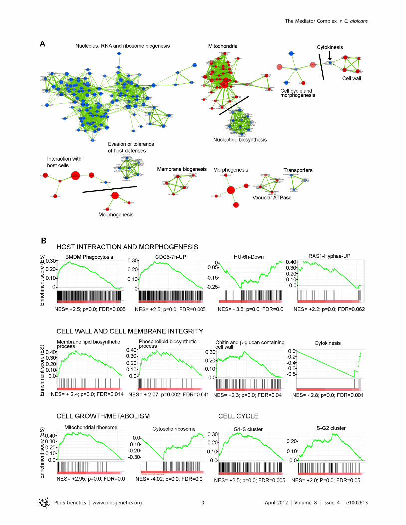
regulated by Med31 in C. albicans, we performed Gene Set
Enrichment Analysis (GSEA) [44,45]. GSEA compares a list from
the transcript profile of interest created by ranking all of the genes
according to the change in their expression (in this case that of a
med31DD mutant) to a predefined gene set, and asks if a specific
gene set is enriched in the top (up-regulated genes) or the bottom
(down-regulated genes) of the ranked list [44,45]. A ranked list of
genes from the transcript profile of med31DD cells was compared to
a custom database of 8123 gene sets (http://candida2.bri.nrc.ca/
andre/GSEA/index.cfm; Sellam and Nantel, submitted) con-
structed using GO annotations and protein interaction data from
CGD (PMID: 19808938), SGD (http://www.yeastgenome.org)
and BioGRID [46], most currently published C. albicans
transcriptional profiling and ChIP-CHIP experiments, our own
TF motif database (PMID: 18342603), and S. cerevisiae genetic-
association data (PMID: 20093466). Since profiles can exhibit
correlations with hundreds of overlapping gene sets, significantly
enriched gene sets (p,0.005, FDR,25%) were further organized
and visualized using the Cytoscape: Enrichment Map plug-in
(PMCID: PMC2981572), which produces networks of gene sets
that share significant overlaps with each other (Figure 1A shows
the most prominent networks of genes; the complete network is
shown in Figure S2 where the details can be visualised by using the
‘‘zoom in’’ function in the pdf document). Figure 1B shows
examples of enrichment plots for selected gene sets. The complete
GSEA output can be found at http://dl.dropbox.com/u/
7211133/Med31%20GSEA%20Results.zip.
GSEA detected enrichment for nucleolar functions, rRNA and
ribosome biogenesis genes, and genes involved in nucleotide
biosynthesis in the set of genes down-regulated in the med31DDmutant, while genes required for mitochondrial function were up-
regulated (Figure 1A and 1B). Enrichment was also found in gene
sets important for virulence-promoting function in C. albicans.
Those include genes differentially expressed during C. albicans-host
interactions with mouse macrophages [47], reconstituted human
oral epithelial cells [48] and polymorphonuclear leukocytes [49],
as well as genes differentially expressed in conditions which alter
cellular morphogenesis, such as the induction of hyphal growth
[50,51], mutations in the Ras-cAMP morphogenesis pathway (ras1
and cdc35/cyc1) [52], and inhibition of cell cycle progression that
Author Summary
In this study, we compared the roles of Mediator, a centraltranscriptional regulator in all eukaryotes, between thepathogenic fungus Candida albicans and the non-patho-genic model yeasts Saccharomyces cerevisiae and Schizo-saccharomyces pombe. We discovered that Mediator hasboth shared and species-specific functions in the threeyeasts. The shared functions include regulation of genesrequired for cell separation after cell division by the Middledomain subunit Med31. The species-specific functionsinclude transcriptional regulation of the cell wall adhesins,which play key roles in the pathogenesis of C. albicans. InC. albicans, the Mediator subunits Med31, Med20, andSrb9/Med13 are activators of the ALS cell wall adhesins. InS. cerevisiae, our results and previous reports suggest anopposite, repressive role in the expression of the FLOgenes and in adhesion-dependent phenotypes. The C.albicans Med31, Med20, and Srb9/Med13 contribute toprocesses highly important for disease: the switch tofilamentous morphology and biofilm formation. Moreover,Med31 impacts on virulence in an invertebrate infectionmodel. Our study has implications for understanding theregulation over virulence-associated genes in C. albicansand the roles of a key transcriptional regulator in thisprocess.
The Mediator Complex in C. albicans
PLoS Genetics | www.plosgenetics.org 2 April 2012 | Volume 8 | Issue 4 | e1002613
The Mediator Complex in C. albicans
PLoS Genetics | www.plosgenetics.org 3 April 2012 | Volume 8 | Issue 4 | e1002613
causes pronounced polarised growth (treatment with hydroxyurea
or down-regulation of the polo-like kinase CDC5) [53] (Figure 1A
and 1B). Genes expressed at the G1-S and S-G2 transition of the
cell cycle [54] were up-regulated in the med31DD mutant, as were
those required for membrane and cell wall biosynthesis (Figure 1A
and 1B). Genes required for cytokinesis were down-regulated (as
shown by the black arrow in Figure 1A, and in the enrichment plot
in Figure 1B).
Modulation of several gene sets enriched in the med31DDmutant, for example down-regulation of genes required for protein
synthesis and up-regulation of those required for cell wall
biogenesis, is part of a more general stress response in C. albicans
[55]. Mediator has been previously implicated in stress responses
in yeasts [7,28], and it is therefore possible that some of the
differences in the med31DD transcriptome are due to activation of
stress responses upon loss of Med31 function. However, our
analysis indicates that this is unlikely to be the cause for much of
the differential gene expression in the mutant. There was little
correlation between the med31DD transcriptional profile and our
large database of transcriptional profiles produced from stressed
cells when analysed by GSEA (of note, profile to profiles
comparisons such as those done by GSEA tend to produce the
strongest correlations and therefore if a correlation existed it is
very likely that it would have been detected by GSEA). We further
used scatter plots to directly compare the med31DD profile with
stressful conditions, such as osmotic or oxidative stress, and these
comparisons confirmed lack of extensive correlation between the
med31DD transcriptome and differential gene expression upon
stress (Figure S1B).
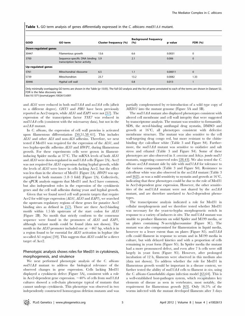
Analysis of gene ontology terms using the GO term finder tool
at the Candida Genome database and the genes up- or down-
regulated by at least 1.5 fold in med31DD cells (see Dataset S1)
confirmed that genes related to morphogenesis, mitochondrial
function and the cell wall were differentially expressed (Table 1
and Dataset S2). Interestingly, several central regulators of
filamentous differentiation, such as the transcription factors
Tec1, Efg1, Cph1 and Nrg1, were amongst the down-regulated
genes, as were six out of the eight genes from the FGR6
(Filamentous Growth Regulator) family located in the RB2 repeat
sequence (Dataset S2). The FGR6 family is one of the gene
families found to be enriched in pathogenic yeast species, and
specifically expanded in C. albicans [56]. While GSEA scored the
cell wall gene set as up-regulated, we noticed that there were also
several genes in this group that appeared at the bottom of the list,
in the down-regulated group. In fact, another Candida-specific gene
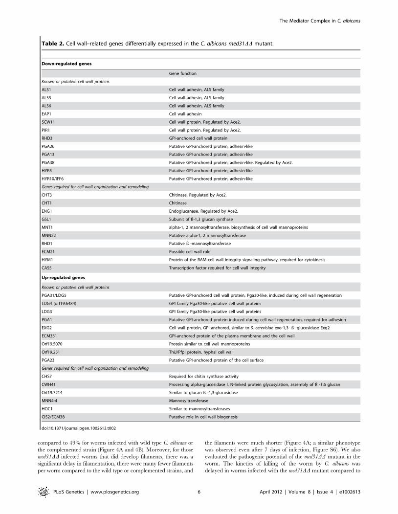
family expanded in pathogens was down-regulated in the med31DDmutant, that encoding the ALS cell wall adhesins [56] (Table 2).
The major C. albicans adhesin ALS1 was one of the most down-
regulated genes in the mutant (5 fold down-regulation, Table 2
and Dataset S1). ALS5 and ALS6 were also down-regulated
(Table 2), but of note, these genes are expected to be expressed at
very low levels in the wild type. Additionally, several other genes
encoding cell wall proteins were down-regulated in the mutant, as
were genes necessary for cell wall construction and remodelling, in
particular those required for cytokinesis and regulated by the
transcription factor Ace2 (e.g. the chitinase CHT3 and the
endoglucanase ENG1) [57] (Table 2; notably GSEA also scored
the cytokinesis genes as down-regulated and this is shown in
Figure 1). The existence of several down-regulated cell wall genes
indicates that the up-regulation of genes with roles in cell wall
integrity that is detected in the med31DD mutant (Figure 1 and
Table 2) likely reflects a compensatory feedback regulation due to
a defective cell wall structure in the absence of Med31. That
med31DD mutants have altered cell walls is supported by
phenotypic analysis demonstrating changes in sensitivity to the
cell wall targeting drugs congo red and calcofluor white (Table 3).
In conclusion, the transcriptome analysis indicated a broad role
for Med31 in cell physiology in C. albicans, with functions in
morphogenesis and cell cycle progression, growth and metabolism,
cell wall integrity, the expression of the cytokinesis genes under the
control of the transcription factor Ace2 and those regulated by the
interaction of C. albicans with host cells. Finally, two gene families
enriched in pathogenic yeasts, the FGR family of filamentous
growth regulators and the ALS adhesins, required Med31 for wild
type expression levels.
Regulation of Ace2-dependent genes and cell walladhesins by Med31 in yeast and hyphal growth
Down-regulation of Ace2 target genes in the absence of Med31
in C. albicans is in agreement with a role for Med31 and other
Mediator subunits in Ace2-dependent gene expression that is
conserved between C. albicans, Schizo. pombe and S. cerevisiae (this
study and [28]). Comparing more broadly the genes affected in
med31DD cells with those reported to be differentially expressed in
the C. albicans ace2 mutant [57] revealed that differential expression
of 35 genes is shared between these two transcription factors
(Table S1). Genes involved in cytokinesis and cell wall functions
were predominant in the shared ‘‘down-regulated’’ group, whereas
mitochondrial biogenesis genes were predominant in the shared
‘‘up-regulated’’ group (Table S1). This analysis suggests that the
functions of Med31 in cell wall integrity and metabolism are
mediated, at least in part, by Ace2-dependent roles.
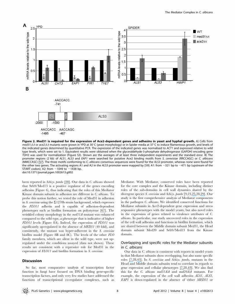
To explore this further, we used quantitative PCR (qPCR) to
directly compare the expression levels of candidate genes in the
med31DD and ace2DD mutants (Figure 2A). Under yeast growth
conditions (as was done for the transcriptome analysis) the mRNA
levels for the chitinase CHT3 and the cell wall proteins PIR1, EAP1
Figure 1. GSEA analysis of the genes differentially expressed in the absence of Med31 in C. albicans. A) The network of functional groupsof gene regulated by Med31 was constructed with GSEA and Enrichment Map. Blue circles are down-regulated gene sets, while the up-regulatedgene sets are represented by red circles. The diameter of the circle reflects the number of modulated gene transcripts in each gene set. Wheredifferent functional groups of genes are linked in the network, they are separated by grey lines to indicate the functions. The full network is presentedas a pdf document in Figure S2, where the details can be visualized using the ‘‘zoom in’’ function. B) Example enrichment plots for selected genes setsdifferentially expressed in the med31DD mutant are presented. On the x-axis are genes ranked according to their expression in the med31DD mutant,starting with the up-regulated genes on the left hand side, and all the way down to the down-regulated genes on far right. The position of theindividual genes in the gene set are shown by black vertical lines. The cumulative value of the enrichment score (y-axis) is represented by the greenline. A positive normalised enrichment score (NES) indicates enrichment in the up-regulated group of genes in the med31DD mutant, while anegative NES indicates prevalence of the genes in the down-regulated group. The title for each of the graphs indicates the genes set used tocompare to the med31DD set. BMDM phagocytosis: the set of genes up-regulated in C. albicans upon phagocytosis by bone-marrow derivedmonocytes [47]; CDC5-7h-UP: gene set up-regulated in the C. albicans 7 h post depletion of the polo-like kinase CDC5 [53]; HU-6h-Down: gene setdown-regulated upon a 6 h treatment of cells with the DNA replication inhibitor hydroxyurea (HU) [53]; RAS1-Hyphae-up: gene set up-regulated inras1 mutants under hyphal growth [52]; G1-S and S-G2: genes expressed at the G1-S and S-G2 phases of the cell cycle in C. albicans [54]. p value of 0.0represents ,0.001, FDR is the false discovery rate.doi:10.1371/journal.pgen.1002613.g001
The Mediator Complex in C. albicans
PLoS Genetics | www.plosgenetics.org 4 April 2012 | Volume 8 | Issue 4 | e1002613
and ALS1 were reduced in both med31DD and ace2DD cells (albeit
to a different degree). CHT3 and PIR1 have been previously
reported as Ace2-targets, while ALS1 and EAP1 were not [57]. The
expression of the transcription factor TYE7 was reduced in
med31DD cells (consistent with the microarray data), but not in the
ace2DD mutant.
In C. albicans, the expression of cell wall proteins is activated
upon filamentous differentiation [50,51,58–61]. This includes
ALS1 and other ALS and non-ALS adhesins. Therefore, we next
tested if Med31 was required for the expression of the ALS1, and
two hypha-specific adhesins ALS3 and HWP1, during filamentous
growth. For these experiments cells were grown in filament-
inducing Spider media at 37uC. The mRNA levels of both ALS1
and ALS3 were down-regulated in med31DD cells (Figure 2A). Ace2
was not required for ALS1 expression during hyphal growth, while
the levels of ALS3 were lower in cells lacking Ace2, but the effect
was less than in the absence of Med31 (Figure 2A). HWP1 was up-
regulated in both mutants (1.8–3 fold) (Figure 2A). Collectively,
the qPCR analysis suggests that Med31 and Ace2 have common,
but also independent roles in the expression of the cytokinesis
genes and the cell wall adhesins during yeast and hyphal growth.
Given that we found novel cell wall protein targets that require
Ace2 for wild type expression (ALS1, ALS3 and EAP1), we searched
the upstream regulatory regions of these genes for putative Ace2
binding sites as defined in [57]. There are three Ace2-binding
motifs within 1.5 kb upstream of the start codon for ALS3
(Figure 2B). No motifs that strictly conform to the consensus
sequence were found in the promoters of ALS1 and EAP1,
although variant motifs could be found (data not shown). The
motifs in the ALS3 promoter included one at 2467 bp, which is in
a region found to be essential for ALS3 activation in hyphae (the
so-called A1 region) [59]. This suggests that ALS3 could be a direct
target of Ace2.
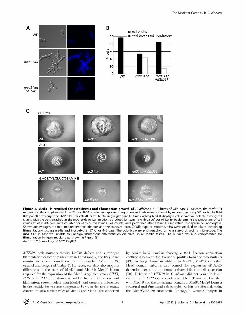
Phenotypic analysis shows roles for Med31 in cytokinesis,morphogenesis, and virulence
We next performed phenotypic analysis of the C. albicans
med31DD mutant to address the biological relevance of the
observed changes in gene expression. Cells lacking Med31
displayed a cytokinesis defect (Figure 3A), consistent with a role
in Ace2-dependent gene expression. ,40% of cells from med31DDcultures showed a cell-chain phenotype typical of mutants that
cannot undergo cytokinesis. This phenotype was observed in two
independently constructed homozygous deletion mutants and was
partially complemented by re-introduction of a wild type copy of
MED31 into the mutant genome (Figure 3A and 3B).
The med31DD mutant also displayed phenotypes consistent with
altered cell membrane and cell wall integrity that were suggested
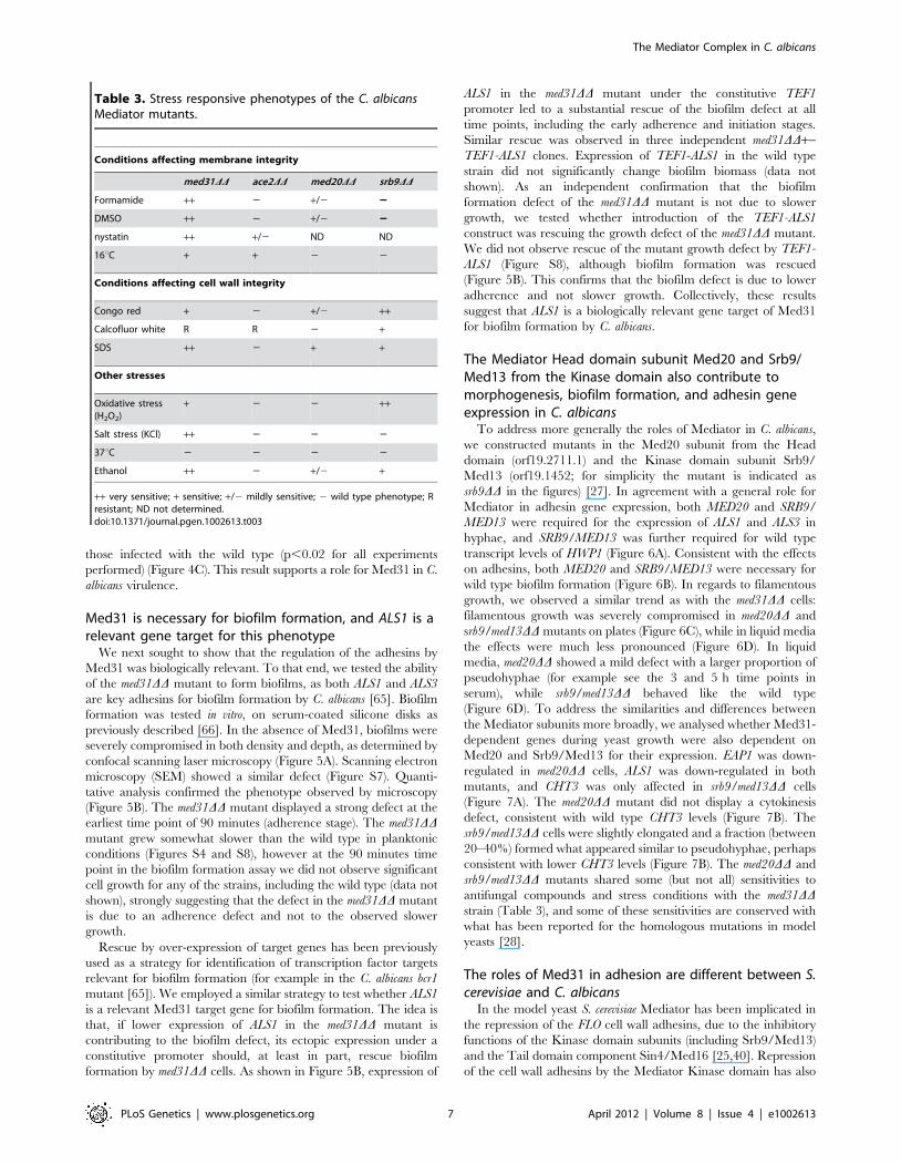
by transcriptome analysis. The mutant was sensitive to formamide,
SDS, the sterol-binding antifungal drug nystatin, DMSO and
growth at 16uC, all phenotypes consistent with defective
membrane structure. The mutant was also sensitive to the cell
wall-targeting drug congo red, but more resistant to the chitin-
binding dye calcofluor white (Table 3 and Figure S4). Further-
more, the med31DD mutant was sensitive to oxidative and salt
stress and ethanol (Table 3 and Figure S4). Some of these
phenotypes are also observed in S. cerevisiae and Schizo. pombe med31
mutants, suggesting conserved roles [28,43]. We also tested the C.
albicans ace2DD mutant side by side with med31DD for tolerance to
the various compounds (Table 3 and Figure S4). Resistance to
calcoflour white was also observed in the ace2DD mutant (Table 3
and [62]), as was a mild sensitivity to nystatin and growth at 16uC,
indicating that these phenotypes could be due to the role of Med31
in Ace2-dependent gene expression. However, the other sensitiv-
ities of the med31DD mutant were not shared by the ace2DDmutant, and are therefore unrelated to Ace2-dependent pheno-
types.
The transcriptome analysis indicated a role for Med31 in
cellular morphogenesis and we therefore tested whether Med31
was necessary for the yeast-to-hypha morphogenetic switch in
response to a variety of inducers in vitro. The med31DD mutant was
unable to produce filaments on solid Spider and M199 media, or
on plates containing N-acetylglucosamine (Figure 3C). The
mutant was also compromised for filamentation in liquid media,
however to a lesser extent than on plates (Figure S5). med31DDcells could filament in response to serum and in M199 media in
culture, but with delayed kinetics and with a proportion of cells
remaining in yeast form (Figure S5). In Spider media the mutant
had a more pronounced defect, and even after 7 h cells were still
largely in yeast form (Figure S5). However, after prolonged
incubation of 12 h, filaments were observed in this medium also
(data not shown). To address whether the role for Med31 in
filamentous growth would be important in a disease context, we
further tested the ability of med31DD cells to filament in vivo, using
the C. albicans-Caenorhabditis elegans infection model [63,64]. This is
a well-established host-pathogen system, which recapitulates key
elements of disease as seen in vertebrates, most notably, the
requirement for filamentous growth [63]. Only 16.5% of the
worms infected with the mutant developed filaments after 3 days
Table 1. GO term analysis of genes differentially expressed in the C. albicans med31DD mutant.
GOID GO term Cluster frequency (%)Background frequency(%) p value FDR (%)
Down-regulated genes
30447 Filamentous growth 13.6 6.6 0.0031 0
3700 Sequence-specific DNA bindingtranscription factor activity
5.9 2.1 0.006 0
Up-regulated genes
5761 Mitochondrial ribosome 6.5 1.1 0.00011 0
5739 Mitochondrion 23.7 13.2 0.0082 1.33
30446 Hyphal cell wall 4.3 0.8 0.013 1
Only minimally overlapping GO terms are shown in the Table (p,0.05). The full GO analysis and the list of gene annotated to each of the terms are shown in Dataset S2.FDR is the false discovery rate.doi:10.1371/journal.pgen.1002613.t001
The Mediator Complex in C. albicans
PLoS Genetics | www.plosgenetics.org 5 April 2012 | Volume 8 | Issue 4 | e1002613
compared to 49% for worms infected with wild type C. albicans or
the complemented strain (Figure 4A and 4B). Moreover, for those
med31DD-infected worms that did develop filaments, there was a
significant delay in filamentation, there were many fewer filaments
per worm compared to the wild type or complemented strains, and
the filaments were much shorter (Figure 4A; a similar phenotype
was observed even after 7 days of infection, Figure S6). We also
evaluated the pathogenic potential of the med31DD mutant in the
worm. The kinetics of killing of the worm by C. albicans was
delayed in worms infected with the med31DD mutant compared to
Table 2. Cell wall–related genes differentially expressed in the C. albicans med31DD mutant.
Down-regulated genes
Gene function
Known or putative cell wall proteins
ALS1 Cell wall adhesin, ALS family
ALS5 Cell wall adhesin, ALS family
ALS6 Cell wall adhesin, ALS family
EAP1 Cell wall adhesin
SCW11 Cell wall protein. Regulated by Ace2.
PIR1 Cell wall protein. Regulated by Ace2.
RHD3 GPI-anchored cell wall protein
PGA26 Putative GPI-anchored protein, adhesin-like
PGA13 Putative GPI-anchored protein, adhesin-like
PGA38 Putative GPI-anchored protein, adhesin-like. Regulated by Ace2.
HYR3 Putative GPI-anchored protein, adhesin-like
HYR10/IFF6 Putative GPI-anchored protein, adhesin-like
Genes required for cell wall organization and remodeling
CHT3 Chitinase. Regulated by Ace2.
CHT1 Chitinase
ENG1 Endoglucanase. Regulated by Ace2.
GSL1 Subunit of ß-1,3 glucan synthase
MNT1 alpha-1, 2 mannosyltransferase, biosynthesis of cell wall mannoproteins
MNN22 Putative alpha-1, 2 mannosyltransferase
RHD1 Putative ß -mannosyltransferase
ECM21 Possible cell wall role
HYM1 Protein of the RAM cell wall integrity signaling pathway, required for cytokinesis
CAS5 Transcription factor required for cell wall integrity
Up-regulated genes
Known or putative cell wall proteins
PGA31/LDG5 Putative GPI-anchored cell wall protein, Pga30-like, induced during cell wall regeneration
LDG4 (orf19.6484) GPI family Pga30-like putative cell wall proteins
LDG3 GPI family Pga30-like putative cell wall proteins
PGA1 Putative GPI-anchored protein induced during cell wall regeneration, required for adhesion
EXG2 Cell wall protein, GPI-anchored, similar to S. cerevisiae exo-1,3- ß -glucosidase Exg2
ECM331 GPI-anchored protein of the plasma membrane and the cell wall
Orf19.5070 Protein similar to cell wall mannoproteins
Orf19.251 ThiJ/PfpI protein, hyphal cell wall
PGA23 Putative GPI-anchored protein of the cell surface
Genes required for cell wall organization and remodeling
CHS7 Required for chitin synthase activity
CWH41 Processing alpha-glucosidase I, N-linked protein glycosylation, assembly of ß -1,6 glucan
Orf19.7214 Similar to glucan ß -1,3-glucosidase
MNN4-4 Mannosyltransferase
HOC1 Similar to mannosyltransferases
CIS2/ECM38 Putative role in cell wall biogenesis
doi:10.1371/journal.pgen.1002613.t002
The Mediator Complex in C. albicans
PLoS Genetics | www.plosgenetics.org 6 April 2012 | Volume 8 | Issue 4 | e1002613
those infected with the wild type (p,0.02 for all experiments
performed) (Figure 4C). This result supports a role for Med31 in C.
albicans virulence.
Med31 is necessary for biofilm formation, and ALS1 is arelevant gene target for this phenotype
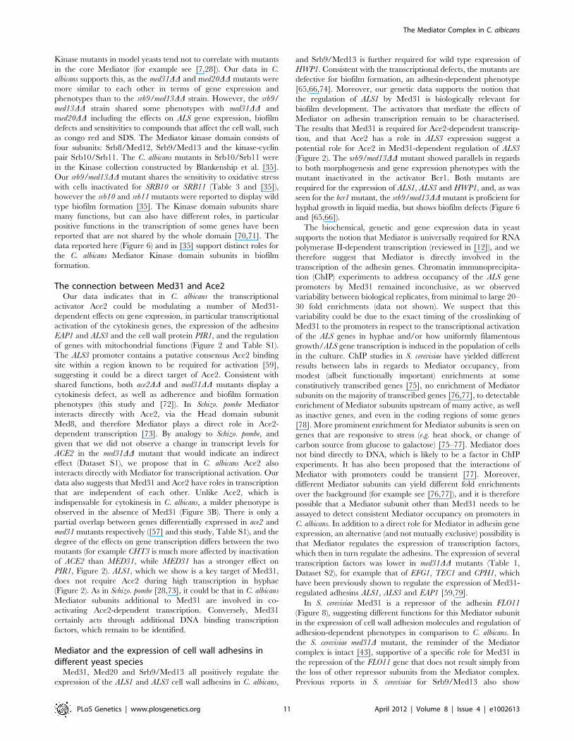
We next sought to show that the regulation of the adhesins by
Med31 was biologically relevant. To that end, we tested the ability
of the med31DD mutant to form biofilms, as both ALS1 and ALS3
are key adhesins for biofilm formation by C. albicans [65]. Biofilm
formation was tested in vitro, on serum-coated silicone disks as
previously described [66]. In the absence of Med31, biofilms were
severely compromised in both density and depth, as determined by
confocal scanning laser microscopy (Figure 5A). Scanning electron
microscopy (SEM) showed a similar defect (Figure S7). Quanti-
tative analysis confirmed the phenotype observed by microscopy
(Figure 5B). The med31DD mutant displayed a strong defect at the
earliest time point of 90 minutes (adherence stage). The med31DDmutant grew somewhat slower than the wild type in planktonic
conditions (Figures S4 and S8), however at the 90 minutes time
point in the biofilm formation assay we did not observe significant
cell growth for any of the strains, including the wild type (data not
shown), strongly suggesting that the defect in the med31DD mutant
is due to an adherence defect and not to the observed slower
growth.
Rescue by over-expression of target genes has been previously
used as a strategy for identification of transcription factor targets
relevant for biofilm formation (for example in the C. albicans bcr1
mutant [65]). We employed a similar strategy to test whether ALS1
is a relevant Med31 target gene for biofilm formation. The idea is
that, if lower expression of ALS1 in the med31DD mutant is
contributing to the biofilm defect, its ectopic expression under a
constitutive promoter should, at least in part, rescue biofilm
formation by med31DD cells. As shown in Figure 5B, expression of
ALS1 in the med31DD mutant under the constitutive TEF1
promoter led to a substantial rescue of the biofilm defect at all
time points, including the early adherence and initiation stages.
Similar rescue was observed in three independent med31DD+-TEF1-ALS1 clones. Expression of TEF1-ALS1 in the wild type
strain did not significantly change biofilm biomass (data not
shown). As an independent confirmation that the biofilm
formation defect of the med31DD mutant is not due to slower
growth, we tested whether introduction of the TEF1-ALS1
construct was rescuing the growth defect of the med31DD mutant.
We did not observe rescue of the mutant growth defect by TEF1-
ALS1 (Figure S8), although biofilm formation was rescued
(Figure 5B). This confirms that the biofilm defect is due to lower
adherence and not slower growth. Collectively, these results
suggest that ALS1 is a biologically relevant gene target of Med31
for biofilm formation by C. albicans.
The Mediator Head domain subunit Med20 and Srb9/Med13 from the Kinase domain also contribute tomorphogenesis, biofilm formation, and adhesin geneexpression in C. albicans
To address more generally the roles of Mediator in C. albicans,
we constructed mutants in the Med20 subunit from the Head
domain (orf19.2711.1) and the Kinase domain subunit Srb9/
Med13 (orf19.1452; for simplicity the mutant is indicated as
srb9DD in the figures) [27]. In agreement with a general role for
Mediator in adhesin gene expression, both MED20 and SRB9/
MED13 were required for the expression of ALS1 and ALS3 in
hyphae, and SRB9/MED13 was further required for wild type
transcript levels of HWP1 (Figure 6A). Consistent with the effects
on adhesins, both MED20 and SRB9/MED13 were necessary for
wild type biofilm formation (Figure 6B). In regards to filamentous
growth, we observed a similar trend as with the med31DD cells:
filamentous growth was severely compromised in med20DD and
srb9/med13DD mutants on plates (Figure 6C), while in liquid media
the effects were much less pronounced (Figure 6D). In liquid
media, med20DD showed a mild defect with a larger proportion of
pseudohyphae (for example see the 3 and 5 h time points in
serum), while srb9/med13DD behaved like the wild type
(Figure 6D). To address the similarities and differences between
the Mediator subunits more broadly, we analysed whether Med31-
dependent genes during yeast growth were also dependent on
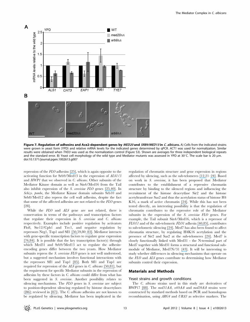
Med20 and Srb9/Med13 for their expression. EAP1 was down-
regulated in med20DD cells, ALS1 was down-regulated in both
mutants, and CHT3 was only affected in srb9/med13DD cells
(Figure 7A). The med20DD mutant did not display a cytokinesis
defect, consistent with wild type CHT3 levels (Figure 7B). The
srb9/med13DD cells were slightly elongated and a fraction (between
20–40%) formed what appeared similar to pseudohyphae, perhaps
consistent with lower CHT3 levels (Figure 7B). The med20DD and
srb9/med13DD mutants shared some (but not all) sensitivities to
antifungal compounds and stress conditions with the med31DDstrain (Table 3), and some of these sensitivities are conserved with
what has been reported for the homologous mutations in model
yeasts [28].
The roles of Med31 in adhesion are different between S.cerevisiae and C. albicans
In the model yeast S. cerevisiae Mediator has been implicated in
the repression of the FLO cell wall adhesins, due to the inhibitory
functions of the Kinase domain subunits (including Srb9/Med13)
and the Tail domain component Sin4/Med16 [25,40]. Repression
of the cell wall adhesins by the Mediator Kinase domain has also
Table 3. Stress responsive phenotypes of the C. albicansMediator mutants.
Conditions affecting membrane integrity
med31DD ace2DD med20DD srb9DD
Formamide ++ 2 +/2 22
DMSO ++ 2 +/2 22
nystatin ++ +/2 ND ND
16uC + + 2 2
Conditions affecting cell wall integrity
Congo red + 2 +/2 ++
Calcofluor white R R 2 +
SDS ++ 2 + +
Other stresses
Oxidative stress(H2O2)
+ 2 2 ++
Salt stress (KCl) ++ 2 2 2
37uC 2 2 2 2
Ethanol ++ 2 +/2 +
++ very sensitive; + sensitive; +/2 mildly sensitive; 2 wild type phenotype; Rresistant; ND not determined.doi:10.1371/journal.pgen.1002613.t003
The Mediator Complex in C. albicans
PLoS Genetics | www.plosgenetics.org 7 April 2012 | Volume 8 | Issue 4 | e1002613
been reported in Schizo. pombe [28]. Our data in C. albicans showed
that Srb9/Med13 is a positive regulator of the genes encoding
adhesins (Figure 6), thus indicating that the roles of this Mediator
Kinase domain subunit in adhesion are different in C. albicans. To
probe this notion further, we tested the role of Med31 in adhesion
in S. cerevisiae using the S1278b strain background, which expresses
the FLO11 adhesin and is capable of adhesion-dependent
phenotypes such as biofilm formation on polystyrene [67]. The
wrinkled colony morphology in the med31D mutant was enhanced
compared to the wild type, a phenotype that is indicative of higher
FLO11 levels (Figure 8A). Indeed, the expression of FLO11 was
significantly up-regulated in the absence of MED31 (40 fold), and
consistently, the mutant was hyper-adherent in the S. cerevisiae
biofilm model (Figure 8B and 8C). The levels of the other FLO
family members, which are silent in the wild type, were not up-
regulated under the conditions assayed (data not shown). These
results are consistent with a repressive role for Med31 in the
expression of FLO11 and biofilm formation in S. cerevisiae.
Discussion
So far, most comparative analysis of transcription factor
function in fungi have focused on DNA binding gene-specific
transcription factors, and only very few studies have addressed the
functions of transcriptional co-regulator complexes, such as
Mediator. With Mediator, conserved roles have been reported
for the core complex and the Kinase domain, including distinct
roles of the sub-domains in cell wall dynamics shared by the
divergent species S. cerevisiae and Schizo. pombe [9,23,25,28,29]. Our
study is the first comprehensive analysis of Mediator components
in the pathogen C. albicans. We identified conserved functions for
Mediator subunits in Ace2-dependent gene expression and stress
responsive phenotypes with the model yeasts, but also novel roles
in the expression of genes related to virulence attributes of C.
albicans. In particular, our study uncovered roles in the expression
of the cell wall adhesins and functions in biofilm formation, which
are shared between the Middle domain subunit Med31, the Head
domain subunit Med20 and Srb9/Med13 from the Kinase
domain.
Overlapping and specific roles for the Mediator subunitsin C. albicans
Our data in C. albicans is consistent with reports in model yeasts
in that Mediator subunits show overlapping, but also some specific
roles [7,28,43]. In S. cerevisiae and Schizo. pombe, mutants in the
Head and Middle domain subunits tend to correlate in regards to
gene expression and cellular phenotypes [7,28,43]. We also find
this for the C. albicans med31DD and med20DD mutants. For
example, the expression of the cell wall adhesins ALS1, ALS3,
EAP1 is down-regulated in the absence of either MED31 or
Figure 2. Med31 is required for the expression of Ace2-dependent genes and adhesins in yeast and hyphal growth. A) Cells frommed31DD or ace2DD mutants were grown in YPD at 30uC (yeast morphology) or in Spider media at 37uC to induce filamentous growth, and levels ofthe indicated genes determined by quantitative PCR. The expression of the indicated genes was normalised to ACT1 and expressed relative to wildtype levels, which were set to 1. Equivalent results were obtained when the glyceraldehyde-3-phosphate dehydrogenase (GAPDH) encoding geneTDH3 was used for normalization (Figure S3). Shown are the averages of at least three independent experiments and the standard error. B) Thepromoter regions (2 kb) of ALS1, ALS3 and EAP1 were searched for putative Ace2 binding motifs from S. cerevisiae (RRCCAGC) or C. albicans(MMCCASC) [57]. The three motifs conforming to C. albicans consensus sequences were found for the ALS3 promoter, whereas none were found forthe other two genes. The activating regions A1 and A2 in the ALS3 promoter were mapped by [59]: A1: from 2321 bp to 2471 bp (upstream of theSTART codon); A2: from 21049 to 21438 bp.doi:10.1371/journal.pgen.1002613.g002
The Mediator Complex in C. albicans
PLoS Genetics | www.plosgenetics.org 8 April 2012 | Volume 8 | Issue 4 | e1002613
MED20, both mutants display biofilm defects and a stronger
filamentation defect on plates than in liquid media, and they share
sensitivities to compounds such as formamide, DMSO, SDS,
ethanol and congo red (Table 3). However, our data also supports
differences in the roles of Med20 and Med31: Med20 is not
required for the expression of the Med31-regulated genes CHT3,
PIR1 and TYE7, it shows a milder biofilm formation and
filamentous growth defect than Med31, and there are differences
in the sensitivities to some compounds between the two mutants.
Shared but also distinct roles of Med20 and Med31 are supported
by results in S. cerevisiae showing a 0.41 Pearson correlation
coefficient between the transcript profiles form the two mutants
[43]. In Schizo. pombe, in addition to Med31, Med20 and other
Head domain subunits also control the expression of Ace2-
dependent genes and the mutants show defects in cell separation
[28]. Deletion of MED20 in C. albicans did not result in lower
expression of CHT3 or a cytokinesis defect (Figure 7). Together
with Med18 and the C-terminal domain of Med8, Med20 forms a
structural and functional sub-complex within the Head domain,
the Med8C/18/20 submodule [28,68,69]. Genetic analysis in
Figure 3. Med31 is required for cytokinesis and filamentous growth of C. albicans. A) Cultures of wild type C. albicans, the med31DDmutant and the complemented med31DD+MED31 strain were grown to log phase and cells were observed by microscopy using DIC for bright field(left panel) or through the DAPI filter for calcofluor white staining (right panel). Strains lacking Med31 display a cell separation defect, forming cellchains with the cells attached at the mother-daughter junction, as judged by staining with calcofluor white. B) To determine the proportion of cellchains at least 200 cells were counted for each of the strains. Cell counts were performed after a brief 1 s sonication to disperse cell aggregates.Shown are averages of three independent experiments and the standard error. C) Wild type or mutant strains were streaked on plates containingfilamentation-inducing media and incubated at 37uC for 4–5 days. The colonies were photographed using a stereo dissecting microscope. Themed31DD mutant was unable to undergo filamentous differentiation on plates in all media tested. The mutant was also compromised forfilamentation in liquid media (data shown in Figure S5).doi:10.1371/journal.pgen.1002613.g003
The Mediator Complex in C. albicans
PLoS Genetics | www.plosgenetics.org 9 April 2012 | Volume 8 | Issue 4 | e1002613
Schizo. pombe supports the idea that Med18 can compensate for the
loss of Med20 [28]. It is therefore possible that Med18 would need
to be inactivated in C. albicans to uncover the roles of the Mediator
Head domain in Ace2-dependent transcription and cytokinesis.
In contrast to the core Mediator complex which functions
predominantly in transcriptional activation, the Kinase domain is
mainly a repressor of transcription and the phenotypes of the
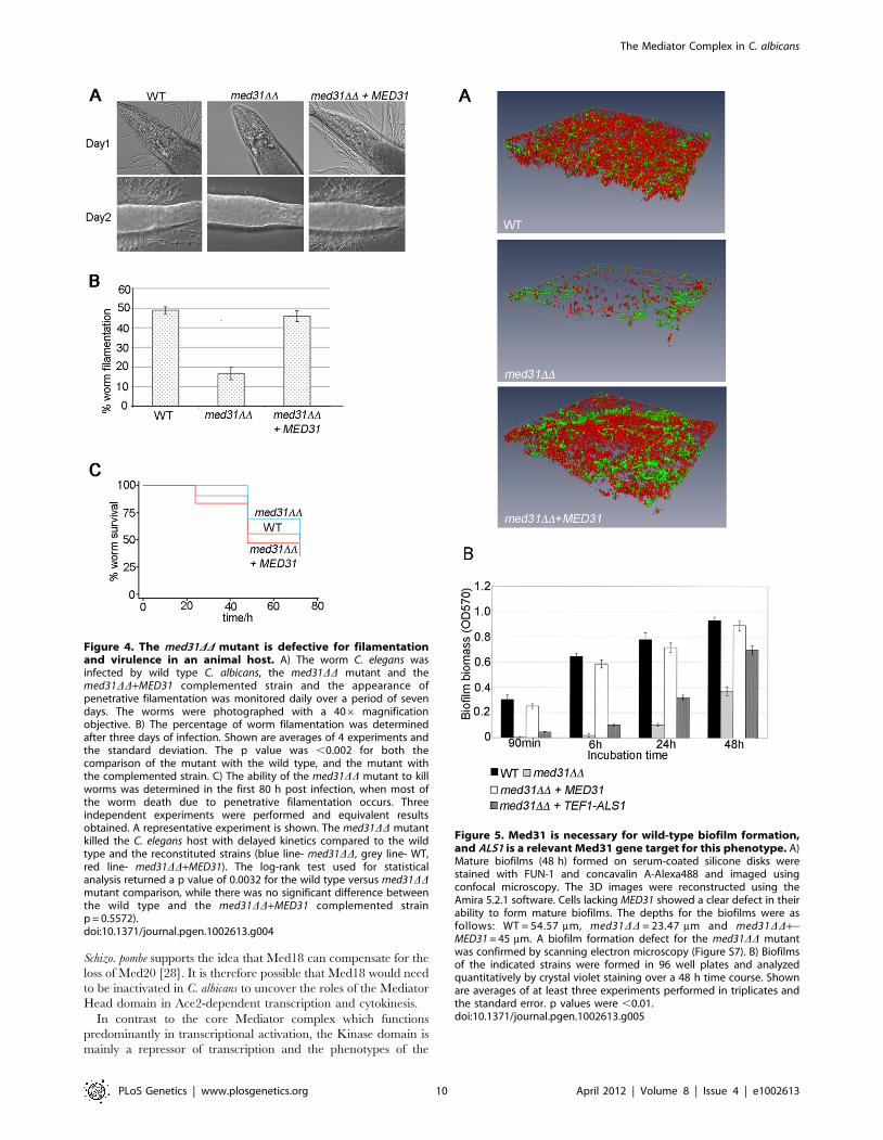
Figure 4. The med31DD mutant is defective for filamentationand virulence in an animal host. A) The worm C. elegans wasinfected by wild type C. albicans, the med31DD mutant and themed31DD+MED31 complemented strain and the appearance ofpenetrative filamentation was monitored daily over a period of sevendays. The worms were photographed with a 406 magnificationobjective. B) The percentage of worm filamentation was determinedafter three days of infection. Shown are averages of 4 experiments andthe standard deviation. The p value was ,0.002 for both thecomparison of the mutant with the wild type, and the mutant withthe complemented strain. C) The ability of the med31DD mutant to killworms was determined in the first 80 h post infection, when most ofthe worm death due to penetrative filamentation occurs. Threeindependent experiments were performed and equivalent resultsobtained. A representative experiment is shown. The med31DD mutantkilled the C. elegans host with delayed kinetics compared to the wildtype and the reconstituted strains (blue line- med31DD, grey line- WT,red line- med31DD+MED31). The log-rank test used for statisticalanalysis returned a p value of 0.0032 for the wild type versus med31DDmutant comparison, while there was no significant difference betweenthe wild type and the med31DD+MED31 complemented strainp = 0.5572).doi:10.1371/journal.pgen.1002613.g004
Figure 5. Med31 is necessary for wild-type biofilm formation,and ALS1 is a relevant Med31 gene target for this phenotype. A)Mature biofilms (48 h) formed on serum-coated silicone disks werestained with FUN-1 and concavalin A-Alexa488 and imaged usingconfocal microscopy. The 3D images were reconstructed using theAmira 5.2.1 software. Cells lacking MED31 showed a clear defect in theirability to form mature biofilms. The depths for the biofilms were asfollows: WT = 54.57 mm, med31DD = 23.47 mm and med31DD+-MED31 = 45 mm. A biofilm formation defect for the med31DD mutantwas confirmed by scanning electron microscopy (Figure S7). B) Biofilmsof the indicated strains were formed in 96 well plates and analyzedquantitatively by crystal violet staining over a 48 h time course. Shownare averages of at least three experiments performed in triplicates andthe standard error. p values were ,0.01.doi:10.1371/journal.pgen.1002613.g005
The Mediator Complex in C. albicans
PLoS Genetics | www.plosgenetics.org 10 April 2012 | Volume 8 | Issue 4 | e1002613
Kinase mutants in model yeasts tend not to correlate with mutants
in the core Mediator (for example see [7,28]). Our data in C.
albicans supports this, as the med31DD and med20DD mutants were
more similar to each other in terms of gene expression and
phenotypes than to the srb9/med13DD strain. However, the srb9/
med13DD strain shared some phenotypes with med31DD and
med20DD including the effects on ALS gene expression, biofilm
defects and sensitivities to compounds that affect the cell wall, such
as congo red and SDS. The Mediator kinase domain consists of
four subunits: Srb8/Med12, Srb9/Med13 and the kinase-cyclin
pair Srb10/Srb11. The C. albicans mutants in Srb10/Srb11 were
in the Kinase collection constructed by Blankenship et al. [35].
Our srb9/med13DD mutant shares the sensitivity to oxidative stress
with cells inactivated for SRB10 or SRB11 (Table 3 and [35]),
however the srb10 and srb11 mutants were reported to display wild
type biofilm formation [35]. The Kinase domain subunits share
many functions, but can also have different roles, in particular
positive functions in the transcription of some genes have been
reported that are not shared by the whole domain [70,71]. The
data reported here (Figure 6) and in [35] support distinct roles for
the C. albicans Mediator Kinase domain subunits in biofilm
formation.
The connection between Med31 and Ace2Our data indicates that in C. albicans the transcriptional
activator Ace2 could be modulating a number of Med31-
dependent effects on gene expression, in particular transcriptional
activation of the cytokinesis genes, the expression of the adhesins
EAP1 and ALS3 and the cell wall protein PIR1, and the regulation
of genes with mitochondrial functions (Figure 2 and Table S1).
The ALS3 promoter contains a putative consensus Ace2 binding
site within a region known to be required for activation [59],
suggesting it could be a direct target of Ace2. Consistent with
shared functions, both ace2DD and med31DD mutants display a
cytokinesis defect, as well as adherence and biofilm formation
phenotypes (this study and [72]). In Schizo. pombe Mediator
interacts directly with Ace2, via the Head domain subunit
Med8, and therefore Mediator plays a direct role in Ace2-
dependent transcription [73]. By analogy to Schizo. pombe, and
given that we did not observe a change in transcript levels for
ACE2 in the med31DD mutant that would indicate an indirect
effect (Dataset S1), we propose that in C. albicans Ace2 also
interacts directly with Mediator for transcriptional activation. Our
data also suggests that Med31 and Ace2 have roles in transcription
that are independent of each other. Unlike Ace2, which is
indispensable for cytokinesis in C. albicans, a milder phenotype is
observed in the absence of Med31 (Figure 3B). There is only a
partial overlap between genes differentially expressed in ace2 and
med31 mutants respectively ([57] and this study, Table S1), and the
degree of the effects on gene transcription differs between the two
mutants (for example CHT3 is much more affected by inactivation
of ACE2 than MED31, while MED31 has a stronger effect on
PIR1, Figure 2). ALS1, which we show is a key target of Med31,
does not require Ace2 during high transcription in hyphae
(Figure 2). As in Schizo. pombe [28,73], it could be that in C. albicans
Mediator subunits additional to Med31 are involved in co-
activating Ace2-dependent transcription. Conversely, Med31
certainly acts through additional DNA binding transcription
factors, which remain to be identified.
Mediator and the expression of cell wall adhesins indifferent yeast species
Med31, Med20 and Srb9/Med13 all positively regulate the
expression of the ALS1 and ALS3 cell wall adhesins in C. albicans,
and Srb9/Med13 is further required for wild type expression of
HWP1. Consistent with the transcriptional defects, the mutants are
defective for biofilm formation, an adhesin-dependent phenotype
[65,66,74]. Moreover, our genetic data supports the notion that
the regulation of ALS1 by Med31 is biologically relevant for
biofilm development. The activators that mediate the effects of
Mediator on adhesin transcription remain to be characterised.
The results that Med31 is required for Ace2-dependent transcrip-
tion, and that Ace2 has a role in ALS3 expression suggest a
potential role for Ace2 in Med31-dependent regulation of ALS3
(Figure 2). The srb9/med13DD mutant showed parallels in regards
to both morphogenesis and gene expression phenotypes with the
mutant inactivated in the activator Bcr1. Both mutants are
required for the expression of ALS1, ALS3 and HWP1, and, as was
seen for the bcr1 mutant, the srb9/med13DD mutant is proficient for
hyphal growth in liquid media, but shows biofilm defects (Figure 6
and [65,66]).
The biochemical, genetic and gene expression data in yeast
supports the notion that Mediator is universally required for RNA
polymerase II-dependent transcription (reviewed in [12]), and we
therefore suggest that Mediator is directly involved in the
transcription of the adhesin genes. Chromatin immunoprecipita-
tion (ChIP) experiments to address occupancy of the ALS gene
promoters by Med31 remained inconclusive, as we observed
variability between biological replicates, from minimal to large 20–
30 fold enrichments (data not shown). We suspect that this
variability could be due to the exact timing of the crosslinking of
Med31 to the promoters in respect to the transcriptional activation
of the ALS genes in hyphae and/or how uniformly filamentous
growth/ALS gene transcription is induced in the population of cells
in the culture. ChIP studies in S. cerevisiae have yielded different
results between labs in regards to Mediator occupancy, from
modest (albeit functionally important) enrichments at some
constitutively transcribed genes [75], no enrichment of Mediator
subunits on the majority of transcribed genes [76,77], to detectable
enrichment of Mediator subunits upstream of many active, as well
as inactive genes, and even in the coding regions of some genes
[78]. More prominent enrichment for Mediator subunits is seen on
genes that are responsive to stress (e.g. heat shock, or change of
carbon source from glucose to galactose) [75–77]. Mediator does
not bind directly to DNA, which is likely to be a factor in ChIP
experiments. It has also been proposed that the interactions of
Mediator with promoters could be transient [77]. Moreover,
different Mediator subunits can yield different fold enrichments
over the background (for example see [76,77]), and it is therefore
possible that a Mediator subunit other than Med31 needs to be
assayed to detect consistent Mediator occupancy on promoters in
C. albicans. In addition to a direct role for Mediator in adhesin gene
expression, an alternative (and not mutually exclusive) possibility is
that Mediator regulates the expression of transcription factors,
which then in turn regulate the adhesins. The expression of several
transcription factors was lower in med31DD mutants (Table 1,
Dataset S2), for example that of EFG1, TEC1 and CPH1, which
have been previously shown to regulate the expression of Med31-
regulated adhesins ALS1, ALS3 and EAP1 [59,79].
In S. cerevisiae Med31 is a repressor of the adhesin FLO11
(Figure 8), suggesting different functions for this Mediator subunit
in the expression of cell wall adhesion molecules and regulation of
adhesion-dependent phenotypes in comparison to C. albicans. In
the S. cerevisiae med31D mutant, the reminder of the Mediator
complex is intact [43], supportive of a specific role for Med31 in
the repression of the FLO11 gene that does not result simply from
the loss of other repressor subunits from the Mediator complex.
Previous reports in S. cerevisiae for Srb9/Med13 also show
The Mediator Complex in C. albicans
PLoS Genetics | www.plosgenetics.org 11 April 2012 | Volume 8 | Issue 4 | e1002613
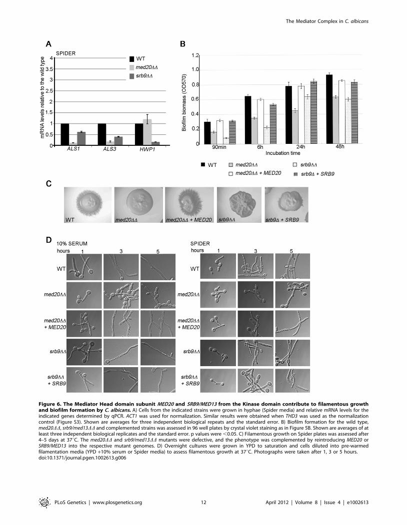
Figure 6. The Mediator Head domain subunit MED20 and SRB9/MED13 from the Kinase domain contribute to filamentous growthand biofilm formation by C. albicans. A) Cells from the indicated strains were grown in hyphae (Spider media) and relative mRNA levels for theindicated genes determined by qPCR. ACT1 was used for normalization. Similar results were obtained when THD3 was used as the normalizationcontrol (Figure S3). Shown are averages for three independent biological repeats and the standard error. B) Biofilm formation for the wild type,med20DD, srb9/med13DD and complemented strains was assessed in 96 well plates by crystal violet staining as in Figure 5B. Shown are averages of atleast three independent biological replicates and the standard error. p values were ,0.05. C) Filamentous growth on Spider plates was assessed after4–5 days at 37uC. The med20DD and srb9/med13DD mutants were defective, and the phenotype was complemented by reintroducing MED20 orSRB9/MED13 into the respective mutant genomes. D) Overnight cultures were grown in YPD to saturation and cells diluted into pre-warmedfilamentation media (YPD +10% serum or Spider media) to assess filamentous growth at 37uC. Photographs were taken after 1, 3 or 5 hours.doi:10.1371/journal.pgen.1002613.g006
The Mediator Complex in C. albicans
PLoS Genetics | www.plosgenetics.org 12 April 2012 | Volume 8 | Issue 4 | e1002613
repression of the FLO adhesins [25], which is again opposite to the
activating function for Srb9/Med13 in the expression of ALS1/3
and HWP1 that we observed in C. albicans. Other subunits of the
Mediator Kinase domain as well as Sin4/Med16 from the Tail
also inhibit expression of the S. cerevisiae FLO genes [25,40]. In
Schizo. pombe, the Mediator Kinase domain subunits Srb10 and
Srb8/Med12 also repress the cell wall adhesins, despite the fact
that some of the affected adhesins are not related to the FLO genes
[28].
While the FLO and ALS gene are not related, there is
conservation in terms of the pathways and transcription factors
that regulate their expression in S. cerevisiae and C. albicans
respectively. Examples include positive regulation by activators
Flo8, Ste12/Cph1 and Tec1, and negative regulation by
repressors Nrg1, Tup1 and Sfl1 [50,59,80–83]. Mediator interacts
with gene-specific transcription factors to regulate gene expression
[76,84]. It is possible that the key transcription factor(s) through
which Med31 and Srb9/Med13 act to regulate the adhesin-
encoding genes differ between the two yeasts. How Mediator
subunits repress the S. cerevisiae FLO genes is not well understood,
but a suggested mechanism involves functional interactions with
the repressors Sfl1 and Tup1 [85]. Both Sfl1 and Tup1 are
required for repression of the ALS genes in C. albicans [50,81], but
the requirement for specific Mediator subunits in the repression of
adhesins by these factors in C. albicans could differ from what has
been suggested in S. cerevisiae. Another possibility relates to
silencing mechanisms. The FLO genes in S. cerevisiae are subject
to position-dependent silencing regulated by histone deacetylases
([86]; reviewed in [87]). The C. albicans adhesins are not known to
be regulated by silencing. Mediator has been implicated in the
regulation of chromatin structure and gene expression in regions
affected by silencing, such as the sub-telomeres [12,21–24]. Based
on work in S. cerevisiae, it has been proposed that Mediator
contributes to the establishment of a repressive chromatin
structure by binding to the silenced regions and influencing the
recruitment of the histone deaceytlase Sir2 and the histone
acetyltransferase Sas2 and thus the acetylation status of histone H4
K16, a mark of active chromatin [24]. While this has not been
tested directly, an interesting possibility is that the regulation of
chromatin contributes to the repressive role of the Mediator
subunits in the expression of the S. cerevisiae FLO genes. For
example, the Tail subunit Sin4/Med16, which is a repressor of
FLO11 and of the sub-telomeric FLO1 adhesin [40,85], contributes
to sub-telomeric silencing [24]. Med7 has also been found to affect
chromatin structure, by regulating H4K16 acetylation and the
presence of Sir2 and Sas2 at the sub-telomeres [24]. Med7 is
closely functionally linked with Med31 - the N-terminal part of
Med7 together with Med31 forms a structural and functional sub-
module of Mediator, Med7N/31 [43]. It will be interesting to
study whether differences in silencing mechanisms that operate on
the FLO and ALS genes contribute to determining how Mediator
subunits control their expression.
Materials and Methods
Yeast strains and growth conditionsThe C. albicans strains used in this study are derivatives of
BWP17 [88]. The med31DD, srb9DD and med20DD strains were
constructed by standard methods based on PCR and homologous
recombination, using ARG4 and URA3 as selective markers. The
Figure 7. Regulation of adhesins and Ace2-dependent genes by MED20 and SRB9/MED13 in C. albicans. A) Cells from the indicated strainswere grown in yeast form (YPD) and relative mRNA levels for the indicated genes determined by qPCR. ACT1 was used for normalization. Similarresults were obtained when THD3 was used as the normalization control (Figure S3). Shown are averages for three independent biological repeatsand the standard error. B) Yeast cell morphology of the wild type and Mediator mutants was assessed in YPD at 30uC. The scale bar is 20 mm.doi:10.1371/journal.pgen.1002613.g007
The Mediator Complex in C. albicans
PLoS Genetics | www.plosgenetics.org 13 April 2012 | Volume 8 | Issue 4 | e1002613

complemented strains were constructed by re-introducing a wild
type copy of MED31, SRB9 or MED20 under own promoter and
terminator into the HIS1 locus of the respective mutants using the
integrative plasmid pDDB78. To make matched HIS1+ mutant
strains, an empty pDDB78 vector was integrated into the genome
of the respective strains. The ace2 mutant is a homozygous mutant
in the BWP17 strain background and was a generous gift from
Aaron Mitchell (this strain is also URA3+ ARG4+ HIS1+). The
med31DD +TEF1-ALS1 overexpression strain was constructed
using the plasmid pCJN498, as described in [65]. The S. cerevisiae
med31D mutant was constructed in the g1278b strain using the
KANMX4 cassette. The wt and flo11D mutant of S1278b were a
generous gift from Todd Reynolds and are described in [67].
Standard growth conditions were YPD (2% glucose, 2%
peptone, 1% yeast extract), at 30uC, 200 rpm. For ura2 strains
the media was supplemented with 80 mg/ml uridine. The mutants
were selected using minimal media lacking the appropriate amino
acids. The TEF1-ALS1 overexpression strains have a nourseo-
thricin resistance cassette (NAT) and were selected on 400 mg/ml
NAT plates (nourseothricin was from Werner Bioagents). The S.
cerevisiae mutant was selected on 200 mg/ml G418 plates.
For cell morphology analysis (Figure 3A), cells were classified as
being in a chain if 3 or more cells were attached. An average of
200 cells per sample were counted, and the experiments were
repeated at least with 3 independent cultures. The average and the
standard error are shown in the figure. To observe the mother-bud
junctions, cells were stained with calcofluor white (1 mg/ml) for
8 min in the dark, followed by washes in phosphate buffered saline
(PBS). Imaging was done using an Olympus IX81 microscope with
the Olympus cell‘M software, using the 1006objective with DIC
or the DAPI filter for calcofluor white stained cells.
Filamentous growth was tested by dilution of cells from
overnight cultures grown to OD600 = 0.1–0.2 into pre-warmed
YPD+10% calf serum, Spider media (1% nutrient broth, 1% D-
mannitol, 2 g K2HPO4), M199 or N-acetylglucosamine media
(9 g NaCl, 6.7 g yeast nitrogen base and 0.56 g N-acetylglucosa-
mine per liter) and incubated at 37uC for the times indicates in the
figures. All cell imaging was done using an Olympus IX81
microscope with the Olympus cell‘M software. For testing
filamentation on plates, C. albicans strains were re-streaked on
plates containing filamentous-growth inducing media. Plates were
incubated for up to five days at 37uC and colonies examined and
photographed with a stereo dissecting microscope (Olympus SZX
16).
For analysis of sensitivities to various drugs and chemicals, ten
fold serial dilutions of cultures from wild type and mutant strains
were dropped on control plates, or plates containing the
compounds indicated in Figure S4 and Table 3. Plates were
incubated at 30uC for three days (unless growth was assessed at
37uC or 16uC), and photographed.
Biofilm assays and imagingC. albicans biofilms were grown in 96-well microtiter plates or on
silicone disks for quantitative or qualitative analysis respectively.
Quantitative biofilm assays were performed as described [89,90].
100 ml of cultures of C. albicans wild type or mutant strains (107
cells/ml in Spider media) were added to wells and incubated at
37uC with gentle shaking (75 rpm) for 90 min (adhesion phase).
Non-adherent cells were discarded and 100 ml of fresh Spider
media were added to each of the wells. Biofilms were allowed to
develop for a future 4.5 h (6 h in total), 24 h, and 48 h,
representing the early, intermediate or mature stage of biofilm
development respectively. The medium was replenished after 24 h
by aspiration and addition of fresh medium. Biofilm biomass was
determined at the different time points using crystal violet staining.
Wells containing only Spider medium with no yeast served as
negative controls. For qualitative studies, biofilms were formed in
vitro on serum-treated silicone disks, which is a well-established
system for biofilm analysis [66]. Sterile silicone disks were
pretreated with fetal bovine serum (Sigma) overnight at 37uCwith gentle shaking (75 rpm). The silicone disks were then washed
twice with PBS and transferred to a 12-well plate containing 2 ml
of freshly prepared cell suspensions (107 cells/ml in Spider media).
The plate was incubated for 1.5 h at 37uC with gentle shaking
Figure 8. The roles of Med31 in adhesion are different betweenS. cerevisiae and C. albicans. A) Colonies of wild type S. cerevisiaeS1278b and the med31D mutant were grown on YPD plates at 30uCand photographed. The flo11D strain was used as the negative control,to show smooth colony morphology in the absence of FLO11. B)Expression of FLO11 was tested by qPCR after 90 min in 0.2% glucosesynthetic complete media, the condition used for biofilm formation inC). Levels of FLO11 were normalized to ACT1, and expressed related tothe wild type, which was set to 1. Shown are averages from at leastthree independent biological repeats and the standard error. C) Theability of the S. cerevisiae med31D mutant to adhere to polystyrene wasassessed in 0.2% glucose synthetic complete media as described inMaterials and Methods. Quantification was performed by crystal violetstaining. At least three independent cultures were used, assayed inquadruplicates. For the mutant, two independently constructeddeletion strains were used and gave equivalent results. The flo11Dmutant was assayed in parallel as a negative control and showed noadherence at any of the time points (not shown). p,0.001.doi:10.1371/journal.pgen.1002613.g008
The Mediator Complex in C. albicans
PLoS Genetics | www.plosgenetics.org 14 April 2012 | Volume 8 | Issue 4 | e1002613
(75 rpm) to allow the yeast to adhere to the disk surfaces. The
silicone disks were then washed in PBS and transferred to a new
12-well plate with Spider media followed by incubation for 48 h at
37uC with shaking at 75 rpm. The established biofilms were
examined with SEM or CLSM. For SEM, biofilms were fixed with
glutaraldehyde (2.5%, v/v, in 0.1 M cacodylate buffer, pH 7.0)
and 1% osmium tetraoxide at room temperature, and dehydrated
with gradually increased ethanol (50%, 75%, 95%, 100%, and
absolute 100%) and hexamethyldisilazane (HMDS) (50%, 75%,
95%, 100%, and absolute 100%). Samples were coated with gold
with a Balzers SCD005 sputter coater and viewed under a Hitachi
S570 scanning electron microscope. For CSLM, biofilms were
stained with FUN-1 (10 mM, Molecular Probes) and Concanavalin
A–Alexafluor488 conjugate (Con A, 25 mg ml21; Molecular
Probes) for 45 min at 37uC. Stained biofilms were observed with
a Leica SP5 CLSM, and images were captured and processed
using the softwares Leica LAS AF and Amira 5.2.1. All
experiments for quantitative analysis were repeated at least three
times in triplicate, and qualitative assays were repeated at least 3
times. One-way ANOVA was used to compare the difference in
biofilm biomass produced by different C. albicans strains. p values
of ,0.05 were considered to be statistically significant.
For S. cerevisiae biofilms (Figure 8), overnight cultures were
growth in synthetic complete media supplemented with 2%
glucose. The cells were then resuspended into 96 well polystyrene
plates to an OD600 = 1.0 using synthetic 0.2% glucose media [67].
Adherence was assayed at the time points indicated in the figure
using crystal violet staining as described above.
RNA extraction, quantitative PCR, and microarray analysisRNA was extracted using the hot-phenol method from cultures
grown in YPD at 30uC to log phase (OD600 = 1). Following hot-
phenol extraction, 100 mg of RNA samples were further purified
using the RNAeasy kit and following the manufacturer’s
instructions. The microarray analysis was performed as described
[91], on microarrays spotted with 6459 70-mer oligonucleotides
(GEO Platform GPL9818). Data normalization and analysis was
conducted in GeneSpring GX version 7.3 (Agilent Technologies).
The microarray data set has been deposited in GEO, under
accession number GSE31632. Genes that were up- or down-
regulated in the med31DD mutant by 1.5 fold or more (p#0.05)
were selected from a Volcano Plot and considered to be
differentially expressed. Gene ontology analysis was performed
at the Candida genome database (CGD, candidagenome.org)
[92]. GSEA analysis ([93] and Sellam et al, submitted) was
performed using the GseaPreranked tool and the weighted
enrichment statistics on 6387 (for C. albicans) gene sets each
containing 5–500 genes. Statistical significance was estimated from
1000 permutations. Enrichment maps were constructed with
Cytoscape 2.8 (http://www.cytoscape.org; [94]) and the Enrich-
ment Map 1.1 plug-in (http://baderlab.org/Software/
EnrichmentMap) using the default settings. For quantitative
PCR, reverse transcription was performed using the Transcriptor
High Fidelity cDNA synthesis kit from Roche. qPCR reactions
were prepared using Fast-Start Sybr Green Master (Roche) on an
Eppendorf Realplex master cycler and analysed by absolute
quantification. The expression levels of the mRNAs were
normalized to the level of ACT1 or the GAPDH encoding gene
TDH3. Three independent cultures were analyzed, with two
technical replicates each. qPCR primers that enable differential
amplification of the ALS1 and ALS3 genes were from Green et al
[95]. Sequences for all qPCR primers used in this study are listed
in Table S2. For analysis of gene expression in YPD, cultures were
grown under the same condition as for the microarrays analysis, to
mid log phase (OD600 = 1). For assaying expression of genes under
hyphal growth, strains were grown in Spider media as described in
[65].
Worm infection assaysThe worm-C. albicans infection assay was performed as
described previously [64]. Briefly, young adult nematodes were
allowed to feed for 4 h on lawns of C. albicans grown on solid BHI
media (Difco) containing ampicillin (100 mg/ml), kanamycin
(50 mg/ml) and streptomycin (200 mg/ml). Worms were washed
with M9 media and transferred into wells of a six-well microtiter
dish (Corning) containing 2 ml of liquid media (80% M9 and 20%
BHI) at 60 to 80 worms per well. The plates were incubated at
25uC, and worms were qualitatively assessed at 24 h intervals for
penetrative C. albicans filamentation using a DIC microscope and
photographed using an Olympus IX81 microscope with the
Olympus cell‘M software. The % of worms with penetrative
filamentation was determined from four independent experiments
at day 3 of the infection. Means and the standard error were
calculated, and the p value was determined using the student t-test.
The killing assays (Figure 4C) were performed three times and
equivalent results were obtained.
Supporting Information
Dataset S1 The complete transcriptome analysis data for the
med31DD mutant.
(XLS)
Dataset S2 Gene ontology and other functional analysis of the
med31DD mutant transcriptome.
(XLSX)
Figure S1 Analysis of the med31DD transcriptome data A)
Chromosomal view of the transcriptional profile of the med31DDmutant of C. albicans. B) Scatter plot comparisons of the med31DDtranscriptome to changes in gene expression observed upon
oxidative or osmotic stress in C. albicans.
(TIF)
Figure S2 A complete GSEA network of genes differentially
expressed in the med31DD mutant of C. albicans. The gene
categories can be viewed by zooming in.
(PDF)
Figure S3 qPCR analysis of gene expression in Mediator
mutants of C. albicans. Cells were grown in YPD at 30uC for yeast
growth or Spider at 37uC for hyphal growth and gene expression
analysed as described in the Materials and Methods. The levels of
the indicated genes were normalised to the levels of the
glyceraldehyde phosphate dehydrogenase (GAPDH)-encoding
gene TDH3. Shown are averages of three independent experi-
ments and the standard error.
(TIF)
Figure S4 Sensitivities of the C. albicans Mediator mutants to
various stresses. 10 fold serial dilutions of the wild type, Mediator
mutants and complemented strains were dropped on YPD plates
containing the indicated compounds. The plates were incubated at
30uC (unless stated otherwise) for 3–4 days and photographed.
The mutants were scored as sensitive or resistant and the results
are presented in Table 3.
(TIF)
Figure S5 Filamentation defect of the med31DD mutant in liquid
media. Overnight cultures of the indicated strains were diluted
into media pre-warmed at 37uC and the appearance of
The Mediator Complex in C. albicans
PLoS Genetics | www.plosgenetics.org 15 April 2012 | Volume 8 | Issue 4 | e1002613
filamentous cells was monitored over time. The images were taken
with a 1006magnification objective and the scale bar represents
10 mm. In Spider media, the mutant has a pronounced
filamentation defect, while in the other media filamentation by
the mutant is somewhat delayed, and a larger number of cell
chains and pseudohyphae is observed than in the complemented
strain.
(TIF)
Figure S6 The med31DD mutant has a filamentation defect in
the worm infection assay even after prolonged incubation. Worms
infected with the wild type, the med31DD mutant or the
complemented med31DD+MED31 strain were imaged 7 days post
infection using an Olympus IX81 microscope.
(TIF)
Figure S7 Scanning electron microscopy of wild type and
med31DD mutant biofilms. Biofilms were formed in vitro on serum
coated silicone disks. Mature biofilms (48 h) were imaged by
scanning electron microscopy (SEM) as described in Materials and
Methods. The SEM experiments confirmed the biofilm defect of
the med31DD mutant observed by confocal microscopy (Figure 5).
(TIF)
Figure S8 Ectopic expression of ALS1 does not complement the
growth defect of the med31DD mutant. Cultures from the indicated
strains were grown in either rich YPD (upper panel) or minimal
synthetic complete media (lower panel). Growth was assessed by
measuring OD600 at regular intervals over an 8 h time course.
Three independent clones of the med31DD+TEF1-ALS1 strain
were tested, all of which rescued the biofilm formation defect of
the med31DD mutant, but not the growth defect.
(TIF)
Table S1 Comparison of the transcriptome changes in the C.
albicans med31DD mutant and those observed upon inactivation of
Ace2. The ace2 data is from [57].
(DOC)
Table S2 The list of primers used in this study.
(DOC)
Author Contributions
Conceived and designed the experiments: NU YQ BJ AN AYP AT.
Performed the experiments: NU YQ BJ TLL FB AYP JC GR CB AT.
Analyzed the data: NU YQ BJ THB TQ AN AYP AT PRB. Wrote the
paper: AT.
References
1. Kim YJ, Bjorklund S, Li Y, Sayre MH, Kornberg RD (1994) A multiprotein
mediator of transcriptional activation and its interaction with the C-terminalrepeat domain of RNA polymerase II. Cell 77: 599–608.
2. Conaway RC, Conaway JW (2011) Function and regulation of the Mediatorcomplex. Curr Opin Genet Dev 21: 225–230.
3. Dotson MR, Yuan CX, Roeder RG, Myers LC, Gustafsson CM, et al. (2000)Structural organization of yeast and mammalian mediator complexes. Proc Natl
Acad Sci U S A 97: 14307–14310.
4. Davis JA, Takagi Y, Kornberg RD, Asturias FA (2002) Structure of the yeast
RNA polymerase II holoenzyme: Mediator conformation and polymerase
interaction. Mol Cell 10: 409–415.
5. Guglielmi B, van Berkum NL, Klapholz B, Bijma T, Boube M, et al. (2004) A
high resolution protein interaction map of the yeast Mediator complex. NucleicAcids Res 32: 5379–5391.
6. Cai G, Imasaki T, Takagi Y, Asturias FJ (2009) Mediator structural conservation
and implications for the regulation mechanism. Structure 17: 559–567.
7. van de Peppel J, Kettelarij N, van Bakel H, Kockelkorn TT, van Leenen D, et
al. (2005) Mediator expression profiling epistasis reveals a signal transduction
pathway with antagonistic submodules and highly specific downstream targets.Mol Cell 19: 511–522.
8. Collins SR, Miller KM, Maas NL, Roguev A, Fillingham J, et al. (2007)Functional dissection of protein complexes involved in yeast chromosome
biology using a genetic interaction map. Nature 446: 806–810.
9. Holstege FC, Jennings EG, Wyrick JJ, Lee TI, Hengartner CJ, et al. (1998)
Dissecting the regulatory circuitry of a eukaryotic genome. Cell 95: 717–728.
10. Borggrefe T, Davis R, Erdjument-Bromage H, Tempst P, Kornberg RD (2002)
A complex of the Srb8, -9, -10, and -11 transcriptional regulatory proteins fromyeast. J Biol Chem 277: 44202–44207.
11. Mo X, Kowenz-Leutz E, Xu H, Leutz A (2004) Ras induces mediator complexexchange on C/EBP beta. Mol Cell 13: 241–250.
12. Malik S, Roeder RG (2010) The metazoan Mediator co-activator complex as anintegrative hub for transcriptional regulation. Nat Rev Genet 11: 761–772.
13. Bjorklund S, Gustafsson CM (2005) The yeast Mediator complex and itsregulation. Trends Biochem Sci 30: 240–244.
14. Mittler G, Kremmer E, Timmers HT, Meisterernst M (2001) Novel critical roleof a human Mediator complex for basal RNA polymerase II transcription.
EMBO Rep 2: 808–813.
15. Baek HJ, Malik S, Qin J, Roeder RG (2002) Requirement of TRAP/mediator
for both activator-independent and activator-dependent transcription inconjunction with TFIID-associated TAF(II)s. Mol Cell Biol 22: 2842–2852.
16. Wang G, Balamotis MA, Stevens JL, Yamaguchi Y, Handa H, et al. (2005)Mediator requirement for both recruitment and postrecruitment steps in
transcription initiation. Mol Cell 17: 683–694.
17. Malik S, Barrero MJ, Jones T (2007) Identification of a regulator of transcription
elongation as an accessory factor for the human Mediator coactivator. Proc NatlAcad Sci U S A 104: 6182–6187.
18. Guglielmi B, Soutourina J, Esnault C, Werner M (2007) TFIIS elongation factorand Mediator act in conjunction during transcription initiation in vivo. Proc
Natl Acad Sci U S A 104: 16062–16067.
19. Donner AJ, Ebmeier CC, Taatjes DJ, Espinosa JM (2010) CDK8 is a positive
regulator of transcriptional elongation within the serum response network. Nat
Struct Mol Biol 17: 194–201.
20. Yudkovsky N, Ranish JA, Hahn S (2000) A transcription reinitiation
intermediate that is stabilized by activator. Nature 408: 225–229.
21. Black JC, Choi JE, Lombardo SR, Carey M (2006) A mechanism forcoordinating chromatin modification and preinitiation complex assembly. Mol
Cell 23: 809–818.
22. Ding N, Zhou H, Esteve PO, Chin HG, Kim S, et al. (2008) Mediator links
epigenetic silencing of neuronal gene expression with x-linked mental
retardation. Mol Cell 31: 347–359.
23. Lenstra TL, Benschop JJ, Kim T, Schulze JM, Brabers NA, et al. (2011) The
specificity and topology of chromatin interaction pathways in yeast. Mol Cell 42:536–549.
24. Zhu X, Liu B, Carlsten JO, Beve J, Nystrom T, et al. (2011) Mediator influences
telomeric silencing and cellular life span. Mol Cell Biol 31: 2413–2421.
25. Chang YW, Howard SC, Herman PK (2004) The Ras/PKA signaling pathway
directly targets the Srb9 protein, a component of the general RNA polymerase IItranscription apparatus. Mol Cell 15: 107–116.
26. Hallberg M, Polozkov GV, Hu GZ, Beve J, Gustafsson CM, et al. (2004) Site-specific Srb10-dependent phosphorylation of the yeast Mediator subunit Med2
regulates gene expression from the 2-microm plasmid. Proc Natl Acad Sci U S A
101: 3370–3375.
27. Bourbon HM (2008) Comparative genomics supports a deep evolutionary origin
for the large, four-module transcriptional mediator complex. Nucleic Acids Res36: 3993–4008.
28. Linder T, Rasmussen NN, Samuelsen CO, Chatzidaki E, Baraznenok V, et al.
(2008) Two conserved modules of Schizosaccharomyces pombe Mediator regulatedistinct cellular pathways. Nucleic Acids Res 36: 2489–2504.
29. Chang YW, Howard SC, Budovskaya YV, Rine J, Herman PK (2001) The ryemutants identify a role for Ssn/Srb proteins of the RNA polymerase II
holoenzyme during stationary phase entry in Saccharomyces cerevisiae. Genetics
157: 17–26.
30. Wheeler RT, Kombe D, Agarwala SD, Fink GR (2008) Dynamic, morphotype-
specific Candida albicans beta-glucan exposure during infection and drugtreatment. PLoS Pathog 4: e1000227. doi:10.1371/journal.ppat.1000227.
31. Wang LI, Lin YS, Liu KH, Jong AY, Shen WC (2011) Cryptococcus neoformans
Mediator protein Ssn8 negatively regulates diverse physiological processes and isrequired for virulence. PLoS ONE 6: e19162. doi:10.1371/journal.-
pone.0019162.
32. Shim WB, Woloshuk CP (2001) Regulation of fumonisin B(1) biosynthesis and
conidiation in Fusarium verticillioides by a cyclin-like (C-type) gene, FCC1. ApplEnviron Microbiol 67: 1607–1612.
33. Bluhm BH, Woloshuk CP (2006) Fck1, a C-type cyclin-dependent kinase,
interacts with Fcc1 to regulate development and secondary metabolism inFusarium verticillioides. Fungal Genet Biol 43: 146–154.
34. Zhou X, Heyer C, Choi YE, Mehrabi R, Xu JR (2010) The CID1 cyclin C-likegene is important for plant infection in Fusarium graminearum. Fungal Genet Biol
47: 143–151.
35. Blankenship JR, Fanning S, Hamaker JJ, Mitchell AP (2010) An extensivecircuitry for cell wall regulation in Candida albicans. PLoS Pathog 6: e1000752.
doi:10.1371/journal.ppat.1000752.
36. Nelson C, Goto S, Lund K, Hung W, Sadowski I (2003) Srb10/Cdk8 regulates
yeast filamentous growth by phosphorylating the transcription factor Ste12.
Nature 421: 187–190.
The Mediator Complex in C. albicans
PLoS Genetics | www.plosgenetics.org 16 April 2012 | Volume 8 | Issue 4 | e1002613
37. Cooper KF, Mallory MJ, Strich R (1999) Oxidative stress-induced destruction of
the yeast C-type cyclin Ume3p requires phosphatidylinositol-specific phospho-
lipase C and the 26S proteasome. Mol Cell Biol 19: 3338–3348.
38. Thakur JK, Arthanari H, Yang F, Pan SJ, Fan X, et al. (2008) A nuclear
receptor-like pathway regulating multidrug resistance in fungi. Nature 452:
604–609.
39. Paul S, Schmidt JA, Moye-Rowley WS (2011) Regulation of the CgPdr1
transcription factor from the pathogen Candida glabrata. Eukaryot Cell 10:
187–197.
40. Fichtner L, Schulze F, Braus GH (2007) Differential Flo8p-dependent regulation
of FLO1 and FLO11 for cell-cell and cell-substrate adherence of S. cerevisiae
S288c. Mol Microbiol 66: 1276–1289.
41. Arbour M, Epp E, Hogues H, Sellam A, Lacroix C, et al. (2009) Widespread
occurrence of chromosomal aneuploidy following the routine production of
Candida albicans mutants. FEMS Yeast Res 9: 1070–1077.
42. Miklos I, Szilagyi Z, Watt S, Zilahi E, Batta G, et al. (2008) Genomic expression
patterns in cell separation mutants of Schizosaccharomyces pombe defective in the
genes sep10 (+) and sep15 (+) coding for the Mediator subunits Med31 and
Med8. Mol Genet Genomics 279: 225–238.
43. Koschubs T, Seizl M, Lariviere L, Kurth F, Baumli S, et al. (2009)
Identification, structure, and functional requirement of the Mediator submodule
Med7N/31. EMBO J 28: 69–80.
44. Subramanian A, Tamayo P, Mootha VK, Mukherjee S, Ebert BL, et al. (2005)
Gene set enrichment analysis: a knowledge-based approach for interpreting
genome-wide expression profiles. Proc Natl Acad Sci U S A 102: 15545–15550.
45. Mootha VK, Lindgren CM, Eriksson KF, Subramanian A, Sihag S, et al. (2003)
PGC-1alpha-responsive genes involved in oxidative phosphorylation are
coordinately downregulated in human diabetes. Nat Genet 34: 267–273.
46. Stark C, Breitkreutz BJ, Reguly T, Boucher L, Breitkreutz A, et al. (2006)
BioGRID: a general repository for interaction datasets. Nucleic Acids Res 34:
D535–539.
47. Marcil A, Gadoury C, Ash J, Zhang J, Nantel A, et al. (2008) Analysis of PRA1
and its relationship to Candida albicans-macrophage interactions. Infect Immun
76: 4345–4358.
48. Spiering MJ, Moran GP, Chauvel M, Maccallum DM, Higgins J, et al. (2010)
Comparative transcript profiling of Candida albicans and Candida dubliniensis
identifies SFL2, a C. albicans gene required for virulence in a reconstituted
epithelial infection model. Eukaryot Cell 9: 251–265.
49. Fradin C, De Groot P, MacCallum D, Schaller M, Klis F, et al. (2005)
Granulocytes govern the transcriptional response, morphology and proliferation
of Candida albicans in human blood. Mol Microbiol 56: 397–415.
50. Kadosh D, Johnson AD (2005) Induction of the Candida albicans filamentous
growth program by relief of transcriptional repression: a genome-wide analysis.
Mol Biol Cell 16: 2903–2912.
51. Nantel A, Dignard D, Bachewich C, Harcus D, Marcil A, et al. (2002)
Transcription profiling of Candida albicans cells undergoing the yeast-to-hyphal
transition. Mol Biol Cell 13: 3452–3465.
52. Harcus D, Nantel A, Marcil A, Rigby T, Whiteway M (2004) Transcription
profiling of cyclic AMP signaling in Candida albicans. Mol Biol Cell 15:
4490–4499.
53. Bachewich C, Nantel A, Whiteway M (2005) Cell cycle arrest during S or M
phase generates polarized growth via distinct signals in Candida albicans. Mol
Microbiol 57: 942–959.
54. Cote P, Hogues H, Whiteway M (2009) Transcriptional analysis of the Candida
albicans cell cycle. Mol Biol Cell 20: 3363–3373.
55. Enjalbert B, Nantel A, Whiteway M (2003) Stress-induced gene expression in
Candida albicans: absence of a general stress response. Mol Biol Cell 14:
1460–1467.
56. Butler G, Rasmussen MD, Lin MF, Santos MA, Sakthikumar S, et al. (2009)
Evolution of pathogenicity and sexual reproduction in eight Candida genomes.
Nature 459: 657–662.
57. Mulhern SM, Logue ME, Butler G (2006) Candida albicans transcription factor
Ace2 regulates metabolism and is required for filamentation in hypoxic
conditions. Eukaryot Cell 5: 2001–2013.
58. Hoyer LL, Green CB, Oh SH, Zhao X (2008) Discovering the secrets of the
Candida albicans agglutinin-like sequence (ALS) gene family–a sticky pursuit. Med
Mycol 46: 1–15.
59. Argimon S, Wishart JA, Leng R, Macaskill S, Mavor A, et al. (2007)
Developmental regulation of an adhesin gene during cellular morphogenesis in
the fungal pathogen Candida albicans. Eukaryot Cell 6: 682–692.
60. Kim S, Wolyniak MJ, Staab JF, Sundstrom P (2007) A 368-base-pair cis-acting
HWP1 promoter region, HCR, of Candida albicans confers hypha-specific gene
regulation and binds architectural transcription factors Nhp6 and Gcf1p.
Eukaryot Cell 6: 693–709.
61. Heilmann CJ, Sorgo AG, Siliakus AR, Dekker HL, Brul S, et al. (2011) Hyphal
induction in the human fungal pathogen Candida albicans reveals a
characteristic wall protein profile. Microbiology 157: 2297–2307.
62. Homann OR, Dea J, Noble SM, Johnson AD (2009) A phenotypic profile of the
Candida albicans regulatory network. PLoS Genet 5: e1000783. doi:10.1371/
journal.pgen.1000783.
63. Breger J, Fuchs BB, Aperis G, Moy TI, Ausubel FM, et al. (2007) Antifungal
chemical compounds identified using a C. elegans pathogenicity assay. PLoS
Pathog 3: e18. doi:10.1371/journal.ppat.0030018.
64. Peleg AY, Tampakakis E, Fuchs BB, Eliopoulos GM, Moellering RC, Jr., et al.
(2008) Prokaryote-eukaryote interactions identified by using Caenorhabditis elegans.
Proc Natl Acad Sci U S A 105: 14585–14590.
65. Nobile CJ, Andes DR, Nett JE, Smith FJ, Yue F, et al. (2006) Critical role of
Bcr1-dependent adhesins in C. albicans biofilm formation in vitro and in vivo.
PLoS Pathog 2: e63. doi:10.1371/journal.ppat.0020063.
66. Nobile CJ, Mitchell AP (2005) Regulation of cell-surface genes and biofilm
formation by the C. albicans transcription factor Bcr1p. Curr Biol 15: 1150–1155.
67. Reynolds TB, Fink GR (2001) Bakers’ yeast, a model for fungal biofilmformation. Science 291: 878–881.
68. Lariviere L, Geiger S, Hoeppner S, Rother S, Strasser K, et al. (2006) Structure
and TBP binding of the Mediator head subcomplex Med8-Med18-Med20. Nat
Struct Mol Biol 13: 895–901.
69. Lariviere L, Seizl M, van Wageningen S, Rother S, van de Pasch L, et al. (2008)
Structure-system correlation identifies a gene regulatory Mediator submodule.
Genes Dev 22: 872–877.
70. Carrera I, Janody F, Leeds N, Duveau F, Treisman JE (2008) Pygopus activates
Wingless target gene transcription through the mediator complex subunits
Med12 and Med13. Proc Natl Acad Sci U S A 105: 6644–6649.
71. Shahi P, Gulshan K, Naar AM, Moye-Rowley WS (2010) Differential roles of
transcriptional mediator subunits in regulation of multidrug resistance gene
expression in Saccharomyces cerevisiae. Mol Biol Cell 21: 2469–2482.
72. Kelly MT, MacCallum DM, Clancy SD, Odds FC, Brown AJ, et al. (2004) The
Candida albicans CaACE2 gene affects morphogenesis, adherence and virulence.
Mol Microbiol 53: 969–983.
73. Mehta S, Miklos I, Sipiczki M, Sengupta S, Sharma N (2009) The Med8
mediator subunit interacts with the Rpb4 subunit of RNA polymerase II and
Ace2 transcriptional activator in Schizosaccharomyces pombe. FEBS Lett 583:
3115–3120.
74. Nobile CJ, Schneider HA, Nett JE, Sheppard DC, Filler SG, et al. (2008)
Complementary adhesin function in C. albicans biofilm formation. Curr Biol 18:1017–1024.
75. Ansari SA, He Q, Morse RH (2009) Mediator complex association with
constitutively transcribed genes in yeast. Proc Natl Acad Sci U S A 106:
16734–16739.
76. Fan X, Chou DM, Struhl K (2006) Activator-specific recruitment of Mediator in
vivo. Nat Struct Mol Biol 13: 117–120.
77. Fan X, Struhl K (2009) Where does mediator bind in vivo? PLoS ONE 4:
e5029. doi:10.1371/journal.pone.0005029.
78. Andrau JC, van de Pasch L, Lijnzaad P, Bijma T, Koerkamp MG, et al. (2006)
Genome-wide location of the coactivator Mediator: binding without activation
and transient Cdk8 interaction on DNA. Mol Cell 22: 179–192.
79. Li F, Palecek SP (2003) EAP1, a Candida albicans gene involved in binding human
epithelial cells. Eukaryot Cell 2: 1266–1273.
80. Bruckner S, Mosch HU (2012) Choosing the right lifestyle: adhesion and
development in Saccharomyces cerevisiae. FEMS Microbiol Rev 36: 25–58.
81. Bauer J, Wendland J (2007) Candida albicans Sfl1 suppresses flocculation and
filamentation. Eukaryot Cell 6: 1736–1744.
82. Murad AM, Leng P, Straffon M, Wishart J, Macaskill S, et al. (2001) NRG1
represses yeast-hypha morphogenesis and hypha-specific gene expression in
Candida albicans. EMBO J 20: 4742–4752.
83. Cao F, Lane S, Raniga PP, Lu Y, Zhou Z, et al. (2006) The Flo8 transcription
factor is essential for hyphal development and virulence in Candida albicans. Mol
Biol Cell 17: 295–307.
84. Kuras L, Borggrefe T, Kornberg RD (2003) Association of the Mediator
complex with enhancers of active genes. Proc Natl Acad Sci U S A 100:
13887–13891.
85. Conlan RS, Tzamarias D (2001) Sfl1 functions via the co-repressor Ssn6-Tup1
and the cAMP-dependent protein kinase Tpk2. J Mol Biol 309: 1007–1015.
86. Halme A, Bumgarner S, Styles C, Fink GR (2004) Genetic and epigenetic
regulation of the FLO gene family generates cell-surface variation in yeast. Cell
116: 405–415.
87. Verstrepen KJ, Fink GR (2009) Genetic and epigenetic mechanisms underlying
cell-surface variability in protozoa and fungi. Annu Rev Genet 43: 1–24.
88. Wilson RB, Davis D, Mitchell AP (1999) Rapid hypothesis testing with Candida
albicans through gene disruption with short homology regions. J Bacteriol 181:
1868–1874.
89. Jin Y, Samaranayake LP, Samaranayake Y, Yip HK (2004) Biofilm formation of
Candida albicans is variably affected by saliva and dietary sugars. Arch Oral Biol
49: 789–798.
90. Jin Y, Yip HK, Samaranayake YH, Yau JY, Samaranayake LP (2003) Biofilm-
forming ability of Candida albicans is unlikely to contribute to high levels of oral
yeast carriage in cases of human immunodeficiency virus infection. J Clin
Microbiol 41: 2961–2967.
91. Dagley MJ, Gentle IE, Beilharz TH, Pettolino FA, Djordjevic JT, et al. (2011)
Cell wall integrity is linked to mitochondria and phospholipid homeostasis in
Candida albicans through the activity of the post-transcriptional regulator Ccr4-
Pop2. Mol Microbiol 79: 968–989.
92. Skrzypek MS, Arnaud MB, Costanzo MC, Inglis DO, Shah P, et al. (2010) New
tools at the Candida Genome Database: biochemical pathways and full-text
literature search. Nucleic Acids Res 38: D428–D432.
93. Hussein B, Huang H, Glory A, Osmani A, Kaminskyj S, et al. (2011) G1/S
transcription factor orthologues Swi4p and Swi6p are important but not essential
The Mediator Complex in C. albicans
PLoS Genetics | www.plosgenetics.org 17 April 2012 | Volume 8 | Issue 4 | e1002613

for cell proliferation and influence hyphal development in the fungal pathogen
Candida albicans. Eukaryot Cell 10: 384–397.94. Smoot ME, Ono K, Ruscheinski J, Wang PL, Ideker T (2011) Cytoscape 2.8:
new features for data integration and network visualization. Bioinformatics 27:
431–432.
95. Green CB, Zhao X, Yeater KM, Hoyer LL (2005) Construction and real-time
RT-PCR validation of Candida albicans PALS-GFP reporter strains and their use
in flow cytometry analysis of ALS gene expression in budding and filamenting
cells. Microbiology 151: 1051–1060.
The Mediator Complex in C. albicans
PLoS Genetics | www.plosgenetics.org 18 April 2012 | Volume 8 | Issue 4 | e1002613
Copyright © 2022 FDOKUMEN