
Endocrinology of pregnancy in the cow: embryonic signals, placental hormones and proteins
The endocrinology of pregnancy and fetal loss in wild baboons
Jacinta C. Beehner a,⁎, Nga Nguyen a, Emmanuel O. Wango b,d,Susan C. Alberts c,d, Jeanne Altmann a,d,e
a Department of Ecology and Evolutionary Biology, Princeton University, Princeton, NJ 08544, USAb Department of Animal Physiology and Veterinary Medicine, University of Nairobi, Nairobi, Kenya
c Department of Biology, Duke University, Durham, NC 27701, USAd Institute of Primate Research, National Museums of Kenya, Nairobi, Kenya
e Department of Conservation Biology, Chicago Zoological Society, Brookfield, IL 60513, USA
Received 15 September 2005; revised 10 December 2005; accepted 13 December 2005Available online 17 February 2006
Abstract
An impressive body of research has focused on the mechanisms by which the steroid estrogens (E), progestins (P), and glucocorticoids (GC)ensure successful pregnancy. With the advance of non-invasive techniques to measure steroids in urine and feces, steroid hormones are routinelymonitored to detect pregnancy in wild mammalian species, but hormone data on fetal loss have been sparse. Here, we examine fecal steroidhormones from five groups of wild yellow baboons (Papio cynocephalus) in the Amboseli basin of Kenya to compare the hormones of successfulpregnancies to those ending in fetal loss or stillbirth. Using a combination of longitudinal and cross-sectional data, we analyzed three steroidhormones (E, P, GC) and related metabolites from 5 years of fecal samples across 188 pregnancies. Our results document the course of steroidhormone concentrations across successful baboon pregnancy in the wild and demonstrate that fecal estrogens predicted impending fetal lossstarting 2 months before the externally observed loss. By also considering an additional 450 pregnancies for which we did not have hormonal data,we determined that the probability for fetal loss for Amboseli baboons was 13.9%, and that fetal mortality occurred throughout gestation (91 lossesoccurred in 656 pregnancies; rates were the same for pregnancies with and without hormonal data). These results demonstrate that ourlongstanding method for early detection of pregnancies based on observation of external indicators closely matches hormonal identification ofpregnancy in wild baboons.© 2006 Elsevier Inc. All rights reserved.
Keywords: Fetal loss; Miscarriage; Fecal steroids; Estrogens; Progestins; Glucocorticoids; Baboon; Papio; Pregnancy
Introduction
In human and non-human primates, steroid hormones arecritically important for regulating key physiological eventsessential to the maintenance of pregnancy and development ofthe fetus. Consequently, considerable research has focused onthe mechanisms by which the steroids estrogen, progesterone,and glucocorticoids ensure successful pregnancy (reviewed inPepe and Albrecht, 1995). In primate pregnancy, progesteroneis essential for many homeostatic mechanisms necessary forgestation, including suppressing maternal immunity to preventthe rejection of the developing fetus and promoting
quiescence to ensure that pregnancy is maintained untilterm (Albrecht and Pepe, 1985, 1990). Estrogen has a centralintegrative role in the developmental regulation of several keyaspects critical to pregnancy maintenance and fetal matura-tion, including enhancing uteroplacental blood flow, stimu-lating the biosynthesis of progesterone (Albrecht et al., 1980,1991; Albrecht and Pepe, 1984, 1990), and activating thehypothalamic–pituitary–adrenocortical (HPA) axis in the fetusand thereby the initial production of fetal adrenal glucocorti-coids (Pepe and Albrecht, 1995). Glucocorticoids, in turn, arenecessary for maintaining an environment to support fetalgrowth and development, and glucocorticoid excess orinsufficiency can result in disturbances in fetal homeostasis(Keller-Wood and Wood, 2001). Moreover, elevated gluco-corticoids are related to the initiation of labor in humans, a
Hormones and Behavior 49 (2006) 688–699www.elsevier.com/locate/yhbeh
⁎ Corresponding author. Fax: +1 609 258 2712.E-mail address: [email protected] (J.C. Beehner).
0018-506X/$ - see front matter © 2006 Elsevier Inc. All rights reserved.doi:10.1016/j.yhbeh.2005.12.016
mechanism that may also be present in other primates (Smithet al., 1993).
In addition to sufficient steroid hormone concentrations, themaintenance of pregnancy and fetal development in primatesalso depends on the timely orchestration of maternal, placental,and fetal physiology. Early pregnancy relies on a temporaryperiod of steroid secretion by the corpus luteum, to be replacedentirely by placental steroid production by mid-gestation (Bosuand Johansson, 1975; Castracane and Goldzieher, 1986; Csapoand Pulkkinen, 1978; Hodges et al., 1984; Oakey, 1970). Withadvancing pregnancy, the placenta is joined by the fetal adrenalin producing large amounts of steroid hormones or indispens-able C19 steroid precursors (Pepe and Albrecht, 1995), andproper fetal development is not possible without a fullyfunctioning fetoplacental unit. Consequently, any failure inthe close communication between the developing placenta andfetus in the biosynthesis and actions of steroid hormones canresult in pregnancy failure. In other words, pregnancy failure(i.e., embryonic or fetal loss) can be a direct consequence ofdisruptions in the functional interaction between the placentaand fetal adrenal with respect to steroid biosynthesis. As such,changes in steroid hormone profiles will carry the first signs ofpregnancy failure.
Alternatively, even if the underlying mechanisms ofpregnancy failure are unknown, steroid hormones can stillreflect early symptoms of impending fetal loss. Measurementsof hormones are used routinely for detecting pregnancy in bothcaptive and wild mammalian species, and with the advance ofnon-invasive techniques to measure steroid hormones in urineand feces, more studies have come to rely on hormone profilesto detect fetal loss as well (Campbell et al., 2001; Exner et al.,2003; Fortman et al., 1993; Guo et al., 1999; Kuehl et al., 1992;Lasley et al., 1995; Schwarzenberger et al., 1996; Tardif et al.,2005). However, despite the impact that fetal loss has onindividual reproductive success, the reproductive endocrinolo-gy of fetal loss in wild populations remains largely undescribed(but see Curtis et al., 2000; Hackländer and Arnold, 1999;Schwarzenberger et al., 1996). Documenting fetal loss as wellas generating an adequate sample size for detailed analyses isboth labor-intensive and requires years of reproductive data,particularly for species with long gestation periods. Becauseover 5 years of fecal hormone data in addition to over 30 yearsof background reproductive and life-history data have beencollected on the yellow baboons (Papio cynocephalus) of theAmboseli basin in Kenya, this dataset is ideal for comparing thesteroid hormones of successful pregnancies with those ofpregnancy failures.
Thus, the objectives of the present study were (1) to describethe normative patterns of fecal steroid hormones (estrogens,progestins, and glucocorticoids) across pregnancy in wildbaboons, (2) to use fecal hormones to validate our longstandingvisual method of pregnancy detection, (3) to investigatewhether fecal steroids can be used to detect fetal loss, and ifso (4) to determine at which stage of pregnancy failure can bedetected, and finally (5) to document the rate and timing of fetalloss across pregnancy in wild baboons. To meet theseobjectives, we examined fecal steroid hormones from 188
pregnancies across 5 years from five groups of Amboselibaboons.
Methods
Study site and study population
The data for this study come from five groups of yellow baboons in theAmboseli basin of Kenya. Individual life-history data for members of thesestudy groups cover more than three decades (e.g., Alberts and Altmann, 1995;Alberts et al., 1996, 2003; Altmann and Alberts, 2003b, see http://www.princeton.edu/~baboon for a complete bibliography and the Baboon ProjectMonitoring Guide, which outlines data collection protocols; Altmann et al.,1988; Pereira, 1988; Shopland, 1987). Subjects included all females with at leastone pregnancy between Dec 1999 and Dec 2004. For this population, the meanage for first pregnancy is 5.5 years, and the ages of all subjects ranged from 4.8to 23.2 years.
Hormone data
The fecal sample collection, storage, and extraction were as describedpreviously (Khan et al., 2002; Lynch et al., 2003). In brief, samples were mixedthoroughly, placed in 95% ethanol and stored in a charcoal refrigerator (∼20–25°C) until shipped to the University of Nairobi (once every 2 weeks), where theethanol was evaporated, and samples were freeze dried. Following freezedrying, samples were stored at −20°C until shipped to the US. After transport toPrinceton University, each fecal sample was sifted to remove vegetative matter,and 0.2 g fecal powder was extracted into 2 ml 90% methanol and then runthrough a prepped Oasis cartridge for solid phase extraction. Prior to assay, allsamples were stored at −20°C.
As part of the continuing Amboseli baboon research, repeated fecal samplesare collected opportunistically from all group members (starting December1999). In particular, we attempted to collect every fecal sample that we observedbeing deposited over the course of the day. All data collection proceduresadhered to the Institutional Animal Care and Use Committee guidelines ofPrinceton University. For this study, we analyzed fecal samples for all pregnantfemales from December 1999 to June 2004.
A total of 1388 fecal samples from 188 pregnancies (75 different females)were assayed for estrogen (E), progestin (P), and glucocorticoid (GC)metabolites using modified protocols of commercially available radioimmu-noassay kits (see protocols in Altmann et al., 2004; Khan et al., 2002; Lynchet al., 2003). The primary antibody in the Total Estrogens Kit (ICNDiagnostics, Costa Mesa, CA) cross-reacts 100% with estradiol-17β andestrone, 9.0% with estriol, 7.0% with estradiol-17α, and 2.5% with equilin(ICN Diagnostics). Inter-assay coefficients of variation for the estrogen assays(n = 10) were 13% and 7% for a low and high control and 14% and 11% for alow and high fecal pool, respectively. The primary antibody in the 125I DirectProgesterone Kit (Pantex, Santa Monica, CA) cross-reacts 100% withprogesterone, 0.5% with 17a-hydroxyprogesterone, and 0.1% with androste-nedione (Pantex). For progesterone assays (n = 10), the inter-assay coefficientsof variation were 12% for a control standard, and 12% and 9% for a low andhigh fecal pool. The primary antibody in the Corticosterone Kit for Rats andMice (ICN Diagnostics, Costa Mesa, CA) has high cross-reactivities with themajor cortisol metabolites present in baboon feces (Wasser et al., 2000). Inter-assay coefficients of variation for the corticosterone assays (n = 10) were 12%and 10% for a low and high control standard and 8% and 12% for a low and ahigh fecal pool. Intra-assay coefficients of variation for all assays were below15% (any above 15% were re-assayed). All hormones are expressed asnanograms per gram dry feces.
Determination of pregnancy
Demographic and reproductive data were extracted from the long-termdatabase, BABASE. All pregnancies had been previously scored by one of us (J.A.) based on the same criteria for the past 30 years of the project. These criteriainclude primarily the cessation of sexual cycling (N40 days) without evidence ofmenstruation (i.e., vaginal bleeding followed by sexual swelling within a week).
689J.C. Beehner et al. / Hormones and Behavior 49 (2006) 688–699
The first day of pregnancy was counted as the day a female's sexual swellingbegan to deturgesce (d-date).
Determination of fetal loss
In most cases, assigned pregnancies that did not result in a live birth wereconsidered to be fetal losses. In a few cases (see below), the post hoc hormoneanalysis revealed either an additional fetal loss (a previously undetectedpregnancy) or a false positive pregnancy (female was not pregnant). Femalesthat disappeared during pregnancy were not included in any analyses except theanalysis on the timing of fetal loss across gestation (see below).
For observed (dead infant found) and suspected stillbirths (no dead infantfound, but female retained external signs of pregnancy until expected birthdate), the loss date was recorded as the observed birth date or the expectedbirth date, respectively. When blood was observed in the vaginal region offemales that eventually miscarried, the loss date was assigned post hoc as thefirst day the blood was observed. When a fetal loss was not accompanied byblood or a dead infant, the loss date was estimated based on data extractedfrom BABASE from over 70 pregnancies (from previous years, 1971 to 2004)where blood was observed in conjunction with fetal loss. We used fetal lossdata from all pregnancies with unambiguous loss dates (i.e., thoseaccompanied by vaginal bleeding or a dead infant) to calculate the meannumber of days between fetal loss and the resumption of sexual swelling(turgescence or ‘t-date’). We calculated separate means for first trimester (10days, N = 13), second trimester (18 days, N = 23), and third trimester (22 days,N = 35) fetal losses. The loss date was estimated as t-date minus the mean forthat trimester. Using this estimation method, we estimated loss dates for eightpregnancies from which we had hormonal data but no external indicators offetal loss. We excluded four possible first trimester fetal losses because we didnot have any fecal samples during the critical determination period (i.e., 3weeks after d-date to 10 days before t-date).
Data analysis
Hormone profiles of successful pregnancyWe analyzed fecal estrogen (fE), progestin (fP), and glucocorticoid (fGC)
metabolites across all successful pregnancies (pregnancies resulting in livebirth). Because hormone measures were not distributed normally, we used a logtransformation on all raw hormone values, which produced an approximatelynormal distribution for each hormone data set. The hormone data set is a mixedlongitudinal–cross-sectional one, with sampling across gestation weeks andacross females. To avoid any bias of an uneven fecal sample distribution, wedivided gestation into 1-week periods and calculated hormone means for eachpregnant female per week. Then we calculated hormone means (±SE) for each ofthe 26 weeks of gestation (mean gestation is 178 days) and 2 weeks postpartum,using only one mean per female per week. Each weekly hormone mean does notnecessarily contain a hormone value from all females because fecal samplingwas of necessity ad libitum.
We then used multiple regression to determine whether four demographicvariables, age (in years), dominance rank, parity (number of pregnancies), andnumber of females in the group, predicted steroid hormone residuals for fE,fP, or fGCs. We used hormone residuals to control for time into gestation (seebelow).
Validating visual method of pregnancy detectionRegardless of how females had been scored (pregnant or not), we identified
all sexual cycles where the luteal phase (i.e., d-date to menstruation) was longerthan the average luteal phase for the population and no menstruation wasobserved. We then explored these ‘possible’ pregnancies in more detail by usingovarian hormones to determine whether females were correctly scored. Wecalculated a threshold value for fE and fP concentrations using hormone samplesof 117 known, non-conceptive cycles (only samples from non-swollen femaleswere included in the calculation). This threshold was 61.87 ± 11.49 ng/g for fEand 88.07 ± 4.28 ng/g for fP. Note that the ovarian hormones of conceptive andnon-conceptive cycles are indistinguishable until the third week of pregnancyfor fE (Fig. 1) and the second week of pregnancy for fP (Fig. 2). Therefore, weexamined the hormones of possible pregnancies only during or after the thirdweek of pregnancy. If samples collected from a female with a possible
pregnancy were higher than the fE and fP thresholds, we assigned pregnancy tothat female.
Detecting fetal lossWe calculated a mean hormone value per female per trimester for four
different categories (Table 1): (1) successful pregnancies, (2) early fetal loss(loss in the first trimester), (2) mid-fetal loss (loss in the second trimester), and(4) late fetal loss (loss in the third trimester or stillbirth). We refer to firsttrimester miscarriages as fetal (rather than embryonic) loss on the assumptionthat physiological maturation of the placenta has already taken place (the fetal–placental shift in progesterone production in the baboon occurs around 21–25days into gestation, Castracane and Goldzieher, 1986). Fecal samples collectedon or after the estimated loss date were not included in the calculation of lossmeans. We used one-way ANOVA to compare the hormones of successfulpregnancies to those of loss for each trimester.
Predictive value of steroid hormones in detecting lossTo control for time into gestation, we generated residual hormone values
using a locally weighted regression procedure (LOWESS) on the full set of rawhormone values. After removing hormone outliers (2 SD above or below themean for each trimester), we plotted the hormone concentration of each sampleas a function of days into gestation (where onset of deturgescence equals day 0).We used a sampling proportion of 0.35 (we lowered the sampling proportion in0.05 increments from 0.5 until all expected values were non-negative) in SigmaPlot 8.0 (SPSS Inc., 2002). The residuals were calculated for each sample as theratio of the observed to the predicted values as determined by the LOWESSregression. We then log transformed all residuals to achieve a normaldistribution. As such, females with relatively high hormone values for thatstage of pregnancy will have positive log residuals, and those with low valueswill have negative ones. First, we used ANOVA to compare the hormoneresiduals of fetal loss with those from successful pregnancies. Next, using onlythe residuals from fetal losses, we calculated mean residual values for each weekprior to fetal loss and used a binomial test to assess whether hormones weresignificantly different from zero for different weeks prior to loss (hormoneresiduals from successful pregnancies were not significantly different fromzero). Where significant differences existed, we conducted a logistic regressionanalysis with maximum likelihood estimators (SPSS, version 11.0) to determinethe likelihood that a female's hormone levels (or hormone residuals, to controlfor gestation stage) could predict an impending fetal loss. For each pregnancy,one hormone mean was calculated per trimester and entered into the logisticanalysis.
Rate and timing of fetal lossTo assess the timing of fetal loss across pregnancies, we constructed
Kaplan–Meier failure time curves and conducted a stratified analysis based onthe loss dates of fetal losses for pregnancies from two different periods: (1)January 1976–November 1999 and (2) December 1999–December 2004. Thefirst period includes many more pregnancies (N = 440), while the second periodrepresents pregnancies from which we had hormone data (N = 188) and werethus able to include additional, hormonally detected cases of early fetal loss.Including pregnancies without hormone data from the latter period (N = 28), weexamined a total of 656 pregnancies. In our analysis, an ‘event’ was fetal loss,and a ‘non-event’ was a pregnancy resulting in live birth. Females thatdisappeared before parturition were included as censored cases (1971–1999:N = 17; 1999–2004: N = 3).
Results
Hormone profiles of successful pregnancy
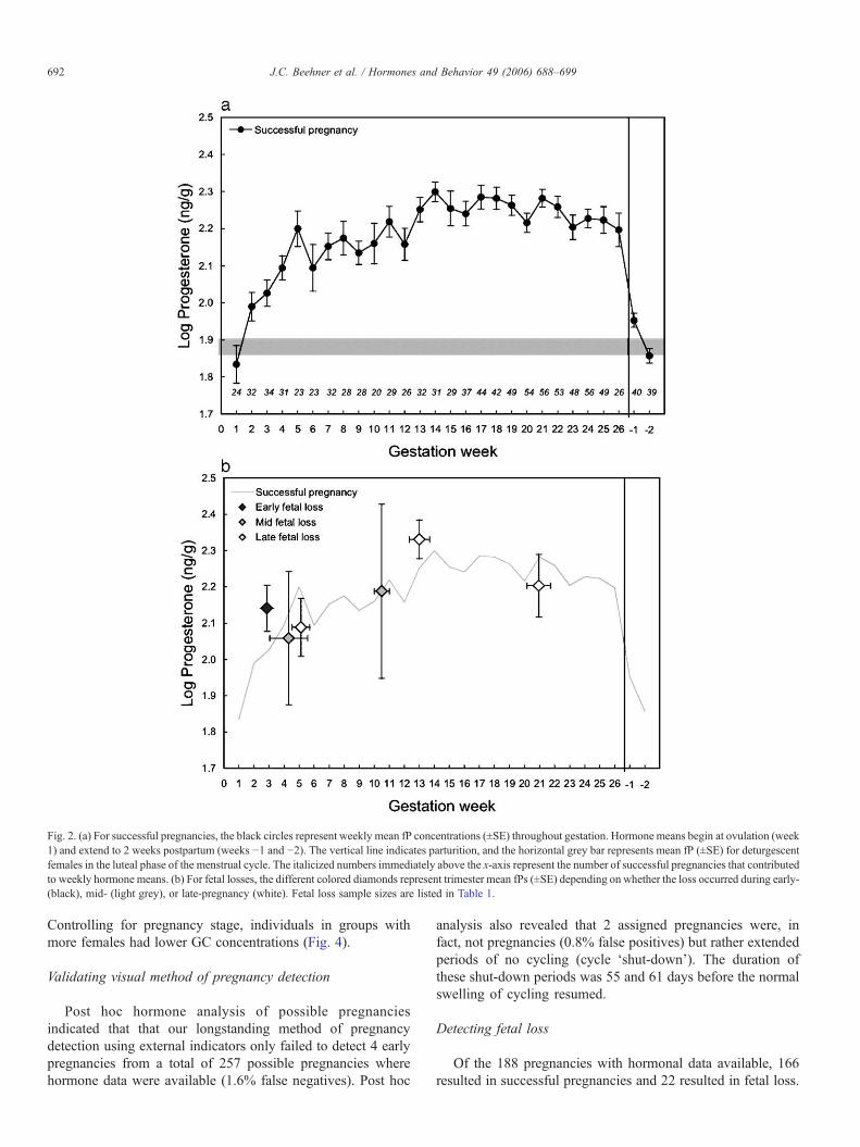
Both fE and fP concentrations increased across pregnancy,with the most rapid elevations occurring in the first trimester(Figs. 1a, 2a). By the end of the first trimester, fE hadincreased nearly 8-fold, and fP was double that of pre-pregnancy. Although fE continued to rise across eachtrimester, no further elevation in fP occurred after the second
690 J.C. Beehner et al. / Hormones and Behavior 49 (2006) 688–699
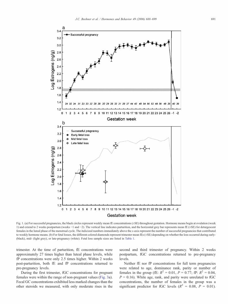
trimester. At the time of parturition, fE concentrations wereapproximately 27 times higher than luteal phase levels, whilefP concentrations were only 2.5 times higher. Within 2 weekspost-parturition, both fE and fP concentrations returned topre-pregnancy levels.
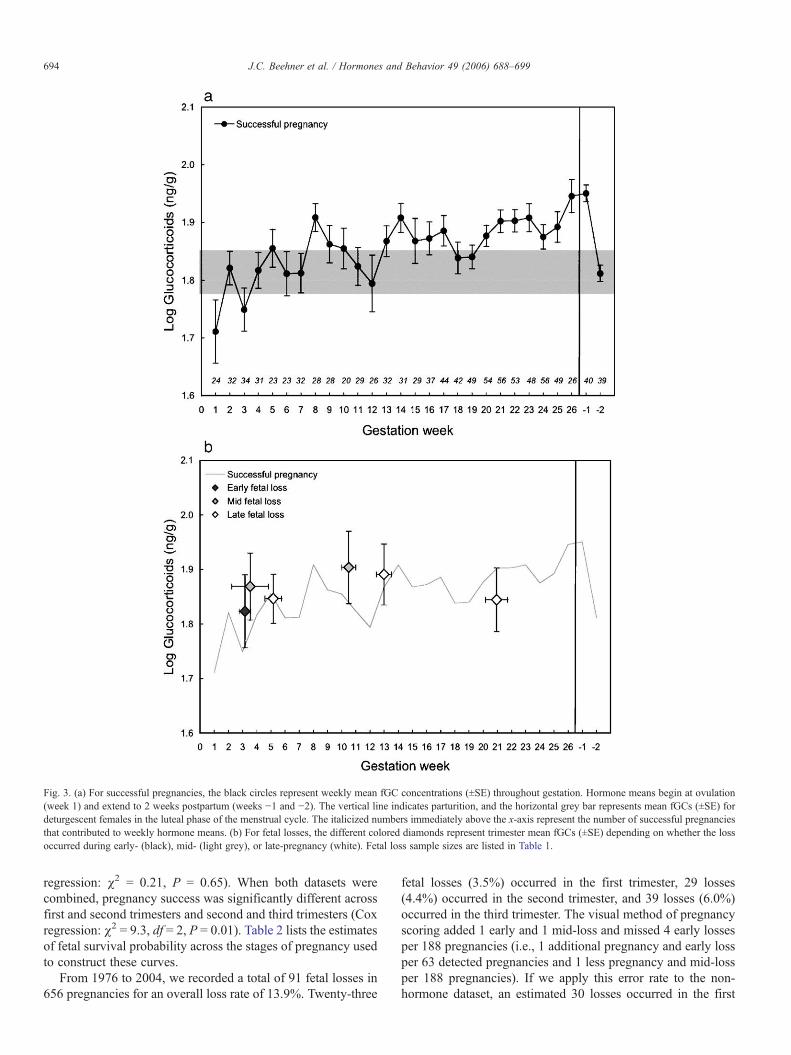
During the first trimester, fGC concentrations for pregnantfemales were within the range of non-pregnant values (Fig. 3a).Fecal GC concentrations exhibited less marked changes than theother steroids we measured, with only moderate rises in the
second and third trimester of pregnancy. Within 2 weekspostpartum, fGC concentrations returned to pre-pregnancylevels.
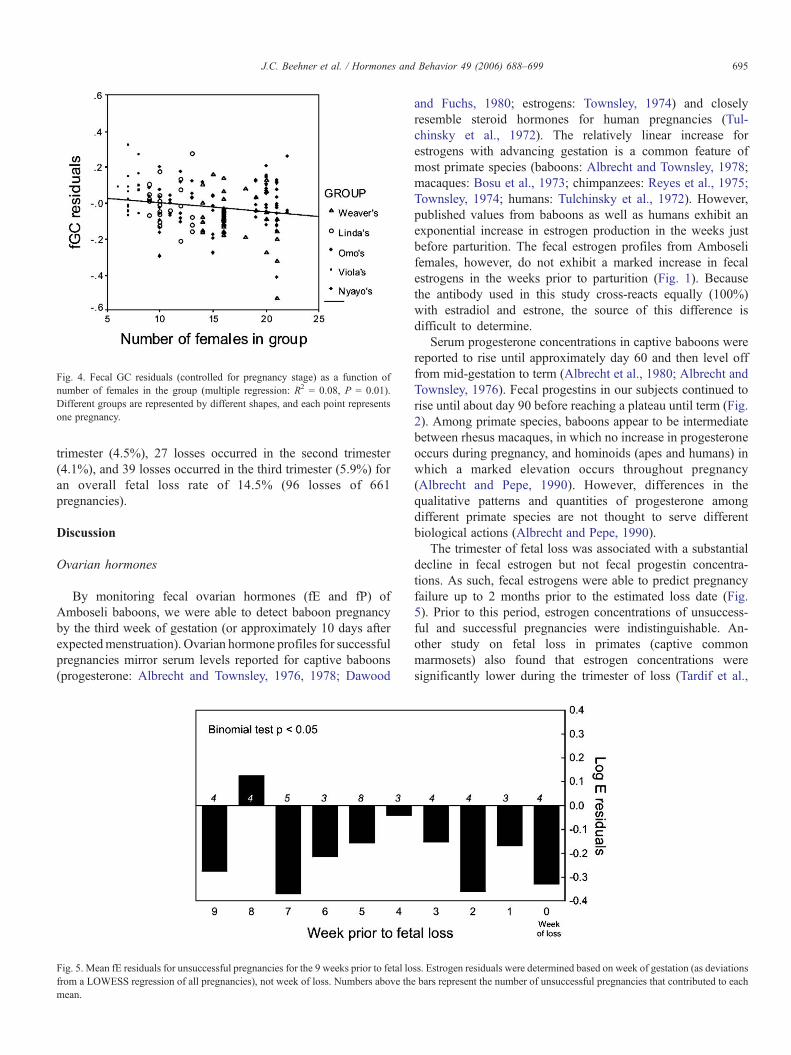
Neither fE nor fP concentrations for full term pregnancieswere related to age, dominance rank, parity or number offemales in the group (fE: R2 = 0.01, P = 0.77; fP: R2 = 0.04,P = 0.16). While age, rank, and parity were unrelated to fGCconcentrations, the number of females in the group was asignificant predictor for fGC levels (R2 = 0.08, P = 0.01).
Fig. 1. (a) For successful pregnancies, the black circles represent weekly mean fE concentrations (±SE) throughout gestation. Hormone means begin at ovulation (week1) and extend to 2 weeks postpartum (weeks −1 and −2). The vertical line indicates parturition, and the horizontal grey bar represents mean fE (±SE) for deturgescentfemales in the luteal phase of the menstrual cycle. The italicized numbers immediately above the x-axis represent the number of successful pregnancies that contributedto weekly hormone means. (b) For fetal losses, the different colored diamonds represent trimester mean fEs (±SE) depending on whether the loss occurred during early-(black), mid- (light grey), or late-pregnancy (white). Fetal loss sample sizes are listed in Table 1.
691J.C. Beehner et al. / Hormones and Behavior 49 (2006) 688–699
Controlling for pregnancy stage, individuals in groups withmore females had lower GC concentrations (Fig. 4).
Validating visual method of pregnancy detection
Post hoc hormone analysis of possible pregnanciesindicated that that our longstanding method of pregnancydetection using external indicators only failed to detect 4 earlypregnancies from a total of 257 possible pregnancies wherehormone data were available (1.6% false negatives). Post hoc
analysis also revealed that 2 assigned pregnancies were, infact, not pregnancies (0.8% false positives) but rather extendedperiods of no cycling (cycle ‘shut-down’). The duration ofthese shut-down periods was 55 and 61 days before the normalswelling of cycling resumed.
Detecting fetal loss
Of the 188 pregnancies with hormonal data available, 166resulted in successful pregnancies and 22 resulted in fetal loss.
Fig. 2. (a) For successful pregnancies, the black circles represent weekly mean fP concentrations (±SE) throughout gestation. Hormone means begin at ovulation (week1) and extend to 2 weeks postpartum (weeks −1 and −2). The vertical line indicates parturition, and the horizontal grey bar represents mean fP (±SE) for deturgescentfemales in the luteal phase of the menstrual cycle. The italicized numbers immediately above the x-axis represent the number of successful pregnancies that contributedto weekly hormone means. (b) For fetal losses, the different colored diamonds represent trimester mean fPs (±SE) depending on whether the loss occurred during early-(black), mid- (light grey), or late-pregnancy (white). Fetal loss sample sizes are listed in Table 1.
692 J.C. Beehner et al. / Hormones and Behavior 49 (2006) 688–699
Mean fE concentrations from mid- and late-pregnancy fetallosses were significantly lower than those of successfulpregnancies, but only during the trimester of loss (Fig. 1b,Table 1). Fecal E concentrations from all previous trimesters aswell as those from early fetal losses were not significantlydifferent from successful pregnancy means (Table 1). None ofthe fetal loss fP concentrations were significantly different thansuccessful pregnancy levels, and the same was also true for fGCconcentrations (Figs. 2b, 3b, Table 1).
Predictive value of steroid hormones in detecting loss
Fecal E residuals for fetal losses (controlling for time intogestation) were significantly lower than successful pregnancies(ANOVA: F1,1275 = 4.92, P = 0.03). When all weeks prior tofetal loss were included in the analysis, mean fE residuals didnot differ significantly from zero (binomial test: N = 25,P = 0.11). However, when we evaluated only the weeks leadingup to the miscarriage (up to 9 weeks, or approximately onetrimester prior to loss), mean fE residuals were significantlylower than zero (binomial test: N = 9, P = 0.02, Fig. 5). Mean fPresiduals for fetal losses did not differ significantly fromsuccessful pregnancies (ANOVA: F1,1313 = 2.44, P = 0.12) orfrom zero when we included all weeks (binomial test: N = 26,P = 0.56) or just the 9 weeks prior to fetal loss (binomial test:N = 9, P = 0.11). Similarly, mean fGC residuals for fetal losses
did not differ from successful pregnancies (ANOVA:F1,1310 = 0.37, P = 0.55) or from zero when all weeks (binomialtest: N = 26, P = 1.00) or 9 weeks prior to loss were evaluated(binomial test: N = 9, P = 0.75).
A binary logistic regression model with fetal loss as thedependent variable (loss/no loss) and all trimester means for fEresiduals as the predictor variable was not significantly differentfrom a model with only the intercept (model χ2 = 1.12, df = 1,P = 0.29, −2 log likelihood = 248.64, Cox and SnellR2 = 0.003). However, when only fE residuals from thetrimester of the fetal loss were included in the logistic analysis(fE residuals from other trimesters were discarded for femaleswith losses), the model was significant (model χ2 = 4.15, df = 1,P = 0.04, −2 log likelihood = 129.63, Cox and Snell R2 = 0.01);pregnancies characterized by lower fE residuals were morelikely to result in fetal loss (fE residuals: B = −1.35, SE = 0.60,Wald score = 5.04, df = 1, P = 0.03, Exp(B) = 0.26; constant:B = −3.36, SE = 0.29, Wald score = 133.74, df = 1, P b 0.01,Exp(B) = 0.04).
Rate and timing of fetal loss
The Kaplan–Meier estimate of the overall probability ofpregnancy success is 0.87 and 0.88 for the 1976–1999 and the1999–2004 datasets, respectively (Fig. 6). Pregnancy successwas not significantly different across time periods (Cox
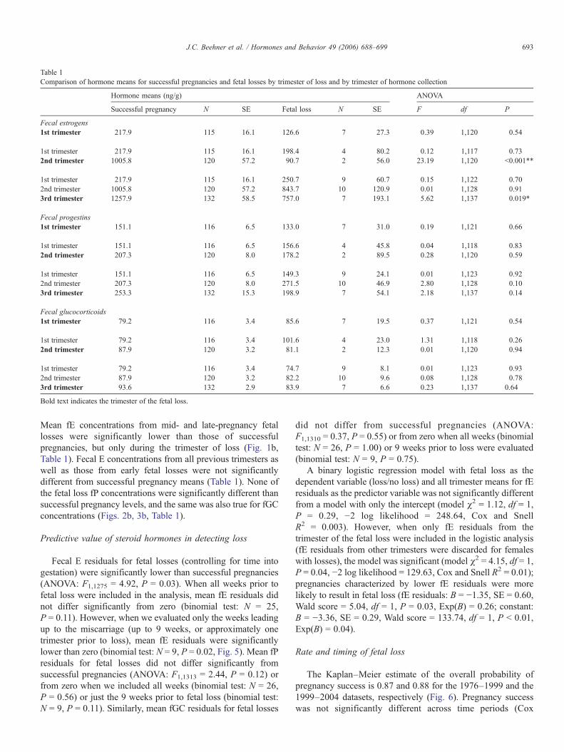
Table 1Comparison of hormone means for successful pregnancies and fetal losses by trimester of loss and by trimester of hormone collection
Hormone means (ng/g) ANOVA
Successful pregnancy N SE Fetal loss N SE F df P
Fecal estrogens1st trimester 217.9 115 16.1 126.6 7 27.3 0.39 1,120 0.54
1st trimester 217.9 115 16.1 198.4 4 80.2 0.12 1,117 0.732nd trimester 1005.8 120 57.2 90.7 2 56.0 23.19 1,120 b0.001**
1st trimester 217.9 115 16.1 250.7 9 60.7 0.15 1,122 0.702nd trimester 1005.8 120 57.2 843.7 10 120.9 0.01 1,128 0.913rd trimester 1257.9 132 58.5 757.0 7 193.1 5.62 1,137 0.019*
Fecal progestins1st trimester 151.1 116 6.5 133.0 7 31.0 0.19 1,121 0.66
1st trimester 151.1 116 6.5 156.6 4 45.8 0.04 1,118 0.832nd trimester 207.3 120 8.0 178.2 2 89.5 0.28 1,120 0.59
1st trimester 151.1 116 6.5 149.3 9 24.1 0.01 1,123 0.922nd trimester 207.3 120 8.0 271.5 10 46.9 2.80 1,128 0.103rd trimester 253.3 132 15.3 198.9 7 54.1 2.18 1,137 0.14
Fecal glucocorticoids1st trimester 79.2 116 3.4 85.6 7 19.5 0.37 1,121 0.54
1st trimester 79.2 116 3.4 101.6 4 23.0 1.31 1,118 0.262nd trimester 87.9 120 3.2 81.1 2 12.3 0.01 1,120 0.94
1st trimester 79.2 116 3.4 74.7 9 8.1 0.01 1,123 0.932nd trimester 87.9 120 3.2 82.2 10 9.6 0.08 1,128 0.783rd trimester 93.6 132 2.9 83.9 7 6.6 0.23 1,137 0.64
Bold text indicates the trimester of the fetal loss.
693J.C. Beehner et al. / Hormones and Behavior 49 (2006) 688–699
regression: χ2 = 0.21, P = 0.65). When both datasets werecombined, pregnancy success was significantly different acrossfirst and second trimesters and second and third trimesters (Coxregression: χ2 = 9.3, df = 2, P = 0.01). Table 2 lists the estimatesof fetal survival probability across the stages of pregnancy usedto construct these curves.
From 1976 to 2004, we recorded a total of 91 fetal losses in656 pregnancies for an overall loss rate of 13.9%. Twenty-three
fetal losses (3.5%) occurred in the first trimester, 29 losses(4.4%) occurred in the second trimester, and 39 losses (6.0%)occurred in the third trimester. The visual method of pregnancyscoring added 1 early and 1 mid-loss and missed 4 early lossesper 188 pregnancies (i.e., 1 additional pregnancy and early lossper 63 detected pregnancies and 1 less pregnancy and mid-lossper 188 pregnancies). If we apply this error rate to the non-hormone dataset, an estimated 30 losses occurred in the first
Fig. 3. (a) For successful pregnancies, the black circles represent weekly mean fGC concentrations (±SE) throughout gestation. Hormone means begin at ovulation(week 1) and extend to 2 weeks postpartum (weeks −1 and −2). The vertical line indicates parturition, and the horizontal grey bar represents mean fGCs (±SE) fordeturgescent females in the luteal phase of the menstrual cycle. The italicized numbers immediately above the x-axis represent the number of successful pregnanciesthat contributed to weekly hormone means. (b) For fetal losses, the different colored diamonds represent trimester mean fGCs (±SE) depending on whether the lossoccurred during early- (black), mid- (light grey), or late-pregnancy (white). Fetal loss sample sizes are listed in Table 1.
694 J.C. Beehner et al. / Hormones and Behavior 49 (2006) 688–699
trimester (4.5%), 27 losses occurred in the second trimester(4.1%), and 39 losses occurred in the third trimester (5.9%) foran overall fetal loss rate of 14.5% (96 losses of 661pregnancies).
Discussion
Ovarian hormones
By monitoring fecal ovarian hormones (fE and fP) ofAmboseli baboons, we were able to detect baboon pregnancyby the third week of gestation (or approximately 10 days afterexpectedmenstruation). Ovarian hormone profiles for successfulpregnancies mirror serum levels reported for captive baboons(progesterone: Albrecht and Townsley, 1976, 1978; Dawood
and Fuchs, 1980; estrogens: Townsley, 1974) and closelyresemble steroid hormones for human pregnancies (Tul-chinsky et al., 1972). The relatively linear increase forestrogens with advancing gestation is a common feature ofmost primate species (baboons: Albrecht and Townsley, 1978;macaques: Bosu et al., 1973; chimpanzees: Reyes et al., 1975;Townsley, 1974; humans: Tulchinsky et al., 1972). However,published values from baboons as well as humans exhibit anexponential increase in estrogen production in the weeks justbefore parturition. The fecal estrogen profiles from Amboselifemales, however, do not exhibit a marked increase in fecalestrogens in the weeks prior to parturition (Fig. 1). Becausethe antibody used in this study cross-reacts equally (100%)with estradiol and estrone, the source of this difference isdifficult to determine.
Serum progesterone concentrations in captive baboons werereported to rise until approximately day 60 and then level offfrom mid-gestation to term (Albrecht et al., 1980; Albrecht andTownsley, 1976). Fecal progestins in our subjects continued torise until about day 90 before reaching a plateau until term (Fig.2). Among primate species, baboons appear to be intermediatebetween rhesus macaques, in which no increase in progesteroneoccurs during pregnancy, and hominoids (apes and humans) inwhich a marked elevation occurs throughout pregnancy(Albrecht and Pepe, 1990). However, differences in thequalitative patterns and quantities of progesterone amongdifferent primate species are not thought to serve differentbiological actions (Albrecht and Pepe, 1990).
The trimester of fetal loss was associated with a substantialdecline in fecal estrogen but not fecal progestin concentra-tions. As such, fecal estrogens were able to predict pregnancyfailure up to 2 months prior to the estimated loss date (Fig.5). Prior to this period, estrogen concentrations of unsuccess-ful and successful pregnancies were indistinguishable. An-other study on fetal loss in primates (captive commonmarmosets) also found that estrogen concentrations weresignificantly lower during the trimester of loss (Tardif et al.,
Fig. 5. Mean fE residuals for unsuccessful pregnancies for the 9 weeks prior to fetal loss. Estrogen residuals were determined based on week of gestation (as deviationsfrom a LOWESS regression of all pregnancies), not week of loss. Numbers above the bars represent the number of unsuccessful pregnancies that contributed to eachmean.
Fig. 4. Fecal GC residuals (controlled for pregnancy stage) as a function ofnumber of females in the group (multiple regression: R2 = 0.08, P = 0.01).Different groups are represented by different shapes, and each point representsone pregnancy.
695J.C. Beehner et al. / Hormones and Behavior 49 (2006) 688–699

2005). The precipitous decrease in fecal estrogens bothfollowing parturition and during the weeks prior to fetal losspoints to the dependence of estrogen production on afunctioning fetus and/or placenta (see also Townsley, 1974).As reviewed by Pepe and Albrecht (1995), estrogenstimulates key steps in progesterone biosynthesis in thesyncytiotrophoblasts of the placenta, as well as uteroplacentalblood flow. By stimulating progesterone production, estrogensalso indirectly promote uterine myometrial quiescence andpermit implantation of the placenta and developing embryo—all processes critical for the maintenance of pregnancy.Specifically with respect to baboons, a 50% miscarriage ratewas observed among captive baboons in which maternalserum estrogen concentrations were experimentally sup-pressed (Albrecht et al., 2000), while pregnancy wasmaintained in all baboons in which the estrogen levels wererestored to normal levels.
One of the physiological functions of the primate fetaladrenal cortex is to provide C19 substrate to the placenta for theformation of estrogens, a strategy for estrogen formation that isunique to primate species (Diczfalusy, 1964; Novy, 1977). Ifimpaired placental or fetal function occurs during gestation, thebiosynthesis of estrogens will thus be compromised. In pregnantbaboons, following the removal of the fetus while leaving theplacenta in utero (fetectomy), progesterone concentrationsdecreased by 20–45%, and maternal estradiol concentrationsdropped to basal values (Albrecht et al., 1980; Albrecht andPepe, 1985). Similar results were also found followingfetectomy in macaques (Tullner and Hodgen, 1974) andintrauterine fetal death or fetal anencephaly in humans(reviewed in Albrecht and Pepe, 1990). Although the fetus inprimate pregnancy is essential to the maternal production ofestrogen, it is not a prerequisite for the production ofprogesterone (reviewed in Albrecht and Pepe, 1990). In primate
pregnancy, cholesterol is taken up from the maternal circulationand utilized by the placenta for progesterone biosynthesis(Hellig et al., 1970; Winkel et al., 1980). Nevertheless,estrogens themselves play an indirect role in the stimulationof progesterone production and regulation of progesteronereceptors (Castracane et al., 1983; Henson, 1998). Therefore, inthe absence of compensatory systems, fetal demise willeventually lead to subnormal levels of progesterone in maternalcirculation (Albrecht and Pepe, 1990).
In sum, our results indicate that fecal estrogens, and notprogestins, signal the first signs of fetal loss in this population ofwild baboons. Although the nature of our data does not permitus to determine whether the decline in estrogens was causal oronly symptomatic of fetal loss, the significant decrease in fE butnot fP concentrations suggest a fetal and not a placental originof failure in our subjects.
Glucocorticoids
Fecal glucocorticoid concentrations for pregnant Amboselifemales also corresponded to serum and urinary glucocorticoidprofiles reported for humans and several non-human primates(e.g., Carr et al., 1981; Smith et al., 1993; Tardif et al., 2005;Wintour et al., 1978). An elevation in glucocorticoidconcentrations was observed during the second trimester,followed by only small changes in late gestation untilparturition (Fig. 3). In both human and non-human primates,pregnancy results in an activation of the HPA axis, whichincreases glucocorticoid secretion overall (Keller-Wood andWood, 2001). In primate pregnancy, increased HPA activity isnecessary for the maintenance of intrauterine homeostasis andthe maturation of essential organ systems including the lungs,liver, and the gut (Bolt et al., 2001; Mesiano and Jaffe, 1997).Because the fetal adrenal does not produce glucocorticoidsuntil late gestation (Jaffe et al., 1979; Voutilainen and Miller,1987), hormones secreted by the placenta play a central role inmediating the increased HPA activity. As such, pregnancyshould be considered a physiological state in which increasedglucocorticoids (over non-pregnant levels) are of adaptivevalue.
Nevertheless, animal experiments have demonstrated thatexposure of pregnant females to stressful conditions can stillresult in a compromised pregnancy or fetal demise (Clark et al.,1993; reviewed in de Catanzaro and Macniven, 1992; Johnsonet al., 1991; see also Wildt et al., 1977). Additionally, well-controlled studies in humans also suggest a direct relationship
Table 2Kaplan–Meier estimates for survival of pregnancies across trimesters for twodata sets, one without (1971–1999) and one with (1999–2004) hormone data forpregnancy validation
1971–1999 1999–2004
Survivalprobability
SE No. inrisk set
Survivalprobability
SE No. inrisk set
1st trimester 0.972 0.007 569 0.964 0.013 2182nd trimester 0.927 0.011 552 0.936 0.017 2113rd trimester 0.864 0.014 515 0.876 0.022 203
Fig. 6. Kaplan–Meier survival curves for fetal losses between the years 1971–1999 (solid line) and 1999–2004 (dotted line). If the mother disappeared beforeparturition, the pregnancy was censored (black diamonds). Hormonal dataavailable for the 1999–2004 dataset were used to correct for errors in pregnancydetection. Survival curves for the two periods are not significantly different (Coxregression: χ2 = 0.21, P = 0.65).
696 J.C. Beehner et al. / Hormones and Behavior 49 (2006) 688–699
between prenatal maternal stress and a number of pregnancycomplications (reviewed in Mulder et al., 2002). The relation-ship between pregnancy failure and the hormone mechanismsthat mediate loss in stressful situations are not completelyunderstood. Although increasing evidence suggests that mater-nal stress can critically alter ovarian steroids and thus endangerpregnancy maintenance (reviewed in de Catanzaro andMacniven, 1992), many other studies found an associationbetween conventional “stress” hormones and elevated fetalmortality or morbidity across a wide array of taxa (mice:Baumgartner and Chrisman, 1987; rats: Davis and Plotz, 1954;Hansen et al., 1999; La Borde et al., 1992; humans:Nepomnaschy, 2005; sheep: Quinlivan et al., 1998; Yang etal., 1969).
For the Amboseli baboons, pregnancy failure was associatedwith neither higher nor lower fecal glucocorticoid concentra-tions. However, pregnant glucocorticoid concentrations werenegatively related to the number of females in the group (Fig.4). As group size increased, females exhibited lower gluco-corticoids for their stage of pregnancy than other females,possibly a sign of a compromised pregnancy. For manypopulations of baboons, including Amboseli, reproductivecosts among females have been associated with larger groups(Altmann and Alberts, 2003a; Bulger and Hamilton, 1987;Rhine et al., 1988; Wasser and Starling, 1988). Although we didnot find a direct association between glucocorticoids and fetalloss with the current dataset, an association between fetal lossand lower glucocorticoid concentrations may be revealed asmore hormone data are collected. In common marmosets, Tardifet al. (2005) demonstrated that food restriction resulted indecreased maternal cortisol and ultimately to fetal demise andpregnancy loss. They suggest that food restriction does not actas a classical stressor with elevated HPA activity, but thatendocrine function in the placenta is rapidly impaired by dietaryrestrictions (Tardif et al., 2005). In wild feeding baboons, largergroups may have decreased food intake, particularly duringseasons when food resources are scarce; and drought conditionsdo indeed result in a higher probability of fetal loss in theAmboseli population (Beehner et al., submitted).
Validating visual method of pregnancy detection
By measuring steroid hormone profiles across reproductivefemales, we demonstrated that our longstanding external basisfor early detection of pregnancies in the baboons closelymatches hormonal identification of pregnancy. For decades, themethod of pregnancy determination for the Amboseli baboonshas of necessity been visual; mainly, one of us (J.A.) hasassigned pregnancies when females ceased cycling and failed tomenstruate prior to the resumption of cycling. Although thismethod of pregnancy detection requires frequent monitoringwith very few gaps in data, we show here that it identifies 97%of pregnancies. Neither fecal hormone sampling nor visualdetection identifies pregnancies that are terminated within thefirst 3 weeks, although urinary steroids are reported to detectpregnancy as early as day 13 post-ovulation, 1 week earlier(Hodges et al., 1986).
Rate and timing of fetal loss
The results from over 30 years of pregnancy data at Amboseliindicate that the (uncorrected) pregnancy failure rate is 13.9%(91 losses of 656 pregnancies). This loss rate is higher than thatreported from two other long-term studies of wild yellow (10%or 17 losses of 170 pregnancies: Wasser, 1995) and anubisbaboons (9.6% or 56 losses of 584 pregnancies: Packer et al.,1995), even if we do not adjust for detection error. In most wildbaboon populations, reddening of the paracallosal skin is used toindicate pregnancy. However, this change does not occur untilapproximately mid-gestation and is highly variable. Forexample, Amboseli females exhibit some reddening of theparacallosal skin at approximately day 61 of gestation (N = 199pregnancies, SE = 1.34 days, range = 10–113 days). Therefore,studies that rely on skin coloration for pregnancy detection maymiss early-gestation losses that our method was able to pick up.Alternatively, ecological factors may play a role in thisdifference. For example, in Gombe, anubis baboons live inrich areas of evergreen forest where food resources are relativelyabundant (Packer et al., 2000). In contrast, the Amboselibaboons spend the majority of the day foraging and movingbetween widely spaced food patches, particularly in dry years(Bronikowski and Altmann, 1996). Ecological factors canproduce high variability in female reproduction on the proximatescale because reproductive physiology is sensitive to theavailability of oxidizable metabolic fuels (Wade and Schneider,1992) and on an evolutionary scale because decreased foodintake must be balanced by less energy expenditure on activitiessuch as reproduction that are not essential for immediate survival(Bronson, 1989).
Very little information about the timing of fetal mortalityacross gestation is available for wild baboons. Packer et al.(1995) report that miscarriages among anubis baboons occur atapproximately equal rates across trimesters. Similarly our dataindicate that fetal mortality in the baboon occurs throughoutgestation.
Overall, the rate of miscarriage in Amboseli was remarkablysimilar whether hormone data were available or not. However,when we evaluated only the hormone dataset (dotted line in Fig.6), the addition of early-gestation losses combined with theremoval of mid-gestation losses revealed three critical periodsmarked by an increase in the rate of loss (i.e., a steeper curve):20–25 days, 50–75 days, and 175–180 days into gestation. Thefirst of these periods coincides with placentation in the baboon.Placentation, where the placenta becomes a significant source ofprogesterone, occurs at approximately days 20–25 of baboonpregnancy (Castracane and Goldzieher, 1986). The extent towhich placental development occurs in this period may have asignificant influence on subsequent fetal growth and survival.The second period may reflect a problem with the initial fetaladrenal production of C19 precursors for estrogen biosynthesisby the placenta (at approximately 40 days, Albrecht and Pepe,1990). The primate placenta is capable of converting C19
steroids to estrogens but cannot produce C19, while the fetaladrenal cortex produces large amounts of C19 steroids (reviewedin Mesiano and Jaffe, 1997). Thus, appropriate development
697J.C. Beehner et al. / Hormones and Behavior 49 (2006) 688–699
and function of the fetal adrenal cortex are critical for fetalmaturation and perinatal survival. The last period marks normalparturition in the baboon (mean gestation in this population is178 days). As far as these critical periods for fetal mortality inthe present study are concerned, it is notable that increased ratesof loss coincide with periods of evident changes in steroidsynthesis and/or function.
Acknowledgments
Supported by NSF IBN-0322613, NSF BCS-0323553, andR03 MH65294, and the Chicago Zoological Society. We thankthe Office of the President, Republic of Kenya, the KenyaWildlife Services, its Amboseli staff and Wardens, the Instituteof Primate Research, the National Museums of Kenya, and themembers of the Amboseli-Longido pastoralist communities.Particular thanks go to the Amboseli fieldworkers whocontributed to sample and data collection, especially R.S.Mututua, S. Sayialel, and J.K. Warutere. Additionally, we wantto thank P. Ogola and T. Wango for sample processing at theUniversity of Nairobi, C. Markham for database assistance andL. Gesquiere, C. Smith, L. Okpala, L. Shek, and C. Ihunna forlaboratory assistance. All protocols were non-invasive andcomply with relevant regulations in Kenya (Kenya ResearchPermit MOEST 13/001/C351 Vol. II) and the United States(IACUC 1456, renewed 12 November 2002).
References
Alberts, S.C., Altmann, J., 1995. Balancing costs and opportunities: dispersal inmale baboons. Am. Nat. 145, 279–306.
Alberts, S.C., Altmann, J., Wilson, M.L., 1996. Mate guarding constrainsforaging activity of male baboons. Anim. Behav. 51, 1269–1277.
Alberts, S., Watts, H., Altmann, J., 2003. Queuing and queue-jumping: long-term patterns of reproductive skew in male Savannah baboons, Papiocynocephalus. Anim. Behav. 65, 821–840.
Albrecht, E.D., Pepe, G.J., 1984. Effect of the antiestrogen ethamoxytriphetol(MER-25) and luteectomy on serum progesterone concentrations inpregnant baboons. Endocrinology 115, 1717–1721.
Albrecht, E.D., Pepe, G.J., 1985. The placenta remains functional followingfetectomy in baboons. Endocrinology 116, 843–845.
Albrecht, E.D., Pepe, G.J., 1990. Placental steroid hormone biosynthesis inprimate pregnancy. Endocr. Rev. 11, 124–150.
Albrecht, E.D., Townsley, J.D., 1976. Serum progesterone in the pregnantbaboon (Papio papio). Biol. Reprod. 14, 610–612.
Albrecht, E.D., Townsley, J.D., 1978. Serum estradiol in mid and late gestationand estradiol/progesterone ratio in baboons near parturition. Biol. Reprod.18, 247–250.
Albrecht, E.D., Haskins, A.L., Pepe, G.J., 1980. The influence of fetectomy atmidgestation upon the serum concentrations of progesterone, estrone, andestradiol in baboons. Endocrinology 107, 766–770.
Albrecht, E.D., Henson, M.C., Pepe, G.J., 1991. Regulation of placental lowdensity lipoprotein uptake by baboons by estrogen. Endocrinology 128,450–458.
Albrecht, E.D., Aberdeen, G.W., Pepe, G.J., 2000. The role of estrogen inthe maintenance of primate pregnancy. Am. J. Obstet. Gynecol. 182,432–438.
Altmann, J., Alberts, S.C., 2003a. Intraspecific variability in fertility andoffspring survival in a non-human primate: behavioral control of ecologicaland social sources. In: Wachter, K.W. (Ed.), Offspring: Human FertilityBehavior In a Biodemographic Perspective. National Academy Press,Washington, D.C., pp. 140–169.
Altmann, J., Alberts, S.C., 2003b. Variability in reproductive success viewedfrom a life-history perspective in baboons. Am. J. Hum. Biol. 15, 401–409.
Altmann, J., Hausfater, G., Altmann, S.A., 1988. Determinants ofreproductive success in Savannah baboons, Papio cynocephalus. In:Clutton-Brock, T.H. (Ed.), Reproductive Success: Studies of IndividualVariation in Contrasting Breeding Systems. University of Chicago Press,Chicago, pp. 403–418.
Altmann, J., Lynch, J.W., Nguyen, N., Alberts, S.C., Gesquiere, L.R., 2004.Life-history correlates of steroid concentrations in wild peripartum baboons.Am. J. Primatol. 64, 95–106.
Baumgartner, A.P., Chrisman, C.L., 1987. Embryonic mortality caused bymaternal heat stress during mouse oocyte maturation. Anim. Reprod. Sci.14, 309–316.
Bolt, R.J., van Weissenbruch, M.M., Lafeber, H.N., Delemarre-van de Waal, H.A., 2001. Glucocorticoids and lung development in the fetus and preterminfant. Pediatr. Pulmonol. 32, 76–91.
Bosu, W.T.K., Johansson, E.D.B., 1975. Implantation and maintenance ofpregnancy in mated rhesus monkeys following bilateral oophorectomy orluteectomy with and without progesterone replacement. Acta Endocrinol.(Copenh.) 79, 598–609.
Bosu, W.T.K., Johansson, E.D.R., Gemzell, C., 1973. Patterns of circulatingoestrone, oestradiol-17ß and progesterone during pregnancy in the rhesusmonkey. Acta Endocrinol. (Copenh.) 74, 743–755.
Bronikowski, A.M., Altmann, J., 1996. Foraging in variable environment:weather patterns and the behavioural ecology of baboons. Behav. Ecol.Sociobiol. 39, 11–25.
Bronson, F.H., 1989. Mammalian Reproductive Biology. University of ChicagoPress, Chicago.
Bulger, J.B., Hamilton, W.J., 1987. Rank and density correlates of inclusivefitness measures in a natural chacma baboon (Papio ursinus) population. Int.J. Primatol. 8, 635–650.
Campbell, C.J., Shideler, S.E., Todd, H.E., Lasley, B.L., 2001. Fecal analysis ofovarian cycles in female black-handed spider monkeys (Ateles geoffroyi).Am. J. Primatol. 54, 79–89.
Carr, B.R., Parker, C.R., Madden, J.D., MacDonald, P.C., Porter, J.C., 1981.Maternal adrenocorticotropin and cortisol relationships throughout humanpregnancy. Am. J. Obstet. Gynecol. 139, 416–422.
Castracane, V.D., Goldzieher, J.W., 1986. Timing of the luteal–placental shift inthe baboon (Papio cynocephalus). Endocrinology 118, 506–512.
Castracane, V.D., Kuehl, T.J., Goldzieher, J.W., 1983. The effect of estrogenantagonism on progesterone production in early pregnancy in the baboon.Am. J. Obstet. Gynecol. 145, 725–729.
Clark, D.A., Banwatt, D., Chaouat, G., 1993. Stress-triggered abortion in miceprevented by alloimmunization. Am. J. Reprod. Immunol. 29, 141–147.
Csapo, A.I., Pulkkinen, M., 1978. Indispensability of the human corpus luteumin the maintenance of early pregnancy luteecotomy evidence. Obstet.Gynecol. Surv. 33, 69–81.
Curtis, D.J., Zaramody, A., Green, D.I., Pickard, A.R., 2000. Non-invasivemonitoring of reproductive status in wild mongoose lemurs (Eulemurmongoz). Reprod. Fertil. Dev. 12, 21–29.
Davis, M.E., Plotz, E.J., 1954. The effects of cortisone acetate on intact andadrenalectomized rats during pregnancy. Endocrinology 54, 384–395.
Dawood, M.Y., Fuchs, F., 1980. Estradiol and progesterone in the maternal andfetal circulation in the baboon. Biol. Reprod. 22, 179–184.
de Catanzaro, D., Macniven, E., 1992. Psychogenic pregnancy disruptions inmammals. Neurosci. Biobehav. Rev. 16, 43–53.
Diczfalusy, E., 1964. Endocrine functions of the human fetoplacental unit. Fed.Proc. 23, 791–798.
Exner, C., Wehrend, A., Hospes, R., Einspanier, A., Hoffmann, B., Heldmaier,G., 2003. Hormonal and behavioural changes during the mating season andpregnancy in Alpine marmots (Marmota marmota). Reproduction 126,775–782.
Fortman, J.D., Herring, J.M., Miller, J.B., Hess, D.L., Verhage, H.G., Fazleabas,A.T., 1993. Chorionic gonadotropin, estradiol, and progesterone levels inbaboons (Papio anubis) during early pregnancy and spontaneous abortion.Biol. Reprod. 49, 737–742.
Guo, Y., Hendrickx, A.G., Overstreet, J.W., Dieter, J., Stewart, D., Tarantal, A.F., Laughlin, L., Lasley, B.L., 1999. Endocrine biomarkers of early fetal loss
698 J.C. Beehner et al. / Hormones and Behavior 49 (2006) 688–699
in cynomolgus macaques (Macaca fascicularis) following exposure todioxin. Biol. Reprod. 60, 707–713.
Hackländer, K., Arnold, W., 1999. Male-cause failure of female reproductionand its adaptive value in alpine marmots (Marmota marmota). Behav. Ecol.10, 592–597.
Hansen, D.K., La Borde, J.B., Wall, K.S., Holson, R.R., Young, J.F., 1999.Pharmacokinetic considerations of dexamethasone-induced developmentaltoxicity in rats. Toxicol. Sci. 48, 230–239.
Hellig, H., Gattereau, D., Lefebvre, Y., Bolte, E., 1970. Steroid production fromplasma cholesterol. I. Conversion of plasma cholesterol to placentalprogesterone in humans. J. Clin. Endocrinol. Metab. 30, 624–631.
Henson, M.C., 1998. Pregnancy maintenance and the regulation of placentalprogesterone biosynthesis in the baboon. Hum. Reprod. Updat. 4, 389–405.
Hodges, J.K., Tarara, R., Wangula, C., 1984. Circulating steroids and therelationship between ovarian and placental secretion during early and midpregnancy in the baboon. Am. J. Primatol. 7, 357–366.
Hodges, J.K., Tarara, R., Hearn, J.P., Else, J.G., 1986. The detection of ovulationand early pregnancy in the baboon by direct measurement of conjugatedsteroids in urine. Am. J. Primatol. 10, 329–338.
Jaffe, R.B., Seronferre, M., Mitchell, B.F., 1979. Perinatal regulation of cortisolin the primate. J. Steroid Biochem. Mol. Biol. 11, 549–555.
Johnson, E.O., Kamilaris, T.C., Carter, S., Gold, P.W., Chrousos, G.P., 1991.Environmental-stress and reproductive success in the common marmoset(Callithrix jaccus jaccus). Am. J. Primatol. 25, 191–201.
Keller-Wood, M., Wood, C.E., 2001. Pituitary–adrenal physiology duringpregnancy. Endocrinologist 11, 159–170.
Khan, M.Z., Altmann, J., Isani, S.S., Yu, J., 2002. A matter of time: evaluatingthe storage of fecal samples for steroid analysis. Gen. Comp. Endocrinol.128, 57–64.
Kuehl, T.J., Kang, I.S., Siler-Khodr, T.M., 1992. Pregnancy and earlyreproductive failure in the baboon. Am. J. Primatol. 28, 41–48.
La Borde, J.B., Hansen, D.K., Young, J.F., Sheehan, D.M., Holson, R.R., 1992.Prenatal dexamethasone exposure in rats: effects of dose, age at exposure,and drug-induced hypophagia on malformations and fetal organ weights.Fundam. Appl. Toxicol. 19, 545–554.
Lasley, B.L., Lohstroh, P., Kuo, A., Gold, E.B., Eskenazi, B., Samuels, S.J.,Stewart, D.R., Overstreet, J.W., 1995. Laboratorymethods for evaluating earlypregnancy loss in an industry-based population. Am. J. Ind.Med. 28, 771–781.
Lynch, J.W., Khan, M.Z., Altmann, J., Njahira, M.N., Rubenstein, N., 2003.Concentrations of four fecal steroids in wild baboons: short-term storageconditions and consequences for data interpretation. Gen. Comp. Endocri-nol. 132, 264–271.
Mesiano, S., Jaffe, R.B., 1997. Developmental and functional biology of theprimate fetal adrenal cortex. Endocr. Rev. 18, 378–403.
Mulder, E.J.H., Robles de Medina, P.G., Huizink, A.C., Van den Bergh, B.R.H.,Buitelaar, J.K., Visser, G.H.A., 2002. Prenatal maternal stress: effects onpregnancy and the (unborn) child. Early Hum. Dev. 70, 3–14.
Nepomnaschy, P.A., 2005. Stress and female reproduction in a rural Mayanpopulation. PhD, The University of Michigan.
Novy, M.J., 1977. Endocrine and pharmacological factors which influence theonset of labour in rhesus monkeys. Ciba Found. Symp. 47, 259–295.
Oakey, R.E., 1970. The progressive increase in estrogen production in humanpregnancy: an appraisal of the factors responsible. Vitam. Horm. 28, 1–36.
Packer, C., Collins, D.A., Sindimwo, A., Goodall, J., 1995. Reproductiveconstraints on aggressive competition in female baboons. Nature 373, 60–63.
Packer, C., Collins, D.A., Eberly, L.E., 2000. Problems with primate sex ratios.Philos. Trans. R. Soc. Lond., B Biol. Sci. 355, 1627–1635.
Pepe, G.J., Albrecht, E.D., 1995. Actions of placental and fetal adrenal steroidhormones in primate pregnancy. Endocr. Rev. 16, 608–648.
Pereira, M.E., 1988. Agonistic interactions of juvenile savanna baboons: I.Fundamental features. Ethology 79, 195–217.
Quinlivan, J.A., Archer, M.A., Dunlop, S.A., Evans, S.F., Beazley, L.D.,Newnham, J.P., 1998. Fetal growth retardation, particularly within lynphoidorgans, following repeated maternal injections of beta methasone in sheep. J.Obstet. Gynaecol. Res. 24, 173–182.
Reyes, F.I., Winter, J.S.D., Faiman, C., Hobson, W.C., 1975. Serial serum levelsof gonadotropins, prolactin and sex steroids in the non-pregnant andpregnant chimpanzee. Endocrinology 96, 1447–1455.
Rhine, R.J., Wasser, S.K., Norton, G.W., 1988. Eight-year study of social andecological correlates of mortality among immature baboons of MikumiNational Park, Tanzania. Am. J. Primatol. 16, 199–212.
Schwarzenberger, F., Mostl, E., Palme, R., Bamberg, E., 1996. Faecal steroidanalysis for non-invasive monitoring of reproductive status in farm, wild andzoo animals. Anim. Reprod. Sci. 42, 515–526.
Shopland, J.M., 1987. Food quality, spatial deployment, and the intensity offeeding interference in yellow baboons (Papio cynocephalus). Behav. Ecol.Sociobiol. 21, 149–156.
Smith, R., Chan, E.C., Bowman, M.E., Harewood, W.J., Phippard, A.F., 1993.Corticotropin-releasing hormone in baboon pregnancy. J. Clin. Endocrinol.Metab. 76, 1063–1068.
Tardif, S.D., Ziegler, T.E., Power, M., Layne, D.G., 2005. Endocrine changes infull-term pregnancies and pregnancy loss due to energy restriction in thecommon marmoset (Callithrix jacchus). J. Clin. Endocrinol. Metab. 90,335–339.
Townsley, J.D., 1974. Estrogen excretion of the pregnant baboon (Papio papio).Endocrinology 95, 1759–1762.
Tulchinsky, D., Hobel, C.J., Yeager, E., Marshall, J.R., 1972. Plasma estrone,estradiol, estriol, progesterone, and 17-hydroxyprogesterone in humanpregnancy. Am. J. Obstet. Gynecol. 112, 1095–1100.
Tullner, W.W., Hodgen, G.D., 1974. Effects of fetectomy on plasmaestrogens and progesterone in monkeys (Macaca mulatta). Steroids 24,887–897.
Voutilainen, R., Miller, W.L., 1987. Coordinate trophic hormone regulation ofmRNAs for insulin-like growth factor II and the cholesterol side-chaincleavage enzyme, P450SCC in human steroidogenic tissues. Proc. Natl.Acad. Sci. 84, 1590–1594.
Wade, G.N., Schneider, J.E., 1992. Metabolic fuels and reproduction in femalemammals. Neurosci. Biobehav. Rev. 16, 235–272.
Wasser, S.K., 1995. Costs of conception in baboons. Nature 376, 219–220.Wasser, S.K., Starling, A.K., 1988. Proximate and ultimate causes of
reproductive suppression among female yellow baboons at Mikumi NationalPark, Tanzania. Am. J. Primatol. 16, 97–121.
Wasser, S., Hunt, K., Brown, J., Cooper, K., Crockett, C., Bechert, U.,Millspaugh, J., Larson, S., Monfort, S., 2000. A generalized fecalglucocorticoid assay for use in a diverse array of non-domestic mammalianand avian species. Gen. Comp. Endocrinol. 120, 260–275.
Wildt, D.E., Doyle, L.L., Stone, S.c., Harrison, R.M., 1977. Correlation ofperineal swelling with serum ovarian hormone levels, vaginal cytology, andovarian follicular development during the baboon reproductive cycle.Primates 18, 261–270.
Winkel, C., Snyder, J., MacDonald, P., Simpson, E., 1980. Regulation ofcholesterol and progesterone synthesis in human placental cells in culture byserum lipoproteins. Endocrinology 106, 1054–1060.
Wintour, E.M., Coghlan, J.P., Oddie, C.J., Scoggins, B.A., Walters, W.A., 1978.A sequential study of adrenocorticosteroid level in human pregnancy. Clin.Exp. Pharmacol. Physiol. 5, 399–403.
Yang, W.H., Yang, W.P., Lin, L.L., 1969. Interruption of pregnancy in the rat byadministration of ACTH. Endocrinology 84, 1282–1285.
699J.C. Beehner et al. / Hormones and Behavior 49 (2006) 688–699
Copyright © 2022 FDOKUMEN