BLM helicase stimulates the ATPase and chromatin-remodeling activities of RAD54
Upload
independentCategory
view
2download
0
Royal College of Surgeons in Irelande-publications@RCSI
Anatomy Articles Department of Anatomy
2008
The effects of increased intracortical remodeling onmicrocrack behaviour in compact bone.Oran D KennedyRoyal College of Surgeons in Ireland
Orlaith BrennanRoyal College of Surgeons in Ireland
Peter MauerRoyal College of Surgeons in Ireland
Susan M RackardUniversity College Dublin
Fergal J O'BrienRoyal College of Surgeons in Ireland
David TaylorTrinity College Dublin
T Clive LeeRoyal College of Surgeons in Ireland
This Article is brought to you for free and open access by the Departmentof Anatomy at e-publications@RCSI. It has been accepted for inclusion inAnatomy Articles by an authorized administrator of [email protected] more information, please contact [email protected].
CitationKennedy OD, Brennan O, Mauer P, Rackard SM, O'Brien FJ, Taylor D, Lee TC. The effects of increased intracortical remodeling onmicrocrack behaviour in compact bone. Bone. 2008;43(5):889-93.
— Use Licence —
Attribution-Non-Commercial-ShareAlike 1.0
You are free:• to copy, distribute, display, and perform the work.• to make derivative works.
Under the following conditions:• Attribution — You must give the original author credit.• Non-Commercial — You may not use this work for commercial purposes.• Share Alike — If you alter, transform, or build upon this work, you may distribute the resulting work onlyunder a licence identical to this one.
For any reuse or distribution, you must make clear to others the licence terms of this work. Any of theseconditions can be waived if you get permission from the author.
Your fair use and other rights are in no way affected by the above.
This work is licenced under the Creative Commons Attribution-Non-Commercial-ShareAlike License. Toview a copy of this licence, visit:
URL (human-readable summary):• http://creativecommons.org/licenses/by-nc-sa/1.0/
URL (legal code):• http://creativecommons.org/worldwide/uk/translated-license
This article is available at e-publications@RCSI: http://epubs.rcsi.ie/anatart/7

Elsevier Editorial System(tm) for Bone
Manuscript Draft
Manuscript Number: BONE-D-08-00152R2
Title: The Effects of Increased Intracortical Remodeling on Microcrack Behaviour in Compact Bone.
Article Type: Original Full Length Article
Keywords: Bone turnover; Remodeling; Fatigue; Microcrack; Ovariectomy
Corresponding Author: Dr. Oran Kennedy, Ph.D
Corresponding Author's Institution: Royal College of Surgeons in Ireland
First Author: Oran Kennedy, PhD
Order of Authors: Oran Kennedy, PhD; Orlaith Brennan, PhD; Peter Mauer; Susan M Rackard, MVB PhD;
Fergal J O'Brien, PhD; David Taylor, PhD DSc; Clive Lee, MD PhD
Abstract: The behaviour of microdamage in bone is related to its microstructural features and thus has an
important role in tissue structural properties. However, it is not known how cracks behave in areas of
increased intracortical remodeling. More remodeling creates wider variation in the properties of the primary
microstructural features of cortical bone, namely osteons. This situation may occur after treatment involving
parathyroid hormone or events such as menopause/ovariectomy. High turnover was modelled in this study
by using ovariectomy (OVX) to induce surgical menopause in sheep. We hypothesized that osteon age
would influence microcrack behaviour during propagation. Five fluorochrome dyes were administered
intravenously at different time-points over 12 months post-OVX to label remodeling sites and all animals
were then euthanized. Compact bone specimens (2x2x36mm) were harvested from the right metatarsal.
Samples were cyclically loaded to failure and then histological analyses were carried out. Cracks were
categorized by length into three groups; short (<100mirons), intermediate (100-300microns) and long
(>300microns). Numerical crack density (Cr.Dn) of long cracks was greater in controls compared with OVX.
Controls also displayed a higher crack surface density (Cr.S.Dn) compared with OVX (p<0.05). The
behaviour of short cracks did not differ between old and new osteons, but intermediate and long cracks
preferentially stopped at newer osteons compared with older ones (p<0.05). This mechanism may have an
important role in terms of prolonging fatigue life. We conclude that recently formed secondary osteons have

a unique influence on propagating microcracks compared with older osteons. Therefore localised
remodeling levels should be considered when studying microcrack behaviour in bone.
Suggested Reviewers: Associate Editor Associate Editor
In the last communication from the Associate Editor he mentioned that he would review the response
himself, so I think there is no need to put reviewers here? Thanks. Oran
Associate Editor Associate Editor
In the last communication from the Associate Editor he mentioned that he would review the response
himself, so I think there is no need to put reviewers here? Thanks. Oran
Opposed Reviewers:

RCSIRoyal College of Surgeons in IrelandColáist e Ríoga na Máinlia in Éir inn RCSIRCSIRoyal College of Surgeons in IrelandColáist e Ríoga na Máinlia in Éir inn
09th July, 2008
Dear Sir/Madam,
Please find my revisied manuscript attached with this letter. This is my first time to submit
an article to your Journal, I hope that everything is in order.
Many Thanks,
Oran Kennedy
Oran Kennedy, PhDDepartment of Anatomy123 St. Stephen’s Green, Dublin 2, IrelandTel: +353 1 402 2147 Fax: +353 1 402 2355Email: [email protected]
Cover Letter

1
TITLE: The Effects of Increased Intracortical Remodeling on Microcrack
Behaviour in Compact Bone.
AUTHORS: Oran D. Kennedy,1,2 Orlaith Brennan,1,2 Peter Mauer, 1,2 Susan M.
Rackard,3 Fergal J. O’Brien,1,2 David Taylor,1 and T. Clive Lee1,2
EMAIL: [email protected], [email protected], [email protected]
[email protected], [email protected], [email protected],
AFFILIATIONS: 1: Trinity Centre for Bioengineering, Department of Mechanical and Manufacturing Engineering, Trinity College Dublin, Dublin 2, Ireland. Tel: +353 1 8961383 Fax: +353 1 6795554 2: Department of Anatomy, Royal College of Surgeons in Ireland, Dublin 2, Ireland. Tel: +353 1 4022147 Fax: +353 1 4022355 3: School of Agriculture, Food Science and Veterinary Medicine, University College Dublin, Dublin 4, Ireland. Tel: +353 1 7166056 Fax: +353 1 7166005
Corresponding Author:
Oran Kennedy Department of Anatomy, Royal College of Surgeons in Ireland, Dublin 2, Ireland. Tel: +353 1 4022147 Fax: +353 1 4022355
* ManuscriptClick here to view linked References

2
ABSTRACT: The behaviour of microdamage in bone is related to its microstructural
features and thus has an important role in tissue structural properties. However, it is not
known how cracks behave in areas of increased intracortical remodeling. More
remodeling creates wider variation in the properties of the primary microstructural
features of cortical bone, namely osteons. This situation may occur after treatment
involving parathyroid hormone or events such as menopause/ovariectomy. High turnover
was modelled in this study by using ovariectomy (OVX) to induce surgical menopause in
sheep. We hypothesized that osteon age would influence microcrack behaviour during
propagation. Five fluorochrome dyes were administered intravenously at different time-
points over 12 months post-OVX to label remodeling sites and all animals were then
euthanized. Compact bone specimens (2x2x36mm) were harvested from the right
metatarsal. Samples were cyclically loaded to failure and then histological analyses were
carried out. Cracks were categorized by length into three groups; short (<100µm),
intermediate (100-300µm) and long (>300µm). Numerical crack density (Cr.Dn) of long
cracks was greater in controls compared with OVX. Controls also displayed a higher
crack surface density (Cr.S.Dn) compared with OVX (p<0.05). The behaviour of short
cracks did not differ between old and new osteons, but intermediate and long cracks
preferentially stopped at newer osteons compared with older ones (p<0.05). This
mechanism may have an important role in terms of prolonging fatigue life. We conclude
that recently formed secondary osteons have a unique influence on propagating
microcracks compared with older osteons. Therefore localised remodeling levels should
be considered when studying microcrack behaviour in bone.
Keywords: Bone turnover; Remodeling; Fatigue; Microcrack; Ovariectomy
3
Introduction
Traditionally only changes in bone mineral density (BMD), as measured by dual energy
x-ray absorptiometry (DEXA), have been considered to be a significant predictor of
fracture risk [1]. However, this only accounts for 60-70% of the variation in bone
strength, thus some other important factors are not captured by DEXA in the progression
of diseases such as osteoporosis [2]. Clinical trials have shown that the fracture risk of a
75 year old woman is 4-7 times greater than that of a 45 year old woman with an identical
BMD [3]. It is now widely accepted that bone quality is a combination of parameters
which contribute to bone strength, independently of BMD [4, 5]. One important
contributor to bone quality is microdamage accumulation and its subsequent behaviour
under load.
One form of microdamage which occurs in vivo is linear microcracking, caused by the
bone being subjected to repetitive sub-maximal loads. These microcracks are normally
found in the matrix and are typically 100µm long in the transverse direction [6-11]. The
ability of bone to withstand fatigue loading is a function of its ability to resist crack
initiation and propagation. These two factors are in turn highly dependent on the
microstructural characteristics of the material. The nature of microdamage and its
location in the bone matrix plays a role in determining age-related fragility of cortical
bone.
The process of damage accumulation in bone under fatigue loading has been extensively
investigated in the literature. Early studies analysed fatigue-induced microdamage by
4
stopping the tests prior to failure to allow for histological analyses of damage which
occurred before fracture [12, 13]. Other researchers have studied the process of initiation
and propagation of individual microcracks [14]. It was found that the microdamage
burden increased rapidly in the early fatigue life of compact bone, the process then
stabilized for much of the second phase of the bone’s fatigue life, and then increased
rapidly again before failure. This demonstrates that bone is able to sustain a significant
amount of sub-critical damage before failure. O’Brien et al [15] found that microcrack
behaviour on encountering osteons depended on their length.
However, it is still not known how microdamage behaves in areas of increased bone
remodeling, such situations may occur after the menopause and in early stage
osteoporosis. This situation was modelled in this study by carrying out ovariectomy
(OVX) procedures on skeletally mature sheep. It has been shown in a related study on the
same animals that OVX increased the amount of intracortical bone remodeling sites, as
measured by the numerical density of osteons that had been labeled with fluorochrome
dyes [17]. We hypothesize that the age and nature of secondary osteons encountered by
cracks during propagation affect their behaviour. It is known that increased bone turnover
is a feature of osteoporosis and, although no direct clinical data are available at present on
microdamage accumulation in osteoporosis, recent research has shown an association
between low BMD and aging in bisphosphonate-treated patients [18]. Thus, the overall
aim of this study was to investigate the fatigue properties of bone from control and OVX
animals and to characterize the behaviour of fatigue-induced microdamage in relation to
new secondary osteons at various stages of development.

5
Materials and methods Animal model and experimental procedures
Thirty-eight skeletally mature ewes of mixed breed were randomly divided into an
ovariectomy group (OVX; n=19) and a control group (n=19). The precise animal age was
not known, however all animals were more than 4 years old. Animals in the OVX group
underwent ovariectomy at the beginning of month 0 under Irish Government license.
Subsequent veterinary assessment of the animals showed no deleterious effects in the
OVX group. Housing, feeding and activity levels were the same for both groups at all
times. All animals received an intravenous injection of a fluorochrome dye, via the
jugular vein, at the time of surgery and thereafter at 3 month intervals in order to label
sites of bone remodeling (Table 1). All animals were euthanized at the end of month 12.
Specimen preparation and fatigue testing
Rectangular parallelepiped beams (2x2x36mm) of cortical bone were obtained from the
anterior quadrant of the right metatarsal. All machining was carried out under wet
conditions and the specimens were not allowed to dry out at any time. All specimens
were stored at -20˚C prior to testing. A horizontally configured, low-force materials
testing machine (MTS 250, Tytron, USA) was used to load specimens in 3-point bending
fatigue. Load was applied such that compression would be induced towards the endosteal
surface and tension towards the periosteal surface. The testing rig had a span of 30mm
giving a span/depth ratio of 15. Testing was conducted at a frequency of 3 Hz, with a
stress range of 110 MPa and a stress ratio of 0.1. These parameters were chosen based on
previous work from our lab [19] and produced tests that would last sufficiently long to
demonstrate the 3 phases of fatigue life, but no so long as to be prohibitive. Initial

6
bending modulus was taken as the maximum slope of the stress-strain curve. Specimens
were continually hydrated with saline solution and testing was carried out until outright
failure occurred.
Histological analysis
Following failure, specimens were stained en bloc with basic fuchsin to label
microdamage which had accumulated during the test. An established protocol was used
to do this [13, 20]. Transverse histological sections were taken as close as possible to the
fracture surface without including any part of it. Sections were then were examined using
bright-field microscopy to measure bone area (IX51, Olympus, Hamburg, Germany).
Epifluorescence microscopy was carried out to identify microdamage. Linear
microcracks were identified using established criteria [8, 15, 21-23].
Microdamage was quantified in terms of crack density (Cr.Dn), which was calculated by
dividing the number of cracks by the measured area. Similarly, crack surface density
(Cr.S.Dn) was calculated by dividing the combined length of all cracks by the measured
area. For the analysis of microdamage interaction with secondary osteons, cracks were
categorized into 3 groups by length; short (<100µm), intermediate (100-300µm) and long
(>300µm). New osteons were defined as those which had fluorochrome labels
incorporated in them, while old osteons were defined as those which had none. New
osteons were labeled with one of five fluorochromes, oxytetracycline (administered at the
time of OVX), alizarin complexone, calcein, xylenol orange and calcein blue
(administered at 3, 6, 9 and 12 months post-OVX). In order to quantify the interaction

7
between microcracks and old and new osteons the number of microcracks which were
arrested at old and each of the differently labeled new osteons was recorded.
Statistical analysis
Variables were expressed as mean ± standard deviation (SD). For statistical analyses,
groups were assessed for normal distribution and then compared using a Student’s t-test
in the case of a two-group comparison. For variables failing the normality test, a
nonparametric Mann-Whitney rank sum test was used. One-way ANOVA was used in
the case of a multiple-group comparison. All pair-wise multiple comparison procedures
were performed using the Holm-Sidak method. SigmaStat 3.0 statistical package
(SYSTAT Software Inc, Chicago, USA) was used for all statistical analyses. A p value of
<0.05 was considered to be significant.
Results
Animal model and experimental procedures
All animals tolerated the surgery and the administration of fluorochrome dyes without
complications. Four animals died during the course of the experiment. Post-mortem
examinations revealed that the cause of death in each case was unrelated to experimental
intervention and the animals were excluded from the study.
Fatigue testing
The initial bending modulus was reduced in the OVX group compared with controls with
values of 17.65 ±2.11 and 20.37 ±7.93 (GPa) (p=0.056). The average number of cycles to
failure was 169,557±121,613 and 158,172 ±119,256 in control and OVX groups,
respectively the difference between groups was not statistically significant (p= 0.785).

8
Crack density and crack surface density
Following fatigue testing, Cr.Dn was 3.17±3.02 and 3.52±2.97 (#/mm2) in control and
OVX respectively, the difference was not statistically significant (p=0.736). When the
crack length categories were taken into consideration there was, as expected, a decrease
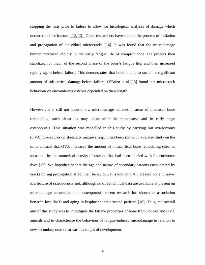
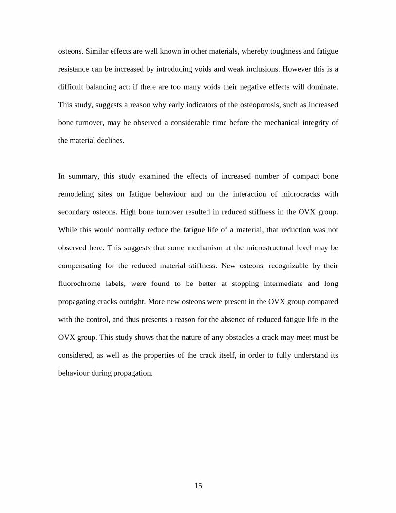
in crack number with increasing length (Figure 1). However, Cr.S.Dn was significantly
higher in the control group compared to the OVX (p=0.03) (Figure 2). Considering that
the control group displayed a higher Cr.S.Dn than the OVX group then it follows,
logically, that the control group must contain a large proportion of relatively long cracks
In order to quantify this, cracks which were 300µm or longer were measured in the
control and OVX groups. The controls contained a significantly higher number of long
cracks (>300µm) compared to the OVX group (p=0.003) (Figure 1).
Crack interaction with secondary osteons
Crack interaction with secondary osteons was quantified by considering both groups
together; this approach was taken because the issue we wished to address was
phenomenological rather than something that resulted from our intervention. Short cracks
(<100µm) which encountered secondary osteons were stopped at the cement-line
regardless whether the osteon was labeled or unlabeled. When the fluorochrome was
taken into consideration, and thus the age of the structure, there was no statistical
difference between the numbers of cracks which stopped at any osteon group. However, a
non-significant trend towards increased number of short cracks being stopped at more
recently formed osteons can be seen (Figure 3a). A higher number of intermediate cracks
(100-300µm) were arrested at the boundary of osteons that were formed in the last 6
months of this experiment. This is shown in figure 3b where the number of cracks

9
stopped at osteons labeled with calcein, xylenol orange and calcein blue are statistically
higher compared with those which were unlabeled. Long cracks (>300µm) were also
shown to stop more often at recently formed osteons compared with unlabeled.
Furthermore, the number of long cracks which stopped at calcein and xylenol orange
labeled osteons was greater compared with other earlier labeled osteons (Figure 3c). This
effect of new osteons acting as good crack stoppers compared with older osteons is
illustrated in Figure 4a where a long crack can be seen to deflect around two unlabeled
osteons before being arrested at the boundary of a labeled one. Figure 4b shows an
intermediate and a long crack which have propagated towards, and stopped at, a labeled
osteon.
Discussion
Understanding the etiology and pathophysiology of osteoporosis is increasingly
important as the world’s elderly population continues to grow. Although fracture risk is
clinically assessed by measuring BMD, the mechanical properties of bone are determined
not only by bone mass but also by bone quality [5]. An important parameter of bone
quality is the ability of the material to withstand microdamage; the subsequent behaviour
of this damage is greatly affected by structure at the microscopic and nanoscopic scales.
It has been shown that secondary osteons in compact bone influence the behaviour of
microcracks at different lengths in the bone matrix [15]. This study sought to develop this
idea further by considering the effects of variation within the properties of the secondary
osteons themselves. Three categories of crack length were defined in this study as this

10
parameter has been shown to be important in studies of microcrack propagation. The
stress intensity of a 300µm crack is higher than that of a 100µm crack, under the same
loading conditions, by a factor of approximately 1.7. This is because if the crack length is
greater by a factor of 3, then the stress intensity is increased by √3, so this observation is
useful in understanding the toughening effect of osteons. We used a fluorochrome
labeling system in order to differentiate between new osteons and old ones and thus to
assess whether the nature of the osteon influences crack behaviour, in addition to crack
length.
This has potential clinical relevance since it is now known that degree of mineralization
of bone tissue (DMB) strongly influences the mechanical resistance of bone and also the
BMD. This issue becomes relevant in a situation where a course of treatment e.g.
parathyroid hormone (PTH), or an event e.g. ovariectomy/menopause, causes an increase
in the amount of remodeling events. This could decrease the ‘lifespan’ of an osteon,
because it is more likely to be resorbed away before reaching a state of complete
mineralisation [24]. In contrast, anti-resorptive agents (bisphosphonates, estrogens) cause
a reduction in the number of remodeling events which can allow more complete
secondary mineralisation in pre-existing osteons. Given that the biological determinant of
mineralization is the rate of bone turnover, then it follows that understanding how these
parameters interact with microdamage, which is also a determinant of bone quality, is an
important issue.

11
The modulus of bending reported here was similar to that measured in a previous study
on the sheep metatarsus, using specimens of similar dimensions [25]. Based on a typical
slope of an S/N curve in bone [26], the decrease in bending modulus of 13% that we
measured would normally result a much greater reduction in Nf. This is because when the
groups are normalized with respect to modulus, the group with lower stiffness will
effectively see higher stresses. In this study, the groups were normalized by modulus and
it was found that the OVX group saw an effective stress of 126.43 MPa compared to the
nominal value of 110 MPa in the controls. With this increase, the fatigue life was
expected to be reduced by a factor of 11.21. The fact that this reduction was not present,
suggests that other microstructural factors must be involved in prolonging the fatigue life
in the OVX group.
Cr.Dn was not significantly different in the OVX group compared to controls (p=0.736)
However, Cr.S.Dn was significantly higher in the control group compared to the OVX
(p=0.003). Furthermore, the number of long cracks (>300µm) was significantly increased
in the control group compared to the OVX. We found higher Cr.Dn and Cr.S.Dn values
compared with other similar studies in the literature [21], this is primarily because
different types of bone were used. We used aged ovine bone which has a high osteon
density relative to bovine bone. If we consider bone to be a composite fiber material, with
osteons as fibers [27], and fiber-spacing is decreased by increased remodeling activity
then this would provide more crack initiation sites in interstitial regions when the bone is
cyclically loaded. Secondly, our test specimens were smaller (our cross sectional area

12
was approximately half the size) and thus would be expected to have a longer fatigue life
based on the reduced probability of the specimens containing a critical defect [28].
It is known that the behaviour of microcracks depends on crack length, in other words,
the energy that the crack has [15]. It has been shown that a microstructural barrier effect
exists in bone whereby crack length influenced microcrack behaviour on encountering an
osteon during propagation [15,16], we wished to investigate the effect of osteons at
various stages of development, on crack propagation behaviour. In this study, most
microcracks were found in interstitial bone which is consistent with the literature [10, 22,
29,30]. We used a fluorochrome labeling system to differentiate between old and new
osteons. New osteons were sub-divided into 5 groups based on the fluorochrome that they
were labeled with. The behaviour of microcracks in relation to these structures was then
characterized.
Short microcracks (<100µm) were the most numerous category of crack found, those that
encountered osteons were generally found to stop at the cement-line regardless of
whether the osteon was labeled or unlabeled. Statistical analyses showed no differences
between any of the 6 categories of osteons. Many of these cracks remained in interstitial
bone and had no interaction with osteons, labeled or unlabeled. It is interesting to
consider what the mechanism is that causes a short microcrack in interstitial bone to stop
without an obvious feature, such as an osteon or a pore, to aid in doing so. This question
cannot be answered by the present study, however it has been suggested that the presence

13
of cracks will serve to redistribute the stresses in the material and thus reduce the stress
intensity to a point below the threshold for growth [15]. While there were a large number
of short cracks found, mechanically this is not necessarily very detrimental to the material
properties of the bone, particularly in comparison to the effect of the propagation of a
smaller number of longer cracks.
Intermediate cracks (100-300µm) stopped preferentially at osteons that were labeled with
calcein, xylenol orange and calcein blue compared with unlabeled osteons. Osteons that
were labeled with any of these three fluorochromes were, at most, 6 months old. A new
osteon will have relatively low mineralization compared with an older structure. Lower
mineralization may lead to higher localized strains within the osteon, causing newer
osteons to behave more like pores in the matrix rather than a functional load bearing
volume of material. This effect appears to stop with osteons that are older than 6 months
old, as evidenced by the lack of statistical difference between cracks that stop at osteons
labeled with oxytetracycline, alizarin complexone and unlabeled ones. These data
compare well with the literature where it has been shown that the mineral apposition rate
of aged ewes is comparable with that of men and post-menopausal women, which is
between 3-6 months [31].
Long cracks (>300µm) were observed to preferentially stop at calcein labeled osteons
compared with unlabeled and oxytetracycline, at xylenol orange labeled osteons
compared with unlabeled, oxytetracycline and alizarin and finally at calcein blue labeled
osteons with compared with unlabeled. As with the intermediate cracks this can be

14
explained by assuming that newer osteons, containing relatively under-mineralized bone,
do not bear much load and so behave like pores in the matrix. Given a favorable
combination of stress intensity and damage zones at the crack tip and at the pore, the
crack may then propagate towards the osteon and then be blunted by it. This effect has
been described in the literature in relation to other materials [32]. There was a significant
difference between the number of long cracks that stopped at calcein and xylenol orange
labeled osteons compared to other labeled osteons as well as to unlabeled osteons. This
may be explained as follows; the longer a crack is the more energy it has thus, a 9 or 12
month old osteon may still be ‘young’ enough to arrest the growth of an intermediate
crack, but not of a longer crack (>300µm). Thus the difference between early labels (0
and 3 months) and later labels (6-12 months) is only apparent when long cracks are
considered.
The OVX group, having a lower elastic modulus, would be expected to have a shorter
fatigue life, but our results show that this was not the case. We demonstrated that, thanks
to increased numbers of under-mineralized osteons, bone with high numbers of recently
formed remodeling sites has an increased ability to impede the growth of relatively long
cracks, preventing them from growing as easily as they would do in normal bone. The
relationship between microstructure and mechanical properties is a complex one: this
study has highlighted one rather unexpected consequence of an increased number of
compact bone remodeling sites. The material’s integrity has been jeopardized, as
evidenced by its reduced elastic modulus and a slight increase in the number of fatigue
cracks initiated, but this has been compensated by the toughening effect of the new
15
osteons. Similar effects are well known in other materials, whereby toughness and fatigue
resistance can be increased by introducing voids and weak inclusions. However this is a
difficult balancing act: if there are too many voids their negative effects will dominate.
This study, suggests a reason why early indicators of the osteoporosis, such as increased
bone turnover, may be observed a considerable time before the mechanical integrity of
the material declines.
In summary, this study examined the effects of increased number of compact bone
remodeling sites on fatigue behaviour and on the interaction of microcracks with
secondary osteons. High bone turnover resulted in reduced stiffness in the OVX group.
While this would normally reduce the fatigue life of a material, that reduction was not
observed here. This suggests that some mechanism at the microstructural level may be
compensating for the reduced material stiffness. New osteons, recognizable by their
fluorochrome labels, were found to be better at stopping intermediate and long
propagating cracks outright. More new osteons were present in the OVX group compared
with the control, and thus presents a reason for the absence of reduced fatigue life in the
OVX group. This study shows that the nature of any obstacles a crack may meet must be
considered, as well as the properties of the crack itself, in order to fully understand its
behaviour during propagation.

16
ACKNOWLEDGMENTS
This study was supported by the Higher Education Authority, under the Programme for
Research in Third Level Institutions (PRTLI) Cycle 3, and also by the Health Research
Board of Ireland under Grant No: RP/2004/229. The authors thank Peter O’Reilly for
technical assistance. Thanks also to Brian Cloak and the staff at the Lyons Research
Facility, UCD for animal care and handling.
REFERENCES
[1]. Burr DB, Forwood MR, Fyhrie DP, Martin RB, Schaffler MB, and Turner CH Bone microdamage and skeletal fragility in osteoporotic and stress fractures. J. Bone Miner. Res. 1997; 12:6-15.
[2]. Ammann P, and Rizzoli R Bone strength and its determinants. Osteoporos. Int. 2003; 14 Suppl 3:S13-8.
[3]. Hui SL, Slemenda CW, and Johnston CC, Jr. Age and bone mass as predictors of fracture in a prospective study. J. Clin. Invest. 1988; 81:1804-9.
[4]. Bouxsein ML Bone quality: where do we go from here? Osteoporos. Int. 2003; 14 Suppl 5:118-27.
[5]. Burr DB Bone quality: understanding what matters. J Musculoskelet Neuronal Interact 2004; 4:184-6.
[6]. Frost HM Presence of microscopic cracks in vivo in bone. Bull Henry Ford Hospital 1960; 8:27-35.
[7]. Burr DB, Milgrom C, Boyd RD, Higgins WL, Robin G, and Radin EL Experimental stress fractures of the tibia. Biological and mechanical aetiology in rabbits. J. Bone Joint Surg. Br. 1990; 72:370-5.
[8]. Lee TC, Myers ER, and Hayes WC Fluorescence-aided detection of microdamage in compact bone. J. Anat. 1998; 193 (Pt 2):179-84.
[9]. O'Brien FJ, Hardiman DA, Hazenberg JG, Mercy MV, Mohsin S, Taylor D, and Lee TC The behaviour of microcracks in compact bone. Eur. J. Morphol. 2005; 42:71-9.
[10]. Taylor D Fatigue of bone and bones: an analysis based on stressed volume. J. Orthop. Res. 1998; 16:163-9.
[11]. Donahue SW, Sharkey NA, Modanlou KA, Sequeira LN, and Martin RB Bone strain and microcracks at stress fracture sites in human metatarsals. Bone 2000; 27:827-33.
[12]. Forwood MR, and Parker AW Microdamage in response to repetitive torsional loading in the rat tibia. Calcif. Tissue Int. 1989; 45:47-53.
[13]. Burr DB, and Hooser M Alterations to the en bloc basic fuchsin staining protocol for the demonstration of microdamage produced in vivo. Bone 1995; 17:431-3.
[14]. Akkus O, and Rimnac CM Fracture resistance of gamma radiation sterilized cortical bone allografts. J. Orthop. Res. 2001; 19:927-34.

17
[15]. O'Brien FJ, Taylor D, and Clive Lee T The effect of bone microstructure on the initiation and growth of microcracks. J. Orthop. Res. 2005; 23:475-80.
[16]. Mohsin S, O'Brien FJ, and Lee TC Microcracks in compact bone: a three-dimensional view. J. Anat. 2006; 209:119-24.
[17]. Kennedy OD, Brennan O, Mauer P, Rackard SM, O'Brien FJ, Taylor D, and Lee TC Fatigue induced microdamge in compact bone samples from control and ovariectomised sheep. 53rd Meeting of the Orthpaedic Research Society 32. San Diego, USA; 2007.
[18]. Stepan JJ, Burr DB, Pavo I, Sipos A, Michalska D, Li J, Fahrleitner-Pammer A, Petto H, Westmore M, Michalsky D, Sato M, and Dobnig H Low bone mineral density is associated with bone microdamage accumulation in postmenopausal women with osteoporosis. Bone 2007; 41:378-85.
[19]. Taylor D, O'Brien F, Prina-Mello A, Ryan C, O'Reilly P, and Lee TC Compression data on bovine bone confirms that a "stressed volume" principle explains the variability of fatigue strength results. J. Biomech. 1999; 32:1199-203.
[20]. Lee TC, Staines A, and Taylor D Bone adaptation to load: microdamage as a stimulus for bone remodelling. J. Anat. 2002; 201:437-46.
[21]. Diab T, and Vashishth D Effects of damage morphology on cortical bone fragility. Bone 2005; 37:96-102.
[22]. Boyce TM, Fyhrie DP, Glotkowski MC, Radin EL, and Schaffler MB Damage type and strain mode associations in human compact bone bending fatigue. J. Orthop. Res. 1998; 16:322-9.
[23]. Lee TC, Arthur TL, Gibson LJ, and Hayes WC Sequential labelling of microdamage in bone using chelating agents. J. Orthop. Res. 2000; 18:322-5.
[24]. Boivin G, and Meunier PJ Effects of bisphosphonates on matrix mineralization. J Musculoskelet Neuronal Interact 2002; 2:538-43.
[25]. Currey JD The effect of porosity and mineral content on the Young's modulus of elasticity of compact bone. J. Biomech. 1988; 21:131-9.
[26]. Gray RJ, and Korbacher GK Compressive fatique behaviour of bovine compact bone. J. Biomech. 1974; 7:287-92.
[27]. Martin R, Burr D, and Sharkey N Skeletal Tissue Mechanics. New York: Springer Verlag; 1998.
[28]. Taylor D Scaling effects in the fatigue strength of bones from different animals. J. Theor. Biol. 2000; 206:299-306.
[29]. Schaffler MB, Choi K, and Milgrom C Aging and matrix microdamage accumulation in human compact bone. Bone 1995; 17:521-525.
[30]. Norman TL, and Wang Z Microdamage of human cortical bone: incidence and morphology in long bones. Bone 1997; 20:375-9.
[31]. Pearce AI, Richards RG, Milz S, Schneider E, and Pearce SG Animal models for implant biomaterial research in bone: a review. Eur Cell Mater 2007; 13:1-10.
[32]. Janssen D, Aquarius R, Stolk J, and Verdonschot N The contradictory effects of pores on fatigue cracking of bone cement. J Biomed Mater Res B Appl Biomater 2005; 74:747-53.
Cr.Dn at different length categories for Controls and OVX
0
0.5
1
1.5
2
2.5
3
<100 100<300 >300
Length Category (mm)
Cr.
Dn
(#
/mm
2)
CON
OVX
Figure 1: Graph of numerical crack density in control and OVX bone for the 3 different length categories
Figure 2: Graph of crack surface density in control and OVX bone samples.
Crack Surface Density in Control and OVX
0100200300400500600700800900
Control OVX
Cr.
S.D
n (
m/m
m2 )
Cr.S
.Dn
(m
/mm
2 )
* * : p<0.05
* : p<0.05
*
Figure(s)

Mean Number of Cracks (<100mm) Stopped at Unlabelled and Labelled Osteons
0
0.1
0.2
0.3
0.4
0.5
0.6
Unlabelled Oxytet Alizarin Calcein XylenolOrange
CalceinBlue
No
rmal
ized
Cr.
Dn
Mean Number of Cracks (100>300mm) Stopped at Unlabelled and Labelled Osteons
00.05
0.10.15
0.20.25
0.30.35
0.40.45
0.5
Unlabelled Oxytet Alizarin Calcein XylenolOrange
CalceinBlue
No
rma
lize
d C
r.D
n
Mean Number of Cracks (>300mm) Stopped at Unlabelled and Labelled Osteons
0
0.05
0.1
0.15
0.2
0.25
0.3
0.35
0.4
Unlabelled Oxytet Alizarin Calcein XylenolOrange
CalceinBlue
No
rmal
ized
Cr.
Dn
Figure 3: Mean Cr.Dn, of both groups combined, for each category of osteon, normalized by the number of osteons in each case for (a) short cracks (<100m), (b) intermediate cracks (100<300m) and (c) long cracks (>300m).
*
**
**
*
§
§
¥*: Significantly different to Unlabelled§: Significantly different to Oxytet¥ : Significantly different to Alizarin
*: Significantly different to Unlabelled
Pre-OVX 0 3 6 9 12 Months Post-OVX
0 3 6 9 12
Pre-OVX 0 3 6 9 12 Months Post-OVX
Pre-OVX 0 3 6 9 12 Months Post-OVX
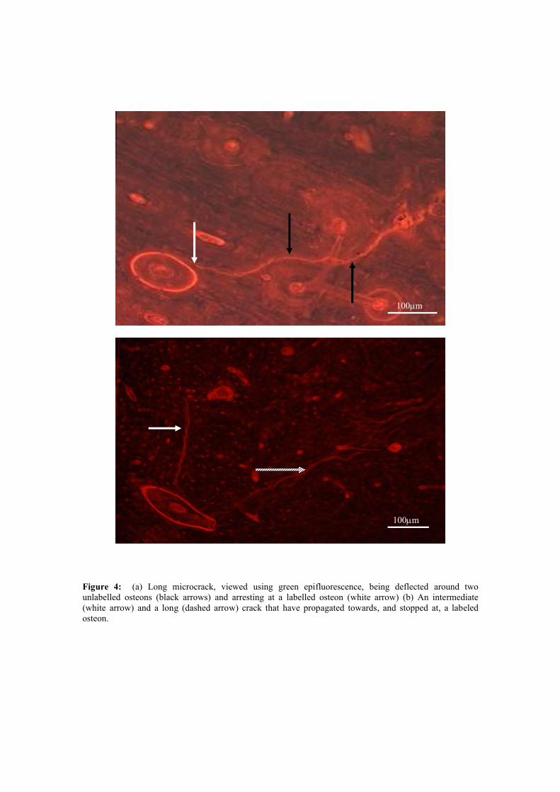
Figure 4: (a) Long microcrack, viewed using green epifluorescence, being deflected around two unlabelled osteons (black arrows) and arresting at a labelled osteon (white arrow) (b) An intermediate (white arrow) and a long (dashed arrow) crack that have propagated towards, and stopped at, a labeled osteon.
100m
100m
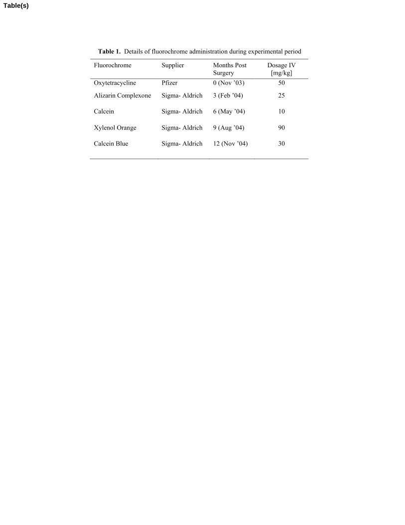
Table 1. Details of fluorochrome administration during experimental period
Fluorochrome Supplier Months Post Surgery
Dosage IV [mg/kg]
Oxytetracycline Pfizer 0 (Nov ’03) 50
Alizarin Complexone Sigma- Aldrich 3 (Feb ’04) 25
Calcein Sigma- Aldrich 6 (May ’04) 10
Xylenol Orange Sigma- Aldrich 9 (Aug ’04) 90
Calcein Blue Sigma- Aldrich 12 (Nov ’04) 30
Table(s)

Reviewers' comments:
Reviewer #1: I am satisfied that the authors have attended to the comments and suggestions made in my review and have improved their manuscript.
Thank you for your help.
Reviewer #2: This draft is a much clearer presentation of what was done, and the additional analyses make for a much more informative paper. A few items should be addressed before publication. Once these are addressed, the paper will make a nice addition to the microdamage literature.
1. The abstract states there was no difference between ovx and control in crack density, but ovx in fact had more >300 um cracks. This should be corrected.
This has been corrected, and the difference has been highlighted in the Abstract.
2. The sentence beginning 'This mechanism may have.' in the abstract should be rewritten to express the point more clearly.
This sentence has been re-written as follows: ‘This mechanism may have an important role in terms of prolonging fatigue life...’
3. In the 'statistical analysis' section, you need to state how the 6 group comparisons (in fig 4) were made. There seems to be very large error bars for getting significant differences.
This section has been amended to include the details of the ANOVA and Holm-Sidak procedures that were carried out for these comparisons.
4. Isn't Fig 3 the same as the right-hand side of fig 1? If so, fig 3 should be removed and significance indicated in fig1.
Yes - Figure 3 has been removed and the significance indicated in Figure 1.
5. Fig 4: the group labels would be more informative if you used the times in relation to ovx instead of the dye color. Not clear if these groups contain data from ovx, control, or both; this needs to be explained here and in text.
The labels on this graph have been changed as suggested. These groups contain data from both groups because we wished to address this as a general phenomenon as opposed to something which resulted from our intervention. This point has now been clarified in the caption and in the text.
* Response to Reviewers
Copyright © 2022 FDOKUMEN