Releasing Differentially Private Trajectories with Optimized ...
Upload
independentCategory
view
1download
0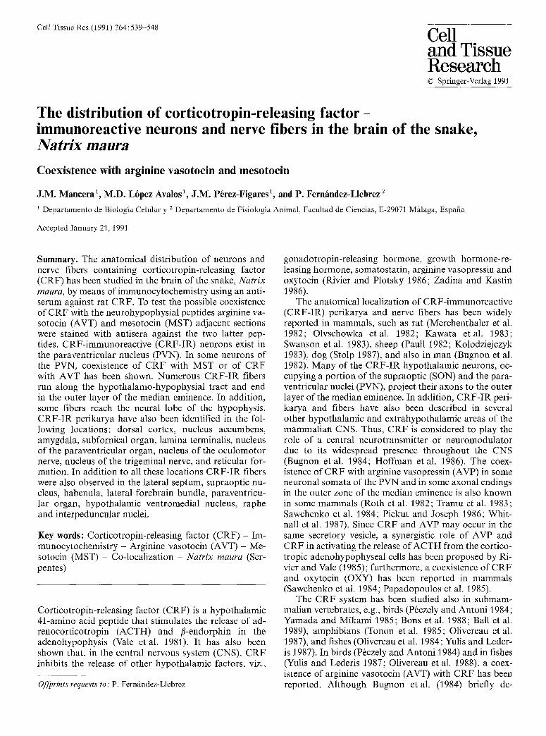
Cell Tissue Res (1991) 264:539-548 Cell and Tissue Research �9 Springer-Verlag 1991
The distribution of corticotropin-releasing factor- immunoreactive neurons and nerve fibers in the brain of the snake, N a t r i x m a ura
Coexistence with arginine vasotocin and mesotocin
J.M. Mancera ~, M.D. L6pez Avalos ~, J.M. P6rez-Figares 1, and P. Fernfindez-Llebrez 2
1 Departamento de Biologia Celular y 2 Departamento de Fisiologia Animal, Facultad de Ciencias, E-29071 Mfilaga, Espafia
Accepted January 21, 1991
Summary. The anatomical distribution of neurons and nerve fibers containing corticotropin-releasing factor (CRF) has been studied in the brain of the snake, N a t r i x maura , by means of immunocytochemistry using an anti- serum against rat CRF. To test the possible coexistence of CRF with the neurohypophysial peptides arginine va- sotocin (AVT) and mesotocin (MST) adjacent sections were stained with antisera against the two latter pep- tides. CRF-immunoreactive (CRF-IR) neurons exist in the paraventricular nucleus (PVN). In some neurons of the PVN, coexistence of CRF with MST or of CRF with AVT has been shown. Numerous CRF-IR fibers run along the hypothalamo-hypophysial tract and end in the outer layer of the median eminence. In addition, some fibers reach the neural lobe of the hypophysis. CRF-IR perikarya have also been identified in the fol- lowing locations: dorsal cortex, nucleus accumbens, amygdala, subfornical organ, lamina terminalis, nucleus of the paraventricular organ, nucleus of the oculomotor nerve, nucleus of the trigeminal nerve, and reticular for- mation. In addition to all these locations CRF-IR fibers were also observed in the lateral septum, supraoptic nu- cleus, habenula, lateral forebrain bundle, paraventricu- lar organ, hypothalamic ventromedial nucleus, raphe and interpeduncular nuclei,
Key words: Corticotropin-releasing factor (CRF) - Im- munocytochemistry - Arginine vasotocin (AVT) - Me- sotocin (MST) - Co-localization - N a t r i x m a u r a (Ser- pentes)
Corticotropin-releasing factor (CRF) is a hypothalamic 41-amino acid peptide that stimulates the release of ad- renocorticotropin (ACTH) and /~-endorphin in the adenohypophysis (Vale et al. 1981). It has also been shown that, in the central nervous system (CNS), CRF inhibits the release of other hypothalamic factors, viz.,
Offprints requests to: P. Fern/mdez-Llebrez
gonadotropin-releasing hormone, growth hormone-re- leasing hormone, somatostatin, arginine vasopressin and oxytocin (Rivier and Plotsky 1986; Zadina and Kastin 1986).
The anatomical localization of CRF-immunoreactive (CRF-IR) perikarya and nerve fibers has been widely reported in mammals, such as rat (Merchenthaler et al. 1982; Olvschowka etal. 1982; Kawata etal. 1983; Swanson et al. 1983), sheep (Paull 1982; Kolodziejczyk 1983), dog (Stolp 1987), and also in man (Bugnon et al. 1982). Many of the CRF-IR hypothalamic neurons, oc- cupying a portion of the supraoptic (SON) and the para- ventricular nuclei (PVN), project their axons to the outer layer of the median eminence. In addition, CRF-IR peri- karya and fibers have also been described in several other hypothalamic and extrahypothalamic areas of the mammalian CNS. Thus, CRF is considered to play the role of a central neurotransmitter or neuromodulator due to its widespread presence throughout the CNS (Bugnon et al. 1984; Hoffman et al. 1986). The coex- istence of CRF with arginine vasopressin (AVP) in some neuronal somata of the PVN and in some axonal endings in the outer zone of the median eminence is also known in some mammals (Roth et al. 1982; Tramu et al. 1983; Sawchenko et al. 1984; Piekut and Joseph 1986; Whit- nall et al. 1987). Since CRF and AVP may occur in the same secretory vesicle, a synergistic role of AVP and CRF in activating the release of ACTH from the cortico- tropic adenohypophyseal cells has been proposed by Ri- vier and Vale (1985); furthermore, a coexistence of CRF and oxytocin (OXY) has been reported in mammals (Sawchenko et al. 1984; Papadopoulos et al. 1985).
The CRF system has been studied also in submam- malian vertebrates, e.g., birds (P6czely and Antoni 1984; Yamada and Mikami 1985; Bons et al. 1988; Ball et al. 1989), amphibians (Tonon et al. 1985; Olivereau et al. 1987), and fishes (Olivereau et al. 1984; Yulis and Leder- is 1987). In birds (P~czely and Antoni 1984) and in fishes (Yulis and Lederis 1987; Olivereau et al. 1988), a coex- istence of arginine vasotocin (AVT) with CRF has been reported. Although Bugnon etal. (1984) briefly de-

540
scribed the system in a turtle, detailed studies in other reptilian orders are missing. Due to their crucial posit ion in the phylogenet ic scale, studies on reptiles are o f great interest. The aim o f the present investigation is to illus- trate the anatomical distr ibution o f C R F - I R per ikarya and nerve fibers in the brain o f the snake N a t r i x m a u r a .
Materials and methods
Five adult animals of either sex were used in this study. All animals were captured in spring and maintained in an aquarium-terrarium at room temperature and natural photoperiod; they were sup- ported with adequate food.
All animals were anesthetized with tricaine (0.292 mg/g body weight) and subsequently perfused transcardiatly with Ringer's so- lution and then with Bouin's fluid for 40 min. The dissected brains were placed for 48 h in the same fixative and then dehydrated and embedded in paraffin.
Sagittal and transverse (8-~tm-thick) sections were hydrated and immunostained according to the PAP method of Sternberger (1986). The following primary rabbit antisera were used: anti-rat- CRF (1 : 500) (kindly provided by Professor E.M. Rodriguez, Valdi- via, Chile), anti-AVT (1:1000) (kindly provided by Professor R.M. Buijs, Amsterdam, Holland), and anti-MST (kindly provided by Professor S. Blfihser, Giessen, FRG). Anti-AVT does not cross- react with isotocin (Dungen et al. 1982) or with mesotocin (Fern/m- dez-Llebrez et al. 1988).
All sections were incubated for 18 h at 22 ~ C in the primary antisera. The second antiserum (from E.M. Rodriguez, Valdivia, Chile) was used at a dilution 1:10 for 30 min at 22~ and the PAP complex (1:75) (Sigma P2030) for 30 min at 22 ~ C. DAB was used as electron donor. All antisera and the PAP complex were diluted in TRIS buffer, pH 7.8, containing 0.7% non-gelling seaweed gelatin, lambda carrageenan (Sigma), and 0_5% Triton X-100 (Sigma), and 0.02% sodium azide. Coplin jars were used for incubation with the first and the second antibody, whereas PAP incubation was carried out in a moist chamber.
Control test: In order to test the specificity of the immunoreaction to anti-CRF antiserum, immunoabsorption with the same CRF preparation as used for raising the antiserum was performed. Ali- quots of the antiserum, diluted 1 : 500, were mixed separately with the CRF preparation at concentrations of 25 and 50 gg/ml. Both preparations were kept for 18 h at 22 ~ C. Then the resulting solu- tions were employed, in the same staining session, for immunocyto- chemistry on sections adjacent to those immunostained with the non-absorbed anti-CRF.
To control the immunoreactive procedure for all the antisera used, adjacent sections were stained according to the described protocol but the primary antisera were ommited.
Neither the use of immunoabsorbed anti-CRF nor immunocy- tochemistry omitting the primary antisera revealed any stained structure in the sections.
Results
N e u r o n a l p e r i k a r y a : Figs. 1 and 2 are schematic draw- ings o f transverse and sagittal sections th rough the brain o f N . m a u r a showing the dis tr ibut ion o f C R F - I R peri- karya and fibers. Immunoreac t ive per ikarya have been found in telencephalic, diencephalic, and mesencephalic regions.
In the telencephalon, some small C R F - I R cells ( ~ 11.52 _+ 1.44 gm in diameter, mean _+ s tandard deviation) were scattered along the inner layer o f the dorsal cortex
(Figs. 1, 5). They occupied a more lateral posi t ion in caudal cortical regions (Fig. 1). These neurons showed a s t rong immunoreac t iv i ty and had round, fus i form or pear-shaped cell bodies. Few small r o u n d cells ( ~ 11.27 _+ 1.36 gm in diameter), displaying nar row, weakly immunoreac t ive perikarya, were also identified in the nucleus accumbens and the amygda la (Fig. 1).
A n appreciable number o f small, fus i form or pear- shaped C R F - I F neurons ( ~ 8 . 6 _ 0 . 8 g m in diameter) appeared in the subfornical o rgan (Figs. 6-8). Mos t o f them were clustered in the vent rocaudal por t ion o f the organ. In addit ion, a few isolated neurons o f the same type occurred in the region o f the lamina terminalis. In the ventral hypo tha lamus , small and medium-sized neurons were scattered in a region between the S O N and the PVN. These cells were endowed with processes and displayed a s t rong immunoreact iv i ty .
Numerous C R F - I R neurons were observed in the P V N (Fig. 13). These magnocel lu lar ( ~ 17.2 +_ 1.4 gm in diameter) or parvocel lular ( ~ 9.1 _ 1.2 gm in diameter) elements occurred close to the ventricle or in deeper por- t ions o f the brain. The parvocel lular fus i form or pear- shaped neurons sometimes emit ted an apical process tha t appeared to contac t the cerebrospinal fluid. Study o f adjacent sections immunos ta ined with an t i -MST (Figs. 12, 15), a n t i - C R F (Figs. 13, 16, 17) and an t i -AVT (Figs. 14, 18) revealed the coexistence o f C R F with A V T and o f C R F with M S T in some magnocel lu lar neurons o f the PVN.
A group o f small, weakly immunoreact ive , r o u n d neurons occupied a region close to the dorsal por t ion o f the paraventr icular o rgan (Fig. 1).
In the mesencephalon, very large bipolar or mul t ipo- lar C R F - I R neurons (~18.66_+1.83 gm in diameter) were found clustered in the nucleus o f the o c u l o m o t o r nerve (Fig. 19) and in the nucleus o f the trigeminal nerve (Fig. 20). These nerve cells showed a s t rong immunore - activity. In addit ion, neurons o f different sizes and shapes displaying variable immunoreac t iv i ty were identi- fied in the reticular fo rma t ion (Fig. 21).
N e r v e f i b e r s : A diagrammat ic representat ion o f the pat- tern o f distr ibution o f C R F - I R fibers is shown in Figs. I and 2. In all regions conta in ing C R F - I R perikarya, C R F - I R fibers were also apparent . In contrast , o ther
Figs. 1, 2. Schematic drawings of sections through the brain of N. maura showing the localization of CRF-immunoreactive cell bodies (dark circles) and fibers (dots). A C Anterior commissure; A L anterior lobe of the hypophysis; A M amygdaloid complex; DC dorsal cortex; H habenula; H H T hypothalamo-hypophysial tract; IL intermediate lobe of the hypophysis; I N interpeduncular nucleus; LFB lateral forebrain bundle; L T lamina terminalis; M E median eminence; NA nucleus accumbens; NPO nucleus of the paraventricular organ; NII I nucleus of the oculomotor nerve; N V nucleus of the trigeminal nerve; O C optic chiasm; O T optic tract; PL posterior lobe of the hypophysis; PO paraventricular organ; P V N paraventricular nucleus; RA nucleus of the raphe; R F nucleus of the reticular formation; S septum; SFO subfornical organ; S O N supraoptic nucleus; VA vestibular area; V H N ventromedial hypo- thalamic nucleus

541
N A
/
A
. 0 . � 9 �9 ' .OO .O - .
F H I J
Fig. 1. Successive rostrocaudal transverse sections (A-J) at the levels shown in Fig. 2
H
N ,
R F ~ , .
Fig. 2. SagittaI section. Note levels corresponding to the transverse section in Fig. 1 A-3

542
Figs. 3, 4. Immunoreactive nerve fibers and endings in the septum. Some negative perikarya are surrounded by immunoreactive fibers (Fig. 4). LV Lateral ventricle. Transverse sections, Fig. 3: • 200; Fig. 4: x 800
Fig. 5. Detail of the inner portion of the dorsal cortex close to the lateral ventricle (LV). Note immunoreactive small neurons. Transverse section, x 320
Figs. 6-8. Subfornical organ showing in its rostral portion (Fig. 8), some immunoreactive perikarya (arrows) and a dense network of selectively stained fibers. The caudal portion of the organ (Fig. 6)
displays scarce fiber elements and numerous small, fusiform peri- karya (detailed in Fig. 7). IS lnterhemispheric connective septum. Transverse sections. Fig. 6: x 330; Fig. 7: • 800; Fig. 8: x 340
Fig. 9. Immunoreactive nerve fibers in the lamina terminalis (LT). IS Interhemispheric connective septum. Transverse section, x 230
Fig. 10. Detail of the ventrolateral hypothalamus showing the su- praoptic nucleus and the lateral forebrain bundle (LFB). The SON displays many CRF-IR nerve fibers and terminals around immuno- negative magnocellular neurons (arrows). OT Optic tract. Trans- verse section, x 230

543
Fig. 11. Sagittal section through the median eminence (ME) and the hypophysis. Note the strong immunoreactivity of the ME especially in its outer zone in juxtaposition to the vessels of the hypophysial portal system (PS). Some fibers run along the hypo- physial stalk (HS) and reach the posterior lobe of the hypophysis (PL). AL Anterior lobe of the hypophysis; H hypothalamus; IL intermediate lobe of the hypophysis, x 80
Figs. 12-14. Adjacent transverse sections through the paraventricu- lar nucleus stained with antisera against mesotocin (MST), cortico-
tropin-releasing factor (CRF) and vasotocin (A VT), respectively. Cells displaying immunoreactivity to anti-MST and anti-CRF (! 5), and to anti-CRF and anti-AVT (A-D) are indicated, x 75
Figs. 15-18. Details of some of the perikarya shown in Figs. 12, 13 and 14 that were immunoreactive to anti-mesotocin (Fig. 15) and anti-CRF (Fig. 16), and to anti-CRF (Fig. 17) and anti-vasoto- cin (Fig. 18). x 550

544
Figs. 19, 20. Large CRF-immunoreactive neurons in the nucleus of the oculomotor nerve (Fig. 19) and the nucleus of the trigeminal nerve (Fig. 20). Transverse sections, x 300
Fig, 21. Reticular formation where, in addition to nerve fibres, perikarya of different sizes and shapes display immunoreactivity for CRF. Transverse section, x 495
Figs. 22, 23. Consecutive transverse sections through the median eminence, immunostained with anti-CRF (Fig. 22) or treated with anti-CRF previously absorbed with CRF (Fig. 23). Immunoab- sorption completely abolished the immunoreaction. V Third ventri- cle. x 160
regions lacking immunoreactive perikarya exhibited im- munoreactive neural tracts.
The lateral septum was rich in CRF-IR fibers (Fig. 3). Some of them displayed varicosities and surrounded im- munonegative perikarya suggesting multiple synaptic contacts (Fig. 4). Numerous selectively stained fibers oc- curred in the lamina terminalis (Fig. 9) and the subforni- cal organ (Figs. 6-8). In the SON, an intense innervation by CRF-IR fibers could be observed at all levels includ- ing the more caudal retrochiasmatic nucleus; these fibers mainly occupied the neuropil among the magnocellular neurons (Fig. 10). Other diencephalic entities such as the lateral forebrain bundle, the habenula, the paraventricu- lar organ and the ventromedial hypothalamic nucleus, contained only small CRF-IR fibers.
Numerous CRF-IR fibers ran along the diencephalic floor and terminated in the outer zone of the median eminence close to the capillary plexus of the portal ves- sels; in this region AVT and MST immunoreactivities were also prominent. In addition, some CRF-IR fibers
passed via the internal zone of the median eminence to the posterior lobe where they ended in the palisade region of the lobules (Fig. 11). The mesencephalic teg- mentum exhibited numerous CRF-fibers in its ventral portion innervating the reticular formation, the raphe, and the interpeduncular nucleus (Fig. 1).
Absorption of anti-CRF with CRF completely abol- ished the immunoreaction in all regions studied (com- pare Figs. 22, 23).
Discussion
The present study is concerned with the localization of neurons and nerve fbers immunoreactive to an antise- rum against rat CRF. Some aspects deserve particular consideration. Several authors have used colchicine to improve the visualization of the CRF-IR cells (Bloom et al. 1982; Merchenthaler et al. 1982; Olschowka et al. 1982; Burlet et al. 1983). Other authors, however, found
545
that pretreatment with colchicine was not essential for the immunostaining of CRF-IR cells (Kawata et al. 1983). The immunocytochemical procedure applied in this study is adequate for the animal species used; experi- ments in our laboratory indicate that colchicine does not actually increase the number of neurons immuno- stained with the anti-CRF employed.
CRF immunoreactivity in the hypothaIamo-hypophysial system." In mammals, there is no agreement among au- thors with regard to the localization of the CRF-produc- ing neurons even in one and in same species, While some authors described a principal population of CRF-IR neurons in the parvocellular portion of the PVN (Bloom et al. 1982; Merchenthaler et al. 1982; Hoffman et al. 1986), others localized the immunoreactivity mainly to the magnocellular component of the PVN and the SON (Paull et al. 1982; Kawata et al. 1983; Stolp et al. 1987). In birds, the hypothalamo-hypophysial CRF-system is similar to that of mammals although the CRF-IR neu- rons extend far beyond the boundary of the PVN proper (P6czely and Antoni 1984; Yamada and Mikami 1985; Bons et al. 1988; Ball et al. 1989). In fishes and amphibi- ans, this system consisting of CRF-IR cells located in the preoptic nucleus projects toward the median emi- nence (Tonon et al. 1987; Olivereau etal. 1987; Yulis and Lederis 1987; Olivereau and Olivereau 1988; Vallar- ino et al. 1989).
To our knowledge, the only report on the CRF-sys- tern in reptiles deals with the situation in the turtle (Bug- non et al. 1984); the system described seems to be quite peculiar since the CRF-IR perikarya, projecting to the median eminence, have been described to occur in the nucleus of the paraventricular organ but not in the PVN. In N. maura, the organization of the paraventriculo-hy- pophysial CRF system resembles that of mammals and is different to that in turtles. Although, as in turtles, in N. maura some neurons displaying weak immunoreac- tivity have been identified in the nucleus of the paraven- tricular organ, the main population of CRF-IR cells oc- cupies the PVN. Axonal extensions from these neurons appear to contribute mainly to the hypothalamo-hypo- physial tract (for comparison, see also Fern/mdez-Lle- brez et al. 1988).
Colocalization of CRF with neurohypophysial peptides : In mammals colocalization of CRF with the neurohypo- physial peptidcs AVP and OXY has been reported for the magnocellular and parvocellular perikarya of the SON and PVN and also for the axonal endings in the median eminence and the neural lobe of the hypophysis (Roth et al. 1982; Burlet et al. 1983; Tramu et al. 1983; Sawchenko et al. 1984; Papadopoulus et al. 1985; Saw- chenko 1987; Piekut and Joseph 1986; Whitnall 1988; Bondy et al. 1989). Coexistence of CRF with AVT has also been reported in birds (P6czely and Antoni 1984). The number of neurons showing coexistence of CRF with neurohypophysial nonapeptides increases after col- chicine or adrenalectomy (Kiss 1984; Whitnall 1988; Bondy 1989). CRF and AVP coexist in the same secreto- ry vesicles within the fiber terminals in the median emi-
hence (Whitnall et al. 1987); both are released simulta- neously, reach the adenohypophysis, and initiate a sys- temic release of ACTH. Thus a synergistic type of action involving AVP and CRF in the release of ACTH has been proposed (Rivier and Vale 1985). Coexistence of CRF with the neurohypophysial peptides seems to be an early phylogenetic event since it has also been re- ported in fishes (Olivereau et al. 1988; Yulis and Lederis 1987). Our observations show that, in N. maura, certain neurons in the PVN appear immunoreactive to anti- CRF and anti-AVT or to anti-CRF and anti-MST in adjacent paraffin sections; thus a coexistence of CRF with both neurohypophysial peptides may also be pro- posed for N. maura.
CRF-IR fibers in the neural lobe of the hypophysis." In fishes, a portion of the CRF-IR fibers of the hypothala- mo-hypophysial tract ends directly in the pars distalis close to the ACTH-producing cells; however, most fibers reach the neurointermediate lobe and terminate near the ACTH and MSH cells. A hypothalamo-hypophysial portal system obviously does not exist, and consequently CRF may act on the responsive cell in a paracrine fash- ion (Bugnon et al. 1983; Olivereau et al. 1984; Yulis and Lederis 1987; Olivereau and Olivereau 1988; Vallarino et al. 1989). In amphibians, most CRF fibers end in the median eminence close to the ACTH cells in the rostral portion of the pars distalis. Here, CRF may approach its target cells mainly via the perivascular spaces (Tonon etal. 1985; Olivereau etal. 1987). In addition, some fibers reach the neurointermediate lobe and, consequent- ly, participation of CRF in the control of neurointerme- diate-lobe functions has been suggested (Tonon et al. 1985). In reptiles (Bugnon et al. 1983; present study), birds (P6czely and Antoni 1984; Boris et al. 1988) and mammals (Bloom et al. 1982; Stolp et al. 1987) most CRF fibers end in the outer zone of the median emi- nence, and some fibers reach the neural lobe. In mam- mals, the presence of the CRF endings in the posterior lobe has been interpreted in different ways by several authors (Kawata et al. 1983; Lengv/tri et al. 1985; Stolp et al. 1987; Bondy and Garnier 1989): (1) CRF may be released to the systemic circulation and behave as a blood-borne hormone with an unknown peripheral target. (2) CRF may reach the adenohypophysis via a hypothetical posterior lobe-adenohypophysial portal system. (3) CRF may act locally and control the release of neurohypophysial peptides. We assume that, in N. maura, in addition to the above-mentioned hypotheses, CRF could also control, in a paracrine fashion, the re- lease of MSH (and/or fl-endorphin) from the intermedi- ate lobe cells, which, in this species, are intermingled with the specific structures of the neural lobules (P6rez and Fern~ndez-Llebrez 1989).
CRF-IR cells and fibers outside the hypothalamo-hypo- physial system." CRF cells and fibers have an extensive hypothalamic and extrahypothalamic distribution in the brain of mammals (Merchenthaler et al. 1982; Olschow- ka et al. 1982), birds (Bons et al. 1988; Ball et al. 1989)

546
and turtles (Bugnon et al. 1984). The evidence available indicates that C R F may act centrally as a neurotransmit- ter or neuromodulator in several brain regions. In con- trast, in amphibians (Tonon et al. 1985; Olivereau et al. 1987) and fishes (Yulis and Lederis 1987; Olivereau and Olivereau 1988), the C R F perikarya appear to be re- stricted to the hypothalamus and the C R F fibers to the neurohypophysis. In N. maura, an extensive extrahypo- physial system has been demonstrated. The following discussion will be focused on those regions in which the presence of C R F cells and/or fibers is of particular inter- est due to their absence in other species, or with respect to the particular physiological significance of a local sys- tem.
In the telencephalon of N. maura, C R F - I R cells have been found in the dorsal cortex, the nucleus accumbens and the amygdala. These results correspond to those in rats (Merchenthaler et al. 1982) pigeons (Boris et al. 1988) and turtles (Bugnon et al. 1984). At least the amyg- dala is involved in the control of the hypophysiotropic axis (Fellmann et al. 1979). The septum receives a strong innervation; this region is connected to the PVN and the subfornical organ (SFO), both structures participate in the control of water balance (Miselis 1981; Tanaka et al. 1988).
In N. maura, the SON lacks C R F - I R cells but dis- plays numerous C R F - I R fibers. It is tempting to specu- late that C R F may influence the release of neurohypo- physial hormones via this extensive innervation of the SON since, in this species, the SON and especially its caudal portion, the retrochiasmatic nucleus, may be the main source of the neurohypophysial fiber tract (cf. Fer- nfindez-Llebrez et al. 1988).
A most intriguing fact is the presence of a patent populat ion of C R F - I R neurons in the subfornical organ (SFO) of N. maura. This fact has never been described before in any of the species studied. In mammals , nerve fibers f rom the SFO innervate the SON and PVN (Lind et al. 1982); these fibers activate the magnocellular secre- tory neurons which release AVP and O X Y in the neuro- hypophysis (Ferguson et al. 1984; Sgro et al. 1984; Tan- aka et al. 1985, 1987). In addition, some parvocellular C R F neurons of the PVN are also activated by fibers f rom the SFO and release C R F into the portal vessels of the median eminence (Ferguson 1988; Plotsky et al. 1988). On the other hand, anatomical studies have re- ported that SFO of mammals send efferent projections to the medial septum (Miselis 1981; Lind et al. 1982) and that certain septal neurons project fibers to the PVN, which are capable of inhibiting the release of AVP (Tanaka et al. 1988). The activation of the SFO neurons occurs mainly by means of circulating angiotensin I I (Simpson 1981; Ferguson 1988). From all these facts it can be concluded that the SFO is a central integrator of water balance and may also participate in the control of the hypothalamo-hypophysia l -adrenal axis. The pres- ence of C R F - I R neurons in the prominent SFO of N. maura could be related to these suggested functions of the SFO; moreover, the CRF fibers found in the septum or the SON might arise f rom the SFO. Future tracing
studies are needed to elucidate the connections of the C R F - I R neurons in the SFO of N. maura.
Although a cytoarchitectonic atlas of the brain of N. maura is not available, our previous experience with this species (cf. Fernfindez-Llebrez et al. 1988) and a comparison of this species with other snakes (Ari~ns Kappers et al. 1967) indicate that the C R F - I R neurons in the mesencephalic tegmentum belong to the oculomo- tot and the trigeminal nuclei. In mammals (Merchen- thaler et al. 1982) and birds (Bons et al. 1988), C R F - I R neurons have been reported to occur in some nuclei of the cranial nerves but never in the oculomotor and trige- minal nuclei, al though these two nuclei show an intense innervation by C R F - I R fibers (Merchenthaler et al. 1982). The nature of the stained neurons and the physio- logical significance of the CRF-immunoreact iv i ty in these neurons are open to discussion.
Acknowledgement. This work has been supported in part by Grants: DGICYT PB87/710, and MAR88-0751, CICYT Madrid.
References
Ari8ns Kappers CU, Huber GC, Crosby EC (1967) The compara- tive anatomy of the nervous system of vertebrates including man. Hafner Publishing Company, New York
Ball GF, Faris PL, Wingfield JC (1989) Immunohistochemical lo- calization of corticotropin-releasing factor in selected brain ar- eas of the European starling (Sturnus vulgaris) and the song sparrow (Melospiza melodia). Cell Tissue Res 257:155-161
Bloom FE, Battenberg ELF, Rivier J, Vale W (1982) Corticotropin releasing factor (CRF): immunoreactive neurons and fibers in rat hypothalamus. Regul Pept 4:43-48
Bondy CA, Whitnall MH, Brady LS, Gainer H (1989) Coexisting peptides in hypothalamic neuroendocrine systems: some func- tional implications. Cell Mol Neurobiol 9:427-446
Bons N, Bouille C, Tonon MC, Guillaume V (1988) Topographical distribution of CRF immunoreactivity in the pigeon brain. Pep- tides 9: 697-707
Bugnon C, Fellmann D, Bresson JL, Clavequin MC (1982) Etude immunocytochimique de l'ontog~n6se du syst~me neuroglan- dulaire a CRF chez l'homme. C R Acad Sci Paris 294:491- 496
Bugnon C, Fellmann D, Gouger A, Bresson JL, Clavequin MC, Hadjiyiassemis M, Cardot J (1984) Corticoliberin neurons: cy- tophysiology, phylogeny and ontogeny. J Steroid Biochem 20:183-195
Burlet A, Tonon MC, Tankosic P, Coy D, Vaudry H (1983) Com- parative immunocytochemical localization of corticotropin re- leasing factor (CRF-41) and neurohypophysial peptides in the brain of Brattleboro and Long-Evans rats. Neuroendocrinology 37 : 64--72
Dungen HM van der, Buijs RM, Pool CW, Terlow M (1982) The distribution of vasotocin and isotocin in the brain of the rain- bow trout. J Comp Neurol 212:146-157
Fellmann D, Bugnon C, Gouget A (1982) Immunocytochemical demonstration of corticoliberin-like immunoreactivity (CLI) in neurons of the rat amygdala central nucleus (ACN). Neurosci Lett 34:253-258
Ferguson AV (1988) Systemic angiotensin acts at the subfornical organ to control the activity of paraventricular nucleus neurons with identified projections to the median eminence. Neuroen- docrinology 47 : 489497

547
Ferguson AV, Day TA, Renaud LP (1984) Subfornical organ affer- ents influence the excitability of neurohypophysial and tuber- oinfundibular paraventricular nucleus neurons in the rat. Neu- roendocrinology 39:423-428
Fernfindez-Llebrez P, P6rez J, Nadales AE, Cifuentes M, Gron- dona JM, Mancera JM, Rodriguez EM (1988) Immunocyto- chemical study of the hypothalamic magnocellular neurosecre- tory nuclei of the snake Natrix maura and the turtle Mauremys caspica. Cell Tissue Res 253:435-445
Hoffman GE, Phelps CJ, Khachaturian H, Sladek JR (1986) Neu- roendocrine projections to the median eminence. Curr Top Neuroendocrinol 7 : 161-196
Kawata M, Hashimoto K, Takahara J, Sano Y (1983) Immunohis- tochemical identification of neurons containing corticotropin- releasing factor in the rat hypothalamus. Cell Tissue Res 230 : 239-246
Kiss JZ, Mezey E, Skirboll L (1984) Corticotropin-releasing factor- immunoreactive neurons of the paraventricular nucleus become vasopressin-positive after adrenalectomy. Proc Natl Acad Sci USA 81:1854-1858
Kolodziejczyk E, Baertschi AJ, Tramu G (1983) Corticoliberin- immunoreactive cell bodies are localized in two distinct areas of the sheep hypothalamus. Neuroscience 9: 261-270
Lengvfiri I, Liposits Z, Vigh S, Schally AV, Flerk6 B (1985) The origin and ultrastructural characteristics of corticotropin-re- leasing factor (CRF)-immunoreactive nerve fibers in the poste- rior pituitary of the rat. Cell Tissue Res 240:467-471
Lind RW, Van Hoesen GW, Johnson AK (1982) An HRP study of the connections of the subfornical organ of the rat. J Comp Neurol 210:265-277
Merchenthaler I, Vigh S, Petrusz P, Schally AV (1982) Immunocy- tochemical localization of corticotropin-releasing factor (CRF) in the rat brain. Am J Anat 165:385-396
Misetis RR (1981) The efferent projections of the subfornical organ of the rat: a circumventricular organ within a neural network subserving water balance. Brain Res 230 : 1-23
Olivereau M, Olivereau J (1988) Localization of CRF-like immuno- reactivity in the brain and pituitary of teleost fish. Peptides 9:13-21
Olivereau M, Ollevier F, Vandesande F, Verdonck W (1984) Im- munocytochemical identification of CRF-like and SRIF-like peptides in the brain and the pituitary of cyprinid fish. Cell Tissue Res 237: 379-382
Olivereau M, Vandesande F, Boucique E, Ollevier F, Olivereau JM (1987) Immunocytochemical localization and spatial rela- tion to the adenohypophysis of a somatostatin-like and a corti- cotropin-releasing factor-like peptide in the brain of four am- phibian species. Cell Tissue Res 247:317-324
Olivereau M, Moons L, Olivereau J, Vandesande F (1988) Coex- istence of corticotropin-releasing factor-like immunoreactivity and vasotocin in perikarya of the preoptic nucleus in the eel. Gen Comp Endocrinol 70:41-48
Olschowka JA, O'Donohue TL, Mueller GP, Jacobowitz DM (1982) Hypothalamic and extrahypothalamic distribution of CRF-like immunoreactive neurons in the rat brain. Neuroen- docrinology 35:305-308
Papadopoulos GC, Karamanlidis AN, Michaloud H, Dinopoulos A, Antonopoulos J, Parnavelas JG (1985) The coexistence of oxytocin and corticotropin-releasing factor in the hypothala- mus: an immunocytochemical study in the rat, sheep and hedgehog. Neurosci Lett 62: 213-218
Paull WK, Schoter J, Arimura A, Meyers CA, Chang JK, Chang D, Shimizu M (1982) Immunocytochemical localization of CRF in the ovine hypothalamus. Peptides 1 : 183-191
P6czely P, Antoni FA (1984) Comparative localization of neurons containing ovine corticotropin releasing factor (CRF)-like and neurophysin-like immunoreactivity in the diencephalon of the pigeon (Columba livia domestica). J Comp Neurol 228:69- 80
P6rez J, Fernfindez-Llebrez P (1989) Immuno- and lectin-electron-
microscopic investigation of the neural lobe of the hypophysis in the snake Natrix maura. Cell Tissue Res 258 : 547-554
Piekut DT, Joseph SA (1986) Co-existence of CRF and vasopressin immunoreactivity in parvocellular paraventricular neurons of rat hypothalamus. Peptides 7 : 891-898
Plotsky PM, Sutton SW, Bruhn TO, Ferguson AV (1988) Evidence supporting a role for the subfornical organ in control of the hypothalamic-pituitary-adrenal axis in the rat. Endocrinology 122:538-545
Rivier C, Plotsky PM (1986) Mediation by corticotropin releasing factor (CRF) of adenohypophysial hormone secretion. Ann Rev Physiol 48 : 475-494
Rivier C, Vale W (1985) Neuroendocrine interaction between corti- cotropin releasing factor and vasopressin on adrenocorticotro- pin hormone secretion in the rat. In: Scbrier RW (ed) Vasopres- sin. Raven Press, New York, pp 181-188
Roth KA, Weber E, Barchas JD (1982) Immunoreactive corticotro- pin releasing factor (CRF) and vasopressin are colocalized in a subpopulation of the immunoreactive vasopressin cells in the paraventricular nucleus of the hypothalamus. Life Sci 31:1857- 1860
Sawchenko PE (1987) Evidence for differential regulation of corti- cotropin-releasing factor and vasopressin immunoreactivities in parvocellular neurosecretory and autonomic-related projections of the paraventricular nucleus. Brain Res 437: 253-263
Sawchenko PE, Swanson LW, Vale WW (1984) Corticotropin-re- leasing factor: co-expression within distinct subsets of oxyto- cin-, vasopressin-, and neurotensin-immunoreactive neurons in the hypothalamus of the male rat. J Neurosci 4:1118-1129
Sgro S, Ferguson AV, Renaud LP (1984) Subfornical organ-su- praoptic nucleus connections: an electrophysiological study in the rat. Brain Res 303 : 7-13
Simpson JB (1981) The circumventricular organs and the central actions of angiotensin. Neuroendocrinology 32: 248-256
Sternberger LA (1986) Immunocytochemistry. Wiley, New York Chichester Brisbane
Stolp R, Steinbusch HWM, Rijnberk A, Croughs RJM (1987) Or- ganization of ovine corticotropin-releasing factor immunoreac- tire neurons in the canine hypothalamo-pituitary system. Neu- rosci Lett 74:337-342
Swanson LW, Sawchenko PE, Rivier J, Vale WW (1983) Organiza- tion of ovine corticotropin-releasing factor immunoreactive cells and fibers in the rat brain: an immunohistochemical study. Neuroendocrinology 36:165-186
Tanaka J, Kaba H, Saito H, Seto K (1985) Electrophysiological evidence that circulating angiotensin II sensitive neurons in the subfornical organ alter the activity of hypothalamic paraventri- cular neurohypophysical neurons in the rat. Brain Res 342:361- 365
Tanaka J, Saito H, Kaba H, Seto K (1987) Subfornical organ neurons act to enhance the activity of paraventricular vasopres- sin neurons in response to intravenous angiotensin I2. Neurosci Res 4: 424-427
Tanaka J, Saito H, Seto K (1988) Involvement of the septum in the regulation of paraventricular vasopressin neurons by the subfornical organ in the rat. Neurosci Lett 92:187-191
Tonon MC, Burlet A, Lauber M, Cuet P, Jegou S, Gouteux L, Ling N, Vaudry H (1985) Immunohistochemical localization and radioimmunoassay of corticotropin-releasing factor in the forebrain and hypophysis of the frog Rana ridibunda. Neuroen- docrinology 40 : 109-119
Tramu G, Croix C, Pillez A (1983) Ability of the CRF immunoreac- five neurons of the paraventricular nucleus to produce a vaso- pressin-like material. Neuroendocrinology 37 : 467-469
Vale W, Spiess J, Rivier C, Rivier J (1981) Characterization of a 41-residue ovine hypothalamic peptide that stimulates secre- tion of corticotropin and /~-endorphin. Science 213:1394- 1397
VaUarino M, Fasolo A, Ottonello I, Perroteau L, Tonon TC, Van- desande F, Vaudry H (1989) Localization of corticotropin-re-

548
leasing hormone (CRF)-like immunoreactivity in the central nervous system of the elasmobranch fish, Scyliorhinus canicula. Cell Tissue Res 258 : 541-546
Whitnall M (1988) Distributions of pro-vasopressin expressing and pro-vasopressin deficient CRH neurons in the paraventricular hypothalamic nucleus of colchicine-treated normal and adrena- lectomized rats. J Comp Neurol 275:13-28
Whitnall MH, Smyth D, Gainer H (1987) Vasopressin coexists in half of the corticotropin-releasing factor axons present in the external zone of the median eminence in normal rats. Neu- roendocrinology 45 : 420-424
Yamada S, Mikami S (1985) Immunohistochemical localization of corticotropin-releasing factor (CRF)-containing neurons in the hypothalamus of the Japanese quail, Coturnix coturnix. Cell Tissue Res 239:299 304
Yulis CR, Lederis K (1987) Co-localization of thc immunoreactivi- ties of corticotropin-releasing factor and arginine vasotocin in the brain and pituitary system of the Catostomus commersoni. Cell Tissue Res 247:267 273
Zadina JE, Kastin AJ (1986) Multi-independent actions of pepti- des in the brain: LHRH-MIF-1 and CRF. Am Zool 26:951- 964
Copyright © 2022 FDOKUMEN