The biofilm matrix destabilizers, EDTA and DNaseI, enhance the susceptibility of nontypeable...
11
ORIGINAL RESEARCH The biofilm matrix destabilizers, EDTA and DNaseI, enhance the susceptibility of nontypeable Hemophilus influenzae biofilms to treatment with ampicillin and ciprofloxacin Rosalia Cavaliere 1 , Jessica L. Ball 2 , Lynne Turnbull 1 & Cynthia B. Whitchurch 1 1 The ithree Institute, University of Technology Sydney, Sydney, New South Wales, Australia 2 Department of Microbiology, Monash University, Clayton, Victoria, Australia Keywords Antibiotic, biofilm, cations, eDNA, matrix. Correspondence Cynthia B. Whitchurch, P.O. Box 123, Broadway, NSW 2007, Australia. Tel: +61 2 9514 4144; Fax: +61 2 9514 4143; E-mail: [email protected] Funding Information C. B. W. was supported by an Australian National Health and Medical Research Council Senior Research Fellowship (571905). L. T. was supported by a UTS Chancellor’s Postdoctoral Fellowship. Confocal laser scanning microscopy was performed at the UTS Microbial Imaging Facility. This study was supported through funding from the Australian National Health and Medical Research Council (491126) and the Financial Markets Foundation for Children. Received: 12 January 2014; Revised: 9 May 2014; Accepted: 28 May 2014 MicrobiologyOpen 2014; 3(4): 557–567 doi: 10.1002/mbo3.187 Abstract Nontypeable Hemophilus influenzae (NTHi) is a Gram-negative bacterial patho- gen that causes chronic biofilm infections of the ears and airways. The biofilm matrix provides structural integrity to the biofilm and protects biofilm cells from antibiotic exposure by reducing penetration of antimicrobial compounds into the biofilm. Extracellular DNA (eDNA) has been found to be a major matrix component of biofilms formed by many species of Gram-positive and Gram-negative bacteria, including NTHi. Interestingly, the cation chelator ethy- lenediaminetetra-acetic acid (EDTA) has been shown to reduce the matrix strength of biofilms of several bacterial species as well as to have bactericidal activity against various pathogens. EDTA exerts its antimicrobial activity by chelating divalent cations necessary for growth and membrane stability and by destabilizing the matrix thus enhancing the detachment of bacterial cells from the biofilm. In this study, we have explored the role of divalent cations in NTHi biofilm development and stability. We have utilized in vitro static and continuous flow models of biofilm development by NTHi to demonstrate that magnesium cations enhance biofilm formation by NTHi. We found that the divalent cation chelator EDTA is effective at both preventing NTHi biofilm for- mation and at treating established NTHi biofilms. Furthermore, we found that the matrix destablilizers EDTA and DNaseI increase the susceptibility of NTHi biofilms to ampicillin and ciprofloxacin. Our observations indicate that DNaseI and EDTA enhance the efficacy of antibiotic treatment of NTHi biofilms. These observations may lead to new strategies that will improve the treatment options available to patients with chronic NTHi infections. Introduction Nontypeable Hemophilus influenzae (NTHi) is an oppor- tunistic pathogen associated with respiratory and ear infections in both children and adults. It has been espe- cially implicated in acute exacerbations and chronic infec- tions in patients with conditions such as otitis media with effusion, chronic obstructive pulmonary disease (COPD), and cystic fibrosis (Ehrlich et al. 2002; Hall-Stoodley et al. 2006; Starner et al. 2006). Strains of NTHi isolated from patients with cystic fibrosis, otitis media, and COPD have been shown to form biofilms in vitro and in vivo (Greiner et al. 2004; Starner et al. 2006; Moriyama et al. 2009; Swords 2012). Biofilms are structured aggregates of bacteria that are encased in a self-produced polymeric matrix (Costerton et al. 1995, 1999). The matrix which holds the bacterial biofilm together is a complex mixture of macromolecules including exopolysaccharides, proteins, and DNA (Suther- land 2001), which are collectively called extracellular ª 2014 The Authors. MicrobiologyOpen published by John Wiley & Sons Ltd. This is an open access article under the terms of the Creative Commons Attribution License, which permits use, distribution and reproduction in any medium, provided the original work is properly cited. 557
Transcript of The biofilm matrix destabilizers, EDTA and DNaseI, enhance the susceptibility of nontypeable...

ORIGINAL RESEARCH
The biofilm matrix destabilizers, EDTA and DNaseI, enhancethe susceptibility of nontypeable Hemophilus influenzaebiofilms to treatment with ampicillin and ciprofloxacinRosalia Cavaliere1, Jessica L. Ball2, Lynne Turnbull1 & Cynthia B. Whitchurch1
1The ithree Institute, University of Technology Sydney, Sydney, New South Wales, Australia2Department of Microbiology, Monash University, Clayton, Victoria, Australia
Keywords
Antibiotic, biofilm, cations, eDNA, matrix.
Correspondence
Cynthia B. Whitchurch, P.O. Box 123,
Broadway, NSW 2007, Australia.
Tel: +61 2 9514 4144;
Fax: +61 2 9514 4143;
E-mail: [email protected]
Funding Information
C. B. W. was supported by an Australian
National Health and Medical Research
Council Senior Research Fellowship (571905).
L. T. was supported by a UTS Chancellor’s
Postdoctoral Fellowship. Confocal laser
scanning microscopy was performed at the
UTS Microbial Imaging Facility. This study
was supported through funding from the
Australian National Health and Medical
Research Council (491126) and the Financial
Markets Foundation for Children.
Received: 12 January 2014; Revised: 9 May
2014; Accepted: 28 May 2014
MicrobiologyOpen 2014; 3(4): 557–567
doi: 10.1002/mbo3.187
Abstract
Nontypeable Hemophilus influenzae (NTHi) is a Gram-negative bacterial patho-
gen that causes chronic biofilm infections of the ears and airways. The biofilm
matrix provides structural integrity to the biofilm and protects biofilm cells
from antibiotic exposure by reducing penetration of antimicrobial compounds
into the biofilm. Extracellular DNA (eDNA) has been found to be a major
matrix component of biofilms formed by many species of Gram-positive and
Gram-negative bacteria, including NTHi. Interestingly, the cation chelator ethy-
lenediaminetetra-acetic acid (EDTA) has been shown to reduce the matrix
strength of biofilms of several bacterial species as well as to have bactericidal
activity against various pathogens. EDTA exerts its antimicrobial activity by
chelating divalent cations necessary for growth and membrane stability and by
destabilizing the matrix thus enhancing the detachment of bacterial cells from
the biofilm. In this study, we have explored the role of divalent cations in
NTHi biofilm development and stability. We have utilized in vitro static and
continuous flow models of biofilm development by NTHi to demonstrate that
magnesium cations enhance biofilm formation by NTHi. We found that the
divalent cation chelator EDTA is effective at both preventing NTHi biofilm for-
mation and at treating established NTHi biofilms. Furthermore, we found that
the matrix destablilizers EDTA and DNaseI increase the susceptibility of NTHi
biofilms to ampicillin and ciprofloxacin. Our observations indicate that DNaseI
and EDTA enhance the efficacy of antibiotic treatment of NTHi biofilms. These
observations may lead to new strategies that will improve the treatment options
available to patients with chronic NTHi infections.
Introduction
Nontypeable Hemophilus influenzae (NTHi) is an oppor-
tunistic pathogen associated with respiratory and ear
infections in both children and adults. It has been espe-
cially implicated in acute exacerbations and chronic infec-
tions in patients with conditions such as otitis media with
effusion, chronic obstructive pulmonary disease (COPD),
and cystic fibrosis (Ehrlich et al. 2002; Hall-Stoodley et al.
2006; Starner et al. 2006). Strains of NTHi isolated from
patients with cystic fibrosis, otitis media, and COPD have
been shown to form biofilms in vitro and in vivo (Greiner
et al. 2004; Starner et al. 2006; Moriyama et al. 2009;
Swords 2012).
Biofilms are structured aggregates of bacteria that are
encased in a self-produced polymeric matrix (Costerton
et al. 1995, 1999). The matrix which holds the bacterial
biofilm together is a complex mixture of macromolecules
including exopolysaccharides, proteins, and DNA (Suther-
land 2001), which are collectively called extracellular
ª 2014 The Authors. MicrobiologyOpen published by John Wiley & Sons Ltd.
This is an open access article under the terms of the Creative Commons Attribution License, which permits use,
distribution and reproduction in any medium, provided the original work is properly cited.
557

polymeric substances (EPS). The EPS matrix is highly
hydrated and has various roles, including adhesion of the
biofilm to surfaces, cell–cell cohesion, sequestering of
toxic substances from the environment, and protection
from predators and the host immune system (Costerton
et al. 1981; Donlan and Costerton 2002).
Biofilm infections are particularly problematic, as they
are not dealt with effectively by the host’s immune system
and are recalcitrant to treatment with antimicrobials.
Indeed, studies have shown that NTHi biofilm cells exhibit
increased resistance to killing by antibiotics compared to
the resistance exhibited by planktonic cells (Slinger et al.
2006; Starner et al. 2006; Izano et al. 2009). Although the
basis of biofilm-associated antibiotic resistance is not fully
understood, it is likely that multiple mechanisms operate
simultaneously in biofilms to contribute to antibiotic resis-
tance. One such protective mechanism is thought to be the
sequestration and restricted penetration of antibiotics
through the biofilm matrix (Brown et al. 1988; de Beer
et al. 1997; Donlan and Costerton 2002; Fux et al. 2005).
Many studies have demonstrated the roles of calcium,
magnesium, and iron divalent cations in promoting cell
growth and cell-to-cell adhesion in biofilms. Divalent
cations appear to stabilize the biofilm matrix of a variety
of organisms by enhancing structural integrity through
electrostatic interactions that serve to cross-link the
matrix. Divalent cations have also been shown to enhance
the interaction of a mucoid strain of Pseudomonas aeru-
ginosa with tracheal epithelium (Marcus et al. 1989). In
some bacteria, divalent cations also enhance EPS produc-
tion. For example, mutations in the magnesium up-take
system of Aeromonas hydrophilia cause a reduction of bio-
film formation and swarming motility (Merino et al.
2001) and divalent cations, in particular Mg2+, were
found to greatly enhance EPS production by Staphylococ-
cus epidermidis strains (Ozerdem Akpolat et al. 2003). In
contrast, in the presence of the cation chelator ethylenedi-
aminetetra-acetic acid (EDTA), EPS production by S. epi-
dermidis was significantly reduced (Ozerdem Akpolat
et al. 2003). As divalent cations also contribute to the
integrity and stability of the outer membrane of Gram-
negative bacteria (Leive and Kollin 1967; Geesey et al.
2000), EDTA likely exerts antimicrobial activity by chelat-
ing cations necessary for growth and membrane stability
and may also display anti-biofilm activity by reducing
EPS production and/or enhancing the detachment of bac-
terial cells from the biofilm (Banin et al. 2006). EDTA
has been shown to have bactericidal activity against both
Staphylococcus aureus and P. aeruginosa biofilm cells and
causes dispersal of P. aeruginosa cells from the biofilm
(Banin et al. 2006). Furthermore, a combination of genta-
micin and EDTA has been shown to result in eradication
of P. aeruginosa biofilms (Banin et al. 2006).
Extracellular DNA (eDNA) has been shown to be
required for biofilm development by P. aeruginosa
(Steinberger et al. 2002; Whitchurch et al. 2002; Matsuk-
awa and Greenberg 2004; Allesen-Holm et al. 2006).
Addition of the eDNA degrading enzyme DNase I inhib-
its biofilm development by P. aeruginosa and established
P. aeruginosa biofilms can be effectively dispersed by
treatment with eDNA degrading enzymes (Whitchurch
et al. 2002; Nemoto et al. 2003). Interestingly, in P.aeru-
ginosa, eDNA has been shown to act as a cation chelator
due to its highly anionic character and electrostatic
interactions between negatively charged DNA, and
cations such as Mg2+ and Ca2+, are thought to stabilize
the eDNA matrix (Mulcahy et al. 2008; Lewenza 2013).
Thus it is conceivable that the ability of EDTA to dis-
perse P. aeruginosa biofilms occurs, at least, in part
through cation sequestration leading to destabilization of
the eDNA matrix.
It is now becoming increasingly apparent that eDNA is
a major matrix component in biofilms formed by both
Gram-positive and Gram-negative bacteria and that the
eDNA in the biofilm matrix plays an important role in
disease pathogenesis and resistance to antimicrobials
(Costerton et al. 1981; Mulcahy et al. 2008; Chiang et al.
2013; Jakubovics et al. 2013; Lewenza 2013). The matrix
of NTHi biofilms is comprised largely of eDNA, which
constitutes the major volumetric component of NTHi
biofilms (Jurcisek and Bakaletz 2007; Izano et al. 2009).
Despite their potentially important role in stabilizing the
eDNA matrix of NTHi biofilms through electrostatic
interactions, divalent cations have not been well studied
with regard to their roles in NTHi biofilms. The purpose
of this study was to firstly investigate the role of divalent
cations in biofilm formation in a clinical isolate of NTHi,
when supplied at physiologically relevant concentrations.
Secondly, we wanted to identify factors that may contrib-
ute to the breakdown of NTHi biofilms by destablilizing
the eDNA matrix and to explore the effects of combining
these matrix destablilizers with antibiotics to treat NTHi
biofilms.
Methods
Bacterial strains and culture conditions
The NTHi strain used in this study (strain 502) was
obtained from an ear infection and was provided by the
Royal Children’s Hospital, Melbourne. NTHi 502 was cul-
tured overnight in brain heart infusion (BHI) broth or in
chemically defined medium pH 9 (CDM) (Coleman et al.
2003), supplemented with 2 lg/mL nicotinamide adenine
dinucleotide (NAD) and 10 lg/mL hemin (sBHI) at
37°C, 5% CO2.
558 ª 2014 The Authors. MicrobiologyOpen published by John Wiley & Sons Ltd.
Magnesium Cations Enhance NTHi Biofilm Development R. Cavaliere et al.

Static biofilm formation assay
Static biofilm formation by NTHi 502 was assessed
using a modified microtitre plate assay with crystal
violet staining (O’Toole et al. 2000). Briefly, an NTHi
502 overnight culture was sub-cultured 1:100 and grown
to an OD595 of 0.4 and then diluted 1:100 in CDM.
For each condition, triplicates of 100 lL/well were dis-
pensed into wells of a 96-well flat-bottomed tissue cul-
ture-treated microtitre plate (Falcon, Becton Dickinson,
Franklin Lakes, NJ, USA). The plates were sealed with
sterile breathable film (Aeraseal; Excel Scientific, Victor-
ville, CA, USA) and incubated at 37°C, 5% CO2. After
24 h incubation, the plates were washed and biofilm
biomass stained with 0.2% (w/v) crystal violet, washed
again and the crystal violet solubilized with 20% acetone
in ethanol. A595 of the solubilized crystal violet was
assayed as an indication of biofilm biomass.
Treatment of established static biofilms
For experiments requiring treatment of an established
biofilm, static biofilms were cultured as above in 96-well
microtitre plates. The wells were washed with sterile H2O
and CDM containing the various treatments added to
each well. Plates were covered with Aeraseal and cultured
for one hour at 37°C, 5% CO2. After incubation, the
plates were washed, stained with crystal violet and
biomass quantified as above.
Microscopy of static biofilms
For microscopy of static biofilms in 96-well plates, bio-
films were formed in lClear� black 96 well polystyrene
cell culture microplates (Greiner bio-one, Frickenhausen,
Germany) under the required conditions at 37°C, 5%
CO2. Biofilms were stained with the nucleic acid
stain, SYTO9� (Life Technologies Corp, Carlsbad, CA,
USA) and visualized microscopically. Confocal laser scan-
ning microscopy images (CLSM) were obtained with a
Nikon A1 confocal microscope with Ζ-series images taken
in 0.5 lm slices. Biofilms were volume rendered using
IMARIS� Software (Bitplane AG, Zurich, Switzerland).
Flow cell biofilms
NTHi 502 biofilms were formed in continuous flow cells
as described previously (James et al. 1995). To inoculate
the chambers, an overnight broth culture of NTHi 502
was diluted in CDM to an optical density at 600 nm of
0.1. Inoculation was performed by back flowing the cul-
ture in the flow cells assembled with plastic coverslips
(ibidi�, Munich, Germany). Media flow was stopped and
cultures were left statically for 2 hours at 37°C, 5% CO2
to allow bacterial attachment. CDM was diluted to 1% in
phosphate-buffered saline (PBS) and flowed through the
channels at a rate of 80 lL/min. The flow cell was placed
in an incubator at 37°C, 5% CO2 and the biofilms
formed for 48 hours. Biofilms were fluorescently stained
with SYTO9� and visualized microscopically. When
applicable, bacterial viability was determined with a live/
dead staining solution. Two stock solutions (SYTO9�:
1 lL in 2 mL, propidium iodide: 1 lL in 1 mL) were
diluted in PBS and injected into the flow channels. Bio-
films were left for 1 h and then washed with PBS and
fixed with 4% paraformaldehyde for 1 h. After washing
with PBS to remove the fixative, CLSM images were taken
with a Nikon A1 confocal microscope with Ζ-seriesimages taken in 0.5 lm slices. For quantitative analysis of
biofilms, image stacks were acquired from equivalent
areas of the flow channel where the flow of medium was
calculated to be laminar. Biofilm images were volume
rendered using IMARIS� software.
Treatment of established flow cell biofilms
In experiments where EDTA, DNaseI or antibiotic treat-
ment was required, 25 mmol/L EDTA, 100 Kunitz units
DNaseI (D5025; Sigma Aldrich Co, St Louis, MO,
USA), 50 lmol/L ampicillin or 1.2 lmol/L ciprofloxacin
were added to 1% CDM in PBS. Biofilms were then
treated with EDTA, DNaseI or antibiotics for 1 h. After
staining with SYTO9�, CLSM images were taken with a
Nikon A1 confocal microscope with Ζ-series images
taken in 0.5 lm slices. Biofilms were volume rendered
using IMARIS� software.
Antimicrobial susceptibility testing
Minimal inhibitory concentrations (MICs) were deter-
mined using a microtitre dilution method. Briefly, over-
night cultures of bacteria were diluted to a final
concentration of 105 cells/mL. Samples were added to
equivalent volumes of various concentration of antibiot-
ics, EDTA and DNaseI distributed on microtitre plates.
After 24 h of incubation at 37°C, 5% CO2, the MIC was
recorded as the lowest concentration of drug in which
there was no visible growth.
Similarly, minimal biofilm eradication concentration
(MBEC) and minimal biofilm inhibitory concentration
(MBIC) were calculated for biofilms grown on microtitre
plates for 24 h. MBIC was defined as the minimal con-
centration of treatment required to completely inhibit
biofilm development. MBEC was defined as the minimal
concentration of treatment required to eradicate the bio-
film after 1 h of treatment.
ª 2014 The Authors. MicrobiologyOpen published by John Wiley & Sons Ltd. 559
R. Cavaliere et al. Magnesium Cations Enhance NTHi Biofilm Development

Checkerboard assay for assessing fractionalinhibitory concentration index
To evaluate the interaction of EDTA and the two antibi-
otics used in this study, ampicillin and ciprofloxacin, the
fractional inhibitory concentration index (FICI) was cal-
culated for each combination. A checkerboard assay was
set up in microtitre plates with the following conditions.
Doubling dilutions of a single antibiotic (amp = 100–6.25 lmol/L) (ciprofloxacin = 10–1.25 lmol/L) were dis-
pensed into successive rows and stepwise dilutions of
EDTA (100–3.125 mmol/L) in successive columns (Lewis
et al. 2002). The fractional inhibition concentration index
(FICI) was calculated for each combination using the
following formula:
FICI ¼ FIC antibioticþ FIC EDTA
where the FIC of the antibiotics is the MBEC of each
antibiotic in combination/MBEC of antibiotic alone and
FIC of EDTA is the MBEC of EDTA in combination/
MBEC of EDTA alone. Similarly, FIC values were calcu-
lated for planktonic cells. MIC values were used for
planktonic cells instead of MBEC values. The results were
interpreted as follows: ≤0.5, synergistic; >0.5 to ≤4, addi-tive; and >4, antagonistic (Odds 2003).
Statistics
Data are presented as mean � SEM. Mean values were
compared by an unpaired two-tailed Student’s t-test for
two groups and P < 0.05 was considered significant.
Quantification of biofilms was performed using COM-
STAT software (Heydorn et al. 2000).
Results
Divalent cations have differential affects onNTHi biofilm formation
In this study, six divalent cations (Fe2+, Mn2+, Ca2+ Zn2+,
Ba2+, and Mg2+) were investigated for their effects on sta-
tic biofilm formation by a clinical NTHI isolate by mea-
suring biofilm biomass in 96-well microtitre tray assays.
To determine the influence of divalent cations on the via-
bility of planktonic cells, colony-forming units were also
determined. These assays showed that addition of Fe2+,
Mn2+, and Zn2+ inhibited biofilm formation and
decreased planktonic cell viability (Fig. 1). This suggests
that these cations were likely to be toxic to the planktonic
cells and that this accounted for the observed inhibition
of biofilm development. In contrast, the addition of Ba2+
and Mg2+ resulted in enhanced biofilm formation,
whereas Ca2+ only caused a mild enhancement of biofilm
formation at high concentration (Fig. 1). The increase in
biofilm biomass in the presence of Ca2+ and Ba2+ was
associated with an increase in planktonic cell growth, sug-
gesting that enhanced growth may account for the
observed increase in biofilm biomass. However, the pres-
ence of Mg2+ resulted in a considerable increase in bio-
film biomass, and was associated with a decrease in
planktonic cell numbers relative to the control media that
had no added Mg2+. This suggests that Mg2+ may be
inducing NTHi biofilm formation and in doing so
reduces the numbers of planktonic cells remaining in the
growth medium.
To further explore the role of Mg2+ in enhancing bio-
film development, biofilms formed under static condi-
tions were examined using CLSM. In these assays the
effect of adding 2.5 mmol/L Mg2+ was assessed as this is
the concentration of Mg2+ found in human serum
(Swaminathan 2003). Biofilms were stained with SYTO9�
and visualized using CLSM (Fig 2). Under static condi-
tions, in the absence of added Mg2+, biofilm microcolon-
ies were sparsely distributed across the coverslip surface
(Fig. 2A). However, with the addition of 2.5 mmol/L
Mg2+, tower-like structures were present (Fig. 2B) and
the biomass was enhanced by ~threefold as compared to
the control biofilms formed in the absence of added
Mg2+ (Fig. 2C).
We also examined the effect of Mg2+ on NTHi biofilm
formation under flow conditions. In the presence of
2.5 mmol/L Mg2+ , the NTHi biofilms were thicker and
denser than those formed without added Mg2+ (Fig. 2D
and E). Quantitative analysis of the flow biofilms showed
an ~twofold increase in biofilm biomass when Mg2+ was
added to the media (Fig. 2F).
Cation chelation reduces NTHi biofilmbiomass
Our observations have shown that addition of Mg2+
enhances NTHi biofilm formation. We hypothesized that
by removing these divalent cations through chelation, the
formation of NTHi biofilms would be inhibited and that
established biofilms would be dispersed. A concentration
range of 0.097–50 mmol/L of the divalent cation chelator
EDTA was tested in the static biofilm assay using a nutri-
ent medium containing 2.5 mmol/L Mg2+. Under these
assay conditions, the minimum inhibitory concentration
(MIC) of EDTA for planktonic growth was determined to
be 7.5 mmol/L, whereas the minimum biofilm inhibitory
concentration (MBIC) of EDTA was 12.5 mmol/L.
The efficacy of EDTA in treating established biofilms
was examined using CLSM. Biofilms were formed in a
static mode for 24 h and EDTA at twice the MBIC con-
centration (25 mmol/L) was used to treat biofilms for
560 ª 2014 The Authors. MicrobiologyOpen published by John Wiley & Sons Ltd.
Magnesium Cations Enhance NTHi Biofilm Development R. Cavaliere et al.

1 h. Biofilms were stained with SYTO9� and imaged
using CLSM. An established static biofilm that was not
treated with EDTA displayed a layer of attached cells with
scattered tower-like biofilm structures (Fig. 3A). In con-
trast, biofilms that were treated with 25 mmol/L EDTA
had no tower-like structures and a reduced attached cell
layer (Fig. 3B). Quantitative analysis of the biofilm bio-
mass revealed a reduction of ~threefold when static bio-
films cultured for 24 h were treated with 25 mmol/L
EDTA for 1 h (Fig. 3C).
We also used a flow biofilm model to examine the
effect that EDTA has on mature NTHi biofilms cultured
for 48 h. Treatment with 25 mmol/L EDTA for 1 h
resulted in a thinner and less structured biofilm (Fig. 3E),
with a 42% biomass reduction (Fig. 3F) as compared to
untreated NTHi biofilms.
EDTA enhances antibiotic efficacy againstNTHi biofilms
Biofilms are notoriously resistant to a wide range of anti-
biotics, including b-lactams and fluoroquinolones
(Walters et al. 2003). Indeed, NTHi biofilms have been
previously shown to have an increased resistance to b-lac-tam antibiotics (Slinger et al. 2006). We hypothesized that
inclusion of EDTA with antibiotics may increase the effi-
cacy of antibiotics against NTHi biofilms by increasing
permeability of the matrix and/or reducing sequestration,
0.11 10
0
100
200
300
400
500
[MgCl2] (mmol/L)
% N
o A
dd
ed c
atio
n
0.11 10
0
50
100
[ZnCl2] (mmol/L)
% n
o A
dd
ed c
atio
n
0.11 10
0
100
200
300
400
500
[BaCl2] (mmol/L)
% n
o A
dd
ed c
atio
n
0.1 1 100
100
200
300
400
500
600
700
[MnCl2] (mmol/L)%
no
Ad
ded
cat
ion
0.11 10
0
200
400
600
800
1000
[CaCl2] (mmol/L)
% n
o A
dd
ed c
atio
n
0.11 10
0
50
100
[Fe2SO4] (mmol/L)
% n
o A
dd
ed c
atio
n
A B
C D
E F
Figure 1. NTHi 502 static biofilm biomass and planktonic cell viability in the presence of divalent cations. Biofilms were formed in the presence
of twofold increases in concentration of Mg+ (A), Ba2+(B), Ca2+(C), Mn2+(D), Zn2+(E), and Fe2+(F) cations in CDM for 24 h at 37°C, 5% CO2.
Circles represent biofilm biomass. Squares represent planktonic cell growth. Planktonic cell viability was measured by CFU counts and biofilm
biomass quantitated with A595 measurement of crystal violet staining. Graphs show values relative to no added cation control. Dotted line
indicates 100% of no added cation control values. In the presence of Fe2+, Mn2+, and Zn2+, biofilm formation and planktonic cell viability
generally decreased as the cation concentration increased. Biofilms were increased in the presence of Ba2+ and Ca2+ in conjunction with an
increase in planktonic cell viability. Mg2+ was the only cation to result in increasing biofilm formation without an increase in planktonic cell
viability. NTHi, nontypeable Hemophilus influenzae; CDM, chemically defined medium pH 9.
ª 2014 The Authors. MicrobiologyOpen published by John Wiley & Sons Ltd. 561
R. Cavaliere et al. Magnesium Cations Enhance NTHi Biofilm Development

thereby enabling better penetration of the antibiotic into
the biofilm. Our observations suggest that EDTA may be
an effective NTHi biofilm matrix destabilizer. We there-
fore tested this in combination with two antibiotics,
ampicillin and ciprofloxacin, to determine if EDTA
increased the efficacy of these antibiotics against estab-
lished NTHi biofilms.
To investigate whether EDTA and ampicillin or cipro-
floxacin have synergistic, additive or antagonistic
behaviors, fractional inhibition concentration index
(FICI) values were calculated for EDTA in combination
with ampicillin and ciprofloxacin, utilizing the respective
MBEC (the minimum biofilm eradication concentration)
antibiotic values or MIC (minimum inhibitory
Static
Flow
A B
D E
Control
Control
+MgCl2
+MgCl2
–Mg
+Mg
–Mg
+Mg
0
5
10
15
Bio
film
biom
ass
(m
3 /m
2 )
***
0
5
10
15
Bio
film
biom
ass
(m
3 /m
2 )
***
C
F
Figure 2. Mg2+ enhances NTHi biofilm formation. Biofilms were formed by NTHi 502 in CDM in the absence (A and D) or presence (B and E) of
2.5 mmol/L Mg2+ using static (A–C) or flow cell (D–F) models of NTHi biofilm develpment. Biofilms were stained with SYTO9� and imaged by
CLSM (A, B, D, E). Biofilm biomass was quantitated from CLSM images with COMSTAT (C and F). Scale bar 50 lm. NTHi, nontypeable
Hemophilus influenzae; CDM, chemically defined medium pH 9; CLSM, confocal laser scanning microscopy.
Static
Flow
A B
D E
Control
Control
EDTA
EDTA
Contro
l
EDTA0
5
10
15
Bio
film
biom
ass
(m
3 /m
2 )
***
Contro
l
EDTA0
5
10
15
20
Bio
film
biom
ass
(m
3 /m
2 )
***
C
F
Figure 3. Cation chelators treat established biofilms. To determine the ability of chelators to treat established biofilms, biofilms were formed
using static (A–C) or flow (D–F) models of biofilm development, in media supplemented with 2.5 mmol/L Mg2+. Biofilms were then treated for
1 h with 25 mmol/L EDTA (B and E) or received no treatment (A and D). Biofilms were stained with SYTO9� and imaged by CLSM (A, B, D, E).
Biofilm biomass was quantitated from CLSM images with COMSTAT (C and F). Scale bar 50 lm. CLSM, confocal laser scanning microscopy,
EDTA, ethylenediaminetetra-acetic acid.
562 ª 2014 The Authors. MicrobiologyOpen published by John Wiley & Sons Ltd.
Magnesium Cations Enhance NTHi Biofilm Development R. Cavaliere et al.

concentration for planktonic growth) antibiotic values as
reference points.
The MBEC values were found to be 2.5 lmol/L for cip-
rofloxacin, 12.5 lmol/L for ampicillin, and 30 mmol/L
for EDTA. We found that the combination of EDTA with
ciprofloxacin against biofilms resulted in an FICI value of
2.71 while EDTA in combination with ampicillin resulted
in an FICI value of 1.41 against biofilms. As these FICI
values were between 0.5 and 4, the interaction of EDTA
with ciprofloxacin and ampicillin are considered to be
additive for biofilm cultures.
The MIC values against planktonic cells were found to
be 1.25 lmol/L for ciprofloxacin, 6.125 lmol/L for ampi-
cillin and 12.5 mmol/L for EDTA. We found that EDTA
in combination with ciprofloxacin resulted in an FICI
value of 2.81 while EDTA in combination with ampicillin
resulted in an FICI value of 3.9 against planktonic cells.
These results show that MBEC values were twice that
of the MIC values for ciprofloxacin, ampicillin, and
EDTA in isolation. The combination of EDTA with cipro-
floxacin or ampicillin was additive against both biofilms
and planktonic cultures of NTHi 502 although the high
FICI value for EDTA with ampicillin (3.9) against plank-
tonic cells is close to the borderline for antagonistic inter-
actions (FICI >4).Confocal laser scanning microscopy (CLSM) was then
conducted on biofilms established for 48 h under flow
conditions and treated for 1 h with either 50 lmol/L
ampicillin alone, 25 mmol/L EDTA alone, or EDTA and
ampicillin combined (Fig. 4). Quantitative analysis
revealed that EDTA reduced biofilm biomass by 31% and
ampicillin treatment reduced biofilm biomass by 41%.
The combination of EDTA and ampicillin was more effec-
tive, reducing biofilm biomass by 86% compared to the
untreated control (Fig. 4A). Similar experiments using
1.2 lmol/L ciprofloxacin showed that ciprofloxacin alone
reduced the biofilm biomass by 26% while the combina-
tion of EDTA and ciprofloxacin resulted in the most
effective treatment with a reduction of 90% of biofilm
biomass when compared to the control (Fig. 4A).
Live/dead staining of NTHi 502 biofilms revealed that
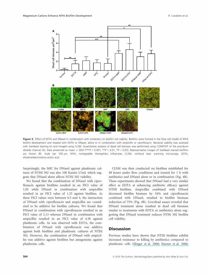
the highest biomass of dead cells was achieved when
EDTA was combined with ciprofloxacin with a 25-fold
increase in dead cells biomass when compared to the
control biofilm (Fig. 5).
Effect of DNaseI on NTHi biofilms
As eDNA has been previously shown to be the major
matrix component in NTHi biofilms (Jurcisek and Baka-
letz 2007), a possible explanation for our observations is
that the Mg2+ cations serve to strengthen the eDNA
matrix through electrostatic interactions with the
negatively charged eDNA as has been shown previously
with P. aeruginosa biofilms (Mulcahy et al. 2008; Lewenza
2013).
We have determined that when NTHi 502 biofilms are
cultured for 48 h in flow conditions and then treated
with DNaseI, that this results in a 30% reduction in bio-
film biomass. These observations suggest that eDNA is a
major component of the matrix of biofilms formed by
NTHi strain 502. Therefore, it is possible that inclusion
of Mg2+ to the biofilm culture media enhances biofilm
development, at least in part, by strengthening the eDNA
matrix through electrostatic interactions and that cation
chelation with EDTA serves to weaken the eDNA matrix.
We therefore performed combinatorial experiments
with DNaseI and antibiotics to determine if DNaseI had a
similar effect as EDTA at enhancing antibiotic efficacy
against NTHi 502 biofilms and planktonic cells. We
found that the MBEC for DNaseI was 100 Kunitz U/mL.
Contro
l
EDTAAM
PCPX
EDTA+AM
P
EDTA+CPX
0
5
10
15
20
Bio
film
biom
ass
(m
3 /m
2 )
********
Buffe
r
DNaseI
AMPCPX
DNaseI+
AMP
DNaseI+
CPX0
5
10
15
Bio
film
biom
ass
(m
3 /m
2 )
********
****
Figure 4. EDTA or DNaseI enhance the efficacy of ampicillin or
ciprofloxacin treatment of established biofilms. Biofilms were formed
in the flow cell model of NTHi biofilm development and treated with
25 mmol/L EDTA alone or in combination with either 50 lmol/L of
ampicillin or 1.2 lmol/L ciprofloxacin (upper) or with DNaseI
(100 Kunitz U/mL) alone or in combination with either 50 lmol/L of
ampicillin or 1.2 lmol/L ciprofloxacin (lower). Biofilm biomass was
quantitated from CLSM images with COMSTAT. Data presented as
mean � SEM (****P < 0.0001). NTHi, nontypeable Hemophilus
influenzae; CLSM, confocal laser scanning microscopy, EDTA,
ethylenediaminetetra-acetic acid.
ª 2014 The Authors. MicrobiologyOpen published by John Wiley & Sons Ltd. 563
R. Cavaliere et al. Magnesium Cations Enhance NTHi Biofilm Development
Surprisingly, the MIC for DNaseI against planktonic cul-
tures of NTHI 502 was also 100 Kunitz U/mL which sug-
gests that DNaseI alone affects NTHi 502 viability.
We found that the combination of DNaseI with cipro-
floxacin against biofilms resulted in an FICI value of
1.05 while DNaseI in combination with ampicillin
resulted in an FICI value of 1.25 against biofilms. As
these FICI values were between 0.5 and 4, the interaction
of DNaseI with ciprofloxacin and ampicillin are consid-
ered to be additive for biofilm cultures. We found that
DNaseI in combination with ciprofloxacin resulted in an
FICI value of 2.13 whereas DNaseI in combination with
ampicillin resulted in an FICI value of 4.58 against
planktonic cells. As was observed with EDTA, the com-
bination of DNaseI with ciprofloxacin was additive
against both biofilms and planktonic cultures of NTHi
502. However, the combination of DNaseI with ampicil-
lin was additive against biofilms but antagonistic against
planktonic cells.
CLSM was then conducted on biofilms established for
48 hours under flow conditions and treated for 1 h with
antibiotics and DNaseI alone or in combination (Fig. 4B).
These experiments showed that DNaseI had a very similar
effect as EDTA at enhancing antibiotic efficacy against
NTHI biofilms. Ampicillin combined with DNaseI
decreased biofilm biomass by 54% and ciprofloxacin
combined with DNaseI, resulted in biofilm biomass
reduction of 75% (Fig. 4B). Live/dead assays revealed that
DNaseI treatment alone resulted in dead cell biomass
similar to treatments with EDTA or antibiotics alone sug-
gesting that DNaseI treatment reduces NTHi 502 biofilm
cell viability.
Discussion
Previous studies have shown that NTHi biofilms exhibit
increased resistance to killing by antibiotics compared to
planktonic cells (Slinger et al. 2006; Starner et al. 2006;
A
B
Figure 5. Effect of EDTA and DNaseI in combination with antibiotics on biofilm cell viability. Biofilms were formed in the flow cell model of NTHi
biofilm development and treated with EDTA or DNaseI, alone or in combination with ampicillin or ciprofloxacin. Bacterial viability was assessed
with live/dead staining kit and imaged using CLSM. Quantitative analysis of dead cell biomass was performed using COMSTAT of the proridium
idodide channel (A). Data presented as mean � SEM (***P < 0.001, **P < 0.01, *P < 0.05). Representative images of live/dead stained biofilms
are shown (B). Scale bar 100 lm. NTHi, nontypeable Hemophilus influenzae; CLSM, confocal laser scanning microscopy, EDTA,
ethylenediaminetetra-acetic acid.
564 ª 2014 The Authors. MicrobiologyOpen published by John Wiley & Sons Ltd.
Magnesium Cations Enhance NTHi Biofilm Development R. Cavaliere et al.

Izano et al. 2009). In this study we have investigated the
role of divalent cations in NTHi biofilm formation. To
gain an understanding of how divalent cations affect
NTHi biofilm formation, we tested the effect of six differ-
ent divalent cations on NTHi biofilm formation and
planktonic cell growth. Most of the divalent cations tested
resulted in dramatically decreased biofilm biomass as well
as decreased planktonic cell numbers at the majority of
concentrations. This strongly suggests that these cations
were toxic to the planktonic cells, and therefore biofilms
were unable to be formed. The remaining three cations
tested, Ba2+, Ca2+, and Mg2+ resulted in an overall
increase in biofilm biomass.
Of particular interest was the increase in biofilm bio-
mass seen in the presence of Mg2+, as Mg2+ was the only
divalent cation tested that increased biofilm biomass dra-
matically without increasing planktonic cell growth. This
suggested that the increases in biomass in the presence of
Mg2+ were due to a direct affect on biofilm formation.
Importantly, we found that inclusion of Mg2+ at physio-
logical concentrations significantly increased NTHi bio-
film biomass. While the influence of Mg2+ on biofilm
formation by NTHi has not been previously investigated,
researchers have found that Mg2+ enhances biofilm for-
mation in a number of other bacterial species including
A. hydrophilia and Pseudomonas fluorescens (Merino et al.
2001; Song and Leff 2006). Furthermore, in a study con-
ducted in mixed biofilms of P. aeruginosa and Klebsiella
pneumoniae, the authors concluded that electrostatic
interactions created in the presence of divalent cations
contribute to biofilm cohesion (Chen and Stewart 2002).
Divalent cations are known to be important for bacte-
rial cell division and cell wall integrity (Gray and Wilkin-
son 1965; Asbell and Eagon 1966). As they have also been
shown to play a role in biofilm formation, it is not sur-
prising that cation chelators have been tested for the pre-
vention and treatment of biofilm formation in many
bacterial species. In particular, the use of the divalent cat-
ion chelator EDTA in preventing biofilm formation, espe-
cially in catheter-related infections has become an
exciting area of research. Indeed, EDTA has been
approved as a treatment of biofilms in humans as a com-
ponent of catheter lock solution (Hancock and Wong
1984). EDTA has been shown to have an effect on at least
11 bacterial biofilm-forming species, including those
formed by P. aeruginosa, S. aureus, and MRSA (Raad
et al. 2002). EDTA in conjunction with the antibiotic
monocycline has been shown to be highly effective against
S. epidermidis, S. aureus, and Candida albicans biofilm on
the surface of catheters (Raad et al. 2002). Prolonged
treatments with EDTA can be lethal to free-living proteo-
bacteria (Gray and Wilkinson 1965; Leive 1974; Hancock
1984; Banin et al. 2006), and short treatments can
increase the permeability of the outer membrane to
hydrophobic molecules (Leive 1974; Nikaido and Vaara
1985). Here, we have shown for the first time that EDTA
has the ability to prevent biofilm formation and remove
established biofilms of a clinical NTHi isolate. CLSM of
biofilms formed under flow conditions revealed that
EDTA treatment of established biofilms resulted in
decreased biofilm biomass.
As eDNA has been shown to be a major component of
the extracellular matrix in NTHi biofilms (Jurcisek and
Bakaletz 2007), it is likely that the anionic nature of
eDNA binds positively charged magnesium cations,
thereby strengthening the biofilm matrix. We hypothe-
sized that matrix destabilizers in combination with
antibiotics would be more effective than the single agents
alone as the matrix destabilizers may enhance accessibility
of these antibiotics into the cell clusters (Stewart 2003).
Indeed, in P. aeruginosa biofilms eDNA has been shown
to shield the cells against aminoglycosides by delaying
their penetration (Chiang et al. 2013).
In this study, we found that established NTHi 502 bio-
films were extremely vulnerable to ampicillin and cipro-
floxacin when combined with EDTA or DNaseI. We have
not determined the mechanism via which these effects
occur, but one possibility is that these may be due to
destabilization of the eDNA matrix, thereby enabling
greater penetration of the antibiotic into the biofilm
either through increased penetration into cell clusters or
via reduced sequestration into the matrix.
Interestingly, we observed that both EDTA and DNaseI
reduced the viability of planktonic and biofilm cells,
which suggests that at least some of the observed effects
could be due to direct action on the bacterial cells by
these agents. The finding that DNaseI is toxic to NTHi
502 was surprising as we and others have shown previ-
ously that DNaseI is not bactericidal against P. aeruginosa
(Whitchurch et al. 2002) or Burkholderia cepacia complex
(Messiaen et al. 2014).
Further understanding of the mechanisms associated
with NTHi biofilm cohesion and antibiotic resistance
could lead to the development of biofilm-specific agents
for the management and treatment of NTHi biofilm-
associated infections. Our observations suggest that DNa-
seI and EDTA enhance the efficacy of antibiotic treatment
of NTHi biofilms. As both DNaseI and EDTA are cur-
rently approved for human use, these observations may
lead to new strategies that will improve the treatment
options available to patients with chronic NTHi infec-
tions. However, the recent report that, unlike the effect
observed in P. aeruginosa, DNaseI treatment does not
increase susceptibility of B. cepacia complex species to
tobramycin (Messiaen et al. 2014) indicates that the
efficacy of the combination therapies such as those
ª 2014 The Authors. MicrobiologyOpen published by John Wiley & Sons Ltd. 565
R. Cavaliere et al. Magnesium Cations Enhance NTHi Biofilm Development

described in our study are likely to be highly dependent
on the antibiotic and species involved.
Acknowledgments
C. B. W. was supported by an Australian National Health
and Medical Research Council Senior Research Fellowship
(571905). L. T. was supported by a UTS Chancellor’s
Postdoctoral Fellowship. CLSM was performed at the
UTS Microbial Imaging Facility. This study was supported
through funding from the Australian National Health and
Medical Research Council (491126) and the Financial
Markets Foundation for Children.
Conflict of Interest
None declared.
References
Allesen-Holm, M., K. B. Barken, L. Yang, M. Klausen, J. S.
Webb, S. Kjelleberg, et al. 2006. A characterization of DNA
release in Pseudomonas aeruginosa cultures and biofilms.
Mol. Microbiol. 59:1114–28.
Asbell, M. A., and R. G. Eagon. 1966. Role of multivalent
cations in the organization, structure, and assembly of the
cell wall of Pseudomonas aeruginosa. J. Bacteriol. 92:380–
387.
Banin, E., K. M. Brady, and E. P. Greenberg. 2006.
Chelator-induced dispersal and killing of Pseudomonas
aeruginosa cells in a biofilm. Appl. Environ. Microbiol.
72:2064–2069.
de Beer, D., P. Stoodley, and Z. Lewandowski. 1997.
Measurement of local diffusion coefficients in biofilms by
microinjection and confocal microscopy. Biotechnol. Bioeng.
53:151–158.
Brown, M. R., D. G. Allison, and P. Gilbert. 1988. Resistance
of bacterial biofilms to antibiotics: a growth-rate related
effect? J. Antimicrob. Chemother. 22:777–780.
Chen, X., and P. S. Stewart. 2002. Role of electrostatic
interactions in cohesion of bacterial biofilms. Appl.
Microbiol. Biotechnol. 59:718–720.
Chiang, W. C., M. Nilsson, P. O. Jensen, N. Hoiby, T. E.
Nielsen, M. Givskov, et al. 2013. Extracellular DNA shields
against aminoglycosides in Pseudomonas aeruginosa biofilms.
Antimicrob. Agents Chemother. 57:2352–2361.
Coleman, H. N., D. A. Daines, J. Jarisch, and A. L. Smith.
2003. Chemically defined media for growth of
Haemophilus influenzae strains. J. Clin. Microbiol.
41:4408–4410.
Costerton, J. W., R. T. Irvin, and K. J. Cheng. 1981. The
bacterial glycocalyx in nature and disease. Annu. Rev.
Microbiol. 35:299–324.
Costerton, J. W., Z. Lewandowski, D. E. Caldwell, D. R.
Korber, and H. M. Lappin-Scott. 1995. Microbial biofilms.
Annu. Rev. Microbiol. 49:711–745.
Costerton, J. W., P. S. Stewart, and E. P. Greenberg. 1999.
Bacterial biofilms: a common cause of persistent infections.
Science 284:1318–1322.
Donlan, R. M., and J. W. Costerton. 2002. Biofilms: survival
mechanisms of clinically relevant microorganisms. Clin.
Microbiol. Rev. 15:167–193.
Ehrlich, G. D., R. Veeh, X. Wang, J. W. Costerton, J. D.
Hayes, F. Z. Hu, et al. 2002. Mucosal biofilm formation on
middle-ear mucosa in the chinchilla model of otitis media.
JAMA 287:1710–1715.
Fux, C. A., J. W. Costerton, P. S. Stewart, and P. Stoodley.
2005. Survival strategies of infectious biofilms. Trends
Microbiol. 13:34–40.
Geesey, G. G., B. Wigglesworth-Cooksey, and K. E. Cooksey.
2000. Influence of calcium and other cations on surface
adhesion of bacteria and diatoms: a review. Biofouling
15:195–205.
Gray, G. W., and S. G. Wilkinson. 1965. The effect of
ethylenediaminetetra-acetic acid on the cell walls of some
gram-negative bacteria. J. Gen. Microbiol. 39:385–399.
Greiner, L. L., H. Watanabe, N. J. Phillips, J. Shao, A. Morgan,
A. Zaleski, et al. 2004. Nontypeable Haemophilus influenzae
strain 2019 produces a biofilm containing
N-acetylneuraminic acid that may mimic sialylated O-linked
glycans. Infect. Immun. 72:4249–4260.
Hall-Stoodley, L., F. Z. Hu, A. Gieseke, L. Nistico, D. Nguyen,
J. Hayes, et al. 2006. Direct detection of bacterial biofilms
on the middle-ear mucosa of children with chronic otitis
media. JAMA 296:202–211.
Hancock, R. E. 1984. Alterations in outer membrane
permeability. Annu. Rev. Microbiol. 38:237–264.
Hancock, R. E., and P. G. Wong. 1984. Compounds which
increase the permeability of the Pseudomonas aeruginosa
outer membrane. Antimicrob. Agents Chemother. 26:48–52.
Heydorn, A., A. T. Nielsen, M. Hentzer, C. Sternberg, M.
Givskov, B. K. Ersboll, et al. 2000. Quantification of biofilm
structures by the novel computer program COMSTAT.
Microbiology 146(Pt 10):2395–2407.
Izano, E. A., S. M. Shah, and J. B. Kaplan. 2009. Intercellular
adhesion and biocide resistance in nontypeable Haemophilus
influenzae biofilms. Microb. Pathog. 46:207–213.
Jakubovics, N. S., R. C. Shields, N. Rajarajan, and J. G.
Burgess. 2013. Life after death: the critical role of
extracellular DNA in microbial biofilms. Lett. Appl.
Microbiol. 57:467–475.
James, G. A., D. R. Korber, D. E. Caldwell, and
J. W. Costerton. 1995. Digital image analysis of growth and
starvation responses of a surface-colonizing Acinetobacter
sp. J. Bacteriol. 177:907–915.
Jurcisek, J. A., and L. O. Bakaletz. 2007. Biofilms formed by
nontypeable Haemophilus influenzae in vivo contain both
566 ª 2014 The Authors. MicrobiologyOpen published by John Wiley & Sons Ltd.
Magnesium Cations Enhance NTHi Biofilm Development R. Cavaliere et al.

double-stranded DNA and type IV pilin protein.
J. Bacteriol. 189:3868–3875.
Leive, L. 1974. The barrier function of the gram-negative
envelope. Ann. N. Y. Acad. Sci. 235:109–129.
Leive, L., and V. Kollin. 1967. Controlling EDTA treatment to
produce permeable Escherichia coli with normal metabolic
processes. Biochem. Biophys. Res. Commun. 28:229–236.
Lewenza, S. 2013. Extracellular DNA-induced antimicrobial
peptide resistance mechanisms in Pseudomonas aeruginosa.
Front. Microbiol. 4:21.
Lewis, R. E., D. J. Diekema, S. A. Messer, M. A. Pfaller, and
M. E. Klepser. 2002. Comparison of Etest, chequerboard
dilution and time-kill studies for the detection of synergy or
antagonism between antifungal agents tested against
Candida species. J. Antimicrob. Chemother. 49:345–351.
Marcus, H., A. Austria, and N. R. Baker. 1989. Adherence of
Pseudomonas aeruginosa to tracheal epithelium. Infect.
Immun. 57:1050–1053.
Matsukawa, M., and E. P. Greenberg. 2004. Putative
exopolysaccharide synthesis genes influence Pseudomonas
aeruginosa biofilm development. J. Bacteriol. 186:4449–4456.
Merino, S., R. Gavin, M. Altarriba, L. Izquierdo, M. E.
Maguire, and J. M. Tomas. 2001. The MgtE Mg2 +transport protein is involved in Aeromonas hydrophila
adherence. FEMS Microbiol. Lett. 198:189–195.
Messiaen, A. S., H. Nelis, and T. Coenye. 2014. Investigating
the role of matrix components in protection of Burkholderia
cepacia complex biofilms against tobramycin. J. Cyst. Fibros.
13:56–62.
Moriyama, S., M. Hotomi, J. Shimada, D. S. Billal, K.
Fujihara, and N. Yamanaka. 2009. Formation of biofilm by
Haemophilus influenzae isolated from pediatric intractable
otitis media. Auris Nasus Larynx 36:525–531.
Mulcahy, H., L. Charron-Mazenod, and S. Lewenza. 2008.
Extracellular DNA chelates cations and induces antibiotic
resistance in Pseudomonas aeruginosa biofilms. PLoS Pathog.
4:e1000213.
Nemoto, K., K. Hirota, K. Murakami, K. Taniguti, H. Murata,
D. Viducic, et al. 2003. Effect of Varidase (streptodornase)
on biofilm formed by Pseudomonas aeruginosa.
Chemotherapy 49:121–125.
Nikaido, H., and M. Vaara. 1985. Molecular basis of bacterial
outer membrane permeability. Microbiol. Rev. 49:1–32.
Odds, F. C. 2003. Synergy, antagonism, and what the
chequerboard puts between them. J. Antimicrob.
Chemother. 52:1.
O’Toole, G., H. B. Kaplan, and R. Kolter. 2000. Biofilm
formation as microbial development. Annu. Rev. Microbiol.
54:49–79.
Ozerdem Akpolat, N., S. Elci, S. Atmaca, H. Akbayin, and
K. Gul. 2003. The effects of magnesium, calcium and EDTA
on slime production by Staphylococcus epidermidis strains.
Folia Microbiol. 48:649–53.
Raad, I., R. Hachem, R. K. Tcholakian, and R. Sherertz. 2002.
Efficacy of minocycline and EDTA lock solution in
preventing catheter-related bacteremia, septic phlebitis, and
endocarditis in rabbits. Antimicrob. Agents Chemother.
46:327–332.
Slinger, R., F. Chan, W. Ferris, S. W. Yeung, M. St Denis,
I. Gaboury, et al. 2006. Multiple combination antibiotic
susceptibility testing of nontypeable Haemophilus influenzae
biofilms. Diagn. Microbiol. Infect. Dis. 56:247–53.
Song, B., and L. G. Leff. 2006. Influence of magnesium ions
on biofilm formation by Pseudomonas fluorescens. Microbiol.
Res. 161:355–361.
Starner, T. D., N. Zhang, G. Kim, M. A. Apicella, P. B.
McCray, Jr. 2006. Haemophilus influenzae forms biofilms on
airway epithelia: implications in cystic fibrosis. Am. J.
Respir. Crit. Care Med. 174:213–220.
Steinberger, R. E., A. R. Allen, H. G. Hansa, and P. A. Holden.
2002. Elongation correlates with nutrient deprivation in
Pseudomonas aeruginosa-unsaturates biofilms. Microb. Ecol.
43:416–423.
Stewart, P. S. 2003. Diffusion in biofilms. J. Bacteriol.
185:1485–1491.
Sutherland, I. W. 2001. The biofilm matrix – an immobilized
but dynamic microbial environment. Trends Microbiol.
9:222–227.
Swaminathan, R. 2003. Magnesium metabolism and its
disorders. Clin. Biochem. Rev. 24:47–66.
Swords, W. E. 2012. Nontypeable Haemophilus influenzae
biofilms: role in chronic airway infections. Front. Cell.
Infect. Microbiol. 2:97.
Walters, M. C., III, F. Roe, A. Bugnicourt, M. J. Franklin,
and P. S. Stewart. 2003. Contributions of antibiotic
penetration, oxygen limitation, and low metabolic activity
to tolerance of Pseudomonas aeruginosa biofilms to
ciprofloxacin and tobramycin. Antimicrob. Agents
Chemother. 47:317–23.
Whitchurch, C. B., T. Tolker-Nielsen, P. C. Ragas, and
J. S. Mattick. 2002. Extracellular DNA required for bacterial
biofilm formation. Science 295:1487.
ª 2014 The Authors. MicrobiologyOpen published by John Wiley & Sons Ltd. 567
R. Cavaliere et al. Magnesium Cations Enhance NTHi Biofilm Development