Microbial Communities Associated with Healthy and White Syndrome-Affected Echinopora lamellosa in...
17
RESEARCH ARTICLE Microbial Communities Associated with Healthy and White Syndrome-Affected Echinopora lamellosa in Aquaria and Experimental Treatment with the Antibiotic Ampicillin David Smith 1,2 *, Peter Leary 1 , Jamie Craggs 3 , John Bythell 1 , Michael Sweet 1,4 1 School of Biology, Newcastle University, Newcastle upon Tyne, NE1 7RU, United Kingdom, 2 School of Biological Sciences, Medical Biology Centre, Queen’s University Belfast, Belfast, BT9 7BL, United Kingdom, 3 Horniman Museum and Gardens Aquarium, Forest Hill, London, SE23 3PQ, United Kingdom, 4 Biological Sciences Research Group, University of Derby, Kedleston Road, Derby, DE22 1GB, United Kingdom * [email protected] Abstract Prokaryotic and ciliate communities of healthy and aquarium White Syndrome (WS)-affect- ed coral fragments were screened using denaturing gradient gel electrophoresis (DGGE). A significant difference (R = 0.907, p < 0.001) in 16S rRNA prokaryotic diversity was found between healthy (H), sloughed tissue (ST), WS-affected (WSU) and antibiotic treated (WST) samples. Although 3 Vibrio spp were found in WS-affected samples, two of these species were eliminated following ampicillin treatment, yet lesions continued to advance, suggesting they play a minor or secondary role in the pathogenesis. The third Vibrio sp in- creased slightly in relative abundance in diseased samples and was abundant in non-dis- eased samples. Interestingly, a Tenacibaculum sp showed the greatest increase in relative abundance between healthy and WS-affected samples, demonstrating consistently high abundance across all WS-affected and treated samples, suggesting Tenacibaculum sp could be a more likely candidate for pathogenesis in this instance. In contrast to previous studies bacterial abundance did not vary significantly (ANOVA, F2, 6 = 1.000, p = 0.422) be- tween H, ST, WSU or WST. Antimicrobial activity (assessed on Vibrio harveyi cultures) was limited in both H and WSU samples (8.1% ±8.2 and 8.0% ±2.5, respectively) and did not dif- fer significantly (Kruskal-Wallis, χ2 (2) = 3.842, p = 0.146). A Philaster sp, a Cohnilembus sp and a Pseudokeronopsis sp. were present in all WS-affected samples, but not in healthy samples. The exact role of ciliates in WS is yet to be determined, but it is proposed that they are at least responsible for the neat lesion boundary observed in the disease. PLOS ONE | DOI:10.1371/journal.pone.0121780 March 20, 2015 1 / 17 OPEN ACCESS Citation: Smith D, Leary P, Craggs J, Bythell J, Sweet M (2015) Microbial Communities Associated with Healthy and White Syndrome-Affected Echinopora lamellosa in Aquaria and Experimental Treatment with the Antibiotic Ampicillin. PLoS ONE 10(3): e0121780. doi:10.1371/journal.pone.0121780 Academic Editor: Jose Luis Balcazar, Catalan Institute for Water Research (ICRA), SPAIN Received: October 3, 2013 Accepted: February 18, 2015 Published: March 20, 2015 Copyright: © 2015 Smith et al. This is an open access article distributed under the terms of the Creative Commons Attribution License, which permits unrestricted use, distribution, and reproduction in any medium, provided the original author and source are credited. Funding: This work was supported by the Natural Environment Research Council grant (NE/H020616/ 1) (www.nerc.ac.uk). The funders had no role in study design, data collection and analysis, decision to publish, or preparation of the manuscript. Competing Interests: The authors have declared that no competing interests exist.
Transcript of Microbial Communities Associated with Healthy and White Syndrome-Affected Echinopora lamellosa in...
RESEARCH ARTICLE
Microbial Communities Associated withHealthy and White Syndrome-AffectedEchinopora lamellosa in Aquaria andExperimental Treatment with the AntibioticAmpicillinDavid Smith1,2*, Peter Leary1, Jamie Craggs3, John Bythell1, Michael Sweet1,4
1 School of Biology, Newcastle University, Newcastle upon Tyne, NE1 7RU, United Kingdom, 2 School ofBiological Sciences, Medical Biology Centre, Queen’s University Belfast, Belfast, BT9 7BL, United Kingdom,3 HornimanMuseum and Gardens Aquarium, Forest Hill, London, SE23 3PQ, United Kingdom, 4 BiologicalSciences Research Group, University of Derby, Kedleston Road, Derby, DE22 1GB, United Kingdom
AbstractProkaryotic and ciliate communities of healthy and aquariumWhite Syndrome (WS)-affect-
ed coral fragments were screened using denaturing gradient gel electrophoresis (DGGE). A
significant difference (R = 0.907, p< 0.001) in 16S rRNA prokaryotic diversity was found
between healthy (H), sloughed tissue (ST), WS-affected (WSU) and antibiotic treated
(WST) samples. Although 3 Vibrio spp were found in WS-affected samples, two of these
species were eliminated following ampicillin treatment, yet lesions continued to advance,
suggesting they play a minor or secondary role in the pathogenesis. The third Vibrio sp in-
creased slightly in relative abundance in diseased samples and was abundant in non-dis-
eased samples. Interestingly, a Tenacibaculum sp showed the greatest increase in relative
abundance between healthy andWS-affected samples, demonstrating consistently high
abundance across all WS-affected and treated samples, suggesting Tenacibaculum sp
could be a more likely candidate for pathogenesis in this instance. In contrast to previous
studies bacterial abundance did not vary significantly (ANOVA, F2, 6 = 1.000, p = 0.422) be-
tween H, ST, WSU or WST. Antimicrobial activity (assessed on Vibrio harveyi cultures) waslimited in both H andWSU samples (8.1% ±8.2 and 8.0% ±2.5, respectively) and did not dif-
fer significantly (Kruskal-Wallis, χ2 (2) = 3.842, p = 0.146). A Philaster sp, a Cohnilembus spand a Pseudokeronopsis sp. were present in all WS-affected samples, but not in healthy
samples. The exact role of ciliates in WS is yet to be determined, but it is proposed that they
are at least responsible for the neat lesion boundary observed in the disease.
PLOS ONE | DOI:10.1371/journal.pone.0121780 March 20, 2015 1 / 17
OPEN ACCESS
Citation: Smith D, Leary P, Craggs J, Bythell J,Sweet M (2015) Microbial Communities Associatedwith Healthy and White Syndrome-AffectedEchinopora lamellosa in Aquaria and ExperimentalTreatment with the Antibiotic Ampicillin. PLoS ONE10(3): e0121780. doi:10.1371/journal.pone.0121780
Academic Editor: Jose Luis Balcazar, CatalanInstitute for Water Research (ICRA), SPAIN
Received: October 3, 2013
Accepted: February 18, 2015
Published: March 20, 2015
Copyright: © 2015 Smith et al. This is an openaccess article distributed under the terms of theCreative Commons Attribution License, which permitsunrestricted use, distribution, and reproduction in anymedium, provided the original author and source arecredited.
Funding: This work was supported by the NaturalEnvironment Research Council grant (NE/H020616/1) (www.nerc.ac.uk). The funders had no role in studydesign, data collection and analysis, decision topublish, or preparation of the manuscript.
Competing Interests: The authors have declaredthat no competing interests exist.
IntroductionProgressively more diseases of scleractinian corals are being reported in the scientific literatureyear on year [1–6]. Despite this, the causal agents of these diseases are often in dispute, with asingle accepted pathogen for a given disease being rare [7]. This is the case for White Syndrome(WS), a disease commonly found in the Indo-Pacific and within aquarium systems [8–12]. WSis characterized by tissue loss and acute lesion progression with a distinct boundary or band be-tween apparently healthy tissue and exposed skeleton [13].
In the past, WS has been attributed to various species from the gammaproteobacteria genusVibrio, including; V. harveyi [14], V.mediterranei [7], V. owensii [15] and V. coralliilyticus[10]. Furthermore, Piskorska, Smith [7], reports an increase in V.mediterranei in the mucus ofWS-affected Echinopora lamellosa, compared to healthy samples. However, Piskorska, Smith[7] used a culture-based method in determining bacteria associated with healthy andWS-af-fected E. lamellosa, an approach that can lead to phylum and class biases and result in inade-quate profiling of microbial communities when not backed up with culture-independenttechniques [16, 17], thus further work is required to confirm the role of Vibrio species inthis disease.
Another proposed causal agent of WS includes Arcobacter sp, found by Sweet and Bythell[18] to be the only bacteria to be absent in non-diseased samples, appear in apparently healthysamples and increase to have a dominant presence in diseased A.muricata samples in a studyscreening the microbial communities associated with WS in this coral. Sweet and Bythell [18]also found V. harveyi to increase in diseased samples, but also to be present in healthy samples.This presence in healthy samples does not rule out V. harveyi as a primary causal agent in WS,as Sussman, Mieog [19] demonstrated that the zinc-metalloprotease (produced by pathogenicVibrio sp) responsible for the breakdown of coral tissue and photo-inactivation of zooxanthel-lae is only expressed once Vibrio sp cell density exceeds 1x10⁹ cells ml-1. This indicates thatVibriomay not be harmful in lower densities, only becoming pathogenic as their numbers in-crease, explaining their presence in healthy samples and increase in diseased samples in theWS study by Sweet and Bythell [18].
The bacterial community associated with the holobiont of healthy corals is considered toplay a potential role in sustaining coral health [20–24]. This community is known to shift instressed corals, lowering coral defence and impeding important processes, such as the cyclingof nitrogen and sulphur. Actinobacteria, for example, are known for their production of sec-ondary metabolites which can be lethal to invasive bacteria [25–27]. Actinobacteria are also as-sociated with healthy coral and have been found to decline in the event of environmental stress[28] and disease [7]. A shift in the natural microbial community associated with corals and adecline in actinobacteria could account for the proliferation of bacteria such as Vibrio sp,which would then become pathogenic in larger cell quantities [19] once their populations canno longer be controlled within the holobiont.
Only relatively recently have other microorganisms been studied with their relation to coraldiseases. For example ciliates such as two scuticociliates from the genus Philaster have been iso-lated fromWS-affected specimens of Acropora muricata and shown to be able to ingest thecorals Symbiodinium and actively burrow into and underneath apparently healthy tissue in ad-vance of the disease lesion [18, 29]. However, what hasn’t been proved to date is the exact roleof these ciliates in coral disease and how many diseases suffer from ciliate infestations. Particu-larly, are the ciliates primary pathogens in WS and involved in the breakdown of host tissue, oralternatively are they secondary pathogens consuming dead and dying tissues. Additionally,the study by Piskorska et al. [7] did not investigate any potential ciliate community associated
Microbial Ecology of White Syndrome
PLOSONE | DOI:10.1371/journal.pone.0121780 March 20, 2015 2 / 17
with WS in E. lamellosa and their roles in disease aetiology, as claimed necessary by Harvell,Jordán-Dahlgren [30].
This study aimed to determine the bacterial and ciliate communities associated with healthyandWS-affected E. lamellosa in aquaria, using culture-independent (16S rRNA gene) tech-niques. Additionally, the effect of experimental ampicillin treatment on the bacterial and ciliatecommunities of WS-affected E. lamellosa was analysed to determine the efficiency of antibioticapplication in aquarium based corals and to infer whether certain bacteria are causal agents de-pending on their elimination from the bacterial community of treated samples.
Methods
Experimental ProcedureAll coral samples used in this work were kindly donated by the Horniman Museum and Gar-dens Aquarium with permission of the senior curator for the aquarium, Jamie Craggs.
Both healthy and White Syndrome-affected Echinopora lamellosa fragments (donated bythe Horniman Museum and Gardens Aquarium, London, UK) were placed into an aquariumtank system (26°C, 34 ppt salinity) at Newcastle University. Samples were characterised as ap-parently healthy (n = 3), untreated WS (n = 3) and treated WS (25 μg/ml ampicillin) (n = 3).Each sample type was placed into independent tanks to prevent contamination between differ-ent sample types during acclimatisation and thus prevent the spread of WS to healthy frags.Fragments were given 24 hours to acclimatise following transportation and disease progressionwas monitored regularly to ensure lesions were advancing in WS specimens before continuingwith the study. Following acclimatisation, fragments were placed into their own independenttanks. These tanks were smaller in size and thus required much greater monitoring of water sa-linity and temperature. Since smaller tanks were not hooked up to the main inflow/outflow sys-tem, tank water was replaced daily with sterile salt water that had been passed through a0.22μm filter using a sterile syringe. Ampicillin treatment was administered (every 4 hours) ton = 3 fragments with the aim of determining the causal agent/s of WS in Echinopora lamellosathrough a process of elimination. Prokaryotic species present in untreated samples that wereeliminated in treated samples (despite WS persisting in treated samples) would be unlikely tobe causal agents of the disease.
Three samples exhibiting what appeared to be a lesion similar to that associated with WSwere sampled immediately upon arrival at Newcastle University. These were characterized as“sloughed tissue” (ST) samples.
Samples were collected prior to complete tissue loss occurring in the WS-affected samplesfor microbial community analysis (stored in 100% ethanol at 4°C).
DNA ExtractionTo determine microbial community shifts in healthy (H), sloughed tissue (ST) untreated WS-affected (WSU) and treated WS-affected (WST) E. lamellosa samples, n = 3 H, n = 3 ST, n = 3WSU, and n = 3WST were analysed. Upon sampling, coral fragments were stored in 50 ml fal-con tubes filled with 100% Ethanol and kept at 4°C until fragments were individually crushedusing a sterilised pestle and mortar. DNA was extracted using the QIAGEN DNeasy Blood andTissue Kit and resultant extracted DNA stored at 4°C.
Prokaryotic 16S rRNA gene diversityUsing the universal prokaryotic primers, as used by Sweet et al, 2010; (357F) (5’-CCTACGG-GAGGCAGCAG-3’) and (518R) (5’ATTACCGCGGCTGCTGG-3’), a segment of the bacterial
Microbial Ecology of White Syndrome
PLOSONE | DOI:10.1371/journal.pone.0121780 March 20, 2015 3 / 17
16S rRNA gene was amplified using a Hybaid PCR Express thermal cycler, prior to DGGEanalysis. PCR cycles were carried out at 94°C for 30 seconds, 53°C for 30 seconds and 72°C for1 minute and a total of 30 cycles were performed, as well as a final extension step at 72°C for 10minutes (Sweet et al, 2010). PCR reaction mixtures were made up to 10 μl, using; 2 ng extractedDNA, buffer incubation mix with 1.5 mMMgCl2 (MP Biomedicals), 0.2 mM dNTP (Qiagen),0.5 mM primer 357F, 0.5 mM primer 518R, 2.5 U of DreamTaq proof-reading DNA Polymer-ase (Fermentas). Each PCR sample mixture was done in triplicate and resulting PCR productscombined to bring about a final volume of 30 μl for each sample. PCR products were verifiedby agarose gel electrophoresis [1% (w/v) agarose] with ethidium bromide staining and visual-ised using a UV transilluminator.
DGGE was performed using the D-code universal mutation detection system (Bio-Rad).PCR products were resolved on 10% (w/v) polyacrylamide gels using a 30 to 60% denaturantgradient for 16 hours at 60°C, with a constant voltage of 50 V. Following DGGE, gels werestained by pouring over a combined solution of 9 μl SYBR Gold (Sigma-Aldrich) and 50 μl1xTAE buffer, and then left covered in the dark for 20 min. Gels were then washed in 500 ml1xTAE buffer for 30 min and visualised using a UV transilluminator. To identify bands ofmost interest in the DGGE gels (those that represented the greatest differences/similarities be-tween samples), representative bands were excised from the gel, left overnight in Sigma molec-ular grade water and then re-amplified using primers 357F and 518R. The products of each re-amplified band were then verified by agarose gel electrophoresis [1% (w/v) agarose] with ethid-ium bromide staining and visualised using a UV transilluminator. PCR products were then pu-rified using the QIAGEN PCR Purification Kit (see main methods section for completedescription) and then labelled using a Big Dye (Applied Biosystems) transformation sequencekit and sent to Genevision (University of Newcastle) for sequencing. Sequences were then com-pared with sequences in the BLAST nucleotide database (NCBI) to allow forgenus/species matching.
Using Bionumerics 3.5 (Applied Maths BVBA) software, bacterial operation taxonomicunits (OTUs) were defined from DGGE band-matching analysis. Standard internal markerlanes were used to allow for gel-to-gel comparisons. Tolerance and optimisation for band-matching was set at 1%.
Ciliate 18S rRNA diversityCiliate-specific 18S rRNA gene amplification was carried out using the same ciliate primers asSweet and Bythell [18]; forward primer CilF (5’-TGGTAGTGTATTGGACWACCA-3’) with a36 bp GC clamp [31] attached to the 5’ end and the reverse primer CilDGGE-r (5’-TGAAAACATCCTTGGCAACTG-3’). PCR reaction mixtures were made up to 10 μl volumes, using; 20 ngextracted DNA, buffer incubation mix T.pol with 1 mMMgCl2 (MP Biomedicals), 100 μMdNTP (Qiagen), 0.2 μM 357F, 0.2 μM 518R, 1 U of DreamTaq DNA Polymerase (Fermentas).PCR was carried out using a Hybaid PCR Express thermal cycler. PCR amplification was car-ried out using an un-nested approach [32], with primary denaturation being carried out at94°C for 5 min, followed by 26 PCR cycles of 94°C for 1 min, 52°C for 1 min and 72°C for1 min. A final elongation step at 72°C was then carried out for 10 min to reduce heteroduplexesin the DGGE [33]. Each PCR reaction was carried out in triplicate and the resulting PCR prod-ucts combined to bring about a final volume of 30 μl for each sample. PCR products were veri-fied by agarose gel electrophoresis [1% (w/v) agarose] with ethidium bromide staining andvisualised using a UV transilluminator. DGGE was performed using the D-code universal mu-tation detection system (Bio-Rad). PCR products were resolved on 6% (w/v) polyacrylamidegels possessing a 32 to 42% denaturant gradient for 16 hours at 60°C, with a constant voltage of
Microbial Ecology of White Syndrome
PLOSONE | DOI:10.1371/journal.pone.0121780 March 20, 2015 4 / 17
50V. Gels were stained and DGGE bands excised and purified as above, but using the ciliateprimers. Samples were sent to Genevision (Newcastle University) for sequencing. DGGEs andsamples were analysed as above. Retrieved ciliate nucleotide sequences have been submitted toGenbank with the following accession codes: KP793000 (C1), KP793001 (C2), KP793002 (C3)and KP792999 (C4).
To confirm the presence of Philaster sp. in diseased samples and clear up any ambiguityfrom the DGGE, a PCR product check was carried out on all samples using the primers BrB-F-171 (5’-TCAAACCCGACTTTACGGAAG-3’) and BrB-R-1721 (5’-TGCAGGTTCACC-TACGGAAAC-3’) [34]. These primers were designed to amplify the ciliate associated withBrown Band Disease identified by Bourne et al. [34] and since described for WS by Sweet andBythell [18], but also amplify a range of similar Philaster species and close relatives. PCR mixeswere as described for the amplification of ciliate 18S rRNA in DGGE. PCR amplification wascarried out at 95°C for 3 minutes, followed by 35 PCR cycles of 95°C for 30 s, 45°C for 45 s and72°C for 2 min. A final elongation step was carried out at 72°C for 10 min [34]. Product checkswere carried out using gel electrophoresis on a 1% agarose gel.
Total Bacterial CountTo assess total bacterial abundance, 700 μl of each sample was vacuum-filtered through a0.22 μm black polycarbonate filter and then stained with 100 μl of 4% PBS (phosphate buffersaline)-buffered paraformaldehyde solution containing 4’6-diamidino-2-phenylindole (at afinal concentration of 5 μg ml-1) for 10 min. The filter was then rinsed with 1 x PBS pH 7.4[35]. The stained filter was mounted onto a microscope slide and viewed under fluorescencemicroscopy, under which 50 images were taken of each sample. The bacteria present in eachimage were counted using the automated cell counter; Cell C [36, 37]. Parameters were set toexclude any objects within an image that were smaller than 0.0314μm2 and bigger than0.6188μm2. Mean abundance of all 50 images per sample was then compared acrossall samples.
Antimicrobial Properties against Vibrio harveyiThe inhibition of bacterial growth was determined from the ethanol-soluble fraction of appar-ently healthy andWS-affected E. lamellosa samples. Each tissue sample was transferred intosterile pre-weighed eppendorfs and then dehydrated in a vacuum centrifuge to remove any eth-anol in which the coral fragments were stored. Each eppendorf was then reweighed to deter-mine the dry weight of tissue and 100% ethanol added to make all samples reach a standardconcentration of 100 mg/ml, to be used as a stock solution in testing bacterial inhibition bycoral extracts.
Pure cultures of Vibrio harveyi, a bacterial species heavily considered the causal agent ofWS, was isolated from an aquarium tank that had previously housed WS-affected corals. V.harveyi was isolated using thiosulfate-citrate-bile-sucrose (TCBS) agar and then subsequentlygrown in LB broth overnight (at 28°C) to get a fresh stock culture to be used in each assay. Theconcentration of bacterial culture to be used in each assay was then standardised to 0.15 opticaldensity (OD) units by diluting the primary stock with additional sterile LB broth accordingly.
Antimicrobial assays were conducted in 96-well plates containing a triplicate control for un-inhibited V. harveyi growth (97.5 μl Mueller Hinton Broth + 97.5 μl of V. harveyi culture), atriplicate control for the effect of 100% ethanol (97.5 μl MHB + 5 μl 100% ethanol + 97.5 μl V.harveyi culture) and a triplicate of each sample (97.5 μl MHB + 5 μl coral extract + 97.5 μl of V.harveyi). Using 5 μl coral extract in a 200 μl volume mix brought the final working concentra-tion of coral extract to 2.5 mg/ml for each sample, as conducted by Sweet et al. [28].
Microbial Ecology of White Syndrome
PLOSONE | DOI:10.1371/journal.pone.0121780 March 20, 2015 5 / 17
The kinetics of bacterial growth was determined by reading the OD every 5 minutes over a24 hour period in a Biotek Power Wave HT plate reader. The rate of bacterial growth duringexponential phase was estimated by plotting OD against time to determine the gradient of eachcurve in a linear regression. Inhibition was then estimated by dividing the mean gradient ofeach sample type over the mean gradient of the ethanol controls. This was then calculated as %inhibition of bacterial growth when compared with ethanol control growth.
Statistical AnalysisFor DGGE profiles, a pairwise ANOSIM based on Bray-Curtis indexes were performed to de-termine differences between gene assemblages associated with the different coral samples (H,ST, WSU andWST). In order to determine which bacterial ribotypes showed the greatest shiftsin presence between sample types, similarity percentages (SIMPER) analyses were conducted.All statistical analyses on bacterial DGGE profiles were conducted using PRIMER V. 6 software[18, 38, 39].
As data was found to be normally distributed and treatment variances found to be approxi-mately equal, bacterial abundance in E. lamellosa samples was compared across all sampletypes with a one-way ANOVA. A Tukey HSD test was further conducted to determine pairwisedifferences between particular samples.
A Levene’s test for homogeneity showed that variances were not equal across treatments forpercentage growth inhibition, even after the data had been transformed using a square-roottransformation. A Kruskal-Wallis test was therefore carried out instead to determine differ-ences in Vibrio harveyi growth inhibition between coral samples.
Results
Prokaryotic 16S rRNA gene diversityA one-way ANOSIM revealed a significant difference (R = 0.907, p< 0.001) in the bacterial/ar-chaeal community profiles associated with healthy, sloughed tissue, untreated white syndrome(WS) and treated WS-affected Echinopora lamellosa samples (based on DGGE band matchinganalysis). Altogether, a total of 40 different 16S rRNA ribotypes were successfully determinedfrom study samples (Fig. 1). Prokaryotic diversity was found to be greater in WS-affected sam-ples than in healthy samples, with 37 identified prokaryotes being found in ST samples, 31 inUWS samples, 30 in TWS and 18 in H samples. Many bacterial ribotypes were detected in WS-affected samples, but found to be absent in healthy tissues thus their grouping as WS-associatedbacteria. The most dominant of these ribotypes includes ones most closely related to; 3 Vibriospecies. (GenBank accession numbers; GQ906358, NR102976 and KC954171), 3 Arcobacterspecies (NR041918, FR870464 and NR025906), 2 Shewanella species (NR036917 andNR044863), aMycobacterium species (AL123456), a Campylobacter species (NR043607), aCaldicellulosiruptor species (NR074807), a Treponema species (NR026247), a Cytophaga spe-cies (AB017048), a Streptomyces species (NR043492) a Novosphingobium species (NR074261)and a Phaeobacter species (NR074144). Ribotypes closely related to Two Tenacibaculum spe-cies (NR024737 and NR044498) and a Garciella species (NR025688) were also found tomarkedly increase in all ST andWSU samples, compared to healthy samples (Fig. 1). Since am-picillin treatment failed to stop lesion progression, WS-associated ribotypes of particular inter-est were those that were still dominant across all samples after the antibiotic treatment. Theseincluded ribotypes related to; 3 Arcobacter species (FR870464, NR025906 and NR102873), aMycobacterium species (AL123456), a Vibrio species (KC954171), a Phaeobacter species(NR074144), a Tenacibaculum species (NR024737), a Streptomyces species (NR043492), and aCytophaga species (AB017048). Of these, only ribotypes closely related to the Tenacibaculum
Microbial Ecology of White Syndrome
PLOSONE | DOI:10.1371/journal.pone.0121780 March 20, 2015 6 / 17
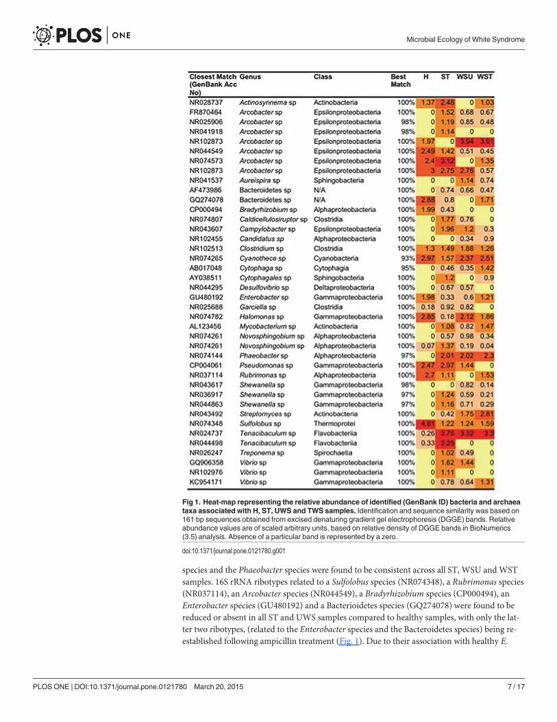
species and the Phaeobacter species were found to be consistent across all ST, WSU and WSTsamples. 16S rRNA ribotypes related to a Sulfolobus species (NR074348), a Rubrimonas species(NR037114), an Arcobacter species (NR044549), a Bradyrhizobium species (CP000494), anEnterobacter species (GU480192) and a Bacterioidetes species (GQ274078) were found to bereduced or absent in all ST and UWS samples compared to healthy samples, with only the lat-ter two ribotypes, (related to the Enterobacter species and the Bacteroidetes species) being re-established following ampicillin treatment (Fig. 1). Due to their association with healthy E.
Fig 1. Heat-map representing the relative abundance of identified (GenBank ID) bacteria and archaeataxa associated with H, ST, UWS and TWS samples. Identification and sequence similarity was based on161 bp sequences obtained from excised denaturing gradient gel electrophoresis (DGGE) bands. Relativeabundance values are of scaled arbitrary units, based on relative density of DGGE bands in BioNumerics(3.5) analysis. Absence of a particular band is represented by a zero.
doi:10.1371/journal.pone.0121780.g001
Microbial Ecology of White Syndrome
PLOSONE | DOI:10.1371/journal.pone.0121780 March 20, 2015 7 / 17
lamellosa samples, these bacteria were considered to potentially have a beneficial role withinthe holobiont. Two dominant ribotypes, similar to a Clostridium species (NR102513) and aCyanothece species (NR074265), were present across all samples, maintaining the same relativeabundance throughout (Fig. 1).
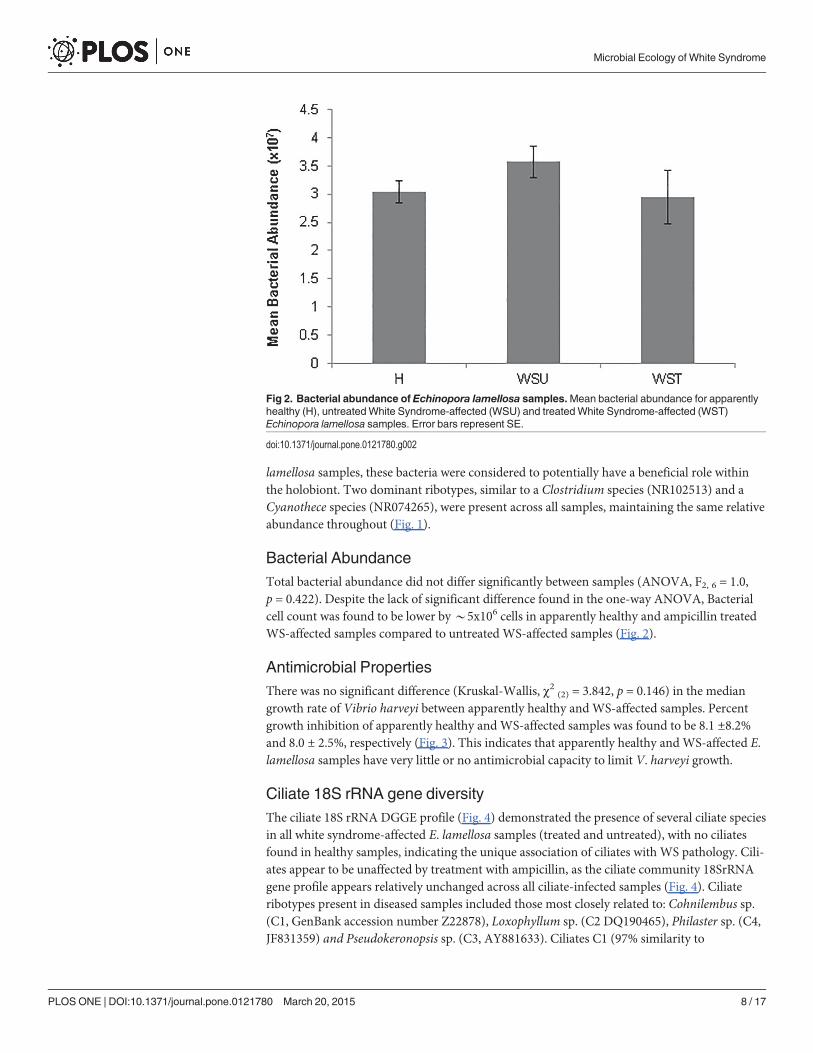
Bacterial AbundanceTotal bacterial abundance did not differ significantly between samples (ANOVA, F2, 6 = 1.0,p = 0.422). Despite the lack of significant difference found in the one-way ANOVA, Bacterialcell count was found to be lower by*5x10⁶ cells in apparently healthy and ampicillin treatedWS-affected samples compared to untreated WS-affected samples (Fig. 2).
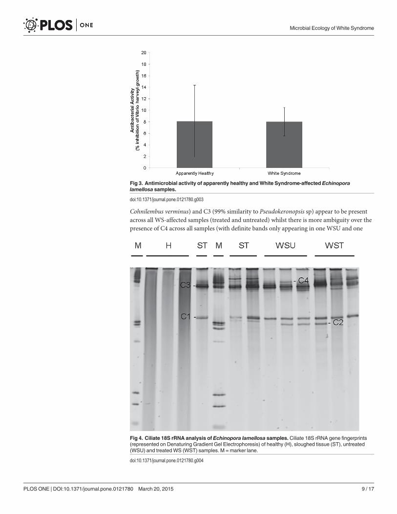
Antimicrobial PropertiesThere was no significant difference (Kruskal-Wallis, χ2 (2) = 3.842, p = 0.146) in the mediangrowth rate of Vibrio harveyi between apparently healthy andWS-affected samples. Percentgrowth inhibition of apparently healthy andWS-affected samples was found to be 8.1 ±8.2%and 8.0 ± 2.5%, respectively (Fig. 3). This indicates that apparently healthy andWS-affected E.lamellosa samples have very little or no antimicrobial capacity to limit V. harveyi growth.
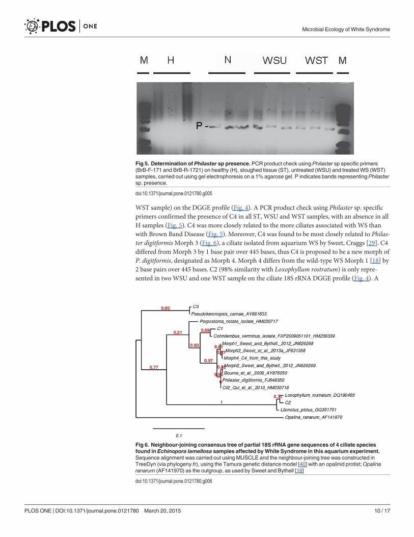
Ciliate 18S rRNA gene diversityThe ciliate 18S rRNA DGGE profile (Fig. 4) demonstrated the presence of several ciliate speciesin all white syndrome-affected E. lamellosa samples (treated and untreated), with no ciliatesfound in healthy samples, indicating the unique association of ciliates with WS pathology. Cili-ates appear to be unaffected by treatment with ampicillin, as the ciliate community 18SrRNAgene profile appears relatively unchanged across all ciliate-infected samples (Fig. 4). Ciliateribotypes present in diseased samples included those most closely related to: Cohnilembus sp.(C1, GenBank accession number Z22878), Loxophyllum sp. (C2 DQ190465), Philaster sp. (C4,JF831359) and Pseudokeronopsis sp. (C3, AY881633). Ciliates C1 (97% similarity to
Fig 2. Bacterial abundance of Echinopora lamellosa samples.Mean bacterial abundance for apparentlyhealthy (H), untreatedWhite Syndrome-affected (WSU) and treatedWhite Syndrome-affected (WST)Echinopora lamellosa samples. Error bars represent SE.
doi:10.1371/journal.pone.0121780.g002
Microbial Ecology of White Syndrome
PLOSONE | DOI:10.1371/journal.pone.0121780 March 20, 2015 8 / 17
Cohnilembus verminus) and C3 (99% similarity to Pseudokeronopsis sp) appear to be presentacross all WS-affected samples (treated and untreated) whilst there is more ambiguity over thepresence of C4 across all samples (with definite bands only appearing in one WSU and one
Fig 3. Antimicrobial activity of apparently healthy andWhite Syndrome-affected Echinoporalamellosa samples.
doi:10.1371/journal.pone.0121780.g003
Fig 4. Ciliate 18S rRNA analysis of Echinopora lamellosa samples. Ciliate 18S rRNA gene fingerprints(represented on Denaturing Gradient Gel Electrophoresis) of healthy (H), sloughed tissue (ST), untreated(WSU) and treated WS (WST) samples. M = marker lane.
doi:10.1371/journal.pone.0121780.g004
Microbial Ecology of White Syndrome
PLOSONE | DOI:10.1371/journal.pone.0121780 March 20, 2015 9 / 17
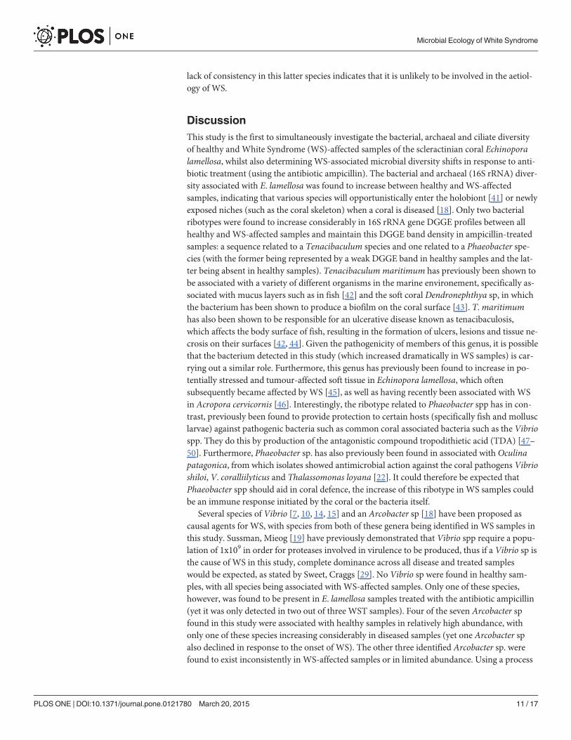
WST sample) on the DGGE profile (Fig. 4). A PCR product check using Philaster sp. specificprimers confirmed the presence of C4 in all ST, WSU and WST samples, with an absence in allH samples (Fig. 5). C4 was more closely related to the more ciliates associated with WS thanwith Brown Band Disease (Fig. 5). Moreover, C4 was found to be most closely related to Philas-ter digitformisMorph 3 (Fig. 6), a ciliate isolated from aquariumWS by Sweet, Craggs [29]. C4differed fromMorph 3 by 1 base pair over 445 bases, thus C4 is proposed to be a new morph ofP. digitformis, designated as Morph 4. Morph 4 differs from the wild-type WS Morph 1 [18] by2 base pairs over 445 bases. C2 (98% similarity with Loxophyllum rostratum) is only repre-sented in twoWSU and one WST sample on the ciliate 18S rRNA DGGE profile (Fig. 4). A
Fig 5. Determination of Philaster sp presence. PCR product check using Philaster sp specific primers(BrB-F-171 and BrB-R-1721) on healthy (H), sloughed tissue (ST), untreated (WSU) and treated WS (WST)samples, carried out using gel electrophoresis on a 1% agarose gel. P indicates bands representing Philastersp. presence.
doi:10.1371/journal.pone.0121780.g005
Fig 6. Neighbour-joining consensus tree of partial 18S rRNA gene sequences of 4 ciliate speciesfound in Echinopora lamellosa samples affected byWhite Syndrome in this aquarium experiment.Sequence alignment was carried out using MUSCLE and the neighbour-joining tree was constructed inTreeDyn (via phylogeny.fr), using the Tamura genetic distance model [40] with an opalinid protist;Opalinaranarum (AF141970) as the outgroup, as used by Sweet and Bythell [18]
doi:10.1371/journal.pone.0121780.g006
Microbial Ecology of White Syndrome
PLOSONE | DOI:10.1371/journal.pone.0121780 March 20, 2015 10 / 17
lack of consistency in this latter species indicates that it is unlikely to be involved in the aetiol-ogy of WS.
DiscussionThis study is the first to simultaneously investigate the bacterial, archaeal and ciliate diversityof healthy and White Syndrome (WS)-affected samples of the scleractinian coral Echinoporalamellosa, whilst also determining WS-associated microbial diversity shifts in response to anti-biotic treatment (using the antibiotic ampicillin). The bacterial and archaeal (16S rRNA) diver-sity associated with E. lamellosa was found to increase between healthy andWS-affectedsamples, indicating that various species will opportunistically enter the holobiont [41] or newlyexposed niches (such as the coral skeleton) when a coral is diseased [18]. Only two bacterialribotypes were found to increase considerably in 16S rRNA gene DGGE profiles between allhealthy and WS-affected samples and maintain this DGGE band density in ampicillin-treatedsamples: a sequence related to a Tenacibaculum species and one related to a Phaeobacter spe-cies (with the former being represented by a weak DGGE band in healthy samples and the lat-ter being absent in healthy samples). Tenacibaculum maritimum has previously been shown tobe associated with a variety of different organisms in the marine environement, specifically as-sociated with mucus layers such as in fish [42] and the soft coral Dendronephthya sp, in whichthe bacterium has been shown to produce a biofilm on the coral surface [43]. T.maritimumhas also been shown to be responsible for an ulcerative disease known as tenacibaculosis,which affects the body surface of fish, resulting in the formation of ulcers, lesions and tissue ne-crosis on their surfaces [42, 44]. Given the pathogenicity of members of this genus, it is possiblethat the bacterium detected in this study (which increased dramatically in WS samples) is car-rying out a similar role. Furthermore, this genus has previously been found to increase in po-tentially stressed and tumour-affected soft tissue in Echinopora lamellosa, which oftensubsequently became affected by WS [45], as well as having recently been associated with WSin Acropora cervicornis [46]. Interestingly, the ribotype related to Phaeobacter spp has in con-trast, previously been found to provide protection to certain hosts (specifically fish and mollusclarvae) against pathogenic bacteria such as common coral associated bacteria such as the Vibriospp. They do this by production of the antagonistic compound tropodithietic acid (TDA) [47–50]. Furthermore, Phaeobacter sp. has also previously been found in associated with Oculinapatagonica, from which isolates showed antimicrobial action against the coral pathogens Vibrioshiloi, V. coralliilyticus and Thalassomonas loyana [22]. It could therefore be expected thatPhaeobacter spp should aid in coral defence, the increase of this ribotype in WS samples couldbe an immune response initiated by the coral or the bacteria itself.
Several species of Vibrio [7, 10, 14, 15] and an Arcobacter sp [18] have been proposed ascausal agents for WS, with species from both of these genera being identified in WS samples inthis study. Sussman, Mieog [19] have previously demonstrated that Vibrio spp require a popu-lation of 1x10⁹ in order for proteases involved in virulence to be produced, thus if a Vibrio sp isthe cause of WS in this study, complete dominance across all disease and treated sampleswould be expected, as stated by Sweet, Craggs [29]. No Vibrio sp were found in healthy sam-ples, with all species being associated with WS-affected samples. Only one of these species,however, was found to be present in E. lamellosa samples treated with the antibiotic ampicillin(yet it was only detected in two out of three WST samples). Four of the seven Arcobacter spfound in this study were associated with healthy samples in relatively high abundance, withonly one of these species increasing considerably in diseased samples (yet one Arcobacter spalso declined in response to the onset of WS). The other three identified Arcobacter sp. werefound to exist inconsistently in WS-affected samples or in limited abundance. Using a process
Microbial Ecology of White Syndrome
PLOSONE | DOI:10.1371/journal.pone.0121780 March 20, 2015 11 / 17
of elimination, the results of this study suggest that Vibrio sp and Arcobacter sp are not thecausal agent for WS. An alternative view point may be that they are the causal agent for someWS outbreaks but not others, with WS being a polymicrobial disease caused by any opportu-nistic pathogenic bacteria at any given time. If the latter is true, then this leads to two possiblescenarios for WS: a) WS is not a single disease, but rather different diseases that appear mor-phologically similar, or b) WS is the result of a systemic infection which can be caused by vari-ous pathogenic bacteria [29].
Over recent years, members of the coral holobiont have been found to provide particularservices to their host, carrying out functions necessary for maintaining coral health, such as thecycling of nitrogen and sulphur and the production of secondary metabolites with an antimi-crobial action useful against invasive bacteria [20–24, 51]. Based on the potential importanceof various holobiont members, the loss of particular bacteria or archaea could have an impacton coral health. Three species identified in this study (closely related to a Bradyrhizobium sp., aRubrimonas sp. and a Sulfolobus sp.) were found to decline considerably between healthy sam-ples andWS-affected samples, with their populations failing to recover to healthy levels in re-sponse to treatment (with the prokaryote closely related to Bradyrhizobium sp. beingcompletely lost). Sulfolobus spp. are known to be sulphur oxidisers [52, 53], converting sulphur(S8) into sulphate (SO4
2−), thus a depletion of Sulfolobus spp. within the local microenviron-ment could result in an increase in sulphur and a decline in sulphate, provided there aren’tother members of the holobiont that can persist and carry out this function. An increase in sul-phur ions can result in tissue necrosis in corals, as associated with Black Band Disease [54] andsulphate reduction could result in reduced DMSP output by zooxanthellae, a molecule whichRaina, Tapiolas [55] state could have a significant role in antioxidant and antimicrobial pro-duction, osmoprotection and the shaping of the microbial community within the holobiont.Bradyrhizobium spp are denitrifying bacteria known to completely denitrify nitrates to nitritesto nitrogen gas, thus a reduction in this species could impact on the cycling of nitrogen withinthe microenvironment, resulting in a more nitrogen-limited environment. Nitrogen-limitationhas been found to directly impact zooxanthellae, resulting in a decline in growth and popula-tion density [56].
Bacterial abundance did not differ significantly, implying that in this particular case, WS isnot the result of a general increase in bacterial load. This is in contrast to previous studies suchas Luna, Biavasco [57] and Sweet, Craggs [29] whereby bacterial abundance was found to in-crease in other aquarium corals exhibiting WS. Since treatment with ampicillin impacted bac-terial diversity, yet overall bacterial abundance was not found to decrease in treated samples, itis possible that there was a very rapid colonisation by bacteria once the coral was replaced backinto the tank system. However, Sweet, Croquer [37] showed that bacterial populations took>12 hours to recover under field conditions following treatment of healthy corals with Cipro-floxacin, which suggests that such rapid recolonisation is unlikely. The Coral Probiotic theory[21] proposes that the natural coral holobiont is host to microorganisms that aid in coral healthand defence against invading bacteria via the production of secondary metabolites. This de-fence may be lost during an episode of environmental stress or disease due to a re-shuffling ofthe microbial community [21] or in this instance disease and subsequent treatment. Interest-ingly, however, healthy andWS-affected coral samples in the present study were found to havevery little to no impact on the growth of the reported coral pathogen (more specifically, previ-ously proposed WS pathogen) Vibrio harveyi. This could suggest that antimicrobial capacity isunchanged in healthy and diseased specimens, with E. lamellosa appearing to have a naturallyvery limited antimicrobial capacity against Vibrio harveyi. This could suggest that not all corals,such as Echinopora lamellosa, possess such antimicrobial qualities, perhaps relying more onthe physical defences of the mucus barrier. It is also possible that these corals are more
Microbial Ecology of White Syndrome
PLOSONE | DOI:10.1371/journal.pone.0121780 March 20, 2015 12 / 17
susceptible to certain stresses and diseases, as observed across a range of corals in response toclimate change with certain species found to be more susceptible than others [58, 59]. Howev-er, it could also be argued that the findings of this study support research that demonstrates dif-ferent coral species have different antimicrobial selectivity. Selectivity is a hallmark ofantimicrobial activity in many coral species [60–62] and it is possible that E. lamellosa featuresantimicrobial properties that do not select against Vibrio harveyi. To understand the overallantimicrobial capacity and selectivity of E. lamellosa (and other species), future antimicrobialassays using coral extracts should be carried out against a much broader range of targetprokaryotes.
In the present study, all WS-affected samples (treated and untreated) were found to be hostto a number of ciliate species, whilst no ciliates were identified in healthy E. lamellosa samples,thus making this the first study to identify and directly associate ciliates with WS in the platingcoral E. lamellosa. A Philaster sp was also found to be present in all diseased samples in thepresent study, supporting previous studies associating this particular genus with the consump-tion of the corals Symbiodinium in WS, White Band Disease (WBD) and Brown Band Disease(BBD)[18, 29, 63]. The belief that this ciliate is responsible for the neat lesion edge associatedwith WS is supported by research into the role of another Philaster sp (P. lucinda) in WBD andWS [46, 63]. It was observed that when the ciliate was knocked down using metronidazole inWBD andWS samples, the morphology of lesion interface was altered, yet lesion progressioncontinued. This promotes the theory that P. lucinda is a secondary agent in WBD, which couldalso be the case for Philaster species associated with WS. Other predominant ciliates found inthe present study were Cohnilembus sp. and Pseudokeronopsis sp. There are no previous reportslinking Cohnilembus sp. with corals and disease, despite this ciliate being found in all WS-af-fected E. lamellosa samples in the present study. Cohnilembus sp. is known to exist as a bacteri-vorous benthic suspension feeder which has enhanced feeding when attached to a surface,whereby stronger feeding currents can be created compared to when the ciliate is suspended inthe water column [64]. It is possible therefore that during WS, Cohnilembus sp. attaches toareas of denuded skeleton that can no longer be protected by the host coral. A further ciliatespecies identified in this study, Pseudokeronopsis sp. was also found in WS and BBD samplesby Sweet and Bythell [18], they observed this particular ciliate to be a ciliaphore, consumingother protists and predominantly found in areas of denuded skeleton. This indicates Pseudo-keronopsis sp. does not have a role in WS pathology, but rather exists as part of a ciliate foodchain colonising the exposed skeleton following the removal of tissue due to the disease. It re-mains uncertain whether the association of ciliates with WS is cause or effect, but it appearsvery clear that ciliates play an important role in the aetiology of WS lesions [29]. Interestingly,Mesanophrys spp., ciliates belonging to the order Philasterida, and morphologically similar toPhilaster, have previously been associated with tissue degradation in different organisms, suchas crabs [65, 66], lobsters [67] and fish (such as the turbot) [68]. Small, Neil [67] found that aMesanophrys sp. isolated from infected lobsters express metalloproteases capable of proteolysisof several host proteins, including structural proteins such as myosin. We propose that futurestudies on coral disease should aim to determine the expression of similar proteases in coraldisease-associated ciliates in order to determine if they are capable of direct host tissue stressand degradation, thus further clarifying their role in diseases such WS.
In conclusion, this study has determined shifts in the microbial rRNA gene profiles betweenhealthy and WS-affected E. lamellosa. Given its association with the formation of lesions andulceration, as well as tissue necrosis in various fish species, and based on its strong associationwith WS-affected samples in the present study, the bacterium Tenacibaculum sp. is presentedas a novel potential causal agent for lesion formation and tissue necrosis in Echinopora lamel-losa. Since this bacterium has not been reported as a causal agent for WS in the past, this
Microbial Ecology of White Syndrome
PLOSONE | DOI:10.1371/journal.pone.0121780 March 20, 2015 13 / 17
supports the view that there is no specific causal agent for this disease, but rather various op-portunistic necrotizing bacteria capable of tissue breakdown may have the capacity for initiat-ing WS [29], thus the disease is likely polymicrobial. However, future studies focusing onfulfilling Koch’s postulates with regards to the role of Tenacibaculum sp in WS are required.This study further supports recent work showing that ciliates play an active role in disease mor-phology in many different species of coral, giving the disease its characterizing neat boundaryat the lesion interface between healthy tissue and denuded skeleton. Future work should focuson the isolation of proteases and other molecules involved in tissue stress and degradationfrom associated ciliate isolates (particularly zooxanthellae consumers), thus determiningwhether or not they actively impair and break down healthy host tissue or whether they arepassively feeding on already weakened tissue. Additionally, this study appears to demonstratethat not all corals possess probiotic qualities that aid in their defence against invasive microor-gansims, which could contribute to why some corals are more susceptible to stress and diseasethan others.
AcknowledgmentsAll coral samples used in this work were donated by the Horniman Museum and Gardens,with the permission of aquarium manager Jamie Craggs.
Author ContributionsConceived and designed the experiments: DS JB MS. Performed the experiments: DS PL JC.Analyzed the data: DS PL. Contributed reagents/materials/analysis tools: JC MS. Wrote thepaper: DS. Reviewed the manuscript: PL JB MS.
References1. Hayes RL, Goreau NI. The significance of emerging diseases in the tropical coral reef ecosystem.
Revista de Biologia Tropical. 1998; 46(5):173–85.
2. Harvell CD. Emerging Marine Diseases—Climate Links and Anthropogenic Factors. Science. 1999;285(5433):1505–10. doi: 10.1126/science.285.5433.1505 PMID: 10498537
3. Harvell CD, Mitchell CE, Ward JR, Altizer S, Dobson AP, Ostfeld RS, et al. Climate warming and dis-ease risks for terrestrial and marine biota. Science. 2002; 296(5576):2158–62. doi: 10.1126/science.1063699 PubMed PMID: PMID: 12077394.
4. Rosenberg E, Ben-Haim Y. Microbial diseases of corals and global warming. Environmental Microbiolo-gy. 2002; 4(6):319–26.
5. Pollock JS, Morris PJ, Willis BL, Bourne DG. The urgent need for robust coral disease diagnostics.PloS Pathogens. 2011; 7(10). doi: 10.1371/.
6. Sokolow S. Effects of a changing climate on the dynamics of coral infectious disease: a review of theevidence. Diseases of Aquatic Organisms. 2009; 87(1–2):5–18. doi: 10.3354/dao02109 PMID:20099414
7. Piskorska M, Smith G, Weil E. Bacteria associated with the coral Echinopora lamellosa (Esper 1795) inthe Indian Ocean—Zanzibar region. African Journal of Environmental Science and Technology. 2007;1(5):93–8.
8. Sweet M, Jones R, Bythell J. Coral diseases in aquaria and in nature. Journal of the Marine BiologicalAssociation of the United Kingdom. 2011:1–11. doi: 10.1017/s0025315411001688
9. Dalton SJ, Godwin S, Smith SDA, Pereg L. Australian subtropical white syndrome: a transmissible tem-perature-dependent coral disease. Marine and Freshwater Research. 2010; 61:342–50.
10. SussmanM, Willis BL, Victor S, Bourne DG. Coral pathogens identified for white syndrome (WS) epizo-otics in the Indo-Pacific. PLoS ONE. 2008; 3(6):1–14.
11. Bruno JF, Selig ER, Casey KS, Page CA, Willis BL, Drew Harvell C, et al. Thermal stress and coralcover as drivers of coral disease outbreaks. PLoS Biol. 2007; 5(6):1220–7.
Microbial Ecology of White Syndrome
PLOSONE | DOI:10.1371/journal.pone.0121780 March 20, 2015 14 / 17
12. Aeby GS, Ross M, Williams GJ, Lewis TD, Work TM. Disease dynamics of Montipora white syndromewithin Kaneohe Bay, Oahu, Hawaii: distribution, seasonality, virulence, and transmissibility. Diseasesof Aquatic Oragnisms. 2010; 91:1–8.
13. Willis BL, Page CA, Dinsdale EA. Coral disease on the Great Barrier Reef. In: Rosenberg E, Loya Y, ed-itors. Coral Health and Disease: Springer, Berlin; 2004. p. 69–104.
14. Luna GM, Bongiorni L, Gili C, Biavasco F, Danovaro R. Vibrio harveyi as a causative agent of the WhiteSyndrome in tropical stony corals. Environmental Microbiology Reports. 2010; 2(1):120–7. doi: 10.1111/j.1758-2229.2009.00114.x PMID: 23766006
15. Ushijima B, Smith A, Aeby GS, Callahan SM. Vibrio owensii Induces the Tissue Loss DiseaseMonti-poraWhite Syndrome in the Hawaiian Reef CoralMontipora capitata. PLoS ONE. 2012; 7(10):e46717.doi: 10.1371/journal.pone.0046717.g001 PMID: 23056419
16. Wagner M, Amann R, Lemmer H, Schleifer K. Probing activated sludge with oligonucleotides specificfor proteobacteria: inadequacy of culture-dependent methods for describing microbial community struc-ture. Applied and Environmental Microbiology. 1993; 59(5):1520–5. PMID: 8517747
17. Scanlan PD, Marchesi JR. Micro-eukaryotic diversity of the human distal gut microbiota: qualitative as-sessment using culture-dependent and -independent analysis of faeces. ISME J. 2008; 2(12):1183–93.doi: 10.1038/ismej.2008.76 PubMed PMID: PMID: 18670396.
18. Sweet M, Bythell J. Ciliate and bacterial communities associated with White Syndrome and BrownBand Disease in reef-building corals. Environmental Microbiology. 2012. doi: 10.1111/j.1462-2920.2012.02746.x
19. SussmanM, Mieog JC, Doyle J, Victor S, Willis BL, Bourne DG. Vibrio zinc-metalloprotease causesphotoinactivation of coral endosymbionts and coral tissue lesions. PLoS ONE. 2009; 4(2):1–14. doi:10.1371/journal.pone.0004511.t001 10.1371/journal.pone.0004511.t002
20. Rosenberg E, Koren O, Reshef L, Efrony R, Zilber-Rosenberg I. The role of microorganisms in coralhealth, disease and evolution. Nature Reviews Microbiology. 2007; 5(5):355–62. doi: 10.1038/nrmicro1635 PMID: 17384666
21. Reshef L, Koren O, Loya Y, Zilber-Rosenberg I, Rosenberg E. The Coral Probiotic Hypothesis. Environ-mental Microbiology. 2006; 8(12):2068–73. doi: 10.1111/j.1462-2920.2006.01148.x PMID: 17107548
22. Nissimov J, Rosenberg E, Munn CB. Antimicrobial properties of resident coral mucus bacteria of Ocu-lina patagonica. FEMSmicrobiology letters. 2009; 292(2):210–5. Epub 2009/02/05. doi: 10.1111/j.1574-6968.2009.01490.x PubMed PMID: PMID: 19191871.
23. Shnit-Orland M, Kushmaro A. Coral mucus-associated bacteria: a possible first line of defense. FEMSMicrobiology Ecology. 2009; 67(3):371–80. doi: 10.1111/j.1574-6941.2008.00644.x PMID: 19161430
24. Kvennefors ECE, Sampayo E, Kerr C, Vieira G, Roff G, Barnes AC. Regulation of Bacterial Communi-ties Through Antimicrobial Activity by the Coral Holobiont. Microbial ecology. 2012; 63(3):605–18. doi:10.1007/s00248-011-9946-0 PMID: 21984347
25. Kumar KS, Haritha R, Mohan YSYJ, Ramana T. Screening of marine actinobacteria for antimicrobialcompounds. Research Journal of Microbiology. 2011; 6(4):385–93.
26. Kennedy J, Baker P, Piper C, Cotter PD, Walsh M, Mooij MJ, et al. Isolation and analysis of bacteriawith antimicrobial activities from the marine sponge Haliclona simulans collected from Irish waters. MarBiotechnol (NY). 2009; 11(3):384–96. doi: 10.1007/s10126-008-9154-1 PubMed PMID: PMID:18953608.
27. Manivasagan P, Gnanam S, Sivakumar K, Thangaradjou T, Vijayalakshmi S, Balasubramanian T. Anti-microbial and cytotoxic activities of an actinobacteria (Streptomyces sp. PM-32) isolated from an off-shore sediments of the Bay of Bengal in Tamilnadu. Advances in Biological Research. 2009; 3(5–6):231–6.
28. Sweet MJ, Smith D, Bythell JC, Craggs J. Changes in microbial diversity associated with two coral spe-cies recovering from a stressed state in a public aquarium. Journal of Zoo and Aquarium Research.2013; 1(2):1–8.
29. Sweet MJ, Craggs J, Robson J, Bythell JC. Assessment of the microbial communities associated withwhite syndrome and brown jelly syndrome in aquarium corals. Journal of Zoo and Aquarium Research.2013; 1(1):1–8.
30. Harvell D, Jordán-Dahlgren E, Merkel S, Rosenberg E, Raymundo L, Smith G, et al. Coral disease, en-vironmental drivers, and the balance between coral and microbial associates. Oceanography. 2007; 20(1):172–95.
31. Muyzer G, Smalla K. Application of denaturing gradient gel electrophoresis (DGGE) and temperaturegradient gel electrophoresis (TGGE) in microbial ecology. Antonie van Leeuwenhoek. 1998; 73:127–41. PMID: 9602286
Microbial Ecology of White Syndrome
PLOSONE | DOI:10.1371/journal.pone.0121780 March 20, 2015 15 / 17
32. Jousset A, Lara E, Nikolausz M, Harms H, Chatzinotas A. Application of the denaturing gradient gelelectrophoresis (DGGE) technique as an efficient diagnostic tool for ciliate communities in soil. The Sci-ence of the total environment. 2010; 408(5):1221–5. doi: 10.1016/j.scitotenv.2009.09.056 PubMedPMID: PMID: 19896703.
33. Janse I, Bok J, Zwart G. A simple remedy against artifactual double bands in denaturing gradient gelelectrophoresis. Journal of microbiological methods. 2004; 57(2):279–81. doi: 10.1016/j.mimet.2003.12.006 PubMed PMID: PMID: 15063068.
34. Bourne DG, Boyett HV, Henderson ME, Muirhead A, Willis BL. Identification of a ciliate (Oligohymeno-phorea: Scuticociliatia) associated with brown band disease on corals of the Great Barrier Reef. ApplEnviron Microbiol. 2008; 74(3):883–8. doi: 10.1128/AEM.01124-07 PubMed PMID: PMID: 18083868;PubMed Central PMCID: PMC2227702.
35. Sweet MJ, Croquer A, Bythell JC. Temporal and spatial patterns in waterborne bacterial communitiesof an island reef system. Aquatic Microbial Ecology. 2010; 61(1):1–11.
36. Selinummi J, Seppälä J, Yli-Harja O, Puhakka J. Software for quantification of labeled bacteria from dig-ital microscope images by automated image analysis. BioTechniques. 2005; 39(6):859–63. doi: 10.2144/000112018 PMID: 16382904
37. Sweet MJ, Croquer A, Bythell JC. Dynamics of bacterial community development in the reef coral Acro-pora muricata following experimental antibiotic treatment. Coral Reefs. 2011; 30(4):1121–33. doi: 10.1007/s00338–011–0800–0
38. Clarke KR, Warwick RM. A further biodiversity index applicable to species lists—variation in taxonomicdistinctness. Marine Ecology Progress Series. 2001; 216:265–78.
39. Cróquer A, Bastidas C, Elliott A, Sweet M. Bacterial assemblages shifts from healthy to yellow band dis-ease states in the dominant reef coralMontastraea faveolata. Environmental Microbiology Reports.2012. doi: 10.1111/j.1758-2229.2012.00397.x
40. Dereeper A, Guignon V, Blanc G, Audic S, Buffet S, Chevenet F, et al. Phylogeny. fr: robust phylogenet-ic analysis for the non-specialist. Nucleic acids research. 2008; 36(suppl 2):W465–W9.
41. Sunagawa S, DeSantis TZ, Piceno YM, Brodie EL, DeSalvo MK, Voolstra CR, et al. Bacterial diversityandWhite Plague Disease-associated community changes in the Caribbean coral Montastraea faveo-lata. The ISME Journal. 2009; 3(5):512–21. doi: 10.1038/ismej.2008.131 PMID: 19129866
42. Avendano-Herrera R, Toranzo AE, Magarinos B. Tenacibaculosis infection in marine fish caused byTenacibaculum maritimum: a review. Diseases of Aquatic Oragnisms. 2006; 71:255–66. PMID:17058606
43. Harder T, Lau SCK, Dobretsov S, Fang TK, Qian P. A distinctive epibiotic bacterial community of thesoft coral Dendronephthya sp and antibacterial activity of coral tissue extracts suggest a chemicalmechanism against bacterial epibiosis. FEMSMicrobiology Ecology. 2008; 43:337–47.
44. El-Galil MAA, HashemM. Epidemiological and bacteriological studies on tenacibaculosis in some RedSea fishes, Egypt. International Journal of Environmental Science and Engineering. 2012; 3:25–32.
45. Smith D, Leary P, Bendall M, Flach E, Jones R, Sweet M. A Novel Investigation of a Blister-Like Syn-drome in Aquarium Echinopora lamellosa. PloS one. 2014; 9(5):e97018. doi: 10.1371/journal.pone.0097018 PMID: 24827734
46. Sweet M, Bythell J. White Syndrome in Acropora muricata: Non‐specific bacterial infection and ciliatehistophagy. Molecular ecology. 2015;In Press.
47. D'Alvise PW, Lillebø S, Wergeland HI, Gram L, BerghØ. Protection of cod larvae from vibriosis byPhaeobacter spp.: A comparison of strains and introduction times. Aquaculture. 2013; 384–387:82–6.doi: 10.1016/j.aquaculture.2012.12.013
48. Prado S, Montes J, Romalde JL, Barja JL. Inhibitory activity of Phaeobacter strains against aquaculturepathogenic bacteria. International Microbiology. 2009; 12:107–14. doi: 10.2436/20.1501.01.87 PMID:19784930
49. Porsby CH, Nielsen KF, Gram L. Phaeobacter and Ruegeria species of the Roseobacter clade colonizeseparate niches in a Danish Turbot (Scophthalmus maximus)-rearing farm and antagonize Vibrio angu-illarum under different growth conditions. Applied Environmental Microbiology. 2008; 74(23):7356–64.doi: 10.1128/AEM.01738-08 PubMed PMID: PMID: 18952864; PubMed Central PMCID:PMC2592939.
50. D'Alvise PW, Lillebo S, Prol-Garcia MJ, Wergeland HI, Nielsen KF, Bergh O, et al. Phaeobacter gallae-ciensis reduces Vibrio anguillarum in cultures of microalgae and rotifers, and prevents vibriosis in codlarvae. PLoS One. 2012; 7(8):e43996. doi: 10.1371/journal.pone.0043996 PubMed PMID: PMID:22928051; PubMed Central PMCID: PMC3425499.
51. Radjasa OK, Sabdono A. Ecological role of a softcoral-associated bacterium Arthrobacter sp. on ma-rine biofilm-forming bacteria. Microbiology Indonesia. 2008; 2(2):84–8.
Microbial Ecology of White Syndrome
PLOSONE | DOI:10.1371/journal.pone.0121780 March 20, 2015 16 / 17
52. Shivvers DW, Brock TD. Oxidation of elemental sulfur by Sulfolobus acidocaldarius. Journal of Bacteri-ology. 1973; 114(2):706–10. PMID: 4706192
53. Brock TD, Brock KM, Belly RT, Weiss RL. Sulfolobus: A new genus of sulfur-oxidizing bacteria living atlow pH and high temperature. Archives of Microbiology. 1972; 84:54–68. PMID: 4559703
54. Sato Y, Willis BL, Bourne DG. Successional changes in bacterial communities during the developmentof black band disease on the reef coral,Montipora hispida. ISME J. 2010; 4(2):203–14. Epub 2009/09/25. doi: 10.1038/ismej.2009.103 PubMed PMID: PMID: 19776765.
55. Raina JB, Tapiolas D, Willis BL, Bourne DG. Coral-associated bacteria and their role in the biogeo-chemical cycling of sulfur. Appl Environ Microbiol. 2009; 75(11):3492–501. Epub 2009/04/07. doi: 10.1128/AEM.02567-08 PubMed PMID: PMID: 19346350; PubMed Central PMCID: PMC2687302.
56. Muscantine L, Falkowski PG, Dubinsky Z, Cook PA, McCloskey LR. The effect of external nutrient re-sources on the population dynamics of zooxanthellae in a reef coral. Proceedings of the Royal SocietyB: Biological Sciences. 1989; 236:311–24. PMID: 2760652
57. Luna GM, Biavasco F, Danovaro R. Bacteria associated with the rapid tissue necrosis of stony corals.Environ Microbiol. 2007; 9(7):1851–7. doi: 10.1111/j.1462-2920.2007.01287.x PubMed PMID: PMID:17564618.
58. FodenW, Mace GM, Vié J-C, Angulo A, Butchart SHM, DeVantier L, et al. Species susceptibility to cli-mate change impacts. Wildlife in a changing world–an analysis of the 2008 IUCNRed List of threatenedspecies2009. p. 77.
59. Harvell CD, Altizer S, Cattadori IM, Harrington L, Weil E. Climate change and wildlife diseases: whendoes the host matter the most? Ecology. 2009; 90(4):912–20. PMID: 19449685
60. Gunthorpe L, Cameron AM. Widespread but variable toxicity in scleractinian corals. Toxicon. 1990; 28(10):1199–219. PMID: 1979891
61. Gochfeld DJ, Pappas KE, Lee S, Aeby GS. Variability in Antibacterial Activity in Hawaiian Corals. 2011.
62. Marquis CP, Baird AH, De Nys R, Holmström C, Koziumi N. An evaluation of the antimicrobial proper-ties of the eggs of 11 species of scleractinian corals. Coral Reefs. 2005; 24(2):248–53.
63. Sweet MJ, Croquer A, Bythell JC. Experimental antibiotic treatment identifies potential pathogens ofwhite band disease in the endangered Caribbean coral Acropora cervicornis. Proceedings of the RoyalSociety of London B: Biological Sciences. 2014; 281(1788):20140094. doi: 10.1098/rspb.2014.0094PMID: 24943374
64. Shimeta J, Starczak VR, Ashiru OM, Zimmer CA. Influences of benthic boundary-layer flow on feedingrates of ciliates and flagellates at the sediment-water interface. Limnology and oceanography. 2001; 46(7):1709–19.
65. Morado JF, Giesecke RH, Syrjala SE. Molt related mortalities of the Dungeness crab Cancer magistercaused by a marine facultative ciliate Mesanophrys pugettensis. Diseases of aquatic organisms. 1999;38(2):143–50.
66. Messick GA, Small EB. Mesanophrys chesapeakensis n. sp., a histophagous ciliate in the blue crab,Callinectes sapidus, and associated histopathology. Invertebrate Biology. 1996:1–12.
67. Small HJ, Neil DM, Taylor AC, Coombs GH. Identification and partial characterisation of metallopro-teases secreted by a Mesanophrys-like ciliate parasite of the Norway lobster Nephrops norvegicus.Diseases of Aquatic Organisms. 2005; 67:225–31. PMID: 16408838
68. Qin L, Wang Y, Zhang L, Dai J. Histopathology of turbot associated with Mesanophrys carcini parasite.Acta Hydrobiologica Sinica. 2007; 31(5):622.
Microbial Ecology of White Syndrome
PLOSONE | DOI:10.1371/journal.pone.0121780 March 20, 2015 17 / 17