The bedside dysmorphologist
305
Transcript of The bedside dysmorphologist


The Bedside Dysmorphologist

The Bedside DysmorphologistClassic Clinical Signs in Human Malformation Syndromes
and Their Diagnostic Significance
William Reardon, M.D.
12008

1Oxford University Press, Inc., publishes works that further
Oxford University’s objective of excellence
in research, scholarship, and education.
Oxford New York
Auckland Cape Town Dar es Salaam Hong Kong Karachi
Kuala Lumpur Madrid Melbourne Mexico City Nairobi
New Delhi Shanghai Taipei Toronto
With offices in
Argentina Austria Brazil Chile Czech Republic France Greece
Guatemala Hungary Italy Japan Poland Portugal Singapore
South Korea Switzerland Thailand Turkey Ukraine Vietnam
Copyright � 2008 by Oxford University Press, Inc.
Published by Oxford University Press, Inc.
198 Madison Avenue, New York, New York 10016
www.oup.com
Oxford is a registered trademark of Oxford University Press
All rights reserved. No part of this publication may be reproduced,
stored in a retrieval system, or transmitted, in any form or by any means,
electronic, mechanical, photocopying, recording, or otherwise,
without the prior permission of Oxford University Press.
Library of Congress Cataloging-in-Publication Data
Reardon, William, 1960–
The bedside dysmorphologist / William Reardon.
p. ; cm.
Includes bibliographical references.
ISBN 978-0-19-530045-1
1. Abnormalities, Human—Diagnosis—Handbooks, manuals, etc.
2. Physical diagnosis—Handbooks, manuals, etc. I. Title.
[DNLM: 1. Abnormalities—diagnosis—Handbooks.
2. Pediatrics—Handbooks. 3. Child. 4. Infant. WS 39 R288b 2007]
QM691.R43 2007
616'.043—dc22 2006028127
9 8 7 6 5 4 3 2 1
Printed in China
on acid-free paper

Preface
Although it has not always been so, dysmorphology is nowadays well served
with several fine books on the subject. Many of the leading practitioners of our
discipline have committed their wisdom and experience to the page to the
great benefit of their colleagues and admirers all around the world, not to
mention the incalculable service to countless patients. Consequently, there
exist now several reputable and authoritative texts and computerized data-
bases on the diagnostic approach to the dysmorphic child, on summarized
published information relating to individual syndromes, on classic examples of
known syndromes, and on various other aspects of dysmorphology practice.
Notwithstanding these considerable aids, reaching a specific diagnosis in the
dysmorphic individual remains, for most of us, a formidable barrier.
Embarking as a very junior trainee in clinical genetics almost 20 years ago,
I often found myself confused as to what exactly constituted an abnormal
clinical sign, so often the springboard to final diagnosis. The vivid admiration
I harbored then for colleagues possessed of the clinical gifts and experience
underlying the recognition of such signs still burns bright. Many of my early
days in training were spent looking up articles in journals seeking that perfect
demonstration of a seminal clinical sign as a means of clarifying my confused
state of mind. This should not in any way reflect upon the mentoring offered
to me, which could not have been bettered, but rather should reflect on my
own inadequacies faced with the struggle to master the language, the litera-
ture, and the seemingly limitless variation in clinical subtlety with which
human malformation syndromes are wont to clothe themselves.
Although mastery has proven elusive, experience has offered both com-
pensation and concern—compensation in that I have now learned enough to
accept that nobody has all the answers in dysmorphology, but concern that
the exact same problems that I personally recollect as a youthful trainee are

still cited by young colleagues undertaking training in clinical genetics—for
example, ‘‘How can I be sure if the ears are low set?’’ ‘‘Is it significant if there
are deep plantar creases?’’ ‘‘What should I be thinking of if the patient has
micropenis?’’
Experienced dysmorphologists will hear in these questions the panicky
voice of inexperience, and their diagnostic insight is absolutely correct! How-
ever, other clinicians, and perhaps pediatricians above all others, will hear in
these questions their own authentic voices, the concerns that bedevil their
daily practices as they examine their patients, and wonder whether an indi-
vidual has a dysmorphic sign suggestive of an underlying syndrome or not. It
has surprisedme that, in all the excellent dysmorphology books published over
the last two decades, so little emphasis has been placed on assisting the non-
specialist, whether trainee geneticist, pediatrician, or other clinical colleague, to
recognize dysmorphic signs and to understand their possible significance. It
appears to me that the literature of clinical genetics and dysmorphology is now
so sophisticated and well organized as to be quite daunting for most nonspe-
cialists. Listening to pediatrician colleagues, I hear that the gulf between our
specialty and our nongenetic colleagues can appear insurmountable, notwith-
standing our shared interests in the diagnosis of and care for the malformed
patient. This is the background to the concept of the book I wish to write—one
that seeks to make the clinical signs of malformation and the reasoning process
deriving from their observation less arcane than currently appears to be the case.
For trainees in clinical genetics, there is an exigency to become comfortable with
these concepts before progressing to a more advanced state of knowledge, while
for nongeneticist colleagues, it is my hope that this book will answer some of the
questions most frequently posed as to why genetic diagnoses may seem so
inaccessible.
Prefacevi
Acknowledgments
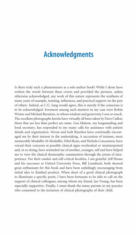
Is there truly such a phenomenon as a sole-author book? While I alone have
written the words between these covers and provided the pictures, unless
otherwise acknowledged, any work of this nature represents the synthesis of
many years of example, training, influences, and practical support on the part
of others. Indeed, as C.G. Jung would agree, this is merely if the conscious is
to be acknowledged. Foremost among such mentors in my case were Robin
Winter andMichael Baraitser, to whose wisdom and generosity I owe somuch.
The excellent photographs herein have virtually all been taken by Dave Cullen;
those that are less than perfect are mine. Lisa Malone, my longstanding and
loyal secretary, has responded to my many calls for assistance with patient
details and organization. Nevan and Isolt Reardon have continually encour-
aged me by their interest in the undertaking. A succession of trainees, most
memorably Mudaffer Al-Mudaffer, Ethel Ryan, and Nichola Concannon, have
voiced their concerns at possible clinical signs overlooked or misinterpreted
and, in so doing, have reminded me of another, younger, self and have helped
me to view the clinical dysmorphic examination through the prism of inex-
perience. For their candor and self-critical faculties, I am grateful. Jeff House
and his successor at Oxford University Press, Bill Lamsback, both showed
great enthusiasm for this book and have been unfailingly encouraging from
initial idea to finished product. When short of a good clinical photograph
to illuminate a specific point, I have been fortunate to be able to call on the
support of clinical colleagues, among whom my friend, Ian Young, has been
especially supportive. Finally, I must thank the many parents in my practice
who consented to the inclusion of clinical photographs of their child.

Contents
Chapter 1 The Skull
1.1 Plagiocephaly 4
1.2 Metopic Ridge 6
1.3 Scalp Defects 8
1.4 Macrocephaly 10
1.5 Microcephaly 12
1.6 Frontal Hairline Variants—High
Hairline and Frontal Upsweep 14
1.7 Frontal Hairline Variants—Low
Hairline and Widow’s Peak 16
1.8 Low Posterior Hairline 18
1.9 Anencephaly 20
Chapter 2 The Face
2.1 Hypertelorism 24
2.2 Hypotelorism 26
2.3 Abnormal Nasal Bridge 28
2.4 Abnormal Alae Nasi and
Nasal Tip 30
2.5 Abnormal Nasal Columella
and Nares 32
2.6 Abnormal Nasal Septum 34
2.7 Abnormal Nasal Appendages 36
2.8 Synophrys 38
2.9 Midfacial Hypoplasia 40
2.10 Micrognathia 42
2.11 Facial Asymmetry 44
2.12 Myopathic Facies 46
2.13 Abnormalities of
the Philtrum 48

Chapter 3 The Eye and Related Structures
3.1 Epicanthus 52
3.2 Deep-Set Eyes 54
3.3 Almond-Shaped Eyes 56
3.4 Blepharophimosis 58
3.5 Palpebral Fissure Slant 60
3.6 Proptosis 62
3.7 Ptosis of the Eyelid 64
3.8 Corneal Clouding 66
3.9 Ectopic Pupil 68
3.10 Blue Sclerae 70
3.11 Iris Coloboma 72
3.12 Anophthalmia/Microphthalmia 74
3.13 Iris Variants 76
3.14 Ectropion of the Lower Eyelid 78
3.15 Eyebrow Variants 80
3.16 Eyelash Variants 82
3.17 Epibulbar Dermoid 84
Chapter 4 The Ear
4.1 Low-Set Ears 88
4.2 Posteriorly Rotated Ears 90
4.3 Auricular Pits 92
4.4 Microtia 94
4.5 External Ear Variants 96
4.6 Ear Lobe Variants 98
4.7 External Auditory Canal Atresia
or Stenosis 100
4.8 Auricular Tags 102
Chapter 5 The Mouth and Oral Cavity
5.1 Microstomia 106
5.2 Upper Lip Clefts 108
5.3 Lip Pits 110
5.4 Thick and Thin Upper Lips 112
5.5 Lip and Oral Mucosa
Pigmentation 114
5.6 Macroglossia 116
5.7 Accessory Oral Frenula 118
5.8 Isolated Cleft Palate 120
5.9 Persistent Drooling 122
5.10 Gum Hyperplasia 124
Chapter 6 The Neck
6.1 Neck Webbing 128
6.2 Short Neck 130
6.3 Goiter 132
6.4 Neck Sinuses/Fistulae/Pits 134
6.5 Occipital Horns 136

Chapter 7 The Chest
7.1 Poland Anomaly 140
7.2 Accessory Nipples 142
7.3 Athelia/Hypothelia (Absent or
Hypoplastic Nipples) 144
7.4 Gynecomastia 146
7.5 Sloping Shoulders 148
7.6 Small Thorax 150
7.7 Pectus Excavatum and
Carinatum 152
7.8 Scoliosis 154
Chapter 8 The Abdomen and Perineum
8.1 Minor Anomalies of
the Umbilicus 158
8.2 Umbilical Hernia/
Omphalocele 160
8.3 Inguinal Hernia 162
8.4 Small Penis/Micropenis 164
8.5 Ambiguous Genitalia 166
8.6 Hypospadias 168
8.7 Shawl Scrotum and Penoscrotal
Transposition 170
8.8 Anal Atresia, Anal Stenosis, and An-
terior Displacement of
the Anus 172
8.9 Caudal Appendage 174
Chapter 9 The Hands
9.1 Postaxial Polydactyly 178
9.2 Preaxial Polydactyly/ Thumb
Duplication 180
9.3 Syndactyly of the Fingers 182
9.4 Clinodactyly of the Fingers 184
9.5 Arachnodactyly of the
Fingers 186
9.6 Camptodactyly 188
9.7 Brachydactyly 190
9.8 Tapering Fingers 192
9.9 Puffy Fingers 194
9.10 Overlapping Fingers 196
9.11 Ectrodactyly 198
9.12 Broad Thumbs 200
9.13 Small and Hypoplastic Thumbs 202
9.14 Long and Triphalangeal Thumbs 204
9.15 Trident Hand 206
9.16 Fetal Finger Pads 208
9.17 Ulnar Ray Defects 210
9.18 Palmar Crease Patterns of Special
Diagnostic Significance 212
Chapter 10 The Feet
10.1 Preaxial Polydactyly 216
10.2 Broad Halluces 218
10.3 Postaxial Polydactyly 220
10.4 Syndactyly of Toes 2/3 222

10.5 Hypoplastic and Missing
Toes 224
10.6 Longitudinal Plantar Creases 226
10.7 Foot Edema 228
10.8 Short Fourth and Fifth
Metatarsals 230
Chapter 11 The Limbs
11.1 Limb Asymmetry 234
11.2 Symmetric Short Limbs 236
11.3 Limb Reduction Defects 238
11.4 Localized Lumps on
the Limb 240
11.5 Arthrogryposis Congenita 242
11.6 Joint Enlargement 244
11.7 Generalized Joint
Hypermobility 246
11.8 Small or Absent Patella 248
11.9 Pseudoarthrosis 250
11.10 Madelung Deformity 252
Chapter 12 The Nails, Hair, and Skin
12.1 Absent or Hypoplastic Nails 256
12.2 ‘‘Tale of a Nail’’ Sign 258
12.3 Longitudinal Splitting of
the Nails 260
12.4 Short Nails 262
12.5 Temporal Balding 264
12.6 Sparse Hair/Alopecia 266
12.7 Poliosis 268
12.8 Cafe-au-Lait Patches 270
12.9 Acanthosis Nigricans 272
12.10 Diffuse Hypopigmented
Streaky Lesions 274
12.11 Piebaldism 276
12.12 Cutis Marmorata 278
12.13 Capillary Hemangioma 280
12.14 Telangiectasia 282
12.15 Ichthyosis 284
12.16 Excess Skin 286
Appendix: Sources of Further Information 288
Index 291

The Bedside Dysmorphologist

Chapter 1 The Skull
Plagiocephaly
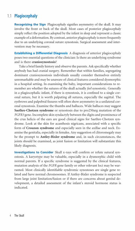
Recognizing the Sign Plagiocephaly signifies asymmetry of the skull. It may
involve the front or back of the skull. Most cases of posterior plagiocephaly
simply reflect the position adopted by the infant in sleep and represent a classic
example of a deformation. By contrast, anterior plagiocephaly ismore frequently
due to an underlying coronal suture synostosis. Surgical assessment and inter-
vention may be necessary.
Establishing a Differential Diagnosis A diagnosis of anterior plagiocephaly
poses two essential questions of the clinician: Is there an underlying syndrome
and is there craniosynostosis?
Take a brief family history and observe the parents. Ask specifically whether
anybody has had cranial surgery. Remember that within families, segregating
dominant craniosynostosis individuals usually consider themselves entirely
unremarkable and may be unaware of clinical features considered dysmorphic
in a hospital setting. In examining the baby, important considerations to re-
member are whether the sutures of the skull actually feel synostotic. Generally
in a plagiocephalic infant, if there is synostosis, it is confined to a single cor-
onal suture, but it is worth palpating all sutures, especially the sagittal. The
eyebrows and palpebral fissures will often show asymmetry in a unilateral cor-
onal synostosis. Examine the thumbs and halluces. Wide halluces may suggest
Saethre-Chotzen syndrome or synostosis due to pro250arg mutation of the
FGFR3 gene. Incomplete skin syndactyly between the digits and prominence of
the crus helicis of the ears are good clinical signs for Saethre-Chotzen syn-
drome. Look at the skin for acanthosis nigricans, associated with a specific
form of Crouzon syndrome and especially seen in the axillae and neck. Ex-
amine the genitalia, especially in females. Any suggestion of clitoromegaly may
be the prompt to Antley-Bixler syndrome and, in such circumstances, the
joints should be examined, as joint fusion or limitation will substantiate this
likely diagnosis.
Investigations to Consider Skull x-rays will confirm or refute sutural syn-
ostosis. A karyotype may be valuable, especially in a dysmorphic child with
normal parents. If a specific syndrome is suggested by the clinical features,
mutation analysis of the FGFR gene family or other relevant loci may be war-
ranted. Most clinically identifiable syndromic synostoses are single gene re-
lated and have normal chromosomes. If Antley-Bixler syndrome is suspected
from large joint limitation/fusion or if there are concerns about genital de-
velopment, a detailed assessment of the infant’s steroid hormone status is
indicated.
1.1
The Skull4

Figure 1.1A and B A right-sided plagiocephaly, caused by isolated coronal suture fusion, isshown. There is orbital asymmetry, the right eyebrow is distorted, and there is flattening ofthe right temporoparietal region. The preoperative photograph from above shows thefrontal asymmetry more clearly.
Plagiocephaly 5

Metopic Ridge
Recognizing the Sign The metopic suture is in the vertical plane in the
midline of the forehead. Prominence of the metopic ridge is a normal, non-
pathologic feature in more than 10% of children. However, it is also a non-
specific feature seen in some dysmorphic disorders and, therefore, is not to be
dismissed. Viewed from above, the anterior skull takes on a triangular ap-
pearance (trigonocephaly). From the front, the eyes may appear to be close
together. An alternative descriptive term sometimes used is ‘‘keel-shaped
skull.’’ Most cases are not related to synostosis.
Establishing a Differential Diagnosis Diagnostic considerations associated
with this sign and that merit directed history taking and examination are rel-
atively limited. About 5% of cases of trigonocephaly with synostosis will have
a positive family history of craniosynostosis, usually trigonocephalic. Some
20% of nonfamilial cases are associated with other malformations, often non-
specific. Particular attention should be paid to a history of sodium valproate
exposure in pregnancy, metopic ridging being a cardinal feature of the re-
sultant malformation pattern. Poor growth and coarse features, often with lip
fullness, should lead to evaluation for I-cell disease (mucolipidosis type II),
particularly if gingival thickening or hepatosplenomegaly is observed. Facial
hemangioma, particularly over the glabellar region, is an excellent clue to the
diagnosis of Oberklaid-Danks syndrome. Other diagnostically valuable clin-
ical signs in this condition are hypertelorism, cleft or deeply ridged palate, and
abnormal wrist position, often with camptodactyly. Iris coloboma may signify
an underlying diagnosis of Baraitser-Winter syndrome.
Investigations to Consider Radiologic examination may be necessary to
exclude metopic suture synostosis. Basic karyotype is valuable, many chromo-
somal abnormalities being associated with trigonocephaly. The most common
of these is Jacobsen syndrome, chromosome 11q deletion. Chromosome 9p
deletion is also over-represented in trigonocephalic presentations, and it is
worth drawing the particular attention of the cytogenetic laboratory to chro-
mosomes 9 and 11 in the trigonocephalic presentation. As with any chromo-
somal deletion, a formal examination for othermalformations, such as cardiac,
should be undertaken. Vacuolated lymphocytes or cytoplasmic inclusions on
fibroblast microscopy will be consistent with I-cell disease. Notwithstanding
their association with craniosynostosis, FGFR gene mutations are very un-
common in trigonocephaly and rarely warranted. Magnetic resonance imag-
ing (MRI) scan for frontal pachygyria or other evidence of neuronal migration
defect will support a clinical suspicion of Baraitser-Winter syndrome.
The Skull6
1.2

Figure 1.2A Trigonocephaly as a result ofmetopic sutural synostosis is demonstrated.
Figure 1.2B Terminal deletion of chromosome 11q is shown from a trigo-nocephalic, developmentally delayed child.
Metopic Ridge 7

Scalp Defects
Recognizing the Sign The term is self-explanatory, the real issue being to
determine whether or not it has a wider diagnostic significance. Note that
some defects are not confined to the scalp and may involve skull vault bony
deficiencies, and severe bleeding events have been reported from the site of
the defect. Even in adult life, the healed scalp defect is usually hairless. Most
scalp defects are benign and do not signify intracranial pathology or an as-
sociated syndromic diagnosis.
Establishing a Differential Diagnosis Benign isolated scalp defects may be
familial, so take a family history and examine the affected child’s parents.
Specific inquiry of antenatal events may reveal maternal ingestion of aspi-
rin. The clinical examination should direct specific attention to the skin for
possible areas of aplasia cutis, which will suggest the condition of aplasia cutis
congenita. Examine the hands and feet for signs of transverse limb defects,
often subtle, which may signify Adams-Oliver syndrome. In that event, the
heart merits careful evaluation, there being as association with congenital
heart disease. Do not dismiss ring-like skin folds of a digit or limb, resembling
amniotic bands, as these are well reported in Adams-Oliver syndrome. Assess
the nipples for absence or hypoplasia and observe the external ears for thick-
ening, overfolding of helices and absence of the tragus, any of which signs should
prompt a diagnosis of Finlay-Marks syndrome, another autosomal dominant
condition. Partial syndactyly of the fingers or toes would also be consistent with
this diagnosis. The presence of epibulbar dermoids is diagnostically significant,
prompting the closer examination of the skin for pigmentary abnormalities.
Some children with epibulbar dermoids and scalp defects have developed giant
cell granulomas. The pattern of hair growth, especially in the presence of a fron-
tal upsweep, is a clue to Johanson-Blizzard syndrome. A history of failure to
thrive and the observation of a small nose, specifically with hypoplastic alae nasi,
would further support this autosomal recessive diagnosis as the reason for the
scalp defect.
Investigations to Consider Do a cardiac assessment if the signs suggest
Adams-Oliver syndrome. Karyotype is indicated in sporadic cases, and chro-
mosomal abnormalities of trisomy 13 and deletion 4p are especially common.
Audiologic assessment for deafness is indicated in Johanson-Blizzard cases, as
are thyroid function measures and assessment for pancreatic insufficiency.
Longitudinal clinical follow-up of cases with epibulbar dermoids is needed lest
this be a cancer predisposition condition, evidence for which is currently sug-
gestive but inconclusive.
The Skull8
1.3

Figure 1.3A Healing scalp defect with surround-ing hairless area is shown in a 3-year-old girlwith aplasia cutis congenita.
Figure 1.3B Localized region of aplasia cutis congenita in the mother of thechild shown in Figure 1.3A.
Scalp Defects 9

Macrocephaly
Recognizing the Sign The sign itself is not difficult; rather, it’s a question of
definition. Its identification prompts concerns with respect to likely signifi-
cance for brain development. History, examination, and investigation com-
bine to assess this possible impact.
Establishing a Differential Diagnosis History and parental examination may
offer evidence for a familial characteristic. Examination should assess body pro-
portions, lest there is an underlying skeletal dysplasia, macrocephaly being
a common finding in hypochondroplasia and many other similar disorders.
Noteworthy frontal bossing and large chin are clues to Sotos syndrome; patients
with this condition often have rather ruddy cheeks, in addition to developmental
delay. Limb or trunk asymmetry may suggest Proteus or Klippel-Trenaunay-
Weber syndromes. Polydactyly is important, possibly suggesting Greig syn-
drome, especially if there is parental macrocephaly. A parental history of sur-
gical resection of polydactyly may be invaluable in securing this diagnosis.
Although not invariable, the polydactyly of Greig syndrome is most commonly
postaxial in the hands and preaxial in the feet. The skin may offer etiologic
clues—facial hemangioma, especially of the upper lip, suggestingmacrocephaly-
cutismarmorata telangiectasia congenita syndrome, oftenwith associated limb
asymmetry and/or polydactyly. Verrucous lesions and/or lipomata raise con-
cernswith respect to possible Proteus syndrome.The cutaneous signs ofCowden
spectrum and Proteus syndromes overlap. Examine the glans penis for macules,
seen in the Bannayan-Zonana syndrome form of the Cowden syndrome spec-
trum. Similarly,mucocutaneous papules, especially around themouth and anus,
involving hair follicles are good clues toCowden syndrome. Be aware that these
may not present until the second decade. The presence of cafe-au-lait patches
should prompt a search for other clues to neurofibromatosis type 1, while ac-
cessory nipplesmay signal Simpson-Golabi-Behmel syndrome. Additional evi-
dence for the latter might be gleaned from small nails on the index fingers, a
deep midline groove on the tongue, or a history of diaphragmatic hernia. Look
for hepatosplenomegaly, general coarseness, and gingival thickening as possible
clues to a metabolic cause.
Investigations to Consider Skeletal radiology is indicated if a dysplasia is
thought likely and bone age is indicated if Sotos syndrome is suspected. Organic
acids for glutaric aciduria, mucopolysaccharide screen, and very-long-chain
fatty acids may be indicated by concerns for a biochemical disorder. Basic
karyotype will have a low yield but fragile X DNA evaluation should not be
overlooked in the absence of clinical signs of other etiologies. A Dandy-Walker
malformation on brain scan should prompt consideration of Meckel and
Bardet-Biedl syndromes, while ventriculomegaly and thinning of the corpus
callosum are known in several conditions such as Sotos and Greig syndromes.
The Skull10
1.4

Figure 1.4A This macrocephalicpatient has no specific diagnos-tic clinical features, as is oftenthe case, but the macrocephalyis caused by a mutation of thePTEN gene, indicating the un-derlying diagnosis of the Cowdensyndrome spectrum. The occipito-frontal head circumference(OFC) is 4 cm above the 97thpercentile for age at 4 years.
Figure 1.4B A typical example of Sotos syndrome is shown. Note themacrocephaly, prominent chin, and ruddy cheeks, often observedin Sotos syndrome patients.
Macrocephaly 11

Microcephaly
Recognizing the Sign Microcephaly is another easy sign to determine by
definition, but is so very important prognostically and so diagnostically be-
wildering as to be something of a clinician’s nightmare.
Establishing a Differential Diagnosis Several ‘‘pure’’ microcephaly condi-
tions are autosomal recessive, and the history must address this possibility by
specifically seeking evidence for consanguinity. Likewise, antenatal exposure
to infection or teratogenic agents needs to be eliminated. Assess whether the
microcephaly is genuine or reflective of a general growth disturbance. Orga-
nomegaly, macroglossia, gingival hyperplasia, or unusual smell may suggest a
biochemical disorder as the basic underlying pathology. Specific dysmorphic
signs may pinpoint a known syndrome—bitemporal narrowing and 2/3 toe
syndactyly suggest Smith-Lemli-Opitz syndrome, while more extensive syn-
dactyly of toes and fingers is consistent with Filippi syndrome. Thumb hy-
poplasia prompts thoughts of Fanconi syndrome, and a skin examination for
pigmented areas should follow. Short first metacarpals and synophrys suggest
de Lange syndrome, while telangiectasia may be the clue to Bloom syndrome.
Examine the eyes—blood vessels visible in the lateral canthus should lead to
examination of the eyebrows for sparse areas or interruption, characteristics of
Kabuki syndrome. Deep-set eyes should provoke inquiry about sun sensitivity
and photophobia, often the clues to Cockayne syndrome. Small ears suggest
Meier-Gorlin syndrome, confirmed by absent patellae, while large ears should
prompt evaluation of calcium, often low in Richardson-Kirk syndrome. Inver-
sion of the nipples and unusual fat distribution are clues to the carbohydrate-
deficient glycoprotein (CDG) disorders.
Investigations to Consider Investigation will be led by the conclusions of
history and examination. A basic karyotype is valuable, nowadays often sup-
plemented by specific fluorescence in situ hybridization (FISH) analysis or
DNA mutation search, depending on the clinical signs. Premature chromo-
some condensation is an important observation, being specific to pure auto-
somal recessive microcephaly type 1 associated with mutation at the MCPH1
locus on chromosome 8. Sister chromatid exchange is indicated in the pres-
ence of telangiectasis, while diepoxybutane challenge is warranted if Fanconi
syndrome is considered likely. Basic blood parameters may show pancyto-
penia in the latter. Disordered blood indices are also suggestive of CDG dis-
orders, for which conditions transferrin isoelectric focusing is the investiga-
tion of choice. Increased levels of 7-dehydrocholesterol are characteristic of
Smith-Lemli-Opitz syndrome. Neuroradiology may give a descriptive diag-
nosis of a brain malformation disorder, many of which are due to single gene
mutation, and the relevant search at the specific locus may be repaid with a
confirmatory diagnosis.
The Skull12
1.5

Figure 1.5A and B The clinical clue to the underlyingdiagnosis of Richardson-Kirk syndrome, an autosomalrecessive disorder, in this microcephalic child lies in theobservation of low-set, prominent ears. Note also therather deep-set eyes. Serum calcium was low in theneonatal period.
Microcephaly 13

Frontal Hairline Variants—High Hairline and Frontal Upsweep
Recognizing the Sign One does not measure the position of the frontal
hairline, but rather forms an impression of its relative position in terms of
other landmarks, such as the orbit. A wide degree of interindividual variation
is normal and, even if noticeably high, there is not a consistent relationship
with pathologic syndromes. If the hairline is ‘‘high,’’ the temporoparietal skin
is more visible than usual. In general, the hair lies forward at the frontal
hairline, and a reversal of this, with the hair swept backward, is frontal up-
sweep, also know as cowlick.
Establishing a Differential Diagnosis High hairline may be familial, espe-
cially in tricho-rhino-phalangeal (TRP) syndrome, which is inherited in an
autosomal dominant manner. Clinical signs of particular diagnostic value
to assess in the parents and child if this diagnosis is being considered are a
bulbous nose and wide middle phalanges of the fingers, which may also show
ulnar deviation. Syndromes involving craniosynostosis can distort the frontal
hairline, so be aware of skull shape and familial appearances in assessing fron-
tal hairline in a baby. Persisting temporal absence of hair in an older child
should prompt thoughts of Pallister-Killian syndrome, especially if devel-
opmental delay is likely. In the older child with short stature and apparent
failure to thrive, a high hairline may be the clue toMulibrey nanism, in which
case relative macrocephaly is common.
Frontal upsweep is unmistakable, being a normal variant in most instances.
However, it is a characteristic finding in Johanson-Blizzard syndrome, in
which condition the very pinched nose with hypoplastic alae nasi will secure
the diagnosis. There is often an associated scalp defect and a range of cardiac
malformations described make cardiac consultation advisable if this diagnosis
is being considered. In the presence of severe developmental delay in a male
child with noteworthy frontal upsweep, consider the likelihood of X-linked
a-thalassemia and mental retardation (ATRX). The family history may offer
further encouragement to pursue this possible diagnosis. Upsweep is also seen
in Rubinstein-Taybi syndrome (R-TS), most such cases showing the char-
acteristic wide, deviated thumbs. FG syndrome patients whose main problems
are constipation and developmental delay may often show a frontal upsweep.
Investigations to Consider Karyotype for chromosome 8q deletion is valu-
able in TRP syndrome, while skin chromosome analysis may be necessary to
unmask the tetrasomy 12p in Pallister-Killian syndrome, chromosomes usu-
ally being normal in lymphocytes. Retinal pigment dots and fibrous dysplasia
of the long bones, particularly the fibula, would support Mulibrey nanism.
Diagnostic concerns for ATRX should prompt a search for HbH inclusion
bodies using freshly constituted brilliant cresyl blue stain. Mutation analysis is
available for ATRX and R-T syndromes.
The Skull14
1.6

Figure 1.6A High hairline isdemonstrated in a typical caseof tricho-rhino-phalangealsyndrome. Observe howsparse the hair is frontally.
Figure 1.6B Frontal up-sweep, in this instance ina patient with Johanson-Blizzard syndrome.
Figure 1.6 C A typicalcowlick of the anteriorhairline is shown in thispatient with X-linkeda-thalassemia mentalretardation syndrome.
Frontal Hairline Variants—High Hairline and Frontal Upsweep 15

Frontal Hairline Variants—Low Hairline and Widow’s Peak
Recognizing the Sign In fetal life, the extension of scalp hair growth onto the
face is suppressed by circular regions around the eyes and ears, thus forming
the normal scalp anterior margin. Eyelashes and eyebrows are exempt from
this regulation. Widow’s peak is the term used to describe a midline prom-
inence of the frontal hairline, while a low frontal hairline refers to the down-
ward extension of hair onto the temple toward the lateral eyebrow.
Establishing a Differential Diagnosis Widow’s peak is a normal observation
in most instances, often the consequence of mild hypertelorism, due to lateral
shift of the hair growth suppression zones around the orbit. Consequently,
a slight tongue of hair extends forward in the midline. If present, there are
specific features to now look out for—a nasal tip groove suggests a diagnosis
of frontonasal dysplasia, while the presence of craniosynostosis, often uni-
lateral coronal fusion, will signify a diagnosis of craniofrontonasal dysplasia
(CFND). Longitudinal ridges along the nails, which often split in this plane,
are characteristic findings in CFND and diagnostically valuable is demarcating
this condition from other disorders with widow’s peak. Pigmentary irregu-
larities of the iris or skin may be the clues toWaardenburg syndrome, usually
type I, in which associated widow’s peak is often observed. A family history
of early hair greying, dying of specific areas of grey hair, and deafness will
support this diagnosis. A shawl configuration to the scrotum in a boy with
widow’s peak could well be the clue to a diagnosis of Aarskog syndrome, es-
pecially if he is of short stature.
A low frontal hairline should draw the clinician’s attention to the eyes, as
it is an almost invariable presence in Fraser syndrome. Interruption to the
margin of the alae nasi in the form of a notch, dysplastic ears, and skin syn-
dactyly of the fingers would further support this autosomal recessive diag-
nostic conclusion, as would genital anomalies. Hairline distortion may reflect
underlying craniosynostosis, so attention to head shape in the individual and
family is warranted. Check specifically for an antenatal history of phenytoin
exposure, the temporal extension of hair being documented. A cheekward
extension of a tongue-like projection of hair is common in Treacher Collins
syndrome, so it is worth looking for pinna abnormalities, lower eyelid co-
lobomata, and maxillary hypoplasia.
Investigations to Consider Skull radiology is indicated if craniosynostosis
is suspected. In Fraser syndrome, consider ophthalmologic evaluation, renal
ultrasound, brain neuroimaging, and, possibly, DNA studies. Computed to-
mography (CT) scan of the petrous temporal bone in Treacher Collins should
be obtained to establish the degree of ossicular malformation.
The Skull16
1.7

Figure 1.7A Widow’s peak is dem-onstrated in a patient with cranio-frontonasal dysplasia. Note also thehypertelorism, narrow palpebralfissures, and the bifid nasal tip.
Figure 1.7B Low frontal hairline isdemonstrated. Note also the sig-nificant infraorbital skin groove,betraying the underlying diagnosisof Schinzel-Gideon syndrome inthis patient.
Frontal Hairline Variants—Low Hairline and Widow’s Peak 17

Low Posterior Hairline
Recognizing the Sign Scalp hair extending downward onto the neck is
thought to reflect prenatal nuchal edema. Unsurprisingly then, posterior hair-
line anomalies often accompany excess skin, referred to as neck webbing, but
may equally represent the clinical clue to structural malformations of the
cervical vertebrae. The term ‘‘trident hairline’’ is sometimes used, meaning
three-pronged appearance, and simply reflects a particular pattern of low
posterior hairline, wherein an M-shaped margin to the hairline is discerned.
Establishing a Differential Diagnosis Once sure that the posterior hairline is
indeed low, assess the neck for redundant skin. In a neonate, the additional
presence of puffy feet is suggestive of lymphoedema and a likely diagnosis of
Turner syndrome. Examine the heart and check for brachiofemoral delay,
specifically in view of the common association between Turner syndrome and
coarctation of the aorta. Significant clinical overlap exists with Noonan syn-
drome, in which condition a history of a parent having had a congenital heart
lesion may be elicited or other clinically supportive evidence, such as ptosis,
adduced. Parental auscultation for unsuspected cardiac murmur is often valu-
able. Beware of bleeding defects in Noonan cases; a history of prolonged gum
bleeding following tooth extraction in the parent of a child with low posterior
hairline can alert the astute clinician to this diagnosis. The emergence of cafe-
au-lait skin patches in the preschool child is significant and probably indic-
ative of Noonan-NF syndrome, a variant of neurofibromatosis type 1, which
therefore puts the child at risk for optic glioma, renal artery stenosis, pheo-
chromocytoma, and other complications attendant upon NF1. Segmentation
defects of the cervical spine should be considered in patients with low posterior
hairline. Confirmation of cervical vertebral fusion, Klippel-Feil syndrome,
should be followed by investigation of the renal tract, agenesis, hypoplasia, and
ureteric duplication being common associations. Vaginal atresia and/or absent
uterus are frequent in females with cervical vertebral fusions, while bilateral ab-
sence of the vas deferens, cryptorchidism, and similar renal anomalies represent
the typical male profile.
Investigations to Consider Formal cardiac evaluation is indicated if Tur-
ner syndrome or Noonan syndrome is considered. This may extend to other
family members in Noonan syndrome. Coagulation assessment is useful in
Noonan syndrome, particularly if minor surgical procedures are being con-
templated outside of a hospital environment. Karyotype will be definitive in
Turner syndrome. Neck x-ray to assess cervical vertebrae is valuable in non-
Turner, non-Noonan cases. Ultrasonic evaluation of the genitourinary tract
structures is important in Klippel-Feil and related syndromes.
The Skull18
1.8

Figure 1.8 Low posterior hairline is associated with verynoteworthy neck webbing in this 3-year-old girl withNoonan syndrome who had first come to attention whenan antenatal scan at 26 weeks’ gestation showed hydropsfetalis.
Low Posterior Hairline 19

Anencephaly
Recognizing the Sign Conventional wisdom represents the primary event in
anencephaly as failure of closure of the anterior neural groove. In conse-
quence, forebrain development is incomplete with calvarial defects and pos-
sible malformations of the face and its structures. The spectrum of resultant
clinical phenotypes represents a sequence of disordered embryologic and fetal
developmental processes arising from this single primary event. It follows that
a natural degree of clinical variability is to be anticipated between cases. Most
cases represent variants of the spina bifida disease spectrum and are not as-
sociated with specific recognizable single gene syndromes or recurrence risks
attendant upon single gene disorders. The purpose of clinical examination is
to identify cases that represent clinical presentation of syndromes, many of
them of autosomal recessive inheritance, whose recognition will have pro-
found consequences for genetic counseling.
Establishing a Differential Diagnosis A range of environmental agents, al-
cohol, valproate, and rubella are all confirmed causes of meningocele, though
less convincingly of anencephaly. This association will not be substantiated
without dutiful antenatal history taking in future cases! Specifically examine the
hands and feet. Small thumbs or absent thumbs lead to evaluation of the radii.
The forearms may be shortened clinically and radiologic radial malformations
identified. Such observations will be consistent with a likely diagnosis of the XK
aprosencephaly syndrome. Check for anal atresia or stenosis, which will help
secure the diagnosis, if present. Likewise, hand and foot malformations attend
acrocallosal syndrome, the ‘‘classical’’ pattern being preaxial polydactyly of the
feet with or without postaxial polydactyly of the hands. This pattern is not
absolute but emphasizes the diagnostic value of well-documented examination.
Meckel-Gruber syndrome more typically presents a posterior encephalocele
but anencephalic presentation is well described. The polydactyly is typically
postaxial, but the most consistent clinical finding, and therefore that of greatest
diagnostic value, is renal and hepatic cysts. Documentation of cysts will afford
distinction from hydrolethalus syndrome in which preaxial and/or postaxial
polydactyly with encephalocele or anencephaly are typical features, but often
this diagnosis is signaled by the antenatal history of polyhydramnios.
Investigations to Consider Radiology of limb malformations, if present, and
autopsy examination of infants with clinical features that prompt concerns
about a syndromic presentation of anencephaly are the most important in-
vestigative considerations.
The Skull20
1.9
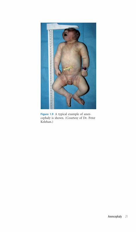
Figure 1.9 A typical example of anen-cephaly is shown. (Courtesy of Dr. PeterKelehan.)
Anencephaly 21

Chapter 2 The Face

Hypertelorism
Recognizing the Sign This term refers to widely spaced eyes and, by defi-
nition, the interpupillary distance is increased. A clinical impression of hy-
pertelorism can be created by epicanthic folds, by a depressed nasal bridge, or
by telecanthus, in which the inner canthus is displaced laterally, the inner
canthus being the meeting point of the upper and lower eyelids on the medial
side of the eye. A useful clinical guide is to assess whether an imaginary
vertical line through the lacrimal punctum actually overlaps the iris. Should
this be the case, then there is telecanthus. Thus, one’s first clinical impression
of hypertelorism needs to be more carefully assessed for possible confound-
ing factors. Once satisfied that the patient does indeed manifest hypertelor-
ism, the purpose of further examination is to evaluate the likely significance
of this confirmed clinical sign.
Establishing a Differential Diagnosis A mild degree of hypertelorism, often
with associated widow’s peak, may be familial and benign. In terms of syn-
dromic associations, the presence of a vertical groove of the nasal tip suggests
the diagnosis of frontonasal dysplasia, while craniosynostosis and a nasal tip
groove are consistent with craniofrontonasal dysplasia. Hoarseness of the
voice should prompt questioning with respect to stridor, a history of neonatal
feeding difficulty, or aspiration. These represent possible pointers to an un-
derlying laryngeal cleft; and cause the clinician to examine for hypospadias—
these being the cardinal features of Opitz G syndrome. Being an X-linked
disorder, asymptomatic mothers may show mild clinical features. In the
context of significant developmental delay in a male and increasing facial
coarseness, best seen on serial photographs, with gathering prominence of the
lips, thoughts of Coffin-Lowry syndrome would be justified. Many early cases
of a-thalassemia mental retardation syndrome of the X-linked type (ATRX)
were confused with Coffin-Lowry syndrome, the clinical features being very
similar. Both are X-linked and may show mild features in carriers. The digits
of Coffin-Lowry syndrome are often tapering and many geneticists point to
the frequent observation of an accessory transverse hypothenar crease (Figure
9.9). In cases of mild developmental delay, especially if associated with short
stature, the hypertelorism may be associated with Aarskog syndrome. Con-
firm clinically by observing the digits for brachydactyly, often with mild skin
syndactyly; the scrotum will generally have a shawl configuration.
Investigations to Consider DNA mutation analysis is indicated for ATRX
and Coffin-Lowry syndromes. HbH inclusion bodies on staining with freshly
constituted brilliant cresyl blue is a good screening measure for ATRX and
identifies 90% of cases. Skeletal survey in Aarskog syndrome often shows mul-
tiple epiphyseal dysplasia of mild degree.
2.1
The Face24

Figure 2.1A Note the hypertelorismin this patient with Aarskog syndrome.It is worth noting the wide philtrum.Contrast with the telecanthus of Figure2.1B, where the inner canthus is later-ally displaced, giving an impression ofwide-spaced eyes.
Figure 2.1B Note the medially flaredeyebrows and synophrys, in addition totelecanthus, in this patient who hasWaardenburg syndrome.
Hypertelorism 25

Hypotelorism
Recognizing the Sign By definition, the interpupillary distance is reduced.
The clinical impression of hypotelorism should lead the experienced clinician
to examine the patient for other clinical signs consonant with this feature and
that may enhance the likelihood of identifying specific syndromes known to
be associated with hypotelorism.
Establishing a Differential Diagnosis Metopic suture fusion, resulting in
trigonocephaly, causes hypotelorism (1.2). Likewise, hypotelorism may be
seen in nonspecific clinical situations such as chromosomal trisomies and
deletion syndromes, which, by their nature, are of widely varying phenotype.
Cyclopia, a single centrally placed eye, represents an extreme form of hypo-
telorism, and it is not surprising that varying degrees of hypotelorism are seen
in holoprosencephaly, in which condition incomplete development of the
frontal lobes of the brain is often associated with developmental failure of the
olfactory tracts. Many of these children do not survive the neonatal period,
but careful clinical examination of other family members may show clinical
clues, in particular a single central incisor or anosmia. In association with 2/3
syndactyly of the toes, the autosomal recessive condition of Smith-Lemli-
Opitz syndrome is an important differential diagnosis to investigate. Olfactory
tract maldevelopment, resulting in anosmia, is also a feature ofKallmann syn-
drome, mostly an X-linked disorder and with associated clinical findings of
cleft palate or uvula, mirror movements, and micropenis or failure of pubertal
development, which is often the presenting feature. Renal agenesis is also
associated. In the presence of skin syndactyly, particularly of fingers 3/4/5,
think of oculo-dento-digital syndrome and seek a history of premature tooth
decay. It is also worth examining for signs of spasticity, as white matter changes
onmagnetic resonance imaging (MRI) are well described, and asking if others in
the family have needed digital surgery—the condition is autosomal dominant.
In Hallermann-Streiff syndrome, the face is dominated by the narrow, pro-
minent nose, but a high forehead, hypotelorism, and small chin will all support
the likely diagnosis, as will cataracts.
Investigations to Consider Some 40% of holoprosencephaly cases have a
chromosomal abnormality, most commonly trisomy 13. Mutations at six loci
have been demonstrated on DNA analysis. 7-Dehydrocholesterol levels are
indicated in suspected Smith-Lemli-Opitz syndrome, elevation thereof being
diagnostic of this autosomal recessive condition. Xp chromosome deletion by
fluorescence in situ hybridization (FISH) analysis or, if normal, mutation
analysis of KALL1 and FGFR1 genes is indicated for likely cases of Kallmann
syndrome.
The Face26
2.2

Figure 2.2B The hands of the patient in Figure 2.2Aare demonstrated postsurgery for 3/4/5 syndactyly.
Figure 2.2A Note the narrow palpebral fissures inthis boy with oculo-dento-digital syndrome. Theinner canthi are slightly displaced, which amelioratesthe impression of hypotelorism, but the interpupil-lary distance is reduced. Other important features toobserve in this patient are synophrys and the hypo-plastic alae nasi, which gives the nares a somewhatanteverted appearance (see 2.4 and 2.5).
Hypotelorism 27

Abnormal Nasal Bridge
Recognizing the Sign The nasal bridge refers to the bony element of the nose
between the orbits. The evaluation of this structure for dysmorphic signs is
complicated by the range of normal variation in the population, shared fa-
milial traits, and age-related phenomena, specifically the observation that de-
pression of the nasal bridge is the norm in infancy. Unsurprisingly then, a
clinical impression of raised or depressed nasal bridge needs to be substan-
tiated by other evidence if it is to enjoy diagnostic significance.
Establishing a Differential Diagnosis Widening of the nasal bridge is usual
in hypertelorism, for which reason assessment of the nasal tip, the skin,
general facial appearance, and height remain relevant (2.1). Nasal bridge
widening is characteristic of Waardenburg syndrome, for which reason the
clinician should evaluate the irides for heterochromia and eyelashes and hair
for hypopigmented areas, in addition to querying the use of hair coloring
products, a comment which equally applies to parents of an affected child.
Observe for hearing or speech difficulties. A high nasal bridge, particularly
in the context of developmental delay and truncal obesity, is characteristic of
Cohen syndrome (Figure 9.8). The philtrum is usually short and the frontal
teeth prominent. Assess the hands, as the fingers are usually tapering; retinal
examination will often show pigmentary changes. In the newborn, promi-
nence of the nasal bridge is exceptional. A high nasal bridge in an infant with
unusual tone, poor feeding, or tremulousness suggestive of seizure activity
could be the clue toWolf-Hirschhorn syndrome (chromosome 4p deletion).
Extreme flattening of the nasal bridge and midfacial region is seen in chon-
drodysplasia punctata and in fetal warfarin embryopathy. In the absence of
warfarin exposure, clinical findings of limb asymmetry, ichthyosis, or patchy
areas of rough skin with large pores, resembling orange peel, strongly suggest
chondrodysplasia punctata. Patients with Stickler syndrome, an autosomal
dominant condition, usually have depressed nasal bridge as a familial charac-
teristic. Suspect Stickler syndrome as the underlying pathology in infants with
Pierre-Robin sequence of cleft palate and micrognathia; in older children, ex-
cessive joint laxity, conductive deafness, myopia, and retinal detachment are
regularly observed. It is worth observing the size of the joints, as large knees are a
good clinical clue to the diagnosis of Stickler syndrome.
Investigations to Consider Unexplained granulocytopenia is often seen in
Cohen syndrome and is a cheap diagnostic adjunct in clinically suspicious
cases. Most 4p- cases are not seen on routine chromosome preparation and
need specific FISH analysis for 4p16 band to confirm the diagnosis. Neonatal
radiology is important in chondrodysplasia punctata as the epiphyseal stip-
pling can be transient.
The Face28
2.3

Figure 2.3A Note the prominent nasal bridge and glabellar regionin this 9-month-old baby with Wolf-Hirschhorn syndrome. Inthis condition, the nose is sometimes described as having a ‘‘Greekhelmet profile.’’ The feeding tube betrays the continuing feedingdifficulties.
Figure 2.3B Contrast the deeply depressed nasal bridge in thispicture with that in Figure 2.3A. The girl in this photograph showsepicanthic folds, especially noteworthy on the right, due to thenasal bridge depression. The underlying diagnosis in this case ischondrodysplasia punctata.
Abnormal Nasal Bridge 29

Abnormal Alae Nasi and Nasal Tip
Recognizing the Sign The alae nasi comprise the lateral walls of the nostrils,
descending from the nasal tip to merge with the upper lip. In the event of hy-
poplasia, the nose acquires a pinched appearance. If combined with a wide nasal
tip, the shape of the nose is described as cylindrical.
Establishing a Differential Diagnosis Prominence of the nasal tip with nar-
row ala nasi represents an excellent diagnostic clue and the examination must
reflect this. Look at the hairline, which may be sparse temporally in tricho-
rhino-phalangeal (TRP) syndrome, and seek a history of delayed hair growth
in this scalp region. The hands usually show evidence of short metacarpals and
the nails may be hypoplastic. As an autosomal dominant condition, assessment
of other family members is important for supportive diagnostic signs. By
contrast, a prominent nasal configuration against a background of hypocalce-
mia or congenital heart disease should prompt consideration of velo-cardio-
facial syndrome (VCFS). Beware of the extreme variability of history and
clinical findings in this disorder; a low threshold for investigation is advised.
Nasal speech, delayed speech, a poor neonatal feeding history, and prolonged
drooling may represent clues to pharyngeal incompetence, which attends many
of these cases. The fingers are often long and hyperextensible. Absent joint skin
crease of the fingers may be the clue to symphalangism, which, in the context of
a large nasal tip and prominent nose should lead to examination of the wrists
and ankles for carpal or tarsal fusion. An accompanying history of conductive
deafness would seal a diagnosis of WL symphalangism syndrome, an autoso-
mal dominant condition, justifying wider family inquiry and examination.
Hypoplastic alae nasi with frontal upsweep of hair typify Johanson-Blizzard
syndrome (Figure 1.6B), while notching of the alae nasi should prompt ex-
amination for lip pits, skin syndactyly, and knee webbing/pterygia, which are
hallmarks of Van der Woude syndrome. Syndactyly, in the context of hypo-
telorism and hypoplastic alae nasi, signals oculodentodigital (ODD) syndrome
(Figure 2.2).
Investigations to Consider FISH of 22q11 is the gold-standard investiga-
tion for VCFS, though if normal, 22q13 and 10p13 deletions may show clin-
ical overlap. Extended cytogenetic band examination of chromosome 8q24 is
warranted if TRP syndrome is clinically suspected, a deletion often being
demonstrable. Mutation of the NOG gene underlies WL symphalangism but,
like Van der Woude syndrome, the diagnosis is clinically identifiable in most
cases.
The Face30
2.4
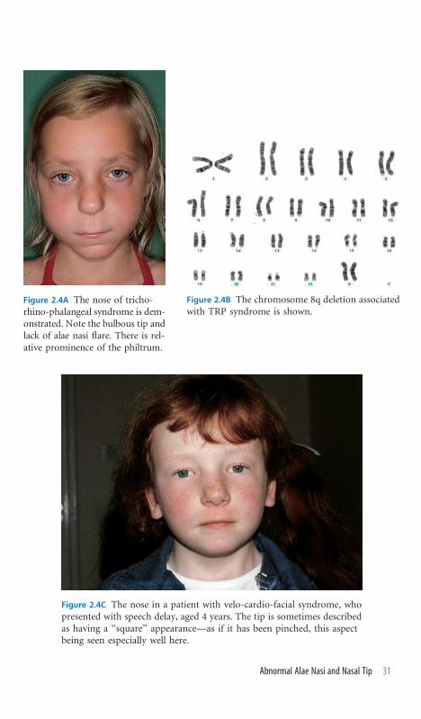
Figure 2.4A The nose of tricho-rhino-phalangeal syndrome is dem-onstrated. Note the bulbous tip andlack of alae nasi flare. There is rel-ative prominence of the philtrum.
Figure 2.4B The chromosome 8q deletion associatedwith TRP syndrome is shown.
Figure 2.4C The nose in a patient with velo-cardio-facial syndrome, whopresented with speech delay, aged 4 years. The tip is sometimes describedas having a ‘‘square’’ appearance—as if it has been pinched, this aspectbeing seen especially well here.
Abnormal Alae Nasi and Nasal Tip 31

Abnormal Nasal Columella and Nares
Recognizing the Sign The columella is the visible outer plane of the nasal
septum, dividing the nostrils, whose aperture is the nares. The columella is
usually situated at approximately the same level as the alae nasi and the nares
not visualized. Should the length of the nose be reduced, then the columella
slants from the foreshortened nasal tip and the nares become visible to frontal
view, or ‘‘anteverted.’’
Establishing a Differential Diagnosis Anteverted nares do not necessarily
signify an underlying syndromic diagnosis, but are particularly associated
with a few well-established syndromes. De Lange syndrome will be recog-
nizable by the characteristic synophrys of the eyebrows, low birth weight,
short limbs, and, characteristically, short first metacarpal. Anteverted nares in
Williams syndrome are often seen in the context of a neonatal cardiac mur-
mur of supravalvular aortic stenosis, perhaps with documented hypercalce-
mia and a stellate pattern to the iris. Somewhat coarse features and hoarseness
of the voice are typical of older Williams syndrome cases, but the anteversion
of the nares remains a characteristic. Anteversion in a hyperteloric child and
short stature are hallmark features of Robinow syndrome. Genital exami-
nation will often show micropenis in males, females being less noteworthy. In
the newborn period gum hypertrophy can be a valuable sign to the diagnosis
of Robinow syndrome. A different configuration, with extension of the col-
umella below the alae nasi, is characteristic of Rubinstein-Taybi syndrome.
Microcephaly and long eyelashes are associated facial characteristics but the
clinical clue par excellence is broad thumbs and halluces, often deviated
(Figure 9.12A). In Floating-Harbor syndrome, the columella is often simi-
larly prominent, but the clinical background in this disorder is of short stature
and mild developmental delay. Characteristically, the eyes are deep set. Prom-
inence of the columella, with a full, rounded nasal tip, represents the quin-
tessential description of the face in Mowat-Wilson syndrome, in which con-
dition, severe developmental delay, often with microcephaly, and constipation
define the clinical presentation. Other useful clinical signs include uplifted
earlobes and an unusual vermillion border to the upper lip, which is full
centrally but very thin at the outer aspects of the lip.
Investigations to Consider FISH of the ELN gene of chromosome 7q is in-
dicated for Williams syndrome. Bone age delay is a sine qua non of Floating-
Harbor, in which antigliadin antibodies and abnormal thyroid function
should be sought. FISH is unrewarding in most cases of Rubinstein-Taybi
syndrome, though mutation analysis is available in clinically doubtful in-
stances. 2q22-23 deletions on karyotype typify a minority of Mowat-Wilson
cases, with diagnosis relying on ZFHX1B mutation in all other instances.
The Face32
2.5

Figure 2.5A The anteverted naresof a patient with Robinow syn-drome are demonstrated.
Figure 2.5B Note the rounded nasal tipand the columella extending below the levelof the ala nasi in this patient who hasRubinstein-Taybi syndrome.
Figure 2.5C The prominent nasal columella in this patientin conjunction with the rounded nasal tip and the vermil-lion border of the upper lip demonstrating extreme thinningtoward the margins of the mouth betray the diagnosisof Mowat-Wilson syndrome.
Abnormal Nasal Columella and Nares 33

Abnormal Nasal Septum
Recognizing the Sign Shortening of the columella causes a flat nose but,
provided the septum is straight, the nares are usually symmetric in shape. A
half-moon configuration to the nares is commonly observed if the columella
is short. A short columella is often associated with flattening of the nasal
bridge, maxillary hypoplasia frequently accompanying and conferring a gen-
erally flat midfacial appearance. Deviation of the nasal septum results in
asymmetric nares.
Establishing a Differential Diagnosis Flattening of the midface in a neo-
nate should lead to inquiry as to intrapartum history, specifically relating to
environmental and teratogenic agents. Fetal warfarin, fetal alcohol, and fetal
rubella syndromes may all confer this presentation. A flat midface is also re-
corded in a few instances of infants born to mothers suffering systemic lupus
erythematosus (SLE) during pregnancy, a further important aspect for spe-
cific historical inquiry. Nasal septum deviation and flattening of the nose is
common in oligohydramnios, as a consequence of fetal constriction, of which
other features such as arthrogryposis or cranial asymmetry should be sought.
Older children with a flat nose and short septum are often termed as having
Binder syndrome. The anterior nasal spine is missing and this can be palpated
clinically by putting the finger inside the upper lip and pressing in the midline,
at the base of the frenulum. Try it on yourself first, but the absence of this
osseous landmark results in a soft, cartilaginous, mobile sensation. If absent,
or in doubt, examine the patient’s fingers. The nails are often small, reflecting
an underlying hypoplasia of the terminal digits. Stature is often reduced, with
symmetric limb shortening. As this can be familial, a family history can assist
in demarcating from overlapping pathologies. A particular condition to be
mindful of is acrodysostosis, an autosomal dominant form of short stature,
in which relative macrocephaly and short fingers can lead to a clinical mis-
diagnosis of achondroplasia. The face in the older patient with acrodysostosis
is sometimes described as ‘‘pugilistic.’’ A short columella in a child with
congenital heart disease or neonatal feeding difficulties may be indicative of
Kabuki syndrome (Figure 3.14). Examination of the palpebral fissures for
lower eyelid ectropion, of the lip for pits, and of the eyebrow for exaggerated
arching or interruption would all lend diagnostic support.
Investigations to Consider Early x-rays often show epiphyseal stippling and
suggest mutation of the X-linked ARSE gene in males with Binder syndrome.
Cone-shaped epiphyses are characteristic of acrodysostosis.
The Face34
2.6

Figure 2.6 A case of Binder syndrome is shown. Notethe flat midface and shallow septum of the nose.Viewed from beneath, the nares have a characteristichalf-moon shape.
Abnormal Nasal Septum 35

Abnormal Nasal Appendages
Recognizing the Sign These signs are not the subject of clinical doubt. The
nose itself is normally formed, the cause of clinical disquiet resting upon
attendant features that are recognized as abnormal. Suchmay comprise a swell-
ing at the base of the nose, a sinus, a polyp, a hair-bearing pit, or the emer-
gence of papillomata around the nares.
Establishing a Differential Diagnosis Particularly if located in the midline, a
sinus, whether hair bearing or blind ending, needs careful evaluation because
of the possibility of direct communication to the central nervous system
(CNS). In a neonate, observe the baby crying and specifically assess whether
there is any swelling of the glabellar region, which may signify an anterior
encephalocele. In addition, there are specific syndromic considerations. Care-
ful evaluation of the upper lip and intraoral structures for Pai syndrome is
indicated by the presence of a sinus or polyp. The upper lip may show minor
midline clefting or upper frenulum reduplication. A pedunculated intraoral
mass may be identified. Evidence of cleft palate or uvula should be sought. By
contrast, the development of discrete lumpy masses around the nares, sig-
nifying nasal papillomata, typically being noted around 5 or 6 years of age, is
strongly indicative of Costello syndrome. There will be a background history
of severe failure to thrive following normal or slightly increased birth weight,
merging into later concerns as to an underlying storage disease due to the de-
velopmental delay and coarse appearance of the patient. There is usually
excess palmar skin with palmar and plantar hyperkeratosis, and the position
of the fingers can show significant ulnar drift. Examine specifically for sco-
liosis. In the older patient, papules on the nose should provoke inquiry as to
family history of breast or thyroid cancer. The concern is that the papules
represent clues to a diagnosis of Cowden syndrome, an autosomal dominant
cancer predisposition syndrome of broad clinical embrace. The examination
should assess for macrocephaly and seek familial evidence for this feature.
Examine for papules around the eyes, mouth, and upper lip, and intraorally.
Likewise, palmoplantar keratosis supports this likely diagnosis, as does a ru-
gosity of the tongue, sometimes described as ‘‘scrotal.’’
Investigations to Consider Nasal sinuses come with a low threshold for
cranial neuroimaging to exclude associated encephalocele. Lipomata, some-
times of the corpus callosum, are frequent in Pai syndrome, but rarely impact
adversely on CNS development. Rhabdomyosarcomata have been described
in older Costello patients, and appropriate screening measures are recom-
mended. The Cowden syndrome disease spectrum is best confirmed by PTen
mutation analysis.
The Face36
2.7

Figure 2.7A The nasal polyp of a case of Pai syndrome is dem-onstrated. Note the midline upper lip cleft and the sinus at the rootof the nose.
Figure 2.7B This photograph demonstrates the nasalpapillomata of Costello syndrome, which only developedwhen the patient was 6 years old.
Abnormal Nasal Appendages 37

Synophrys
Recognizing the Sign Synophrys is the term used to describe eyebrows that
meet in the midline over the nasal root. It is a normal finding in some indi-
viduals, but is seen more frequently in association with hypotelorism (see 2.2
and Figure 2.2A).
Establishing a Differential Diagnosis In the newborn period, noteworthy
synophrys, particularly in a low-birth-weight infant, is an almost invariable
finding in de Lange syndrome. The nasal bridge is usually depressed, the nares
anteverted, the philtrum long, and the limbs short, with oligodactyly (absent
fingers) in upward of a quarter of all cases. Evenwith no digital deficit, the hands
have a characteristic appearance, the thumb being proximally inserted due to
an underlying short first metacarpal (Figure 9.13B). In an older child with syn-
ophrys, a history of early normal milestones and then gradual developmental
concern, often with unexplained diarrhea, should lead the examiner to consider
Sanfilippo syndrome. Unlike other mucopolysaccharidoses, hepatosplenome-
galy and other features of storage disorders including coarse facies, corneal
clouding, and gingival thickening are often absent. An easily overlooked diag-
nosis, Sanfilippo syndrome is often flagged by parental accounts of behavioral
concerns and worsening aggression but requires careful, targeted history tak-
ing. Synophrys, perhaps with associated flaring of the medial region of the
eyebrows, is seen in some cases of Waardenburg syndrome, for which reason
examination for pigmentary disturbance and telecanthus as well as clinical as-
sessment of hearing are advised (Figure 2.1B). Frontal upsweep of the hair, male
pseudohermaphroditism, and severe developmental delay in a patient with syn-
ophrys might cause ATRX syndrome to be considered as the underlying di-
agnosis. Likewise, a flat midface appearance or fleeting impression of a Down
syndrome appearance are very common in the newly emerging chromosome
9q34 microdeletion syndrome. Besides synophrys, brachycephaly, macroglos-
sia, and truncal hypotonia are other commonly observed clinical findings to be
aware of in this diagnosis, as is the development of truncal obesity.
Investigations to Consider Classic de Lange syndrome is a clinical diagnosis.
Cases of ‘‘mild’’ de Lange and atypical cases may need to have diagnosis con-
firmed molecularly by DNA analysis of the NIPBL gene on chromosome 5p.
Bone age may be advanced in Sanfilippo syndrome, though features of dysos-
tosis are not consistently present. Urinary mucopolysaccharides may be within
normal range. Oligosaccharides may be increased, but enzymatic analysis in
white cells or fibroblasts is usually reliable. 9q34 microdeletion syndrome re-
quires comparative genome hybridization techniques in specialized laboratories
for diagnosis. Waardenburg syndrome should be assessed audiologically. If
molecular confirmation is required, PAX3 analysis generally demonstrates mu-
tation provided telecanthus is present.
The Face38
2.8

Figure 2.8 Synophrys is clearly seen in this patient, who hasa diagnosis of Cohen syndrome, a condition not usuallyassociated with synophrys. Note the prominent nasalbridge, narrow palpebral fissures, and short philtrum,which, in the context of her developmental delay, signalthe diagnosis.
Synophrys 39
Midfacial Hypoplasia
Recognizing the Sign There is much scope for confusion here, the terms
‘‘malar hypoplasia,’’ referring to the zygomatic region immediately below the
eye, and ‘‘maxillary hypoplasia,’’ referring to the immediately inferior region
of the face, sometimes being used interchangeably. Essentially, this feature is
present if the cheek bones are flat.
Establishing a Differential Diagnosis Check whether this is familial by
observing facial characteristics of the parents. Midface hypoplasia is the hall-
mark of Treacher Collins syndrome, often with a tongue-shaped projection
of scalp hair onto the cheek. Examine the ears, the pinnae of which are usually
malformed, often with accompanying tags of skin or fistulae, and look for
lower eyelid coloboma. As should always be undertaken in midface hypo-
plasia, an inspection of the palate will show clefting in about a third of
Treacher Collins cases. Beware of the close resemblance in facial and even ear
features to Nager syndrome, which is distinguished from Treacher Collins by
the presence of radial hand malformations, including thumb hypoplasia.
Concerns that early development is not proceeding normally, often associated
with abnormalities of muscle tone or feeding problems in a baby whose ap-
pearance is not frankly dysmorphic but in whom midface hypoplasia is def-
initely present, may suggest the autosomal recessive disorder of Schinzel-
Giedion syndrome. The forehead is tall and usually there is a noteworthy
infraorbital skin groove (Figure 1.7B). Choanal stenosis occurs frequently and
should be sought. An infraorbital skin crease is also an invaluable clinical sign
in fetal valproate syndrome, running downward and laterally from the inner
canthus region. Seek a confirmatory history of valproate exposure and, if
confirmed, look further for metopic ridging and long philtrum. Preaxial poly-
dactyly of the foot or thenar eminence hypoplasia are solid supportive signs,
frequently not present but of strong diagnostic value if observed. Midface
hypoplasia, with a smooth philtrum and thin upper lip, characterize fetal
alcohol syndrome, though the history of fetal exposure can be harder to elicit.
Look for aberrant skin creases of the hands, skin syndactyly of the fingers,
short palpebral fissures, and poor growth, otherwise unexplained.
Investigations to Consider Deafness, usually conductive, is the norm in
Treacher Collins cases. Mutation analysis is reserved for clinically subtle or
doubtful cases, but demonstration of hypoplastic zygomatic processes on skull
x-ray can obviate the need for DNA analysis. Computed tomography (CT) scan
of the petrous temporal bone usually demonstrates ossicular malformations,
consistent with a conductive deafness. Renal ultrasound demonstrating cysts
or hydronephrosis would strongly support the likely diagnosis in Schinzel-
Giedion syndrome. Demonstration of an occipital synchondrosis on skull x-ray
is confirmatory.
The Face40
2.9

Figure 2.9A In this image of a patient withTreacher Collins syndrome, the zygomaticregion immediately below the palpebralfissure is hypoplastic bilaterally.
Figure 2.9B The characteristiclower eyelid coloboma ofTreacher Collins syndrome isdemonstrated in the samepatient.
Midfacial Hypoplasia 41
Micrognathia
Recognizing the Sign Best viewed in profile, this clinical sign is caused by a
small mandible that has not grown out. When opening the lips, there is
usually a malalignment of the alveolar margins in neonates and of the teeth in
older patients. The chin is small or, in the adult patient, often grows out but
may have a receding profile. Pierre-Robin sequence refers to a cleft palate
consequent upon the posterior displacement of the tongue within the devel-
oping oral cavity occasioned by a primary failure of the mandible to advance.
Establishing a Differential Diagnosis In any case of Pierre-Robin sequence,
Stickler syndrome should be a diagnostic concern, and evidence from the
family history of high myopia, retinal detachment, early-onset arthropathy, or
deafness would certainly strongly advance this diagnostic likelihood. Clini-
cally, observe if the nasal bridge is depressed and look for excess joint laxity.
Ophthalmic examination may show congenital cataract. Experienced oph-
thalmologists will specifically seek vitreous abnormalities. In other cases of
micrognathia with cleft palate, examine the fingers. A misshapen index finger
in a micrognathic patient, usually with ulnar deviation of that digit, is diag-
nostic of Catel-Manzke syndrome. Intelligence is generally normal in these
cases, but there is a known association with cardiac defects, for which reason
formal cardiac evaluation is indicated. Micrognathia as the striking facial
feature of a child who has unexplained growth deficiency and a history of
low birth weight may signify Meier-Gorlin syndrome (Figure 11.8). Confir-
matory clinical support should be sought by palpating the patellae, which are
absent, and by observing the ears, which are small. In contrast, children with
cerebro-costo-mandibular syndrome will generally present with a Pierre-
Robin picture at birth, often with associated respiratory problems, and emer-
ging developmental concerns will cloud the clinical picture. Micrognathia is
a nonspecific finding in many different forms of chromosomal aneuploidy.
The author has personal experience of extreme micrognathia, requiring pro-
longed tracheotomy, as the sole presenting finding in patients who later proved
to have chromosomal mosaicism. Accordingly, careful skin examination for
areas of pigmentary differences and areas of localized keratosis pilaris and
evaluation of skin under Wood’s lamp is advised.
Investigations to Consider X-ray of the hands in Catel-Manzke syndrome
will show an accessory metacarpal bone at the base of the index finger, while
chest x-ray is the key to cerebrocostomandibular syndrome diagnosis, show-
ing the discontinuity in rib outline, ‘‘rib gaps,’’ diagnostic of the condition.
Karyotype and even skin karyotype are indicated in cases without a recognized
syndrome diagnosis in an attempt to formally exclude mosaicism.
The Face42
2.10

Figure 2.10A Micrognathia, requiring trache-ostomy for airway maintenance, is shown in apatient with slightly coarse facial features andpoor temporal hair growth at age 2 years.Wood’s lamp examination showed streakypigmentary disturbance consistent with chro-mosomal mosaicism. Karyotype from lym-phocytes was normal, as was fibroblast kar-yotype. A second skin biopsy eventuallydemonstrated the chromosomal mosaicism.Keratinocyte culture is technically much moredemanding and not routinely available forthis reason, but is reported to have a higheryield in identifying occult mosaicism. SeeFigure 12.10 for Wood lamp skin findings inthis same patient.
Figure 2.10B A typical micrognathic pre-sentation is seen in this patient whoseophthalmic examination showed featuresconsistent with Stickler syndrome.
Micrognathia 43

Facial Asymmetry
Recognizing the Sign Some normal variation between the left and right sides
of the face is to be anticipated. Neonatally, the clinician must be aware of the
scope for facial asymmetry representing intrauterine forces, such as oligo-
hydramnios, or normal molding in transit through the birth canal. Most such
instances represent transient asymmetry and self-correct.
Establishing a Differential Diagnosis The possibility of an underlying cra-
niosynostosis needs to be borne in mind (1.1), and a search for clinical signs of
syndromic forms of craniosynostosis is worthwhile. As many are autosomal
dominant, do observe the parental facial features. Broad thumbs and halluces
are typical of Pfeiffer syndrome, while Apert syndrome is easily recognized
through the extreme syndactyly (‘‘mitten’’) of both hands and feet. Some mild
skin syndactyly of fingers 3/4 is characteristic in Saethre-Chotzen syndrome. If
oligohydramnios is suspected, look at the joints for limitation of movement
and carefully observe the joint skin creases, their absence signifying paucity of
movement in utero. Ear malformations in the context of facial asymmetry, such
as microtia or preauricular skin tags, are likely to represent a diagnosis of
Goldenhar syndrome. A parent may draw the clinician’s attention to an infant
whose face looks normal at rest but who, on crying, manifests significant facial
asymmetry, especially around themouth, which is typical of asymmetric crying
facies syndrome (ACFS). The eye movement and eyelid closure are usually
normal, the movement disorder being confined to the lower face. Careful ex-
amination of the heart for murmur and of the anus for stenosis is indicated in
ACFS. Facial asymmetry with upper lip hemangioma is characteristic of the
macrocephaly-cutismarmorata-telangiectasia congenita (MCTC) syndrome,
in which condition, 2/3 toe syndactyly, limb asymmetry, and delayed devel-
opment should be anticipated (Figure 12.12). In the absence of hemangiomata,
lipomata, or other cutaneous manifestations, facial asymmetry with accompa-
nying limb asymmetry is usually classed as isolated hemihyperplasia. Such
children generally have normal developmental outcomes, but screening for
Wilms tumor is advocated, certainly until the age of 7 years. Evolving facial
asymmetry in an older child is characteristic of Parry-Romberg syndrome, in
which a history of a previously symmetric face is usual. There is a progressive
hemifacial atrophy, often affecting the scalp with localized areas of hair loss.
Investigations to Consider Skull radiology should be undertaken to look for
sutural synostosis if craniosynostosis is suspected. Vertebral x-rays are im-
portant in Goldenhar syndrome to assess for hemivertebrae. ACFS overlaps
clinically with deletion 22q.11 syndrome and FISH for this deletion is ap-
propriate, as is differential white cell count. Transient hypocalcemia has been
observed occasionally.
The Face44
2.11

Figure 2.11A This baby has asymmetriccrying facies syndrome. There is very littlemovement on the left side of the face onopening the mouth, smiling or crying.
Figure 2.11B Right-sided facial hemihyperplasia in an otherwisenormal 5-year-old is shown.
Facial Asymmetry 45

Myopathic Facies
Recognizing the Sign Poor muscle tone of the facial muscles results in a
somewhat narrow face. Often the clue to a muscle pathology is fairly obvious,
there being drooling, poor feeding, or swallowing and ptosis.
Establishing a Differential Diagnosis Ptosis in infancy can be difficult to
elicit; remember to sit the baby upright before concluding that this sign is
absent. Many children with weak facial musculature have an underlying
primary muscle disease, in consequence of which general muscle bulk, tone,
reflexes, and observations on handling the baby are all of potential signif-
icance. Likewise, family history may be invaluable and hold the key to the
diagnosis. Myotonic dystrophy represents a case in point, the congenitally
affected infant frequently having a history of polyhydramnios but the signs in
the mother being more subtle and requiring formal questioning and/or ex-
amination to elicit the characteristic myotonia. The best objective clinical test
in such females is percussion of the thenar eminence, with the thumb failing
to relax back across the hand but staying in the cross-palmar position for a few
minutes due to the myotonia. Mitochondrial disease may also cause facial
muscle weakness, and a family history consistent with mitochondrial inher-
itance may be pertinent. A maternal history of squint or epilepsy should not
be discounted. The signature of Moebius syndrome is facial weakness char-
acterized by congenital bilateral sixth and seventh nerve palsy. However, since
bulbar involvement is common, examination of the lower cranial nerves is no
less important. Continue the examination by careful attention to the upper
limbs; brachydactyly, syndactyly, and oligodactyly are consistent with the
Moebius diagnosis. The chest wall musculature should be formally exam-
ined, particularly the pectoralis major, the sternal head of which may be defi-
cient inMoebius syndrome. Cleft palate is also described inMoebius syndrome,
but cleft palate and myopathic facies are more likely to signify a distinct auto-
somal recessive condition,Carey-Fineman-Ziter syndrome. In the event of this
syndrome being considered, a good clinical sign to look for is the presence of
dimples at the wrist. Myopathic facies in a tall male with arachnodactyly and
a marfanoid habitus but who is developmentally delayed is typical of the rare
X-linked condition, Lujan-Fryns syndrome.
Investigations to Consider If a primary muscle disease is suspected, muscle
biopsy, specialized staining, and targeted DNA investigation will likely form
the basis of diagnosis. For myotonic dystrophy, expansion of the triplet repeat
mutation is confirmatory of the clinical findings. Specialized staining with
Gomori trichrome of muscle, muscle enzyme biochemistry, and mitochon-
drial DNA analysis may be required to confirm mitochondrial disease.
The Face46
2.12

Figure 2.12 A typical example of a patient with myopathic facies.Note the downslanting palpebral fissures, bilateral ptosis, facialweakness, and sloping shoulders. (Reproduced with permission ofWiley-Liss from Baraitser and Reardon, Am J Med Genet 1994;53:163–164.)
Myopathic Facies 47

Abnormalities of the Philtrum
Recognizing the Sign The philtrum occupies that region of the upper lip
between the nasal columella and the vermilion of the lip. The appearance of
the philtrum is governed to some extent by the relationship to the nose, a
short nose resulting in a long philtrum and vice versa. Considered in isolation,
characteristics of the philtrum rarely represent signatures of specific syn-
dromes, but observations of the philtrum can consolidate and support ad-
ditional clinical findings to cumulatively suggest a likely clinical diagnosis.
Establishing a Differential Diagnosis Though perhaps the most celebrated,
fetal alcohol syndrome (FAS) is not the only cause of a smooth philtrum, the
philtral pillars being indistinct. However, this appearance is seen in many
children with a firm history of excess alcohol exposure in utero. Even if the
antenatal history is not immediately forthcoming, a low birth weight; poor
postnatal growth; short palpebral fissures, perhaps with a degree of ptosis; and
a thin upper lip may suggest the diagnosis. Optic nerve hypoplasia should be
sought, and retinal arterial tortuosity, if observed, will further consolidate the
diagnosis. Clinical evidence of radioulnar or carpal synostosis is worth seeking,
as is partial syndactyly of the fingers and abnormal palmar crease patterns
(Figure 9.18D). Remember that a short and smooth philtrum in the context of
unexplained growth failure is also characteristic of Floating-Harbor syndrome.
Celiac disease or hypothyroidism would consolidate the latter condition as the
more likely diagnosis. Both Robinow syndrome and Aarskog syndrome are
further short stature syndromes associated with noteworthy philtral features,
being long in Robinow syndrome and wide in Aarskog syndrome (Figure
2.1A). The key to the correct diagnosis is genital examination, the scrotum
being shawl shaped in Aarskog syndrome and there being micropenis in Ro-
binow syndrome. In fetal valproate syndrome the philtrum is generally long
and smooth, the most characteristic facial finding being the infraorbital skin
crease. The rather bulbous nose of tricho-rhino-phalangeal (TRP) syndrome,
particularly accentuated by the ala nasi hypoplasia, draws the examiner’s eye,
giving the impression of a prominent philtrum. The philtrum of Cohen syn-
drome is typically short (Figure 2.8). Prominent incisors, a high nasal bridge,
narrow palpebral fissures, developmental delay, and truncal obesity combine
to suggest this autosomal recessive diagnosis. Examination of the hands gen-
erally shows long, tapering fingers, perhaps with some excess joint laxity.
Investigations to Consider Delayed bone age is common to both FAS and
Floating-Harbor syndromes, the diagnosis being clinical in both instances.
Chromosomal examination, specifically of 8q24, may disclose a localized de-
letion in TRP syndrome. Granulocytopenia is a useful supportive clue to the
diagnosis of Cohen syndrome.
The Face48
2.13

Figure 2.13A and B The patients shown in these illustrations both demonstratesmooth philtrum and thin upper lip, in both these instances associated with fetalalcohol syndrome. Prominence of the philtrum is well demonstrated in Figure2.4A, a wide philtrum is shown in Figure 2.1A, and a short philtrum isexemplified by Figure 2.8.
Abnormalities of the Philtrum 49

Chapter 3 The Eye and Related Structures

Epicanthus
Recognizing the Sign Epicanthus refers to a vertical fold of redundant skin
between the eye and the nose, originating below the eye and extending up-
ward, eventually blending with the upper lid, the inner canthus occasionally
being obscured by this skin fold.
Establishing a Differential Diagnosis Epicanthus is a normal finding in
many infants, due to a depressed nasal bridge, and will usually disappear by
the second birthday, as the nasal bridge develops. Family history can be im-
portant, with surgery for ptosis in a parent suggesting blepharophimosis-
ptosis-epicanthus inversus syndrome (BPES). Infertility in adult females in
the family is another useful clue to this disorder. The associated presence of
ptosis, often with compensatory frontalis muscle overactivity giving trans-
verse creasing of the forehead, is a useful clinical sign to observe. Likewise,
look for a compensatory backward tilt of the head. The palpebral fissure may
be somewhat short and, in a minority of cases, there can be developmental
delay. Be aware that some children can be diagnosed with developmental de-
lay prematurely and surgical intervention for ptosis can result in develop-
mental catch-up. Epicanthic folds are well known through the association with
Down syndrome, but are nonspecifically observed in a whole range of chro-
mosomal aneuploidies, all of which will give rise to developmental concerns.
Epicanthus persisting into late childhood and beyond is characteristic of
Stickler syndrome, due to the depressed nasal bridge, so do check for cleft
palate repair, excess joint laxity, and a history of retinal detachment. Other
connective tissue associations to bear in mind when observing epicanthus
includeEhlers-Danlos syndrome (EDS), in particular types I, II, andVI. Excess
joint laxity, scarring in response to minor skin trauma, keloid formation, skin
hyperextensibility, prominent nasolabial folds, and a generally ‘‘jowly’’ ap-
pearance are important clinical clues, as is the family history. Patients with
Williams syndrome frequently have epicanthus. The neonatal presentation
with murmur and hypercalcemia is easily diagnosed, but the later presentation
with only slight developmental concerns can be deceiving. The best clinical
clues in the older age group are hoarseness of voice with periorbital fullness
and a stellate pattern to the iris (Figure 3.13).
Investigations to Consider In the presence of abnormal tone, concerns that
there may be delayed development, or feeding difficulties, epicanthus most
likely is a harbinger of a chromosomal aneuploidy and karyotyping is man-
datory. For Williams syndrome, demonstration of a 7q deletion by fluores-
cence in situ hybridization (FISH) of ELN is diagnostic. A small number of
cases of BPES have chromosome 3q23 deletions, but most are due to mutation
within the FOXL2 gene in this cytogenetic region.
3.1
The Eye and Related Structures52

Figure 3.1 Epicanthus with associated mild bilateralptosis is demonstrated in this 3-year-old child. Notealso the nonspecific finding of lateral flaring ofthe eyebrows.
Epicanthus 53

Deep-Set Eyes
Recognizing the Sign An important sign, deep-set eyes are frequently the
harbinger of wider concerns. The neonate generally opens his or her eyes
voluntarily in the first day or two of life. Failure to do so may reflect photo-
phobia or underlying eye malformation, such as microphthalmia. Tightly shut
eyes, which require a speculum to expose the eye, are typical of deep-set eyes
and should prompt a formal pediatric ophthalmologic evaluation. Deep-set
eyes are best appreciated by viewing the face from the side. The usual bulge
created by the globe is reduced in volume and there is often an associated skin
crease extending laterally from the margin of the palpebral fissure.
Establishing a Differential Diagnosis In a neonate, convincingly deep-set
eyes must prompt specific examination. Think of Freeman-Sheldon syn-
drome, in which microstomia and camptodactyly of the hands are the clinical
giveaway features (Figure 5.1), and Rubinstein-Taybi syndrome, in which
the broad thumbs and prominent nasal columella should secure the clinical
diagnosis. Secure that you have clinically excluded these two recognizable
phenotypes, consider now two specific genetic conditions, Lowe syndrome
and chromosome 1p36 deletion. Lowe syndrome is an X-linked condition
of mental retardation, renal tubulopathy, and cataract, often associated with
glaucoma. Hence, family history is important, as are urinary investigation for
nonspecific aminoaciduria and ophthalmologic evaluation. Female carriers
generally have cortical opacities of no visual significance identifiable on slit
lamp examination. In contrast, chromosome 1p36 deletion is the second
most common microdeletion syndrome after 22q11. Look for low-set or
horizontal eyebrows with loss of normal arched shape. Unexplained obesity is
a frequent observation. The helices of the ears should be examined for
asymmetry, thickening of the helices, or pinna malformations. Hypotonia,
neonatal feeding concerns, or delayed motor milestones in a child with deep-
set eyes should stimulate specific investigation for this condition. Lenz mi-
crophthalmia syndrome classically presents with deep-set eyes. Inherited in
X-linked dominant manner, a family history of coloboma, lens surgery, or
retinal detachment offers valuable diagnostic support. Oligodontia should be
sought in adult cases, and occasional skeletal features such as radioulnar
synostosis or polydactyly are within the spectrum of malformations expected.
Investigations to Consider In Lowe syndrome be aware of the initial absence
of aminoaciduria in urine during the neonatal period and repeat urinalysis
after a few months. Elevated urinary retinol binding protein is the best sin-
gle screening measure. Chromosome 1p36 deletions need specific cytogenetic
analysis by targeted subtelomere studies, so discuss your clinical concerns
with your laboratory. Lenz microphthalmia can be confirmed by mutation
analysis of the BCOR gene on Xp27.
The Eye and Related Structures54
3.2
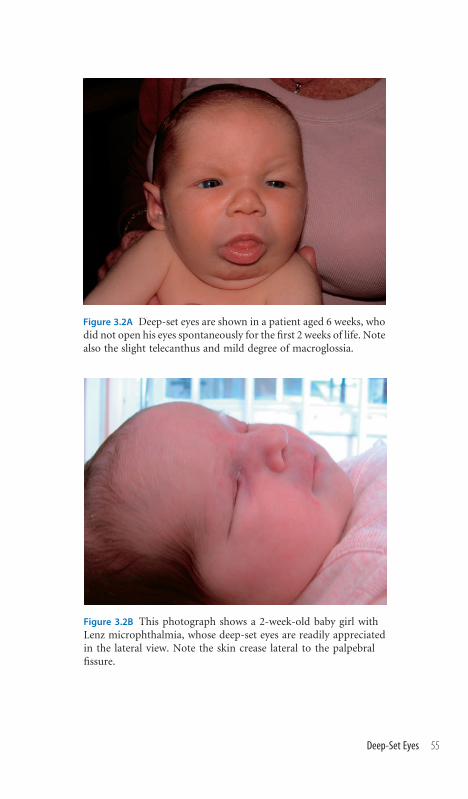
Figure 3.2A Deep-set eyes are shown in a patient aged 6 weeks, whodid not open his eyes spontaneously for the first 2 weeks of life. Notealso the slight telecanthus and mild degree of macroglossia.
Figure 3.2B This photograph shows a 2-week-old baby girl withLenz microphthalmia, whose deep-set eyes are readily appreciatedin the lateral view. Note the skin crease lateral to the palpebralfissure.
Deep-Set Eyes 55
Almond-Shaped Eyes
Recognizing the Sign The almond comparison is a good one. Palpebral
fissure dimensions, the length of the palpebral fissure from the inner canthus
to the outer canthus of each eye, may be reduced, giving a false clinical im-
pression of microphthalmia, with the size of the globe being normal.
Establishing a Differential Diagnosis The almond-shaped eye sign is
quintessentially associated with Prader-Willi syndrome, and the description of
a palpebral fissure as being almond shaped must lead to evaluation of the other
clinical aspects of this condition. In the neonate, hypotonia, poor sucking,
transient need of special feeding techniques, and a history of reduced fetal
movement are frequently recorded in Prader-Willi syndrome patients. The skull
shape is often dolichocephalic, the upper lip may be thin, and themouthmay be
small, with downturned corners. None of these signs is definitive but, in
combination and with a supportive history, may well be signaling a diagnosis of
Prader-Willi syndrome. Examine the genitalia; undescended testes and reduced
penile length are well described in the male. Emerging developmental concerns
and failure to achieve motormilestones are to be expected.Weight gain is a later
feature but is always present by 6 years, usually associated with hyperphagia and
food-related obsessional behavior. Other clinical clues that can be very useful are
small hands and feet and a straight ulnar border to the hands; it is worth asking
the parents whether the child picks at his or her skin, particularly at minor skin
lesions. On direct questioning, a history of excess daytime sleeping may be
forthcoming. The other main condition to consider in relation to this clinical
sign is chromosome 1p36 deletion, in which asymmetric, sometimes thickened
ear helices are good clinical clues, as are the eyebrows, which are somewhat
horizontal with little by way of normal arching. Severe hypotonia neonatally
and developmental delay in the older child are characteristic, with many af-
fected individuals also having congenital heart disease of no specific diagnostic
association.
Investigations to Consider Careful cytogenetic analysis is the approach to
chromosome 1p36 deletion; specific subtelomere FISH or array hybridization
are required if no deletion is microscopically visible in cases of strong clinical
suspicion. Prader-Willi syndrome shows 15q11-13 deletion of the paternal
chromosome in about 70% of cases. Uniparental maternal disomy of 15q11-
13 accounts for 25% and requires DNA analysis of the patient and parents for
confirmation.
The Eye and Related Structures56
3.3

Figure 3.3A Note the almond-shaped eyes in this 2-year-oldgirl with confirmed Prader-Willi syndrome. The initialpresentation in this girl was of neonatal feeding problems,requiring nasogastric intubation for several weeks. Neuro-logic evaluation confirmed hypotonia andmuscle biopsywassuggestive of a congenital myopathy. After the age of 1 year,the rapid weight gain and global developmental concernsgave the clues to the true diagnosis.
Figure 3.3B A typical neonatal presentation of Prader-Willisyndrome is pictured. Note the nasogastric feeding re-quirement and the dolichocephaly.
Almond-Shaped Eyes 57

Blepharophimosis
Recognizing the Sign The clinical impression is of small eyes, but the globe
is structurally and functionally normal in most instances. Blepharophimosis
or short palpebral fissure, as it is also called, is a reduction of palpebral fissure
length in the horizontal axis, measured from the inner canthus to the outer
canthus of one eye. If present, this is generally bilateral. Ophthalmologic eval-
uation may reveal associated microphthalmia.
Establishing a Differential Diagnosis Observe the parents and obtain a
family history of any surgery for ptosis. The autosomal dominant condition
of blepharophimosos-ptosis-epicanthus inversus syndrome (BPES) tends to
restrict the palpebral fissure, and such a finding in another family member is
probably of diagnostic significance. Examine for epicanthus and ptosis. Ob-
serve the head position carefully for signs of the latter. In the absence of a
family or suggestive history of BPES, examine the rest of the face. Look for
micrognathia and cleft palate. These features may indicate a likely diagnosis
of Catel-Manzke syndrome, in which blepharophimosis is typical. The clin-
ching of this diagnosis will be by observation of the hands and seeing the
unusual shape of the index fingers, which generally show ulnar deviation
bilaterally. This is caused by an accessory metacarpal bone in the index fingers.
Blepharophimosis may be a nonspecific indicator of chromosomal aneuploi-
dies, particularly if associated with malformations, such as congenital heart
disease, for which reason clinical evaluation of the heart is important. There
are a few rare but diagnosable conditions in which blepharophimosis is char-
acteristic and consequently merit specific consideration—Hallermann-Streiff
syndrome presents a thin, prominent, and pointed nose; small chin; and small
palpebral fissures. Neonatal cataracts or microphthalmia are almost universal.
Short palpebral fissures are seen in many cases of fetal alcohol syndrome, in
which the outer and inner canthi may be displaced toward the midline.
Ptosis and/or epicanthus may be present also, but look for other clues such as
a smooth philtrum, a thin upper lip, and aberrant creases of the palms and
forearms. A history of low birth weight is usual.
Investigations to Consider A routine karyotype, without need for specific
cytogenetic refinements, is appropriate if a generic ‘‘chromosomal’’ basis is to
be excluded. Specifically, chromosome 3q23 abnormality should be sought in
BPES, particularly if there is developmental delay, which may signify an as-
sociated loss of other genetic material by deletion of adjacent loci. Most BPES
cases show intragenic mutation of the FOXL2 gene on 3q23 by DNA analysis.
Hand radiology demonstrating the accessory metacarpal bones of the index
fingers is confirmatory of Catel-Manzke syndrome.
The Eye and Related Structures58
3.4

Figure 3.4A The classic signs ofBPES comprising blephar-ophimosis, ptosis, and epi-canthus inversus are readilyappreciated.
Figure 3.4B In profile, themicrognathia, beaked nose ofblepharophimosis of this babywith Hallermann-Streigg syn-drome are appreciated.
Figure 3.4C Short palpebralfissures are demonstrated inthis boy with fetal alcoholsyndrome. Note the clinicalimpression of small eyes. Thereis microcephaly and malarhypoplasia. Unlike manyother cases of this disorder,the philtral pillars arequite well formed and the up-per lip is not thin. Other di-agnostically useful signs in thispatient were partial 3/4 fingersyndactyly and abnormal pal-mar creases.
Blepharophimosis 59

Palpebral Fissure Slant
Recognizing the Sign Should the outer canthi be sited above an imaginary
line connecting the two inner canthi, then the palpebral fissures slant upward
(mongoloid); below such an imaginary line, the palpebral fissures slant down-
ward (antimongoloid). Some variation is observed in up to 5% of the normal
population.
Establishing a Differential Diagnosis Upslanting palpebral fissures suggest
reduced skull growth relative to the midface, so assessment for microcephaly
and associated signs is sensible (1.2). Apart from the well-established associ-
ation with Down syndrome, upslanting palpebral fissures are a nonspecific
finding in many cases of chromosomal aneuploidy. Of more specific diag-
nostic value, this sign is frequently seen in Pallister-Killian syndrome, tet-
rasomy chromosome 12p, in which condition additional clinical signs to look
for are temporal balding, with poor hair growth in this region, and severe
developmental delay. Accessory nipples and regional skin pigmentation ab-
normalities are important diagnostically supportive clinical signs, but not
universally present. With age, affected patients develop a coarse appearance,
reminiscent of storage disorders. Upslanting palpebral fissures are often noted
in early childhood in Kabuki syndrome, the other ophthalmic signs being
ectropion of the lower eyelid and hairless ‘‘gaps’’ in the eyebrows. Down-
slanting palpebral fissures frequently accompany midface hypoplasia and are
common in conditions thus characterized. If the striking characteristic of
a face is downslanting palpebral fissures, think specifically of Noonan syn-
drome and examine the heart; assess for familial short stature and low hairline
on the neck, and ask about a personal or family history of bleeding disorders,
as many Noonan cases have an unexplained coagulopathy. Downslanting
palpebral fissures with associated hypertelorism and developmental delay in
a male suggest Coffin-Lowry syndrome (Figure 9.9), especially if a history
compatible with X-linked mental retardation can be established. Carrier fe-
males may show mild clinical signs. Particularly useful clinical signs to seek
are rather full lips and tapering, fleshy fingers. The face of Robinow syndrome
may also show hypertelorism and downslanting palpebral fissures, but there is
usually little cause for developmental concern. Look specifically for gum hy-
pertrophy and tongue tie, both valuable clues to this diagnosis, and, in males,
for micropenis. The nose is usually anteverted and there is short stature.
Investigations to Consider Pallister-Killian syndrome patients almost in-
variably need fibroblast analysis to establish the diagnosis and, in some in-
stances, need repeated skin biopsies to achieve this. Keratinocyte culture,
rarely available, has a better yield. Spinal radiology is important in Robinow
syndrome to assess for hemivertebrae or fusions, which may necessitate sur-
gical management.
The Eye and Related Structures60
3.5

Figure 3.5A The upslant of the palpebral fissures inthis baby with Kabuki syndrome is beyond dispute.
Figure 3.5B This photograph shows mildlyupslanting palpebral fissures in a 13-year-old boy with Pallister-Killian syndrome.
Figure 3.5C The same patient is demon-strated as in Figure 3.5B. Seen in profile, thetemporal balding, typical of Pallister-Killiansyndrome, has been minimized by brushingthe hair forward onto the hairless region.
Figure 3.5D A striking exampleof downslanting palpebral fissuresis shown.
Palpebral Fissure Slant 61

Proptosis
Recognizing the Sign The globe of the eye protrudes inordinately. It is pos-
sible to measure the degree of orbital protrusion, but this is rarely undertaken
in practice. A clinical impression of inordinate prominence of the eyes will
lead the clinician to consider this sign and its related conditions.
Establishing a Differential Diagnosis First establish whether the proptosis is
unilateral or bilateral. In craniosynostosis the premature fusion of the skull
base causes reduced orbital volume and proptosis. With the exception of uni-
coronal synostosis, the proptosis is bilateral, even if asymmetric. Proptosis is
the abiding clinical memory of the clinician’s first glance at Crouzon syn-
drome. The absence of significant digital features on hand and foot exami-
nation will establish Crouzon syndrome as the likely diagnosis, while broad
halluces and thumbs in the context of proptosis and clinical evidence of cra-
niosynostosis on skull palpation will point toward Pfeiffer syndrome. Apert
syndrome patients, readily recognizable by the syndactyly of fingers and toes,
tend to have less significant degrees of proptosis than Crouzon syndrome, but
do remember to look for cleft palate seen in 75% of Apert cases. By contrast,
unilateral proptosis should prompt the astute clinician to examine the child
for clinical signs of neurofibromatosis type 1 (NF1). Specifically look for
cafe-au-lait patches and macrocephaly. Observe if the eye is pulsatile, usually
reflecting a deficiency of the greater wing of the sphenoid bone in the orbital
wall. Peripheral neuromas, plexiform neuromas, and axillary and groin
freckling should all be sought in the older child. It is important to examine for
scoliosis and for hypertension, in view of the known association with renal
artery stenosis and with pheochromocytoma. Bilateral eye prominence is also
seen in some forms of Ehlers-Danlos Syndrome (EDS). Further clues to this
condition may emerge from the family history. Examine the skin and joints
for hyperlaxity. Look for evidence of bruising or exaggerated response to
minimal trauma. An important clinical feature to establish is scoliosis, which
generally signifies type VI, in which there is a propensity to perforation of the
globe and retinal detachment.
Investigations to Consider Mutation analysis of the appropriate FGFR
(Fibroblast Growth Factor Receptor) locus will usually offer molecular con-
firmation of a specific clinical syndrome of craniosynostosis. Slit lamp ex-
amination for Lisch nodules on the iris and retinal examination for signs of
optic glioma are important elements of the assessment of NF1 in childhood.
In EDS type VI, lysyl hydroxylase deficiency may be demonstrated on fibro-
blasts, but an easier screening test is to undertake urinary pyridinoline
cross-links.
The Eye and Related Structures62
3.6
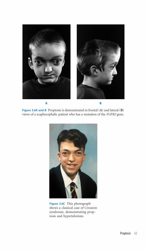
A B
Figure 3.6A and B Proptosis is demonstrated in frontal (A) and lateral (B)views of a scaphocephalic patient who has a mutation of the FGFR2 gene.
Figure 3.6C This photographshows a classical case of Crouzonsyndrome, demonstrating prop-tosis and hypertelorism.
Proptosis 63

Ptosis of the Eyelid
Recognizing the Sign The upper eyelid normally covers 2 cm of cornea,
which approximates with the outer margin of the iris. Hence, sclera is usually
not seen between the iris and the upper lid and, if present, suggests lid retrac-
tion. The eyes are wide open, sometimes described as ‘‘startled’’ appearance.
Conversely, ptosis of the eyelid narrows the palpebral fissure.
Establishing a Differential Diagnosis Establish whether the ptosis is uni-
lateral or bilateral and whether it is congenital or of later onset. Observe any
compensatory head tilt of frontalis muscle overactivity. Blepharophimosis-
ptosis-epicanthus inversus syndrome (BPES) has already been outlined (2.1).
Affected individuals do not manifest all the signs in all cases, so family history
allied with careful examination will be useful in diagnosis of this autosomal
dominant condition. Establish whether the patient has the full range of eye
movements and examine the cranial nerves. Evidence of VI andVII nerve palsy,
often with signs of more extensive lower cranial nerve deficiencies, suggests a
likely diagnosis of Moebius syndrome and attention should be given to the
hands—syndactyly or brachydactyly being common in this condition. Clin-
ical awareness, neurologic history and physical signs are the key to diagnosis
of the mitochondrial myopathies, in which conditions the gradual develop-
ment of ptosis with age is common. A family history, particularly with atten-
tion to symptoms in the maternal lineage, may offer clues, though many cases
are sporadic. Examine for ophthalmoplegia and general muscle weakness. Seek
a history of stroke-like episodes or myoclonic epilepsy. Ptosis in association
with short stature and/or a cardiac murmur may be the signals to a case of
Noonan syndrome. Look for a low posterior hairline and evidence of excess
nuchal skin, which will further support this diagnosis, if present. Often seen in
Saethre-Chotzen type craniosynostosis, the true nature of the ptosis is be-
trayed by the unusually upright forehead shape, prominent ear crus, and partial
skin syndactyly of the fingers. The frequent observation of ptosis in the con-
dition ofWilms-aniridia-genital abnormality-retardation (WAGR) empha-
sizes the need for careful ophthalmic and general evaluation of the patient
with ptosis in the interest of the true diagnosis emerging. Cryptorchidism and
hypospadias are useful clinical prompts in cases of WAGR syndrome.
Investigations to Consider Investigation of mitochondrial myopathies in-
cludes electrocardiogram (ECG) for block, audiologic evaluation for deafness,
and retinal examination for pigmentary retinopathy. Muscle biopsy for rag-
ged red fibers by Gomori trichrome stain may be indicated. Mitochondrial
DNA study for deletions/duplications or point mutations may be valuable.
Karyotype usually shows 11p13 deletion in WAGR, whereas karyotype in
BPES may show 3q deletion.
The Eye and Related Structures64
3.7

Figure 3.7C This photograph shows ptosisin a patient with Noonan syndrome; ob-serve the compensatory backward tilt ofthe head.
Figure 3.7D This photograph demonstratesunilateral ptosis. Note also the macro-glossia in this boy.
Figure 3.7A and B Bilateral ptosis is demonstrated in a patient withMoebius syndromeof bilateral facial diplegia. The paucity of facial movement is demonstrated betweenthe facial expression at rest (A) and smiling (B). Note the degree to which the uppereyelid obscures the iris and pupil. Additionally, the upper eyelid crease is absent, afurther sign of ptosis. (Reproduced with permission ofWiley-Liss from Reardon et al.,Am J Med Genet 2003;122A:84–88.)
Ptosis of the Eyelid 65

Corneal Clouding
Recognizing the Sign A generalized hazy appearance of the cornea is pres-
ent, usually bilaterally. Specific forms of corneal clouding are described, most
notably buphthalmos (‘‘bull’s eye’’), in which congenital glaucoma causes en-
largement of the anterior chamber and cloudy cornea. Sclerocornea is a term
used in describing congenital, nonprogressive corneal opacification.
Establishing a Differential Diagnosis Formal ophthalmologic evaluation is
essential in determining the likely cause and management of corneal clouding.
Essentially, three main causes should be considered: (1) glaucoma; (2) primary
anterior eye chamber diseases, which sometimes have other clinical manifesta-
tions; and (3) metabolic disease. While glaucoma is well described in Sturge-
Weber and Rubinstein-Taybi syndromes, the corneal clouding is not the primary
clinical finding in such instances and the underlying diagnosis is evident from
the characteristic facial hemangioma of the former and broad, deviated thumbs
and characteristic nasal columella of the latter. Likewise, congenital glaucoma
can be seen in Stickler syndrome, other clues to the true diagnosis coming from
the enlarged joints, excess joint laxity, and cleft palate, if present, in addition to
the family history.Most glaucoma is nonsyndromic and nonocular signs will not
be established. Among primary anterior eye chamber disorders, clinical clues
to such a diagnosis will be eccentric siting of the pupil or absence of the iris.
Extended neonatal jaundice in an infant with cloudy cornea should highlight
the diagnostic likelihood of Alagille syndrome. The finding of congenital heart
disease, particularly peripheral pulmonary stenosis, would support this diagno-
sis. Rieger syndrome presents with congenital iris dysplasia, often resulting in
glaucoma. The clinical clues to the diagnosis lie in the frequent history of anal
stenosis, hypodontia, the incisors often being absent, and failure of involution
of umbilical skin, frequently resulting in surgery for umbilical hernia. Ptosis,
cryptorchidism, and hypospadias will signal WAGR (Wilm’s Tumor, Aniridia,
Genitalia Retardation) syndrome, while a family history of autosomal dominant
anterior chamber anomalies is often available on inquiry in isolated aniridia
cases. Corneal clouding in biochemical diseases can represent mucopolysac-
charidosis, mucolipidosis, or gangliosidosis. Useful clinical clues may comprise
organomegaly, gum hypertrophy, developmental delay, and short stature.
Investigations to Consider If Alagille syndrome is suspected, look for hemi-
vertebrae or butterfly vertebrae radiologically and get an echocardiograph. In
WAGR, chromosome 11p deletions should be sought, but basic karyotype is
warranted in the absence of a specific ophthalmic diagnosis. PAX6 gene mu-
tation analysis may be valuable in isolated/familial cases of anterior chamber
malformations without signs of Rieger syndrome. Increased urinary muco-
polysaccharides, oligosaccharides, and vacuolated lymphocytes on blood
smear are appropriate basic screening measures for biochemical conditions.
The Eye and Related Structures66
3.8
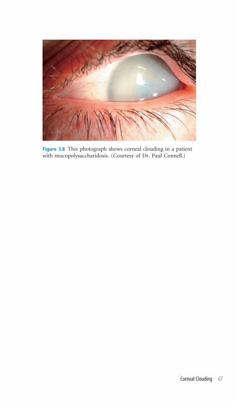
Figure 3.8 This photograph shows corneal clouding in a patientwith mucopolysaccharidosis. (Courtesy of Dr. Paul Connell.)
Corneal Clouding 67

Ectopic Pupil
Recognizing the Sign Usually central within the iris, eccentric siting of the
pupil is visible on inspection of the eye, generally affecting both eyes, though
there may be asymmetry in the degree of involvement. In extreme cases, the
displacement of the pupil may give the clinical appearance of iris coloboma.
Establishing a Differential Diagnosis As applies to corneal clouding, formal
ophthalmologic evaluation is mandatory to establish the primary diagnosis for
this clinical sign. Be aware that there may be associated underlying lens dis-
location, cataracts, or retinal detachment. In many cases the clinical signs are
confined to the eye. It is worth taking a family history, as there are sibships
reported with ectopic pupils, often associated with lens and other anterior
chamber malformations. In conducting the rest of the clinical examination,
particular reference to those conditions, already outlined in relation to cor-
neal clouding, that cause anterior chamber malformations is advised. Spe-
cifically, look for a history of extended neonatal jaundice or established bile
duct paucity on histologic examination in Alagille syndrome or in such a
patient who also has a cardiac murmur. Seek the history of anal stenosis, often
requiring surgical intervention, or surgical treatment for umbilical ‘‘hernia,’’
which may betray the diagnosis of Rieger syndrome. Since these are auto-
somal dominant disorders, an extended family history may uncover other,
previously unremarked upon, valuable corroborative evidence of either di-
agnosis. In Peter’s anomaly, there is generally a central corneal opacification,
sometimes associated with microphthalmia. Such a presentation in the pres-
ence of a prominent forehead, long philtrum, and dysplastic ears may be the
manifestations of Peter’s plus syndrome, in which short stature and mental
retardation may be expected to emerge with time. More commonly, think of
unusual presentations of Marfan syndrome in such patients and examine for
arachnodactyly, excess joint laxity, and a family history suggestive of dis-
secting aneurysm. Likewise, be aware of the association with aniridia and the
possibility of a chromosome 11p–related predisposition to Wilms tumour-
Aniridia-Genital anomaly-Retardation syndrome (WAGR), in which ptosis
and genital anomalies are common.
Investigations to Consider As a nonspecific finding in many karyotypic
abnormalities, in the absence of a specific diagnosis from ophthalmic exam-
ination, chromosomal evaluation is indicated. 11p deletion and 20p dele-
tion are described in WAGR and Alagille syndromes, respectively, but are not
consistent reliable findings. Radiologic demonstration of butterfly vertebrae is
valuable in securing an Alagille diagnosis. Mutation analysis for Fibrillin1
(FBNI ) may be useful should a Marfan syndrome diagnosis seem likely but
insecure clinically, while PAX6 mutations are reported in some familial cases
of ectopic pupils.
The Eye and Related Structures68
3.9

Figure 3.9 The clinical presentation of left-sided ectopic pupil isshown in a developmentally normal 9-month-old child. The familyhistory of glaucoma in her father and grandaunt suggested a di-agnosis of Rieger syndrome.
Ectopic Pupil 69

Blue Sclerae
Recognizing the Sign The darkly pigmented choroid layer often confers a
rather blue appearance to the naturally thin sclera of the neonate. As a denser
connective tissue construction to the sclera develops during the first several
months of life, this natural phenomenon of transient blueness of the neonatal
sclera disappears, usually by the age of 1 year. Persistence of the clinical sign
beyond this age may be diagnostically significant and, obviously, blue sclera
in a younger child may be diagnostically important and not just a transient
phenomenon.
Establishing a Differential Diagnosis Most clinicians will be aware of the
association between blue sclera and osteogenesis imperfecta (OI) and will
seek a history of fractures and delayed tooth eruption and examine for bony
deformation, signs of scoliosis, and joint hyperextensibility. Useful clues may
be derived from the family history, particularly in cases where multiple frac-
tures are not a major clinical problem and the diagnosis may have been hith-
erto unsuspected. However, blue sclera should also prompt examination for
other connective tissue diseases, specifically Marfan syndrome and Ehlers-
Danlos syndrome (EDS). In the case ofMarfan syndrome, observe for the tall,
slim appearance; joint hyperextensibility; arachnodactyly; and sternal mal-
formations, either carinatum or excavatum. Parental height and family his-
tory, particularly with a view to sudden unexplained death in early adult life,
may offer corroborative evidence. Many of these features will overlap with the
Ehlers-Danlos syndrome complex of phenotypes, but the family history and
clinical demonstration of skin hyperextensibility, excessive scarring in re-
sponse to a relatively trivial injury, and easy bruising will alert the wary to a
diagnosis of EDS type I. Blue sclera, skin scarring, and extreme joint laxity
will not distinguish between EDS types I and EDS type VI, an autosomal
recessive condition, but scoliosis and multiple criss-cross lines on the palms
and soles are redolent of EDS type VI, in which eye examination is important
because of associated glaucoma and propensity to rupture of the globe. Blue
sclera in a small baby, particularly if the growth concerns predate birth, may
be the signal to Russell-Silver syndrome. The examination should focus on
seeking other clues such as skeletal asymmetry, relative macrocephaly, cafe-
au-lait patches, and a clammy sweatiness, best identified by direct questioning
of the mother.
Investigations to Consider X-ray evaluation looking for evidence of healed
fractures, bony deformities, and wormian bones in the cranial sutures is the
mainstay of diagnosis in OI. If clinical doubt exists, mutation analysis of the
FBNI gene may be undertaken in Marfan syndrome, while lysyl hydroxy-
lase deficiency in cultured skin fibroblasts is demonstrable in many cases of
EDS type VI. A good screening test is urinary pyridinoline cross-linkages.
The Eye and Related Structures70
3.10

Figure 3.10 Blue sclera in a severe case of osteogen-esis imperfecta.
Blue Sclerae 71

Iris Coloboma
Recognizing the Sign The term coloboma signifies a fissure in the iris. Since
the iris derives embryologically from the neural retina and the pigment epi-
thelium of the optic cup, the clinical significance of an iris coloboma is that it
may be the visible sign of an embryologically associated underlying coloboma
of the retina.
Establishing a Differential Diagnosis Ophthalmologic examination is cru-
cial not only to establish the extent of embryologic defect, but also to identify
any associated abnormalities such as cataract or retinal detachment. In terms
of the general clinical examination of the baby with iris colobomata, specific
evaluation for clinical signs of CHARGE association (coloboma, heart defects,
atresia choana, retardation of growth and development, genital anomaly, and
ear malformation) is important. Frequently, the external ear is the most note-
worthy sign, often comprising a simple, cup-shaped or, less commonly, an
overfolded helix. Iris coloboma and dysplastic ears will also be encountered
in Catel-Manzke syndrome, but the cleft palate and bizarrely deviated index
finger will betray this specific diagnosis. The presence of preauricular pits or
tags will alert the clinician to the possible diagnosis of cat-eye syndrome, and
examination should specifically be extended to assess for anal stenosis and
possible evidence, depending on age at examination, of developmental delay
(Figure 4.8). The observation of hypertelorism in association with iris colo-
boma should prompt evaluation for Wolf-Hirschhorn syndrome, looking
specifically at the glabellar region for prominence and at the nose for the Greek
helmet profile (Figure 2.3A). Microcephaly, cleft lip, or a midline scalp defect
would strongly support such diagnostic thoughts. By contrast, Gorlin syn-
drome, in which iris coloboma is seen in the context of macrocephaly and
there is a propensity to basal cell carcinoma of the skin, is rarely diagnosable
by examination of the newborn in isolation, but the family history in this
autosomal dominant condition will explain the significance of the iris colo-
boma, and the later emergence of the nevoid basal cell carcinomas should be
anticipated. A rare but important association is neurofibromatosis type I
(NF1), signifying the need for longitudinal follow-up lest cafe-au-lait and
other characteristic signs emerge.
Investigations to Consider Cardiac evaluation is mandatory if CHARGE,
cat-eye, or Wolf-Hirschhorn syndromes are suspected. Computed tomogra-
phy (CT) scan of the petrous temporal bone in CHARGE shows hypoplasia of
the semicircular canals. FISH is indicated for 4p deletion in Wolf-Hirschhorn
syndrome, while karyotype in cat-eye syndrome usually shows additional
chromosome 22material. Occasionally, FISH needs to be undertaken to estab-
lish the cause of the clinical signs. X-ray in Catel-Manzke will show an ac-
cessory phalanx at the base of the index finger.
The Eye and Related Structures72
3.11

Figure 3.11 Bilateral iris colobomata are demon-strated in this patient.
Iris Coloboma 73

Anophthalmia/Microphthalmia
Recognizing the Sign Anophthalmia signifies complete absence of out-
growth from the optic vesicle and failure of the eye to form. This very rarely
occurs and the more likely situation clinically is of ‘‘cryptophthalmos,’’ where
a fusion of the eyelid obscures the orbit beneath, which may itself be ab-
normal, perhaps colobomatous, or microphthalmic. Microphthalmia refers
to an abnormally small eye, often seen in conjunction with narrow palpebral
fissures and deep-set eyes. Obviously, full ophthalmic characterization of the
lesions is important.
Establishing a Differential Diagnosis The history, particularly of antenatal
exposure or intrauterine infection, may be significant, TORCH agents, al-
cohol, and warfarin all being established causes of microphthalmia. Likewise,
pure familial forms are well reported, so the family history may give a clue. In
the established absence of such issues, examine the ears for signs of CHARGE
association and consider evidence for choanal atresia/stenosis in the form of
mouth breathing or persistent runny nose. Simple ear helices, cleft lip, or
sloping shoulders due to hypoplasia of the lateral element of the clavicles are
the clinical clues to Lenz microphthalmia syndrome. Microcephaly is usual
and will be associated with developmental delay. Being X-linked, this is an
important diagnosis to secure. The same applies to Lowe syndrome, again
inherited as an X-linked disorder, in which the deep-set, microphthalmic eyes
of the neonate may be the clue to diagnosis. In oculo-dento-digital syn-
drome, the microphthalmia will generally be associated with hypotelorism
and skin syndactyly, typically of fingers 3 to 5 (Figure 2.2). Syndactyly of a
more general nature is characteristic of Fraser syndrome, in which cryp-
tophthalmia is generally observed, often with a protrusion of the anterior
hairline toward the orbital margin. Look for genital malformations, typically
hypospadias or clitoromegaly, and low-set ears for corroborative evidence
of this autosomal recessive condition. Finally, examine the skin carefully, as
linear areas of dermal hypoplasia, perhaps erythematous in the neonate and
pigmented or atrophic in the older child are likely to signify the diagnosis of
Goltz syndrome (focal dermal hypoplasia).
Investigations to Consider Aminoaciduria is usual in Lowe syndrome,
though it may be normal neonatally. Urinary retinol binding protein may be a
better assay for screening. Ophthalmic examination of carrier females for
visually insignificant cortical opacities is valuable, almost all carriers showing
this sign. Ultrasound of the kidneys may show renal agenesis in Fraser syn-
drome. Mutation analysis of the BCOR gene on Xq27 is required to confirm
Lenz syndrome. Chromosomal evaluation has shown Xp22 microdeletions in
a handful of cases of linear skin lesions with microphthalmia.
The Eye and Related Structures74
3.12
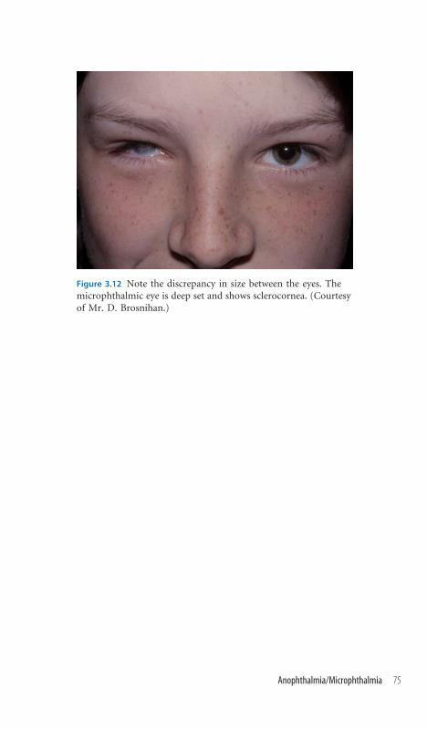
Figure 3.12 Note the discrepancy in size between the eyes. Themicrophthalmic eye is deep set and shows sclerocornea. (Courtesyof Mr. D. Brosnihan.)
Anophthalmia/Microphthalmia 75

Iris Variants
Recognizing the Sign Within the normal population, the iris demonstrates
variation in color and pattern. However, there are some well-delineated,
syndromically important, specific iris patterns: (1) Brushfield spots are white
or yellowish speckled dots on the iris, usually toward the periphery; (2) stellate
iris, meaning a lacy pattern, sometimes described as resembling the outward
radiation of the spokes of a bicycle wheel; (3) cloverleaf iris (also known as
Lester’s sign), in which a collarette of dark pigmentation around the central
part of the iris is surrounded by lighter pigment; (4) Lisch nodules, which are
hamartomatous in nature and clinically present brown or cream colored well-
defined elevations, randomly distributed on the surface of the iris, and gen-
erally visible on close inspection, though confirmation of whose absence ne-
cessitates slit lamp examination; and (5) heterochromia iridum in which the
color of one iris differs from the other (but the term is also used to denote
color variation within the same iris).
Establishing a Differential Diagnosis Brushfield spots are invariably linked
in the clinical mind toDown syndrome, and associated signs of macroglossia,
brachycephaly, single palmar creases, and clinodactyly will automatically be
assessed. Remember that these iris pigmentary phenomena are also seen in
10% of normal neonates. Beware of potential confusion with Zellweger syn-
drome, in which hepatomegaly and extreme hypotonia also occur. The stellate
iris is also a phenomenon of 10% of the normal population, but noted in up to
75% of blue-eyed Williams syndrome cases. Examine for the supra-valvular
aortic stenosis and other cardiac signs and look clinically for the wide mouth
and long, usually smooth philtrum. Lester’s sign is seen in 50% of cases of
nail-patella syndrome, in which absent or hypoplastic nails affect the thumb
most commonly (Figure 12.1). Small patellae, tending to recurrent subluxa-
tion, are a feature of older children. Lisch nodules betoken neurofibromatosis
type 1 (NF1) and a search for other clinical signs, such as cafe-au-lait patches,
freckling, and neurofibromata, is indicated. Do check for optic glioma, sco-
liosis, and hypertension. Heterochromia iridium should prompt evaluation
of other hypopigmentation signs, such as premature greying in the family,
assessment of hearing, and specific examination for telecanthus, all charac-
teristic of Waardenburg syndrome.
Investigations to Consider Karyotype will confirm Down syndrome. FISH
for ELN on chromosome 7q is required for Williams syndrome confirmation.
Very-long-chain fatty acids are generally abnormal in Zellweger cases, and
Waardenburg syndrome diagnosis is confirmed by PAX3 mutation analysis if
dystopia canthorum is present (type I) and MITF mutation analysis if absent
(type II) (Figure 12.7A).
The Eye and Related Structures76
3.13

Figure 3.13A Brushfield spots.
Figure 13.3B Stellate iris.
Figure 13.3C Lester’ sign. (Courtesyof Dr. Elizabeth Sweeney.)
Figure 13.3D Lisch nodule.
Figure 13.3E Heterochromia iridum.
Iris Variants 77

Ectropion of the Lower Eyelid
Recognizing the Sign The normal position of the lower eyelid is such that it
sits against the globe. Ectropion is present when the lower eyelid, through
excess tissue or loss of elasticity, no longer approximates to the globe and a
gap is visible between the eyelid margin and the globe. There may be eversion
of the eyelid margin, whose capillary bed becomes visible.
Establishing a Differential Diagnosis Particular attention to the examina-
tion of the skin is advised. In a newborn, ectropion accompanies lamellar
ichthyosis (collodion baby syndrome; Figure 12.15). The skin looks shiny
and cracks, peeling away to reveal normal skin beneath over a period of a few
weeks. The ectropion usually resolves. Persisting ectropion may betoken
cutis laxa syndrome. Look for redundant folds of excess skin, often best
observed over the neck, the dorsum of the hands, and the chest. Tachypnea is
an important clue, as many of these patients develop early-onset emphysema
and succumb to cor pulmonale. Other valuable clinical clues are scars of
surgical treatment for umbilical or inguinal herniae. The presence of a cleft
lip and/or palate is likely to signify the autosomal dominant condition of
blepharo-cheilo-dontic (BCD)syndrome, andparental examination foroligo-
dontia, ectropion, and double row of eyelashes (distichiasis) may secure this
diagnosis in the parent and child. Cardiac abnormalities should be sought if
the diagnosis is Kabuki syndrome, in which the facial characteristics are
of wide palpebral fissures and ectropion. Mild developmental delay is usual.
Specifically helpful clinical features are persisting fetal finger pads (Figure
9.16) and eyebrows that may be excessively arched and are often deficient of
hair in the middle. A history of early feeding difficulty, perhaps requiring na-
sogastric supplementation, is common. The author has twice been prompted
to this diagnosis by persisting blue sclerae in children aged 2 years. Family
history of parathyroid, medullary thyroid, or pheochromocytoma in a child
with ectropion is almost certain to represent multiple endocrine neoplasia
type IIB (MEN IIB). The lips may be thickened and nodular and the tongue
likewise. Malar hypoplasia, downslanting palpebral fissures, and ectropion,
the facial features of Miller syndrome, should prompt examination of the
hands, the little fingers and toes usually being absent.
Investigations to Consider Clinical examination is often adequate to secure
the diagnosis. Bladder diverticula may be radiologically demonstrable in cutis
laxa. RET gene mutations are demonstrable in MEN IIB.
The Eye and Related Structures78
3.14
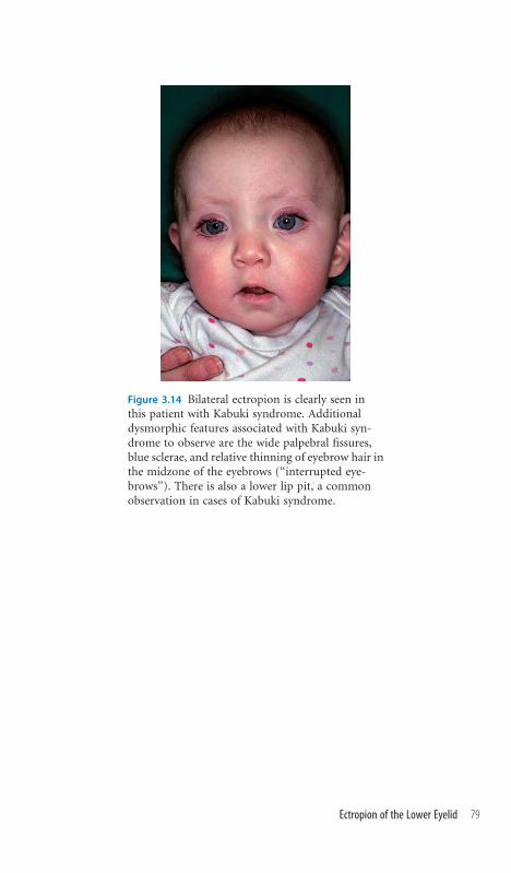
Figure 3.14 Bilateral ectropion is clearly seen inthis patient with Kabuki syndrome. Additionaldysmorphic features associated with Kabuki syn-drome to observe are the wide palpebral fissures,blue sclerae, and relative thinning of eyebrow hair inthe midzone of the eyebrows (‘‘interrupted eye-brows’’). There is also a lower lip pit, a commonobservation in cases of Kabuki syndrome.
Ectropion of the Lower Eyelid 79
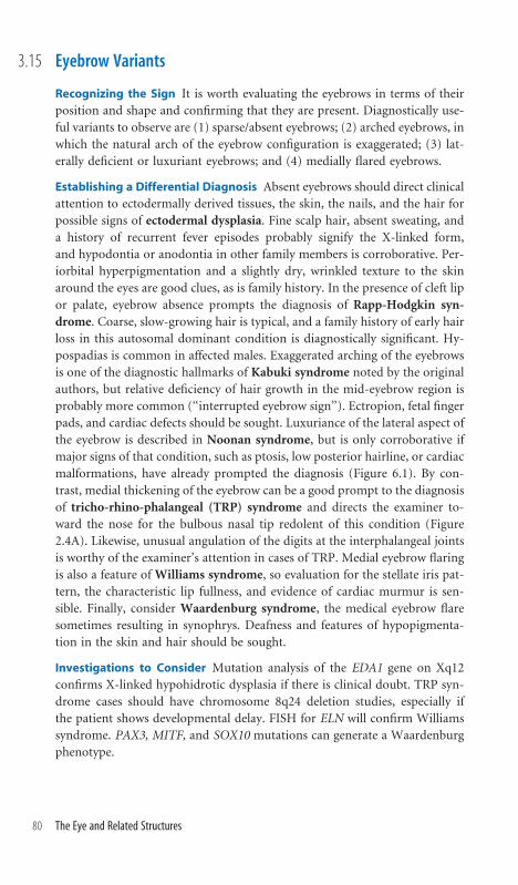
Eyebrow Variants
Recognizing the Sign It is worth evaluating the eyebrows in terms of their
position and shape and confirming that they are present. Diagnostically use-
ful variants to observe are (1) sparse/absent eyebrows; (2) arched eyebrows, in
which the natural arch of the eyebrow configuration is exaggerated; (3) lat-
erally deficient or luxuriant eyebrows; and (4) medially flared eyebrows.
Establishing a Differential Diagnosis Absent eyebrows should direct clinical
attention to ectodermally derived tissues, the skin, the nails, and the hair for
possible signs of ectodermal dysplasia. Fine scalp hair, absent sweating, and
a history of recurrent fever episodes probably signify the X-linked form,
and hypodontia or anodontia in other family members is corroborative. Per-
iorbital hyperpigmentation and a slightly dry, wrinkled texture to the skin
around the eyes are good clues, as is family history. In the presence of cleft lip
or palate, eyebrow absence prompts the diagnosis of Rapp-Hodgkin syn-
drome. Coarse, slow-growing hair is typical, and a family history of early hair
loss in this autosomal dominant condition is diagnostically significant. Hy-
pospadias is common in affected males. Exaggerated arching of the eyebrows
is one of the diagnostic hallmarks of Kabuki syndrome noted by the original
authors, but relative deficiency of hair growth in the mid-eyebrow region is
probably more common (‘‘interrupted eyebrow sign’’). Ectropion, fetal finger
pads, and cardiac defects should be sought. Luxuriance of the lateral aspect of
the eyebrow is described in Noonan syndrome, but is only corroborative if
major signs of that condition, such as ptosis, low posterior hairline, or cardiac
malformations, have already prompted the diagnosis (Figure 6.1). By con-
trast, medial thickening of the eyebrow can be a good prompt to the diagnosis
of tricho-rhino-phalangeal (TRP) syndrome and directs the examiner to-
ward the nose for the bulbous nasal tip redolent of this condition (Figure
2.4A). Likewise, unusual angulation of the digits at the interphalangeal joints
is worthy of the examiner’s attention in cases of TRP. Medial eyebrow flaring
is also a feature of Williams syndrome, so evaluation for the stellate iris pat-
tern, the characteristic lip fullness, and evidence of cardiac murmur is sen-
sible. Finally, consider Waardenburg syndrome, the medical eyebrow flare
sometimes resulting in synophrys. Deafness and features of hypopigmenta-
tion in the skin and hair should be sought.
Investigations to Consider Mutation analysis of the EDA1 gene on Xq12
confirms X-linked hypohidrotic dysplasia if there is clinical doubt. TRP syn-
drome cases should have chromosome 8q24 deletion studies, especially if
the patient shows developmental delay. FISH for ELN will confirm Williams
syndrome. PAX3, MITF, and SOX10 mutations can generate a Waardenburg
phenotype.
The Eye and Related Structures80
3.15

Figure 3.15A Sparse eye-brows.
Figure 3.15B Interruptedeyebrow, lower lid ectro-pion, and wide palpebralfissure in Kabuki syn-drome.
Figure 3.15C Medial flareof the eyebrow and dys-topia canthorum inWaardenburg syndrometype I.
Figure 3.5D Absenceof the eyebrows is dem-onstrated in a patientwith X-linked hypohy-drotic ectodermal dys-plasia syndrome. Notethe very characteristicfine periorbital wrinkling.

Eyelash Variants
Recognizing the Sign The normal configuration of the eyelashes follows a
gentle outward curve from the eyelid, with the upper and lower lashes meeting
but not interdigitating on lid closure.
Establishing a Differential Diagnosis Straight eyelashes in a neonate who is
feeding poorly are a feature of myopathic conditions and are observed in
situations that can present as a myopathy, such as Prader-Willi syndrome.
This diagnosis is not made on the configuration of the eyelash alone, but
the clinical observation can drive the investigation in a particular direction.
Generalized hirsutism is frequently associated with long, perhaps eccentrically
oriented, eyelashes. Look over the back for hair whorls. Think specifically of
de Lange syndrome and assess for microcephaly and generalized increased
tone and examine the hands for malformations and specifically for proximally
placed thumbs due to the characteristically short first metacarpal (Figure
9.13B). Long eyelashes in a patient in whom you think there is floppiness or
evidence of delayed motor milestones might prompt you to look for retinal
pigmentary disorders, as these features comprise a rare autosomal recessive
mental retardation disorder associatedwith variously disordered pituitarymal-
function, chorioretinitis-pituitary dysfunction syndrome. A family history
of lymphoedema from around the age of puberty should prompt examination
for distichiasis, a double row of eyelashes, which can scratch the conjunc-
tiva and be a severe irritant to patients. The condition of lymphoedema-
distichiasis syndrome is autosomal dominant, and congenital heart disease
should be sought. Absence of eyelashes is frequently observed in ectodermal
dysplasias, in consequence of which a careful evaluation of the skin, nails,
hair, and dentition as well as family history may give clues to the underlying
diagnosis. Absent eyelashes, especially if there are dermatologic signs such
as palmoplantar skin thickening, knuckle thickening, or leukonychia (white
nails), should prompt assessment of hearing, as there are a range of deafness
syndromes associated. Short limbs in a baby with sparse hair and eyebrow
and eyelash absence may be the clues toMcKusick-Cartilage-Hair hypoplasia
syndrome, an important autosomal recessive condition to recognize because
of the associated immunodeficiency and particular susceptibility to varicella
infection, often with fatal outcome.
Investigations to Consider Paternal deletion of chromosome 15q is de-
monstrable either cytogenetically or by DNA analysis in most cases of Prader-
Willi syndrome. If deafness is suspected, connexin 26 and 30 (GJB2 and GJB6)
mutation should be sought. Radiologic characteristics of cartilage hair hypo-
plasia apart from short, wide bones are metaphyseal dysplasia, particularly of
the lower end of the femur, and cone-shaped epiphyses of the phalanges.
The Eye and Related Structures82
3.16

Figure 3.16A The normal curvature of the eyelashesis shown.
Figure 3.16B A double row of eyelashes is shown ina patient with lymphoedema-distichiasis syndrome.Note the bizarre course of some of the hairs thatare directed toward the cornea and frequentlycause corneal irritation and abrasion. (Courtesyof Mr. D. Brosnihan.)
Eyelash Variants 83

Epibulbar Dermoid
Recognizing the Sign These are solid tumors of the ocular surface, usually a
pearly grey color, comprising collagen with an epidermal covering or, oc-
casionally, adipose tissue. They may be unilateral or bilateral and are most
likely to be noticed if at the limbus. However, most occur on the bulbar con-
junctiva and as such, may be identified only by retraction of the eyelid for
examination.
Establishing a Differential Diagnosis Examine the eye thoroughly. Note
whether the iris looks normal, whether the pupil is central, and whether there
is any evidence of corneal opacity, and seek evidence of other dermoid tumors
on the conjunctiva. Look at the eyelids for coloboma. Pediatric ophthalmo-
logic evaluation is indicated to assess normality of ocular structures. Assess
the face for evidence of asymmetry and the ears for preauricular tags and/or
microtia. The presence of epibulbar dermoid and asymmetric facial weakness
suggests a likely diagnosis of Goldenhar syndrome spectrum, in which con-
dition examination for congenital heart disease should form part of the as-
sessment, as should examination of hearing. Awareness of the high likelihood
of associated cervical hemivertebra is important to investigation and future
management. Isolated corneal dermoids are described in autosomal domi-
nant, recessive, and X-linked pedigrees, for which reason family history and
parental examination can be useful. Careful examination of the skin is im-
portant in isolated cases of epibulbar dermoid lest this be the presenting
feature of linear sebaceous nevus syndrome. The signs in this condition
can be protean, but look out for slightly elevated, yellow or tan skin plaques,
which often acquire a verrucous appearance with time. The association of the
condition with progressive macrocephaly, cranial nerve palsy, mental retar-
dation, and bone cysts should be borne in mind. Deafness concerns in a child
with epibulbar dermoid are not confined to the Goldenhar spectrum, but
a history of anal stenosis suggests Townes-Brocks syndrome (TBS). Look
carefully at the thumbs, where the terminal phalanx is often at an odd angle
with respect to the proximal phalanx, due to the presence of an accessory
phalanx (Figure 9.14D). Likewise, the anus may be anteriorly placed. Many of
these features, including preauricular pits, congenital heart disease, and anal
stenosis, overlap with marker chromosome 22 syndrome (Figure 4.8).
Investigations to Consider Neck radiology is indicated if Goldenhar syn-
drome seems likely. Chromosomal studies, if necessary of the fibroblasts, should
be undertaken if marker 22 is suspected. Mutation of SAL1 is demonstrable
in most patients with TBS and even a few cases that clinically overlap with
Goldenhar syndrome.
The Eye and Related Structures84
3.17

Figure 3.17 Note the grey colored dermoid demonstrated atthe limbus.
Epibulbar Dermoid 85

Chapter 4 The Ear

Low-Set Ears
Recognizing the Sign Low-set ears are one of the most commonly attributed
and least specific of clinical signs in dysmorphology, and an endless source of
confused argument. The definition of the sign is governed by the drawing
of an imaginary horizontal line through the outer canthi of the eyes, and the
ears are adjudged to be low set if the root of the helix falls below this line
(Figure 4.1A).
Establishing a Differential Diagnosis Bear inmind that hydrocephalus,mac-
rocephaly, and unusually small but normally shaped ears distort the normal
relative proportions between these anatomic features, resulting in low-set ears.
In view of the nonspecificity of the clinical sign, the purpose of clinical ex-
amination is to establish the presence of other anomalies that may facilitate the
emergence of an underlying diagnosis. Observe whether the ear is normal or
has other anomalous features, such as of helical shape, which are dealt with in
later sections of this chapter. Relatively common genetic syndromes in which
low-set ears are frequently encountered include Noonan syndrome and velo-
cardio-facial (VCF) syndrome. The associated features in Noonan syndrome
of family history of short stature, ptosis, heart murmur, typically pulmonary
stenosis, low posterior hairline, and excess nuchal skin will betray the true di-
agnosis. VCF syndrome is more variable, but the presence of a heart murmur;
suspected palatal incompetence or cleft; feeding problems, especially nasal re-
gurgitation of milk; hypocalcemia; or facial palsy may all serve to prompt the
diagnosis. The identification of hypospadias in a patient with low-set ears
should raise the possibility that the patient has Smith-Lemli-Opitz syndrome
(S-L-O), further substantiated if 2/3 toe syndactyly, ptosis, and microcephaly
and developmental concerns complete the clinical presentation. Microceph-
aly and low-set ears are also common in Rubinstein-Taybi syndrome, but the
characteristic nasal configuration, the septum extending below the ala nasi, and
the broad thumbs will clearly signal this diagnosis in all but the most subtle of
cases. The finding of joint dislocations and/or restricted joint movement with
low-set ears may be the clues to the autosomal recessive condition of Pena-
Shokeir syndrome; early diagnosis is important as neonatal death frequently
follows from associated pulmonary hypoplasia.
Investigations to Consider The identification of associated clinical find-
ings, unless syndrome specific, merit karyotype investigation. Fluorescence
in situ hybridization (FISH) of 22q11 will confirm or refute concerns for
VCF syndrome. S-L-O patients have low plasma cholesterol but raised 7-
dehydrocholesterol. Identification of the associated coagulopathy is valuable
if Noonan syndrome cases are undergoing surgery. Only a minority of
Rubinstein-Taybi (R-T) cases show a chromosome 15 deletion by FISH, and if
confirmation of diagnosis is required, molecular analysis must be resorted to.
4.1
The Ear88

Figure 4.1A A scematic representation demonstrating ear place-ment relative to a horizontal line through the outer canthi ofthe eyes. The ears are considered low-set when the root of thehelix is below this line. (With acknowledgment to Stevensonand Hall, Human Malformations and Related Anomalies, secondedition. Oxford University Press, 2006.)
Figure 4.1B Low-set ears are demonstrated as a secondary phe-nomenon in a boy with hydrocephalus.
Low-Set Ears 89

Posteriorly Rotated Ears
Recognizing the Sign This term describes the position of the external ear
whose vertical axis has been rotated posteriorly. It therefore follows that a
posteriorly rotated ear is likely to be low set, and in fact, posterior rotation is
the most common cause of a seemingly low-set ear.
Establishing a Differential Diagnosis Posteriorly rotated ears are a com-
mon, nonspecific finding on clinical examination and their significance has to
be weighted with respect to other clinical findings. However, be aware of the
posteriorly rotated ears as an easy clue to Noonan syndrome and look out for
ptosis, excess nuchal skin, and other corroborative signs. There are two closely
overlapping syndromes to bear in mind: cardio-facio-cutaneous (CFC) syn-
drome and Costello syndrome. CFC syndrome should be considered in a
Noonan-like child, usually with pulmonary stenosis or an atrial septal defect
(ASD) but in whom the hair growth is poor and the skin dry, usually itchy and
even ichthyotic. Loose skin on the hands and feet and unexplained liver
enlargement are other clinical findings of diagnostic value. Costello syn-
drome shares the features of excess loose skin on the palms and soles, often
with areas of extreme hyperkeratosis on pressure areas. Characteristically,
nasal papillomata develop around the age of 6. Developmental delay is the
norm in Costello cases, and an additional finding is the ulnar deviation of the
rest position of the hands. Examination of the skin is also important in
suspecting the diagnosis of carbohydrate-deficient glycoprotein (CDG)
syndrome, with unusual distribution of fat pads around the pubis or buttocks
being important to assess. Inversion of the nipples against a background
history of probable developmental delay in a nondysmorphic child, often with
posterior rotation of the ears, may be a valuable prompt to this diagnosis. By
contrast, an extra nipple should prompt consideration of Simpson-Golabi-
Behmel (SGB) syndrome, and inquiry for high birth weight and examination
of the tongue for a deep midline groove, the heart for murmur, and the hands
for evidence of surgically removed polydactyly and small nails on the index
fingers are the way to proceed. Large, posteriorly rotated ears in a baby who is
failing to thrive may signify Richardson-Kirk syndrome, an autosomal re-
cessive disorder with hypoparathyroidism and immunocompromise. Check
the fontanelle for delayed closure.
Investigations to Consider Echocardiography should be undertaken if
Noonan, CFC, Costello, or SGB syndromes are clinically suspected. Mutation
analysis of GPC3 on Xq27 will confirm diagnosis in SGB. Radiology of the
long bones usually shows medullary stenosis and cortical thickening in
Richardson-Kirk. Hypoparathyroidism should be sought biochemically. Mu-
tation analysis of the TBCE gene on chromosome 1 is confirmatory.
The Ear90
4.2

Figure 4.2 Posterior rotation of the ear is demonstrated in a boywith cardiofaciocutaneous syndrome. Other clues to this diagnosisin his case are shown—note the sparse scalp hair, the rash, thethick bushy eyebrows, the long and somewhat chaotic eyelashes,and his high cheek color.
Posteriorly Rotated Ears 91

Auricular Pits
Recognizing the Sign These commonly observed clinical features com-
prise blind-ending, shallow invaginations whose entrance is typically seen just
anterior to the insertion of the helix of the ear. Bilateral in approximately 20%
of cases, they also occur in front of the tragus of the ear, sometimes associated
with preauricular appendages. Pits on the helical rim are rare and are spe-
cifically associated with Beckwith-Wiedemann syndrome.
Establishing a Differential Diagnosis While most preauricular pits are of no
diagnostic significance, the clinical examination should specifically address the
possibility that the patient may have branchio-oto-renal (BOR) syndrome,
preauricular pits being especially redolent of that disorder. Observe the neck for
sinuses and seek a family history of neck sinus surgery or use of hearing aids,
whichmight suggest this autosomal dominant condition. Examine for deafness,
typically conductive. The external ear may be misshapen and hypoplastic, while
the ear canal may be narrow and upslanting. Much phenotypic overlap can exist
between BOR syndrome and a genetically distinct syndrome, Townes-Brocks
Syndrome (TBS), in which lop ears (malformation of the superior portion of
the ear and associated floppiness of the helix), microtia, and ear pits are asso-
ciated with deviated distal phalanges of the thumb, due to the presence of
an additional phalanx (Figure 9.14D). Classically, affected individuals have anal
stenosis and, even if absent, excess perianal skin, anterior placement of the anus,
and midline perianal raphe extending between the scrotum and the anus are
important discriminating clues to the diagnosis of TBS. Deafness should
be sought by history and assessment of hearing. Ear pits along a line from the
tragus to the mouth are seen in cases of Treacher Collins syndrome, but the
narrow face of the hypoplastic zygomas and lower eyelid colobomata will allow
distinction of this cause of pits from other syndromes. The facial asymmetry,
often with asymmetric microtia and, possibly, epibulbar dermoid ofGoldenhar
syndrome, is certainly associated with auricular pits in a minority of instances.
Examine behind the ear for hemangioma, which signals the rare autosomal
dominant condition branchio-oculo-facial (BOF) syndrome and for colobo-
mata of the eye, lip pits, and very prominent philtral pillars giving an impression
of treated cleft lip, which are additional valuable signs.
Investigations to Consider Renal ultrasound for renal aplasia, hypoplasia,
and collecting system malformations is indicated in BOR syndrome and TBS.
Biochemical indices of renal function are indicated in BOR syndrome and
TBS. Hand radiology in TBS assesses for possible triphalangeal thumbs. Com-
puted tomography (CT) scan of the petrous temporal bone in BOR syndrome
usually shows middle ear flattening and ossicular malformations. Mutation of
SALL1 confirms TBS, while EYA1 is the main locus for BOR syndrome.
The Ear92
4.3

Figure 4.3A Typical preauricular position of the pit in a patientwith branchio-oto-renal syndrome. Though slightly cupped, theexternal ear is essentially normal. (See also Figure 4.4.)
Figure 4.3B Several discrete depressions are visible on the posteriorsurface of the helical rim in a case of Beckwith-Wiedemannsyndrome. (See also Figure 4.4.)
Auricular Pits 93

Microtia
Recognizing the Sign Microtia is a widely embracing term signifying a spec-
trum of external ear malformations from a structurally normal but small ear to
complete absence of auricular structures, most commonly represented by some
identifiable cartilaginous presence, often with preauricular tags or accessory
auricles.
Establishing a Differential Diagnosis Observe whether the external auditory
canal is atretic or patent. Atresia is common in cases of microtia and conductive
hearing disruption attends, irrespective of any associated underlying cochlear
malformation. Look at the face, specifically for facial asymmetry and/or facial
nerve palsy, both of which suggest a possible underlying diagnosis of Goldenhar
syndrome (oculoauriculovertebral). Examine the eye for an epibulbar dermoid,
which confirmsGoldenhar syndrome. A prenatal history ofmaternal diabetes or
alcohol exposure is significant,microtia being described in both situations. Assess
for other clinical features of embryopathy, congenital heart defects, and preaxial
polydactyly of the feet in diabetes, and microcephaly, short palpebral fissures,
smooth philtrum, andmild degrees of finger syndactyly in alcohol exposure. Both
Nager and Miller syndromes are associated with microtia, the underlying di-
agnosis being identifiable by the limb abnormalities. The facial features in both
these conditions are reminiscent of Treacher Collins syndrome, with malar hy-
poplasia, downslanting palpebral fissures, sometimes lower eyelid colobomata,
and micrognathia, but the distinction in limb malformations affords diagnostic
accuracy—absence or hypoplasia of the thumbs and/or radii characterizes Nager
syndrome, and Miller syndrome typically has fifth digit absence. Abnormal an-
gulation of the thumbs in a child with microtia probably betrays a diagnosis of
Townes-Brocks syndrome (Figure 9.14D) and anterior placement of the anus, or
a history of anal stenosis would be entirely consistent. Likewise, microtia can be
seen in branchio-oto-renal (BOR) syndrome, the family history of or recogni-
tion on clinical examination of branchial sinuses being specific clues (Figure
4.5A). The presence of an iris coloboma should alert the clinician to the possi-
bility that the underlying condition is CHARGE association (coloboma, heart,
atresia choana, retarded growth, genital abnormalities, ear malformations), but
the ear anomalies are generally at the mild end of microtia, with a recognizable
external ear of unusual shape rather than a misshapen cartilaginous rudiment.
Investigations to Consider Audiologic evaluation is essential. Conductive loss
is common, associated with ossicular malformation. Sensorineural loss is rarer,
generally with underlying cochlear malformation, for which reason CT scan of
the petrous temporal bone to assess bothmiddle ear and cochlear morphology is
indicated. Neck radiology should be undertaken to assess possible cervical ver-
tebral fusion if facial asymmetry is seen. Cardiac echography is indicated in cases
of possible CHARGE, and renal investigations if BOR syndrome is suspected.
The Ear94
4.4

Figure 4.4 Severe microtia in a case of Treacher Collins syndrome.Several auricular pits are visible on the malformed ear in thispatient, who also has a pit on the cheek, which is often seen inTreacher Collins cases.
Microtia 95

External Ear Variants
Recognizing the Sign The architecture of the external ear is subject to sig-
nificant variability within the normal range. Some specific configurations do
have particular diagnostic and pathologic implications, though it should be
established that most of these clinical signs are usually isolated and of no
underlying significance: (1) ‘‘lop ear,’’ a term of wide embrace but which
broadly refers to a loss of normal shape of the superior part of the helix, which
then cups anteriorly; (2) crumpled ears, in which additional extra folds in the
cartilage confer a crumpled appearance on the auricle; (3) protruding ears
(‘‘bat ears’’), in which the whole ear stands out prominently from the side of
the head, the distance from the outer rim of the helix to the mastoid being
greater than 2 cm; and (4) cryptotia, in which the superior helical rim is less
well demarcated from adjacent scalp skin than usual, appearing to be con-
tinuous with the lateral scalp.
Establishing a Differential Diagnosis Lop ears should prompt examination
for other possible features of branchio-oto-renal (BOR) syndrome and
Townes-Brocks (TBS) syndrome. Long, abnormal angulation of the thumbs
suggests TBS, as does anal stenosis or anterior placement of the anus, while
branchial sinuses or a family history of such betray BOR. Specifically think
of CHARGE association (Coloboma—Heart defects—Atresia choani—
Retardation—Genital anomalies—Ear malformations), of which lop ear is a
particular but inconstant hallmark. The observation of crumpled ears should
prompt immediate thoughts of Beals syndrome and be followed by exami-
nation of the joints for contractures, typically of the fingers but, less com-
monly, of the knees, elbows, and hips. There may be arachnodactyly and the
habitus is thin and marfanoid. In the older child, remember to examine for
scoliosis and beware of the absence of contractures that may have responded
to physiotherapy; the history, prompted by the ear configuration, will betray
the diagnosis. Protruding ears are a normal variant in the population, but in
the presence of neonatal hypocalcemia may be the clue to Richardson-Kirk
syndrome, or in a male with short stature, to Aarskog syndrome, in which
condition the shawl scrotum and hypertelorism should be sought. Cryptotia
is uncommon but is seen in some cases of Edwards syndrome and
Fraser syndrome, in which the anterior hairline is often irregularly low and
the main clinical focus will be on the eyes, which are cryptophthalmic or
microphthalmic.
Investigations to Consider Renal tract evaluation in possible BOR and TBS
cases as well as biochemical indices of kidney function. Cardiac evaluation is
necessary in CHARGE and Beals, specifically for mitral prolapse in the latter.
The Ear96
4.5
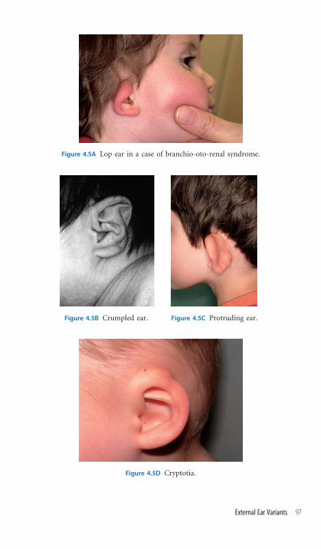
Figure 4.5A Lop ear in a case of branchio-oto-renal syndrome.
Figure 4.5B Crumpled ear. Figure 4.5C Protruding ear.
Figure 4.5D Cryptotia.
External Ear Variants 97

Ear Lobe Variants
Recognizing the Sign Normal variation accommodates a wide range of con-
figurations, but there are a few that may enjoy syndromic association and
therefore are diagnostically noteworthy: (1) ear lobe creases, by which is meant
linear fissures on the lobule of the ear; (2) absence of the earlobe; (3) uplifted
earlobes, in which the insertion of the lobe to the facial skin is above the level
of the lobe so that the lobe hangs, free of medial attachment to the face; and
(4) large, prominent ear lobes, disproportionate to the rest of the external ear.
Establishing a Differential Diagnosis Familial ear lobe variation is com-
mon, so obtain a family history and observe parental ears before ascribing a
diagnostic significance to ear lobe variants. Creases of the ear lobe are
uncommon and generally of no pathologic significance, but are specifically
associated with Beckwith-Wiedemann syndrome (BWS), for which reason
macroglossia, visceromegaly, facial nevus flammeus, and a history of neonatal
hypoglycemia should be specifically sought. The prevalence of the syndrome
is higher among infants conceived by assisted means. Absence of the earlobe is
most frequently seen as a familial, autosomal dominant trait, but is a useful
clinical signal to possible cases of Ehlers-Danlos syndrome (EDS) type IV.
Look for thin skin; prominence of veins, particularly over the chest and
shoulders; and joint laxity, and inquire for a history of easy bruising. In the
clinical context of severe microcephaly, lobeless ears are likely to indicate the
autosomal recessive condition Seckel syndrome. Uplift of the earlobes is a
striking feature of important diagnostic significance in a developmentally
delayed child with constipation and/or Hirschsprung’s disease, indicating a
likely case ofMowat-Wilson syndrome. Seizures, microcephaly, a prominent
philtrum, a large nose, and congenital heart disease strongly support this
diagnosis. Large, prominent lobes are often seen in Costello syndrome, so
seek a history of early feeding difficulty, examine the skin for cutis laxa and the
hands for ulnar drift, and, in the older child, look for nasal papillomata.
Likewise, an early history of poor feeding in a child with large earlobes could
signify a diagnosis of Kabuki syndrome. Look carefully at the lower eyelid for
ectropion, at the eyebrows for possible areas of hair thinning or discontinu-
ity, and at the lower lip for pits. In the older child, unexplained obesity is a
recurrent feature of Kabuki syndrome.
Investigations to Consider Chromosome 11p15 studies for deletion, dis-
omy, or imprinting mutations can confirm BWS if there is clinical doubt but
are complex. If EDS type IV is suspected Col3A1mutation analysis is useful. In
Mowat-Wilson, ZFHX1B mutation confirms the diagnosis.
The Ear98
4.6

Figure 4.6A Ear lobe creases typical ofBeckwith-Wiedemann syndrome.
Figure 4.6B Absent ear lobes in a case ofEhlers-Danlos syndrome type IV.
Figure 4.6C Uplifted earlobe inMowat-Wilson syndrome. This isthe same patient whose face isseen in Figure 2.5C.
Figure 4.6D and E Prominent earlobe is demonstrated in frontal and lateral views of thesame patient with Kabuki syndrome.

External Auditory Canal Atresia or Stenosis
Recognizing the Sign Ninety percent of cases of external auditory canal atre-
sia involve microtia and are addressed in section 4.4. The 10% of cases to which
this section refers are easily missed, with the external ears being normal; the
average age at diagnosis is about 3 years. The external auditory canal may be
completely absent or a small external meatus may be seen, which, on otoscopy,
is blind ending.
Establishing a Differential Diagnosis Establish whether the atresia is uni-
lateral or bilateral. The family history might be useful, rare autosomal dom-
inant kindreds being described in which this disorder segregates. Clinical
assessment of hearing should be expected to demonstrate a significant con-
ductive hearing impairment, but there may also be a sensorineural deafness if
there is associated cochlear malformation. Assess the patient’s body habitus,
specifically the neck for clinical suspicion of a Klippel-Feil malformation
(cervical vertebral fusion). Specifically examine the external ocular move-
ments, with particular reference to the lateral rectus. The condition known as
Wildervanck syndrome is a rather variable syndrome combining features of
Klippel-Feil malformation, sixth nerve palsy, and deafness, in a few of which
cases external auditory canal atresia or extreme stenosis has been observed. In
addition to assessing hearing, a general assessment of development is valuable.
Early evidence of developmental delay in a baby with external auditory canal
atresia should certainly prompt concerns for chromosome 18q deletion
syndrome. Corroborative clinical findings would be microcephaly, cleft pal-
ate, nystagmus, and congenital heart defects. A carp-shaped mouth is said to
be characteristic, by which term is meant a mouth downturned at the corners
with a short philtrum and usually a thickened lower lip.
Investigations to Consider Neck radiology is indicated if cervical spine
malformation is suspected. CT scan of the petrous temporal bones is im-
portant, both to assess likely malformation of the middle ear chamber and
ossicles and to look for cochlear malformations. Karyotype will establish gross
18q 23 deletions, but be aware that there are cases reported with a milder
phenotype in terms of developmental delay in whom the external canal atre-
sia was the clue to submicroscopic deletion of 18q23, not demonstrable by
routine microscopy. The author is aware of one such individual attending
normal school.
The Ear100
4.7

Figure 4.7 External auditorycanal atresia with a normalexternal ear in a patient withchromosome 18q23 deletion.(Courtesy of Dr. MohnishSuri.)
External Auditory Canal Atresia or Stenosis 101

Auricular Tags
Recognizing the Sign These are easily identified and rarely confused. Fleshy,
skin-covered appendages of tissue are seen, usually in front of the tragus of the
ear though occasionally extending along the cheek toward the angle of the
mouth, in which position it is not infrequent to observe an associated scar or
pit. About 90% are unilateral.
Establishing a Differential Diagnosis Fewer than 5% are thought to repres-
ent syndromes, and most auricular tags are isolated unilateral minor mal-
formations of no diagnostic significance. Opinion is divided as to the need
for deafness screening in this group, but if any associated malformation of the
ear is present, an underlying syndrome is more likely and such screening is
certainly justified. In terms of identifying associated syndromes, microtia,
facial asymmetry, epibulbar dermoid, and/or colobomata of the eyelids are
strongly suggestive of Goldenhar syndrome, which is the most common syn-
drome associated with auricular tags. A history suggestive of anal stenosis may
suggest Townes-Brocks syndrome, in which case triphalangeal thumbs
should be sought. The other consideration if anal stenosis is present is cat-eye
syndrome. This rare disorder, consequent on the presence of an additional
chromosome comprising chromosome 22 material, should be suspected if
there is evidence of congenital heart disease, iris coloboma, or horseshoe kid-
ney in a child with auricular tags and/or anal stenosis.
Investigations to Consider Cervical vertebralmalformation should be sought
radiologically in Goldenhar cases. Mutation of SALL1 gene confirms Townes-
Brocks syndrome, while extended chromosomal analysis may be needed to
adequately investigate cat-eye syndrome. Occasionally skin biopsy is required
to confirm the chromosomal mosaicism, as the additional chromosome
22 material can be lost in lymphocytes.
The Ear102
4.8

Figure 4.8A A typical example of an auricular tag, in this instanceassociated with a further tag on the cheek, is shown in a patientwith cat-eye syndrome. Note also the preauricular pit.
Figure 4.8B The arrow shows the marker chromosome 22 material,cytogenetically confirming the clinical diagnosis in the patient inFigure 4.8A.
Auricular Tags 103

Chapter 5 The Mouth and Oral Cavity

Microstomia
Recognizing the Sign Tables are available to establish whether the mouth,
measured from one corner of themouth to its opposite landmark, is enlarged or
reduced outside the norm. In practice, a subjective impression is often formed
looking at the face, which then informs the subsequent examination. A good
rule of thumb is that the angles of the mouth are generally vertically below the
pupils of the eye. Provided there are no concerns about hypotelorism, reference
to the pupils can serve to confirmor refute a clinical impression ofmicrostomia.
Establishing a Differential Diagnosis Consider the obvious by examining for
micrognathia, best done in profile. Observe the symmetry of the mouth both
resting and during movement and consider the associated landmarks, particu-
larly the nasolabial folds, asymmetry of which may signify neuromuscular con-
cerns. Micrognathia causing a small mouth can be identified by examination
of the alveoli with the jaws held together and, instead of meeting evenly, the
lower alveolar margin is well behind the upper. Asymmetric nasolabial folds at
rest and asymmetric crying face on movement are occasional presenting fea-
tures of bothCHARGE (coloboma, heart, atresia choana, retarded growth, genital
abnormalities, ear malformations) syndrome and velo-cardio-facial (VCF) syn-
drome. Hence, reference to the ears for evidence of asymmetry or microtia is
sensible for CHARGE cases, as is seeking a history consistent with choanal atresia/
narrowing. Likewise, cardiac evaluation should form part of the examination in
both these conditions. Carefully evaluate the hands for evidence of digital hy-
poplasia, seen in hypodactyly-hypoglossia syndrome and Moebius syndrome.
Sixth and seventh cranial nerve palsies characterize the latter, and the pectoral
muscles should be assessed for possible absence. A small tongue, perhaps anchored
to the floor of the mouth, is more likely to represent hypodactyly-hypoglossia
syndrome. A tightly contracted mouth usually accompanied by H-shaped puck-
ering of the chin is the hallmark of Freeman-Sheldon syndrome and the diag-
nosis is substantiated by observing camptodactyly with ulnar deviation of the
digits. Additional related clinical signs are deep-set eyes and a long philtrum.
Tight contraction of the mouth in association with joint limitation and increased
tone are likewise the hallmarks of Schwartz-Jampel syndrome, an autosomal
recessive disorder that clinically overlaps with Freeman-Sheldon syndrome.
Investigations to Consider Unadorned clinical examination is the basis of
several of the diagnoses discussed. Echocardiography is important if CHARGE
or VCF syndromes are being considered. Fluorescence in situ hybridiza-
tion (FISH) examination of 22q11 will confirm the characteristic deletion in
VCF syndrome. An electromyograph may be needed to establish myotonia in
Schwartz-Jampel cases. Likewise, radiologic evaluation of the vertebrae for
platyspondyly and coronal clefts and of the hips for epiphyseal dysplasia is
diagnostically valuable in this condition.
The Mouth and Oral Cavity106
5.1

Figure 5.1 Microstomia is well demonstrated in this classical caseof Freeman-Sheldon syndrome. Note additional clinical signs ofthe H-shaped vertical skin pucker on the chin; the deep-set, tightlyclosed eyes; and the camptodactyly of the fingers, showing ulnardeviation.
Microstomia 107

Upper Lip Clefts
Recognizing the Sign Embryologically, clefts derive from failure of fusion of
the maxillary processes with the medial nasal processes, and clinical evidence
of this failure may present with bilateral defects, unilateral defects, midline
defects, a slight defect reminiscent of a scar adjacent to the margin of the
philtrum (pseudocleft), or a slight dimple at the vermillion border of the lip.
Some 20% of clefts are associated with an underlying syndrome.
Establishing a Differential Diagnosis Ensure that the palate is intact as cleft
lip without cleft palate is empirically less likely to be associated with mal-
formations elsewhere. The family history may offer clues consistent with au-
tosomal dominant clefting or other important information—repair of a cleft
palate in a parent should prompt examination of both parent and child for
lower lip pits, which would confirm Van der Woude syndrome. Midline
clefts, though rare, have important implications and the clinician needs to be
aware of the underlying possible diagnosis of holoprosencephaly. Appro-
priate assessment for this should include evaluation for hypotelorism and op-
tic colobomata. Parental assessment should include looking at the teeth for a
single central maxillary incisor. Specific chromosomal syndromes to bear in
mind in examining the infant presenting with cleft lip are Patau syndrome
(trisomy 13), Wolf-Hirschhorn syndrome (4p16 deletion), and 1p36 deletion.
A low birth weight, wide nasal bridge, and Greek helmet profile to the nose
will prompt consideration of 4p deletion, especially if there is a cardiac mur-
mur. Chromosome 1p36 deletion does not have a neonatal ‘‘gestalt’’ appear-
ance, but deep-set eyes and a rather horizontal configuration to the eyebrows
may correctly prompt the examiner. The limbs may offer clues—broad or bifid
halluces in oro-facio-digital syndromes, clinically confirmed by looking for
multiple oral frenula, or ectrodactyly in ectrodactyly-ectodermal dysplasia
syndrome. Sparse eyebrows and eyelashes and dry skin should result in eval-
uation of the parents for possible Rapp-Hodgkin syndrome, an autosomal
dominant disorder, in which wiry hair, absence of secondary sexual hair, and
hypohidrosis are usual. In boys, clefting associated with hypospadias or an
ectopic anus is likely to signify Opitz G syndrome, and the possibility of
laryngeal cleft needs to be formally investigated.
Investigations to Consider Karyotype is indicated only if there are addi-
tional unexplained malformations. Specific FISH for 4p16 in Wolf-Hirsch-
horn and molecular cytogenetic analysis may be necessary to identify subtle
1p36 deletion. The underlying loci are known for ectrodactyly-ectodermal
dysplasia, Rapp-Hodgkin, Opitz G, and Van der Woude syndromes; the same
is true of several holoprosencephaly loci, but the diagnosis is essentially a
clinical issue.
The Mouth and Oral Cavity108
5.2

Figure 5.2A Unilateral cleft lipis seen in this patient. Notethe secondary deformation ofthe nares consequent to theprimary cleft malformation.
Figure 5.2B The vertical right-sided impression of a scar on thispatient’s philtrum is in fact a pseudocleft. He comes of a familywith a strong history of autosomal dominant cleft lip and thisclinical sign indicates that he carries the family mutation, thoughnot affected as obviously as others in the pedigree.
Upper Lip Clefts 109

Lip Pits
Recognizing the Sign Three distinct forms are observed. Commisural pits,
seen in the angles of the mouth on opening the oral cavity, are of no clinical
significance. Upper lip pits generally occupy a midline situation and com-
municate with underlying glands but do not betoken a syndromic or dys-
morphologic significance. Lower lip pits are generally seen as depressions,
occasionally as minor elevations, on either side of the midline and also com-
municate with underlying glands. Most lower lip pits have a syndromic as-
sociation.
Establishing a Differential Diagnosis Lower lip pits are uncommon, the
greatest hazard being that they are easily missed, and it is worth everting the
lower lip to inspect the mucocutaneous junction for their presence. If ob-
served, then three specific syndromes need to be considered in the course
of examination. Van der Woude syndrome is an autosomal dominant con-
dition, so a family history and examination of family members may further
assist the diagnosis. Look for cleft palate, bifid or deviated uvula, and evidence
of repaired cleft lip/palate in the family. Rare features described include
Hirschsprung’s disease and synechiae of the eyelids, but these are not typical.
Cleft lip/palate and lip pits in the patient may signify popliteal pterygium
syndrome, the clinical hallmark being popliteal webs, which limit extension
of the knees. Since this syndrome is autosomal dominant, pertinent ques-
tioning with respect to the medical history of other family members will often
generate additional diagnostic data. Good clinical clues to watch out for on
examination are oligodactyly, syndactyly, hypoplastic nails, and genital mal-
formations, especially micropenis. Finally, lip pits are an inconstant but valu-
able prompt to the sporadically occurring multiple congenital anomaly con-
dition known as Kabuki syndrome. Characteristic facial features to look for
in considering this diagnosis are arched eyebrows, often interrupted (in other
words, a hairless region occurs as a gap in the eyebrow), and wide palpebral
fissures, frequently with ectropion of the lower eyelid (Figure 3.14). Congeni-
tal heart defects are a common presentation in the newborn period and symp-
toms may be cardiac or feeding related, sustained feeding problems being a
particular problem in these children. Variable immune dysfunction–related
presentations in the first year or two of life are described, while presentation in
the preschool child is generally related to delayed development. The history,
facial features, and, if present, lip pits prompt the diagnosis. Confirmatory ad-
ditional signs to be vigilant for during examination include fetal finger pads,
blue sclera, and short stature. Obesity is a feature in adolescence.
Investigations to Consider Analysis of the IRF6 gene has established familial
mutations in both Van der Woude and popliteal pterygium syndromes.
The Mouth and Oral Cavity110
5.3

Figure 5.3B This photograph of a patient withKabuki syndrome shows a pit as a slightly atrophicarea on the patient’s left.
Figure 5.3A Slight swellings are shown on either sideof the midline with the pits visible just behind theswellings. The patient has a diagnosis of Van derWoude syndrome.
Lip Pits 111

Thick and Thin Upper Lips
Recognizing the Sign It is possible to formally measure the thickness of
the lip. The Handbook of Normal Physical Measurements describes a technique
measuring in the midline from the vermillion border to the aperture of the
mouth and provides normal ranges. In practice, this is very rarely undertaken,
but a subjective impression of the child’s face may register the lip as being
thicker or thinner than usual.
Establishing a Differential Diagnosis Unusual degrees of lip thickness can
be familial and nonpathologic, so clinical signs relying on this observation are
supportive of a diagnosis only in the presence of other concerns. Unusually
thin lips noted in an older child under assessment for mild developmental
delay or jitteriness in the newborn period may remind the examiner to
consider fetal alcohol syndrome and other features of that ill-defined entity
should be sought, such asmicrocephaly, unusual palmar creases, and a smooth
philtrum. In assessing an older child presenting with short stature and mild
developmental concerns, a thin upper lip may be the clue to a diagnosis of
Floating-Harbor syndrome. Additional support for that conclusion would
come from observing deep-set eyes and a nasal columella extending below the
ala nasi and confirming from the history that the birth weight was low. When
assessing a developmentally delayed child in whom there is a thick lip, be
mindful in boys of the X-linked conditions Coffin-Lowry syndrome and
a-thalassemia mental retardation X syndrome (ATRX). An especially valu-
able clinical sign in Coffin-Lowry patients are the fingers, which are puffy and
tapering, sometimes described as ‘‘sausage shaped.’’ Obviously a family history
may offer consolidation, but examination of the mother can also be helpful—
particularly the hands of carriers of Coffin-Lowry syndrome. Fullness of the
lip is a helpful clinical feature of Williams syndrome, especially in the slightly
older child, so it is worth checking the iris for a stellate pattern and the history
for hypercalcemia or heart disease. Lip prominence in a newborn with unex-
plained growth retardation should cause the diagnosis of leprechaunism to be
considered and provoke examination for the other clinical signs—acanthosis
nigricans, breast enlargement, prominent ears, and genital enlargement.
Investigations to Consider Significant bone age delay is characteristic in
Floating-Harbor syndrome. HbH inclusion bodies on brilliant cresyl blue
staining are seen in about 90% of ATRX cases, though mutation analysis may
sometimes be necessary to secure a diagnosis. Coffin-Lowry cases have mu-
tations of the RSK2 locus on Xp. Williams syndrome is confirmed by FISH for
ELN deletion on 7q. Leprechaunism is confirmed by insulin receptor gene
mutations, INSR on 19p.
The Mouth and Oral Cavity112
5.4

Figure 5.4A and B Thin upper lip (A) and brachyclinodactyly (B) areshown in this 5-year-old boy with Floating-Harbor syndrome, referredfor developmental concerns and short stature. See Figures 9.9 forCoffin-Lowry syndrome and Figure 1.6C for ATRX syndrome.
Thick and Thin Upper Lips 113

Lip and Oral Mucosa Pigmentation
Recognizing the Sign Spotty pigmentation of the lips and oral mucosa is an
uncommon finding with very specific clinical connotations. The sign is readily
recognizable as brownish macules, of variable diameter, most noticeable on
the lips but sometimes also present on the hard palate, gums, oral mucosa,
and perioral and periorbital regions. Very infrequent at birth or in the new-
born period, these usually develop in later childhood.
Establishing a Differential Diagnosis The family history may betray clues—
neurofibromatosis type 1 is a good example, where the emergence of clinical
features in a child takes place over a period of time but the diagnosis can
generally be suspected and complications sought early by reference to family
history. Assess carefully for optic glioma in the young child and for scoliosis in
the older age group. Blood pressure monitoring is important in all age groups,
there being an association with both pheochromocytoma and renal artery
stenosis. The classical association of lip pigmentation is Peutz-Jeghers syn-
drome (P-JS), in which hamartomatous polyposis and malignant propensity
of the small bowel is increased, as are malignancies of the breasts and re-
productive organs. Episodic colicky abdominal pain in adolescence usually
signals intussusception of small bowel polyps. Macules may also be found on
the palms, soles, volar aspects of the fingers, and genitalia. Laugier-Hunziker
syndrome (L-HS) is a very rare, sporadic condition clinically indistinguish-
able from P-JS in lip and oral pigmentation, but without associated gastro-
intestinal complication. Whereas the onset of the pigmentation in P-JS is
usually complete by puberty, the onset of pigmentation in L-HS is usually in
adult life. In Carney complex, the lip and mucosal pigmentation is spotty, but
usually has a more extensive distribution around the face. This autosomal
dominant diagnosis is important because of the association with atrial myx-
oma. Conjunctival pigmentation and eyelid myxomas are occasional features.
Finally, consider LEOPARD syndrome (lentigines, echocardiographic abnor-
malities, ocular hypertelorism, pulmonary stenosis, abnormal genitalia, retar-
dation of growth, and deafness). Assess for hypospadias, cardiacmurmurs, and
multiple lentigines of the face, neck, and trunk, and assess parents for evidence
of this autosomal dominant condition.
Investigations to Consider Almost all cases can be resolved at a clinical level.
Echocardiography is important for LEOPARD and Carney complex, to spe-
cifically assess for atrial myxomata in the latter disorder, which may be re-
current. Mutation analysis is available for all conditions in the event of clinical
uncertainty.
The Mouth and Oral Cavity114
5.5

Figure 5.5 Numerous discrete pigmented macules, typical of Peutz-Jeghers syndrome, are demonstrated.
Lip and Oral Mucosa Pigmentation 115

Macroglossia
Recognizing the Sign The tongue is usually contained fully within the
oral cavity, and any protrusion of the tongue begs examination. True macro-
glossia signifies a large tongue, while relative macroglossia signifies a small
oral cavity, the tongue protrusion being a secondary effect.
Establishing a Differential Diagnosis Establish whether there is micro-
gnathia, a clue that the macroglossia is secondary. In the neonate, specifically
examine for clinical signs ofDown syndrome—especially useful are hypotonia,
poor Moro reflex, excess nuchal skin, and simian hand creases. In the older
child whose chromosomes are normal but whose development is inexplicably
delayed, relative macroglossia and a flat or dished appearance to the midface
can be the clues to chromosome 9q34 deletion syndrome. The clinical im-
pression of the 9q34-deleted patient among colleagues and family is often of a
patient who looks like he or she has Down syndrome but who has ‘‘normal’’
chromosomes. True macroglossia in the neonate is suggestive of Beckwith-
Wiedemann syndrome (BWS). Corroborative signs to look out for are nevus
flammeus, visceromegaly, ear creases on the lobe or pit-like depressions on the
helical rim, a history of hypoglycemia, and a history of assisted conception,
which is associated with increased prevalence of BWS. There is clinical overlap
between BWS and Simpson-Golabi-Behmel syndrome, an X-linked mental
retardation syndrome comprising large birth weight and macroglossia, often
with a deep midline groove on the tongue, the groove perhaps extending to
the lower lip. Particularly valuable clinical signs that suggest this diagnosis
are accessory nipples and nail hypoplasia, most notably of the index finger.
Asymmetry of a congenitally enlarged tongue probably signifies an underlying
hemihypertrophy as the cause, and careful examination of the limbs may show
evidence for this. Macroglossia in a neonate whose clinical presentation is of
unexplained weakness and/or poor feeding is typical of Pompe’s disease, a gly-
cogen storage disorder. Tongue fasciculation and joint contractures are corrob-
orative signs to assess. An impression of macroglossia combined with emerging
developmental concerns in the first couple of months of life should prompt in-
vestigation of mucopolysaccharidosis (MCP), even before other clinical signs
such as joint stiffness and corneal clouding appear.
Investigations to Consider Karyotype is indicated for suspected Down syn-
drome, but specific molecular cytogenetic assay is required to identify 9q34
deletion. BWS remains in essence a clinical diagnosis, molecular analysis for 11p
imprinting center defects being demonstrable in about 50% of cases. Simpson-
Golabi-Behmel syndrome may be confirmed by mutation analysis of the GPC3
gene on Xq21. An electrocardiogram is valuable in Pompe’s disease, showing
giant QRS complexes, and an echocardiograph shows ventricular enlargement.
Glycosaminoglycan excretion in urine is the screening test for MCP.
The Mouth and Oral Cavity116
5.6

Figure 5.6 True macroglossia is shown in a 3-month-old child withBeckwith-Wiedemann syndrome, conceived by assisted reproduction.
Macroglossia 117

Accessory Oral Frenula
Recognizing the Sign The frenula are the normal folds of mucosal mem-
brane extending between the alveolar ridge and the inside of the upper lip and
the counterparts inside the lower lip and beneath the tongue. Any additional
such mucosal folds are termed accessory frenula or aberrant frenula.
Establishing a Differential Diagnosis Accessory frenula are important clues
to syndrome diagnosis but are not the subject of scrutiny in most general
clinical examinations undertaken and, for this reason, are often found in chil-
dren whose more clinically obvious signs have been noted earlier. In a new-
born with postaxial polydactyly of the hands, the identification of accessory
oral frenula is very suggestive of the autosomal recessive condition Ellis-van
Creveld syndrome. Look for corroborative signs, especially small, deep-set,
hypoplastic fingernails and a murmur consistent with an atrial septal defect,
present in about 50% of cases. Natal teeth are another good clue to this diag-
nosis on intraoral examination, though not invariable. The presence of cleft lip
or palate should prompt a search for lip pits and accessory frenula, which are
common in popliteal pterygium syndrome. The pterygia result in a popliteal
web and there is associated abnormality of the toenail of the hallux, a pyra-
midal skinfold extending from the base to the tip of the toenails. The clinical
value of limb assessment also applies to hypoglossia-hypodactyly syndrome,
in which a small tongue is seen, often with accessory oral frenula, perhaps ex-
tending between the palate and the floor of the mouth. There is associated mi-
crognathia, and cranial nerve defects, especially of the sixth and seventh nerves,
are often demonstrable clinically. Syndactyly, adactyly, and variable micro-
dactyly should be sought. In oro-facio-digital (OFD) syndromes, of which
there are a bewildering variety of clinically and genetically distinct forms, oral
frenula associated with preaxial or postaxial polydactyly signal the diagnosis.
Though not invariable, mental retardation in associated with several forms of
OFD syndromes, as are central nervous system malformations.
Investigations to Consider Karyotype is justified if accessory frenula are
identified as part of a child with multiple congenital anomalies for whom no
unifying syndromic diagnosis is apparent. Neuroimaging of the brain is im-
portant in refining the diagnosis of which form of OFD syndrome a child may
have, the inheritance differing among the several discrete forms. Mutation
analysis for Ellis-van Creveld and popliteal pterygium syndromes is not nec-
essary to the diagnosis of these clinically recognizable disorders.
The Mouth and Oral Cavity118
5.7

Figure 5.7 Multiple accessory alveolar frenula are demonstratedin this patient with a diagnosis of orofaciodigital syndrome, typeII. Note the associated gum hypertrophy, which was secondary tophenytoin medication for epilepsy. (Reproduced with permissionfrom Reardon et al., J Med Genet 1989;26:659–663.)
Accessory Oral Frenula 119

Isolated Cleft Palate
Recognizing the Sign Cleft of the soft and/or hard palate is easily seen and
should be sought in all infants. Less easily identified are more subtle forms,
specifically submucous clefting of the soft palate, wherein the oral mucosa
shows no defect but a notch is palpable at the posterior margin of the hard
palate. The uvula should always be inspected and, if bifid, a submucous cleft
should be suspected. The associated velopharyngeal incompetence may be
suspected in a baby where a history of milk leaking down the nares during
feeding is elicited. Recourse to examination by an expert may be required to
confirm.
Establishing a Differential Diagnosis Fifty percent of cases of isolated cleft
palate have associated congenital malformations and 20% have an identifi-
able monogenic cause, while 1% of cases have a chromosomal abnormality.
Trisomy 13 represents the most recognizable karyotypic abnormality, but
very many others are described. However, most cleft palate cases are isolated
events often associated with micrognathia, resulting in Pierre-Robin sequence.
In seeking monogenic causes, the family history may indicate several affected
males in a kindred, suggesting the nonsyndromic condition of X-linked cleft
palate. Ankyloglossia, where the tongue is anchored to the floor of the mouth,
is a feature in some individuals affected with this disorder, while female
heterozygotes may show bifid uvula and submucous clefting. Lip pits or cleft
lip in the family should suggest Van der Woude syndrome (section 5.3).
A cardiac murmur clinically in a child with cleft palate or a history suggestive
of velopharyngeal incompetence should prompt assessment for velo-cardio-
facial (VCF) syndrome. Look for the prominent nose, the nasal tip being
somewhat square, and seek a corroborative history of hypocalcemia or recur-
rent infections. VCF syndrome is remarkably variable, and a low threshold for
investigation is appropriate. Hypospadias associated with cleft palate, espe-
cially if there is a family history of same, is likely to signify the X-linked
condition of Opitz G syndrome. Female heterozygotes may show mild de-
grees of hypertelorism. Severe short stature present at birth with nasal flat-
tening and associated cleft palate points toward a skeletal dysplasia as the
underlying cause, the exact diagnosis possibly being Kniest syndrome. Joint
laxity in association with cleft palate may be symptomatic of Stickler syn-
drome, especially if there is evidence of myopia and flattening of the nasal
bridge.
Investigations to Consider Karyotype is almost always normal, but FISH of
22q11 is important for diagnosis of VCF cases. Skeletal radiology in associ-
ation with clinical findings is the basis of Kniest and Stickler diagnoses.
The Mouth and Oral Cavity120
5.8

Figure 5.8A The typical V-shaped defect in the soft palate isdemonstrated in this child who had no other malformations andfor whom a diagnosis of nonsyndromic cleft palate was reached.
Figure 5.8B Absent uvula with associated mid-line translucency, betraying absence of musclefusion, is shown. These features are entirelyconsistent with a diagnosis of subclinical cleftpalate. (Courtesy of Mr. David Orr.)
Isolated Cleft Palate 121
Persistent Drooling
Recognizing the Sign This hardly requires any clinical expertise, but a good
history can inform the examination and investigation. The neonatal feeding
history will often be of a poor feeding pattern, perhaps requiring nasogastric
supplementation. Establish whether there was macroglossia at birth, cleft pal-
ate, or micrognathia. Was there any suggestion of velopharyngeal incompetence
on feeding, often represented by nasal regurgitation of milk during feeding?
Gauge the progress of the child with respect to perceptive and expressive speech.
Establishing a Differential Diagnosis Observe the face for evidence of a
generalized myopathy and then assess the patient’s muscle bulk generally.
Specifically, look at the tongue. Establish whether it looks large and symmet-
ric, perhaps with a deep central groove, which might cause you to consider
Simpson-Golabi-Behmel (SGB) syndrome, especially if there was a high birth
weight, a history of X-linkedmental retardation, and/or cardiac malformations.
Evaluate the heart clinically—a murmur will add weight to your diagnostic
concern. Cardiac concerns, a history of nasal regurgitation of milk on feeding,
and speech delay may equate with velo-cardio-facial (VCF) syndrome. Does
the tongue look asymmetric, in which case careful evaluation of the face and
limbs for asymmetry is warranted? Does the tongue move normally and spe-
cifically assess whether the child protrudes the tongue from the oral cavity?
Failure to protrude the tongue with a history of poor feeding and delayed ex-
pressive speech is typical of Worster-Drought syndrome. In this condition,
examine the lower cranial nerves—often there are signs of pseudobulbar palsy.
There may be a striking discrepancy between the general development of the
child, which can be within normal parameters, and the expressive speech, which
is greatly delayed. A history of polyhydramnios, an open-mouth appearance,
and generalized muscle weakness are the classic hallmarks of congenital myo-
tonic dystrophy. Careful evaluation of the family history may establish cases
of symptomatic cataract before the age of 40 years, and maternal examination
in this condition will establish corroborative signs, such as myotonia of the
thenar eminence on percussion. Transmission of the congenital form is virtually
exclusively maternal. Sixth or seventh cranial nerve palsy strongly suggests
Moebius syndrome as the underlying diagnosis, in which case limb asymmetry,
syndactyly, and brachydactyly should be sought and the pectoral wall muscles
be evaluated, since absence is quite common in this condition.
Investigations to Consider GPC3 gene mutations underlie SGB syndrome,
but clinical diagnosis is usually possible. FISH of 22q11 is indicated to establish
VCF syndrome. Myotonic dystrophy is demonstrable as a triplet repeat ex-
pansion in theMD gene on chromosome 19.Magnetic resonance imaging scan
in Worster-Drought cases frequently establishes perisylvian polymicrogyria.
The Mouth and Oral Cavity122
5.9

Figure 5.9A Note the drooling and open-mouth appearance in this3-year-old boy referred for ‘‘unexplained mental retardation.’’The motor milestones were almost within normal parameters, buthe had persistent drooling and could not protrude his tongue.A clinical diagnosis of Worster-Drought syndrome was made.
Figure 5.9B Magnetic resonance imaging scanof the same patient confirms perisylvianpolymicrogyria.
Persistent Drooling 123

Gum Hyperplasia
Recognizing the Sign In the child whose teeth have erupted, gum hyper-
trophy is readily apparent, the teeth being engulfed by the gingiva, which may
themselves have a nodular appearance. In the neonate, this clinical sign is much
more subjective. Repeated observation of the normal during routine exami-
nation will develop the necessary recognition skills to identify the abnormal.
Establishing a Differential Diagnosis The most commonly recognized cause
of gingival hyperplasia is drug related and a history of medications taken may
readily explain the clinical sign. Specifically, phenytoin and cyclosporin are
medications that are commonly associated with this clinical side effect. The
observation of gum hyperplasia in the neonatal stage suggests a few specific
diagnoses—Robinow syndrome, I-cell disease, and leprechaunism. In the
latter, breast enlargement, genital hypertrophy, and acanthosis nigricans
strongly support this possible diagnosis, especially if there is a history of in-
trauterine growth retardation and a general appearance of lipodystrophy—
very little subcutaneous fat. Clinical features that ought to be sought in Ro-
binow syndrome include cleft lip, anteverted nares, and hypertelorism. The
genitalia may not be notably abnormal in the female, but a normally formed
small penis is usual in affected males. It is worth noting the hands and feet—if
there is not clinical evidence of a bifid thumb or hallux, a broad digit may be
the prompt that radiologic investigation might establish evidence of incom-
plete reduplication. I-cell disease follows a typical storage disease pattern,
with concerns that development is not proceeding normally often prompted
by initial feeding difficulties, progressive limitation of joint movement, and
corneal clouding. The author has seen the diagnosis made prior to the emer-
gence of any of these landmarks by preemptive investigation following the
recognition of gum hyperplasia in the neonate. There is clinical overlap be-
tween I-cell disease and a rare autosomal recessive disorder, infantile sys-
temic hyalinosis, in which failure to thrive neonatally, gingival hyperplasia,
and crying due to pain on joint movement, with the early emergence of flex-
ion contractures, represent a fairly typical pattern. Facial papules, perioral
pigmentation, and fleshy nodules at the anal margin allow this condition to be
confidently recognized. Hyperpigmented swellings over the metacarpopha-
langeal joints emerge with time.
Investigations to Consider Hemivertebrae or bifid terminal phalanges of the
thumb/hallux radiologically support the clinical diagnosis of Robinow syn-
drome. Insulin receptor gene mutations confirm leprechaunism, if the clinical
diagnosis is not secure. While urinary oligosaccharides, vacuolated lympho-
cytes, and/or disordered lysosomal enzymes may be observed in I-cell disease,
fibroblast culture is required for demonstration of the characteristic ‘‘inclu-
sion bodies.’’
The Mouth and Oral Cavity124
5.10

Figure 5.10A Gum hypertrophy in a6-year-old child treated with cyclosporin.(Courtesy of Mr. Paddy Fleming.)
Figure 5.10B Gum hypertrophy in a 14-month-old child with I-celldisease.
Gum Hyperplasia 125

Chapter 6 The Neck
Neck Webbing
Recognizing the Sign Neck webbing refers to redundant skin at the back and
sides of the neck and is thought to arise from resolved nuchal edema in utero.
In contrast to a short neck, it is possible to palpate the folds of excess skin
between the fingers. The posterior hairline is often extended downward in as-
sociation with the webs of excess skin. Distinction needs to be drawn between
excess nuchal skin and specific webs of tissue (pterygia) in the neck region,
which frequently accompany limitation of extension in multiple joints as part
of a wider multiple pterygium syndrome, all of them rare.
Establishing a Differential Diagnosis Observe the parents and ask if either
of them have needed cardiac examination/intervention. Noonan syndrome
frequently presents with nuchal edema, indeed even generalized hydrops on
occasion, and much negative investigation may be spared by such parental
evaluation. Short stature, ptosis, and posteriorly rotated ears are all good
clinical signs to watch out for in considering this diagnosis, as well as a car-
diac examination for congenital heart disease, particularly pulmonary ste-
nosis. A history of abnormal bleeding after minor procedures is consistent
with Noonan syndrome. In the older child, multiple cafe-au-lait patches with
facial and other features of Noonan syndrome signify the subtype known as
Noonan-neurofibromatosis type 1 (NF1). These children essentially haveNF1
and need appropriate management measures for that condition. Clinical signs
of coarctation of the aorta in a baby with excess nuchal skin should cause a
likely diagnosis ofTurner syndrome to be considered. The classic clinical signs
of cubitus valgus, short stature, and wide-spaced nipples may not be apparent
until much later, but puffy feet are a good clue in the neonate. Excess nuchal
skinmay occur as part of a generalized cutis laxa presentation. Early feeding dif-
ficulties may presage the later emergence of coarse facial features, developmen-
tal delay, and a diagnosis of Costello syndrome. Likewise, a generalized cutis
laxa presentation, but with normal development, can signal a connective tissue
disorder, most likely one of the forms of Ehlers-Danlos syndrome (EDS). As-
sessment should establish the degree of skin elasticity as well as any scarring,
striae, easy bruising, and a family history of the same.
Investigations to Consider An echocardiogram is mandatory if a diagnosis
of Noonan or Turner syndrome is being considered. Costello syndrome is
open to molecular investigation of the KRAS gene but this is rarely necessary
in experienced clinical hands. Before reaching a diagnosis of EDS, it is worth
assaying tenascin X, easily done on serum, deficiency of which produces a
condition that overlaps with EDS, especially types II and III.
The Neck128
6.1

Figure 6.1 Excess nuchal skin is dem-onstrated in this girl with Noonansyndrome,whoseneonatal presentationwas of generalized hydrops.
Neck Webbing 129

Short Neck
Recognizing the Sign The underlying problem in this clinical situation is
likely malformation or malsegmentation of the cervical vertebrae. Conse-
quently, the head appears to sit directly on the shoulders. Unsurprisingly, the
posterior hairline is usually low and the mobility of the neck reduced in all
directions. Neck webbing may be observed as an associated secondary phe-
nomenon.
Establishing a Differential Diagnosis Empirically, the most likely diagnosis
is that of Klippel-Feil syndrome. In this condition, there will be an under-
lying radiologic fusion of vertebrae, which may extend beyond the cervical
region in severe cases. Clinical examination should assess for evidence of a
sixth cranial nerve palsy (Duane phenomenon) and deafness, both associated
with Klippel-Feil malformation, the combination of all three being known as
Wildervanck syndrome.Curiously, the condition is almost exclusive to females.
Associated malformations of the renal and internal genital tracts are common,
specifically vaginal atresia, absent uterus, renal agenesis, hypoplasia, and ure-
teric reduplication. Mirror movements are a well-described clinical phenom-
enon and should be sought. Epibulbar dermoids should be noted, and their
observation supports the likely diagnosis. Congenital upward displacement of
the scapula, known as Sprengel shoulder, may be unilaterally or bilaterally pres-
ent. The observation of a short neck and/or Sprengel shoulder in a male should
be followed by palpation of the vas deferens, congenital bilateral absence of
the vasa being a frequent phenomenon in boys with renal anomalies and cer-
vical malsegmentation. The association of limb abnormalities, especially the-
nar eminence hypoplasia or thumb reduplication, is more reminiscent of
MURCS association (Mullerian, renal, cervical spine), in which the vertebral
fusion is associated with renal and uterine dysplasia, generally with absence of
the uterus. In the event of a short neck being observed in a child of normal
developmental progress but concerns of short stature, it is useful to be aware
thatMorquio syndrome (mucopolysaccharidosis [MPS] type IV)may so pres-
ent, the limitation of joint movement characteristic of other types of MPS not
being so reliable in this form of storage disease. Corneal clouding and opacities
should be sought, but are not invariable.
Investigations to Consider Neck radiology and renal and pelvic ultrasound
are essential to fully evaluate Klippel-Feil and associated malformations, and
likewise to differentiate MURCS syndrome. Audiologic evaluation should be
considered. Keratan sulphate urinary excretion is usually increased in MPS
type IV. Skeletal radiology will generally show dysostosis multiplex, and od-
ontoid hypoplasia is a particular feature. Enzymatic confirmation is available
by leucocyte study.
The Neck130
6.2

Figure 6.2 Short neck and low poste-rior hairline are shown in a femalepatient who presented at age 14 years,with short stature and menstruationnot having commenced. The uteruswas absent. There was unilateral renalagenesis and cervical vertebrae C3–5were fused. The diagnosis was MURCSassociation.
Short Neck 131

Goiter
Recognizing the Sign Goiter simply signifies enlargement of the thyroid.
Generally in children this is of uniform, as opposed to nodular or localized,
character, as can apply in adults. The bilobar shape of the thyroid, straddling
the midline, is readily apparent.
Establishing a Differential Diagnosis Neonatal goiter is uncommon and
should rightly raise concerns for a biochemical abnormality in thyroxine syn-
thesis. However, do be aware that maternal ingestion of carbimazole can
result in congenital goiter. In the event of a maternal history of carbimazole
ingestion, it is worth looking specifically for choanal atresia, or severe nar-
rowing of the nasal passages, as well as nipple hypoplasia or absence in the
baby, both features well described in carbimazole embryopathy. Likewise,
localized scalp defects have been described in several cases of this teratogenesis
syndrome. In considering possible metabolic causes, it is worth recording
that most causes of neonatal hypothyroidism are associated with aplasia or
aberrant development of the gland andwill not be associated with goiter. How-
ever, congenital or emerging neonatal goiter does require full pituitary eval-
uation, lest a treatable form of mental retardation be missed. Oral thyroxine is
effective in such cases. The most common association of childhood goiter is
with Pendred syndrome, in which deafness is the key feature. Generally the
deafness predates the onset of goiter by a decade or more. The deafness has
particular characteristics that allow the diagnosis to be suspected clinically. The
hearing threshold fluctuates and parents will be aware of day-to-day variation
in the child’s hearing; it is sensitive to atmospheric pressure variation, so it can
deteriorate after airline travel, diving, etc., and it is often associated with var-
iable vestibular symptoms. Acute, irreversible loss of hearing following head
injury is well described. If the deafness has been appropriately evaluated by
computed tomography (CT) scan of the petrous temporal bones, the diagnosis
will be made well before the thyroid enlargement emerges—typically in the
second decade of life. Congenital and early-onset goiter is very uncommon in
Pendred syndrome.
Investigations to Consider Thyroid-stimulating hormone (TSH) evaluation,
thyroid-releasing hormone (TRH) stimulation, and magnetic resonance imag-
ing (MRI) evaluation of the anterior pituitary are urgent considerations in con-
genital goiter. In the deaf child, dilatation of the vestibular aqueducts, onCT scan
of the petrous temporal bones, will disclose the very likely diagnosis of Pendred
syndrome. The only other documented cause of dilated vestibular aqueduct as
an isolated finding is distal renal tubular acidosis with deafness. Less commonly,
a Mondini malformation of the cochlea is identified in Pendred syndrome pa-
tients. Mutation analysis of the SLC26A4 gene discloses multiple mutations in
both goiter-associated and non–goiter-associated Pendred syndrome.
The Neck132
6.3
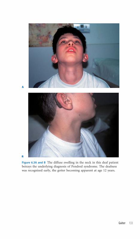
Figure 6.3A and B The diffuse swelling in the neck in this deaf patientbetrays the underlying diagnosis of Pendred syndrome. The deafnesswas recognized early, the goiter becoming apparent at age 12 years.
Goiter 133
Neck Sinuses/Fistulae/Pits
Recognizing the Sign A clinical distinction between a branchial sinus and a
fistula cannot be drawn without surgical exploration. By definition, a fistula
has an external opening onto the cutaneous surface and extends inward,
opening into the pharynx. A sinus, by contrast, is blind ending internally but,
like a fistula, creates a clinically identifiable pit on the skin. These external
openings are generally seen along the anterior border of the sternocleido-
mastoid muscle and are sometimes a focus for secretions and secondary infec-
tion. The only other likely cause of a neck sinus is a thyroglossal cyst, which,
by contrast to branchial cysts/sinuses, is midline and may be found at any po-
sition between the sternal notch and the hyoid bone.
Establishing a Differential Diagnosis Thyroglossal cysts, though occasion-
ally familial, do not betoken a syndromic association and should be viewed as
isolated developmental malformations. Branchial pits need more careful di-
agnostic evaluation. Most occur as sporadic isolated findings, either unilateral
or bilateral, in otherwise normal individuals, without evidence of associated
malformations. The specific diagnostic concern raised by the observation of a
branchial sinus is whether the patient has branchio-oto-renal (BOR) syn-
drome. Preauricular pits; external ear malformations, including microtia;
and evidence of deafness should be assessed. Most affected individuals have
hearing loss, with mixed type being most likely. The external auditory canal
is often narrow; indeed, cholesteatoma is occasionally described. A history of
urinary tract infection will suggest renal involvement and needs to be specifi-
cally investigated, irrespective of symptoms, if this diagnosis is under consid-
eration. Renal agenesis or hypoplasia, cystic renal changes, and vesicoureteric
reflux are all described, though actual renal failure is rare. The identification
of a sixth nerve palsy is specific to deletions involving the BOR locus on chro-
mosome 8. Neck sinuses overlying a hemangiomatous region of tissue, often
along the line of the sternocleidomastoid muscle but also occurring behind
the ear, are typical of branchio-oculo-facial (BOF) syndrome. The philtrum
is narrow but shows thick vertical ridges, producing a pseudocleft appearance.
Ocular involvement is usual and can embrace coloboma, lacrimal gland ob-
struction, and hypertelorism separately or in combination.
Investigations to Consider Biochemical parameters of renal function should
be undertaken in both BOR and BOF syndromes as well as ultrasound eval-
uation of the kidneys and renal tract. CT scan of the petrous temporal bone in
BOR syndrome shows dilated vestibular aqueduct in association with mid-
dle ear malformations, including ossicular anomalies. Mutations of the EYA1
locus on chromosome 8 are demonstrable but rarely necessary to secure the
diagnosis. Patients with Duane syndrome (sixth nerve palsy) need detailed
evaluation molecularly for chromosome 8q deletion.
The Neck134
6.4

Figure 6.4A A typical branchial sinus is shown from a pa-tient with BOR syndrome. Note also the lop ear.
Figure 6.4B The contralateral ear in the same patientdemonstrates the characteristic site of preauricular pit inBOR syndrome.
Neck Sinuses/Fistulae/Pits 135

Occipital Horns
Recognizing the Sign The term reflects occipital exostoses due to calcifica-
tion of the trapezius and sternocleidomastoid muscles at their attachment to
the occipital bone. This is not a sign identifiable in infancy or early childhood,
taking some years to become apparent, but when they occur, occipital horns
are clinically apparent on palpation before the age of 10 years and, radiolog-
ically, are identifiable earlier.
Establishing a Differential Diagnosis The phenomenon of a clinical sign
being ‘‘pathognomonic’’ or signaling one condition exclusively is quite rare in
syndromology, but does apply to occipital horns. This clinical sign is exclusive
to the condition known as occipital horn syndrome, which is synonymous
with Ehlers-Danlos syndrome type IX. Slight ptosis, generalized skin laxity,
easy bruising, and hyperextensible joints, perhaps occasioning delay in walk-
ing, will allow early classification of the patient as likely to represent some form
of Ehlers-Danlos syndrome. Being X-linked, all affected individuals are male.
The family history may offer a clue to the true diagnosis before the occipital
horns become apparent. Most affected individuals are mildly mentally retar-
ded, and occasional instances associated with epilepsy are recorded. Other use-
ful clinical signs that betray the true diagnosis to the alert clinician are the long
neck and trunk, pes planus, and genu valgum. An early history of inguinal
hernia is common, and bladder diverticula resulting in intermittent outlet ob-
struction or recurrent infections are well described. Diarrhea of unknown ori-
gin is also recognized.
Investigations to Consider Hair microscopy may reveal pili torti, in which
the shaft of the hair is rotated 180 degrees along the longitudinal axis. Though
neither consistent nor diagnostic, serum copper levels are usually reduced.
Skeletal x-rays, in addition to the occipital horns, may show short, broad clav-
icles and carpal fusion. Fibroblast evaluation of copper metabolism typically
shows an increased concentration of intracellular copper. The ultimate confir-
mation of this condition, which is difficult to diagnose prior to the identifi-
cation of the pathognomonic clinical sign, is mutation demonstration in the
MKS Menkes syndrome gene on Xq12. The disease is allelic with Menkes
syndrome.
The Neck136
6.5

Figure 6.5 The occipital exostoses that give rise to the palpable ‘‘horns’’ aredemonstrated.
Occipital Horns 137

Chapter 7 The Chest

Poland Anomaly
Recognizing the Sign The essential issue is absence or hypoplasia of the ster-
nal head of the pectoralis major muscle. Hence, the recognition will be trig-
gered by upper chest asymmetry and absence of the usual anterior axillary
fold. If in doubt, get the patient to place the hand on the hip and contract the
muscle, which facilitates easy palpation of the muscle bulk at the anterior ax-
illa. Breast asymmetry is often an easy clue to the muscle hypoplasia. Examine
the hands for the brachydactyly and/or syndactyly, which frequently accom-
panies the chest malformation. Typically there is hypoplasia of the middle
phalanges, often with fusion of the terminal phalanges and skin syndactyly.
Fifth finger clinodactyly is usual but the thumb is generally fairly normal.
Muscle bulk in the arms/forearms may be asymmetric. These observations are
almost always unilateral and on the same side as the chest muscle defect.
Establishing a Differential Diagnosis Assess the heart, as there is an asso-
ciation with dextrocardia. More important in terms of cardiac function, there
is an association with subclavian artery stenosis and aberrant arterial supply,
so assessment of the pulses in the neck and arms may be useful. Cardiac and
upper limb involvement can overlap clinically with the spectrum of abnor-
malities comprising Holt-Oram syndrome (H-OS), an autosomal dominant
disorder of variable cardiac malformation but most commonly causing atrial
septum defects, which also involves pectoral muscles in a minority of cases.
Accordingly, family history, parental history of congenital heart disease, and
cardiac assessment can have a significant impact on the interpretation of what
may initially appear to be a curiosity of isolated malformation. However, most
cases of pure Poland anomaly are sporadic events. Holt-Oram syndrome aside,
the other main diagnostic consideration is the condition known as Poland-
Moebius syndrome, in which disorder the typical chest and hand malfor-
mations of Poland anomaly are associated with congenital bilateral sixth and
seventh cranial nerve palsy. Careful evaluation of the lower cranial nerves is
indicated, as the neural deficit can often involve the bulbar region. Mental
retardation is relatively uncommon (5%–10%).
Investigations to Consider The presence of Poland signs in cases of Moebius
is important because it effectively reassures the clinician that the Moebius
signs are not related to a primary muscle pathology. Moebius syndrome with-
out Poland signs requires more extensive muscle investigation, including
electrical evaluation. Echocardiography and, in occasional cases, angiocardi-
ography comprise the mainstay of investigation in Poland anomaly. Genetic
investigations are not indicated.
The Chest140
7.1
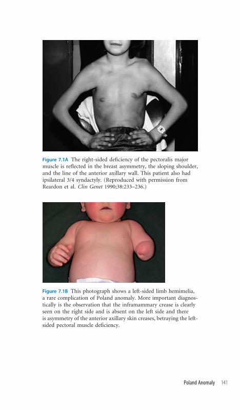
Figure 7.1A The right-sided deficiency of the pectoralis majormuscle is reflected in the breast asymmetry, the sloping shoulder,and the line of the anterior axillary wall. This patient also hadipsilateral 3/4 syndactyly. (Reproduced with permission fromReardon et al. Clin Genet 1990;38:233–236.)
Figure 7.1B This photograph shows a left-sided limb hemimelia,a rare complication of Poland anomaly. More important diagnos-tically is the observation that the inframammary crease is clearlyseen on the right side and is absent on the left side and thereis asymmetry of the anterior axillary skin creases, betraying the left-sided pectoral muscle deficiency.
Poland Anomaly 141

Accessory Nipples
Recognizing the Sign Most commonly sited inferior and medial to the nor-
mal nipple, supernumerary nipples may be identified by their presentation as
small pigmented marks or as dimples. They may be confused with birthmarks,
moles, or nevi. Some 60% show a unilateral single additional nipple, 30%
demonstrate bilaterality, and the remainder show three ormore additional nip-
ples. Mostly these arise as an isolated clinical finding and are devoid of further
significance.
Establishing a Differential Diagnosis It is worth being aware that the feature
can be inherited as an uncomplicated autosomal dominant trait and also that a
specific association with renal tract anomalies is described, embracing hypo-
plastic kidneys, ectopic kidneys, collection system reduplication, etc. Since the
feature is an occasional finding in fetal valproate exposure in utero, a drug
history in pregnancy may offer an explanation. Specific genetic syndromes in
which accessory nipples are identified such as Rubinstein-Taybi syndrome
and Miller syndrome will be more easily identified by their signature clinical
features of broad thumb and prominent beaked nose in the former and malar
hypoplasia with postaxial limb defects in the latter. However, there are sev-
eral syndromes associated with accessory nipples in which this clinical find-
ing may serve as the clue to the underlying diagnosis. Primary among these is
Pallister-Killian syndrome (tetrasomy 12p), a condition that should be sought
in situations of emerging developmental concerns, often associated with sei-
zures and an early history of hypotonia. Macroglossia and poor hair growth
in the temporal regions are valuable clinical signs. Examination under ultra-
violet lightmay show areas of patchy hypopigmentation, signifyingmosaicism.
Simpson-Golabi-Behmel syndrome is an X-linked overgrowth condition of-
ten associated with additional nipples. A family history, especially of high birth
weight, postaxial polydactyly, and mild developmental delay, will all sharpen
the likelihood of this underlying disorder in a child with supernumerary nip-
ples. Hypertelorism; a deep midline groove on the tongue, lower lip; and chin;
and hypoplasia of the index fingernail are all diagnostically supportive clinical
signs to watch out for. Macrocephaly, a family history of the same, lipomata,
and mental retardation in association with accessory nipples should lead to
investigation for Bannayan-Zonana syndrome.
Investigations to Consider Chromosomal analysis on skin fibroblasts is usu-
ally necessary to demonstrate the tetrasomy of chromosome 12p in Pallister-
Killian syndrome, though repeat biopsies may be required. Melanocyte culture
is reputed to be more reliable, though very infrequently available. Mutation
analysis of the GPC3 gene is available for Simpson-Golabi-Behmel confirma-
tion and of PTen for Bannayan-Zonana and overlapping phenotypes.
The Chest142
7.2

Figure 7.2 This patient shows accessory nipples bilaterally,in the midline, but also has accessory nipples at the anterioraxillary wall, which is demonstrated on the right side.Though present on the left side also, the accessory nippleis obscured by the skin creases of the axillary wall.
Accessory Nipples 143

Athelia/Hypothelia (Absent or Hypoplastic Nipples)
Recognizing the Sign Absence of the nipple is easily recognized in infancy
and is generally associated with amastia of the breast tissue, which will not be-
come apparent in the female until puberty. Hypoplasia of the nipple is more
difficult to recognize, but should be considered if there is little pigment around
the nipple and if the areola, which should underlie and surround the nipple, is
absent. The remarks appertaining to Poland anomaly (section 7.1) apply. As-
sess the chest wall musculature, facial movement, and ipsilateral upper limb.
In the event of these being normal, consider the following possibilities.
Establishing a Differential Diagnosis Family history can be diagnostically
significant because both autosomal dominant and recessive forms of athelia
are described, as isolated findings. Likewise, a detailed history of drug inges-
tion can solve the diagnostic mystery, with carbimazole, widely used for treat-
ment of thyrotoxicosis in pregnancy, being associated with absent or hypo-
plastic nipples, often in association with choanal atresia. Scalp defects and
imperforate anus are also described in this syndrome of teratogenesis. Chro-
mosomal aneuploidies can result in this clinical sign, but almost always in
the context of a more significant multiorgan malformation complex. Specifi-
cally, Edwards syndrome (trisomy 18) is often associated with nipple hypo-
plasia. Likewise, though a common feature of de Lange syndrome, the diag-
nosis will be signaled by the characteristic facial and limb findings outlined
elsewhere. Small, invariably inverted nipples are important diagnostic clues to
the carbohydrate-deficient glycoprotein (CDG) syndromes. Other clinical
clues include unusual fat pad distribution over the buttocks and areas of lipo-
atrophy. With time, developmental delay is noted and, later, cerebellar ataxia
can emerge as an important clue to etiology. Limb defects, typically deficient
digits on the ulnar side of the hand, should always prompt consideration of
ulnar-mammary syndrome and nipple absence or hypoplasia strongly sup-
ports this diagnosis. Other signs are abnormal placement of the anus, which
may be stenotic, and small male genitalia. Limb malformations, typically of
ectrodactyly (split hand/foot), in association with nipple hypoplasia signal
ectrodactyly-ectodermal dysplasia syndrome (EEC), or a variant thereof,
the condition being clinically highly heterogeneous.
Investigations to Consider Isoelectric focusing of transferrin is the best way
to detect almost all cases of CDG variants but may not be sensitive in the first
few weeks of life until abnormal forms build up. Ulnar-mammary syndrome
may be confirmed by TBX3 gene mutation analysis and EEC clinical variants
thereof by p63 gene mutations.
The Chest144
7.3

Figure 7.3 A small hypoplastic nipple with little surrounding pig-ment and an almost complete absence of areola is shown in apatient who presented neonatally with choanal atresia and whosemother had been treated for hyperthyroidism with carbimazolethroughout pregnancy.
Athelia/Hypothelia (Absent or Hypoplastic Nipples) 145

Gynecomastia
Recognizing the Sign Excess breast development in male patients must be
considered in terms of normal physiologic gynecomastia, which may be ob-
served neonatally and at adolescence. This is generally bilateral and transient.
Breast enlargement, resembling gynecomastia, is a feature of obesity.Widely de-
scribed in adult male populations with hyperthyroidism, hypothyroidism, liver
failure, and chronic renal failure, gynecomastia is rare in the prepubertal child.
Establishing a Differential Diagnosis An impression of gynecomastia in a
thin baby, particularly if there is a history of intrauterine growth retardation
and failure to thrive, may reflect a generalized lipodystrophy of leprechaun-
ism. Signs of acanthosis nigricans would support the diagnosis of this autoso-
mal recessive condition. These children are noteworthy for the well-defined ap-
pearance of their musculature, the subcutaneous fat being deficient. However,
most gynecomastia is a postpubertal phenomenon, in which instance, signs of
normal sexual development should be sought. One of themost common causes
of gynecomastia is Klinefelter syndrome (47, XXY), in which the associated
small gonads and penis, long limbs, and, occasionally but not reliably, a history
of behavioral or mild developmental concerns may be forthcoming. However,
many Klinefelter cases only present in adult life at infertility clinics. Think spe-
cifically aboutKallmann syndrome, especially if there is developmental delay—
check the patient for repaired cleft palate or subclinical clefting in the form
of bifid uvula. Some Kallman patients may have generalized dry skin from an
X chromosome deletion and the observation of mirror movements on neuro-
logic evaluation is very strongly suggestive of this diagnosis. If observed, specific
neurologic assessment for anosmia is warranted. The presence of skin lentigines
in gynecomastia cases enjoying normal secondary sexual characteristics is im-
portant because of the association with silent but potentially devastating atrial
myxoma—this autosomal dominant association being known as Carney com-
plex; a family history may offer additional diagnostic support. A presentation
of emerging gynecomastia in a previously normal nonobese adult man requires
careful evaluation of neurologic status of the lower cranial nerves. Any perioral
fasciculation, tongue weakness, or swallowing difficulties will strongly suggest
the diagnosis of X-linked spinal muscular atrophy.
Investigations to Consider Karyotype is indicated to look for the 47, XXY of
Klinefelter syndrome. Evaluation of endocrine status is warranted if no other
clinical signs prompt a diagnosis. Echocardiography should be conducted for
myxomata if lentigines are present. Mutations of the androgen receptor gene
are demonstrable in X-linked spinal muscular atrophy (SMA) and of the
insulin receptor gene in leprechaunism; deletion of the Kal1 gene on Xp22 is
the most common molecular cause of Kallman syndrome.
The Chest146
7.4

Figure 7.4 Bilateral gynecomastia is demonstrated in this 14-year-old patient referred for evaluation of pubertal delay, who provedto have Kallmann syndrome. The genitalia were very small andanosmia was confirmed on formal testing.
Gynecomastia 147

Sloping Shoulders
Recognizing the Sign Absence of the normal horizontal configuration of the
shoulder, due to underlying clavicular bony and/or muscular abnormality,
results in shoulders that slope and are almost continuous with the neck.
Establishing a Differential Diagnosis This sign is classically, but not ex-
clusively, seen in the autosomal dominant condition cleidocranial dysostosis
(CCD). This condition is usually identifiable at birth, the facial features of a
high, prominent, bossed forehead often accompanied by very large fontanelles,
hypertelorism, and the characteristic absence of well-demarcated shoulders. In
later life, the patients demonstrate a facility to voluntarily oppose the shoul-
ders, owing to the hypoplasia of the acromial ends of the clavicles. Good cor-
roborative evidence for the condition in the older patient is found in the teeth,
in which delayed loss of deciduous teeth is often followed by an edentulous
period before the very delayed eruption of permanent dentition. Abnormally
short clavicles and associated neck webbing in some patients with cranio-
frontonasal dysplasia (CFND) give a shoulder configuration similar to CCD.
Usually the facial features, with prominent widow’s peak and bifid nasal tip,
signal the true diagnosis. Despite the frequent coincidental absence of the
corpus callosum in CFND patients, intelligence is generally normal. Most pa-
tients with branchiootorenal (BOR) syndrome have entirely normal shoulders,
but one variant with sloping shoulders, typical of CCD in appearance, is the
condition known as oto-facio-cervical syndrome. This should be suspected in
a child with sloping shoulders who also has deafness. Examine specifically for
preauricular pits, branchial cysts, and evidence of underlying renal malforma-
tions. Sloping shoulders are occasionally encountered inHolt-Oram syndrome,
but the diagnosis in this situation will be clear because of associated congenital
heart disease, most commonly atrial septal defect (ASD), and more extensive
upper limb malformations, which are usually bilateral. Sloping shoulders are
also well-recognized within the Noonan syndrome phenotype, the diagnosis
being suspected by the ptosis, curly hair, and cardiac lesion, most commonly
pulmonary stenosis.
Investigations to Consider Radiologic examination of the skeleton in CCD
confirms the diagnosis with wormian bones and areas of calvarial thickening
in the skull vault, hypoplastic or absent clavicles and scapulae, and hypoplasia
of the iliac wings and delayed ossification of the pubis. Mutation of the CBFA1
gene on chromosome 6 confirms CCD, if there is clinical or radiologic doubt.
CFND patients have mutations of the EFNB1 gene on the X chromosome,
while deletion in the region of chromosome 8q around the BOR locus, the
gene EYA1, is described in oto-facio-cervical syndrome.
The Chest148
7.5

Figure 7.5 This child has the oto-facio-cervical syndrome variantof branchiootorenal syndrome. The sloping shoulders are welldemonstrated.
Sloping Shoulders 149

Small Thorax
Recognizing the Sign A narrow chest can be confirmed by measuring the
chest circumference at the nipple level and comparing to normal age-matched
values. This is a sign commonly identified antenatally on ultrasound anomaly
scans. Whether antenatally or postnatally identified, the length, shape, and
radiologic morphology of the long bones will have a major bearing on di-
agnostic conclusions.
Establishing a Differential Diagnosis It needs to be borne in mind that the
age at presentation has a major impact on likely diagnosis—for instance,
thanatophoric dysplasia (TD) is one of the most common causes of a small
thorax ultrasonically, but virtually all these patients die in the immediate neo-
natal period. In examining a deceased infant with a narrow chest, valuable
signs to establish are whether there is clover leaf malformation of the skull
(TD), short limbs (TD and camptomelic dysplasia [CD]), blue sclera (osteo-
genesis imperfecta), postaxial polydactyly (Jeune and Ellis-van Crefeld syn-
drome), and sexual ambiguity (CD). Ultimately, the diagnosis will depend
upon radiologic characteristics, but good clinical documentation can prove
invaluable. In the child who has survived the postnatal period but whose chest
circumference is narrow, look at the hands for polydactyly and in the mouth
for accessory frenula, and examine the heart for a possible ASD, all of which
signs betoken the diagnosis of Ellis-van Crefeld syndrome. Natal teeth are
a very good clue to this diagnosis. Associated findings of short arms and
legs, particularly in a baby whose hair is slow to grow, probably represent
McKusick-cartilage-hair hypoplasia syndrome, which is an important con-
dition to recognize because of the associated susceptibility to varicella infec-
tion and defects of cellular immunity, occasionally necessitating bone marrow
transplantation. Typically the radiologic changes are of metaphyseal dysplasia,
worse at the distal end of the femur. Patients with Jeune asphyxiating tho-
racic dystrophy often do not survive, but if they do, they are at risk of cystic
renal changes, cirrhosis, and retinal dystrophy. Small numbers of Campome-
lic Dysplasia cases have survived into adult life and should be suspected in
narrow-chested individuals with macrocephaly, hypertelorism, hypotonia,
micrognathia, and cleft palate.
Investigations to Consider Full skeletal survey is the cornerstone of diag-
nosis, irrespective of the age at presentation. Chromosomal sex is a valuable
test if there is curvature of the femora in a phenotypic female. 46XY patients
with CD undergo sex reversal, sometimes complete, more commonly resulting
in genital ambiguity, and karyotype can prompt the true diagnosis. Mutation
analysis is available for most of these conditions but is rarely needed for di-
agnostic purposes, radiology in expert hands usually sufficing.
The Chest150
7.6

Figure 7.6 Note the narrowing of the thorax in this surviving caseof Jeune asphyxiating thoracic dystrophy.
Small Thorax 151

Pectus Excavatum and Carinatum
Recognizing the Sign The depression of the sternum and costosternal junc-
tions that signals pectus excavatum is easily identified and the rationale for the
synonymous term, ‘‘funnel chest,’’ clear. The opposite configuration, of ster-
nal protrusion, comprises pectus carinatum. It is worth bearing in mind that
these signs may be no more than an isolated finding and not relate to any
underlying syndrome.
Establishing a Differential Diagnosis Observe and chart the stature. Is there
evidence for excess linear growth, in which case Marfan syndrome will need
to be considered? Look for arachnodactyly, unusual degrees of joint laxity, lens
dislocation (usually upward), and transverse striae in the dorsal thoracic and
lumbar regions. The family history may contain clues, specifically of sudden
death due to dissecting aneurysm. Long fingers should also prompt clinical en-
quiry for Beals syndrome (congenital contractural arachnodactyly), in which
syndrome external ear inspection often discloses a rather crumpled appear-
ance. Careful assessment of the joints, both large and small, may be rewarded
by evidence of joint contracture or limitation of full extension. Spinal assess-
ment for scoliosis is an important element of the examination if this condition
is being considered. Developmental delay with ‘‘marfanoid’’ characteristics,
both of increased linear growth and of arachnodactyly, should cause the ob-
server to consider homocystinuria as a possible diagnosis. Lens dislocation
(usually downward), restriction of joint movement, kyphoscoliosis, and gen-
eralized osteoporosis on x-ray are strong clues to this easily overlooked con-
dition. Pectus excavatum in patients with stature toward the lower end of the
normal range warrants a different diagnostic approach. Cardiac examination
may reveal a murmur and assessment for Noonan syndrome is advised. The
classic chest configuration in Noonan cases is of a pectus carinatum above and
a pectus excavatum below, but not every case obliges by conforming exactly to
the textbook! Ptosis, excess nuchal skin, and low posterior hairline may offer
additional corroboration, as may family history and clinical assessment of the
parents.
Investigations to Consider Echocardiography is essential if the diagnosis of
Marfan syndrome is being considered to assess the aortic root diameter and
evaluate for management implications. Prolonged bleeding time is seen in
many Noonan cases. Elevated methionine and homocysteine levels on amino
acid analysis are suggestive of homocystinuria. FBN1 mutation analysis is oc-
casionally useful inMarfan syndrome, whilemutation of FBN2 is characteristic
of Beals syndrome.
The Chest152
7.7

Figure 7.7 This is the chest of a 44-year-old man whose son was sentfor clinical genetics assessment for short stature. The boy hadundiagnosed Noonan syndrome. The father had had surgery at age28 for pulmonary stenosis (see themidline sternotomy scar) and hissternal configuration of pectus carinatum superiorly and pectusexcavatum inferiorly is classical of Noonan syndrome.
Pectus Excavatum and Carinatum 153

Scoliosis
Recognizing the Sign Lateral curvature of the spine constitutes scoliosis,
which may be isolated or associated with kyphosis, in which posterior angu-
lation of the spine develops. Inspect the spine in the flexed position, and ob-
serve any asymmetry in shoulder height, pelvic tilt, and exaggerated lordosis.
Establishing a Differential Diagnosis Scoliosis is an especially difficult prob-
lem to assess as a possible clue to a dysmorphic syndrome, being such an etio-
logically heterogeneous anomaly. Consider the possible causes and assess whether
the spinal problem is idiopathic. Any other clinical feature, such as abnormal
limb length or developmental delay, should forestall thoughts of an idiopathic
basis! Reduced limb length and limitation of joint movement probably indi-
cate that the primary pathology is a skeletal dysplasia, which can be a primary
disorder or secondary to an underlying biochemical abnormality, such as arises
inmucopolysaccharidoses and other storage disorders. By contrast, an increase
in limb length or excessive joint laxity might be the clinical clue to consider a
connective tissue disorder as the primary disease process. Accordingly, the cli-
nical examination will focus on the skin for signs of fragility, striae, or excess
bruising and the eyes for blue sclera. Specific clinical examination, such as for
evidence of lens dislocation in suspectedMarfan syndrome, may be warranted.
Scoliosis is a frequent late childhood complication of those clinical entities de-
scribed as ‘‘associations’’ on the basis of the frequent nonrandom observation of
the anomalies coinciding in individuals. Hence, a history of the neonatal and
childhood periodmay give a clue—ahistory of cardiac surgerymay indicate that
the child had VATER association (vertebrae, imperforate anus, tracheoesopha-
geal fistula, radial þ/– renal anomalies) and the scoliosis is an understandable
late manifestation of this, hemivertebrae being a frequent finding. Finally, there
are specific syndromes of which scoliosis can be a severe complication. Neu-
rofibromatosis type 1 should be readily identifiable in a scoliotic child by the
skin manifestations of cafe-au-lait patches and axillary freckling. Coffin-Lowry
syndrome should be considered in boys with a history of developmental delay
who present with scoliosis and the rather coarse facial features, full lips, and
characteristically puffy fingers sought at examination (Figure 9.9). Finally, the
author has seen several cases of Sotos syndrome, in whom that diagnosis was
missed in early life, present with scoliosis at 5 or 6 years of age. Early develop-
mental concerns,macrocephaly, ruddy cheeks, and large chins are useful clinical
indicators of this syndrome.
Investigations to Consider A full skeletal survey is necessary to address
whether there is a skeletal dysplasia and possibly identify the underlying con-
dition. Bone age is indicated if Sotos syndrome is suspected—advancement is
the general rule.
The Chest154
7.8

Figure 7.8 Severe kyphoscoliosis is shown in a 15-year-old girl, always considered tall for her age. Examinationshowed prominent, slightly protruding eyes; multipleskin bruises; and the ‘‘criss-cross’’ pattern of the palms,which is typical of Ehlers-Danlos syndrome type VI—the oculoscoliotic form—in which autosomal recessivecondition lysyl hydroxylase deficiency is associatedwith ocular fragility, blue sclera, prominence of theeyes, and severe vertebral problems. See also Figure9.18C for the criss-cross pattern.
Scoliosis 155

Chapter 8 The Abdomen and Perineum

Minor Anomalies of the Umbilicus
Recognizing the Sign The normal position of the umbilicus is at the level of
the top of the iliac crests, opposite the third or fourth lumbar vertebra. The
umbilicus is extraordinary for its variation, there being no ‘‘standard.’’ Minor
anomalies, if recognized, can impart diagnostically important information
and guide the clinician toward an initially unsuspected diagnosis. In this re-
spect, three particular variations are noteworthy: (1) ovoid depression sur-
rounding a prominent, protruding umbilicus; (2) prominent umbilicus with
redundant periumbilical skin; and (3) upward displacement of the umbilicus
toward the xiphisternum.
Establishing a Differential Diagnosis Ovoid depression of the tissue im-
mediately surrounding the umbilicus, resulting in a lozenge-shaped depres-
sion encircling a slightly prominent umbilicus, is entirely typical of the X-
linked short stature condition Aarskog syndrome, being very infrequently
seen in any other situation. Faced with this observation, the clinical exami-
nation will focus on the patient’s stature, but also on other clinical features
typical of this condition—hypertelorism is usual in such cases and ptosis very
common; the hands frequently show mild skin syndactyly and hyperextension
of the fingers at the proximal interphalangeal joints. Brachydactyly is usual
(Figure 9.7B). Genital examination is important, the penis having a charac-
teristic ‘‘shawl’’ configuration, in which the scrotal folds are inserted above the
base of the penis (see 8.7 and Figure 8.7). Family history may support the likely
diagnosis, and carrier females may themselves show short stature and bra-
chydactyly. Redundant periumbilical skin around a prominent umbilicus
often gets mistaken for a small umbilical hernia and is treated surgically. A
history such as this is typical of patients with Rieger syndrome, an autosomal
dominant condition of hypodontia, anterior segment of the eye malformation,
and characteristic failure of involution of the periumbilical skin. A family
history of ‘‘umbilical hernia’’ is an excellent pointer toward this diagnosis.
Examine the teeth for hypodontia and oligodontia. Cleft palate is an occa-
sional finding. Anterior chamber eye malformations need to be specifically
sought by expert ophthalmologic evaluation. Finally, an upwardly displaced
umbilicus demands examination for other features of Robinow syndrome,
this feature being observed in many Robinow patients. A cleft lip, familial
short stature, anteverted nares, and micropenis in the male are all strongly
supportive of this underlying diagnosis (Figure 2.5A).
Investigations to Consider The diagnosis in all instances is primarily clin-
ical. Skeletal radiology is complementary in Aarskog cases, showing non-
specific metaphyseal dysplasia and bone age delay. Hand radiology in Robi-
nowmay show bifid terminal phalanges to the thumbs and halluces. Mutation
analysis is available in all instances for clinically doubtful examples.
The Abdomen and Perineum158
8.1

Figure 8.1A A typical umbilicus of Aarskog syndrome in profile,showing the periumbilical depression surrounding the minimallyprotruding umbilicus. See Figure 2.1A for the facial features ofAarskog syndrome.
Figure 8.1B A cranially displaced umbilicus is shown. Note alsothe shawl scrotum (see section 8.7) and the hypoplasia of thescrotum.
Minor Anomalies of the Umbilicus 159

Umbilical Hernia/Omphalocele
Recognizing the Sign This is an easy sign to identify, being a skin-covered
protrusion of the umbilicus, due to defective closure of the umbilical ring. In
most babies, the umbilical ring closes within the first 2 years of life and
persistence beyond this age is associated with a declining rate of spontaneous
closure. A large umbilical hernia at birth may be, more correctly, an ompha-
locele, in which abdominal contents, covered by peritoneum, herniate into the
cord.
Establishing a Differential Diagnosis Omphalocele is a well-recognized
presentation of Beckwith-Wiedemann syndrome (BWS). A history of poly-
hydramnios, large birth weight, and neonatal hypoglycemia should be sought.
Examination should assess for nevus flammeus, macroglossia, ear lobe creases,
and punched-out depressions of the helical rim and posterior aspect of the
pinna, as well as organomegaly. There is an increased prevalence among babies
conceived by assisted reproductive technologies. Long-term surveillance for
neoplasms is usual. Other conditions to consider in the newborn period are
the trisomies, omphaloceles being well described in trisomy 21, 18, and 13.
The distinction of BWS from Down syndrome, in which a clinical impression
of macroglossia derives from a somewhat reduced oral cavity volume, should
not be taken for granted, proving rather challenging on well-remembered
occasions in the delivery ward! Macroglossia is a rarely recognized feature of
several forms of Ehlers-Danlos syndrome (EDS), in which disorders there is a
propensity to neonatal inguinal hernias developing and, although admittedly
rare, the author has seen three cases of EDS diagnosed neonatally as BWS.
Neonatal umbilical hernia is well recognized in congenital hypothyroidism,
which should be identified by Guthrie screening. Likewise, persisting umbilical
hernia in a child whose development is starting to give cause for concern may
be the presentation of mucopolysaccharidosis (MPS) I and II. Examination
for the characteristic corneal clouding is advised. By contrast, Sanfilippo syn-
drome, MCP III, does not show corneal clouding and the presentation is often
at age 3 or 4 years, with persisting umbilical hernia or hernia that may have
been operated upon, persistent diarrhea, progressively unruly behavior, and,
identifiable only by reviewing old photographs, coarsening of the facies.
Investigations to Consider Molecular testing for BWS is very complex but
mutation of the CDKN1C gene is particularly common among cases pre-
senting with omphalocele. Do basic karyotype if in clinical doubt about tri-
somy. Urinary screen for MCP will identify excess glycosaminoglycan (GAG)
in MCP I and II, but diagnosis of type III can be elusive and the best screening
test is not urinary GAG but vacuolated lymphocytes on peripheral blood
smear.
The Abdomen and Perineum160
8.2
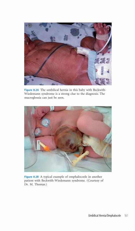
Figure 8.2A The umbilical hernia in this baby with Beckwith-Wiedemann syndrome is a strong clue to the diagnosis. Themacroglossia can just be seen.
Figure 8.2B A typical example of omphalocoele in anotherpatient with Beckwith-Wiedemann syndrome. (Courtesy ofDr. M. Thomas.)
Umbilical Hernia/Omphalocele 161

Inguinal Hernia
Recognizing the Sign Tenfold more common among male than female
children, widening of the external inguinal ring facilitates the herniation of a
loop of bowel into the inguinal canal and scrotum. The resultant hernia is
easily seen on inspection. Ten percent are bilateral.
Establishing a Differential Diagnosis Among phenotypic females, bilater-
ality, being rare, should arouse suspicion. Many androgen insensitivity cases
present with this pattern and careful internal examination ultrasonically and at
laparotomy confirms the diagnosis—but only if the examining clinician is alive
to the likely diagnosis. Of course, most inguinal hernias are nonsyndromic,
especially if related to prematurity. However, several syndromes have an in-
creased prevalence of hernia. Foremost among these are the mucopolysac-
charidoses and a low threshold for investigation of these disorders in children
with inguinal hernia in the first few months of life is strongly advised, even if
corneal clouding and more ‘‘classical’’ syndromic signs have not yet appeared.
The hernia may herald the onset of problems retrospectively identifiable as
MCP related. The second category of syndrome to bear in mind are connective
tissue disorders such as Marfan, Ehlers-Danlos (EDS) complex, and cutis laxa
syndromes. Their differentiation may require expert clinical and/or laboratory
assistance but signs of excess skin, skin fragility, hypermobility of joints, and
thin skin in which veins are easily seen are all strong clues to this group of
conditions. Since many of these disorders are dominant, details of the family
history may not only confirm the likely diagnosis, but also specify one disorder
above others. Unique clinical features, such as the presence of nasal papillomata
in Costello syndrome cases, facilitate the differentiation of particular disorders
from clinically overlapping entities. Finally, the examiner needs to be aware of a
few individual syndromes associated with inguinal hernia. Careful assessment
for Williams syndrome is advised—was there hypercalcemia neonatally? Is
there a cardiac murmur? Do the eyes have a stellate iris pattern? Is there flare of
the eyebrow medially? In the older child, are there developmental concerns?
Compare the facial features with early photographs and assess for any slight
coarsening. If there is short stature, think specifically ofAarskog syndrome and
look for shawl scrotum as a likely clue to diagnosis.
Investigations to Consider Urinary GAG and skeletal radiology are indicated
to assess for MCP. Karyotype in females is warranted to exclude androgen
insensitivity. Urinary pyridinoline cross-links for EDS VI and serum tenascin
X for EDS-like syndromes. Fluorescence in situ hybridization (FISH) of ELN is
indicated for Williams syndrome. Molecular diagnosis for many variants of
connective tissue disorders is available, but suspecting the diagnosis is pri-
marily a clinical exercise.
The Abdomen and Perineum162
8.3

Figure 8.3 Repaired left-sided inguinal hernia is seen in this boywith Aarskog syndrome. Note the shawl scrotum hinting directlyat the underlying syndrome diagnosis.
Inguinal Hernia 163

Small Penis/Micropenis
Recognizing the Sign A subjective impression of a small penis should be
confirmed by measurement and charting against normal values. The mea-
surement is taken from the pubic bone to the tip of the glans.
Establishing a Differential Diagnosis Pure endocrine causes will generally
not result in associated malformations, so the examination needs to focus on
syndromes that are associated withmicropenis. The irismay show a coloboma, or
a cardiac murmur may be heard, which should lead to an evaluation of the
external ear for possible overfolding of the helix, all of which may be indicative of
CHARGE association (coloboma, heart, atresia choana, retarded growth, genital
abnormalities, ear malformations). Unilateral choanal atresia may be present but
unrecognized, bilaterality usually presenting early. Floppiness in the newborn
with micropenis, especially in the presence of a need for assisted feeding and a
poor suck, should prompt concern for Prader-Willi syndrome. In the older case,
the almond-shaped eyes, narrow hands, and straight ulnar border will clinically
suggest the diagnosis, but the characteristic eating behavioral disorder will be the
main clue after the first 2 years. Careful evaluation of the hands and feetmay show
postaxial ‘‘nubbins’’—vestigial polydactyly, which frequently signifies Bardet-
Biedl syndrome. Developmental delay, emerging obesity, and a reduced ability to
concentrate urine will characterize the natural history of this autosomal recessive
disorder, which is associated with retinal dystrophy but usually not before the
second decade of life. Klinefelter syndrome must be considered, especially in
patients who fail to enter puberty. Tall stature is usual and gynecomastia a valu-
able clue, if present. Breast abnormality is likewise the prompt for suspecting cases
of ulnar-mammary syndrome, with gynecomastia but, more likely, breast hy-
poplasia being common. Evaluate the hands for evidence of ulnar defects, miss-
ing fingers, or hypoplasia. Boys with Robinow syndrome (Figure 2.5A) as the
basis of their micropenis will have the typical facial features of hypertelorism and
anteverted nares and the umbilicus may be displaced cranially, while cleft palate
or lip associated with micropenis should prompt concern for Kallmann syn-
drome, in which developmental delay is common; mirror movements are an
excellent clinical clue and anosmia an almost invariable feature.
Investigations to Consider Most patients with CHARGE have semicircle
canal aplasia/hypoplasia and computed tomography (CT) of the petrous tem-
poral bones can secure the diagnosis in clinically doubtful situations. Mutation
of the CHD7gene is established in CHARGE. Paternal 15q deletion studies for
Prader-Willi syndrome sometimes need further molecular supplementation,
while basic karyotype will confirm 47,XXY in Klinefelter cases. Deletion of the
KAL1 gene is the commonest cause of Kallman syndrome but the molecular
diagnosis of Bardet-Biedl syndrome remains extremely complex due to the sheer
number of genes involved.
The Abdomen and Perineum164
8.4

Figure 8.4 Micropenis is demonstrated in a newborn whose pre-sentation was with a heart murmur due to a ventricular septaldefect (VSD). Further examination showed bilateral retinal colo-bomata, hypoplastic semicircular canals, and unilateral choanalatresia. The diagnosis was CHARGE association.
Small Penis/Micropenis 165

Ambiguous Genitalia
Recognizing the Sign The appearance of the external genitalia manifests
elements of both male and female development.
Establishing a Differential Diagnosis The single most common cause is
21a-hydroxylase deficiency, an autosomal recessive nonsyndromic enzy-
matic deficiency requiring investigation of the adrenal gland for possible salt-
losing crisis. This and other biochemical causes of a nonsyndromic nature,
such as androgen insensitivity, are not considered further here. Primary
among the clinician’s concerns must be to establish whether the baby has other
malformations or clinical clues to a syndromic diagnosis. Examine the eyes—
specifically, query whether there is aniridia. Your concern is forWAGR (Wilms
tumor, aniridia, genital anomalies, retardation) syndrome due to deletion of
the WT1 gene on chromosome 11p13. A related condition to also consider is
Drash syndrome, in which early-onset hypertension, proteinuria, and ne-
phropathy develop in children with 46XY genotype whose genitalia are fe-
male or ambiguous and is due to a WT1 gene mutation. Examine the limbs;
2/3 syndactyly of the toes is a good clue to Smith-Lemli-Opitz (S-L-O) syn-
drome and if present, look for facial features of bitemporal narrowing and
anteversion of the nostrils. Limitation of extension at the joints, particularly
the elbow, with evidence of craniosynostosis should prompt consideration of
Antley-Bixler syndrome. This condition can be mimicked fairly exactly by
maternal ingestion of Fluconazole during pregnancy. Exstrophy of the blad-
der, omphalocele, and anal agenesis result in ambiguous or grossly abnormal
genitalia as part of a series of well-recognized patterns of associated mal-
formation but of unknown basis, such as urorectal septum defects and OEIS
(omphalocele, exstrophy of bladder, imperforate anus, and sacral anomalies).
Ambiguous genitalia can be the first clinically recognizable feature of
a-thalassemia mental retardation, X-linked (ATRX), so a family history of
male mental retardation should not be discounted as irrelevant. Assess the face
for coarseness, especially around the upper lip, while small teeth and depressed
nasal bridge are good facial clues in a slightly older age group (see Figure 1.6C).
Investigations to Consider Karyotype is indicated to establish the chro-
mosomal sex. Ambiguous genitalia are a reported feature of several chro-
mosomal abnormalities and an abnormal karyotype may itself offer adequate
explanation of the phenotype. Specifically request 11p FISH studies if WAGR
is suspected. WT1 mutation analysis is indicated for Drash syndrome, but
proteinuria should be consistently present to justify this investigation. Serum
cholesterol and 7-dehydrocholesterol are needed to diagnose S-L-O. Though
mutation analysis is available for ATRX syndrome, HbH inclusion bodies on
staining with freshly constituted brilliant cresyl blue are a very good screening
test, positive in 90% of cases.
The Abdomen and Perineum166
8.5

Figure 8.5 A complex translocation between three chromosomes was the basisof the ambiguous genitalia in this baby who had an XY karyotype.
Ambiguous Genitalia 167

Hypospadias
Recognizing the Sign The condition is easily recognized provided it is looked
for, the urethral meatus being situated on the ventral surface of the penis and
proximal to the glans penis.
Establishing a Differential Diagnosis Hypospadias is a relatively common
malformation, many cases being isolated nonsyndromic events. Assessment of
the kidneys and urinary tract is mandatory in all. Likewise, there are hundreds
of syndromes in which hypospadias is described but in many, Fraser syn-
drome representing a case in point, with cryptophthalmos being the primary
feature the clinician will address, the hypospadias is cast in a supporting role,
in diagnostic terms. The purpose here is to highlight conditions that ought
to be considered if there is a clinical finding of hypospadias. Primary among
these are Smith-Lemli-Opitz (S-L-O) syndrome, an autosomal recessive con-
dition in which the observation of 2/3 syndactyly of the toes, partial or com-
plete, strongly supports the need for biochemical investigation. Bitempo-
ral narrowing, ptosis, and evidence of developmental delay are characteristic,
though not reliably present. Holoprosencephaly is an occasional presentation
of S-L-O, so be aware of this diagnosis in the holoprosencephalic infant with
hypospadias. Hypertelorism should be noted, as it is a cardinal feature ofOpitz
G syndrome, an X-linked condition associated with hypospadias and swal-
lowing difficulties, sometimes with laryngeal cleft. Laryngoscopic examina-
tion is advised. Evaluate the mother, as carriers often show mild degrees of
hypertelorism and such observations can clench a diagnosis in difficult cir-
cumstances, as can a family history. An ectopic or stenotic anus can constitute
part of the Opitz G syndrome spectrum, but may also be significant of Townes-
Brocks syndrome (T-BS) in a hypospadias presentation. Apart from lacking the
hypertelorism of Opitz G, the presence of overfolded ears, preauricular pits, and
deafness betray the true syndrome diagnosis of T-BS. Already discussed under
ambiguous genitalia (section 8.5), assessment for WAGR is warranted likewise
in hypospadias, while a rare autosomal recessive condition, easily missed and
which frequently presents with hypospadias, is that of Schinzel-Giedion syn-
drome. Generalized hirsutism, fleshy earlobes, and narrow and deep-set nails are
features that substantiate this diagnosis, but the main facial features that dys-
morphologists agree on are bitemporal narrowing and an infraorbital groove,
much as described in fetal valproate syndrome.
Investigations to Consider Karyotype, including 11p FISH studies, is indi-
cated for WAGR. Cholesterol and 7-dehydrocholesterol are warranted for
S-L-O. MID1 mutation is confirmed in many cases of Opitz G syndrome.
T-BS is a clinical diagnosis, but mutation analysis of SALL1 confirms or refutes
doubtful cases. The best test for Schinzel-Giedion syndrome is skull x-ray,
which typically shows an occipital ‘‘gap’’ in ossification due to synchondrosis.
The Abdomen and Perineum168
8.6

Figure 8.6 Severe hypospadias is demonstrated in apatient with Smith-Lemli-Opitz syndrome. Apartfrom the genital malformation, the main clinical cluewas partial 2/3 syndactyly of the feet.
Hypospadias 169

Shawl Scrotum and Penoscrotal Transposition
Recognizing the Sign A shawl scrotum gives the appearance of a gothic arch-
shaped fold of skin above the base of the penis. Essentially, the genital tubercle,
whence the penis derives, has been displaced caudally to a minor degree. In
more severe displacement, the penis emerges from the scrotal sac and, at the
most extreme end of the spectrum, the penis and scrotum are transposed.
Establishing a Differential Diagnosis The initial impression on inspection
often is of micropenis, which should be measured and charted against stan-
dard norms. Usually the penis is of normal size on objective assessment. Shawl
scrotum is one of the cardinal diagnostic features of Aarskog syndrome, an
X-linked condition of short stature, delayed bone age, and mild develop-
mental delay in many affected individuals. The facial features are distinctive,
particularly the hypertelorism, widow’s peak, and high forehead (Figure 2.1A).
Look out for the umbilicus, which is often displaced cranially and also shows a
characteristic lozenge-shaped depression around the umbilicus (section 8.1
and Figure 8.1A).Many of these patients are chance diagnoses, identified when
they present for inguinal hernia repair. The hands show brachydactyly, often
with clinodactyly and amild degree of syndactyly (Figure 9.7B). Typically there
is hyperextension of the proximal interphalangeal joints on extension. The
family history and review of photographs of family occasions may reveal
several other affected individuals. Transposition of the penis and scrotum is
extremely uncommon but is seen occasionally in association with anal anom-
alies in urorectal septal defects sequence, where septation of the primitive
cloaca is abnormal. Clinical presentations include absence of the penis, am-
biguous genitalia, a ‘‘smooth’’ perineum, common exits for anal and urethral
channels, in addition to renal malformations. All cases are sporadic and are
frequently complicated by congenital heart disease.
Investigations to Consider Skeletal radiology in Aarskog syndrome will
show delayed bone age; short limbs, predominantly rhizomelic in character;
and mild metaphyseal dysplasia. Mutation analysis of the FGD1 locus on
Xp11.21 confirms mutations in affected individuals if the clinical diagnosis is
in doubt. Karyotype and renal tract evaluation are worth undertaking in
penoscrotal transposition.
The Abdomen and Perineum170
8.7

Figure 8.7A A typical shawl scrotum configurationis shown in this patient with Aarskog syndrome.This is the same patient as in Figure 9.7B. SeeFigure 2.1A for the facial features of Aarskogsyndrome.
Figure 8.7B An even more pronounced instance of shawl scrotumis shown in a boy without a diagnosis.
Shawl Scrotum and Penoscrotal Transposition 171

Anal Atresia, Anal Stenosis, and Anterior Displacementof the Anus
Recognizing the Sign Inspect the perineum. Surface signs of fistula on the
perineum or to the vulva should be sought. The anus may be present but be
almost continuous with the vagina, with little clear separation. In males, hy-
pospadias and bifid scrotum may be observed.
Establishing a Differential Diagnosis The family history will usually identify
the autosomal dominant condition Currarino syndrome, in which anal
malformations and/or chronic childhood constipation may be the presenting
feature. Be aware of the possibility of cerebrospinal fistula, and a history of
deaths from Escherichia colimeningitis in the family should not be discounted
as irrelevant. In the event of a normal family history, assess the palate. Has
there been nasal regurgitation during feeding, or is there a cleft? Anal steno-
sis is an overlooked aspect of velo-cardio-facial (VCF) syndrome, so do lis-
ten to the heart and look for asymmetric crying facies. Cat-eye syndrome,
due to mosaic trisomy 22, is another chromosomal cause of anal stenosis/
displacement. Preauricular tags and pits, cardiac defects, and coloboma of the
iris comprise the full clinical spectrum, but the diagnosis can be especially
elusive in the absence of coloboma and is made only by heightened clinical
awareness and diligent investigation. Cat-eye syndrome cases can manifest
significant clinical overlap with Townes-Brocks syndrome (T-BS), and ear
and anal malformations are common to both these conditions. However,
triphalangeal thumbs and distal thumb deviation offer a specific diagnostic
clue to T-BS, if present, but the distinction between these two disorders is oc-
casionally beyond clinical resolution. VATER association (vertebrae, imper-
forate anus, tracheoesophageal fistula, radial þ/– renal anomalies) is well
known, comprising the association of vertebral, anal, tracheoesophageal, and
radial malformations in varying degrees. Less well recognized but equally
associated with anal malformations is Pallister-Hall syndrome (P-HS), in
which a presentation of an anal malformation can mask underlying hypopi-
tuitarism and a hypothalamic hamartoblastoma. Polydactyly, either postaxial
or mesoaxial (an extra middle finger), should alert the clinician to this im-
portant diagnosis. Hypertelorism and hypospadias will signify Opitz G syn-
drome and the need for formal assessment for laryngeal cleft.
Investigations to Consider Sacral radiology shows a characteristic ‘‘crescent’’
shape in 75% of Currarino cases. Magnetic resonance imaging (MRI) scan of
the sacrum may show presacral mass. Mutation of the HLXB9 gene is possible
for doubtful or sporadic cases. Karyotype in anal malformations needs to focus
on 22q11 FISH; 7qter for tiny deletions and for marker chromosome 22, often
lost in blood and fibroblast studies may be needed for diagnosis. SALLI mu-
tation confirms T-BS and GLI3 mutation establishes P-HS.
The Abdomen and Perineum172
8.8

Figure 8.8A The anterior placement of the anus in thisexample has displaced the anal opening right nextto the vestibule. (Courtesy of Professor Prem Puri.)
Figure 8.8B An example of ‘‘vestibular anus’’ isshown, in which the anal opening is within thevestibule, the vagina and anus sharing a commonwall for about 3 cm. (Courtesy of ProfessorPrem Puri.)
Anal Atresia, Anal Stenosis, and Anterior Displacement 173

Caudal Appendage
Recognizing the Sign A tail is attached to the lumbar or coccygeal region in
the midline.
Establishing a Differential Diagnosis Most such instances comprise non-
bony appendages of fat, muscle, nerve, and blood vessels and arise as rare spo-
radic anomalies without syndromic associations. However, in the same way
that a hairy patch, dimple, lipoma, or sinus over the lower spine can signify
underlying diastematomyelia, a caudal appendage should be viewed as po-
tentially signifying an underlying anomaly and the spinal cord of such indi-
viduals merits investigation. There are specific syndromic associations—for
instance, with severe forms of craniosynostosis and in cases of Crouzon and
Pfeiffer syndromes, the patient’s abnormal cranial morphology will be the first
clinical issue to command attention. The finding of caudal appendage should
be recognized as comprising an element of the same underlying genetic dis-
order and not a distinct pathology. The observation of caudal appendage in a
baby who was otherwise ‘‘normal’’ should prompt re-evaluation, as there are a
few well-recognized syndromes to which this sign can be a clue. Relook at the
face for signs of Pallister-Killian syndrome, particularly hypertelorism, coarse
features, and temporal balding. Is there evidence for developmental delay?
Assess the eyes and particularly the shape of the palpebral fissure for possible
Kabuki syndrome—you are looking for an elongated fissure, often best rec-
ognized by seeing blood vessels in the lateral margin of the fissure (Figure
3.15B). Observe also for ectropion of the lower eyelid and blue sclerae. Lip pits,
if present, should be noted and fetal finger pads, are solid, though subtle, clues
to this elusive clinical diagnosis. In a boy with large birth weight, think of
Simpson-Golabi-Behmel (S-G-B) syndrome and assess for cleft palate, deep
midline tongue and lower lip groove, congenital heart disease, hypospadias,
and polydactyly, or evidence of surgically resected polydactyly. Since this is
X-linked, the family history and observation of other family members may
betray important diagnostic clues.
Investigations to Consider MRI of the spine is indicated for evidence of
cord tethering. In association with craniosynostosis, a syndromic diagnosis
can be reached clinically and molecular confirmation of mutations at FGFR1/
FGFR2/FGFR3 loci can be undertaken, dependant on the particular pheno-
type. Skin karyotype is necessary to identify the tetrasomy 12p of Pallister-
Killian syndrome, while mutation analysis of GPC3 on Xq will confirm sus-
pected cases of S-G-B syndrome.
The Abdomen and Perineum174
8.9
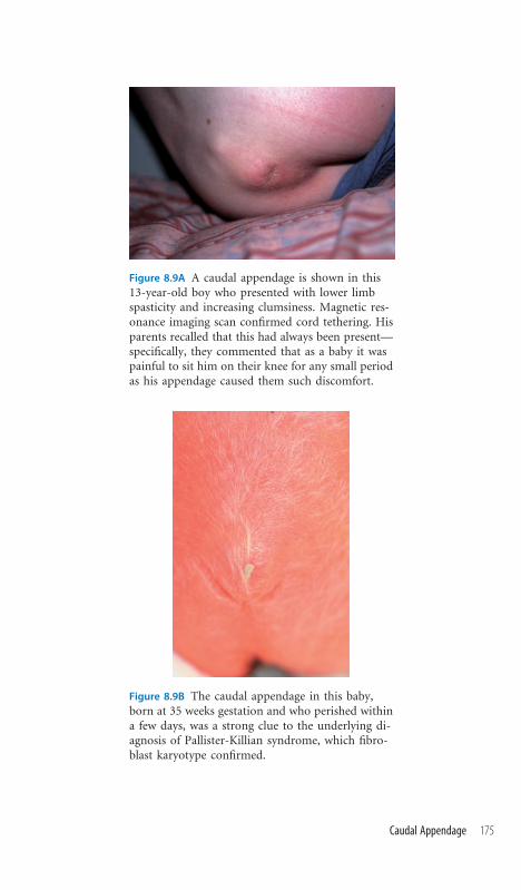
Figure 8.9A A caudal appendage is shown in this13-year-old boy who presented with lower limbspasticity and increasing clumsiness. Magnetic res-onance imaging scan confirmed cord tethering. Hisparents recalled that this had always been present—specifically, they commented that as a baby it waspainful to sit him on their knee for any small periodas his appendage caused them such discomfort.
Figure 8.9B The caudal appendage in this baby,born at 35 weeks gestation and who perished withina few days, was a strong clue to the underlying di-agnosis of Pallister-Killian syndrome, which fibro-blast karyotype confirmed.
Caudal Appendage 175

Chapter 9 The Hands

Postaxial Polydactyly
Recognizing the Sign An extra digit is present on the ulnar side of the hand.
This can be subtle and consist of no more than a small projection of tissue,
usually between the metacarpophalangeal and proximal interphalangeal
creases.
Establishing a Differential Diagnosis This is one of the most common of all
congenital anomalies and can run in families as a harmless autosomal dom-
inant trait. In the event of a normal family history, syndromic consideration
must be addressed. Remember to assess for Patau syndrome (trisomy 13). The
presence of cleft lip and/or a cardiac murmur will increase your concerns that
this is the real diagnosis. A cardiac murmur might also make you think about
Ellis-van Creveld syndrome (EvC). To substantiate this diagnosis, look in the
mouth for multiple oral frenula. The additional observation of natal teeth is
very strong evidence for this condition. The nails may be small and deep set
and the thorax narrow. A narrow thorax is the defining clinical characteristic
of Jeune asphyxiating thoracic dystrophy, most cases of which expire shortly
after birth and many of whom also have a postaxial polydactyly. Survivors can
develop a nephronophthisis and retinal dystrophy. The polydactyly ofMeckel-
Gruber syndrome, a lethal autosomal recessive condition, is usually noted
by way of confirming the diagnosis in a patient presenting with a Potter-type
history of oligohydramnios, encephalocele, and grossly enlarged cystic kidneys.
Skull malformation, suggestive of craniosynostosis, is the best prompt to Car-
penter syndrome, in which postaxial polydactyly of the hand is associated with
syndactyly and short fingers. Duplication of the hallux is an unreliable clinical
sign in the feet, being absent in several reported cases. Postaxial polydactyly
is often the only clue to Bardet-Biedl syndrome (BBS) at the time of birth.
Emerging obesity in the first year of life, despite normal appetite, consolidates
the clinical concerns. The signature retinal dystrophy is often a phenomenon of
the second decade and therefore not diagnostically reliable in the young. Hypo-
genitalism is universal in males and should be sought clinically to support the
likely diagnosis in obese males with polydactyly.
Investigations to Consider A basic karyotype will demonstrate the addi-
tional chromosome 13 in Patau syndrome. Skeletal radiology in E-vC and
Jeune syndromes both show a hook-like downward projection from the me-
dial and lateral acetabular margins, in addition to narrow thorax. Electroret-
inography (ERG) is often helpful in establishing the diagnosis in BBS before
retinal signs become clinically apparent. BBS mutations are known in several
genes but have not been a diagnostically valuable investigation in individual
cases.
The Hands178
9.1

Figure 9.1A Classic postaxial polydactyly in a case of autosomaldominant nonsyndromic familial postaxial polydactyly, the baby’sfather and grandmother having had the same anomaly.
Figure 9.1B Postaxial polydactyly in a 1-year-old boy referred toclinic because of emerging concerns with respect to development.His weight was increased, being above the 97th percentile, andwith this history, the polydactyly and hypogenitalism pointedtoward a diagnosis of Bardet-Biedl syndrome.
Postaxial Polydactyly 179

Preaxial Polydactyly/Thumb Duplication
Recognizing the Sign This uncommon phenomenon is characterized by
complete or partial reduplication of a normal biphalangeal thumb. Clinically,
you may see a second thumb adjacent to the normal, but if the duplication is
confined to the distal phalanx, the clinical presentation may be of a broad nail
on the thumb, perhaps with a central line of fusion between the two nails
forming the fused nail. It is an uncommon clinical sign.
Establishing a Differential Diagnosis Examine both hands and feet for other
evidence of polydactyly, syndactyly, or developmentalmalformation of the digits.
Family history may offer clues, in that rare pedigrees are known with this mal-
formation segregating in autosomal dominant manner. Teratogenic influences
should be sought, reduplication, more typically involving the hallux but occa-
sionally of the thumb, being described in maternal diabetes. Assuming that the
thumbmalformation is the sole sign, assess forRobinow syndrome, in which this
specific feature is well described. Look at the face for hypertelorism, anteverted
nares, and evidence of cleft lip (Figure 2.5A). During infancy, gum hypertrophy
is an especially good clue to this diagnosis. The umbilicus may be shifted toward
the xiphisternum and males will show micropenis. Assess the ears. Simple, cup-
shaped ears are suggestive of lacrimo-auriculo-dento-digital (LADD) syndrome,
in which deafness, thumb reduplication, and hypodontia occur with nasolacri-
mal duct obstruction and consequent epiphora. Parents may volunteer that the
child never sheds tears on crying—a very good pointer to the diagnosis. Although
thumb reduplication is not the most typical hand malformation in Townes-
Brocks syndrome (T-BS), triphalangeal thumbs being more often encountered
clinically, this conditionmay be the correct diagnosis in the deaf patient with anal
malformations, lifelong constipation, and lop ears, perhapswith preauricular pits
or skin tags. Careful evaluation of external eye movements is valuable. The ob-
servation of a Duane phenomenon, a unilateral sixth nerve palsy with contralat-
eral enophthalmos, strongly suggests that the thumbduplication is the presenting
feature of acro-reno-ocular (ARO) syndrome, in which deafness and urinary
tract anomalies are common. A congenital anemia, especially if associated with
cleft lip/palate and the thumb anomaly, urges that the plausible diagnosis of
Blackfan-Diamond syndrome be considered.
Investigations to Consider Most of the conditions alluded to are clinical
diagnoses. Audiologic evaluation is obviously important in LADD, T-BS, and
acrorenoocular syndromes. Ophthalmic evaluation is appropriate for possible
chorioretinal colobomata in ARO. Mutation analysis of SALL1 identifies
mutations in T-BS and those in the related gene SALL4 confirm ARO if there
is clinical doubt. There is a high risk of renal failure among T-BS cases, so
longitudinal renal biochemistry assessment is advisable. Hematologic char-
acterization of anemia will assist identification of Blackfan-Diamond cases.
The Hands180
9.2

Figure 9.2 Complete thumb duplication is shown in this child witha provisionally unique syndrome.
Preaxial Polydactyly/Thumb Duplication 181

Syndactyly of the Fingers
Recognizing the Sign Syndactyly refers to partial or complete fusion between
the fingers, which can be osseous or involve the skin only. The interdigital web
between fingers 2 and 3 lies at the same level as that between fingers 3 and 4,
and any advance in the position of the web signifies cutaneous syndactyly of
that web space.
Establishing a Differential Diagnosis Complete syndactyly, often involving
the thumb, is typical of Apert syndrome, the appearance being termed ‘‘mit-
ten hand’’ malformation. The accompanying skull and facial malformations
with eye prominence, beaked nose, and high, turret-shaped skull are easily rec-
ognizable. Careful oral examination is warranted, the palate frequently being
cleft. Cutaneous syndactyly involving fingers 3 and 4 in a low-birth-weight
infant may be the best clinical clue to triploidy or diploid/triploid mosaicism.
Other clinically recognizable conditions in which syndactyly may be the pres-
enting clinical feature are Poland anomaly and Moebius syndrome. Accord-
ingly, chest examination for asymmetry of muscle is warranted in the former
and cranial nerve evaluation, most particularly of sixth and seventh nerves, in
the latter; the examiner is advised to be aware that a small number of cases can
have features of both conditions, the Moebius-Poland phenomenon. A spe-
cific appearance of syndactyly of fingers 3/4/5 should raise the possibility of
Timothy syndrome, in which long QT syndrome and a high propensity to ar-
rhythmias and sudden death apply. This same pattern of syndactyly is also seen
in oculo-dento-digital (ODD) syndrome, the diagnosis being prompted by
the hypotelorism, small nose, and enamel hypoplasia (Figure 2.2). Though
poorly understood, spasticity of the lower limbs is commonly observed in
affected individuals. The most common form of syndactyly is familial, involv-
ing fingers 3/4 and toes 2/3, so parental assessment in an infant with syndactyly
may exclude syndromic concerns. In association with plagiocephaly, or un-
usual skull shape, syndactyly is often observed in Saethre-Chotzen syndrome.
Being autosomal dominant, parental examination may also reveal forehead
flattening or other features of craniosynostosis. Ptosis, frontalis overactivity,
and prominence of the horizontal ear crus are other valuable clinical signs in
securing this diagnosis.
Investigations to Consider Karyotype will establish triploidy (69 chromo-
somes). Electrocardiography (ECG) and specifically QT-interval evaluation is
indicated if Timothy syndrome is being considered. CACNA1C gene mutation
is established in all cases. Apert syndrome constitutes a clinical diagnosis. FGFR2
mutation is present in all affected individuals. ODD is caused by GJA1 muta-
tion. Magnetic resonance imaging (MRI) examination has shown white matter
changes in some ODD affected individuals with spasticity. Mutations of the
TWIST locus on 7p are demonstrable in cases of Saethre-Chotzen syndrome.
The Hands182
9.3

Figure 9.3A Complete syndactyly of fingers 3 and 4 isshown in a patient with triploidy.
Figure 9.3B The hands shown are of a 24-year-old womanwho has Saethre-Chotzen syndrome. Note the bilateral,symmetric cutaneous syndactyly of fingers 2 and 3.
Figure 9.3C Symbrachydactyly associated with Polandanomaly is demonstrated in an adult female patient. Inaddition to the finger fusion, the digits are short, hencethe descriptive term ‘‘symbrachydactyly’’

Clinodactyly of the Fingers
Recognizing the Sign This term refers to deflection of one or more fingers
medially or laterally. The most common presentation is of medial deviation of
the fifth finger due to hypoplasia of the middle phalanx. A good clinical
barometer of clinodactyly is to look at the flexion creases of the interpha-
langeal joints, which slope toward one another in clinodactyly rather than
pursuing parallel courses, as is the norm.
Establishing a Differential Diagnosis Clinodactyly is common and fre-
quently familial. Parental evaluation may dissipate syndromic concerns. Alter-
natively, a form of autosomal dominant brachydactyly may be established and
the clinodactyly thus explained. Clinodactyly, especially of the fifth finger, is
common, and this minor anomaly simply heightens the awareness of the ex-
aminer for other similar signs of abnormal development. Hence, 60% of Down
syndrome cases will have fifth finger clinodactyly, but the clinical diagnosis of
Down syndrome will rest on the presence of other characteristic signs. Likewise,
clinodactyly is a nonspecific finding in several chromosomal deletion and du-
plication syndromes beyond clinical recognition and reliant for determination
on karyotypic analysis. However, several short stature syndromes are associated
with clinodactyly—in Russell-Silver syndrome, the first clue to diagnosis may
be the low birth weight and length but relatively normal head circumference.
Cafe-au-lait skin patches often emerge in time. Careful evaluation for asym-
metry of the limbs is useful, asymmetry being an associated feature, but absence
of limb asymmetry in an otherwise typical Russell-Silver presentation is char-
acteristic of a subgroup of children whose short staturemay be due to disomy of
chromosome 7. Clinodactyly is also common in Aarskog syndrome, in which
an X-linked history of short stature should be sought. Look for other charac-
teristic findings of hypertelorism, shawl scrotum, and periumbilical depression
(Figures 8.1 and 8.7). In the older child referred for developmental concerns,
clinodactyly is a good pointer toward tricho-rhino-phalangeal (TRP) syndrome
(Figures 2.4 and 12.5). On questioning it will be established that the hair is fine,
growing poorly in the frontotemporal regions, while evaluation of the nose will
often show a bulbous tip. There may be prominent interphalangeal joints with
slight deviation of the phalanges causing clinodactyly of other fingers.
Investigations to Consider Karyotype is indicated if trisomy 21 is suspected
or clinical signs warrant exclusion of other chromosomal conditions. Disomy
7 studies in symmetric short stature but with a clinical resemblance to Russell-
Silver syndrome require parental DNA samples in addition to that of the
patient. Hand x-rays may show cone-shaped epiphyses in TRP syndrome and
exostoses are sometimes seen on long bones. Deletion of chromosome 8q
should be sought if TRP is suspected.
The Hands184
9.4
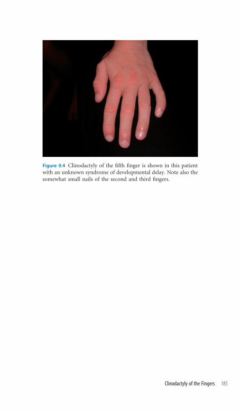
Figure 9.4 Clinodactyly of the fifth finger is shown in this patientwith an unknown syndrome of developmental delay. Note also thesomewhat small nails of the second and third fingers.
Clinodactyly of the Fingers 185

Arachnodactyly of the Fingers
Recognizing the Sign This description refers to long, thin fingers.
Establishing a Differential Diagnosis Measure the height of the patient and
assess the skeletal features clinically. The major concern is to establish whether
the patient may haveMarfan syndrome (MS). Excess joint laxity, pectus cari-
natum or excavatum, scoliosis, reduced upper-to-lower segment height ratio,
and the ability to protrude the thumb below the ulnar border of the enclosed
fist are all features that strongly support a likely diagnosis ofMarfan syndrome.
A positive family history, if procured, can be confirmatory. Ocular evaluation
for lens dislocation, skin examination for striae, evidence of hernia repair, and
cardiac evaluation for aortic root dilatation or mitral valve prolapse should
be undertaken. The misdiagnosis of Marfan syndrome, both diagnosis missed
and cases wrongly identified, represents the most common cause of referral to
the author’s clinic. A common error arises with respect to lens dislocation, of-
ten absent if evaluated in the newborn period and assumed therefore to be
normal. This feature can develop later, and serial ophthalmic re-evaluation is
diagnostically useful in cases where the diagnosis remains in doubt. The closely
related condition of Beals contractural arachnodactyly usually shows camp-
todactyly of the fingers and joint contractures of the knees. An unusual ‘‘crum-
pled’’ appearance to the ear should be sought, the helix being flattened su-
periorly, and confirms the diagnosis, which is not associated with aortic root
weakness. Homocystinuria mimics the skeletal features of Marfan syndrome
and characteristic downward lens dislocation may not occur until the age of
10 years. Mild developmental delay, the absence of cardiac findings, and a
history of seizures or evidence of spasticity clinically represent the best clinical
parameters for distinction from MS. A malar flush is often observed. Occa-
sionally Stickler syndrome patients demonstrate arachnodactyly. The family
history of myopia and/or retinal detachment, cleft palate and micrognathia,
and early-onset osteoarthritis will usually serve as a good clinical basis for
distinction from MS. The presence of a flat malar region and depressed nasal
bridge in a patient should alert the examiner to the likely diagnosis and avert
the misdiagnosis of MS.
Investigations to Consider Echocardiography and specific evaluation of
aortic root diameter is mandatory if MS is being considered, having both
diagnostic and management value. FBN1mutation analysis is available in MS,
but the diagnosis is essentially based on clinical evaluation. FBN2 mutation
confirms Beals syndrome. Serum amino acids demonstrating elevated me-
thionine and homocysteine and low cysteine is the recommended screening
test for homocystinuria, but false negatives do arise.
The Hands186
9.5

Figure 9.5 Arachnodactyly is clearly present in this patient, referred as a case ofMarfan syndrome, but whose other clinical findings did not substantiate thediagnosis.
Arachnodactyly of the Fingers 187
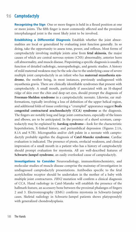
Camptodactyly
Recognizing the Sign One or more fingers is held in a flexed position at one
or more joints. The fifth finger is most commonly affected and the proximal
interphalangeal joint is the most likely joint to be involved.
Establishing a Differential Diagnosis Establish whether the joint abnor-
malities are local or generalized by evaluating joint function generally. In so
doing, take the opportunity to assess tone, power, and reflexes. Most forms of
camptodactyly involving multiple joints arise from fetal akinesia, the major
causes of which are central nervous system (CNS) abnormality, anterior horn
cell abnormality, andmuscle disease. Pinpointing a specific diagnosis is usually a
function of detailed radiologic, neuropathologic, and genetic studies. A history
of mildmaternal weakness may be the sole clue to the underlying cause of severe
multiple joint camptodactyly in an infant who has maternal myasthenia syn-
drome, the mother being, in most instances, previously undiagnosed with
myasthenia gravis. There are clinically identifiable syndromes that present with
camptodactyly. A small mouth, particularly if associated with an H-shaped
ridge of skin over the chin and deep-set eyes, should prompt the diagnosis of
Freeman-Sheldon syndrome in a camptodactylic neonate. Likewise, ear mal-
formations, typically involving a loss of definition of the upper helical region,
and additional folds of tissue conferring a ‘‘crumpled’’ appearance suggest Beals
congenital contractural arachnodactyly (CCA) syndrome as the diagnosis.
The fingers are notably long and large joint contractures, especially of the knees
and elbows, are to be anticipated. In the presence of a shawl scrotum, camp-
todactyly may be explained by Aarskog syndrome—look for the characteristic
hypertelorism, X-linked history, and periumbilical depression (Figures 2.1A,
8.1A and 9.7B). Micrognathia and/or cleft palate in a neonate with campto-
dactyly probably signifies the diagnosis of Catel-Manzke syndrome. Cardiac
evaluation is indicated. The presence of ptosis, orofacial weakness, and clinical
impression of a small mouth in a patient who has a history of camptodactyly
should prompt evaluation for myotonia. All are well-described features of
Schwartz-Jampel syndrome, an easily overlooked cause of camptodactyly.
Investigations to Consider Neuroradiology, immunohistochemistry, and
molecular studies of muscle disease comprise the mainstay of investigation in
undiagnosed camptodactyly presentations. Antibodies specific to the fetal
acetylcholine receptor should be undertaken in the mother of a baby with
multiple joint contractures. FBN2 mutation will confirm a clinical diagnosis
of CCA. Hand radiology in Catel-Manzke will establish the presence of the
hallmark feature, an accessory bone between the proximal phalanges of fingers
2 and 3. Electromyography (EMG) confirms myotonia in Schwartz-Jampel
cases. Skeletal radiology in Schwartz-Jampel patients shows platyspondyly
with generalized chondrodysplasia.
The Hands188
9.6

Figure 9.6A Bilateral camptodactyly is demonstrated.
Figure 9.6B The hand of a patient with Freeman-Sheldon syndromeis shown. See also Figure 5.1 for the typical facial appearance andanother example of associated camptodactyly.
Camptodactyly 189

Brachydactyly
Recognizing the Sign One or more fingers are short. Generally, the short-
ening is phalangeal and there may be associated clinodactyly, but the term also
covers metacarpal shortening both in combination with phalangeal involve-
ment and in isolation thereof.
Establishing a Differential Diagnosis Both family and pregnancy histories
are important. There are several autosomal dominant patterns of brachy-
dactyly, some associated with generalized short stature, which may come to
light through family history and examination and explain the clinical features
in the patient without recourse to investigation. Teratogens tend to cause a
brachydactyly of the distal phalanges, important and common culprits being
alcohol and phenytoin. Alcohol may be denied, so do assess for accessory pal-
mar creases, additional nipples, short palpebral fissures, thin upper lip, and
smooth philtrum, which may lend support to this difficult diagnosis. These
causes excluded, think about specific syndromic considerations as follows. If
the brachydactyly only involves the thumb, evaluate the birth weight and face
forde Lange syndrome, in which short firstmetacarpal is characteristic (Figure
9.13B). Look especially for synophrys, generalized hirsutism and a thin up-
per lip. Micropenis and/or cleft lip are suggestive of Robinow syndrome—
evaluate the umbilicus to see if it is displaced toward the sternum and look at
the face for hypertelorism. Broad thumbs are frequent, due to duplication of
the terminal phalanx. Hypertelorism and brachydactyly but with normal-sized
penis should prompt the thought of Aarskog syndrome. Specifically, look for
the periumbilical depression so typical of this condition and the shawl con-
figuration of the scrotum. Get the patient to close the fist and look for the
short fourth metacarpal by observing the knuckle-knuckle-dimple-knuckle
sign (the ring finger has a depression over the joint secondary to the shortness
of the fourth metacarpal). This sign is the hallmark of familial brachydactyly
type E, in which development is normal. However, the same sign is often
demonstrated in cases of Albright’s hereditary osteodystrophy (AHO), in
which developmental delay, generalized obesity, and signs of hypothyroidism
should be sought. Palpate the skin lightly, looking for areas of subcutaneous
calcification. Stature is generally short and metatarsal shortening, most com-
monly of the fourth and fifth metatarsals should also be sought.
Investigations to Consider Hand x-rays may show terminal phalangeal du-
plication of the thumbs in Robinow cases. FGD1 gene mutation analysis may
confirm a clinical diagnosis of Aarskog syndrome as may ROR2 analysis in
Robinow syndrome. Chromosome 2q37 studies are important in AHO as de-
letion of this region mimics AHO, true AHO cases having variable hypocal-
cemia, raised parathyroid hormone levels, and GNASI mutation.
The Hands190
9.7

Figure 9.7A Generalized brachy-dactyly is shown, the noteworthyclinical feature being short nails.
Figure 9.7B By contrast with Figure9.7A, brachydactyly is associatedwith partial skin syndactyly, clin-odactyly, hyperextension of themetacarpophalangeal (MCP) joints(see how prominent the tendons areover the MCP joints), and flexion ofthe fourth proximal interphalangealjoint bilaterally. This patient hasclassical hand features of Aarskogsyndrome. See also Figures 2.1A,8.1A, and 8.7
Figure 9.7D The radiographof thehand fromFigure 9.7Cconfirms the particular shortening of the fourth
metacarpal.
Figure 9.7C The generalized brachydactyly associ-ated with familial brachydactyly type E is demon-strated in this patient.
Brachydactyly 191

Tapering Fingers
Recognizing the Sign All fingers taper naturally in a proximodistal direction
but this ill-defined clinical sign refers to a progressive narrowing of finger
breadth, which is noteworthy for the degree of narrowing.
Establishing a Differential Diagnosis Of itself, this sign represents a minor
malformation and may be of no clinical significance. Like all minor mal-
formations, the value of the observation is to heighten awareness for other
signs that signify an underlying diagnosis. Tapering fingers are well described
in association with obesity but are especially characteristic of Cohen syn-
drome. This autosomal recessive condition of truncal obesity and develop-
mental delay has characteristic facial features of a short philtrum, conferring
an open-mouthed presentation, prominent central incisors, and a thick head
of hair (Figure 2.8). Early-onset myopia and progressive pigmentary reti-
nopathy are valuable supportive signs, without which the diagnosis should be
infrequently reached. Unless there is an older affected sibling, this is not a
diagnosis for the neonatal period and is more easily made in the 3-year-old
and upward age range, at which stage the tapering fingers and truncal obesity
are more readily recognizable. Tapering fingers in the neonatal period are
uncommon, but if associated with arachnodactyly should result in evaluation
for the neonatal presentation of Marfan syndrome (MS). Cardiac compli-
cations are usually severe and of early onset with aortic root dilatation and
valvular dysfunction. Emphysema is a particular complication of this form of
MS and assists the diagnosis if identified. Extreme hypotonia in the neonatal
period with tapering fingers is the combination of features that facilitates the
early diagnosis of Ehlers-Danlos syndrome (EDS) type VIA before the char-
acteristic kyphoscoliotic signs appear and make diagnosis easier. The unusual
pattern of ‘‘criss-cross’’ lines on the palms and soles is a very valuable, indeed
virtually pathognomic, sign (Figure 9.18C). An impression of tapering fingers
is common in obese older children, usually with developmental delay, and
chromosomal mosaicism should be investigated in such a context. Look for
linear hypopigmentation, clinically and by ultraviolet light examination.
Investigations to Consider Unexplained neutropenia is a common finding in
Cohen syndrome and a valuable aid to diagnosis if present. Retinal examination
is indicated for the characteristic pigmentary retinopathy if Cohen syndrome
is suspected. Mutation analysis of the COH1 locus on chromosome 8 will es-
tablish the diagnosis, but there may be other unidentified genes. Bone age may
be advanced in neonatal EDS VI. Urinary pyridinoline analysis is a very effec-
tive test, obviating the need for lysyl hydroxylase assay on fibroblasts. Fibroblast
culture is usually necessary to secure a diagnosis of chromosomal mosaicism.
Neonatal MS is confirmed by FBN1 mutation, usually in exons 24–32.
The Hands192
9.8

Figure 9.8A A classic example of tapering fingers isshown in this 10-year-old girl with Cohen syndrome.
Figure 9.8B Note the thick hair, the manner in whichthe upper lip retracts on smiling, and the largecentral upper incisors in the face of the same patient(see also Figure 2.8).
Tapering Fingers 193

Puffy Fingers
Recognizing the Sign A purely subjective impression, the fingers appear
swollen. Though a rare sign, due to its diagnostic significance its identification
can be extremely valuable.
Establishing a Differential Diagnosis The exclusivity of a particular clinical
sign to one particular syndrome is unusual, but applies in this instance. Puffy
fingers are virtually pathognomonic of Coffin-Lowry syndrome. This is an X-
linked mental retardation syndrome. Intellectual development is usually se-
verely impaired and early hypotonia and motor milestone delay are typical.
The facial appearance, difficult to recognize in the neonatal period, coarsens
with time, with hypertelorism and thickened, everted, lips being the most con-
sistent features. There is frequently an associated pectus excavatum. As also
occurs in Williams syndrome, there is a propensity to rectal prolapse. The
fingers are puffy in appearance and a telling and useful additional sign is the
presence of an accessory transverse hypothenar crease on the hand. It should
be noted that the fingers are entirely pain free and normally functioning. The
family history may offer substance for the diagnosis with other affected males,
consistent with X-linked inheritance being identified. Older patients frequen-
tly show severe kyphoscoliosis. Clinical examination of females in a pedigree
is also warranted, the puffy fingers often being present in obligate carriers.
Indeed, the author has seen the diagnosis made in a floppy male neonate by
the astute glance that the clinician gave at the carrier mother’s hands.
Puffy fingers are also seen in infantile systemic hyalinosis, an autosomal
recessive condition, but in contrast to Coffin-Lowry syndrome, the fingers
in this condition are swollen and painful. There is usually hyperpigmenta-
tion over the joints and dermatologic evidence of inflammation, particu-
larly periauricular, perioral, and perianal, is another diagnostically useful dis-
tinction.
Investigations to Consider Coffin-Lowry syndrome is a clinical diagnosis.
The main sources of diagnostic error are confusion with Williams syndrome,
in which the fingers are not swollen but the lip prominence can mislead the
inexperienced. Similar scope for confusion arises from the facial resemblance
between Coffin-Lowry syndrome and a-thalassemia mental retardation,
X-linked (ATRX) syndrome. Staining a blood smear with 1% brilliant cresyl
blue in search of hemoglobin H inclusion bodies is a useful screen to exclude
the latter. Molecular confirmation of Coffin-Lowry cases is available by RSK2
mutation analysis.
The Hands194
9.9

Figure 9.9A Note the hypertelorism and lip fullness in the faceof the affected boy with Coffin-Lowry syndrome.
Figure 9.9B The digits arebroad and look swollen inthis hand of a typical femalecarrier of Coffin-Lowrysyndrome.
Figure 9.9C The hypothenarcrease is shown in a Coffin-Lowry affected boy, but itis worth also observing thegeneralized puffiness of thefingers. (Courtesy of Profes-sor Ian Young.)
Puffy Fingers 195

Overlapping Fingers
Recognizing the Sign This sign is hardly open to misinterpretation, though
some overlap with camptodactyly is inevitable. However, camptodactyly is not
invariably associated with overlapping position of the fingers, and the reverse
also holds true.
Establishing a Differential Diagnosis Overlapping fingers, typically of the
index finger over the middle finger and of the fifth finger over the ring finger,
is characteristic of Edwards syndrome (trisomy 18). Generally, the hands are
clenched, the thumb often adducted across the palm. Close inspection fre-
quently shows absence of the distal creases of the fifth fingers. Viewed in profile,
the skull is dolichocephalic, with a prominent occiput and low-set ears, usually
malformed in appearance. Cardiac examination will establish congenital heart
lesions in more than 80% of cases. Much less commonly, overlapping is ob-
served in Patau syndrome (trisomy 13), in which condition postaxial poly-
dactyly of the fingers is the more typical finding, sometimes indeed with asso-
ciated overlapping. The hands of Pena-Shokeir syndrome can exactly resemble
the hands of trisomy 18 syndrome—this is a condition of multiple joint con-
tractures, paucity of facial movement, and pulmonary hypoplasia, often fatal.
Being autosomal recessive, its recognition and clear distinction from trisomy 18,
a sporadically occurring condition, is important. Chromosomal disorders aside,
overlapping fingers are seen in Beals congenital contractural arachnodactyly
(CCA) syndrome, in which cases the crumpled appearance of the upper helix of
the ear and the joint contractures, especially of the knees, will render the con-
dition readily diagnosable. In a similar vein, maternal myasthenia syndrome,
already considered under camptodactyly (section 9.6), can present in an infant
with fingers whose gross appearance is of overlapping position, but the super-
ficial interpretation of this sign will be modified by examination of joint creases,
range of movement, and involvement of other joints in the pathologic process,
most particularly in the form of talipes, hip flexion, and knee extension. Mul-
tiple joint contractures, particularly if there is ulnar deviation of the hand, the
fingers possibly overlapping, is a typical presentation of Freeman-Sheldon
syndrome, an easily made diagnosis from the facial inspection, themouth being
small and pursed, often with a hallmark H-shaped knuckle of skin over the chin
(Figures 5.1 and 9.6). The author has twice made the diagnosis of Smith-Lemli-
Opitz (S-L-O) syndrome by noting an overlapping index finger in an otherwise
nondysmorphic child and investigating biochemically.
Investigations to Consider Basic karyotype is needed for chromosomal triso-
mies. FBN2mutation analysis is indicated for CCA. Antifetal-type acetylcholine
receptor antibodies are warranted in suspected maternal myasthenia syndrome.
Low serum cholesterol and elevated 7-dehydrocholesterol typify S-L-O.
The Hands196
9.10

Figure 9.10A The hands of a case ofEdwards trisomy 18 syndrome areshown. Note the position of the indexfinger overlapping the middle finger,the flexed thumb, and the fifth fingeroverlapping the fourth. The nails arealso hypoplastic. (Courtesy ofProfessor Ian Young.)
Figure 9.10B The hands of a differentcase of Edwards syndrome, demon-strating overlapping fingers, small nails,and syndactyly. (Courtesy of ProfessorIan Young.)
Figure 9.10C The hands of a child with Smith-Lemli-Opitz syndrome are dem-onstrated showing the occasional finding in that condition of the index fingeroverlapping the middle finger.
Overlapping Fingers 197

Ectrodactyly
Recognizing the Sign There is absence of central digits, perhaps extending to
involvement of the metacarpals, and often demonstrating syndactyly, camp-
todactyly, or clinodactyly of the remaining digits.
Establishing a Differential Diagnosis Start with a detailed family history.
The condition of ectrodactyly, ectodermal dysplasia, and clefting (EEC) syn-
drome is an autosomal dominant condition of wide clinical variability, and
many aspects such as dry skin, palmar hyperkeratosis, small brittle nails,
sparse hair, and hypodontia need to be evaluated in attempting to identify a
history of this disorder. Reports of tear duct anomalies in family members are
a valuable diagnostic clue. In examining the patient, evaluate all four limbs.
The extent of limb involvement need not be equally severe in all limbs for this
diagnosis to be reached. Cleft lip is common in affected individuals. Blue
irides and photophobia are useful supportive clues to observe. It is valuable to
examine the hands and feet of parents, whose digits may be normal but have
abnormal skin grooves of the palms of feet, betraying mutation carrier status.
The allelic condition of limb mammary syndrome (LMS) differs in that the
ectrodactyly is associated with hypoplasia of the nipples and mammary glands
but there is no clefting or signs of ectodermal dysplasia and lacrimal duct
anomalies are much less common. Genetically distinct syndromes of split
hand and foot malformations (SHFMs) not involving ectodermal signs are
usually identifiable from the autosomal dominant family history. Cup-shaped
ears are often observed in this situation as is limb shortening, especially of the
tibia and/or ulna. Clinical overlap in terms of limb shortening or asymmetry
between the limbs and the appearance of an ectrodactyly-like malformation
of the hand occurs between SHFM and femur-fibula-ulnar (FFU) syndrome.
The hand appearance in FFU syndrome usually reflects absence of the digits
on the ulnar side of the hand. Likewise, an ectrodactyly-like hand is an
occasional presentation in de Lange syndrome, but the low birth weight,
microbrachycephaly, general hirsutism, and characteristic synophrys should
facilitate differentiation from other causes of ectrodactyly. Seen in association
with holoprosencephaly or anencephaly, ectrodactyly is signaling the diag-
nosis of XK aprosencephaly syndrome, an important condition to recognize
since there is evidence that some such families represent autosomal recessive
inheritance.
Investigations to Consider Mutation analysis of P63 on chromosome 3q27
confirms the diagnosis of EEC syndrome and LMS.
The Hands198
9.11

Figure 9.11 A typical ‘‘lobster-claw’’ malformation of EEC syn-drome showing, in addition to the ectrodactyly, camptodactylyof the digits on either side of the cleft.
Ectrodactyly 199

Broad Thumbs
Recognizing the Sign There is no definition as such, but in addition to
looking broadwith respect to the other digits, the thumbmay present a flattened
appearance, perhaps with abnormal angulation of the terminal phalanx.
Establishing a Differential Diagnosis Though not exclusive of other diag-
noses, broad thumbs are quintessentially associated with two syndromes of
which they represent the hallmark feature. These are Rubinstein-Taybi syn-
drome and Pfeiffer syndrome. In Rubinstein-Taybi syndrome, there is often
radial angulation of the terminal phalanx of the thumbs. Examine the feet for
associated hallucal broadening. Microcephaly is usual, the forehead being pro-
minent and the palpebral fissures downslanting. The nose is absolutely char-
acteristic, the nasal bridge being wide and the columella extending below the ala
nasi (Figure 2.5B). Generally recognizable in the newborn period, these features
become more pronounced with age. A small number of cases have been diag-
nosed molecularly in whom the facial characteristics prompted the diagnosis
but the thumbs were normal. These represent an exception in what remains a
clinical diagnosis. Pfeiffer syndrome comprises the association of craniosyn-
ostosis and broad thumbs. Hallucal broadness is usual and some soft tissue
syndactyly of the hands and feet common. The craniosynostosis is generally a
brachycephaly due to underlying coronal suture fusion, but more extensive
malformation is possible, including the severe clover-leaf skull malformation.
Being autosomal dominant, careful assessment of the parents and family may
identify others affected. Complete syndactyly giving the ‘‘mitten’’ appearance in
association with broad thumbs is diagnostic of Apert syndrome, another of
the craniosynostosis group of disorders, but which represents a sporadically oc-
curring new dominant mutation. Both Pfeiffer and Apert syndromes may have
associated joint fusions, especially of the elbow and radioulnar joints. In asso-
ciation with short stature, broad thumbs are often the clue to the diagnosis
of Robinow syndrome. The genitalia should be examined, often being small,
though sex assignment is usually clear. A very useful sign in the neonatal period
is gum hypertrophy. Finally, the pitfall of interpreting this clinical sign in iso-
lation of a family history and relevant clinical observation is exemplified by three
cases in the author’s experience whose real diagnosis of autosomal dominant
brachydactyly type Dwas misattributed to Rubinstein-Taybi syndrome and the
family wrongly advised of the inevitability of severe mental retardation.
Investigations to Consider Skull x-ray is warranted if craniosynostosis is a
concern. Sleep studies should be conducted for sleep apnea in craniosynos-
tosis syndromes. Hand radiology in Robinow syndrome may show bifid
terminal phalanges of the thumb and index finger. FGFR1/2/3 mutation
analysis is indicated in craniosynostosis depending on the exact phenotype
suspected.
The Hands200
9.12

Figure 9.12A Broad and radially deviated thumb of a typical case ofRubinstein-Taybi syndrome is demonstrated.
Figure 9.12B The short, broad, radially deviated thumbs bilaterally are shown in acase of Pfeiffer syndrome. Note also the brachydactyly of the index fingers.
Broad Thumbs 201

Small and Hypoplastic Thumbs
Recognizing the Sign There is quite a wide range of clinical presentations,
from the clinically small but functionally normal thumb to the functionally
useless pedunculated digit. If in doubt, assess the length of the thumb by
seeing how far it reaches along the radial border of the hand. A thumb that
does not reach the metacarpophalangeal (MCP) joint of the index finger is
short. Thenar eminence hypoplasia is usual with this sign.
Establishing a Differential Diagnosis A prenatal history of polyhydramnios
should be sought, as unilateral thumb hypoplasia may be the sole finding on
external clinical examination in VATER association (vertebrae, imperforate
anus, tracheoesophageal fistula, radial þ/– renal anomalies). Vertebral anom-
alies, anal stenosis/malposition, and cardiac malformations should be the sub-
jects of particular examination, especially if a tracheoesophageal fistula/atresia
is established. Symmetrically hypoplastic but clinically identifiable thumbs
associated with forearm shortening, the consequence of radial aplasia, are typ-
ical of thrombocytopenia-aplastic radii (TAR) syndrome. Neonatally symp-
tomatic thrombocytopenia should be anticipated in most cases of this auto-
somal recessive condition. Examine the lower limbs for corroborative signs
such as limb asymmetry, limb bowing, or hip dislocation. Cow’s milk intol-
erance is a surprising and unexplained feature of many TAR patients. It is
advisable to examine the heart in patients with thumb hypoplasia. A murmur,
particularly an atrial septal defect (ASD), should raise diagnostic consider-
ation of Holt-Oram syndrome (HOS). Bilateral hand involvement is usual
in this autosomal dominant condition, but the hand malformations are quite
variable and can be asymmetric. Associated syndactyly, phocomelia, and
thumb triphalangy are well within the spectrum of the condition and should
be sought in other family members, unsuspecting of their risk of possible car-
diac lesions. In a healthy child with thumb hypoplasia, assess for short stature
and cafe-au-lait patches or abnormal skin pigmentation. These cutaneous signs
are valuable pointers toward the autosomal recessive condition of Fanconi
pancytopenia syndrome, the pancytopenia and associated leukemia rarely
presenting before the age of 8 or 9 years, but the condition can be anticipated
from the other features. Being autosomal recessive, early recognition has clin-
ical implications for predictive testing in siblings. In de Lange syndrome, the
thumbs often appear short, due to the short first metacarpal bone, which is a
hallmark of the condition.
Investigations to Consider Vertebral radiology and echocardiography are
indicated in suspected VATER association, and platelet count in TAR syn-
drome. Echocardiogram and ECG are used to confirm HOS. Chromosome
breakage studies following diepoxybutane (DEB) exposure confirms Fanconi
syndrome.
The Hands202
9.13

Figure 9.13A Unilateral thumb hypoplasia in a boy with VATERassociation. Note the absence of any thenar prominence.
Figure 9.13B The short thumb, consequent on shortening of the firstmetacarpal bone, is shown in a typical example of Cornelia de Langesyndrome. Note that the other fingers are of normal length. The thumbdoes not extend to the MCP joint of the index finger, which is a goodindicator of shortening. (Courtesy of Professor R.C. Hennekam.)
Small and Hypoplastic Thumbs 203

Long and Triphalangeal Thumbs
Recognizing the Sign Thumb length is assessed by holding the thumb in par-
allel with the hand and establishing how far along the index finger the thumb
extends. Long thumbs extend to or beyond the proximal interphalangeal
(PIP) joint of the index finger. Reduced thenar musculature is usual.
Establishing a Differential Diagnosis Digitized thumbs, possessed of a
triphalangeal constitution, are particularly associated with deafness, con-
genital anemia, and structural heart defects. There is generally bilateral hand
involvement, though the nature of thumb malformation can be variable,
perhaps being digitized on the one side and duplicated or hypoplastic on the
other. Examine the external ears—simple or cup-shaped ears with thumb
malformations/digitization suggest a diagnosis of lacrimo-auriculo-dento-
digital (LADD) syndrome. Delayed eruption of teeth, absent teeth, and
failure to produce tears on crying are valuable corroborative evidence of this
diagnosis, in which deafness is to be anticipated. While cup-shaped ears are
more reminiscent of LADD syndrome, the presence of preauricular tags or
pits in the context of thumb triphalangy is more likely to represent Townes-
Brocks syndrome (T-BS). Anal malformations, including stenosis, anterior
placement, and fistulae, are a central element of the diagnosis in patients with
this autosomal dominant condition. While an individual case may personally
show no anal features, the family history may offer the evidence needed to se-
cure the diagnosis. Examination of the cranial nerves is important in cases of
thumb digitization, unilateral sixth nerve palsy with contralateral enophth-
almos (Duane phenomenon) being the best clinical sign to the diagnosis of
Okihiro syndrome. This may present in the neonate as a strabismus. Deafness
and renal malformations should be sought. Thumb elongation can be the
presenting feature of Blackfan-Diamond syndrome, in which a congenital
hypoplastic anemia occurs. While most individuals with this condition have
normal hands, the thumb abnormality, if present, can be the prompt to di-
agnosis. In relation to congenital heart disease, the variable nature of hand
abnormalities in Holt-Oram syndrome (HOS) does extend to long/digitized
thumbs, so careful cardiac evaluation and family history are needed before
this possible diagnosis can be discounted.
Investigations to Consider Audiologic assessment is needed, as is renal ul-
trasound, especially if T-BS is suspected. Hematologic indices are warranted.
Echocardiogram and ECG are indicated for HOS. Mutation analysis of SALL1
for T-BS and SALL4 for Okihiro syndrome should be considered.
The Hands204
9.14
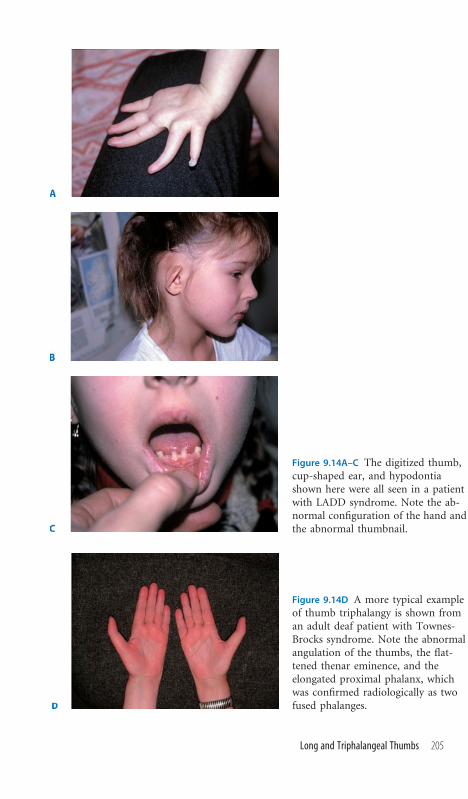
Figure 9.14A–C The digitized thumb,cup-shaped ear, and hypodontiashown here were all seen in a patientwith LADD syndrome. Note the ab-normal configuration of the hand andthe abnormal thumbnail.
Figure 9.14D A more typical exampleof thumb triphalangy is shown froman adult deaf patient with Townes-Brocks syndrome. Note the abnormalangulation of the thumbs, the flat-tened thenar eminence, and theelongated proximal phalanx, whichwas confirmed radiologically as twofused phalanges.
Long and Triphalangeal Thumbs 205

Trident Hand
Recognizing the Sign The description refers to a triangular configuration of
the hand, caused by short broad fingers. The net effect of phalangeal short-
ening is a splaying of the fingers, the index and ring fingers pointing away
from the middle finger. This confers a three-pronged, ‘‘trident,’’ appearance.
Establishing a Differential Diagnosis In contrast to most signs featured be-
tween these covers, the trident hand sign is almost exclusive to achondroplasia.
This is the most common form of chondrodysplasia and usually identifiable at
birth. Birth length can extend into the normal range, an important observation
that limits the diagnostic usefulness of ultrasonic screening for the condition in
pregnancy. Rhizomelic shortening of the humeri is usually noticeable at birth
(Figure 11.2A). Coupled with the typically enlarged occipitofrontal circumfer-
ence, prominent forehead, depressed nasal bridge, and trident hand, skeletal
investigations are indicated to secure the diagnosis. In the older child, leg bow-
ing, limitation of elbow extension, and exaggerated lordosis with protuberance
of the buttocks all comprise elements of the phenotype. While delay in achiev-
ing early motor milestones is usual, normal intellectual outcome is to be antici-
pated. Foramen magnum compression is an important complication of which
to be aware, and sudden death in childhood has been widely reported. Brain-
stem compression may present as obstructive sleep apnea. The best clinical test
for cord compression is to look for clonus and hyperreflexia in the lower limbs.
Mental retardation in achondroplasia should result in examination for acan-
thosis nigricans, seen in SADDAN syndrome (severe achondroplasia, develop-
mental delay, acanthosis nigricans). Thanatophoric dysplasia also causes a tri-
dent hand but the severe compromise of the chest and skeletal system results in
an early perinatal death. A cloverleaf skull appearance, due to craniosynostosis,
is often present, facilitating the diagnosis ultrasonographically during antenatal
life and clinically if the baby is born.
Investigations to Consider Skeletal radiology confirms the diagnosis. The
classical radiologic sign of achondroplasia is absence of the normal increase in
interpedicular distance from the upper lumbar vertebrae caudally. The pedi-
cles are short, seen on a lateral view, with a narrow vertebral canal. The tubular
bones are short and thickened, the fibula often being disproportionately long
relative to the tibia. Mutation of FGFR3 shows a highly specific G1138C,
resulting in a Gly380Arg substitution in over 95% of cases, the mutation
invariably arising on the paternal chromosome and having a positive associ-
ation with advanced paternal age. FGFR3 mutation is also the basis of tha-
natophoric dysplasia and the classic radiologic sign is bowing of the long bones
with ‘‘telephone receiver’’–shaped femora. SADDAN syndrome represents
another form of FGFR3 mutation, occasioning Lys650Met substitution.
The Hands206
9.15

Figure 9.15 The typical appearanceof trident hand is demonstratedin a patient who has achon-droplasia.
Trident Hand 207

Fetal Finger Pads
Recognizing the Sign In human embryology fingertip pads arise on the volar
surfaces of the fingers but regress in early gestation. The absence of normal
regression results in the persistence of fetal finger pads. These are best seen by
looking at the fingertips in profile and the well-defined elevation of tissue
overlying the terminal phalanges is easily identifiable.
Establishing a Differential Diagnosis The sign is a hallmark feature of
two conditions—Kabuki syndrome and Weaver syndrome—but may also be
seen as an occasional finding in other pediatric situations. Characteristic fa-
cial features facilitate the diagnosis of Kabuki syndrome cases—observe the
palpebral fissure and note whether it is wide. A good clue is to look for blood
vessels in the outer canthus of the eye, where they are not usually seen (Figure
3.14). The eyebrows may be unusual, often showing a hairless gap in the
middle. Look for ectropion of the lower eyelid, a most uncommon feature
in pediatric populations but very characteristic in this diagnosis. Lip pits, cleft
palate, and congenital heart disease should be sought. In the older child, short
stature may be the presentation and evidence of developmental delay is to
be expected. Obesity is characteristic of the syndrome in the adolescent age
range. The author has twice made the diagnosis in unsuspected cases of men-
tally retarded females with short stature referred for assessment on foot of
early breast development. By contrast, the history in Weaver syndrome is of
abnormally high birth weight, overgrowth, and macrocephaly. A hoarse cry
is characteristic and developmental delay usual. The facial features are not
specific, but forehead prominence, hypertelorism, and a well-formed philtrum
are usual, while the chin tends to be somewhat small. Apart from the finger
pads, camptodactyly, broad thumbs, and general fleshiness of the hands are
consistent with the diagnosis. This is neither a common nor an easy diagnosis
to make and overlaps with cases of Sotos syndrome. Persistence of finger pads
is also recorded as a finding in several clinically nonspecific chromosomal
deletion syndromes, but one in particular, deletion 22q13 (as distinct from
22q11 deletion), repays specific investigation in children with mild mental
retardation but disproportionate delay in speech.
Investigations to Consider Kabuki syndrome is a clinical diagnosis. Ad-
vanced bone age should be sought if Weaver syndrome is clinically likely.
Mutation analysis of NSD1, the Sotos syndrome locus, is positive in some but
not all cases of Weaver syndrome. 22q13 deletion requires customized cyto-
genetic investigation by subtelomere study or, if available, array-based com-
parative genomic hybridization.
The Hands208
9.16
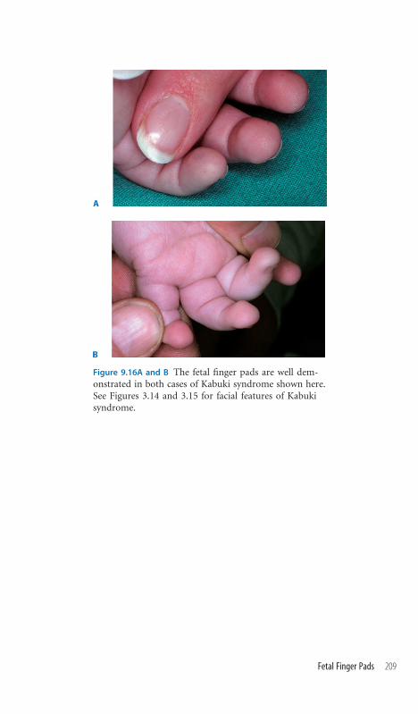
Figure 9.16A and B The fetal finger pads are well dem-onstrated in both cases of Kabuki syndrome shown here.See Figures 3.14 and 3.15 for facial features of Kabukisyndrome.
Fetal Finger Pads 209

Ulnar Ray Defects
Recognizing the Sign There are five digital rays, represented by the meta-
carpal bones and the digits. By definition, ulnar ray defects comprise mal-
formations of the ring and/or little finger, which may extend to involvement
of the metacarpal bones and, occasionally, the ulna.
Establishing a Differential Diagnosis A positive family history of little finger
hypoplasia or absence immediately suggests that the presentation represents
the autosomal dominant condition ulnar-mammary syndrome (UMS). Anal
examination may establish abnormal position or stenosis, both corroborative
features of the diagnosis. Look specifically at the nipples and breasts, usually
hypoplastic, occasionally absent. Genital hypoplasia should be sought inmales.
Older patients may have delayed puberty, and the absence of apocrine glands
typically results in reduced axillary sweat production. In the presence of sym-
metric bilateral ulnar ray defects, the main alternative syndromic consider-
ation is of Miller syndrome. This rare autosomal recessive condition is typ-
ified by malar hypoplasia and lower eyelid ectropion, often with an associated
eyelid coloboma. While these facial characteristics are easily confused with
Treacher Collins syndrome, indeed the ears also being dysplastic as in that dis-
order, the absence of the fifth finger, often with some syndactyly of the other
digits, is the clinical clue that seals the true diagnosis. Reflecting the clinical
presentation of the hands, the fifth toes are usually deficient in Miller cases.
Ulnar ray defects are described in diabetic embryopathy, for which reason a
history of maternal diabetes combined with this clinical finding should spark
appropriate investigations for other features of that protean condition. This
form of oligodactyly has occasionally been described in de Lange syndrome,
but proximally placed thumbs (Figure 9.13B) and small hands and fingers are
more typical hand findings. Nonetheless, in the context of low birth weight,
subsequent growth deficiency, and mental retardation, it is not inappropriate
to consider this condition in the presence of ulnar ray defects.
Investigations to Consider Mutation analysis of the TBX3 locus will confirm
UMS if necessary, and endocrine investigations may be required in the event
of delayed puberty. Miller syndrome is a clinical diagnosis but should be
supplemented by echocardiography and radiology of the vertebral column in
view of the occasional reports of nonspecific congenital heart disease and ver-
tebral segmentation defects.
The Hands210
9.17

Figure 9.17 Typical absence of the little finger in a patient with ulnar-mammarysyndrome. The oligodactyly was bilateral. Note the normal thumb and otherdigits, absence of syndactyly, and normal joint creases.
Ulnar Ray Defects 211

Palmar Crease Patterns of Special Diagnostic Significance
Recognizing the Sign Normal palmar crease patterns accommodate a legion
of variation. Typically there are three main creases, a thenar crease, a trans-
verse proximal palmar crease, and a distal palmar crease. No clinical pattern
involving the palmar creases is absolutely pathognomic, but particular syn-
dromic significance appertains to a few: (1) deep palmar creases, (2) accessory
hypothenar crease, (3) criss-cross patterning, (4) rudimentary palmar crease,
and (5) hockey-stick sign.
Establishing a Differential Diagnosis Deep palmar creases, usually with at-
tendant redundancy of the skin, conferring the appearance of an oversized glove
is typical of Costello syndrome. An early history of poor feeding, high birth
weight, and early recognition of developmental delay are useful diagnostic ad-
juncts. From around the age of 6 years, the typical warty papillomata around
the nose and mouth appear and confirm the diagnosis. A clinical overlap with
cardio-facio-cutaneous (CFC) syndrome exists in that deep creases character-
ize both disorders and absolute distinction between these entities can be difficult
in the first year or two of life. Pulmonary stenosis and hepatomegaly are more
likely to signify CFC syndrome. The observation of an accessory hypothenar
crease raises the possibility of Coffin-Lowry syndrome, an X-linked mental re-
tardation condition with attendant facial features of hypertelorism and promi-
nent, everted lips, becoming more prominent with age (Figure 9.9). Criss-cross
patterning of the hands is an unusual appearance often observed in patients
with the oculoscoliotic form of Ehlers-Danlos syndrome type VI. A blue scleral
tinge, prominent eyes that may not close fully during sleep, and evidence of sco-
liosis should be sought. Rudimentary palmar creases, particularly in the mid-
palmar region, are sometimes seen in fetal alcohol syndrome and can be an
important pillar of diagnosis of this clinically elusive condition, but only if sup-
ported by other features that frequently attend this condition. Look especially
for a featureless philtrum, microcephaly, and short palpebral fissures. A history
of neonatal irritability and low birth weight is likely to apply if this is the true
diagnosis. Likewise, a hockey-stick sign is also well described in fetal alcohol
syndrome, referring to the course of the distal palmar crease, pursuing a hor-
izontal course on the ulnar side of the hand and then abruptly angulating to exit
between the index and middle fingers.
Investigations to Consider Echocardiography is indicated in Costello syn-
drome or CFC syndrome for cardiomyopathy or ventricular thickening and
pulmonary stenosis in CFC syndrome. Serial examination for neuroblastoma
and rhabdomyosarcoma is indicated for Costello cases. Mutation analysis
of RSK2 is needed to confirm Coffin-Lowry syndrome and facilitate carrier
detection in females. Urinary pyridinoline cross-links are indicated in EDS
type VI.
The Hands212
9.18

Figure 9.18D Demonstrated is arudimentary palmar crease be-tween the thenar crease and thedistal palmar crease. The usualproximal palmar crease has notformed properly. The distal pal-mar crease, well established onthe ulnar border, represents ahockey-stick configuration. Thispatient has fetal alcoholsyndrome.
Figure 9.18A The hand of a CFCsyndrome case is shown. Notethe deep palmar creases.
Figure 9.18B Excess, redundantskin in Costello syndrome isshown. Though the palmarcreases are not especially deep inthis patient, deep creases are alsocommonly observed in Costellosyndrome. See also Figure 2.7B.
Figure 9.18C This photographshows the criss-cross pattern soredolent of Ehlers-Danlos syn-drome type VI in a patient withthat condition. Note the absenceof the usual three-crease pattern.
Palmar Crease Patterns of Special Diagnostic Significance 213

Chapter 10 The Feet
Preaxial Polydactyly
Recognizing the Sign Hallucal reduplication is unlikely to be missed. How-
ever, subclinical forms should be suspected in halluces that are clinically broad,
the wide toe perhaps representing an underlying duplication of the terminal
phalanx, and the clinical sign might be more accurately interpreted as an in-
complete duplication.
Establishing a Differential Diagnosis Proximal insertion of an additional
hallux that is identifiably distinct from the normally situated hallux is a marker
for diabetic embryopathy, and such an arrangement of the toes should result in
review of the history for maternal diabetes. The family history may yield diag-
nostically significant information, especially inGreig syndrome. The classic de-
scription of this autosomal dominant condition, ‘‘cephalopolysyndactyly,’’ re-
flects the main clinical features of macrocephaly, polydactyly, and syndactyly.
Typically, the polydactyly is postaxial in the hands and preaxial in the feet, the
extra hallux, if fully formed, lying side by side with its companion, perhaps
sharing a nail. However, wide variation is well described and seemingly unre-
lated historical details such as hydrocephalus and absence of the corpus callo-
sum are occasional findings in the syndrome and discounted at the clinician’s
peril! Familial syndactyly and polydactyly should be targeted in history taking
and sought in examining available relatives, as should measurements of occi-
pitofrontal head circumference. Clinically overlapping with Greig syndrome,
acrocallosal syndrome comprises preaxial polydactyly of both feet, mental re-
tardation, and absence of the corpus callosum. Recognition of this autosomal
recessive disorder has important implications for patient and family alike. Ex-
amination in cases of preaxial polydactyly is incomplete without looking in the
mouth—in particular, evidence of clefting, lingual nodules, or accessory oral
frenula raises the possibility of oro-facio-digital (OFD) syndrome, of which
several genetically distinct subtypes exist. Alternatively, the observation of gum
hypertrophy could represent Robinow syndrome and further examination for
hypertelorism, anteverted nares, short stature, and micropenis give substance to
this diagnosis. Plagiocephaly or a family history of craniosynostosis points to a
likely diagnosis of Saethre-Chotzen syndrome, in which hallucal reduplication
is an occasional finding. More typical features to look for are ptosis, incomplete
syndactyly of the fingers (Figure 9.3B), and flat forehead.
Investigations to Consider Neuroradiology constitutes an important element
of evaluation—absence of the corpus callosum being most likely in acrocallosal
syndrome but occasional in Greig cases. Posterior fossa malformations, such as
Dandy-Walker abnormality, are described in some of the OFD syndromes. Mu-
tation analysis of the GLI3 locus establishes the diagnosis of Greig syndrome,
while mutation at this locus is reported, albeit rarely, in acrocallosal syndrome.
Mutation of TWIST confirms Saethre-Chotzen syndrome if in clinical doubt.
The Feet216
10.1

Figure 10.1A Shown is the typical configuration of thepreaxial polydactyly of the foot in diabetic embryopathy.
Figure 10.1B Demonstrated are the broad, incompletelyreduplicated toes in a case of Greig syndrome.
Figure 10.1C This photograph is from a patient withplagiocephaly, which required surgical correction andwas due to Saethre-Chotzen syndrome.
Preaxial Polydactyly 217

Broad Halluces
Recognizing the Sign The hallux is wider than expected, which may simply
be a broad toe or due to underlying phalangeal duplication. The nail, if bifid,
is a good clue to incomplete duplication.
Establishing a Differential Diagnosis Examination of the thumbs is no less
important. Not only do many of the conditions that cause broad halluces also
cause broad thumbs, but also the novice dysmorphologist will often find
confidence for diagnostically significant hallucal broadness by looking at the
thumbs. Rubinstein-Taybi syndrome (R-TS) characteristically shows broad
halluces and thumbs, the latter frequently being medially deviated. In the neo-
natal period, when facial features are less well formed, microcephaly is a valu-
able adjunctive clinical sign of R-TS. Later, the typical facial features of fore-
head prominence, broad nasal bridge, and characteristic nose with the nasal
columella extending below the level of the ala nasi become evident. In Pfeiffer
syndrome, the thumbs and halluces are broad, but this autosomal dominant
form of craniosynostosis is often recognized within an affected family, whose
history may be a valuable aid to correct classification. Look at the skull for evi-
dence of abnormal shape, size of fontanelles, and the configuration of the fore-
head, which may be flattened or asymmetric. Examine the joints—limitation
of elbow movement is an occasional finding in some forms of Pfeiffer syn-
drome. Broad halluces with plagiocephaly and a positive family history of
unusual skull shape or corrective cranial surgery are also typical of a different
form of craniosynostosis, Saethre-Chotzen syndrome (Figure 10.1C). The
clinical distinction between this condition and Pfeiffer syndrome is that the
thumbs are normal or possibly even proximally inserted in Saethre-Chotzen
cases, but the thumbs of Pfeiffer syndrome are broad. Mild degrees of finger
syndactyly are consistent with Saethre-Chotzen syndrome (Figure 9.3B). The
broad hallux seen in some instances of craniofrontonasal dysplasia (CFND)
is invariably accompanied by a longitudinal ridge of the nail, which on cut-
ting, splits neatly at the point of the ridge. Hypertelorism, prominent widow’s
peak, and a bifid nasal tip are the cardinal facial signs that will identify this
condition (Figure 1.7A).
Investigations to Consider R-TS is a clinical diagnosis, but 33% have con-
genital heart disease, so cardiac evaluation is important in a significant minor-
ity. Skull radiology for evaluation of suture patency is fundamental to Saethre-
Chotzen, Pfeiffer, and CFND syndromes. Mutation of FGFR1 or FGFR2 is likely
in Pfeiffer, of TWIST in Saethre-Chotzen, and of EFNB1 in CFND. Sleep apnea
studies and optic nerve pressure concerns requiring retinal evaluation are much
more likely in Pfeiffer cases than in Saethre-Chotzen or CFND syndromes.
The Feet218
10.2

Figure 10.2 The broad toe is shown in the case of a boy who hadPfeiffer syndrome and presented with unusual skull shape andinability to extend the elbow fully at birth.
Broad Halluces 219

Postaxial Polydactyly
Recognizing the Sign An extra digit is manifest on the fibular margin of the
foot. It may be unilateral or associated with other digital abnormalities.
Establishing a Differential Diagnosis Examine all four limbs carefully be-
fore trying to reach any conclusions. Is there evidence of limb shortening,
syndactyly, or brachydactyly? Is the polydactyly mirrored by similar findings
in the hand, even of a subtle nature? Once confident that this is an isolated
postaxial polydactyly of the feet, whether unilateral or bilateral, commence
working toward a diagnosis. From the family history, establish whether this
trait is familial—the incidence of isolated postaxial polydactyly, of no asso-
ciated syndromic connotation, is higher in populations of African descent but
also observed in Caucasians. Of particular diagnostic concern is the possibility
that the extra toe may represent the presenting feature of Bardet-Biedl syn-
drome (B-BS), an autosomal recessive condition. The classic presentation of
this syndrome in the older child is a phenotype comprising developmental
delay, rod-cone dystrophy resulting in night blindness, hypogenitalism, obe-
sity, and ataxia, sometimes with renal impairment or structural abnormalities
of the renal tract predisposing to renal compromise. In full-blown form in the
older child, the diagnosis is easily made, but in the neonate, without a family
history of an older affected sibling, it is simply not possible to establish the
diagnosis, but awareness of this diagnostic possibility and continued obser-
vation of the patient are imperative. This need is heightened by the possible
high risk of recurrence and the need to establish a diagnosis. Meckel-Gruber
syndrome if represented by its full clinical constellation of encephalocele/
anencephaly, microphthalmia, renal and hepatic cysts, cleft lip and palate, and
four-limb postaxial polydactyly is readily identifiable. However, atypical forms
arise, usually presenting with renal cysts, polydactyly, and Dandy-Walker mal-
formation of the brain and are less easily recognized, though still representative
of autosomal recessive disease and with the attendant risks appertaining in
future pregnancies.
Investigations to Consider Serial urea and creatinine levels, as well as uri-
nalysis if abnormal, may be diagnostically important in a possible case of
B-BS, apart from the implications of abnormal indices for management.
Renal ultrasound may establish evidence for structural changes. Neuroimag-
ing should be considered, especially if head growth, tone, or developmental
progress gives cause for concern, and likewise may help secure a diagnosis of
B-BS or Meckel/B-BS overlap cases. Mutation analysis is highly unsatisfac-
tory owing to the number of loci and the complexity of interaction between
mutations at different loci. Electroretinography (ERG) abnormalities may
predate clinical retinal changes indicative of B-BS, not usually seen before age
8 years.
The Feet220
10.3

Figure 10.3 The feet of a 7-year-old boy with Bardet-Biedl syndrome are shown.The clinical features of hypogenitalism, suprapubic fat pad, generalized obesity,and developmental delay are apparent by this age.
Postaxial Polydactyly 221

Syndactyly of Toes 2/3
Recognizing the Sign The partial or complete fusion of toes 2 and 3 is an
easily recognized sign.
Establishing a Differential Diagnosis This is the most common form of
familial syndactyly, often being associated with partial syndactyly of fingers
3 and 4 either unilaterally or bilaterally. Inheritance is autosomal dominant.
Pure syndactyly of toes 2 and 3 without finger involvement also occurs as an
autosomal dominant trait, being more common in males. Consequently, this
clinical sign has to be interpreted in the context of the family history and
appropriate observation of relatives. Though a relatively common clinical
sign and not of specific diagnostic significance, its observation can heighten
the awareness to additional clinical observations that might otherwise pass
unheeded. Smith-Lemli-Opitz (S-L-O) syndrome represents a case in point.
The facial features of bitemporal narrowing and anteverted nares are easily
overlooked on examination, but the combination of hypospadias and 2/3 toe
syndactyly will alert the thinking clinician to the possibility of this elusive
autosomal recessive cause of mental retardation. Cleft palate should be sought
on examination and a particularly valuable clinical clue, if present, is that the
index finger may overlap the third finger. Syndactyly of digits 2 and 3 is com-
monly seen in cases of Pallister-Hall syndrome, but the examination of the
digits will usually show other features, most typically a polydactyly, perhaps
involving both hands and feet. Examination of the anus, which frequently is
imperforate, and the mouth for additional frenula offers further evidence for
this condition. Similarly, the presence of swallowing difficulties prompting
thoughts of tracheoesophageal fistula in a neonate with 2/3 toe syndactyly
may denote Feingold syndrome. Syndactyly of toes 3 and 4, narrow palpebral
fissures, and short index fingers, due to hypoplasia of the middle phalanges,
are typical additional clinical findings in this autosomal dominant disorder,
where a family history of surgery for tracheoesophageal fistula or duodenal
atresia will be strongly suggestive of the diagnosis. Similar clinical findings
occur in patients with deletions of chromosome 13q. This is, however, a very
uncommon condition, and 2/3 toe syndactyly associated with generalized
developmental delay can be observed across a wide number of chromosomal
deletion or unbalanced rearrangement disorders.
Investigations to Consider Cholesterol is low in S-L-O syndrome, while
7-dehydrocholesterol levels are greatly elevated. Brain imaging is appropriate for
Pallister-Hall syndrome to look for the characteristic hypothalamic hamarto-
blastoma. Dynamic studies of pituitary function may also be indicated. For
possible Feingold cases, radiologic and/or endoscopic assessment of the trachea
and esophagus are important, as well as excluding a duodenal web. Detailed
cytogenetic assessment of chromosome 13q should also be undertaken.
The Feet222
10.4

Figure 10.4 Syndactyly of toes 2 and 3, more noteworthy on the right than onthe left, is seen in this child. The clinical sign was not pertinent to his develop-mental delay, as his intellectually normal father also had the same finding.
Syndactyly of Toes 2/3 223

Hypoplastic and Missing Toes
Recognizing the Sign Absent toes and hypoplastic toes with or without nail
reduction will be easily recognized, but the significance of the sign depends on
whether the long bones are normal or otherwise. Short toes may be normal in
some forms of skeletal dysplasia. This section addresses short toes unrelated to
underlying skeletal abnormalities.
Establishing a Differential Diagnosis Assess all four limbs and identify any
other digital abnormalities, in particular absence of fingers, syndactyly, or
absent nails. Look for evidence of leg asymmetry, which might be a clue to an
amniotic band disruption resulting in the digital aplasia. The features will,
naturally, vary from case to case, but careful examination may reveal a band-
like depression around the lower limb, or specifically around the digit in ques-
tion, and querying the mother may yield a history of fibrous material bound
in a constrictive ring around the base of the missing digit at birth. Some cases
overlap with the rare septooptic dysplasia Pagon type, in which condition
optic nerve hypoplasia is to be expected and pituitary dysfunction an unsur-
prising accompanying feature. More commonly, the absence of toes heralds a
diagnosis of Adams-Oliver syndrome (AOS), the observation of aplasia cutis
over the scalp being confirmatory (Figure 1.3A). Some syndactyly of the toes
or apparent constriction rings are well reported in AOS cases and confer an
amniotic band-like configuration on the clinical signs, which may mislead the
unwary. AOS is autosomal dominant, for which reason family history and
examination may be confirmatory. The range of scalp defects can be very mi-
nor, indeed unnoticed by the patient, to severe, requiring surgical interven-
tion. Congenital heart defects have been identified in several cases. Absent fifth
toes, usually bilaterally, characterize Miller syndrome. The facial features of
this condition are reminiscent of Treacher Collins cases, the ears being small
and cupped, in addition to a noteworthy malar hypoplasia and eyelid colo-
bomata, but the absence of the fifth fingers and toes clenches the diagnosis. A
short hallux, perhaps with an absent nail, may be the harbinger of progressive
inappropriate ossification, which characterizes fibrodysplasia ossificans pro-
gressiva, and may not commence until a few years of age, usually following
trauma.
Investigations to Consider If septooptic dysplasia is being considered, neu-
roradiology to assess the septum pellucidum and midline structures is imper-
ative and dynamic investigation of pituitary integrity ought to be considered.
Cardiac evaluation is recommended for cases of AOS, while Miller syndrome
patients have a high prevalence of deafness, for which reason formal audiom-
etry is a sensible precaution.
The Feet224
10.5

Figure 10.5A The feet of a boy with fibrodysplasia ossificansprogressive are shown. The typical bilateral shortening of thehalluces is seen, the other toes being normal.
Figure 10.5B The feet of a boy with Adams-Oliver syndromeare shown.
Hypoplastic and Missing Toes 225

Longitudinal Plantar Creases
Recognizing the Sign Plantar creases are less well defined than the palmar
counterparts, but deep, fissure-like creases in a lengthwise direction along the
sole of the foot denote this sign. On questioning the mother, a history is fre-
quently obtained of the deep crevices thus formed as a repository for fluff and
the need to actually open the creases out to clean them satisfactorily.
Establishing a Differential Diagnosis Seen as the sole clinically abnormal
finding in a neonate, the chances are that the baby has trisomy 8 mosaicism.
Birth weight, antenatal history, and family history are usually noncontribu-
tory in signaling the diagnosis, although the frequent absence of the corpus
callosum in the condition may result in abnormality being recognized dur-
ing antenatal scanning. Occasional cases present clinically with hydrocepha-
lus postnatally. The diagnosis is easily overlooked in the neonatal period, and
the pediatric presentation is likely to be of a globally developmentally delayed,
nondysmorphic child. In addition to the plantar creases, diagnostically valu-
able clinical signs that are specifically associated with the condition are dysplas-
tic ears, small patellae, and stiffness of the finger joints, which may progress to
contractures with time. Deep creases in the palms should also be sought.Beare-
Stevenson syndrome is a very rare condition of craniosynostosis, in which the
striking malformation of the skull will indicate the underlying diagnosis. Ex-
amination of the skin shows acanthosis nigricans and longitudinal wrinkling
of the skin, known as cutis gyratum, and this can extend to the feet conferring
the appearance of deep plantar creases. Likewise, in Costello syndrome, deep
plantar creases on the feet can be seen but are not the sole clinical finding,
excess skin of the hands and feet, a history of poor feeding in infancy, and the
emergence of a coarse countenance toward the end of the first year of life,
reminiscent of mucopolysaccharidosis representing sentinel clinical findings.
A jowly appearance to the face is typical, as is global developmental delay, and
the emergence of nasal papillomata at the age of 5 or 6 years seals the diagnosis
(Figure 2.7B).
Investigations to Consider Karyotype is mandatory and, if normal, skin bi-
opsy for karyotype may be necessary to secure confirmation of mosaicism
of trisomy 8. Trisomy 8 mosicism becomes more difficult to establish with age,
due to the loss of the extra chromosome. Radiology of the knee confirms the
absent or small patellae. Mutation analysis of FGFR2 confirms Beare-Stevenson
syndrome, but the diagnosis is essentially clinical. Tumor surveillance is ad-
vised in Costello syndrome, the diagnosis of which may be confirmed by mu-
tation analysis at the HRAS locus.
The Feet226
10.6

Figure 10.6A and B This boy presented to clinic at age 1 year with ahistory of mild unexplained hydrocephalus and concern that motormilestones were slightly delayed. Indeed, a diagnosis of cerebral palsyhad been suggested. Chromosomal study of the blood had been nor-mal in the pediatric clinic. The clinical findings shown, of deep lon-gitudinal plantar creases and dysplastic external ears, prompted skinbiopsy and fibroblast karyotype, confirming the diagnosis of mosaictrisomy 8.
Longitudinal Plantar Creases 227

Foot Edema
Recognizing the Sign The characteristic puffiness over the dorsum of the
feet is easily recognized. Establish whether there is edema elsewhere, extend-
ing up the legs, in the hands, periorbitally, and in the dependent areas. Ex-
amine for clinical evidence suggestive of intrauterine hydrops by looking for
webbing of the neck and the presence of epicanthic folds.
Establishing a Differential Diagnosis Empirically, the single most likely cause
of puffy feet is Turner syndrome. Short stature, usually apparent at birth; puff-
iness over the dorsum of the hands; broad-spaced nipples; and a low posterior
hairline, often accompanied by neck webbing, all combine to suggest the diag-
nosis. Cardiac examination is important because of the high associated prevalence
of aortic valve dysplasia and coarctation. Milroy’s disease, an autosomal dom-
inant form of lymphoedema, almost never presents before the age of 20 years and
should be suspected on the basis of the family history of other affected individ-
uals. Likewise, lymphoedema-distichiasis syndrome is a condition of postpu-
bertal presentation, in which the autosomal dominant family history and specific
evaluation of the upper and lower eyelashes medially for extra hairs establish the
diagnosis. Puffy feet in the karyotypically normal neonate may signify Noonan
syndrome, and additional evidence for this condition may derive from echo-
cardiography, clinical observation of pectus carinatum above and excavatum at
the lower end of the sternum (Figure 7.7), as well as evaluation of the parents for
history of congenital heart disease and clinical signs of this autosomal dominant
condition. Nephrotic syndrome may present with puffy feet, which usually
transforms into generalized edema fairly quickly, but might be the clinical sign
neonatally of the congenital Finnish form, which is autosomal recessive. The
neonate with puffy feet needs serial evaluation for development, lest carbohy-
drate-deficient glycoprotein (CDG) syndrome become apparent. In addition to
developmental concerns, inversion of the nipples or unusual body fat distribu-
tion patterns, such as fat pads over the buttocks, are strong indicators for in-
vestigation of this complex of disorders. Any infant with unexplained puffy feet
and abnormal tone ought to have an ophthalmic examination looking specifi-
cally for optic atrophy, the hallmark of PEHO syndrome (progressive ence-
phalopathy, hypsarhythmia, optic atrophy), a rare autosomal recessive disorder.
Investigations to Consider Karyotype is needed to establish 45XO or mosa-
icism thereof in Turner syndrome. Renal ultrasound is indicated to establish
if horseshoe kidney, a frequent Turner associated finding, prevails. Echocardi-
ography should be conducted if Noonan or Turner syndrome is suspected. Co-
agulation indices are prolonged in Noonan syndrome. Urinalysis is indicated to
consider nephrotic syndrome and NPHS1 mutation analysis to confirm the
Finnish form. Isoelectric focusing of transferrin is the best screeningmeasure for
CDG syndromes.
The Feet228
10.7

Figure 10.7A and B Puffy hands and feet from siblings with PEHO syndrome aredemonstrated. (Courtesy Dr. Bruce Castle.)
Figures 10.7C and D Invertednipples are shown from apatient with carbohydrate-deficient glycoprotein syn-drome type Ia whose initialpresentation was with delayedmotor milestones and puffyfeet (D).
Foot Edema 229
Short Fourth and Fifth Metatarsals
Recognizing the Sign In this clinical sign, the observant clinician notices
that the hallux, second and third toes are of relatively normal size but the ap-
pearance of the foot is noteworthy for the relative shortness of the fourth and
fifth toes. The phalangeal components of these toes are normal and the short-
ness is established radiologically as a function of short metatarsals.
Establishing a Differential Diagnosis Though not pathognomonic of Alb-
right hereditary osteodystrophy syndrome, this sign is especially associated
with that disorder and examination should be tailored accordingly. Look for
analogous shortening of the metacarpal bones on the ulnar side of the hand.
Ask the patient to close the fist and observe whether the metacarpophalan-
geal joint of the ring finger is foreshortened. In the presence of fourth meta-
carpal shortening, the fist assumes a characteristic ‘‘knuckle-knuckle-dimple-
knuckle’’ configuration. The general body habitus offers clues in that patients
are usually short, about two-thirds of cases being below the third percentile
for age, and obese. Mental retardation is to be anticipated in the majority of
cases. Examination is incomplete without seeking evidence of soft tissue cal-
cification, often concentrated over the scalp and the periarticular regions of
the hands and feet. The family history may be of interest, the condition being
autosomal dominant, and examination of parental hands and feet illumi-
nating. Confusion with brachydactyly type E is the most likely diagnostic
pitfall. In this autosomal dominant condition of short stature and normal in-
telligence, the hands and feet have the exact same appearance as that described
in Albright hereditary osteodystrophy cases. However, the absence of mental
retardation, obesity, and subcutaneous calcification should facilitate discrim-
ination between these two superficially similar but distinct diagnostic entities.
This same clinical sign is observed in the context of mental retardation in a
small number of children with chromosome 2q37 deletion syndrome.
Investigations to Consider Parathyroid biochemistry is usually distorted in
Albright hereditary osteodystrophy. Hypocalcemia and hyperphosphatemia
should be sought. Urinary cyclic adenosine monophosphate following stim-
ulation by exogenous parathyroid hormone is usually depressed. Thyroid
function should be assessed in view of associated hypothyroidism. Radiology
establishes the metacarpal and metatarsal seat of brachydactyly. Mutation
analysis of Gs-a subunit establishes the diagnosis if there is clinical or bio-
chemical doubt. Detailed cytogenetics is advised to exclude 2q37 deletion in
cases where no mutation is demonstrable or mutation analysis not available.
The Feet230
10.8

Figure 10.8 The feet of a patient with familial brachydactyly type E are shown.
Short Fourth and Fifth Metatarsals 231

Chapter 11 The Limbs

Limb Asymmetry
Recognizing the Sign An anatomically normal limb is noticed to be larger
or smaller, longer or shorter than its contralateral counterpart. Having made
this observation or perhaps attention having been drawn to it by a mother,
the examination seeks to establish whether this is an isolated or more gen-
eralized phenomenon. Observe all four limbs for differences. Look at the face
for evidence of possible asymmetry. Document any vascular phenomena—
hemangiomata, cutis marmorata, or nevi. Measure limb length and girth. A
useful question is to establish a discrepancy in shoe size.
Establishing a Differential Diagnosis In the newborn period a hemangioma
on the upper lip is a hallmark characteristic ofmacrocephaly-cutis marmorata-
telangiectasia congenita (MCTC) syndrome (Figure 12.12). Macrocephaly,
limb asymmetry, and cutis marmorata, the persistence of a vascular pattern
conferring a ‘‘marbled’’ appearance on the skin, are usually present, the latter
oftenbecomingmoreapparentwhen thebabycries.Developmentaldelay isusual
in patients with this condition. In contrast, Klippel-Trenaunay-Weber syn-
drome (KTW) is characterized by normal development but limb enlargement,
which may be of arm or leg only, and is associated with vascular malformations
often of a blue or purple color and occasional limb swelling consequent on
lymphatic involvement. Serial observation should facilitate differentiation from
another condition of normal intelligence and development, Proteus syndrome.
While many different dermatologic signs may be associated with the latter, the
essential hallmarks of the condition are the relentless march of the asymmetry,
with progressive overgrowth and resultant disproportion. Macrodactyly of a
single finger may emerge. Likewise, in time, the sole of the affected foot may
develop a warty, rugose appearance giving the characteristic ‘‘moccasin foot.’’
The absence of vascular phenomena and normal intellectual development favors
a diagnosis of isolated hemihyperplasia. Limb asymmetry is an occasional fea-
ture of neurofibromatosis type 1 (NF1), but the positive family history, clinical
signs of axillary and/or groin freckling, and cafe-au-lait skin patches in the older
child will easily facilitate differentiation of this diagnosis from other causes. In
the intrauterine growth–retarded baby, limb asymmetry is a good clue to Silver-
Russell syndrome and relative macrocephaly, poor growth, and the typical
triangular-shaped face should be watched for over time.
Investigations to Consider Specialized radiologic approaches to establish as-
sociated arteriovenous malformations may be necessary in some cases of
KTW syndrome. Wilms tumor screening is recommended for isolated hemi-
hyperplasia cases. NF1 patients need ophthalmic examination in view of the risk
of optic glioma, and regular blood pressure recordings are recommended due to
the associated risks of phaeochromocytoma and renal artery stenosis.
The Limbs234
11.1

Figure 11.1A Note the discrepancy in size between theenlarged right leg and the normal left leg in this boyaged 18 months. Note the compensatory flexion of theenlarged limb. Absence of skin stigmata narrowed hisdiagnosis to either nonsyndromic hemihyperplasiaor Proteus syndrome, which had yet to declare itself.
Figure 11.1B The same patient at age4 years, when a diagnosis of non-syndromic hemihyperplasia had beenreached.
Limb Asymmetry 235

Symmetric Short Limbs
Recognizing the Sign The limbs are short. Note any bowing, perhaps clinically
manifest by skin dimpling, which suggests diaphyseal pathology and enlarge-
ment of joints, more likely to represent a primary meta-epiphyseal process.
Establishing a Differential Diagnosis Birth length is a useful point of refer-
ence by which to assess the changing growth profile. Parental height facilitates
the establishment of a ‘‘likely height’’ prediction. Body proportions such as
upper-to-lower segment ratiosandarmspanarevaluable indeterminingwhether
there is involvement of the axial skeleton in addition to the limbs. Clinically,
assess for scoliosis and joint laxity. Observing the limbs, assess by inspection
whether the shortening is generalized or more pronounced in one particular
region of the limb. Disproportionate rhizomelic shortening, referring to the
humeri and femora, is a particular feature of achondroplasia, which should be
recognizable in the newborn period. In addition to the short limbs, the relative
macrocephaly, prominent forehead, and trident configuration of the hands
betray the diagnosis on clinical evaluation alone. The observation, during serial
review, of atypical features such as acanthosis nigricans should call the original
diagnosis into question. The development of acanthosis nigricans in an ‘‘achon-
droplasia’’ patient indicates SADDAN (severe achondroplasia, developmental
delay, acanthosis nigricans) syndrome. Clinically and molecularly, the latter
condition is closely related to thanatophoric dysplasia, associated with very
early perinatal death from the narrow chest, but in whom the symmetric short
limbs and trident hand closely mimic achondroplasia. Significant chest nar-
rowing also characterizes Jeune asphyxiating thoracic dystrophy syndrome,
which clinically and radiologically resembles Ellis-van Creveld (E-vC) syn-
drome.Cardiac evaluation will often reveal an atrial septal defect (ASD) in E-vC
syndrome. A predominantly mesomelic short-limbed dwarfism, affecting the
radius and ulna disproportionately, is typical of patients with Robinow syn-
drome, in whom hypertelorism, anteverted nares, and small genitalia should be
sought. Broad or bifid thumbs would likewise support this diagnosis, as would
cleft lip. Micrognathia and a persistently palpable fontanelle are the clinical
hallmarks of the rare autosomal recessive condition pycnodysostosis. The
propensity to dental caries, occasional fractures, blue sclerae, andwormianbones
on skull x-ray in pycnodysostosis form an obvious pitfall for confusion with
osteogenesis imperfecta type I, but the symmetry of the short stature, absence of
family history, and skull ossification defect identifiable by persisting patency of
the fontanelle should deflect the thinking clinician from this trap.
Investigations to Consider Skeletal radiology and expert interpretation
thereof are the gateway to diagnosis. Mutation analysis of FGFR3 establishes
mutations in achondroplasia, SADDAN syndrome, and thanatophoric dys-
plasia responsible for the different but overlapping patterns of malformation.
The Limbs236
11.2
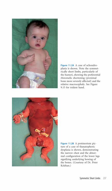
Figure 11.2A A case of achondro-plasia is shown. Note the symmet-rically short limbs, particularly ofthe humeri, showing the preferentialrhizomelic shortening (proximalbone more severely affected) and therelative macrocephaly. See Figure9.15 for trident hand.
Figure 11.2B A postmortem pic-ture of a case of thanatophoricdysplasia is shown, demonstratingthe narrow chest and the abnor-mal configuration of the lower legssignifying underlying bowing ofthe bones. (Courtesy of Dr. PeterKelehan.)
Symmetric Short Limbs 237

Limb Reduction Defects
Recognizing the Sign Essentially, this term refers to short, abnormal limbs
and incorporates several of the clinical signs already addressed in the chapters
dealing with hands and feet. The intention here is to concentrate on condi-
tions predominantly involving the long bones.
Establishing a Differential Diagnosis In addition to a family history, the
antenatal history has particular relevance in this situation, with chorionic villous
sampling during pregnancy, maternal diabetes, and valproate exposure all being
associated with limb reduction defects. Examination of all four limbs is neces-
sary, the intention being to establish whether the abnormality is confined to a
single limb or has a more general distribution. Amniotic bands will be expected
to cause a discernible constriction ring below which the bony and soft tissue
growth is distorted by vascular disruption in the single limb affected, all other
limbs being normal. The possibility of Poland anomaly should be evaluated by
examination of the chest for asymmetry, perhaps manifest by the nipples not
being in line with one another or the anterior axillary creases being asymmetric
between the two sides. In the event of chest asymmetry, examination of the cra-
nial nerves should follow to assess for Poland-Moebius syndrome, in which
condition mental retardation may occur, in contrast to uncomplicated Poland
anomaly. Cranial nerve evaluation is also key to the recognition of Okihiro
syndrome, an autosomal dominant condition characterized by sixth nerve
palsy, deafness, and limb reduction disorders, usually bilateral, but variable. A
useful tip is to examine the range of external eye movement in the parents, even
if both have normal limbs, as the limb defects can be very subtle and the sixth
nerve palsy almost unidentifiable in the baby with the limb reduction defects.
Examine the intraoral cavity and assess tongue size—hypoglossia is likely to
signify Hanhardt hypoglossia-hypodactyly syndrome, and many of these pa-
tients also have signs of sixth and seventh cranial nerve damage. Assessment of
the scalp is advised, specifically looking for areas of hairlessness signifying cutis
aplasia, the hallmark of Adams-Oliver syndrome, if there is limb involvement.
Cardiac examination may reveal congenital heart disease in Adams-Oliver syn-
drome, but unless there is identifiable cutis aplasia, is more likely to represent
Holt-Oramsyndrome (HOS), inwhichautosomaldominant condition the fam-
ily history of structural heart defects and limb malformations usually prompts
the diagnosis.
Investigations to Consider Audiometric assessment is indicated in suspected
cases of Okihiro syndrome. Mutation analysis of the SALL4 gene confirms if
the diagnosis is in doubt. HOS cases need electrocardiography (ECG) and
echocardiography. Mutation analysis of TBX5 may be helpful, especially if a
new mutation is suspected.
The Limbs238
11.3

Figure 11.3A and B The contrasting limb reduction defects in a daughter and motherwith Okihiro syndrome are demonstrated. Lateral rectus palsy on examination of theeye movement was the clue to the diagnosis. See also Figure 7.1B for an instance ofamputation-type reduction defect associated with Poland anomaly.
Limb Reduction Defects 239

Localized Lumps on the Limb
Recognizing the Sign In general, genetic causes of localized lumps result in
onset of signs after a few years rather than neonatally. Unsurprisingly then, the
history is usually of a parent noticing a swelling.
Establishing a Differential Diagnosis The family history may often offer an
explanation. Inquiry as to familial tumors or malignancy is advised. Examine
for other evidence of previously undocumented lumps. Note the position of
lumps, possibly over joints. Establish whether they are mobile, fixed to deeper
tissue, or arising from bone.Neurofibromatosis type 1 is associated with neu-
rofibromata, which present as mobile lumps beneath the skin. Corroborative
clinical signs of cafe-au-lait patches and groin and axillary freckling should be
sought. If the diagnosis is established, continuous evaluation throughout
childhood for scoliosis and hypertension is usually advised. Familial angio-
lipomatosis is a rare condition in which the lumps are subcutaneous, mimic
neurofibromata, and are usually clustered around the joints. The autosomal
dominant family history and the absence of other signs of NF1 make this
condition very likely. All cases in the author’s personal experience have been
misdiagnosed as NF1. Lumps that are fixed and clinically of bony origin
denote likely multiple exostoses. These comprise clinically overlapping au-
tosomal dominant conditions, one of which, on chromosome 11, is associated
with craniosynostosis and enlarged parietal foramina, while deletion of
chromosome 8q, in addition to the bony lumps, may cause Langer-Gideon
syndrome. The attendant ala nasi hypoplasia and wispy hair with little tem-
poral hair growth allow clinical recognition of the latter entity. Both forms
may be associated with mental retardation. Cowden syndrome (CS) is an
extremely variable condition often presenting with macrocephaly and devel-
opmental delay. A family history of macrocephaly, thyroid malignancy, or
fibrocystic breast disease is strongly suggestive. Lumps usually represent ha-
martomata and are very unpredictable as to clinical presentation and location,
some cases being clinically confused with Proteus syndrome. In males, pig-
mented spots on the glans penis are a very useful clinical sign of this condition.
Lumps that are calcified should result in consideration of Albright’s heredi-
tary osteodystrophy (AHO) as a possible diagnosis and the characteristic
brachydactyly of the fourth metacarpal and metatarsal should be sought.
Investigations to Consider Skeletal radiology will confirm multiple exos-
toses. Parietal foramina on skull x-ray are characteristic of a chromosome 11p
deletion. Karyotyping and detailed cytogenetics are appropriate. Deletion 8q
should be sought if a Langer-Gideon phenotype prevails. A low threshold for
PTen mutation analysis is advisable for CS and its clinical variants. Parathy-
roid and thyroid function tests are indicated for AHO.
The Limbs240
11.4

B
Figure 11.4A and B This 8-year-old boy presented acutely due to parentalconcern about the large swelling on the lateral aspect of his right leg justbelow the knee. Examination showed several other, previously unnoticedlumps. The family history established that his uncle had had surgicalremoval of a lump some years previously and histology had shown acartilage-capped exostosis. Radiology confirmed familial multiple exos-toses as the diagnosis. Note the characteristic bony spur on the lowerfemur, in addition to the large exostosis over the superior tibiofibular joint.
Localized Lumps on the Limb 241

Arthrogryposis Congenita
Recognizing the Sign Multiple joint contractures are present from birth.
Establishing a Differential Diagnosis Joint contractures may represent a
physical manifestation of a primary abnormality in the neuromuscular de-
velopment of the baby, or may arise secondary to fetal constraint or maternal
neuromuscular disease. Hence, family history, with particular reference to
talipes or contractures, is important. In particular, maternal history and ex-
amination needs to be undertaken. Myasthenia gravis in a mother can result
in severe arthrogryposis in her offspring. Unsuspected and asymptomatic my-
asthenia gravis in the mother should be investigated in recurrent cases of fetal
arthrogryposis among her offspring. Apart from evaluating the extent of joint
limitation, the range of joints affected, and the presence of pterygia (skin webs
across affected joints), the clinical examination should also take cognizance of
clinical indicators of joint movement in fetal life. Specifically, evaluate the joint
creases of the hands and feet, absence of which indicates likely failure to es-
tablish movement in early development. Likewise, skin dimples overlying con-
tractures probably indicate failure of normal movement and a primary disease
process. General observations with respect to neurologic function may suggest
impairment of tone or absence of muscle bulk. Edwards syndrome, which
frequently presents with clenched hands and overriding fingers as well as
limitation of hip movement, should be excluded as the underlying cause.
Amyoplasia congenita confers a characteristic symmetric malpositioning of
joints with talipes equinovarus, flexion of the wrists and fingers, and internal
rotation of the shoulders. The presence of dimples and absence of skin creases
betrays an absence of normal joint development and movement in utero. A
glabellarhemangiomaisoftenpresent.Spinalmuscularatrophy (SMA)iseasily
overlooked as a possible diagnosis, the classic presentation of the disorder
being of hypotonia. However, contractures are well described in cases having
genetic deletions, so careful attention to feeding history and evaluation for
tongue fasciculation may reveal the true diagnosis. Multiple pterygium syn-
drome, an autosomal recessive condition, is usually recognizable at birth
because, in addition tomultiple large and small joint contractures, pterygia are
generally identifiable across the joints. Beals congenital contractural ara-
chnodactyly syndrome will be clinically discernible by the characteristic
crumpled helix of the ear and arachnodactyly.
Investigations to Consider The anticholinesterase receptor antibody, spe-
cific for the fetal receptor is necessary to establish maternal myasthenia syn-
drome. Karyotype is indicated for Edwards syndrome. Electromyographic
study is necessary for SMA and, if in doubt, mutation analysis of the SMN
locus on chromosome 5 for deletion should be conducted. Mutation analysis
of FBN2 gene confirms Beals syndrome.
The Limbs242
11.5

Figure 11.5A Severe bilateral talipes and dimples over the knees are shown in atypical case of amyoplasia congenita.
Figure 11.5B Note the wrist flexion and absent elbow joint creasesin another patient with the same condition.
Arthrogryposis Congenita 243

Joint Enlargement
Recognizing the Sign The sign is easily identified, the purpose of examina-
tion thereafter being to establish whether the enlargement represents a gener-
alized process involving several joints or whether it is localized to a single joint.
If localized, then possible dysmorphic considerations are forestalled by more
likely inflammatory causation and evidence of effusion should be sought.
Establishing a Differential Diagnosis The family history is useful; in par-
ticular, a family history of conjunctivitis, uveitis, or arthritis should not be
discounted. The autosomal dominant condition of chronic infantile neuro-
logic cutaneous and articular syndrome (CINCA) presents with an episodic
fever and maculopapular rash of neonatal origin, often difficult to diagnose
but much more easily recognized if the family history is taken into diagnostic
account. Typically painful, swollen joints do not set in until age 3 or 4 years,
the knees and ankles being most affected. Stickler syndrome is an autosomal
dominant condition, and large painless knees in a child with short stature
should lead to consideration of this diagnosis. A history of micrognathia, per-
sonal or family history of myopia, or retinal detachment and evidence of joint
laxity or early-onset osteoarthritis all weigh heavily in favor of this diagnosis.
Formal eye evaluation for cataract, vitreous abnormality, and retinal detach-
ment is advised for management as well as diagnostic purposes. In a similar
vein, Kniest syndrome should be suspected in a child with short stature and
large knees. The condition can be suspected neonatally by the relative mac-
rocephaly, symmetrically enlarged knees, and frequent observation of cleft
palate. Generalized joint thickening emerging over the first few years of life,
often with associated restriction in the range of movement, is characteristic
of the joint enlargement that accompanies mucopolysaccharidosis, but the
typical facial features and usual developmental delay invariably facilitate di-
agnosis and predate the emergence of this sign.Multiple exostoses, discussed
in section 11.4, may present with single or multiple periarticular swelling due
to the preferential development of exostosis near the epiphyses or the epiph-
yseal/metaphyseal junction. Finger clubbing associated with symmetric swell-
ing of large joints, particularly the ankles and knees, and excessive palmar
sweating is characteristic of cranio-osteoarthropathy, an autosomal recessive
condition. Cranial suture closure is delayed, with clinical preservation of an
open fontanelle until age 2 or 3 years.
Investigations to Consider Radiologic evaluation in Stickler syndrome
demonstrates metaphyseal widening, coronal clefts, and platyspondyly in the
neonatal period, while a similar finding but with an additional ossification
center of the distal end of the middle phalanges typifies Kniest syndrome.
Mutations of COL2A1 cause Kniest, while Stickler syndrome maybe caused by
either COL2A1 or COL11A1.
The Limbs244
11.6

Figure 11.6A–D The typical finger clubbing, restriction of joint extension at the knees,knee enlargement, and ankle swelling of cranio-osteoarthropathy are demonstrated inthese illustrations from two different patients with the condition. (C and D courtesy ofDr. Mohnish Suri. Figure reproduced with consent from O’Connell et al. Clin Dys-morphol 2004;13:213–219.)

Generalized Joint Hypermobility
Recognizing the Sign Patients will tend to show you that they are ‘‘double-
jointed’’ by demonstrating that they can passively appose the thumb against
the flexor aspect of the forearm. Further objective evidence to look for on
examination includes passive extension of the fifth metacarpophalangeal
joint to greater than 90 degrees, hyperextension of the elbows and knees by
greater than 10 degrees, and ability to touch the floor with the palms while
standing with the knees extended, by flexing the spine.
Establishing a Differential Diagnosis In view of the major management im-
plications relating to aortic root dilatation, it is critical to establish whether
joint hypermobility is indicative of Marfan syndrome (MS). Family history
may assist, the condition being autosomal dominant. Particular awareness of
unexplained deaths in childhood or early adult life is advised, possibly re-
flecting demise from aortic dissection. Ophthalmic evaluation for myopia and
lens dislocation is essential, while clinical signs to observe are pectus; ara-
chnodactyly; an arm span exceeding standing height; high, arched palate; and
transverse striae of the skin best sought over the lumbar region. Joint laxity is
a feature of Stickler syndrome in the pediatric age range, though usually less
so in adult life. A history of Pierre-Robin sequence should be sought and
clinical evidence of depressed nasal bridge, large knee joints, and a personal
and family history of myopia, cataract, or retinal detachment are the central
elements of sifting out this diagnosis from other causes of joint laxity. Liga-
mentous laxity can be familial, and such an observation by history or personal
demonstration of signs in a parent is consistent with the Ehlers-Danlos
syndrome (EDS) complex of disorders. All are genetic, the commoner forms
being autosomal dominant. Gorlin sign—the ability to touch the tip of the
nose with the tongue—is more common in EDS patients than the general
population. Other diagnostically valuable manifestations of EDS are blue
sclerae, skin hyperextensibility, easy bruising, scarring disproportionate to the
degree of trauma, joint dislocation, surgery for inguinal hernia, and a history
of delayed early motor milestones but with normal intelligence. Ophthalmic
examination may indicate keratoglobus in type VI, the cornea being prone to
rupture on minor trauma. Kyphoscoliosis is a clinical finding among most
types, but a signature of type VI and the criss-cross pattern of hand skin in this
particular variant are clinically confirmatory (Figure 9.18C).
Investigations to Consider Ophthalmic and echocardiographic evaluation
are indicated in MS. Radiology is necessary if Stickler is considered likely—
you will see coronal clefts and flared metaphyses in early life. Lysyl hydrox-
ylase assay on fibroblasts for EDS type VI is usually diagnostic, being function-
ally absent. Serum tenascin X assay is indicated for any clinically suspected
cases of EDS.
The Limbs246
11.7

Figure 11.7A Typical hypermobility of the thumb isdemonstrated in a patient with type III Ehlers-Danlossyndrome.
Figure 11.7B Gorlin sign, theability to touch the nasal tipwith the tongue, is a feature ofmany cases of Ehlers-Danlossyndrome but is seen infewer than 10% of the non–Ehlers-Danlos population.
Figure 11.7C Hypermobility of the distal interphalangeal joint is demonstrated inthis baby with Ehlers-Danlos syndrome type II. Note the excess skin over thehands, manifesting as additional skin wrinkling over the fifth finger.
Generalized Joint Hypermobility 247
Small or Absent Patella
Recognizing the Sign Clinically, the clue to patellar absence or hypoplasia is
of a flat anterior aspect to the knee, accentuated on flexion. The patella is not
palpable and is not seen radiologically. A pitfall for the unwary is that in
young children patellar ossification may be delayed until as late as age 6 years
in the normal population.
Establishing a Differential Diagnosis Single gene diseases may be flagged by
the family history. In particular, nail-patella syndrome (NPS), being auto-
somal dominant, may be recognized in other family members. A family history
of renal failure in one or more individuals, typically becoming symptomatic in
early adulthood, in combination with clinical signs of nail malformation, es-
pecially in the thumbs and index fingers, and triangular shape to the lunules
are characteristic of the disorder, effectively confirming the diagnosis. The
absence of skin creases over the distal interphalangeal joints is a common
observation (Figure 12.1). Patellar agenesis is a feature of a more widespread
bone dysplasia representing mild camptomelic dysplasia, a condition that is
usually associated with severe tibial bowing and dimpling and often associated
with respiratory compromise and death. However, in attenuated form, sur-
vivors may show few clinical features apart from patellar hypoplasia. Radial
defects, including absence of the thumbs, in combination with patellar hy-
poplasia are suggestive of a group of molecularly related disorders represented
by Rothmund-Thompson syndrome. Short stature, skin mottling, telangi-
ectasia, and craniosynostosis are other features of this variable group of con-
ditions, which are autosomal recessively inherited. A less disparate phenotype
is that of Meier-Gorlin syndrome, in which autosomal recessive disorder
short stature is associated with hypoplastic patellae. The best guide to the
diagnosis is the small ears, which are unmistakable. In a child with unex-
plained developmental delay, always examine for absent patellae, the identi-
fication of which is very likely to signifymosaic trisomy 8. The diagnosis may
be confirmed clinically by checking for the deep plantar creases (Figure 10.6).
If associated with contractures of the knees and/or severe talipes, do bear in
mind the possibility of genitopatellar syndrome, in which the presence of
scrotal hypoplasia, cryptorchidism, and clitoromegaly are to be anticipated on
examination and severe developmental delay likely.
Investigations to Consider Renal biochemistry and urinary albumin-to-
creatinine ratio annually are recommended for NPS patients as a screen for
incipient renal disease. Mutation of LMX1B confirms the diagnosis. Skeletal
radiology will establish abnormal ischiopubic ossification in surviving ca-
mptomelic dysplasia cases. RECQL4 mutations are established in Rothmund-
Thompson syndrome and related phenotypes. Mosaic trisomy 8 generally
requires fibroblast culture to establish the cytogenetic abnormality.
The Limbs248
11.8

Figure 11.8 Lateral view of a patient with Meier-Gorlin syndrome to demon-strate the typical microtia that identifies the condition from a host of other shortstature disorders.
Small or Absent Patella 249

Pseudoarthrosis
Recognizing the Sign Most typically a feature of tibial fracture, pseudoar-
throsis refers to the nonunion of a long bone following fracture resulting,
usually, in a permanent localized area of discontinuity in the shaft of the bone.
Although congenital presentation is known, the more usual history is of pro-
gressive bowing of the limb in the first year of life and spontaneous fracture on
attempted weight bearing, there being subsequent nonunion of the fracture.
Establishing a Differential Diagnosis Establish whether the long bones are
normal elsewhere with respect to length and absence of deformation, or bow-
ing. Themajor diagnostic concern is forNeurofibromatosis type1 (NF1).While
characteristic skin changes of multiple cafe-au-lait patches, axillary and groin
freckling, and palpable neurofibroma are typical in the older child, readily fa-
cilitating diagnosis, the disorder is easily overlooked when presentation predates
the onset of cutaneous clues. Obviously, parental examination for signs of this
autosomal dominant condition is important in any child presenting with pseu-
doarthrosis. In evaluating such a case, it is useful to be aware that the absence of
Lisch nodules—benign hamartomas of the iris typically seen in NF1—on iris
examination does not reduce the likelihood of NF1, since these are rarely ob-
served in children under the age of 5 years. Since many cases of NF1 arise as de
novo mutations, serial examination for emerging cafe-au-lait patches is appro-
priate in a child presenting with a pseudoarthrosis, as is parental history and
examination. Potential for diagnostic confusion exists in thatMcCune-Albright
syndrome also presents cutaneous pigmentary abnormalities and cystic lesions
of bones, occasionally associated with fracture. The pigmentary abnormalities,
though said to mimic cafe-au-lait patches, are usually irregular in outline, in
contrast with the typically smooth outline of a cafe-au-lait patch in NF1 (Figure
12.8). The presence of endocrine abnormalities, perhaps presenting clinically
with precocious puberty or hyperthyroidism, is a good basis for differentiation
from NF1. Fibrous dysplasia of bone, mimicking pseudoarthrosis and some-
times associated with bowing, is well described in Mulibrey Nanism, an auto-
somal recessive disorder associated with growth failure and which clinically
resembles Silver-Russell syndrome.
Investigations to Consider Optic nerve evaluation for optic glioma is in-
dicated in NF1, and serial blood pressure measurement is recommended in
view of associated renal artery stenosis and pheochromocytoma. Mutation of
GNAS1 is diagnostic of McCune-Albright, and thyroid function tests are im-
portant considerations in management. Skeletal radiology shows slender long
bones with a narrow medullary channel and occasionally fibrous dysplasia of
the fibula in Mulibrey Nanism. Echocardiography or magnetic resonance
imaging is advised for possible constrictive pericarditis.
The Limbs250
11.9

Figure 11.9A A very typical anteriordeformity of the lower limb isshown in a child with neurofibro-matosis type 1–related pseudo-arthrosis. (Courtesy of Dr. SusanHuson.)
Figure 11.9B Radiologic appearance of neurofibromatosis type1–related pseudoarthrosis is shown in a different case.(Courtesy of Dr. J. Kelleher.)
Pseudoarthrosis 251

Madelung Deformity
Recognizing the Sign Madelung deformity is a clinical sign that emerges as
a child grows. It generally takes 5 or 6 years for the feature to become iden-
tifiable. Bony growth is disproportionate in the forearm, the distal end of
the ulna becoming progressively dorsally dislocated. Clinically, the forearm
adopts a very recognizable appearance, sometimes referred to as ‘‘dinner-
fork’’ configuration, there being a dip between the distal end of the ulna and
the wrist. Generally, there is associated mesomelia (middle segment of the
limb, hence forearm and tibiaþ fibula) and short stature. There may be some
restriction of elbow extension. Radiologically, the radius is short with respect
to the ulna and usually bowed, and the distal radial epiphysis shows a sloping
outline.
Establishing a Differential Diagnosis This is the clinical signature par ex-
cellence of Leri-Weill syndrome. The affected child usually presents to the
pediatric clinic with short stature. A background history of short stature may
be noted in the family, and parental examination may establish similar fore-
arm and wrist deformations. Intelligence and other developmental parameters
are normal. The condition is described as autosomal dominant, affecting both
male and female patients, although the gene is on the short arm of the X
chromosome, in the pseudoautosomal region, thus explaining this apparent
deviation from classical genetic principles of inheritance. The main differ-
ential diagnosis is from patients with Turner syndrome and others with lo-
calized deletions of the Xp22 chromosomal region. Both such patient groups
are short and more likely to show Madelung deformity due to absence of the
pseudoautosomal region of the X chromosome.
Investigations to Consider Skeletal radiology confirms the typical appear-
ance of the radius and ulna, often with associated shortening of the tibia and,
even more, of the fibula. Karyotype is useful, not only for the identification of
Turner syndrome, but also because several cases with deletion of Xp22-ter
have been recorded with Madelung deformity. Submicroscopic deletions and
mutations of the SHOX gene on Xp22 have been demonstrated in several
affected families.
The Limbs252
11.10

Figure 11.10A A typical instance of Madelung forearm is shown. Note the‘‘dinner-fork’’ configuration of the wrist.
Figure 11.10B The radiology confirms the radial bowing and the distal radialepiphysis presents a sloping appearance. (Courtesy of Dr. Debbie Shears.)
Madelung Deformity 253

Chapter 12 The Nails, Hair, and Skin

Absent or Hypoplastic Nails
Recognizing the Sign Absent nails are simply a matter of observation, but
hypoplastic nails are a subjective judgment. Complete absence of the nails is
very uncommon. If nail hypoplasia is signifying an underlying syndromic
diagnosis, the nail abnormality will be readily identifiable. Small nails are
usually deeply embedded in the tissue surrounding them, the term ‘‘deep set’’
being apt.
Establishing a Differential Diagnosis In a neonate, consider teratogenic
causes and in particular exposure to alcohol or phenytoin during pregnancy. In
both disorders parallel effects would be anticipated in the feet, and the absence
of such clinical findings should call for caution in attributing the syndrome.
Family history can offer clues—in particular, dominant ectodermal dysplasia
with sparse growth of fine hair, dystrophic nails, and abnormalities of dentition
may explain the clinical findings. The possibility of a diaphragmatic hernia in
a neonate should prompt assessment for Fryns syndrome, one of the cardi-
nal features of which is small nails. Coarse facial features, increased birth weight
above the norm, and corneal clouding are important pointers toward this au-
tosomal recessive condition. Deep-set nails are a cardinal feature of Ellis-van
Creveld (E-vC) syndrome and should prompt evaluation for this possible di-
agnosis. Evidence of postaxial polydactyly, possibly in the form of a tiny ir-
regularity on the ulnar margin of the fifth finger; accessory frenula and natal
teeth; narrow chest; and low birth length are all suggestive of the disorder. If
suspected, then cardiac evaluation is important, the most common lesion being
an atrial septal defect (ASD). Nail dysplasia concentrated on the radial side of
the hand, frequently with normal nails on fingers 3 to 5, is suggestive of nail-
patella syndrome (NPS). Look for the characteristic triangular shape to the
lunules, loss of creases over the distal interphalangeal (DIP) joints of the fingers,
and absent, impalpable, or easily dislocated patella. There is a significant risk
(>10%) of developing kidney disease. Nail hypoplasia associated with neonatal
hypotonia and difficulty in establishing feeding is a typical presentation of
deafness-onycho-osteodystrophy retardation (DOOR) syndrome. Neonatal
seizures complicate many cases of this autosomal recessive condition. Recog-
nizing the importance of the nail hypoplasia as the key to the diagnosis is the
seminal diagnostic maneuver.
Investigations to Consider Echocardiography is indicated in E-vC syn-
drome, in addition to radiology for narrow thorax and hypoplastic iliac wings.
The risk of renal disease in NPS requires annual screening by blood pressure
and urine analysis. Urine albumin-to-creatinine ratio is the recommended
assay. Mutation of the LMX1B gene confirms the diagnosis, if there is clini-
cal doubt. Brainstem auditory-evoked responses show no response to high-
intensity stimulation in DOOR syndrome.
The Nails, Hair, and Skin256
12.1

Figure 12.1B–D Diagnostically valuable signs in the hand of patients with nail-patella syndrome are demonstrated. Note the normal nails on fingers 3 to 5 butthe small index finger nail. The triangular outline of the lunules in fingers 3 to5 is also well demonstrated. Distal interphalangeal joint creases are also absent, asis expected. The dysplastic thumb nails are also typical, the thumbs and indexfingers characteristically showing more severe degrees of nail dysplasia. (Courtesyof Prof. Ian Young and Dr. Ernie Bongers.)
Figure 12.1A Shown are nailsthat are small and deep set.The nails themselves look nor-mal, but observe how smallthey are relative to the distancefrom the distal interphalan-geal joint crease.
Absent or Hypoplastic Nails 257

‘‘Tale of a Nail’’ Sign
Recognizing the Sign A hooked nail of the fifth finger, specifically, of un-
usual shape due to its adherence to a hypoplastic distal phalanx of the finger
comprises the ‘‘tale of a nail’’ sign. Minor variations of this arise, sometimes
extending to small volar nails on the palmar surface of the fifth finger. The
muscle pulp of the distal fifth finger is often significantly and noticeably re-
duced.
Establishing a Differential Diagnosis In almost all cases, this rare sign is sig-
nifying deletion of chromosome 4q34. Very few cases are described, but swal-
lowing difficulties, frequently complicated by aspiration, are especially common
among affected individuals. Associated congenital heart disease is also well rec-
ognized. It is worth obtaining a family history, since the condition, in less florid
form, can be associated with essentially normal development and parent-to-
child transmission is known, the parent having been spared the aspiration and
associated respiratory difficulties that may bedevil the child. As with any chro-
mosomal deletion syndrome, the clinical features are variable, but developmen-
tal delay is frequent among those who are recognized and survive the aspiration-
related risks. It is likely that many pediatric cases of recurrent aspiration have
this little-known condition.
Investigations to Consider The basic karyotype is often reported as normal.
Specific direction of cytogenetic attention may reveal a submicroscopic dele-
tion of the 4q34 region on higher band scrutiny, but occasionally molecu-
lar cytogenetics, perhaps by way of subtelomeric fluorescent in situ hybridiza-
tion (FISH) or array hybridization may have to be resorted to, if the deletion is
to be revealed.
The Nails, Hair, and Skin258
12.2

Figure 12.2A and B Thenails of two childrenwith 4q34 deletions areshown. Note the varia-tion in the clinical signand the demonstrationof the hypoplasia of themuscle pulp on thepalmar surface ofthe digit.
Figure 12.2C The min-ute deletion of chro-mosome 4q34 is shownon the karyotype; theoriginal report, at lowerlevels of investigation,was reported as normal.
‘‘Tale of a Nail’’ Sign 259

Longitudinal Splitting of the Nails
Recognizing the Sign Most characteristic of the thumb but also seen in other
digits, including the feet, longitudinal ridges, which are both visible and
palpable, traverse the length of the nail and the patient or parent will report
that the nail routinely breaks at this fault line. In the neonatal period, it is
often easier to feel this sign than to see it.
Establishing a Differential Diagnosis Few signs are pathognomonic, but the
recognition of this sign in a female patient is strongly suggestive of cranio-
frontonasal dysplasia (CFND). In florid form, the patient will present with
hypertelorism and plagiocephaly at birth and a diagnosis of craniosynostosis
will be reached without specifying a particular syndrome. The recognition of
this clinical sign identifies the underlying condition but is also prognostically
important, CFND being generally associated with a normal developmental
outcome, not the universal experience in other forms of craniosynostosis. Di-
agnostic and prognostic confusion can be exponentially increased in the un-
recognized case by the identification on imaging of an absent corpus callosum,
usually a poor prognostic indicator in developmental terms but a normal find-
ing in many patients with CFND. Other clinical features that, if present, sup-
port this likely diagnosis are a bifid nasal tip, widow’s peak hair line, and wiry
texture to the hair in the older patient. Breast asymmetry and sloping shoul-
ders, with enhanced shoulder joint hypermobility, mimicking cleidocranial
dysostosis, are also well recognized within the CFND clinical spectrum. Almost
all cases described are female, there being few convincing male cases.
Investigations to Consider Skull x-rays may be indicated to establish whe-
ther there is synostosis of the coronal or other sutures. Mutation analysis of
the EFNB1 gene confirms if there is a clinical doubt.
The Nails, Hair, and Skin260
12.3

Figure 12.3A A typical instance of longitudinal splitting of a thumbnail is demonstrated in an adult patient with craniofrontonasaldysplasia.
Figure 12.3B The facial features of this 3-year-old child withcraniofrontonasal dysplasia are unlikely to be confused with anyother syndrome. Note the hypertelorism and bifid nasal tip.The corpus callosum is absent but development is normal.
Longitudinal Splitting of the Nails 261
Short Nails
Recognizing the Sign There is not a clear-cut distinction between short nails
and small nails, which will obviously overlap somewhat with nails described as
dysplastic. However, what the clinician notes is that the nails are short in the
longitudinal axis. Obviously this is to be anticipated in cases of brachydactyly
(see 9.7), where the nail shortening reflects a generalized shortness of the
phalanges. In contrast with dysplastic nails, the nail growth and strength are
normal.
Establishing a Differential Diagnosis Though rare, cartilage-hair hypo-
plasia (CHH) syndrome is an important condition to establish in infancy be-
cause of the associated susceptibility to varicella. Short stature is usually ap-
parent at birth or shortly afterward, but the head circumference is normal.
Scalp hair is sparse and fine, though this sign tends to be more valuable in the
postneonatal period. Recurrent infections or low white cells can offer addi-
tional clues to the diagnosis. In all cases seen by the author, short nails were a
valuable clinical sign noted on the first examination, predating the diagnosis
and signaling appropriate investigations. Short nails are common inWilliams
syndrome, though it is the typical facial features of anteverted nostrils with a
long philtrum and progressive thickening of the lips that are more likely to
betray this diagnosis to a clinician. However, in a child with a murmur, es-
pecially if related to supravalvular aortic stenosis or pulmonary stenosis, the
recognition of short nails should accentuate the case for specific genetic inves-
tigation ofWilliams syndrome. Progressive loss of energy in a clinically normal
child should always prompt investigation of renal biochemistry. This is even
more appropriate in the context of short nails, which may the only clinically
identifiable abnormality inMainzer-Saldino syndrome. Chronic renal failure
due to nephronophthisis is the usual presentation, but a pigmentary retinop-
athy should be sought, as it foretells the predestined eventual tunnel vision. In
view of the occasional association with anatomic malformations of the cere-
bellum, such as Dandy-Walker malformation, it is worth seeking a history of
delayed motor milestones and examining clinically for ataxia.
Investigations to Consider Differential white cell count in CHH syndrome
may disclose a neutropenia or lymphopenia. Skeletal radiology shows meta-
physeal dysplasia, most characteristically at the lower end of the femora. FISH
of ELN on chromosome 7 confirms the localized deletion typical of Williams
syndrome. Urinary concentration defects usually predate abnormal urea and
creatinine in Mainzer-Saldino syndrome, so look for polyuria, nonspecific
aminoaciduria, and glycosuria.
The Nails, Hair, and Skin262
12.4
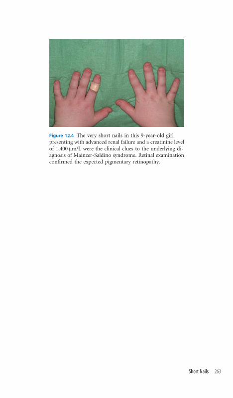
Figure 12.4 The very short nails in this 9-year-old girlpresenting with advanced renal failure and a creatinine levelof 1,400 mm/L were the clinical clues to the underlying di-agnosis of Mainzer-Saldino syndrome. Retinal examinationconfirmed the expected pigmentary retinopathy.
Short Nails 263

Temporal Balding
Recognizing the Sign Although the hair grows well elsewhere without lo-
calized areas of alopecia, there is poor growth of hair on the temporal regions,
which have a high anterior hairline in consequence.
Establishing a Differential Diagnosis Sparse temporal hair growth is one of
the classical clinical hallmarks of Pallister-Killian syndrome. This condition
is caused by tetrasomy of chromosome 12p, but the abnormal chromosome
finding rarely manifests on routine chromosomal analysis. Hence, clinical
recognition and directed karyotypic investigation are critical if the diagnosis is
to be established. Temporal balding is a cornerstone of that clinical recog-
nition process (Figure 3.5C). Affected children will present with severe devel-
opmental delay and do not usually acquire speech. The family history is non-
contributory. The face tends to be slightly coarse, becoming more so with age,
and the cheeks can assume a somewhat sagging appearance. Accessory nipples
are frequent and lend support to the likely diagnosis. The condition may pres-
ent prenatally with polyhydramnios or neonatally with diaphragmatic hernia.
A much better developmental prognosis attends tricho-rhino-phalangeal
(TRP) syndrome, in which autosomal dominant condition evaluation of the
parents may signal the diagnosis. In addition to sparse wispy hair, notably
deficient at the temples, the nose is often slightly unusual with a prominent tip
and relative deficiency of the ala nasi (Figure 2.4). Examination of the fingers
is valuable because of a slightly crooked appearance, not invariably present
but diagnostically valuable if noted, due to deviation at the proximal inter-
phalangeal joints. Temporal balding and a prominent nose of rather pinched
appearance are the hallmarks of Hallermann-Streiff syndrome. The face is
small but the nose striking. In particular, there is noteworthy micrognathia.
The eyes appear small and examination almost always reveals cataract. Such
hair as there is on the scalp is thin and eyebrows and lashes are likewise sparse.
The condition is diagnosed clinically and most cases are intellectually normal.
Investigations to Consider Fibroblast examination for tetrasomy chromo-
some 12p is the usual means of establishing the diagnosis in Pallister-Killian
syndrome. The condition is mosaic and the abnormal isochromosome is not
present in all cells. With age, it becomes less likely that the isochromosome
will be identified. Hence, repeated analysis can be necessary for confirmation.
Hand x-rays in TRP syndrome show short phalanges and cone-shaped epi-
physes. A proportion of cases show exostoses, particularly if associated with a
microscopically visible deletion at chromosome 8q24.12.
The Nails, Hair, and Skin264
12.5

Figure 12.5A and B Note the large area of temporofrontal regiondevoid of any hair growth in this 3-year-old girl who had tri-chorhinophalangeal syndrome. The hands show the typical pha-langeal crookedness at the proximal interphalangeal joint. (See alsoFigures 2.4A and B.)
Temporal Balding 265

Sparse Hair/Alopecia
Recognizing the Sign Subtlety is not required. The hair either does not grow
or else is sparse, the scalp being easily visible.
Establishing a Differential Diagnosis Family history may hold the key.
Ectodermal dysplasias frequently involve poor hair growth, alopecia, or thin,
wiry hair, and several of these syndromes are transmitted from generation
to generation. The most common form, X-linked hypohidrotic ectodermal
dysplasia (Figure 3.15D), is associated with inability to sweat and affected
boys often present in infancy with unexplained high fevers. Sparse hair, absent
teeth, and deficient eyebrows and eyelashes are good indicators of other af-
fected individuals in the pedigree. Clinical signs are less reliable in carrier
females. In the neonatal period, scaling of skin in affected males can lead to
confusion with ichthyosis, but the family history and examination offer a
ready means of differentiation. An erythematous or seborrheic rash emerging
after several months in a child whose skin had previously been normal and
alopecia or very sparse hair are the usual harbingers of biotinidase deficiency.
Often there will have been background concerns about hypotonia and, per-
haps, developmental delay. With later presentation the neurologic issues will
be more overt and seizures will be recorded. A good clue from the history is to
establish whether there have been recurrent fungal infections, to which chil-
dren with this condition are prone. Neurologic issues again dominate the his-
tory in children with Coffin-Siris syndrome, severe mental retardation being
a consistent finding among these cases. Good clinical pointers to this, apart
from the sparse scalp hair, are hypertrichosis over the back and absent fifth
fingernails and toenails. The diagnosis is essentially confined to female patients
and, though the exact cause is not known, males are rarely diagnosed with this
disorder. A history of a cardiac murmur or echocardiographic evidence of in-
terventricular septal thickening and sparse hair growth should pose the ques-
tion as to whether the childmay have cardiofaciocutaneous (CFC) syndrome.
The skin might be dry, indeed appear ichthyotic, and the liver and spleen en-
larged. There is poor hair growth. Often the hair that does grow is curly and
the face resembles that of Noonan syndrome, ptosis being common. In the
context of a short stature neonate, sparse hair should raise the diagnostic
thought of cartilage–hair-hypoplasia (CHH), an important diagnosis because
of the associated susceptibility to varicella and the immunization implications.
Investigations to Consider Organic aciduria is usually present in biotinidase
deficiency. Skeletal radiology shows metaphyseal changes, characteristically
most severe at the lower end of the femur in CHH. Mutation analysis of
RMRP gene confirms.
The Nails, Hair, and Skin266
12.6

Figure 12.6 The 3-year-old girl pictured here presented with severeeczema in the neonatal period, sparse hair that hardly grew, and athickened interventricular septum on echocardiography. Unex-plained hepatomegaly led to a suggestion that liver biopsy wasnecessary. However, the recognition of cardio-facio-cutaneoussyndrome offered a unifying diagnosis for all these features.
Sparse Hair/Alopecia 267

Poliosis
Recognizing the Sign Loss of melanin production in hair follicles is normal
from the fourth decade of life resulting in greying. Poliosis refers to premature
greying in childhood or early adulthood.
Establishing a Differential Diagnosis As an aid to diagnosis, poliosis is
valuable in Waardenburg syndrome (WS), in which deafness, segregating in
autosomal dominant manner, should be carefully sought. Several different
phenotypes of WS are described, but the important distinction clinically is to
establish whether dystopia canthorum is present. This refers to lateral dis-
placement of the inner canthi of the eyes, conferring an impression of hy-
pertelorism on inspection of the face. Examine for slight medial fusion of the
eyelids, consequent reduction in the sclera visible medially and lateral dis-
placement of the lacrimal punctum toward the iris. Dystopia canthorum is the
hallmark of WS type I, which is genetically distinct from type II, characterized
by the absence of this finding. Other diagnostically useful signs to observe are
pigment abnormalities, such as heterochromia iridum and hypopigmented
skin patches, as well as synophrys, all of which are more common in WS type
I–affected individuals and lend diagnostic support. Useful tricks in estab-
lishing the diagnosis are to question adult members of the family as to whether
they employ hair dyes and to examine the eyelashes, which may be the sole site
of poliosis in some affected individuals. The main scope for clinical error is
confusion with piebaldism (12.11). Patients affected by Williams syndrome
are unlikely to present with poliosis in the early childhood period, the cardiac
murmur, hypercalcemia, and constipation being more likely features to bring
them to attention. However, cases missed in childhood who present in ado-
lescence for diagnosis of unexplained developmental delay are usually recog-
nizable by their somewhat coarse facial features and frequent poliosis. Ataxia
telangiectasia cases generally develop normally until walking, at which time
ataxia becomes apparent. Scleral telangiectasia, a history of sun sensitivity, and
poliosis should be sought if this diagnosis is being considered.
Investigations to Consider Good audiologic profiling is obviously impor-
tant if WS is being considered and may need to be extended to other family
members. Mutation of PAX3 gene is confirmatory of type I, mutation of
MITF being seen in type II. Mutation at other loci, such as SOX10, can also
present a WS-like picture clinically. If Williams syndrome is suspected, the
FISH of ELN on chromosome 7q should establish the characteristic deletion.
Elevated serum a-fetoprotein is a good screening test for ataxia telangiectasia,
and confirmation is established by demonstrating increased chromosome
breakage following irradiation of cultured cells.
The Nails, Hair, and Skin268
12.7
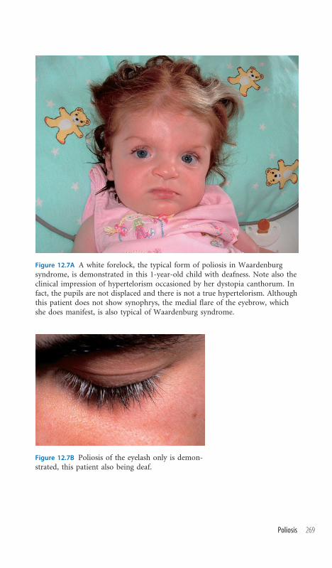
Figure 12.7A A white forelock, the typical form of poliosis in Waardenburgsyndrome, is demonstrated in this 1-year-old child with deafness. Note also theclinical impression of hypertelorism occasioned by her dystopia canthorum. Infact, the pupils are not displaced and there is not a true hypertelorism. Althoughthis patient does not show synophrys, the medial flare of the eyebrow, whichshe does manifest, is also typical of Waardenburg syndrome.
Figure 12.7B Poliosis of the eyelash only is demon-strated, this patient also being deaf.
Poliosis 269

Cafe-au-Lait Patches
Recognizing the Sign These are well-circumscribed brown macules that can
arise anywhere on the body. As one or two such pigmentary marks are present
in up to 20% of the normal population, the recognition of cafe-au-lait patches
does not, of itself, signify an underlying syndrome but rather begs the ques-
tion of the clinician as to whether there may be an associated syndrome. An
adequate clinical examination needs to address this possibility.
Establishing a Differential Diagnosis The primary condition associated with
this clinical finding is neurofibromatosis type 1 (NF1). A positive background
family history and multiple cafe-au-lait patches in a child secure the diagnosis
in clear-cut instances. However, de novo cases or the unavailability of a family
history requires adherence to diagnostic criteria. Caf�ee-au-lait patches should
exceed 0.5 cm in prepubertal individuals to be considered significant. Other
cutaneous stigmata to seek out are axillary or groin freckling, usually apparent
by the age of 5 years; dermal neurofibromata, which tend to be a feature of late
childhood and early adulthood; and plexiform neurofibromata. Ophthalmic
evaluation should establish whether there is evidence of optic glioma and
should also include slit lamp examination for Lisch nodules, the presence of
which further contributes to establishing the diagnosis. Both renal artery ste-
nosis and pheochromocytoma are associated with NF1, and blood pressure
evaluation forms an important element of clinical examination. Likewise, spi-
nal assessment for scoliosis is recommended in view of the aggressive nature
of scoliosis in NF1 patients. McCune-Albright syndrome is characterized by
large cafe-au-lait skin marks that usually have an irregular (coast of Maine)
outline and are generally unilateral, respecting the midline. Clinical evidence
of precocious puberty should be sought as well as radiologic confirmation of
polyostotic fibrous dysplasia. Cafe-au-lait patches in a patient with short stat-
ure should alert the clinician to possible Russell-Silver syndrome, about 25%
of whom show this feature. Examination for signs of asymmetry and evalu-
ation of the history for low birth weight and characteristically fussy eating
may add to the evidence for this diagnosis. Pancytopenia is the ultimate fate
of children with Fanconi syndrome, but recognition of this autosomal re-
cessive condition prior to hematologic crisis is sometimes possible. Caf�ee-au-
lait patches, especially if associated with thumb or radial hypoplasia, may be
the essential clues.
Investigations to Consider Skeletal radiology is indicated to demonstrate
polyostotic lesions in McCune-Albright syndrome, in which evaluation of
thyroid function is recommended. Cytogenetic analysis is occasionally war-
ranted, as ring chromosomes can present clinically with caf�ee-au-lait patches,
almost inevitably associated with developmental delay. Diepoxybutane chal-
lenge to chromosomes is the diagnostic test for Fanconi syndrome.
The Nails, Hair, and Skin270
12.8

Figure 12.8A Several cafe-au-lait patches are seen on the abdomenin this patient with neurofibromatosis type 1. Note the oval shapeand smooth margin of the pigmented regions.
Figure 12.8B The large cafe-au-lait patch shown in this child,offspring of consanguineous parents, was the clinical feature thatfacilitated the early diagnosis of the autosomal recessive conditionFanconi anemia.
Cafe-au-Lait Patches 271
Acanthosis Nigricans
Recognizing the Sign This term describes hyperpigmented regions of skin,
of velvety appearance, typically distributed in the neck, axillae, and ante-
cubital and popliteal regions. The accentuation of the skin markings differ-
entiates this feature from mere hyperpigmentation.
Establishing a Differential Diagnosis Nowadays, the most common cause of
acanthosis nigricans is obesity and, even in the presence of normal nutritional
status, most instances of acanthosis nigricans are without syndromic associa-
tion. However, acanthosis nigricans is an important diagnostic feature in some
syndromes. Specifically, leprechaunism is associated with this finding. Intra-
uterine growth retardation, facial hirsutism, and enlarged breasts and genitalia
are the usual features of this disorder. A prominent facial feature is lip thick-
ening, and intraoral examination usually reveals gum hypertrophy. Insulin re-
ceptor gene mutations underlie leprechaunism, and insulin resistance in any
form is associated with this clinical sign. Lipodystrophy, whether total or par-
tial, is generally associated with insulin resistance and acanthosis nigricans. The
clinical hallmark of lipodystrophy in childhood is the apparent muscularity
of the subject, occasioned by the absence of subcutaneous fat. Hepatomegaly,
early-onset diabetes mellitus, and hypertrophic cardiomyopathy are all consis-
tent with the diagnosis. Acanthosis nigricans is well described in Costello syn-
drome, in which the typical history of neonatal feeding difficulty, followed by
a progressive facial coarsening in the second year of life, prompts the diagnosis
on looking at sequential photographs of the child. Excess skin of the hands,
mimicking an oversized glove, is a very good clue to this diagnosis, in which
cardiomyopathy and increased risk of malignancy is well documented. Absence
of the characteristic nasal papillomata is not a bar to diagnosis, as these do not
appear until the age of 5 or 6 years (Figure 2.8B). In a child with a clinical and
radiologic diagnosis of achondroplasia, the emergence of acanthosis nigricans
signifies the more severe form, known as SADDAN syndrome (severe achon-
droplasia, developmental delay, and acanthosis nigricans). Mutation of the
FGFR3 gene causes SADDAN syndrome, but other phenotypes of FGFR3 mu-
tation are also associated with acanthosis nigricans, specifically a mild form of
Crouzon syndrome of craniosynostosis.
Investigations to Consider Hyperinsulinemia is characteristic of both le-
prechaunism and lipodystrophy. Raised lipid levels should be sought in li-
podystrophy. Leprechaunism may be confirmed by INSR mutation analysis.
Costello syndrome is diagnosed clinically, but molecular confirmation by
HRAS gene on chromosome 11p is available. Like achondroplasia, SADDAN
syndrome reflects mutation at the FGFR3 locus, all reported cases of the lat-
ter sharing the A1949T mutation. Mutations elsewhere at this locus cause
Crouzon syndrome associated with acanthosis nigricans.
The Nails, Hair, and Skin272
12.9
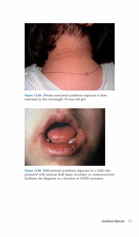
Figure 12.9A Obesity-associated acanthosis nigricans is dem-onstrated in this overweight 10-year-old girl.
Figure 12.9B Mild perioral acanthosis nigricans in a child whopresented with unusual skull shape secondary to craniosynostosisfacilitates the diagnosis as a function of FGFR3 mutation.
Acanthosis Nigricans 273

Diffuse Hypopigmented Streaky Lesions
Recognizing the Sign The striking aspect of this sign is the pigmentary dis-
turbance, with linear streaks of hypopigmented regions seen over the limbs
and whorls of hypopigmentation seen over the trunk. Skin texture may be
slightly more rough or dry over the affected region and, in subtle cases in
whom the pigmentary disturbance is not readily identifiable, it is the textural
skin differences that may draw the examiner’s attention. In such instances,
examination with a Wood’s lamp in a darkened room is required to reveal the
hypopigmentation. Although often present at birth or noted shortly thereafter,
hypomelanosis may develop up to the age of 1 year.
Establishing a Differential Diagnosis This is the cardinal clinical sign of
genetic mosaicism and is most commonly encountered in cases of unex-
plained developmental delay. Typically, blood karyotype will be normal but
fibroblast karyotype may reveal the mosaicism. Other clinical features, such as
micrognathia, syndactyly of the fingers, and obesity of adolescent onset, are
likely to be associated with the mosaic phenotype. Obesity is particularly
noted in patients mosaic for diploid/triploid mosaicism (46XX/69XXX). Most
commonly, the mosaicism is chromosomal, there being two discernible and
distinct cell lines demonstrable on fibroblast culture, though rarely on lym-
phoblast culture. However, hypomelanosis is also described in mosaicism for
single gene disorders, most notably in heterozygote female carriers of the X-
linked condition Menkes disease.
Investigations to Consider Karyotype, especially on fibroblasts, is indicated.
It should be noted that repeated normal karyotypes have been recorded in
some affected individuals. As patients get older, it appears that the likelihood
of positive findings on karyotype become reduced, presumably due to selective
loss of the abnormal chromosomal cell line in skin cells. Keratinocyte culture
is reputed to have a higher yield in terms of identifying abnormal karyotypes
among patients with this clinical phenomenon but is not available in most
centers.
The Nails, Hair, and Skin274
12.10

Figure 12.10A Typical hypopigmented linear streakylesions are seen on the back of the calves in thisteenage girl who presented with unexplained devel-opmental delay and seizures. The lesions could befollowed upward onto the buttocks and were alsovisible on the back.
Figure 12.10B This photograph shows subclinicalhypopigmentation, identified under Wood’s light,in a patient whose presentation was with severemicrognathia, requiring prolonged tracheostomy,and developmental delay of moderate degree. Thisis the same patient as shown in Figure 2.10.
Diffuse Hypopigmented Streaky Lesions 275

Piebaldism
Recognizing the Sign Local areas of skin hypopigmentation are present from
birth, affecting any region of the body. In equine parlance, a ‘‘piebald’’ refers
to a horse of two different colors, typically a blotchy combination of black and
white.
Establishing a Differential Diagnosis Blotchy hypopigmentation is typical
of autosomal dominant familial piebaldism, which is usually readily diag-
nosable by reference to family history and observation or clinical examination
of immediate relatives of the index case. In contrast to vitiligo, the hypo-
pigmented regions of piebaldism are present from birth, vitiligo being an ac-
quired form of hypopigmentation. Apart from the cutaneous hypopigmen-
tation, patients with piebaldism almost invariably have a central poliosis. The
main differential diagnosis is of Waardenburg syndrome, in which patients
have poliosis, often with associated dystopia canthorum and deafness, as well
as hypopigmented skin regions (Figure 12.7). Since deafness is an occasional
but inconstant finding in piebaldism, confident clinical distinction between
the two diagnoses may not always be easy. However, in general, the hypo-
pigmentation of pure piebaldism is more noteworthy than that seen in most
cases of Waardenburg syndrome. Additional clinical signs in Waardenburg
syndrome of heterochromia iridum or brilliant blue eyes with hypoplastic
stroma can also assist in differentiation. Hypopigmented skin lesions, occa-
sionally associated with poliosis, are a rare form of clinical presentation of
Fanconi anemia predating the characteristic pancytopenia by several years
but offering an opportunity to the clinically aware of recognizing this auto-
somal recessive condition in a presymptomatic stage. Sometimes careful ex-
amination of the hands may establish mild degrees of thumb hypoplasia,
which further lends weight to this possible diagnosis.
Investigations to Consider The diagnosis is generally clinical. Mutation
analysis is possible in familial piebaldism, the relevant genes being KIT and
SNAI2. Karyotype with diepoxybutane challenge is warranted if Fanconi ane-
mia is suspected.
The Nails, Hair, and Skin276
12.11

Figure 12.11 Although the hypopigmented regions so dramatically demonstratedin this patient from a pedigree segregating piebaldism as an autosomal domi-nant trait are symmetric, this need not necessarily be the case in all affectedindividuals.
Piebaldism 277

Cutis Marmorata
Recognizing the Sign This is a vascular phenomenon, present at birth and
which often fades with time, becoming less noticeable. Essentially, the skin has
a ‘‘marbled’’ appearance consequent on a reticular vascular pattern. The mot-
tling of the skin can be exacerbated by changes in temperature or by emotion,
so that direct questioning of parents as to the effect of bathing or of crying on
the skin pigmentation can be a useful aspect of history taking.
Establishing a Differential Diagnosis The skin marbling appearance may be
localized or general and, in the course of assessment, be alive to the possibility
of associated limb atrophy or hypertrophy conferring limb asymmetry. While
localized limb atrophy associated with cutis marmorata is well established as
an isolated and nonsyndromic phenomenon, the finding of limb asymmetry
also raises the possibility of macrocephaly-cutis marmorata-telangiectasia
congenita (MCTC) syndrome. In this distinct overgrowth syndrome, macro-
cephaly and cutis marmorata are associated with limb asymmetry, occasional
polydactyly, a very typical 2/3 syndactyly of the toes, and a degree of develop-
mental delay. The very characteristic facial feature is of a hemangioma on the
philtrum. A similar but clinically distinct disorder is Klippel-Trenaunay-
Weber syndrome (KTW), in which limb asymmetry is associated with mac-
ular vascular nevi and intelligence and developmental parameters are normal.
The philtral hemangioma and 2/3 toe syndactyly of MCTC syndrome are
lacking on examination in K-T-W cases. Cutis marmorata in the distribution
of the trigeminal branch of the fifth cranial nerve is well described in asso-
ciation with glaucoma and, as such, overlaps clinically with Sturge-Weber
syndrome (S-WS), the typical trigeminal lesion of which is a hemangioma. In
view of this overlap, the possibility of associated meningeal angiomata, sei-
zures, and neurologic deficit should be borne in mind if the distribution of
the skin mottling is confined to this region. Neonatal hypotonia, difficulty in
establishing feeding, the impression of macroglossia, and cutis marmorata
should alert the clinician to the possibility of Down syndrome, and these
same clinical features may also betoken congenital hypothyroidism. Con-
stipation is a frequent complaint in both these situations.
Investigations to Consider Karyotype is indicated if Down syndrome is
clinically suspected. Neonatal screening will establish congenital hypothyroi-
dism in most instances. Skull radiographs are warranted if Sturge-Weber is
suspected. The distribution of the vascular pattern often shows ‘‘tram line’’
calcification, which may be identified in the cortex or in the angiomatous
vessels.
The Nails, Hair, and Skin278
12.12

Figure 12.12A The characteristic marbled appear-ance of cutis marmorata of no syndromic signifi-cance is demonstrated in this neonate.
Figure 12.12B and C This photograph shows a 5-year-old girl who presented with developmental delay andlimb asymmetry. Note the faded but still discernibletriangular-shaped hemangioma over the philtrum.Observe also the prominent vascular marking of thehands and the left forearm especially. The diagnosisin this child is macrocephaly-cutis marmorata-telangiectasia congenita syndrome.
Cutis Marmorata 279

Capillary Hemangioma
Recognizing the Sign More colloquially known as ‘‘strawberry marks,’’ cap-
illary hemangiomas are soft, red areas of raised skin, of which the related
history is frequently of a pale or slightly red localized region of skin at birth,
which rapidly grows into the typical elevated strawberry nevus during the first
year of life, generally regressing thereafter.
Establishing a Differential Diagnosis Most strawberry marks are devoid
of any associated diagnostic implications. Indeed, it is estimated that up to
45% of infants have signs of benign vascular marks of this sort. Concern that
the hemangioma may have a more particular significance arises if there is a
positive family history or if multiple such lesions are observed. Multiple le-
sions are usually present from birth and likely to be indicative of disseminated
hemangiomatosis (DH). Such patients are at risk of visceral hemangiomata
developing in the liver, gastrointestinal tract, and respiratory tract, the author
being personally familiar with recurrent massive hemoptysis in two affected
individuals in which the hemangiomatosis involved the trachea. Both pa-
tients had only two or three skin lesions, and these were quite small and had
been dismissed as ‘‘normal’’ in the neonatal period. It appears that recognition
of DS cases from background ‘‘normal’’ capillary hemangioma requires care
and continued surveillance. Thrombocytopenia resulting from an associated
Kasabach-Merritt phenomenon is a well-established complication, as is car-
diac failure. Familial forms are rare and sometimes clinically overlap with a
group of genetic disorders that cause familial predisposition to cerebral hem-
orrhage and retinal hemorrhage, which are transmitted in autosomal domi-
nant mode and associated with specific gene mutations. However, other forms
of familial strawberry nevus are devoid of these catastrophic consequences and
appear only to segregate cutaneous signs. Maffucci syndrome combines the
association of hemangioma with enchondromata of the bones, most com-
monly manifest as bowing clinically of the affected bone. Careful, serial clinical
evaluation of the hands, feet, and limbs is required to recognize this condition,
with many patients being recognized only subsequent to fracture related to
enchondroma.
Investigations to Consider Platelet count and cardiac function need con-
tinuous monitoring if Kasabach-Merritt phenomenon arises with consequent
thrombocytopenia. Magnetic resonance imaging scanning for individuals at
risk of cerebral bleeding is appropriate if the family history suggests one of
the dominant forms with this complication.
The Nails, Hair, and Skin280
12.13

Figure 12.13 A typical capillary hemangioma, which regressed over the next3 years, is shown in this 11-month-old boy who presented with developmentaldelay due to a chromosomal disorder. The hemangioma was of no diagnosticsignificance.
Capillary Hemangioma 281

Telangiectasia
Recognizing the Sign Prominent blood vessels, of capillary origin, are visible
on the skin, conjunctiva, nail beds, and tongue or around the lips. With age,
these tend to become more prominent and to increase numerically.
Establishing a Differential Diagnosis Telangiectasia is almost always signif-
icant in the pediatric setting. The interpretation of the sign as a diagnostic indi-
cator will depend on the background history. In a child with normal growth
parameters presenting with recurrent epistaxis, the possibility of hereditary
hemorrhagic telangiectasia (HHT) should be considered. Although skin tel-
angiectasia may not develop until adulthood, being autosomal dominant, pa-
rental telangiectasia and/or a history of epistaxis or gastrointestinal bleeding in
the family may offer the key to diagnosis. Careful examination of the mucosal
surfaces for telangiectasia in the index case may confirm the diagnosis clinically.
Be aware of possiblemajor arteriovenousmalformations that can result in central
nervous system presentations or massive hemoptysis if present in the pulmonary
system and for which mechanical embolization may be a treatment option. By
contrast, skin telangiectasia in a child whose growth is the cause of concern opens
the possibility of chromosome breakage syndromes as the underlying cause, all
of which conditions have long-term associations with lymphomas and leukemia.
A history of prenatal growth deficiency, sun sensitivity, and telangiectasia in a
butterfly distribution on themalar region favorsBloom syndrome, while growth
failure of postnatal onset, coupled with developmental delay and ataxia, is more
reminiscent of ataxia-telangiectasia (AT) syndrome. Careful neurologic exam-
inationmay establish evidence of athetosis, dysarthria, or nystagmus, all of which
support the diagnosis of AT syndrome. Conjunctival telangiectasia should be
sought, as these predate the cutaneous signs that, when established, are typically
distributed over the eyelids, malar region, and ears. Sun sensitivity is often the
initial diagnostic clue in Cockayne syndrome, the typical history being of
postnatal growth failure with progressive neurologic concerns, mental retarda-
tion, ataxia, weakness, and deafness being common findings. Evaluating early
photographs can be valuable, demonstrating a progressive loss of periorbital
adipose tissue, the eyes appearing to becomemore sunkenwith time.Ophthalmic
examination often establishes a diagnostically valuable pigmentary retinopathy.
Normal chromosomes characterize dyskeratosis congenita, an X-linked con-
dition also resulting in immune compromise and pancytopenia in the second
decade and associated with telangiectasia. The presence of dysphagia, leuko-
plakia, and tear duct blockage all suggest preferment of this latter diagnosis.
Investigations to Consider Elevated a-fetoprotein is typical in AT syndrome.
X irradiation of chromosomes to demonstrate the enhanced breakage under
such conditions confirms. Sister chromatid exchange is increased in Bloom
syndrome. Ultraviolet irradiation forms the basis of diagnosis in Cockayne
syndrome, mRNA synthesis being greatly reduced.
The Nails, Hair, and Skin282
12.14
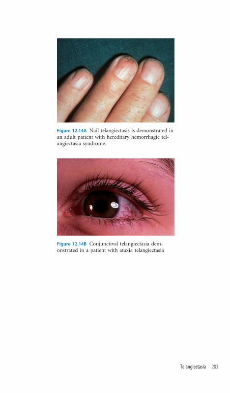
Figure 12.14A Nail telangiectasis is demonstrated inan adult patient with hereditary hemorrhagic tel-angiectasia syndrome.
Figure 12.14B Conjunctival telangiectasia dem-onstrated in a patient with ataxia telangiectasia
Telangiectasia 283

Ichthyosis
Recognizing the Sign This term describes excessively dry, scaly skin reflect-
ing disordered keratinization. Depending on the nature and extent of scaling,
quite clinically disparate conditions may be described by ichthyosis.
Establishing a Differential Diagnosis At its most dramatic, ichthyosis is the
clinical sign par excellence of collodion baby syndrome. The newborn is en-
meshed within a shiny membrane, often fissured, which proceeds to exfoliate
over the succeeding weeks. Despite the dramatic presentation, a normal neu-
rologic and developmental outcome should be anticipated, and the eventual
outlook for the skin is of a mildly dry skin, with normal hair. The condition is
autosomal recessive. A family history of dry skin may betray the autosomal
dominant form of ichthyosis, often associated with atopic dermatitis and
asthma. In evaluating male patients referred for nonspecific developmental de-
lay, the identification of dry skin should remind the clinician to inquire as to the
history of the delivery. Boys affected with the X-linked recessive condition
steroid sulfatase deficiency are usually the product of induced labor for pro-
longed pregnancy, as their carrier mothers produce inadequate estriol and may
not labor spontaneously. This condition may be due to localized deletion of the
Xp chromosome, and chondrodysplasia punctata, short stature, and Kallmann
syndrome may all be observed as part of the related phenotype. However, mu-
tation within the gene results in steroid sulfatase deficiency alone, development
being normal and the only clues being the history of delayed onset of labor and
ichthyosis. In some cases, the ichthyosiform element of the skin manifestation
is quite inapparent, but close inspection may reveal raindrop pigmentation of
the skin. The presence of limb shortening or asymmetry is likely to represent
Conradi-Hunnerman syndrome, anX-linked dominant condition that is rarely
compatible with survival in males and therefore is to be suspected clinically in
females with ichthyosis. A very characteristic and useful sign is follicular atro-
phoderma, conferring the appearance of ‘‘orange peel’’ on the skin of the af-
fected individual. Cataracts are another valuable diagnostic sign, as is punctuate
calcification of the bones, often a transient finding, for which reason early
radiologic evaluation is important if this is suspected.
Investigations to Consider Chondrodysplasia punctata is seen radiologically
in bothConradi-Hunnerman and steroid sulfatase deficiency syndromes. Raised
8-dehydrocholesterol is characteristic of Conradi-Hunnerman cases. FISH of
Xp22 may establish a deletion in patients with the more extensive steroid sul-
fatase phenotype, whilemutationwithin theARSC gene should be anticipated in
males with the phenotype confined to placental estriol inadequacy and derma-
tologic signs of ichthyosis. Plasma steroid sulfatase levels are usually unrecord-
able in both forms of the disorder.
The Nails, Hair, and Skin284
12.15

Figure 12.15A Congenitallamellar ichthyosis causingcollodion presentation isshown in the recovery phase.
Figure 12.15B A mild degree of ichthyosisis seen in this boy, born by induction oflabor after a 43-week gestation, who hassteroid sulfatase deficiency, his motherbeing heterozygous for that condition.
Figure 12.15C Raindrop pigmentation isshown in another boy with an establishedsteroid sulfatase deficiency.
Figure 12.15D Punctuate calcification in-volving the hip joints is demonstrated inthese x-rays from the neonatal period.
Ichthyosis 285
Excess Skin
Recognizing the Sign The clinical impression here will be of a child whose
skin is akin to an inappropriately large glove. There may be extra folds of skin
in addition to skin wrinkles.
Establishing a Differential Diagnosis Poor feeding in the neonatal period
may be the main clue to Costello syndrome, in which condition the excess
skin is sometimes paralleled by gum hypertrophy and perhaps evidence of
cardiomyopathy. By the age of 6 years, the diagnosis becomes apparent with
the appearance of the characteristic nasal papillomata (Figure 2.8B). Both
Noonan syndrome and cardio-facio-cutaneous syndrome (CFC) can pres-
ent with fetal hydrops. However, the more usual presentation is with cardiac
concerns, most classically valvular pulmonary stenosis, in the neonatal pe-
riod. Ptosis, short stature, and prolonged bleeding are characteristic of classic
Noonan syndrome cases, sparse curly hair and skin manifestations such as
ichthyosis or hemangioma marking the distinction in CFC syndrome. The
excess skin is often most apparent over the neck in Noonan syndrome, while
CFC cases, like Costello syndrome, frequently show excess skin on the hands.
Several children previously described as having cutis laxa syndrome more
correctly represent examples of Costello syndrome. Similarly, occipital horn
syndrome, an X-linked condition characterized by easy bruising and scarring
with excess joint laxity, can be misdiagnosed as cutis laxa (Figure 6.5). These
skeletal features are also typical of the Ehlers-Danlos syndrome (EDS) group
of disorders. While hyperelasticity of the skin, often associated with scar for-
mation in response to trauma, and joint hypermobility are more reminis-
cent of types I to III, excess skin is a central element of the diagnosis in type
VII. Hip dislocation is especially common in this form, the diagnosis being
prompted by the skin fragility and the blue sclera that often attend the con-
dition. Excess skin with associated inverted nipples or unusual distribution
of body fat, particularly the observation of fatty pads over the lumbosacral
region, are good clues to carbohydrate-deficient glycoprotein (CDG) syn-
drome, in the case of which signs of developmental delay should be sought on
examination.
Investigations to Consider Echocardiography has a valuable place in es-
tablishing evidence to support the clinical diagnosis of Costello, CFC, and
Noonan syndromes. Reduced serum copper and ceruloplasmin is typical of
occipital horn syndrome, but the presence of occipital exostoses on skull
radiology is the gold-standard investigation (see 6.5). Evidence of a coagu-
lopathy is useful in some forms of CDG syndrome, but the best screening
measure for this group of conditions is transferrin isoelectric focusing.
The Nails, Hair, and Skin286
12.16

Figure 12.16A The deeppalmar creases and gen-eral excess of skin are welldemonstrated in thisteenage patient withCostello syndrome.
Figure 12.16B The facialfeatures of Costello syn-drome in the same pa-tient are shown. Notethe facial coarseness,wiry hair, and nasalpapillomata. (See alsoFigure 2.7.)
Figure 12.16C Deep pal-mar creases, excess skin,and fetal finger pads areclearly seen in this handof a 1-year-old boy withcardiofaciocutaneoussyndrome.
Excess Skin 287

Appendix Sources of Further Information
It was never envisaged that this should serve as an encyclopedic text on mat-
ters dysmorphic and genetic. Rather, this book was conceived as a guide to as-
sist the confident recognition of clinical signs that are often useful in reaching
a dysmorphic diagnosis, and thereafter to offer some basic structure to the
inexperienced clinician in how to proceed, once secure that he or she had in-
terpreted the clinical signs correctly. The text accompanying each clinical sign
is limited by space and, most commonly, this limitation impacts mainly upon
the consideration of investigations. Accordingly, readers not habitually accus-
tomed to using genetic resources may benefit from guidance as to where they
may seek recourse for further information on specific conditions, journal ref-
erences, and detailed reports of investigations and their uses in specific clinical
settings. Inevitably, such guidance reflects personal practice and preference,
the following suggestions being particular favorites to which I turn most com-
monly but by no means an exhaustive exploration of the many different types
of information sources and formats with which a geneticist may have regular
cause to interact.
Databases
European Directory of DNA Diagnostic Laboratories (EDDNAL). This is an
easy resource for identifying laboratories offering diagnostic DNA testing for
specific loci and attendant clinical conditions.
www.eddnal.com
GeneTests. This is a valuable, publicly funded, and therefore free service that
allows easy identification of laboratories offering specialized investigations for
particular named conditions. It also allows easy access to reviews of particular
conditions.
www.geneclinics.org
The Human Gene Mutation Database (HGMD). This is a database con-
taining in excess of 50,000 known mutations in over 2,000 human genes that
summarizes and references most of the mutations that have been reported in
individual genes and the diseases consequent on mutations therein. It is a
useful reference in establishing whether a DNA sequence change has previ-
ously been recorded in a pathogenic situation. It is free to academic institu-
tions.
www.hgmd.cf.ac.uk
KnutShell.com. This is a recently launched database online search portal that
enables efficient searches of OMIM by symptom or structured list of clinical
signs/symptoms. It includes links to GeneTests and indexes of some well
288

established textbooks. Available by individual use-metered or institutional
subscription only.
www.knutshell.com
London Medical Databases. This is a constantly updated comprehensive
series of linked databases, summarizing all clinical and investigative data on
conditions likely to be encountered in a genetics clinic, whether mendelian,
chromosomal, or environmental. It is supplemented by a superb library of clin-
ical pictures of each disorder. This is a subscription-only service.
www.lmdatabases.com/contact
OMIM (Online Mendelian Inheritance in Man). This is a constantly updated
database, available free of charge, which summarizes clinical and genetic data
on mendelian disorders and affords some examples of specific mutations where
the locus for a disease has been established.
www.ncbi.nlm.nih.gov/omim/
Radiological Atlas of Malformation Syndromes and Skeletal Dysplasias
(REAMS). This is a subscription-only expert system that facilitates searches of
known skeletal abnormalities in malformation syndromes and dysplasias
by radiologic features, with a view to identifying the most likely conditions to
present with these radiologic features.
www.oup.co.uk/ep/cdroms/reams
Books
Aase JM. Diagnostic dysmorphology. Published by Plenum Medical Book Co.,
1990.
Firth HV, Hurst JA. Oxford desk reference: Clinical genetics. Published by Ox-
ford University Press, 2005.
Gardner RJM, Sutherland GR. Chromosome abnormalities and genetic counsel-
ling, 3rd ed. Published by Oxford University Press, 2004.
Gorlin RJ, Cohen MM Jr, Hennekam RCM. Syndromes of the head and neck,
4th ed. Published by Oxford University Press, 2001.
Hall JG, Allanson JE, Gripp KW, Slavotinek AM. Handbook of physical
measurements, 2nd ed. Published by Oxford University Press, 2006.
Rimoin DL, Connor JM, Pyeritz RE, Korf BR. Emery and Rimoin’s principles
and practice of medical genetics, 4th ed. Published by Churchill Livingstone,
2002.
Spranger JW, Brill P, Poznanski A. Bone dysplasias: An atlas of genetic disorders
of skeletal development, 2nd ed. Published by Oxford University Press, 2002.
Appendix 289

Tabyi H, Lachman RS. Radiology of syndromes, metabolic disorders and skeletal
dysplasias, 4th ed. Published by Mosby, 1996.
Toriello HV, Reardon W, Gorlin RJ. Hereditary hearing loss and its syndromes,
2nd ed. Published by Oxford University Press, 2004.
Traboulsi EI. Genetic diseases of the eye. Published by Oxford University Press,
1998.
Appendix290

Index
Aarskog syndromebrachydactyly, 190camptodactyly, 188clinodactyly, 184ear variation, external, 96hair graying, early, 16hypertelorism, 24, 25, 190inguinal hernia, 162, 163ovoid depression aroundumbilicus, 158, 159
philtral features, 48shawl scrotum, 170, 171short stature, 96
Abdomen/perineum, 158–175anal atresia/stenosis, 172–173caudal appendage, 174–175genitalia. See Genitaliahypospadias. See Hypospadiasinguinal hernia, 162–163minor anomalies of umbilicus,158–159
umbilical hernia/omphalocele, 158,160–161
Acanthosis nigricans, 4, 272–273ACFS (asymmetric crying facies
syndrome), 44, 45Achondroplasia, 34, 236, 237Acrocallosal syndrome, 20, 216Acrodysostosis, 34Acrondroplasia, tridenthand,206,207
Acrorenoocular syndrome (ARO),180
Adams-Oliver syndrome (AOS)limb reduction defects, 238scalp defects, 8toes, hypoplastic or missing,224, 225
AHO (Albright’s hereditaryosteodystrophy), 190, 230, 240
Alae nasi abnormalities, 30–31Alae nasi and nasal tip abnormalities,
30–31Alagille syndrome, 66, 68Albright’s hereditary osteodystrophy
(AHO), 190, 230, 240Alcohol exposure, intrauterineabsent or hypoplastic nailsand, 256
anophthalmia/microphthalmiaand, 74
brachydactyly and, 190microtia and, 94
Alopecia, 266–267Alpha-fetoprotein elevation, 282Alpha-thalassemia and mental
retardation, X-linked. SeeX-linked a-thalassemia andmental retardation
Ambiguous genitalia, 166–167Aminoaciduria, 74
291

Amniotic band disruption, 224, 238Amyoplasia congenita, 242, 243Anal atresia, 172–173Anal stenosis, 172–173Androgen insensitivity, 162, 166Anencephaly, 20–21Aneuploidy, 42, 144Ankyloglossia, 120Anophthalmia, 74–75Anterior encephalocele, 36Antley-Bixler syndrome, 4, 166Anus, anterior displacement of,
172–173AOS. See Adams-Oliver syndromeApert syndrome, 44, 62, 182, 200Aplasia cutis, 224Aplasia cutis congenita, 8, 9ARO (acrorenoocular syndrome),
180Arthrogryposis congenita,
242–243Aspirin exposure, intrauterine, 8Asymmetric crying facies syndrome
(ACFS), 44, 45Asymmetry, facial, 44–45Ataxia-telangiectasia syndrome, 268,
282, 283Atrial septal defect (ASD), 148, 256ATRX. See X-linked a-thalassemia
and mental retardationAuricular pits, 92–93Auricular tags, 102–103Autosomal dominant form of
icthyosis, 284
Balding, temporal, 264–265Bannayan-Zonana syndrome,
10, 142Baraitser-Winter syndrome, 6Bardet-Biedl syndromeDandy-Walker malformation, 10postaxial polydactyly, 178, 179,220, 221
small penis/micropenis, 164‘‘Bat ears’’ (protruding ears), 96, 97BCD (blepharocheilodontic
syndrome), 78BCOR gene, 74
Beals syndrome (congenitalcontractural arachnodactyly)
arachnodactyly, 186, 242camptodactyly, 186, 188external ear variants, 96, 242overlapping fingers, 196pectus excavatum and carinatum,152
Beare-Stevenson syndrome, 226Beckwith-Wiedemann syndrome
(BWS)auricular pits, 92, 93earlobe creases, 98, 99macroglossia, 116, 117umbilical hernia/omphalocele,160–161
Binder syndrome, 34, 35Biochemical diseases, corneal
clouding in, 6Biotindase deficiency, 266Blackfan-Diamond syndrome, 180, 204Bladder diverticula, 78Blepharocheilodontic syndrome
(BCD), 78Blepharophimosis, 58–59Blepharophimosis-ptosis-epicanthus
inversus syndrome (BPES), 52,58, 64
Bloom syndrome, 12, 282Blue sclerae, 70–71BOF (branchiooculofacial syndrome),
134BOR. See Branchiootorenal
syndromeBPES (blepharophimosis-ptosis-
epicanthus inversus syndrome),52, 58, 64
Brachydactylyin Aarskog syndrome, 158, 191autosomal dominant or type D,190, 200
familial or type E, 190, 191, 230,231
Branchial sinus, 134Branchiooculofacial syndrome
(BOF), 134Branchiootorenal syndrome (BOR)
auricular pits, 92, 93brachial sinus, 134, 135
Index292

lop ears, 96microtia, 94sloping shoulders, 148
Breast asymmetry, pectoral musclehypoplasia and, 140, 141
Brushfield spots, 76, 77‘‘Bull’s eye’’ (buphthalmos), 66Butterfly vertebrae, 66BWS. See Beckwith-Wiedemann
syndrome
CACNA1C gene mutation, 182Cafe-au-lait skin patches
neurofibromatosis type 1, 10Russell-Silver syndrome, 184,270–271
Campomelic dysplasia, 150Camptodactyly, 188–189Camptomelic dysplasia, 248Capillary hemangioma, 280–281Carbimazole exposure, intrauterine,
132, 144, 145Carbohydrate-deficient glycoprotein
syndrome (CDG)ears, posteriorly rotated, 90fat distribution, unusual, 12,90, 286
foot edema, 228, 229nipple inversion, 12, 90, 144
Cardiac murmur, 120Cardiofaciocutaneous syndrome (CFC)
excess skin, 286, 287palmar crease patterns, 212, 213posteriorly rotated ears in, 90, 91sparse hair, 266, 267
Carey-Fineman-Ziter syndrome, 46Carinatum, 152–153Carney complex, 114, 146Carpenter syndrome, 178Cartilage-hair hypoplasia syndrome
(CHH), 262, 266Catel-Manzke syndrome, 42, 58,
72, 188Cat-eye syndrome, 72, 102, 103, 172Caudal appendage, 174 –175CBFA1 gene, 148CDG. See Carbohydrate-deficient
glycoprotein syndrome
Cerebrocostomandibularsyndrome, 42
Cerebrospinal fistula, 172Cervical vertebral fusion
(Klippel-Feil malformation),18, 100, 130
Cervical vertebral malformation, 102CFC. See Cardiofaciocutaneous
syndromeCFND. See Craniofrontonasal
dysplasiaCHARGE associationear malformations, 74, 94, 96iris coloboma, 72microstomia, 106small penis/micropenis, 164, 165
Chest, 140–155accessory nipples, 142–143athelia/hypothelia, 144–145gynecomastia, 146–147pectus excavatum and carinatum,152–153
Poland anomaly, 140–141scoliosis, 154–155sloping shoulders, 148–149small thorax, 150–151
CHH (cartilage-hair hypoplasiasyndrome), 262, 266
Cholanal stenosis, 40Cholesterol, low, 196Chondrodysplasia punctata, 28, 29Chorioretinitis-pituitary dysfunction
syndrome, 82Chromosomal sex determination, 150Chromosome breakage syndromes,
282Chromosome deletions1p36, 54, 56, 1084p, 8, 729p, 611p, 6820p, 682q37, 2304q34, 258–2595q, 82, 1648q, 14, 1489q34, 11611q, 6, 713q, 222
Index 293

Chromosome deletions (continued)18q, 10018q23, 100, 10122q13, 208
Chromosome 22 material, cat-eyesyndrome and, 102, 103
Chronic infantile neurologiccutaneous and articularsyndrome (CINCA), 244
Cleft lip, 108–109, 198Cleft palateisolated, 120–121Moebius syndrome, 46Pai syndrome, 36Rieger syndrome and, 158subclinical, 121
Cleft uvula, 36Cleidocranial dysostosis, 148Clinodactyly of fingers, 184–185Clitoromegaly, 4Cloverleaf iris (Lester’s sign), 76, 77Cloverleaf skull malformation, 206Cockayne syndrome, 12, 282Coffin-Lowry syndromefingers in, 24, 112, 154, 194, 195hypertelorism, 24, 195hypothenar crease, 24, 195lip fullness, 112, 154palmar crease patterns, 212palpebral fissure slant, 60scoliosis and, 154
Coffin-Siris syndrome, 266Cohen syndromenasal bridge abnormality, 28philtrum abnormalities, 48synorphrys, 39tapering fingers, 192, 193
COH1 locus, 192Col3A1 mutation, 98Colloidion baby syndrome, 284Columella, shortening of, 34Conductive hearing loss, 94Congenital bilateral absence of the
vasa, 130Congenital contractural
arachnodactyly syndrome.See Beals contracturalarachnodactyly syndrome
Congenital heart defects, 66, 110
Congenital hypothyroidism,160, 278
Congenital iris dysplasia, 66Congenital lamellar ichthyosis, 285Congenital myotonic dystrophy, 122Conjunctival telangiectasia, 283Connective tissue disorders, 70,
154, 162Connexin 26, 82Connexin 30, 82Conradi-Hunnerman syndrome, 284Contractural arachnodactyly
syndrome, congenital. See Bealscontractural arachnodactylysyndrome
Corneal clouding, 66–67Cornelia de Lange syndrome.
See De Lange syndromeCostello syndrome
acanthosis nigricans, 272earlobe variants, 98early feeding problems, 128ears, posteriorly rotated, 90excess skin, 286, 287nasal papillomata, 36, 37palmar crease patterns, 212, 213plantar creases, longitudinal, 226
Cowden syndrome, 10, 11, 36, 240Craniofrontonasal dysplasia (CFND)
frontal hairline variations, 16, 17hallucal broadness, 218longitudinal splitting of nails,260, 261
sloping shoulders, 148Cranioosteoarthropathy, 244, 245Craniosynostosis
acanthosis nigricans, 273caudal appendage, 174facial asymmetry, 44frontal hairline variations, 14, 16plagiocephaly, 4
Creases, earlobe, 98, 99Crouzon syndrome
acanthosis nigricans, 4, 272caudal appendage, 174plagiocephaly, 4proptosis, 62, 63
Crumpled ears, 96, 97Cryptophthalmos, 74
Index294

Cryptorchidism, 64Cryptotia, 96, 97Currarino syndrome, 172Cutis laxa syndrome, 78, 286Cutis marmorata, 278–279Cyclopia, 26Cyclosporin exposure, intrauterine,
124, 125
Dandy-Walker malformation, 10, 262Databases, 288–289Deafness
with epibulbar dermoid, 84Pendred syndrome, 132, 133
Deafness-onycho-osteodystrophyretardation syndrome (DOOR),256
DEB (diepoxybutane), 2027-Dehydrocholesterol, 12, 26, 196De Lange syndrome
brachydactyly, 190ectrodactyly, 198eyelash variants, 82limb malformations, 32low-birth weight, 32, 38, 190short first metacarpal, 12,32, 190
synorphrys, 12, 32, 38thumbs variations, 190, 202, 203ulnar ray defects, 210
Delayed bone age, 48Diabetic embryopathy
preaxial polydactyly, 216, 217ulnar ray defects, 210
Diepoxybutane (DEB), 202Diepoxybutane challenge test, 270Diffuse hypopigmented streaky
lesions, 274–275‘‘Dinner-fork’’ configuration, of
wrist, 252, 253Disomy of chromosome 7, 184Disseminated hemangiomatosis,
280Distichiasis (double row of
eyelashes), 78, 82, 83DOOR (deafness-onycho-
osteodystrophy retardationsyndrome), 256
Down syndromebrushfield spots, 76clinodactyly in, 184cutis marmorata, 278epicanthus, 52micrognathia with secondarymacroglossia, 116
Drash syndrome, 166Drooling, persistent, 122–123Duane syndrome (sixth cranial nerve
palsy), 130, 134Dyskeratosis congenita, 282Dystopia canthorum, 268
Ears, 88–103auricular pits, 92–93auricular tags, 102–103earlobe variants, 98–99external auditory canal atresia orstenosis, 100–101
external variants, 96–97low-set, 88–89malformations, with facialasymmetry, 44
microtia, 94–95posteriorly rotated, 90–91
Ectodactyly-ectodermal dysplasiasyndrome, 144
Ectodermal dysplasiaeyelash variants, 82nails, absent or hypoplastic, 256X-linked hypohydrotic, eyebrowabsence in, 80, 81
Ectrodactyly, 198–199Ectrodactyly-ectodermal dysplasia, 108Ectropion of lower eyelid, 78–79EDA1 gene, 80EDDNAL (European Directory of
DNA Diagnostic Laboratories),289
Edema, foot, 228–229EDS. See Ehlers-Danlos syndromeEdwards syndrome (trisomy 18)arthrogryposis congenita, 242cryptotia, 96nipple hypoplasia, 144omphalocele, 160overlapping fingers, 196, 197
Index 295

EEC (ectrodactyly, ectodermaldysplasiaandclefting syndrome),198
EFNB1 gene, 148Ehlers-Danlos syndrome (EDS)blue sclerae, 70earlobe absence, 98, 99epicanthus, 52excess skin, 286inguinal hernia, 162joint hypermobility, 246, 247neck webbing, 128palmar crease patterns, 212proptosis, 62tapering fingers, 192type VI, 155, 212umbilical hernia, 160
Ellis-van Creveld syndromedeep-set nails, 256frenula, accessory, 118limb defects, 236small thorax, 150
ELN gene, 32, 80, 112Endocrine causes, of micropenis, 164Environmental agentsanencephaly and, 20meningocele and, 20midface flattening and, 34
Epibulbar dermoids, 84–85, 130Epicanthus, 52–53Escherichia coli meningitis, 172European Directory of DNA
Diagnostic Laboratories(EDDNAL), 289
Excess skin, 286–287External auditory canal atresia or
stenosis, 100–101EYAl1 gene mutation, 92EYA1 locus, 134Eye. See also Eyebrows; Eyelashes;
Eyelidsalmond-shaped, 56–57anophthalmia/microphthalmia,74–75
blepharophimosis, 58–59blue sclerae, 70–71corneal clouding, 66–67deep-set, 54–55ectopic pupil, 68–69
epibulbar dermoid, 84–85epibulbar dermoids, 84–85, 130epicanthus, 52–53iris coloboma, 72–73iris variants, 76–77palpebral fissure slant, 60–61proptosis, 62–63
Eyebrowsinterrupted, 79, 80sparse, 80, 81variants, 80–81
Eyelashesdouble row, 78variants, 82–83
Eyelidsectropion, 78–79ptosis, 64–65
Face, 24–49abnormal nasal bridge, 28–29alae nasi and nasal tipabnormalities, 30–31
asymmetry, 44–45hypertelorism, 24–25hypotelorism, 26–27micrognathia, 42–43midfacial hypoplasia, 40–41myopathic facies, 46–47nasal appendages, abnormal,36–37
nasal columella and naresabnormalities, 32–33
nasal septum abnormalities,34–35
philtrum abnormalities, 48–49synophrys, 25, 38–39
Facial hemangioma, 10Familial angiolipomatosis, 240Familial brachydactyly type E,
190, 191Familial multiple exostoses, 241Familial piebaldism, 276, 277Familial postaxial polydactyly, 220Familial type E brachydactyly, 190,
191, 230, 231Fanconi anemia, 276Fanconi pancytopenia syndrome, 202Fanconi syndrome, 12, 270, 271
Index296

FBN1 mutation, 68, 152, 186FBN2 mutation, 152, 186Feet, 216–231. See also Toes
broad halluces, 218–219edema, 228–229longitudinal plantar creases,226–227
short fourth and fifth metatarsals,230–231
Feingold syndrome, 222Femur-fibula-ulnar syndrome (FFU),
198Fetal akinesia, 188Fetal alcohol syndrome (FAS)
blepharophimosis, 58, 59lip defects, 112midfacial hypoplasia, 40palmar crease patterns, 212, 213philtrum abnormalities, 48, 49
Fetal valproate syndrome, 40, 48, 142Fetal warfarin embryopathy, 28a-Fetoprotein elevation, 282FFU (femur-fibula-ulnar syndrome),
198FGFR gene mutations, 4, 6FG syndrome, 14Fibrodysplasia ossificans progressiva,
224, 225Filippi syndrome, 12Fingers
arachnodactyly, 186–187brachydactyly, 190–191camptodactyly, 188–189clinodactyly, 184–185fetal finger pads, 208–209overlapping, 196–197puffy, 194–195‘‘sausage-shaped,’’ 112syndactyly, 4, 182–183tapering, 192–193
Finlay-Marks syndrome, 8Floating-Harbor syndrome
nasal columella abnormalities, 32philtrum abnormalities, 48thin upper lip, 112, 113
FOXL2 gene mutation, 52, 58Fraser syndrome
external ear variants, 96low frontal hairline, 16
renal agenesis, 74syndactyly, 74
Freeman-Sheldon syndromecamptodactyly, 188, 189deep-set eyes, 54microstomia, 106, 107overlapping fingers in, 196
Frenula, accessory, 118–119Frontonasal dysplasia, 16, 24Fryns syndrome, 256‘‘Funnel chest,’’ 152–153
GAG (glycosaminoglycan), urinary,116, 160, 162
GeneTests, 289Genetic mosaicism, 274Genitaliaambiguous, 166–167penis. See Penispentoscrotal transposition, 170–171shawl scrotum, 170–171
Genitopatellar syndrome, 248Glaucoma, congenital, 66GLI3 gene mutation, 172Glycosaminoglycan (GAG), urinary,
116, 160, 162GNASI mutation, 190Goiter, 132–133Goldenhar syndrome
(oculoauriculovertebral), 44, 84,92, 94, 102
Goltz syndrome, 74Gorlin sign, 246, 247Gorlin syndrome, 72Granulocytopenia, 28, 48Greig syndrome, preaxial
polydactyly, 10, 216, 217Gum hyperplasia, 119, 124–125Gynecomastia, 146–147
Hairpoliosis, 268–269sparse or alopecia, 266–267temporal balding, 264–265
Hairlinehigh, with frontal upsweep, 14–15low frontal, 16–17
Index 297

Hairline (continued)low posterior, 18–19widow’s peak, 16–17
Hallermann-Streiff syndrome, 26,58, 264
Halluces, broad, 218–219Handhardt hypoglossia-hypodactyly
syndrome, 238Hands, 178–213. See also Fingers;
Thumbsbrachydactyly, 190–191camptodactyly, 188–189ectrodactyly, 198–199palmar crease patterns, withdiagnostic significance, 212–213
preaxial polydactyly/thumbduplication, 180–181
trident, 206–207ulnar ray defects, 210–211
HbH inclusion bodies, 14, 24,112, 166
Hemihyperplasia (hemihypertrophy)facial, 44, 45isolated, 234, 235macroglossia, 116
Hemivertebrae, 66Hereditary hemorrhagic
telangiectasia (HHT), 282, 283Herniainguinal, 162–163umbilical, 158, 160–161
Heterochromia iridum, 76, 77, 268HGMD (The Human Gene Mutation
Database), 288–289HLXB9 gene mutation, 172‘‘Hockey-stick sign,’’ in fetal alcohol
syndrome, 212, 213Holoprosencephaly, 26, 108, 168Holt-Oram syndromeatrial septal defect, 140, 202limb reduction defects, 238sloping shoulders, 148thumb defects, 202, 204
Homocystinuria, 152, 186The Human Gene Mutation
Database (HGMD), 288–289Humerus, rhizomelic shortening
of, 206Hydrocephalus, 88, 89
Hydrolethalus syndrome, 20Hypertelorism, 24–25, 158Hypochondroplasia, 10Hypoglossia-hypodactyly syndrome,
106, 118Hypoplasia
of muscle pulp on palmar surfaceof digit, 258, 259
of nails, 256–257of nipples, 144–145patellar, 248–249
Hypospadias, 168–169with cleft palate, 120Smith-Lemli-Opitz syndrome, 88WAGR, 64
Hypotelorism, 26–27Hypothyroidism, neonatal, 132
I-cell disease, gum hyperplasia,124, 125
Icthyosis, 284–285Infantile systemic hyalinosis, 124, 194Information resources, 288–290Inguinal hernia, 162–163Insulin receptor gene mutations,
112, 272Interpupillary distance
decreased or hypotelorism, 26–27increased or hypertelorism, 24–25
‘‘Interrupted eyebrow sign,’’ 79,80, 81
IRF6 gene, 110Iris coloboma, 72–73Isolated hemihyperplasia, 44
Jacobsen syndrome, 6Jeune asphyxiating thoracic
dystrophypostaxial polydactyly, 178small thorax, 150, 151, 178, 236
Johanson-Blizzard syndrome, 8, 14,15, 30
Joint enlargement, 244–245
Kabuki syndromecaudal appendage, 174
Index298
earlobe prominence in, 98, 99ectropion of lower eyelid, 78, 79fetal finger pads, 208, 209‘‘interrupted eyebrow sign,’’ 12, 79,80, 81
lip pits, 110, 111palpebral fissure slant, 60, 61short columella, 34
Kal1 gene, 146Kallmann syndrome
cleft palate or lip with micropenis,164
gynecomastia, 146, 147olfactory tract maldevelopment, 26
Kasabach-Merritt phenomenon, 280Keratan sulfate excretion, urinary,
130Klinefelter syndrome, 146, 164Klippel-Feil malformation (cervical
vertebral fusion), 18, 100, 130Klippel-Trenaunay-Weber syndrome
(KTW), 10, 234, 278Kniest syndrome, 120, 244KnutShell.com, 288KRAS gene, 128KTW (Klippel-Trenaunay-Weber
syndrome), 10, 234, 278Kyphoscoliosis, 155, 246
Lacrimo-auriculo-dento-digitalsyndrome (LADD), 180, 204,205
Lamellar ichthyosis (collodion babysyndrome), 78
Langer-Gideon syndrome, 240Laugier-Hunziker syndrome, 114Lenz microphthalmia syndrome,
54, 55, 74LEOPARD syndrome, 114Leprechaunism, 112, 124, 146, 272Leri-Weill syndrome, 252Lester’s sign (cloverleaf iris), 76, 77Limb mammary syndrome (LMS),
198Limbs
asymmetry, 234–235generalized joint hypermobility,246–247
joint enlargement, 244–245localized lumps on, 240–241Madelung deformity, 252–253malformations, 130, 144, 234–253pseudoarthrosis, 250–251reduction defects, 238–239small or absent patella, 248–249symmetric short, 236–237
Linear sebaceous nevus syndrome, 84Lipodystrophy, 272Lipomata, 36Lipscleft, 108–109, 198and oral mucosa pigmentation,114–115
pits, 110–111upper, thick and thin, 112–113
Lisch nodules, 62, 76, 77, 270LMS (limb mammary syndrome), 198‘‘Lobster-claw’’ malformation, 199London Medical Databases, 288Long QT syndrome, 182‘‘Lop ear,’’ 96, 97Lowe syndrome, 54, 74Lujan-Fryns syndrome, X-linked, 46Lymphoedema-distichiasis syndromeeyelash variants, 82, 83foot edema, 228
Lysyl hydroxylase assay, 246Lysyl hydroxylase deficiency, 70
Macrocephaly, 10–11, 88Macrocephaly-cutis
marmorata-telangiectasiacongenita (MCTC)
cutis marmorata, 278, 279facial asymmetry, 44facial hemangioma, 10, 44limb asymmetry, 234
Macroglossia, 55, 116–117Madelung deformity, 252–253Maffucci syndrome, 280Mainzer-Saldino syndrome, 262,
2634Malar hypoplasia, 40Marfan syndromearachnodactyly, 186, 187blue sclerae, 70
Index 299

Marfan syndrome (continued)ectopic pupil, 68inguinal hernia, 162joint hypermobility, 246pectus excavatum and carinatum,152
scoliosis, 154tapering fingers, 192
Maternal diabetes, 94Maternal myasthenia syndrome,
188, 196Maxillary hypoplasia, 40McCune-Albright syndrome, 250, 270McKusick-cartilage-hair hypoplasia
syndrome, 82, 150MCPH1 locus mutation, 12MCTC syndrome. See
Macrocephaly-cutismarmorata-telanagiectasiacongenita
Meckel-Gruber syndrome, 20,178, 220
Meier-Gorlin syndrome, 12, 42,248, 249
Menkes syndrome, 136, 274Metabolic causes, of goiter, 132Metopic ridge, 6–7Microcephaly, 12–13, 74Micrognathia, 42–43, 106Micropenis, 164–165Microphthalmia, 74–75Microstomia, 106–107Microtia, 94–95Midfacial hypoplasia, 40–41Miller syndrome, 78, 94, 142,
210, 224Milroy’s disease, foot edema, 228Minor malformations of no
diagnostic significance,unilateral, 102
MITF mutation, 76, 80Mitochondrial myopathies, 46, 64Moebius syndromeeyelid ptosis, 64, 65hand defects, 106microstomia, 106myopathic facies, 46persistent drooling, 122syndactyly, 182
Morquio syndrome(mucopolysaccharidosestype IV), 130
Mosaicism, 192Mosaic trisomy 8, 248Mouth/oral cavity, 106–125. See also
Cleft palate; Lipsaccessory oral frenula, 118–119drooling, persistent, 122–123frenula, accessory, 118–119gum hyperplasia, 119, 124–125macroglossia, 55, 116–117microstomia, 106–107upper lip clefts, 108–109
Mowat-Wilson syndrome, 32, 33,98, 99
MPS. See MucopolysaccharidosesMucolipidosis type II, 6Mucopolysaccharidoses (MPS)
corneal clouding, 67inguinal hernia, 162joint enlargement, 244macroglossia, 116scoliosis, 154synophrys, 38type III or Sanfilippo syndrome,38, 160
type IV or Morquio syndrome, 130umbilical hernia/omphalocele, 160
Mulibrey nanism, 14, 250Multiple endocrine neoplasia type
IIB (MEN IIB), 78Multiple exostoses, 240, 244Multiple pterygium syndrome,
128, 242MURCS association, 130, 131Myasthenia gravis, 242Myopathic conditions, 82Myotonic dystrophy, 46
Nager syndrome, 40, 94Nail-patella syndrome, 76, 248,
256, 257Nails
absent or hypoplastic, 256–257longitudinal splitting of, 260–261short, 262–263‘‘tale of a nail’’ sign, 258–259
Index300

Nares abnormalities, 32–33Nasal appendages, abnormal, 36–37Nasal bridge, abnormal, 28–29Nasal columella abnormalities, 32–33Nasal septum abnormalities, 34–35Nasal tip abnormalities, 30–31Natal teeth, 118Neck, 128–137
goiter, 132–133occipital horn syndrome,136–137, 286
short, 130–131sinuses/fistulae/pits, 134–135webbing, 18, 19, 128–129
Nephrotic syndrome, foot edema, 228Neurofibromatosis type 1
cafe-au-lait patches, 18, 154,270, 271
limb asymmetry, 234Lisch nodules, 76localized lumps on limbs, 240proptosis, 62pseudoarthrosis, 250, 251scoliosis, 154
Nipplesabsent or hypoplastic, 144–145accessory, 142–143
NOG gene mutation, 30Noonan-neurofibromatosis type
1, 18, 128Noonan syndrome
ear variants, 88, 90excess skin, 286eyebrow variants, 80eyelid ptosis, 64, 65foot edema, 228low posterior hairline, 18, 19neck webbing, 128, 129palpebral fissure slant, 60pectus excavatum and carinatum,152, 153
sloping shoulders, 148Nose. See nasal entriesNuchal skin, excess, 128, 129
Oberklaid-Danks syndrome, 6Obesity-associated acanthosis
nigricans, 273
Occipital horn syndrome, 136–137,286
Occipitofrontal head circumference(OFC), 11
Oculo-dento-digital syndrome(ODD)
hypoplastic alae nasi, 30hypotelorism, 26, 27, 30microphthalmia, 74syndactyly, 30, 182
OEIS syndrome, 166OFC (occipitofrontal head
circumference), 11OI (osteogenesis imperfecta), 70, 71Okihiro syndrome, 204, 238, 239Oligohydramnios, 34, 44OMIM (Online Mendelian
Inheritance in Man), 288Omphalocele, 160–161Online Mendelian Inheritance in
Man (OMIM), 288Opitz G syndrome, 24, 108, 120,
168, 172Optic nerve hypoplasia, 48Oral cavity. See Mouth/oral cavityOral mucosa, and lip pigmentation,
114–115‘‘Orange peel’’ skin, 284Orofaciodigital syndromesaccessory frenula, 118, 119preaxial polydactyly, 216upper lip clefts, 108
Osteogenesis imperfecta (OI), 70, 71Otofaciocervical syndrome, sloping
shoulders, 148, 149Overlapping fingers, 196–197
Pai syndrome, 36, 37Palate, cleft. See Cleft palatePallister-Hall syndrome, 222Pallister-Killian syndrome
(tetrasomy 12p)accessory nipples, 142anal deformities, 172caudal appendage, 174, 175frontal hairline variants, 14palpebral fissure slant, 60, 61temporal balding, 264
Index 301

Palms, crease patterns on, 192,212–213
Palpebral fissure slant, 60–61Parry-Romberg syndrome, 44Patau syndrome (trisomy 13)cleft palate, 120lip clefts, 108omphalocele, 160overlapping fingers, 196postaxial polydactyly, 178scalp defects, 8
Patella, small or absent, 248–249PAX 3 mutation, 76, 80PAX6 mutation, 68Pectus excavatum and carinatum,
152–153PEHO syndrome, 228, 229Pena-Shokeir syndrome, 88, 196Pendred syndrome, 132, 133Penishypospadias. See Hypospadias‘‘shawl’’ configuration, 158, 159small/micropenis, 164–165
Pentoscrotal transposition, 170–171Perineum. See Abdomen/perineumPersylvian polymicrogyria, 123Peter’s anomaly, 68Peter’s plus syndrome, 68Peutz-Jeghers syndrome, 114, 115Pfeiffer syndromebroad thumbs in, 44, 200, 201caudal appendage, 174facial asymmetry, 44hallucal broadness, 44, 218proptosis, 62
p63 gene mutation, 144Phenytoin exposure, intrauterineabsent or hypoplastic nails, 256brachydactyly, 190frontal hairline variants, 16gum hyperplasia and, 124
Philtrum abnormalities, 48–49Piebaldism, 268, 276–277Pierre-Robin sequence, 28, 42,
120, 246Pitsauricular, 92–93branchial, 134lip, 110–111
Plagiocephaly, 4–5Plantar creases, longitudinal,
226–227Poland anomaly, 140–141, 182,
183, 238Poland-Moebius syndrome, 140, 238Poliosis, 268–269Polydactyly
Greig syndrome, 10postaxial, 178–179, 220–221preaxial, 180–181, 216–217
Polyhydramnios, 122Pompe’s disease, 116Popliteal pterygium syndrome,
110, 118Postaxial polydactyly
fingers, 178–179toes, 220–221
Prader-Willi syndrome, 56, 57, 82, 164Preaxial polydactyly
fingers, 180–181toes, 216–217
Preaxial polydactyly/thumbduplication, 180–181
Prematurity, 162Primary anterior eye chamber
disorders, 66Primary muscle disease, 46Proptosis, 62–63Proteus syndrome, 10, 234, 235Pseudoarthrosis, 250–251Pseudocleft, 109PTEN gene mutation, 11, 34, 142Ptosis, in infancy, 46Pulmonary hypoplasia, 88Pycnodysostosis, 236
Radiological Atlas of MalformationSyndromes and SkeletalDysplasias (REAMS), 289
Raindrop pigmentation, 285Rapp-Hodgkin syndrome, 80, 108REAMS (Radiological Atlas of
Malformation Syndromes andSkeletal Dysplasias), 289
RET gene mutations, 78Retinol binding protein, urinary, 74Rhabdomyosarcomata, 36
Index302

‘‘Rib gaps,’’ 42Richardson-Kirk syndrome, 12, 13,
90, 96Rieger syndrome, 66, 68, 69, 158Robinow syndrome
abnormal nasal columella andnares, 32, 33
brachydactyly, 190broad thumbs, 200gum hyperplasia, 124palpebral fissure slant, 60philtrum abnormalities, 48preaxial polydactyly, 216small penis/micropenis, 164symmetric short limbs in, 236thumb malformation, 180umbilicus anomalies, minor, 158
Rothmund-Thompson syndrome,248
Rubinstein-Taybi syndromeabnormal nasal columella andnares, 32, 33
accessory nipples, 142broad thumbs, 200, 201corneal clouding, 66deep-set eyes, 54frontal hairline variants, 14hallucal broadness, 218low-set ears, 88
Russell-Silver syndrome, 70, 184, 270
SADDAN syndrome, 206, 236, 272Saethre-Chotzen syndrome
craniosynostosis, 64facial asymmetry, 44hallucal broadness, 218plagiocephaly, 4preaxial polydactyly, 216, 217syndactyly of fingers, 182, 183
SALL1 gene, 92, 102, 168, 172, 180SALL4 gene, 180Sanfilippo syndrome (MCP III), 38,
160Scalp defects, 8–9. See also HairlineScaphocephaly, with FGFR2 gene
mutation, 63Scapula, congenital upward
displacement, 130
Schinzel-Giedion syndrome, 17,40, 168
Schwartz-Jampel syndrome, 106, 188Sclerocornea, 66Scoliosis, 154–155Seckel syndrome, 98Septooptic dysplasia Pagon type, 224SGB. See Simpson-Golabi-Behmel
syndromeShawl scrotum, 170–171SHFMs (split hand and foot
malformations), 198Shoulders, sloping, 148–149Silver-Russell syndrome, 234, 250Simpson-Golabi-Behmel syndrome
(SGB)accessory nipples, 142caudal appendage, 174macrocephaly, 10macroglossia, 116persistent drooling, 122posteriorly rotated ears, 90
Sixth cranial nerve palsy (Duanesyndrome), 130, 134
Skeletal dysplasia, 154Skinacanthosis nigricans, 272–273cafe-au-lait patches, 270–271capillary hemangioma, 280–281cutis marmorata, 278–279diffuse hypopigmented streakylesions, 274–275
excess, 286–287ichthyosis, 284–285lentigines, 146piebaldism, 276–277telangiectasia, 282–283
Skull, 4–21anencephaly, 20–21clover-leaf malformation, 200hairline defects. See Hairlinemacrocephaly, 10–11metopic ridge, 6–7microcephaly, 12–13, 74plagiocephaly, 4 –5scalp defects, 8–9
SLC26A4 gene, 132S-L-O. See Smith-Lemli-Opitz
syndrome
Index 303
SMA (spinal muscular atrophy), 242Smith-Lemli-Opitz syndrome
(S-L-O)ambiguous genitalia, 166hypospadias, 168, 169hypotelorism, 26low-set ears, 88microcephaly, 12overlapping fingers in, 196, 197syndactyly of toes 2/3, 222
Sodium valproate exposure,intrauterine, 6
Soles, ‘‘criss-cross’’ lines on, 192Sotos syndrome, 10, 11, 154, 208SOX10 mutation, 80Spinal muscular atrophy (SMA), 242Spine, scoliosis of, 154–155Split hand and foot malformations
(SHFMs), 198Sprengel shoulder, 130Stellate iris, 76, 77Steroid sulfatase deficiency, 284, 285Stickler syndromearachnodactyly of fingers, 186cleft palate, 120corneal clouding, 66epicanthus, 52joint defects, 244, 246micrognathia, 42, 43nasal bridge abnormality, 28
‘‘Strawberry marks,’’ 280–281Sturge-Weber syndrome, 66, 278Symbrachydactyly, 183Syndactylyof fingers, 4, 44, 182–183Fraser syndrome, 74of toes, 44, 222–223Van der Woude syndrome, 30
Synophrys, 25, 38–39Systemic lupus erythermatosus,
maternal, 34
‘‘Tale of a nail’’ sign, 258–259TAR (thrombocytopenia-aplastic
radii syndrome), 202TBCE gene, 90TBS. See Townes-Brocks syndromeTBX3 gene mutation, 144
TBX3 locus, 210TD (thanatophoric dysplasia), 150,
206, 236Telangiectasia, 282–283Telecanthus, 55Temporal balding, 264–265Tenascin X deficiency, 128Teratogens. See also specific teratogens
brachydactyly and, 190midface flattening and, 34
Tetrasomy 12p. See Pallister-Killiansyndrome
a-Thalassemia and mentalretardation, of X-linked type(ATRX). See X-linkeda-thalassemia and mentalretardation
Thanatophoric dysplasia (TD), 150,206, 236
Thrombocytopenia-aplastic radiisyndrome (TAR), 202
Thumbsbroad, 200–201duplication, 180–181long and triphalangeal, 2024–205small and hypoplastic, 202–203
Thyroglossal cysts, 134Thyroid-stimulating hormone
(TSH), 132Timothy syndrome, 182Toes
hypoplastic or missing, 224–225postaxial polydactyly, 220–221preaxial polydactyly, 216–217syndactyly, 44, 222–223
Tonguefailure to protrude, 122macroglossia, 55, 116–117
TORCH, 74Townes-Brocks syndrome (TBS)
anal stenosis, 84, 102, 168, 172ear deformities, 92, 96, 102epibulbar dermoid, 84hypospadias, 168microtia, 92, 94thumb deformities, 92, 172, 180,204, 205
Tracheotomy, for micrognathia, 43‘‘Tram line’’ calcification, 278
Index304

Treacher Collins syndromeauricular pits, 92frontal hairline variants, 16microfacial hypoplasia, 40, 41microtia, 94, 95ulnar ray defects, 210
Tricho-rhino-phalangeal syndrome(TRP)
abnormal alae nasi and nasal tip,30, 31
clindactyly, 184eyebrows, medial thickening of, 80frontal hairline variants, 14, 15nasal abnormalities, 48temporal balding, 264
‘‘Trident hairline,’’ 18Trident hand, 206–207Trigonocephaly, 6–7Triploidy, 182, 183Trisomy 13. See Patau syndromeTrisomy 18. See Edwards syndromeTrisomy 21, 160Trisomy 8 mosaicism, 226, 227TRP. See Trichorhinophalangeal
syndromeTSH (thyroid-stimulating hormone),
132Turner syndrome
foot edema, 228low posterior hairline, 18Madelung deformity, 252neck webbing, 128
Ulnar-mammary syndrome (UMS),144, 164, 210, 211
Ulnar ray defects, 210–211Umbilical hernia, 158, 160–161Umbilicus, minor anomalies,
158–159UMS. See Ulnar-mammary syndromeUrorectal septum defects, 166, 170Uvula, cleft, 36
Valproate exposure, intrauterine, 6Van der Woude syndrome
alae nasi notching, 30cleft palate, 120
lip defects, 30, 108, 110, 111, 120syndactyly and knee webbing/pterygia, 30
Vasa, congenital bilateral absence of,130
VATER associationanal defects, 172scoliosis, 154small/hypoplastic thumbs in,202, 203
Velo-cardio-facial syndrome(VCF syndrome)
anal stenosis, 172cleft palate, 120low-set ears, 88microstomia, 106nasal tip abnormality, 30, 31persistent drooling, 122
Velopharyngeal incompetence, 120‘‘Vestibular anus,’’ 173Vitiligo, 276
Waardenburg syndromemedial eyebrow flare, 25, 80, 81nasal bridge widening, 28piebaldism, 276pigmentary irregularities, 16, 76poliosis, 268, 269synophrys, 25, 38telecanthus, 25, 76
WAGR (Wilm’s Tumor, Aniridia,Genitalia Retardation)syndrome, 66, 68, 166, 168
Warfarin exposure, intrauterine, 28, 74Weaver syndrome, 208Widow’s peak hairline, 16–17Wildervanck syndrome, 130Williams syndromeanteverted nares, 32epicanthus, 52inguinal hernia, 162iris variants, 76lip defects, 112medial eyebrow flaring, 80poliosis, 268puffy fingers, 194short nails, 262
Wilms tumor, 44
Index 305

WL symphalangism syndrome, 30Wolf-Hirschhorn syndrome, 28, 29,
72, 108Woods lamp evaluationdiffuse hypopigmented streakylesions, 274, 275
micrognathia, 42, 43Worster-Drought syndrome,
122, 123Wrist, ‘‘dinner-fork’’ configuration
of, 252, 253WT1 gene, 166
XK aprosencephaly syndrome, 20, 198X-linked a -thalassemia and mental
retardation (ATRX)ambiguous genitalia, 166
frontal upsweep hairline, 14,15, 38
hypertelorism, 24lip defects, 112puffy fingers, 194synophrys, 38
X-linked cleft palate, 120X-linked hypohidrotic ectodermal
dysplasia, 266X-linked spinal muscular atrophy,
146Xp chromosome deletion, 26Xp22 microdeletions, 74Xq12, 80
Zellweger syndrome, 76ZFHX1B mutation, 32, 98
Index306