The barley chloroplast genome - Springer Link
25
C. POULSEN: Barley chloroplast genome Carlsberg Res. Commun. Vol. 48, p. 57-80, 1983 THE BARLEY CHLOROPLAST GENOME: PHYSICAL STRUCTURE AND TRANSCRIPTIONAL ACTIVITY IN VIVO by CARSTEN POULSEN Department of Physiology, Carlsberg Laboratory, Gamle Carlsberg Vej 10, DK-2500 Copenhagen Valby, Denmark Keywords: Chloroplast DNA, restriction endonucleases, the large subunit of ribulose bisphosphate carboxylase, chloroplast RNA, formaldehyde agarose gels, Northern blotting, filter hybridization, light induced transcription. DNA has been isolated from barley chloroplasts and analyzed with restriction endonucleases PstI, Sail, PvulI and HindIII. Fragments obtained by digestion with PstI and HindIII have been inserted in the bacterial transfor- mation vectors pBR325 and pBR322 and amplified in Escherichia coli. The inserts of the recombinant plasmids have been mapped with the same restriction enzymes as the intact chloroplast DNA yielding a physical map for the barley chloroplast genome. It is a circular molecule about 133,000 basepairs in size, which is equivalent to the chloroplast genomes of other gramineae such as wheat and maize. Like these it has an inverted repeat, of about 20,900 basepairs, containing the genes for the ribosomal RNAs. There are many similarities of the barley, wheat and maize genomes with respect to recognition sites for the enzymes PstI, SalI and PvuII. Heterologous hybridiza- tion with a probe containing parts of the genes for the large subunit of ribulose bisphosphate carboxylase and for the dicistronic mRNA for two ATPsynthetase CF~ subunits, reveal that the position and organization of these genes in the barley chloroplast DNA are the same as found for maize and wheat. Selected chloroplast DNA fragments isolated from the recombinant plasmids and covering about 80% of the genome have been used for hybridization to RNA purified from plastids of dark grown and 8 hours illuminated seedlings. The RNA was electrophoretically separated in denaturing agarose gels and subsequently Northern blotted onto DBM-paper. The DNA fragments were nick translated and hybridized to the filterbound RNA. A total of 70 transcripts ranging in size from 350 nucleotides to more than 6,000 were identified. Of these 11 were synthesized only after illumination of the seedlings. The transcripts encoding the large subunit of ribulose bisphosphate carboxylase and the ATP synthetase CFt subunits [3and e have been tentatively identified. About 30 transcripts are larger than 3,000 nucleotides, indicating either the existence of a large number of polycistronic mRNAs or various forms of precursor RNAs. Abbreviations: bp = basepairs; DBM- = diazobenzyloxymethyl-; DCCD = dicyclohexylcarbodiimide; DCMU = dichlorophenyl-dimethyl-urea; kb = kilobases; kbp = ldlobasepairs; LS = large subunit; NBM- = nitrobenzylox- ymethyl-; NBPC = nitrobenzyloxy-methylpyridinium chloride;pHvC = recombinant plasmids carrying Hordeum vulgare chloroplast DNA fragments; RuBPCase = ribulose bisphosphate carboxylase. Genes: rbcL = the gene for RuBPCase LS; atpA = the gene for ATP synthetase CFt subunit ct; atpB = the gene for ATP synthetase CF~ subunit [3; atpE = the gene for ATP synthetase CF 1 subunit e; atpH = the gene for ATP synthetase CFo subunit III; psbA = gene for a 32,000 dalton photosystem II protein; rrs, rrl and rrf= 16S, 23S and 5S rRNA genes; trn = tRNA genes; pet = genes for photosynthetic electron transport proteins, cytochrome f. 0105-1938/83/0048/0057/$ 05.20
-
Upload
khangminh22 -
Category
Documents
-
view
0 -
download
0
Transcript of The barley chloroplast genome - Springer Link

C. POULSEN: Barley chloroplast genome
Carlsberg Res. Commun. Vol. 48, p. 57-80, 1983
THE BARLEY CHLOROPLAST GENOME: PHYSICAL STRUCTURE AND TRANSCRIPTIONAL ACTIVITY IN VIVO
by CARSTEN POULSEN
Department of Physiology, Carlsberg Laboratory, Gamle Carlsberg Vej 10, DK-2500 Copenhagen Valby, Denmark
Keywords: Chloroplast DNA, restriction endonucleases, the large subunit of ribulose bisphosphate carboxylase, chloroplast RNA, formaldehyde agarose gels, Northern blotting,
filter hybridization, light induced transcription.
DNA has been isolated from barley chloroplasts and analyzed with restriction endonucleases PstI, Sail, PvulI and HindIII. Fragments obtained by digestion with PstI and HindIII have been inserted in the bacterial transfor- mation vectors pBR325 and pBR322 and amplified in Escherichia coli. The inserts of the recombinant plasmids have been mapped with the same restriction enzymes as the intact chloroplast DNA yielding a physical map for the barley chloroplast genome. It is a circular molecule about 133,000 basepairs in size, which is equivalent to the chloroplast genomes of other gramineae such as wheat and maize. Like these it has an inverted repeat, of about 20,900 basepairs, containing the genes for the ribosomal RNAs. There are many similarities of the barley, wheat and maize genomes with respect to recognition sites for the enzymes PstI, SalI and PvuII. Heterologous hybridiza- tion with a probe containing parts of the genes for the large subunit of ribulose bisphosphate carboxylase and for the dicistronic mRNA for two ATPsynthetase CF~ subunits, reveal that the position and organization of these genes in the barley chloroplast DNA are the same as found for maize and wheat.
Selected chloroplast DNA fragments isolated from the recombinant plasmids and covering about 80% of the genome have been used for hybridization to RNA purified from plastids of dark grown and 8 hours illuminated seedlings. The RNA was electrophoretically separated in denaturing agarose gels and subsequently Northern blotted onto DBM-paper. The DNA fragments were nick translated and hybridized to the filterbound RNA. A total of 70 transcripts ranging in size from 350 nucleotides to more than 6,000 were identified. Of these 11 were synthesized only after illumination of the seedlings. The transcripts encoding the large subunit of ribulose bisphosphate carboxylase and the ATP synthetase CFt subunits [3 and e have been tentatively identified. About 30 transcripts are larger than 3,000 nucleotides, indicating either the existence of a large number of polycistronic mRNAs or various forms of precursor RNAs.
Abbreviations: bp = basepairs; DBM- = diazobenzyloxymethyl-; DCCD = dicyclohexylcarbodiimide; DCMU = dichlorophenyl-dimethyl-urea; kb = kilobases; kbp = ldlobasepairs; LS = large subunit; NBM- = nitrobenzylox- ymethyl-; NBPC = nitrobenzyloxy-methylpyridinium chloride;pHvC = recombinant plasmids carrying Hordeum vulgare chloroplast DNA fragments; RuBPCase = ribulose bisphosphate carboxylase. Genes: rbcL = the gene for RuBPCase LS; atpA = the gene for ATP synthetase CFt subunit ct; atpB = the gene for ATP synthetase CF~ subunit [3; atpE = the gene for ATP synthetase C F 1 subunit e; atpH = the gene for ATP synthetase CFo subunit III; psbA = gene for a 32,000 dalton photosystem II protein; rrs, rrl and rrf= 16S, 23S and 5S rRNA genes; trn = tRNA genes; pet = genes for photosynthetic electron transport proteins, cytochrome f.
0105-1938/83/0048/0057/$ 05.20

C. POULSEN: Barley chloroplast genome
1. INTRODUCrlON The DNA found in the chloroplasts of green
algae and higher plants has been isolated and studied for a number of species including Chlamydomonas reinhardti (30), Euglena gra- cilis (18), Spinacia oleracea (50), Zea mays (2), Triticum aestivum (9) and Pisum sativum (12). Fragment pattern analyses with restriction endo- nucleases have revealed that chloroplast DNAs (cpDNA) are circular, consist of 124,000 to 180,000 basepairs and frequently contain an in- verted repeat of 20,000 to 25,000 basepairs. DNA-RNA hybridization studies showed the in- verted repeat to house the genes for ribosomal RNAs (23S, 16S, 5S and others), the rrn-genes (6). One exception to this general organization is Euglena gracilis, which contains a triple tandem repeat of the rRNA cistrons and an extra copy of the 16S rRNA gene. The Leguminosae on the other hand contain only a single copy of the rRNA cistrons (35). Positions of the genes for some of the 30-40 chloroplast tRNAs (trn-genes) have been determined by hybridization of radio- activity labelled tRNAs to cpDNA fragments (15).
Restriction endonuclease fragments of cpDNA from these species have been cloned in bacterial vectors and used to identify additional genes on the cpDNAs. The cloned fragments were employed both in coupled transcription- translation assays and for hybrid-selection of mRNAs followed by in vitro translations to iden- tify the structural genes for the following chlo- roplast polypeptides: The large subunit of ribulosebisphosphate carboxylase (rbcL) (32, 54), ATP synthetase CF~ subunit ct (atpA) (50), subunit [3 and subunit 8 (atpB/atpE) (27, 53), cytochrome f (pet) and the subunitIII of ATP synthetase CF 0 (atpH) (21), and one light induci- ble 32,000 dalton thylakoid protein (psbA) (6). The nucleotide sequences for several of these genes have been or are currently being estab- lished (42, 55). The above types of genes are lim- ited to products for which antibodies are avail- able or to genes which are expressed in non-ho- mologous systems.
In vitro hybridizations between chloroplast RNA and chloroplast DNA has indicated that 40-60% of the chloroplast genome is expressed as transcripts (6, 33). This would correspond to
transcription of about one strand of the genome. In an average genome of 140,000 basepairs, 15,000 can be assumed to encode rRNAs and tRNAs, while 125,000 basepairs are available to accomodate genes for proteins, with an upper total limit of 30,000 to 40,000 amino acids. If an average protein contains 300 residues, a coding capacity for more than 100 proteins is to be ex- pected. In order to find out if such a large num- ber of transcripts indeed are made, I have used 21 mapped restriction endonuclease fragments comprising 80% of the barley cpDNA as hybrid- ization probes for electrophoretically separated chloroplast RNA molecules. For distinction of transcripts made in the dark, in the light or under both conditions the chloroplast RNA was iso- lated from dark grown seedlings and from seed- lings greened for 8 hours.
2. MATERIALS AND METHODS 2.1. Chemicals and enzymes
All chemicals used were analytical grade. Phe- nol was freed of impurities by distillation. Buffer containing 5 M-guanidinium thiocyanate (Fluka, Buchs, Switzerland) was sterilized by passing through disposable 0.22 p.m filters (Millipore, Bedford, Ma., USA). Formamide AnalAR (BDH, Poole, England) was deionized with AG501x8 from Bio-Rad (Richmond, Cal., USA). 37% formaldehyde AnalAR was also from BDH. Agarose for gel electrophoresis was from Sigma, St. Louis, Mo., USA. The use of Seakem HGT agarose from Marine Colloids, Rockland, Me., USA will be specified in the text. Nitrobenzyloxy-methylpyridinium chloride (NBPC) was kindly provided by professor K. MARr DNase I (DN100) lysozyme and bovine serum albumin was purchased from Sig- ma and pronase from Calbiochem, S. Diego, Cal., USA. The pronase was dissolved in H20 at a concentration of 5 mg/ml and self-digested for two hours at 37 "C before use. The restriction endonucleases HindIII, PstI, SalI, EcoRI, BamH I and PvuII were generally from Boehringer, Mannheim, W. Germany. Sall and PvuII, ob- tained from New England Biolabs, Beverly, Ma., USA and BamH 1 from Bethesda Research Labo- ratories, Rockville, Md., USA was also used oc- casionally. For DNA/DNA ligations, T4 DNA ligase from New England Biolabs was used.
58 Carlsberg Res. Commun. Vol. 48, p. 57-80, 1983

C. POULSEN: Barley chloroplast genome
DNA polymerase I was from Boehringer. All DNA modifying enzymes were used according to the instructions provided by the companies, unless other conditions are described.
2.2. Plant material Barley plastid nucleic acids were isolated from
seedlings of Hordeum vulgare L. var Bonus. Seeds were germinated and grown in moist ver- miculite for six days in the dark at 20 *C. Before plastid DNA isolation, seedlings corresponding to about one kg of leaf material were illuminated in white light (2400 lux) overnight. For plastid RNA extraction, etiolated seedlings correspond- ing to about 100 g of leaf material were used either directly or after illumination with white light (2400 lux) for 2, 4, 6, 8, 10 and 15 hours. In one experiment the seedlings were transferred back into the dark for 13 hours after 2 hours of illumination, and thereafter used for RNA ex- traction. In all experiments the uppermost 5-8 cm of the seedling leaves were used.
2.3. Preparation of developing plastids The procedure for isolating barley plastids
from seedlings was a slight modification of that used by KOLODNER and TEWARI (25, 26). Homo- genization was performed in buffer A: 0.3 M- mannitol, 50 mM-Tris-HC1 pH 8.0, 3 mM-EDTA, 10 mM-2-mercaptoethanol and 1 mg/ml bovine serum albumin. Batches of 100 g of detached seedling leaves, precooled on ice, were homoge- nized in the modified Braun homogenizer (23) with 500 ml of icecold buffer A. Homogeniza- tion was performed with three five-second bursts at maximal speed. A crude filtration was made through one layer ofmiracloth (Calbiochem) fol- lowed by an additional filtration through four layers of miracloth. All operations were at 0 ~ *C. For the etiolated tissue, operations were per- formed in a dark room under green safelight.
For chloroplast DNA isolation, five such ho- mogenates were combined before centrifugation (GS-3, Sorvall, 6,000 rpm, 4 oC, 20 minutes). During the first centrifugation another five ho- mogenates were made. After miracloth filtra- tions, these replaced the supernatants of the first centrifugation. All plastids were subsequently pelleted by a second spin. After decanting the supernatants, pellets were resuspended in buffer
A (0 *C) with the help of a soft paintbrush. A total volume of 100 ml ofplastid suspension was obtained. This could now be used for further treatment before DNA extraction (cf. section 2.4.).
For plastid RNA extraction the filtrate of one homogenate of 100 g of seedling leaves was cen- trifuged in four GSA tubes (Sorvall, 6,000 rpm, 4 *C, l0 minutes). Supernatants were discarded whereafter the pellets were resuspended to a total volume of l0 ml with buffer A (0 *C). RNA was extracted from these plastids after another step of purification (cf. section 2.5.).
2.4. Extraction of plastid DNA To crush or remove contaminating nuclei, the
100 ml of plastid suspension was filtered through a sheet of 31 ~tm nylon cloth into an Erlenmeyer flask placed in ice. One ml of 1M-MgC12 was ad- ded. Ten mg of DNaseI, which had been preincu- bated in 2 ml of 5 mM-MgC12, 50 mM-Tris-HC1 pH 8.0, was added to the flask. Most of the free DNA was degraded during a one hour incuba- tion at 0 ~ The suspension was occasionally gently swirled. Subsequently the suspension was evenly distributed on top of six sucrose cushions, each consisting of 200 ml of 0.5 M-sucrose, 50 mM-Tris-HCl pH 8.0, 20 mM-EDTA whereafter the plastids were pelleted (GSA, Sorvall, 6,000 rpm, 4 *C, 20 minutes) through the cushions. Supernatants were discarded and the plastids re- suspended to a total volume of 200 ml with buf- fer B (0.3 M-sucrose, 50 mM-Tris-HC1 pH 8.0, 20 mM-EDTA). The plastids were repelleted as de- scribed above. This resuspension-repelleting step was repeated two times. Hereby most remaining DNA and DNaseI activity in the medium sur- rounding the plastids was removed. The last pel- let was resuspended in 20 ml of buffer B (0 *C). Five ml of a 10% (w/v) Na-N-lauroyl-sarcosinate solution and 0.3 ml of the selfdigested pronase was added. This mixture was incubated at 37 *C for one hour with gentle shaking. During this process the plastids were lysed. Meanwhile 200 ml ofredistilled phenol was equilibrated and sat- urated with 0.1 M-Tris-HCl pH 8.0. After plastid lysis, the suspension was extracted four to six times with the buffer-equilibrated phenol. Phase separation was performed in polypropylene cen- trifuge tubes in the HB-4 rotor (Sorvall, 10,000
Carlsberg Res. Commun. Vol. 48, p. 35-56, 1983 59

C. POULSEN: Barley chloroplast genome
rpm, 20 ~ 10 minutes). The last upper phase was pink and slightly turbid. This chloroplast DNA containing solution was distributed evenly in four siliconized corex centrifuge tubes, where- after 2.5 volumes of ethanol (-20 *C) was added to each. Nucleic acids precipitated overnight at -20 ~ followed by pelleting in the Sorvall (SS- 34, 10,000 rpm, 0 ~ 10 minutes). The pellets were washed with 65% ethanol (-20 ~ and re- pelleted. After removal of the supernatants, the pellets were dried for 30 minutes in open air and then redissolved in one ml of 50 mM-Tris-HCl pH 8.0, 20 mM-EDTA. The plastid nucleic acids were now spun through sucrose gradients in the following way: Four 13 ml gradients of 5-30% sucrose were formed in SW40 cellulose nitrate tubes. The gradients contained 100 mM-Tris-HCl pH 8.0, 10 mM-EDTA. A fourth of the nucleic acid solution was placed on top of each gradient. These were centrifuged for 14 hours at 21,000 rpm and 4 ~ in the SW40 of a Beckman ultra- centrifuge. Subsequently, all but the bottom four cm of the gradients were removed and replaced by 10 mM-Tris-HC1 pH 8.0, 1 mM-EDTA. Ultra- centrifugation was continued for 16 hours at 35,000 rpm and 4 ~ The sucrose gradients were then poured into a beaker by rapid flipping over the tubes. This left a DNA pellet in the bottom of the tubes. Each pellet was carefully redissolved in 100 l.tl of 10 mM-Tris-HCl pH 8.0, l mM-EDTA. The four solutions were combined and could now be used for analyses. The yield and purity of the DNA was estimated by measurement of ab- sorbance at 260 and 280 nm. An absorbance of 1.0 at 260 nm in a 1 cm pathway is assumed to correspond to 50 ixg/ml. The DNA was stored at 4~
2.5. Extraction of RNA from developing plastids Crude plastids from 100 g of etiolated or de-
veloping seedlings, suspended in 10 ml of buffer A, were filtered through small 31 ~tm nylon fil- ters. The filtrate was then layered on a 200 ml sucrose cushion, whereafter the plastids were pel- leted like before (cf. section 2.4.). The pellet was resuspended in two ml of buffer B (0 ~ and transferred to an SS-34 polypropylene centrifuge tube. In order to rupture plastids and imme- diately inactivate contaminating ribonuclease activity, 10 ml of 5 M-guanidinium thiocyanate,
50 mM-Tris-HC1 pH 7.5, 10 mM-EDTA, 5% 2- mercaptoethanol was added together with 8 ml of 10% Na-N-lauroyl-sarcosinate and 3 g of CsC1. After careful mixing, denatured protein was re- moved by centrifugation (Sorvall, 15,000 rpm, 4 ~ 20 minutes). Four ml fractions of the yellow to green supernatants were loaded on one ml cushions of 5.7 M-CsC1, 100 mM-EDTA pH 7.5 in SW50.1 polyallomer centrifuge tubes. The RNA was spun through the CsCl-cushions in the SW50.1 rotor in a Beckman ultracentrifuge (37,000 rpm, 20 ~ 18 hours). Supernatants were discarded and the glass clear pellets rapidly washed with 3 M-Na-acetate pH 5.5 (0 ~ All salt solution was removed and the RNA dis- solved in two ml of 10 mM-Tris-HC1 pH 7.5. The RNA was further cleaned by precipitation with 2.5 volumes of ethanol. RNA was collected by centrifugation, dried in a stream of nitrogen and subsequently dissolved in sterile H20 at a con- centration of 3 mg/ml as estimated by absor- bance at 260 nm. An absorbance of 1.0 was as- sumed to correspond to a concentration of 40 lag/ml. The RNA preparations were stored at -20 ~ until required for analysis. Centrifuge tubes and all buffers, except the guanidinium thiocya- nate, were autoclaved and all glassware was heat sterilized at 240 ~ for at least two hours before u s e .
2.6. Concentrating DNA and change of assay conditions
When new experimental steps or new assay conditions were required, ethanol precipitations of the nucleic acids were employed. The follow- ing general procedure was used: Addition of one tenth volume of 3 M-Na-acetate pH 7.0 and of 2.5 volumes of 96% ethanol (-20 ~ The pre- cipitation mixtures were stored at -20 ~ over- night or at -70 ~ for 30-60 minutes. In case of large volume precipitations, siliconized 15 ml or 30 ml corex tubes were used. Precipitated mate- rial was collected by centrifugation (SS-34, Sor- vail, 10,000 rpm, 0 ~ 10 minutes). Collection of nucleic acids on a small scale was performed in microcentrifuge tubes in an Eppendorf micro- centrifuge placed in a cold room. After cen- trifugations, supernatants were discarded and nucleic acid pellets washed with 65% ethanol (-20 ~ Pellets were recollected by centrifuga-
60 Carlsberg Res. Commun. Vol. 48, p. 57-80, 1983

C. POULSEN: Barley chloroplast genome
tion as before. The pellets were dried in a stream of nitrogen or in an evacuated desiccator. At last the nucleic acids were redissolved to accomodate the subsequent conditions.
2.7. Cloning of chloroplast DNA For cloning of chloroplast DNA (cpDNA)
fragments, the restriction endonucleases HindIII and PstI were used. HindIII fragments were lig- ated into the HindIII site in the tetracycline-re- sistance gene of pBR322 and PstI fragments were ligated into the PstI site in the ampicillin- resistance gene of pBR325 (7). In both cases, cpDNA and pBR were mixed in a 1:1 molar ratio before restriction. A total of 5 lag of DNA was digested in 50 lal. After two hours of digestion, the mixtures were heated to 65 *C for five min- utes. As a control, 10 lal of the digestion assay was removed for gel electrophoresis. The residual 40 lal were diluted to 100 lal with 40 lal of H20, l0 lal 4 mM-ATP and 10 ~1 often times concentrated ligase buffer. Between 30 and 40 units of T4 DNA ligase in 0.1 lal was added. Ligation was carried out for four to six hours at 22 ~ After ligation, 25 lal was removed for gel elec- trophoresis. The residual amount of ligated DNA was precipitated and redissolved in 100 lal of transformation buffer (20 mM-Tris-HCl pH 8.0, 20 mM-NaCl, I mM-EDTA). Transformation into competent Escherichia coli HB 101 (recA-, mt-) was performed as described by COHEN et al. (13). Transformants containing recombinant plasmids were grown and selected on growth me- dia containing the appropriate antibiotics. Sam- ples of pBR322 and pBR325 were kindly provided by dr. S. HOLMBEgG. E. coli HB 101 is in general circulation in the department. When containing recombinant plasmids, E. coli was handled under PI laboratory conditions (19).
2.8. Screening recombinant plasmids and preparation of plasmid DNA
Recombinant plasmids were isolated by the small scale procedure described by BIRNBOIM and DoLY (5). Plasmids isolated from 5-10 ml of liq- uid culture, contained in 100 lal of 10 mM-Tris- HCI pH 8.0, 1 mM-EDTA, were further purified by centrifugation through 0.4 ml beds of Sephadex G-50 equilibrated in l0 mM-Tris-HCl pH 8.0, l mM-EDTA. Aliquots of recombinant
pBR322-derived plasmids were then digested with HindIII, whereafter gel electrophoresis was run with HindlIl digested cpDNA included as a marker, pBR325-derived plasmids were screened in a similar way, except that PstI was used. Elec- trophoresis was run in 0.7% agarose gels. Elec- trophoresis buffer was 90 mM-Tris-borate, 20 mM-EDTA, pH 8.3. Gels contained 1 lag ofeth- idium bromide per ml. DNA fragments in the gels were visualized with transilluminating UV- light and photographed.
Recombinant plasmids containing cpDNA fragments were subsequently prepared by a 50- fold upscaled version of the BmNBOIM and DOLY procedure (5). The bacterial nucleic acids ob- tained this way were separated in CsCl-gradients in 50Ti cellulose nitrate tubes. Dried nucleic acids were dissolved in 8 ml of 0.15 M-NaC1, 0.015 M-Na-citrate, 1 mM-EDTA pH 7.0. Eight g of CsCI was added and ethidium bromide was included at a final concentration of 0.2 lag/ml. Gradients were formed by 48-60 hours of cen- trifugation at 37,000 rpm and 18 *C in a Beck- man ultracentrifuge. The plasmid bands were collected under UV-light and ethidium bromide removed by six extractions with equal volumes of n-butanol. The plasmids were then dialyzed for 24 hours against l0 mM-Tris-HCl pH 8.0, 1 mM-EDTA. The DNA was precipitated and re- dissolved in 10 mM-Tris-HC1 pH 8.0, 1 mM- EDTA at a concentration of 1 mg/ml and was now ready for analyses. The maize chloroplast DNA recombinant plasmid pZmC3711 was pre- pared in this manner also.
2.9. Restriction endonuclease analysis of cpDNA and recombinant plasmids
Barley cpDNA was digested with the restric- tion endonucleases PstI, SalI and PvuII. Aliquots containing two lag of digested DNA were run on 0.7% agarose gels (cf. section 2.8.). The digestion patterns were compared to published data for similar digestions of the wheat cpDNA (9) and maize cpDNA (2, 35). The similarity of the di- gestion patterns was a guideline to establish the physical structure of the barley chloroplast gen- o m e .
Recombinant PstI-plasmids containing nine of eleven fragments were mapped with the aid of single, double and triple digestions using com-
Carlsberg Res. Commun. Vol. 48, p. 57-80, 1983 61

C. POULSEN: Barley chloroplast genome
binations of the four restriction endonucleases PstI, SalI, PvuII and HindIIl. During elec- trophoresis, standardized fragments from ~. DNA digested with EcoRl, HindlII and EcoRI+HindllI were used as size markers. Mark- ers were also made from the 5.9 kilobasepair (kbp) pBR325, which was digested in a fashion similar to the pBR325-derived recombinant plasmids. This helped to locate pBR325-derived fragments in digests of the recombinant mole- cules, when electrophoresed on the same gel.
The apparent similarity in the structure of bar- ley cpDNA with that of wheat and maize, and the physical maps of the PstI-plasmids, allowed the construction of a preliminary map of the bar- ley cpDNA. These data were further confirmed by control for consistency between the map and the band patterns obtained with single, double and triple digestions of intact cpDNA using combinations of PstI, Sail and PvuII according to the procedures described in (17). Sites for these three enzymes were also located in some of the pBR322-derived HindIII plasmids. Additional allocation of cloned HindIIl fragments were ob- tained by comparison of sizes and RNA-hybrid- ization patterns (cf. section 2.14. and 3.6.) with those of the HindIII fragments isolated from the PstI plasmids. At last, consistency between the location of Hindlll sites and the band pattern obtained after HindIII digestion of intact cpDNA was required.
The gene for the large subunit of RuBPCase was located in the following way: Samples of two lag of barley cpDNA were digested with HindIII, SalI, BamHI and EcoRI. The fragments were separated by electrophoresis on 0.8% Seakem agarose gels. Subsequently, the separated frag- ments were transferred to nitrocellulose filters (Millipore) as described by SOUTHERN (43). TWO sets of filters were prepared of which one was without the SalI fragments. The filters were hybridized with two different radioactive probes according to the procedure described by DEN- HARDT (16). The filter with the four sets of frag- ments was hybridized with 1 lag of nick trans- lated pZmC3711 whereas the other filter was hybridized with a probe prepared by nick trans- lation of 0.2 lag of a 567 basepair PstI fragment from within the rbcL region. BamHl and EcoRI sites were mapped in the two PstI plasmids shar-
ing the barley rbcL gene. These experiments in- cluded single, double and triple digestions in combinations with HindIlI and Pstl as well as subdigestions of specific fragments isolated from these plasmids (cf. section 2.10. and 3.2).
2.10. Isolation of individual fragments To isolate selected restriction endonuclease
fragments of the barley cpDNA, 10-20 lag of the different PstI plasmids were digested with com- binations of the appropriate restriction endo- nucleases. The digested plasmids were elec- trophoresed on 0.7% Seakem agarose gels. After separation, slices containing the wanted frag- ments were excized from the gels and subse- quently the DNA was electroeluted in dialysis tubing covered wells in another agarose gel, ac- cording to WIENAND et al. (51). The eluted DNA was precipitated and redissolved in 100 lal 10 mM-Tris-HC1 pH 8.0, 1 mM-EDTA. The solu- tions were then passed over Sephadex G-50 beds as previously described. Homogeneity and yield of the fragments was checked by agarose gel elec- trophoresis ofaliquots of the Sephadex filtrates.
Inserts of the HindIII plasmids were prepared in a similar way, after digestion of 5-10 lag of the selected plasmids with HindIlI. The 567 bp PstI fragment from pZmC3711 was obtained in this manner also after digestion of l0 gg of plasmid with PstI.
DNA fragments thus purified could be used for nick translation and hybridization (cf. sec- tions 2.13 and 2.14) or for subdigestions with other enzymes.
2.11. Electrophoresis of RNA For electrophoresis of RNA on denaturing
agarose gels, 2 lal aliquots of RNA preparations (cf. section 2.5.) was combined with 3.5 lal of 37% formaldehyde. Also added was l0 I.tl of de- ionized formamide, 2 lal 0.1% (w/v) brom- phenolblue, 2 ml of l0 times concentrated elec- trophoresis buffer and 0.5 lal of H~0. Before elec- trophoresis, samples were heated to 60 *C for 15 minutes followed by rapid cooling in an ethanol/ ice-bath (-10 *C). Gels for electrophoresis consis- ted of 1.5% Seakem agarose, 6% formaldehyde and 20 mM-Na-morpholinopropane sulphonate pH 7.0, 5 mM-Na-acetate, 1 mM-EDTA (dec- trophoresis buffer) (31). After loading of the
62 Carlsberg Res. Commun. Vol. 48, p. 57-80, 1983

C. POULSEN: Barley chloroplast genome
cooled samples, electrophoresis was run until the bromphenolblue tracking dye had migrated to about 1 cm from the bottom of the gel. Gels were subsequently washed in a stream of cold tap water for removal of excess formaldehyde (20-30 minutes).
The RNA could now be stained for photogra- phy. This was performed in 0.1 M-Tris-HCI pH 9.0 containing ethidium bromide (1 lag/ml) for 30 minutes. Destaining was also in 0.1 M-Tris- HC1 pH 9.0 (30-60 minutes). If gels were to be used for transfer of RNA to hybridization filters staining was avoided.
For calibration of the gels, the following RNA markers were used: Total RNA from E. coli, bar- ley endosperm RNA (courtesy of drs. E. HoPP and A. BRANDT) and globin mRNA (New Eng- land Nuclear, Boston, Ma., USA).
2.12. Preparation of RNA-paper for hybridization
Modification of Whatman 540 filter paper (Whatman, Maidstone, Kent, England) into ni- trobenzyloxy-methyl- (NBM-) paper and its sub- sequent modification into diazobenzyloxy- methyl- (DBM-) paper, was performed as de- scribed by ALWINE et al. (1). RNA-gels (cf. section 2.11.) for blotting were immersed into 50 mM- NaOH for 60 minutes (20 ~ whereby partial RNA-degradation was obtained. The gels were subsequently neutralized in 1 M-Na-acetate pH 4.0 (two exchanges, each for 15 minutes).
The RNA was then blotted onto freshly pre- pared DBM-paper (1, 49) employing I M-Na-ace- tate pH 4.0 as the blotting agent. Blotting was overnight at room temperature. The RNA-filters were then prehybridized in plastic boiling bags in a shaking waterbath at 40 ~ for 6-12 hours. Con- ditions were: 50% (v/v) deionized formamide, 0.75 M-NaCI, 75 mM-Na-citrate, 50 mM-Na- phosphate pH 6.5, 0.2% (w/v) Na-dodecylsul- phate, 0.02% (w/v) bovine serum albumin, 0.02% (w/v) ficoll, 0.02% (w/v) polyvinylpyr- rolidone, 1% (w/v) glycine and 0.5 mg/ml of sonicated, phenol-extracted, heat-denatured salmon sperm DNA (hybridization grade).
2.13. Nick translation of plastid DNA sequences In vitro incorporation of 32p-labelled deoxy-
nucleotides into DNA was performed essentially
as described by RIGBY et al. (38). The radioactive component was triethylammonium-ct-32P-deox- yadenosine-5'-triphosphate at a specific activity of 400 Ci/mmol (NEG-012A, New England Nu- clear, Boston, Ma., USA). Assay mixture con- centrations of the three cold deoxynucleoside- triphosphates were 0.5 mM for each of dCTP, dGTP and dTTP. The nick translations were per- formed at 14 *C.
For intact plasmids conditions were 5 ng/lal DNA, 1 laCi/lal of 32P-dATE and 0.05 u/lal of DNApolymerase I for two hours. For fragments conditions were 2 ng/lal DNA, 1 laCi/~tl of 32p_ dATP and 0.1 u/lal ofDNApolymerase I for only one hour. Incorporation was measured as TCA- precipitable counts per minute. Precipitations over l0 minutes at 0 *C contained 1 lal assay mix, 50 lag of salmon sperm DNA and 1 ml of 10% trichloroacetic acid. Precipitated DNA was col- lected on glassfiber filters (Schleicher and Schull, Keene, N.H., USA). The filters were counted in vials also containing 5 ml of Dimilume scin- tillant (Packard, Downers Grove, Ill., USA) em- ploying the 32P-channel of a Beckman liquid scintillation counter. Nick translations were en- ded by addition of 5 lal of 0.5 M-EDTA pH 7.5. Free nucleotides and oligonucleotides were re- moved by two precipitations with two volumes of ethanol after the addition of a tenth volume of 3 M-Na-acetate and l0 lag of hybridization grade salmon sperm DNA. The second precipitation was performe d after resuspension of the first pel- let in 0.3 M-Na-acetate (cf. section 2.6.).
2.14. Hybridization of radioactive DNA-probes to filterbound RNA
Conditions for hybridization of nick trans- lated DNA to filterbound RNA were essentially as described for the prehybridization (cf. section 2.12.), except for the following changes: Con- centration of salmon sperm DNA was reduced to 0.1 mg/ml, glycine was omitted and dextran sulphate (Pharmacia, Uppsala, Sweden) was added to a final concentration of 10% (w/v) (49). Precipitated, dried radioactive probe was dis- solved in a fraction of the hybridization solution and subsequently denatured by heating to 95 *C for five minutes. The denatured probe was added to the residual amount of hybridization solution,
Carlsberg Res. Commun. Vol. 48, p. 57-80, 1983 63

C.PouLSEN: Barley chloroplast genome
which had been preheated to 60 ~ The hybrid- ization filter was moved to a new plastic boiling bag, the hybridization mixture was added and the plastic bag sealed. Hybridization was carried out in a shaking waterbath at 44 ~ over a period of 16-20 hours. The amount of probe used corre- spond to about 50-100 ng per lane on the filter. One ml of hybridization mixture was used per lane. The specific activity of the DNA was gener- ally 5-10 x l07 cpm per p.g and 2-5 x 10⁷ cpm per p.g for fragments. After hybridization, filters were washed as described by ALWINE et al. (l) and subsequently dried and covered with plastic wrap. Autoradiography on Kodak RP X-ray film
employing intensifying screens was carried out at -70 ~
3. RESULTS AND DISCUSSION 3.1. The physical structure of the barley
chloroplast genome In Figure 1 the PvuII/SalI/PstI-map obtained
for barley cpDNA is presented and compared to the PvuII/SalII-map of maize (35) and to the PstI/SalI-map of wheat cpDNA (9). When bar- ley cpDNA is restricted with SalI fourteen frag- ments are obtained with the molecular sizes in kbp listed in Figure 1 and Table I. They range from 28.1 kbp to 0.8 kbp. It is apparent that 13 of
Figure 1. Comparison of restriction endonuclease maps of the chloroplast genomes of maize (ZmCDNA), wheat (TaCDNA) and barley (HvCDNA).
The circles show the fragments obtained by digestion with Pvull (outer circle), SalI (middle circle) and Pstl (inner circle). Fragment sizes are given in kilobasepairs. The cross hatched areas indicate the location of the gene for the large subunit of ribulose bisphosphate carboxylase.
64 Carlsberg Res. Commun. Vol. 48, p. 57-82, 1983

C.PoULSEN: Barley chloroplast genome
the fragments have very similar sizes to 13 wheat SalI fragments. If the barley fragments are ar- ranged in the map order established for wheat the fourteenth fragment of barley with a size of 14.2 kbp has to be placed at 7 o'clock between the 1.2 kbp and the 6.0 kbp SalI fragments. It is then seen that in wheat one extra SalI site is present giving rise to the 6.8 and 7.2 kbp fragments lo- cated in this position.
Cleavage of barley cpDNA with PstI yields 11 fragments with the sizes given in Figure 1 and Table I. Comparison with the map of wheat re- veals that seven barley and wheat PstI fragments are homologous in size. The barley 20.4 kbp PstI
130 0
fragment at 8 o'clock is represented in wheat by three fragments of 14.5, 5.2 and 1.4 kbp. On the other hand the 33.0 kbp fragment of wheat cor- responds to a 20.7 and a 13.5 kbp fragment in barley. The neighboring barley fragment of 10.0 kbp is split in wheat into an 8.1 and 1.9 kbp frag- ment.
The deviation of the barley map from that of wheat by fragment size comparison could be substantiated by mapping of restriction frag- ments in the following way. Nine of the eleven Pstl restriction fragments have been cloned in pBR325. These are presented with their clone numbers in Table I and Figure 2. Each plasmid
105
loo'
95.
~ Hindllt Pvull Sail Pst I pHvC
,30
90'
85'
80 " ~ " 55
75 60
70 65 Figure 2. A physical map of the barley cpDNA.
Restriction fragment sizes can be read on the circular scale. Well characterized fragments found in recombinant plasmids are designated with their pHvC numbers. The black shaded fragments hybridize with the maize rbcL probes.
Carlsberg Res. Commun. Vol. 48, p. 57-82, 1983 65

C.PouLSEN: Barley chloroplast genome
was cut with the restriction endonucleases Pstl, Sail and Pvull, and the sites mapped with the aid of double and triple digestions. The subfrag- ments thus obtained from the cloned Pstl frag- ments are identified in Table I, where they are listed according to size. With the exception of the regions covered by the 20.7 kbp and 18.4 kbp PstI fragments (which have not been cloned) this
permits the construction of the PstI/SalI/PvulI map presented in Figure 1. Cross-checking was done against restriction fragment patterns pro- duced from intact chloroplast DNA with the three endonucleases (Figure 3). The restriction sites in the 20.7 kbp and 18.4 kbp PstI fragments were mapped with the aid of this figure.
Like maize and wheat the barley cpDNA has
a b c d e f g h i j k
Figure 3. Restriction endonuclease digestions of barley chloroplast DNA. Approximate sizes of restriction fragments (kbp) can be obtained by the scale to the right. Digestions were
performed with a) and i) PstI; c) Sail; g) PvulI; b) and d) PstI + Sail; f) SalI+ PvulI; h) and j) PstI + Pvull; e) and k) Pstl + Sail + PvulI.
66 Carlsberg Res. Commun. Vol. 48, p. 57-82, 1983

C.PoULSEN: Barley chloroplast genome
an inverted repeat, a small and a large single copy
region. Summation of fragment sizes give ap-
proximately 134,000 bp for the barley cpDNA,
137,000 bp for the maize cpDNA and 135,000
bp for the wheat cpDNA. Whether these dif-
ferences are significant cannot be determined at
present.
When the PvulI-maps for the barley and
maize cpDNAs are compared the following dif-
ferences are found: In maize, the small single
copy region is covered by two PvulI fragments of
15.3 kbp and 3.1 kbp. In barley, the small single
copy region is covered by a single 18.4 kbp frag-
ment. Inside the inverted repeat, the maize frag-
ments of 4.15 kbp and 15.3 kbp are homologous
to the 4.1 kbp and 14.0 kbp fragments, while site
Table I. List of restriction endonuclease fragments obtained from the barley chloroplast DNA arranged according to approximate size in kbp.
PstI PstI PstI SalI SalI
PstI pHvC SalI PvulI SalI pHvC PvulI pHvC PvulI PvulI pHvC
20.7 - 28.1 36.9 20.4 208 20.3 28.6 18.4 - 14.2 18.4 13.5 186 13.9 14.0 12.7 238 13.8 14.0 10.9 192 11.8 7.2 10.0 209 7.2 5.7 8.4 205 7.2 4.1 8.4 205 6.0 4.1 5.6 203 4.5 0.3 5.3 222 4.5 0.3
1.2 1.1 0.8
18.4 - 18.6 208 28.1 13.5 186 13.5 186 13.5 186 13.8 10.0 208 12.7 - 11.7 - 10.9 10.0 - 11.9 208 10.0 - 10.0 8.4 - 9.3 238 10.0 209 9.4 8.4 - 7.5 209 9.2 192 9.4 7.5 209 7.2 205 8.4 - 7.2 7.2 205 7.2 205 8.4 - 6.0 7.2 205 7.2 - 7.2 238 4.6 6.7 192 6.7 192 5.6 205 4.6 6.0 208 6.0 208 5.6 205 4.5 5.3 222 5.3 222 5.3 222 4.3 4.6 205 4.2 192 3.7 203 4.1 4.6 205 4.2 203 3.7 238 2.5 4.5 - 2.6 209 2.8 205 2.5 2.6 209 2.1 238 2.8 205 2.2 2.5 205 1.4 203 2.0 208 2.2 2.5 205 1.2 238 1.7 238 1.7 2.2 203 1.1 208 1.4 203 1.7 2.2 192 1.0 205 1.4 192 1.2 2.1 238 1.0 205 0.3 203 1.1 2.0 208 0.9 208 0.3 192 0.8 1.7 238 0.8 208 0.3 1.4 203 0.3 205 0.3 1.4 192 0.3 205 0.2 1.4 203
1.2 238 1. i 208 1.1 205 1.0 205 0.9 208 0.8 208 0.3 192 0.3 203 0.3 205 0.3 205 0.2 238
134.3 134.4 133.6 133.9 133.4 133.8 133.3
Carlsberg Res. Commun. Vol. 48, p. 57-82, 1983 67

C.PoULSEN: Barley chloroplast genome
differences appear in the domain towards the single copy region.
Three PvulI fragments of 21.2 kbp, 7.3 kbp and 6.6 kbp in the large single copy region of maize cpDNA are fused into one very large frag- ment in the barley cpDNA. Also in maize, a 13 kbp PvulI fragment is split into two fragments in barley (7.2 and 5.7 kbp).
The region of the 20.4 kbp PstI fragment in barley is quite different in maize with respect to Sail sites. Five fragments are present in barley but only three are found in maize and six in wheat. A high degree of polymorphism is found among the three species in this part of the chlo- roplast genome.
On the whole, wheat and barley cpDNA organisations are more similar to one another than to the maize cpDNA. This is not surprising, since wheat and barley are both eurasian species and are taxonomically closely placed in the same subfamily (Hordeae) whereas maize in the sub- family Maydeae originated on the American continent.
3.2. Localization of the rbcL gene on the barley chloroplast genome
Figure 1 also shows the approximate position of the gene for the large subunit of ribulose bisphosphate carhoxylase (rbcL) of maize (24, 35) and wheat (9) cpDNAs. The two genes are in equivalent positions in the two genomes. In order to map the rbcL gene on the barley gen- ome, heterologous hybridization was performed between radioactive probes prepared from the
Figure 4. Localization of the gene for the large subunit of ribulose bisphosphate carboxylase on the barley cpDNA.
First, third and fifth lanes show the fragment pat- tern of cpDNA digested with EcoRI, BamHI and HindllI, respectively, after electrophoresis on an 0.8% agarose gel. The gel was Southern blotted and hybridized with a 567 bp PstI fragment from the maize cpDNA recombinant plasmid containing part of the maize rbcL gene. Autoradiographs of the hybridized and washed filters are shown in the second, fourth and sixth lane. The scale on the right shows fragment sizes in kbp. The ethidium bromide stained gel shows a background of nuclear and mitochondrial DNA which does not interfere in this experiment.
maize LS clone pZmC3711 and filterbound re- striction endonuclease fragments of the barley cpDNA. When nick translated pZmC3711 was hybridized to SalI, BamHl, EcoRI and HindIII fragments of barley cpDNA, the following pat-
I- '-4 I--.4
' - ' ,~ "r 7- t~ I~t r r o r o o E E 'o
W W 03 rn T T ,4
-16.5 -12.5
-9.2
-7.2
-6 .0
-5 .0
-4.0
-3.3 -2.9
-2 .3 -2.1
-1.7
.1.4
.1.2
0.8
0.7
68 Carlsberg Res. Commun. Vol. 48, p. 57-82, 1983

C.PoULSEN" Barley chloroplast genome
tern was observed: Only the largest BamHI and Sail fragments, of 16.5 kbp and 28 kbp, respec- tively, were binding radioactive pZmC3711. The hybridization to the 28 kbp Sail fragment reveals that the barley rbcL gene is located in the same region of the genome as in maize and wheat (cf. Figure 1). When hybridized to EcoRI fragments, radioactivity was about equally shared between two fragments of about 5.7 and 3.1 kbp. Of the HindlII fragments two hybridized with this probe, the fragment of 6.0 kbp giving a ten-fold stronger response than that with a size of 3.3 kbp.
Table II. List of the HindllI endonuclease fragments of barley chloroplast DNA. IR = fragments found within the inverted repeat. n.d. not determined.
HindlII Size Coordinates pHvC fragment kbp
1 9.5 80.4- 90.0 208 2a IR 9.2 10.3- 19.5 205 2b IR 9.2 113.0-122.2 205 3 8.8 127.5 -0-3.4 222+192 4 8.4 25.3- 33.7 73 5 8.3 39.0- 47.5 46+186 6 7.4 64.0- 71.4 21+238 7arbcL 6.0 50.8- 5 6 . 8 209+186 7b 6.0 57.4- 6 3 . 4 209+238 7c 6.0 n.d. 8aiR 5.8 19.5- 25.3 18 8b IR 5.8 107.2 - 113.0 18 9 5.0 3.4- 8.4 192 10 4.0 90.0- 94.0 35 lla 3.3 47.5- 50.8 186 lib 3.3 75.4- 78.7 208 llc 3.3 124.2-127.5 28 12 2.9 n.d. 13 2.3 71.4- 73.7 180+238 14 2.1 n.d. 115 15a 1.7 73.7- 75.4 174+208 15b 1.7 78.7- 80.4 208 16 1.4 37.6- 39.0 16+186 17 1.2 n.d. 147 18aiR 0.8) 19aiR 0.7) 8.4- 1 0 . 3 192+205+68 20a IR 0.5) 18b IR 0.8) 19b IR 0.7) 122.2 - 124.2 203+205+68 20b IR 0.5)
To~l 126.2 114.0
The data obtained with a more specific rbcL probe are given in Figure 4 as this permitted a positioning of the barley rbcL gene. Here, the 567 bp PstI fragment from pZmC3711 (cf. 2.9) hybridizes only with the 3.1 kbp EcoRI fragment and with the 6.0 kbp HindIII fragment.
The results of the EcoRI, BamHI and HindIII restriction endonuclease analyses of the recom- binant plasmids pHvC209 and 186 are included in the map shown in Figure 2 (cf. section 3.3.). It was possible to locate the BamHI sites and the EcoRI sites in the 6.0 kbp HindIII fragment and in the neighboring regions of the barley genome. The fragments shaded black in Figure 2 are those hybridizing to the maize rbeL probe, e.g. the E5 (3.1 kbp) and the B1 (16.5 kbp) fragments are shown. The position of the EcoRI site separating the E3 (5.7 kbp) and E5 (3.1 kbp) fragments sug- gests a direction of transcription which is the same as that of the maize and wheat rbcL genes. This EcoRI site is about 200 bp inside the insert of the recombinant plasmid pHvC209, con- taining the l0 kbp PstI fragment. In the maize rbcL sequence there is a PstI site 167 bp down- stream from the ATG start codon and there is an EcoRI site 33 bp upstream from this triplet. The hybridizations suggest that these two sites are equivalent to the sites now found in the barley cpDNA and thus that they are transversed by the 5'-end of the LS mRNA. The 567 bp maize rbcL probe starts at the mentioned PstI site and pro- ceeds downstream to nucleotide pair 734, that is in the opposite direction relative to the EcoRI site. This interpretation is in agreement with the wheat data for this region of the cpDNA (22, 24). The wheat rbcL gene has been associated with an EcoRI fragment of 2.8 kbp. This fragment is shorter than the barley E5 fragment probably due to the position of another EcoRI site about 100 bp downstream from the PstI site (22). Such a site has eluded identification in the barley cpDNA. To this should be added, that the second PstI site creating the 567 bp rbcL probe from maize is absent in wheat as well as in barley cpDNA. Concerning the genes atpB and atpE encoding a 2.2 kb transcript partly contained in pZmC3711 (27) and the corresponding 2.4 kb transcript encoded by pTac39 of wheat (22), it appears that the equivalent sequences in the bar- ley cpDNA are contained in E3 (5.7 kbp).
Carlsberg Res. Commun. Vol. 48, p. 57-82, 1983 69

C. POULSEN" Barley chloroplast genome
3.3. Recombinant plasmids and the location of Hindlll sites
When the barley cpDNA is digested with HindlII and the fragments are separated by gel electrophoresis, 15 groups of fragments are sepa- rated in the molecular size region between 700 and 10,000 bp (Figure 4). As can be seen in Fig- ure 4, seven groups contain two or more frag- ments. Scanning of silver grain intensity on en- larged negatives and normalization to fragment size, yields a HindlII fragment collection of 28 fragments for this region (Table II). These add up to 126.2 kbp, while the rest of the genome has to be accounted for by small fragments below 700 bp. Thus, HindlIl appears to restrict the barley cpDNA into at least 40 fragments.
The restriction endonuclease analyses of the many recombinant plasmids containing either PstI or HindlII fragments, permits the addition of a partial map of HindlII sites in the chlo- roplast DNA (Figure 2). The map discloses the coordinates for 26 of the 30 fragments listed in Table II. The coordinate 0 corresponds to the PstI site in the middle of the small single copy region. Two small 300 bp fragments, 20a and 20b, were found during analysis ofpHvC192 and pHvC203. They were added to the list beside the 28 distinguishable fragments mentioned above. Likewise, two other small fragments accounting for 1.2 kbp were found in pHvC238 and in pHvC209 (cf. Figure 2). Hereby 127.4 kbp of the barley cpDNA are accounted for as HindlII frag- ments. Of these, four fragments of 6.0, 2.9, 2.1 and 1.4 kbp have not yet been localized in the map. They are assumed to occupy the major part of the open HindlII sections, with coordinates 33.7-37.6 and coordinates 94.0- 107.2, i.e. 12.2 kbp out of 18.1 kbp. Thus about 6 kbp are left for the group of small HindlII fragments.
There are still unclear points in the HindlII map. For instance, it is not yet clear whether the fragment represented by pHvC18 is immediately neighboring the 9.2 kbp fragment of the inverted repeat, or whether the fragment represented by pHvC79 is neighboring the pHvC 18 fragment on the right side of the circle. It cannot be excluded either, that very small HindlII fragments have escaped detection in the analyses of the PstI plas- mids.
3.4. The RNA isolated from developing plastids Figure 5 shows the picture obtained after eth-
idium bromide staining of a gel with plastid
< < ,,~ ,< Z Z Z Z tm n" n" n" E o. 0~
o E 1,,,_ (1) "0 0.. 4"
0 0 0
"o i'r" o ..O o t.-- z uJ w ~ z
-3900 -2900
-1900
-1500
-1100
- 6 5 0
t . _ v
Figure 5. Agarose gel electrophoresis of RNA de- natured with formaldehyde.
HbmRNA: 3 lal globin mRNA (650 nucleotides), cpRNA: 6 lag of total RNA isolated from developing plastids, E. coli RNA: 10 lag of total RNA isolated from Escherichia coli, endosperm RNA: 3 lag of total RNA from developing barley endosperms. MR: Mobility rel- ative to E. coli 16S rRNA. The number of ribonucleotides in the visible RNA species are shown in the scale to the right.
70 Carlsberg Res. Commun. Vol. 48, p. 57-80, 1983

C.PoULSEN" Barley chloroplast genome
RNAs separated in a denaturing system together with a set of marker RNAs. Three predominant species of RNA are present in the cpRNA sam- ple. These are the 16S rRNA (1500 nucleotides) and the two fragments of the 23S rRNA (1900 and 1000-1100 nucleotides). The formaldehyde system used here, as well as urea-polyacrylamide and glyoxal-agarose gel systems, failed to reveal visible amounts of 5S rRNA and the 4S tRNAs. Accordingly, they are lost in the procedure, probably during the pelleting through the CsC1- cushions. On the other hand, a very faint band is visible in the region corresponding to the 25S rRNA (3900 nucleotides) of the barley cytoplasmic ribosomes (endosperm RNA), sug- gesting that the cpRNA preparations are slightly contaminated with cytoplasmic RNA.
The integrity of the three rRNA bands indi- cates, that the RNA prepared by the guanidinium thiocyanate procedure is of ade- quate quality for the intended DNA/RNA-paper hybridizations. Examples of such hybridizations are shown in Figure 6. Lane a shows a hybridiza- tion experiment with nick translated purified pBR322, indicating that pBR sequences con- taminating the cpDNA fragments used as probes (cf.section 2.10.), will not significantly contrib- ute to the hybridization responses. Lane b shows another control hybridization in which a recom- binant plasmid, with an insert of 9.0 kbp, was used as a probe. Restriction endonuclease map- ping of this plasmid excluded that the insert orig- inates from cpDNA. Thus the two faint RNA bands of about 2.5 kb, which appeared after six days of autoradiography, could be either large cytoplasmic mRNAs from membrane bound polyribosomes, hnRNAs from nuclei or even mitochondrial RNAs. Lanes c and d show the hybridizations of filterbound RNA with nick translated recombinant plasmids containing in- serts of 2.1 kbp (pHvCll5) and 1.2 kbp (pHvC147) (Table [I). After autoradiography for 24 hours lane c reveals that the 2.1 kbp fragment hybridizes with a major band of 4.0 kb and three minor bands between 2.5 and 3.5 kb. The single transcript of 1.3 kb found to hybridize with pHvC147 (lane d) must be an abundant chlo- roplast transcript, since the shown intensity ap- peared after only 8 hours of autoradiography.
It is indicated by these experiments that auto-
,fl
,f2
a b c d Figure 6. Autoradiographs of radioactive DNA- cpRNA hybrids.
Two samples of cpRNA were separated on for- maldehyde-agarose gels, blotted onto DBM-paper and subsequently hybridized with nick translated DNA: lane a, 0.2 lag of pBR322, lane b, 0.2 lag of a non- cpDNA recombinant plasmid, lane c, 0.1 lag pHvC 115, lane d, 0.1 lag of pHvC147. The designations 16, 23fl and 23t2 indicate the positions of chloroplast 16S rRNA and the two fragments fl and f2 of the chlo- roplast 23S rRNA. Hybridization to DNA con- taminating the cpRNA is seen at the top of the hybrid- ization strips.
radiography for less than 24 hours is unlikely to reveal hybridization between DNA sequences contaminating the plasmid inserts and the RNA from the plastids. They also fail to reveal hybrid- ization between plasmid inserts and RNA not of plastid origin. Therefore in the subsequent ex- periments autoradiography was limited to 24 hours or less.
Yield of RNA preparations from 100 g seed- ling leaves greened for various length of time (cf. section 2.2) varied possibly due to changes of plastid stability during development. The RNA preparations were dissolved in H20 at con-
Carlsberg Res. Commun. Vol. 48, p. 57-82, 1983 71
C.PoULSEN" Barley chloroplast genome
centrations close to 3 mg/ml, and these adjusted for equal content of the three rRNA bands as judged by ethidium bromide staining. Thereby the rRNAs served as internal standards for con- tent of the other transcripts.
3.5. The hybridization probes' location in the chloroplast genome
Guided by the restriction endonuclease map al a2
appropriate restriction endonuclease fragments b were isolated from the collection of recombinant c plasmids and used for the detection of chlo- d roplast transcripts blotted onto DBM-paper. The el recombinant plasmid sources, the restriction en- e2 donucleases used for excision and the size in kbp fl of the excised fragments are listed in Table III. In t2 order to facilitate the description of the hybrid- gl ization patterns, each probe was designated with g2
h a small letter. As seen, the fragments isolated
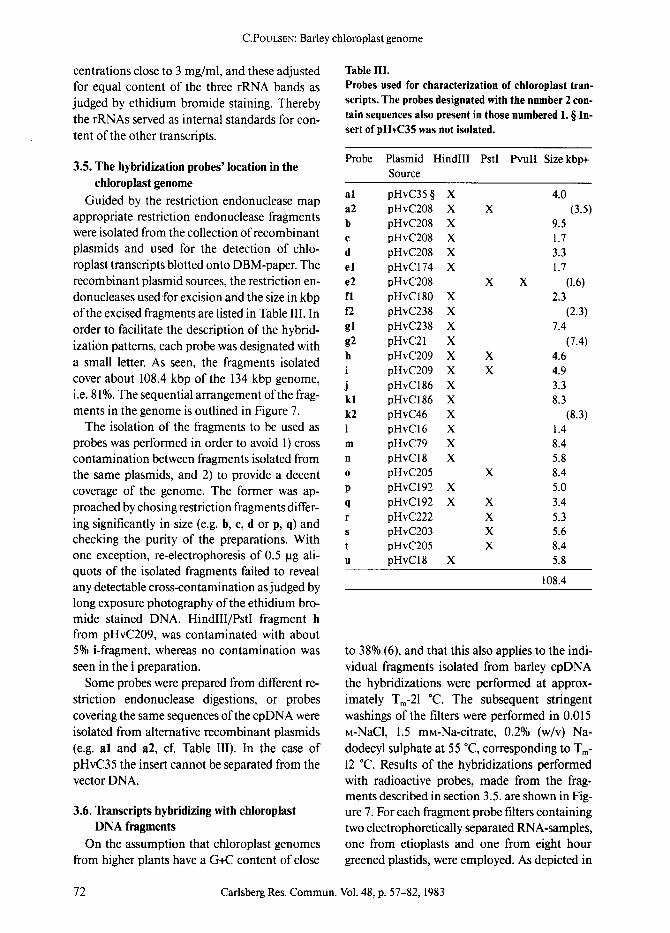
i cover about 108.4 kbp of the 134 kbp genome, j i.e. 81%. The sequential arrangement of the frag- kl ments in the genome is outlined in Figure 7. k2
The isolation of the fragments to be used as I probes was performed in order to avoid 1) cross m contamination between fragments isolated from n the same plasmids, and 2) to provide a decent o coverage of the genome. The former was ap- P proached by chosing restriction fragments differ- q ing significantly in size (e.g. h, c, d or p, q) and r
S checking the purity of the preparations. With t one exception, re-electrophoresis of 0.5 p.g ali- u quots of the isolated fragments failed to reveal any detectable cross-contamination as judged by long exposure photography of the ethidium bro- mide stained DNA. HindlII/PstI fragment h from pHvC209, was contaminated with about 5% i-fragment, whereas no contamination was seen in the i preparation.
Some probes were prepared from different re- striction endonuclease digestions, or probes covering the same sequences of the cpDNA were isolated from alternative recombinant plasmids (e.g. al and a2, cf. Table III). In the case of pHvC35 the insert cannot be separated from the vector DNA.
3.6. Transcripts hybridizing with chloroplast DNA fragments
On the assumption that chloroplast genomes from higher plants have a G+C content of close
Table III. Probes used for characterization of chloroplast tran- scripts. The probes designated with the number 2 con- tain sequences also present in those numbered I. w In- sert of pHvC35 was not isolated.
Probe Plasmid HindlII PstI Pvull Size kbp+ Source
pHvC35 w X 4.0 pHvC208 X X (3.5) pHvC208 X 9.5 pHvC208 X 1.7 pHvC208 X 3.3 pHvC174 X 1.7 pHvC208 X X (1.6) pHvC180 X 2.3 pHvC238 X (2.3) pHvC238 X 7.4 pHvC21 X (7.4) pHvC209 X X 4.6 pHvC209 X X 4.9 pHvC186 X 3.3 pHvC186 X 8.3 pHvC46 X (8.3) pHvC16 X 1.4 pHvC79 X 8.4 pHvCl8 X 5.8 pHvC205 X 8.4 pHvC192 X 5.0 pHvC192 X X 3.4 pHvC222 X 5.3 pHvC203 X 5.6 pHvC205 X 8.4 pHvC18 X 5.8
108.4
to 38% (6), and that this also applies to the indi- vidual fragments isolated from barley cpDNA the hybridizations were performed at approx- imately Tm-21 *C. The subsequent stringent washings of the filters were performed in 0.015 M-NaC1, 1.5 mM-Na-citrate, 0.2% (w/v) Na- dodecyl sulphate at 55 *C, corresponding to T m- 12 ~ Results of the hybridizations performed with radioactive probes, made from the frag- ments described in section 3.5. are shown in Fig- ure 7. For each fragment probe filters containing two electrophoretically separated RNA-samples, one from etioplasts and one from eight hour greened plastids, were employed. As depicted in
72 Carlsberg Res. Commun. Vol. 48, p. 57-82, 1983

C.PouLSEN: Barley chloroplast genome
130 0 _
HindU
a2
Hindl l l Pvu II Sai l Pst I
III
t30
35
907
85
Pvu II Hindll l ,dill Hindlll ~ , \ ~ Y / . a l ~ . L "45
80 fl ....... <~" " " g 55
6O 70 65
Figure 7. The position of the restriction endonuclease fragments a to u isolated in order to identify transcripts of the chloroplast genome.
Figure 8, the data suggest that hybridization con- ditions may have been too stringent in some cases (b, c, d) and maybe not overly stringent in others (a, e, f, k etc.). This might be indicative of low and high G+C contents, respectively. Since coding sequences (protein-encoding) are nor- mally more G - ~ rich than non-coding se- quences, there does seem to be some correlation between hybridization conditions and the hybridization responses with the different pro- bes: The diversity of the responses indicate that certain regions of the genome are more heavily transcribed than others, or that high contents of G+C and possibly partial sequence homology be-
tween certain transcripts may give rise to false hybridization responses. Assuming the former possibility and keeping the second in mind, I will present a systematic description of the data given in Figure 8 and interpreted in Figure 10.
3.6.1. Large single copy region The first hybridization strip (a2) shows that
the 3.5 kbp HindlII/PstI fragment hybridizes to at least five major transcripts. Two of these are larger than 3.9 kb and are larger than the frag- ment itself. They must therefore extend into neighboring fragments. The third transcript is about 2.9 kb and is the second strongest
Carlsberg Res. Commun. Vol. 48, p. 57-82, 1983 73

C. POULSEN: Barley chloroplast genome
hybridizing band. The strongest hybridizing band is found at 0.86 kb. For this transcript stronger hybridization is observed for RNA from eight hour greened plastids than with etioplast RNA. This suggests either that the RNA is syn- thesized preferentially in the light or that there are two species of RNA of this size, one which is synthesized in the dark and in the light and one which is only synthezised in the light. A fifth band is found at 0.60 kb. For comparison Figure 9 shows the hybridization of the slightly larger probe al with the RNA preparations from eight different plastid developmental stages. This 4.0 kbp HindIll fragment shows hybridization to at least two additional bands. The two additional bands correspond to a transcript of 1.8 kb and one of only 350 nucleotides, which seems most intense during the early to middle stages of greening. Figure 10 also shows that the 0.86 kb band hybridizes with increasing intensity as greening proceeds. There seems to be a slightly enhanced synthesis of the 0.60 kb transcript dur- ing greening. Parts of the a-region of the chlo- roplast genome thus are subject to light regulated transcription. Two additional points can be made here. Firstly, the similarity of the al- and a2-hybridization responses, suggest that there are no major contributions to a2 by contaminants of other probes isolated from the same large plas- mid (pHvC208). Secondly, the a probes contain many G+C rich restriction endonuclease sites. To what extent the relatively large number of hybridizing bands are due to incomplete de- naturation cannot be decided, but it can be pointed out that the experimental procedures followed here have not deviated significantly from those described elsewhere (28, 31).
The same transcript complexity is not found in the b , c- and d-region. It appears that the cod- ing for two a-transcripts extend into the DNA sequence of the ~region, one is the largest and the other is a 0.86 kb transcript. These observa- tions support the notion that the transcription complexity from the a-fragment is real. A third presumably overlapping transcript of 2.95 kb is found for the ~ and the c-regions. It seems more strongly synthesized in greening seedlings than in dark grown seedlings. No major transcripts hybridize with the d-probe. Repeated hybridiza- tion and washing at lower stringency did not re-
veal any new RNA species, suggesting that this region is quite inactive in transcription.
The e-, f- and g-probes also illustrate a region complex in transcription. Here the hybridiza- tions gave highly reproducible patterns, when the different e-, f- and g-fragments were used as probes (cf. Table III). Several transcripts hybrid- ize to two or all three fragments, but with dif- ferent intensities. Thus the 1.6 kb e2-probe hybridizes strongly with two to three transcripts, one of 3.2 kb and one or two of 2.6 kb. There are hints of these transcripts in the fl-hybridization pattern. The f-probes show a strong transcript band of 3.5 kb, which seems to extend into the e- fragments and also with low intensity into the g- fragment. Breakdown products of this transcript blur the slightly faster migrating transcripts de- scribed above for the e-region. The Pstl site sepa- rating e and f has been identified in wheat to be inside the atpH gene (21).
Figure 8 lane g, and Figure 9 both identify a very large (above 4 kb) transcript and a low abundance light induced transcript migrating
rrf rrf
IR IR
~ . , ~ ..3 4 b ~ , , , ' ~ rbcL
atpE
Figure l 1. A map of the barley chloroplast genome. The genome is oriented by the inverted repeats ([R)
and the PstI sites. The presumptive positions of the genes rrs, rrl, rrf, atpB, atpE and rbcL are shown with the black bars on the outside of the circle. The putative position of the photogene32 (psbA) and the regions coding for light induced transcripts are shown with open bars on the inside. The numbers associated with these regions refer to the transcripts summarized in Table IV.
74 Carlsberg Res. Commun. Vol. 48, p. 57-80, 1983

C. POULSEN: Badey chloroplast genome
slightly slower than the 3.5 kb transcript. This figure also illustrates the hybridization artefacts frequently observed in the three rRNA bands, which appear light and bordered by unspecific accumulations of hybridizing molecules.
The h- and i-probes also hybridize with several transcript bands. With the h-probe four major transcripts of 2.8 kb, 2.6 kb, 1.8 kb and 0.94 kb and a minor one of 0.70 kb are found. In addi- tion three light induced transcripts of 1.0, 0.86 and 0.74 kb are present. The i-probe hybridizes with transcripts of 2.8 kb, 2.6 kb, 2.1 kb, 1.7 kb and 0.75 kb. Here, the 2.1 kb and the 0.75 kb transcripts appear light induced. As discussed in section 3.5, the i-fragment contaminated the h- fragment. This is reflected by the finding of the 2.8 and 2.6 kb transcripts with both probes, al- though the contaminated h-probe hybridizes much less with the two transcripts. On the other hand the light induced transcripts of 0.74-0.75 kb marked by both probes but weaker with the i- probe cannot be explained by contamination. They are either two different transcripts of simi- lar size, or a single transcript overlapping both fragments. The 1.8 kb and the 1.7 kb transcripts are clearly distinguished in terms of mobility as well as hybridization response.
The i-fragment is a 4.9 kbp subfragment of the 6.0 kbp HindlII fragment, which contains se- quences homologous to pZmC3711, the maize cpDNA clone covering the rbcL gene and parts of the atpB/atpE-cistron (27, 32). According to the analysis reported in section 3.2. the barley 1.7 kb rbcL transcript starts a few hundred nu- cleotides from the PstI end of the i-fragment. Therefore, the most abundant mRNA in the plastids appears only with a moderate hybridiza- tion intensity. The transcript extends into the j- fragment. The dicistronic atpB/atpE-transcript found in this region of the maize cpDNA is 2.2 kb. The i-fragment hybridizes with a transcript of 2.1 kb, which is light induced and therefore cannot be the barley atpB/atpE mRNA. Thus remain the 2.6 kb and 2.8 kb RNAs as candidates for encoding of the ATP synthetase 13- and e- subunits. This situation is reminiscent of that in spinach chloroplasts, where a 1.98 kbp fragment encoding most of the 13- and e-subunits, hybrid- izes to more than one, namely three transcripts of 2.4, 2.6 and 2.8 kb, respectively. In pea (53)
and wheat (22), like in maize, only a single di- cistronic transcript for the two polypeptides is found. The exact nature of the multiple tran- scripts in the different species will have to be clarified. Barley is sofar unique by the finding of the 2.1 kb light induced transcript in this region. This transcript also reaches into the j-fragment. It appears to be transcribed from the same piece of double stranded DNA as the rbcL transcript and to extend into the spacer region between the rbcL and the atpB/atpE genes.
The j-fragment hybridizes to two-three tran- scripts of about 3.9-4.0 kb, which continue into the 8.3 kbp k-fragment. The k-fragment hybrid- izes to five other major transcripts and to a few minor ones. A minor band around 0.78 kb ap- pears light induced. The small (1.4 kbp) I-frag- ment, which neighbors the k-fragment on one side and a not yet cloned section of the barley cpDNA on the other, hybridizes strongly with a 2.2 kb transcript which has to transgress into the un-characterized region.
The 8.4 kbp m-fragment also seems to hybrid- ize to a remarkable large number of transcripts. Some of the weaker ones can be removed by washing at higher temperature. The two major ones are seen here as a 1.6 kb RNA and a 0.76 kb RNA.
3.6.2. The inverted repeat and the small single copy region
Two thirds of the inverted repeat DNA se- quences are covered by two sets of fragments (n,o and t,u). Results of hybridizations with these four probes are shown in the bottom half of Fig- ure 8 and in Figure 9. The 5.8 kbp probes n or u, which are positioned towards the large single copy region, hybridize to a large number of low abundance transcripts in the high molecular size region. Some of these appear light induced. However, prolonged autoradiography shows that most transcripts are also present among etioplast RNA molecules. A single major transcript ofl.6 kb is possibly shared with m, the adjacent frag- ment which contains a portion of the inverted repeat at its end. Fragment n hybridizes to traces of a very large light induced transcript (not visi- ble in Figure 8) that appears to cover a large sec- tion of the inverted repeat. This transcript is clearly seen in the o- and t-hybridizations, which
Cadsberg Res. Commun. Vol. 48, p. 57-80, 1983 75

C. POULSEN: Barley chloroplast genome
also exhibit the powerful responses of the 16S rRNA and the large fragment of the 23S rRNA. In addition to these major transcripts, a set of three transcripts is seen in the 3.1 to 3.5 kb re- gion. Some weakly responding bands (above 3.9 kb) are more pronounced in the light than in the dark and may correspond to transcript bands of the a and n hybridizations.
It thus appears that the population of tran- scripts originating from the inverted repeat is very complex. The possibility for the involve- ment of a processing system creating a large number of semi-stable intermediates or precur- sors from larger primary transcripts cannot be excluded_ A closer analysis of the inverted repeat with smaller probes, Sl-nuclease protection ex- periments and in vitro transcription studies, will improve the understanding of these data.
The small single copy region is covered by the four probes p, q, r and s. There is a gap of 2.5 kbp between the n-fragment and the p-fragment, whereas the s-fragment goes into the inverted re- peat and adjoins the t-fragment. Hybridization results are shown in Figure 8, between the two sets of inverted repeat hybridizations. The s- probe hybridizes to inverted repeat transcripts only. These are the three 3.1-3.5 kb transcripts seen with the o- and t-probes and the small frag- ment of the 23S rRNA. This suggests that the Pstl site separating the t- and the s-fragments is very close to the 23S rRNA fragmentation point. Furthermore, since the s-fragment subclone pHvC68 gives a hybridization response quite similar to the s-fragment itself, it is possible that the three 3.1-3.5 kb transcripts are different 23S rRNA precursors, which may also include a 4.5S rR_NA (6).
Hybridization with. the p-probe was unfertu- nate, bul it does show a number of weak bands in the 3.0 to 4,0 kb size class, a minor transcript of 1.3 kb and a fairly abundant light induced tran- script of 0.90 kb, which may reach into the q- fragment. The q-probe reveals two transcripts of 3.0 kb atad 4.0 kb which may also extend back into the p-fragment. Additionally, three tran- scripts of0~90 kb, 0.?5 kb and 0.68 kb show up. The latter is quite abundant. There might also be two transcripts in the 2.0-2.4 kb size class. The background in the 8 hour track of this hybridiza- tion as well as in the p-hybridization, suggest the
presence of sequences in both fragments which may correspond to an easily degraded or a rapid- turnover transcript of high molecular weight. Since it causes background mainly in the 8 hour track, its synthesis could be light induced.
The r-probe hybridizes with three transcripts of 2.5, 1.2 and 0.84 kb. The 1.2 kb transcript hybridizes quite strongly, suggesting that it is fairly abundant.
3.6.3. Light regulated RNA synthesis Several investigators have reported on the syn-
thesis in chloroplasts of a 32,000 dalton mem- brane polypeptide during development of plas- tids (3, 14, 37, 44, 55). This protein is syr~4;hesized as a larger precursor of species dependent size and is processed to its natural size during incor- poration into the photosynthetic membranes probably as a component of photosystem II. In vitro translation of mRNA yields a polypeptide which shares features with a protein component isolated from photosynthetic membranes or photosystem II particles (3, 44). It also appears to be lhe same protein which will bind photosystem II uncouplers such as DCMU and triazine her- bicides (44). Furthermore it was found that the control for synthesis of the 32,000 dalton protein is on the transcriptional level. G.DNK (29) dem- onstrated that synthesis of the mRNA is under phytochrome control and that the mRNA is ab- sent in dark grown mustard seedlings.
These studies have also resulted in the localiza- tion of the corresponding gene on several gen- omes. According to the new terminology, this gene is now termed psbA (6). In the maize cpDNA, this gene was previously termed pho- togene32 and is associated with a BamHI frag- ment, l~amS~ which is crossing tl~e border be- tween one of the inverted repeat sequences and the large single copy region (3) in the "9 o'clock" position of the map shown in Figure 1. The pre- sence of an equivalent 5 kbp BamHI fragment in the barley cpDNA (cf. Figure 4), the failure in finding sequences corresponding to this frag- ment in the barley cpDNA clones investigated and the absence of an abundant light induced transcript of the correct size (1,2 kb) in the tran- script studies reported here, suggest that the loca- tion of this gene in the barley genome is equiv- alent to that in the maize genome. I have been
76 Carlsberg Res. Commun. Vol. 48, p. 57-80, 1983

C. POULSEN: Barley chloroplast genome
Table IV. The light induced transcripts of the barley chloroplast DNA.
Hybridization Number Fragment Size, kb Response
1 al/a2 0.86 medium to strong 2 b,c 2.95 weak 3 g 3.5 very weak 4 h 1.0 medium 5 h 0.86 weak 6 h 0.74 medium 7 i 0.75 weak 8 i (j?) 2.1 medium 9 k 0.78 very weak 10 n,o+t,u >6.0 strong 11 p,q 0.90 medium
able to identify 11 other photogenes, that is genes only transcribed after illumination. These are lo- cated at various positions throughout the barley cpDNA as shown in Figures 10 and 11. The char- acteristics of these 11 light induced transcripts are listed in Table IV.
It will be interesting to see, if all these pho- togenes are transcribed under the control of the phytochrome light receptor system. Nucleotide sequence analysis of the regions flanking the genes might reveal common features of promo- tor and terminator sequences characteristic for light controlled gene expression.
In these experiments no transcripts have been found which are synthesized in the dark, but not in the light. However, it may be noted that these experiments cover only 81% of the genome and that a single etioplast RNA preparation has been used. Further studies are required to definitively exclude the existence of light induced repression of cpDNA genes.
4. CONCLUDING REMARKS
Conservative analysis of Figure 8 yields 70 transcript bands for the 81% of the barley cpDNA analysed and these have been drawn in Figure 10. Approximately an additional 20 bands have been seen in the hybridizations (36) but need further confirmation. About 25 to 30 of the 70 transcripts are larger than 3,000 nu- cleotides, the coding capacity for a 100,000 mo- lecular weight protein. In analogy with the di-
cistronic mRNA for the 13 and e subunits of the ATP synthetase CF~ the large transcripts could represent polycystronic mRNAs, whereas the majority of the transcripts below 3,000 nu- cleotides most likely encode a single polypeptide.
Overall comparison of the Northern blots make it clear that the different regions of the chloroplast genome are characterized by unique patterns of transcripts. There are some regions giving rise to few transcript bands, but only frag- ment d was devoid of significant hybridization responses. There are several (scattered) cpDNA regions hybridizing to a surprisingly large num- ber of transcripts.
The following possibilities are to be consid- ered in the further elucidation of the multitude of transcripts and the large size of some of them: l) A given region of the genome can be tran- scribed from several promotors on the same or on opposite strands resulting in overlapping and/ or divergent transcripts. 2) The large transcripts of a given region could be stable precursors or intermediates for the smaller ones.
Several observations in maize, spinach and Euglena gracilis have documented that both pos- sibilities are realized in chloroplast genomes. Di- vergent transcripts have been identified for the genome region in maize and spinach containing the atpB/atpE and rbcL genes. The transcription initiation points for a tRNA H~s and an unknown 1.6 kb transcript are found within a few basepairs on opposite strands of the DNA resulting in slightly overlapping transcripts (40). As already mentioned one strand of the region encoding the 13 and ~ subunits of CF~ in spinach is transcribed into three RNA molecules with sizes of 2.8, 2.6 and 2.4 kb, respectively (53). It is not yet known whether the three transcripts originate from sep- arate initiation points or whether post-transcrip- tional processing at the termini takes place. It will be of further interest to see if all three tran- scripts can be translated into the protein prod- ucts. In contrast the atpB/atpE genes of maize (27) are transcribed into a single 2.2 kb mRNA molecule originating at an initiation point 300 bp upstream from the ATG methionine triplet and with promotor consensus sequences at -10 and-35 nucleotides upstream from the initiation point.
Precursor RNA molecules and splicing inter-
Carlsberg Res. Commun. Vol. 48, p. 57-80, 1983 77

C. POULSEN: Barley chloroplast genome
mediates are examplified by the maize (45) and Euglena (18) tRNA genes containing introns. The rbcL gene for the large subunit of ribulose bisphosphate carboxylase in Euglena (46) con- tains introns, while this gene for maize, spinach and tobacco is devoid of intervening sequences. A refined analysis of some of the heavily tran- scribed regions of barley cpDNA will reveal whether the considered possibilities suffice to ex- plain the large number of transcripts and their size distributions.
Tightly clustered transcription of genomes is characteristic for viruses, e.g., cauliflower mosaic virus (20), for the 2 ~tDNA plasmid of yeast (10) and has to be postulated for mam- malian mitochondrial genomes (3). Also pro- karyotic genomes, with which chloroplast gen- omes are often compared, encode overlapping and/or divergent transcripts (39). The large number of transcripts of unknown function ob- served in the mentioned cases as well as in plas- rids tell that many proteins with as yet un- discovered functions are involved in cellular me- tabolism. In vitro translation of total plastid RNA from spinach results in a large number of protein products (8) supporting the notion gain- ed in this work that cpDNA encodes a large number of different proteins, possibly in excess of 100. This is also in agreement with the obser- vations that E. coli RNA polymerase can bind to most restriction endonuclease fragments tested from spinach cpDNA and that most fragments can hybridize with radioactively labelled plastid RNA (52). Nucleotide sequencing ofcpDNA re- gions which by Northern hybridization have given evidence for tightly clustered transcription will provide successively better estimates for the number of proteins encoded by the chloroplast genome.
ACKNOWLEDGEMENTS
The expertise and help provided by dr. S. HOLMBERG, dr. A. BRANDT from the Carlsberg Laboratory and professor K. MARCKER from the Institute of Molecular Biology, University of Aarhus is gratefully acknowledged. Technical as- sistance and assistance in the preparation of this manuscript was kindly provided by Ms. INGA OLSEN, Ms. G. BANK, Ms. A.-S. STEINHOLZ and Ms. N. RASMUSSEN. Professor D.VON WETTSTEIN
is thanked for help in putting this paper together. Financial support for this work has in part been provided by the Commission of the European Communities, contract No. ESD-013-DK of the Solar Energy Programme.
REFERENCES 1. ALWINE, J.C., D.J. KEMP, B.A. PARKER, J. REI-
SER, J. RENART~ G.R. STARK • G.M. WAHL: De- tection of specific RNAs or specific fragments of DNA by fractionation in gels and transfer to diazobenzyloxymethyl paper. In: Methods in Enzymology 68, R. Wu, ed., pp 220-242 (1979) Academic Press New York and London.
2. BEDBROOK, J.R. & L. BOC-ORAD: Endonuclease recognition sites mapped on Zea mays chlo- roplast DNA. Proc. Natl. Acad. Sci. USA 73, 4309-4313 (1976)
3. BEDBROOK, J., G. LINK, D.M. COEN, L. BOGORAD & A. RICH: Maize plastid gene ex- pressed during photoregulated development. Proc. Natl. Acad. Sci. USA 75, 3060-3064 (1978)
4. BIBB, M.J., R.A. VAN ETTEN, C.T. WRIGHT, M.A. WALBERG & D.J. CLAYTON: Sequence and gene organization of mouse mitochondrial DNA. Cell 26, 167-180 (1982)
5. BIRNBOIM, H.C. & J. DOLY: A rapid alkaline extraction procedure for screening recombi- nant plasmid DNA. Nucl. Acids Research 7, 1513-1520 (1979)
6. BOHNERT, H.J., E.J. CROUSE & J.M. SCHMITT: Organization and expression of plastid gen- omes. In: Encycl. Plant Physiol. vol 14B, pp. 475-530, B. Parthier and D. Boulter eds., (1982) Springer Verlag, Berlin, Heidelberg
7. BOLIVAR, F.: Construction and characteriza- tion of new cloning vehicles.III. Derivatives of plasmid pBR322 carrying unique EcoRI sites for selection of EcoRI generated recombinant DNA molecules. Gene 4, 121-136 (1978)
8. BOTTOMLEY, W. & P.R. WHITFELD: Cell-free transcription and translation of total spinach chloroplast DNA. Eur. J. Biochem. 93, 31-39 (1979)
9. BOWMAN, C.M., B. KOLLER, H. DELIUS ~r T.A. DYER: A physical map of wheat chloroplast DNA showing the location of the structural genes for the ribosomal RNAs and the large subunit of ribulose 1,5-bisphosphate carbox- ylase. Mol. gen. Genet. 183, 93-102 (1981)
10. BROACH, J.R., J.E ATKINS, C. McGILL & L. CHOW: Identification and mapping of the tran-
78 Carlsberg Res. Commun. Vol. 48, p. 57-80, 1983

C. POULSEN: Barley chloroplast genome
scriptional products of the yeast plasmid, 2 rt circle. Cell 16, 827-839 (1979)
11. CHU, N.M. & K.K. TEWARI: Arrangement of the ribosomal RNA genes in chloroplast DNA of Leguminosae. Mol. gen. Genet. 186, 23-32 (1982)
12. CHu, N.M., K.K. OISHI & K.K. TEWARI: Physi- cal mapping of the pea chloroplast DNA and localization of the ribosomal RNA genes. Has- mid 6, 279-292 (1981)
13. COHEN, S.N., A.C.Y. CHANG & L. Hsu: Non- chromosomal antibiotic resistance in bacteria: Genetic transformation of Escherichia coli by R-factor DNA. Proc. Natl. Acad. Sci. USA 69, 2110-2114 (1972)
14. DRIESEL, A.J., J. SPEIRS & H.J. BOHNERT: Spin- ach chloroplast mRNA for a 32,000 dalton polypeptide. Size and localization on the phys- ical map of the chloroplast DNA. Biochim. Bi- ophys. Acta. 610, 297-310 (1980)
15. DRIESEL, A.J., E.J. CROUSE, K. GORDON, H.J. BOHNERT, R.G. HERRMANN, A. STEINMETZ, M. MUBUMmLA, M. KELLER, G. BURKARD & J.H. WEIL: Fractionation and identification of spin- ach chloroplast transfer RNAs and mapping of their genes on the restriction map of chlo- roplast DNA. Gene 6, 285-306 (1979)
16. DENHARDT, D.T.: A membrane-filter technique for the detection of complementary DNA. Bit- chem. Biophys. Res. Commun. 23, 641-646 (1966)
17. GORDON, K.H.J., E.J. CROUSE, H.J. BOHNERT & R.G. HERRMANN: Restriction endonuclease cleavage site map of chloroplast DNA from Oenothera parviflora (Euoenothera Plastome IV). Theor. Appl. Genet. 59, 281-296 (1981)
18. GRUISSEM, W., D.M. PRESCOTT, B.M. GREEN- BERG 8~ R.B. HALLICK; Transcription ofE. coli and Euglena chloroplast tRNA gene clusters and processing of polycistronic transcripts in a Hela cell-free system. Cell 30, 81-92 (1982)
19. Guidelines for research involving DNA mole- cules Federal Register U.S. 47 (77) 17180-17198 (1982)
20. GUILLEY, H., R.K. DUDLEY, G. JONARD, E. BAL- AZS & K.E. RICHARDS: Transcription of cau- liflower mosaic virus DNA: Detection of pro- motor sequences and characterization of tran- scripts. Cell 30:763-773 (1982)
21. HOWE, C.J., A.D. AUFFRET, A. DOHERTY, C.M. BOWMAN, T.A. DYER & J.C. GRAY: Location and nucleotide sequence of the gene for proton translocating subunit of wheat chloroplast ATP synthase. Proc. Natl. Acad. Sci. USA 79, 6903- 6908 (1982)
22. HowE, C.J., C.M, BOWMAN, TA. DYER • J.C. GRAY: Localization of wheat chloroplast genes for the beta and epsilon subunits of ATP syn- thane. Mol. gen. Genet. 186, 525-530 (1982)
23. KANNANGARA, C.G., S.P. GOUGH, B. HANSEN, J.N. RASMUSSEN & D.J. SIMPSON; A homoge- nizer with replaceable razor blades for bulk iso- lation of active barley plastids. Carlsberg Res. Commun. 42, 431-440 (1977)
24. KOLLER, B., H. DELIUS & T.A. DYER; The orga- nization of the chloroplast DNA in wheat and maize in the region containing the LS gene. Eur. J. Biochem. 122, 17-23 (1982)
25. KOLODNER, R. & K.K. TEWARI: The molecular size and conformation of the chloroplast DNA from higher plants. Biochim. Biophys. Acta 402, 372-390 (1975)
26. KOLODNER, R. & K.K. TEWARI: Inverted re- peats in chloroplast DNA from higher plants. Proc. Natl. Acad. Sci. USA 76, 41-45 (1978)
27. KREBBERS, E.T., I.M. LARRINUA, L. MCINTOSH & L.BoGORAD: The maize chloroplast genes for the 13 and e subunits of the photosynthetic coup- ling factor CF~ are fused. Nucl. Acids Res. 10, 4085-5002 (1982)
28. LEHRACH, H., D. DIAMOND, J.M. WOZNEY & H. BOEDTKER: RNA molecular weight determina- tions by gel electrophoresis under denaturing conditions, a critical reexamination. Bio- chemistry 16, 4743-4752 (1977)
29. LINK, G.: Phytochrome control of plastid mRNA in mustard (Sinapis alba L.). Planta 154, 81-86 (1982)
30. MALNOE, P., J.-D. ROCHAIX, N.H. CHUg & P.-E SPAHR: Characterization of the gene and mes- senger RNA of the large subunit ofribulose 1,5- bisphosphate carboxylase in Chlamydomonas reinhardii. J. Mol. Biol. 133, 417-434 (1979)
31. MANIATIS, T., E.E FRITSCH & J. SAMBROOK: Electrophoresis of RNA through gels con- taining formaldehyde. In: Molecular cloning. A laboratory manual, pp 202-203, Cold Spring Harbor Laboratory 1982.
32. MCINTOSH, L., C. POULSEN & L. BOGORAD: Chloroplast gene sequence for the large subunit of ribulose bisphosphate carboxylase of maize. Nature 288, 556-560 (1980)
33. OISnl, K., T. SUMNICHT 8s K.K. TEWARK Mes- senger ribonucleic acid transcript of pea chlo- roplast deoxyribonucleic acid. Biochemistry 20, 5710-5717 (1981)
34. PALMER, J.D. & W.E THOMPSON: Clone banks of the mung bean, pea and spinach chloroplast genomes. Gene 15, 21-26 (1981)
35. PALMER, J.D. & W.E THOMPSON: Chloroplast
Carlsberg Res. Commun. Vol. 48, p. 57-80, 1983 79

C. POULSEN" Barley chloroplast genome
DNA rearrangements are more frequent when a large inverted repeat sequence is lost. Cell 29, 537-550 (1982)
36. POOLSEN, C.: The in vivo transcriptional ac- tivity of the barley chloroplast genome. In: Structure and Function of Plant Genomes. O. Ciferri and L. Dure III, eds., Plenum Press, New York (1982), in press
37. REISFELD, A., A.K. MATTOO 8r M. EDELMAN: Processing of a chloroplast-translated mem- brane protein in vivo. Analysis of the rapidly synthesized 32000-dalton shield protein and its precursor in Spirodela oligorrhiza. Eur. J. Biochem. 124, 125-129 (1982)
38. RIGBY, EW.J., M. DIECKMANN, C. RHODES & P. BERG: Labeling deoxyribonucleic acid to high specific activity in vitro by nick translation with DNA polymerase I. J. Mol. Biol. 133, 237- 251 (1977)
39. SCHWARZ, E., G. SCHERER, G. HOBOM & H. KOSSEL: Nucleotide sequence of cro, CII and part of the O gene in phage ~. DNA. Nature 272, 410-414 (1978)
40. SCHWARZ, Z., S.O. JOLLY, A. STEINMETZ L.BoGORAD: Overlapping divergent genes in the maize chloroplast chromosome and in vitro transcription of the gene for tRNA his. Proc. Natl. Acad. Sci. USA 78, 3423-3427 (1981)
41. SHINOZArJ, K. & M. SUGtURA: Sequence of the intercistronic region between the ribulose-l,5- bisphosphate carboxylase/oxygenase large subunit and the coupling factor ~ subunit gene. Nucl. Acids Res. 10, 4923-4934 (1982)
42. SmNOZArO, K. & M. SUG~URA: The nucleotide sequence of the tobacco chloroplast gene for the large subunit of ribulose bisphosphate car- boxylase-oxygenase. Gene 20, 91-102 (1982)
43. SOUTHERN, E.M.: Detection of specific se- quences among DNA fragments separated by gel electrophoresis. J. Mol. Biol. 98, 503-517 (1975)
44. STEINBACK, K.E., L. MCINToSH, L. BOGORAD & C, ARNTZEN: Identification of the triazine re- ceptor protein as a chloroplast gene product. Proc. Natl. Acad. Sci. USA 78, 7463-7467 (1981)
45. STEINMETZ, A., E.J. GUBBiNS & L. BOGORAD: The anticodon of the maize chloroplast gene for tRNA L~u is split by a large intron. Nucl. Acids Res. 10, 3027-3037 (1982)
46. STIEGLER, G.L., H.M. MATTHEWS, S.E. BING- HAM • R.B. HALLICK: The gene for the large subunit of ribulose-l,5-bisphosphate carbox- ylase in Euglena gracilis chloroplast DNA: lo- cation, cloning and evidence for an intervening sequence. Nucl. Acids Res. 10, 3427-3444 (1982)
47. TAKAIWA, E & M. SUGIURA: Nucleotide se- quence of the 16S - 23S spacer region in an rRNA gene cluster from tobacco chloroplast DNA. Nucl. Acids Res. 10, 2665-2676 (1982)
48. TAKAIWA, E t~ M. SUGIURA" The complete nu- cleotide sequence of a 23-S rRNA gene from tobacco chloroplasts. Eur. J. Biochem. 124, 13- 19 (1982)
49. WAHL, G.M., M. STERN & G.R. STARK: Effi- cient transfer of large DNA fragments to di- azobenzyloxymethyl-paper and rapid hybrid- ization by using dextran sulfate. Proc. Natl. Acad. Sci. USA 76, 3683-3687 (1979)
50. WESTHOFF, P., N. NELSON, H. BUNEMANN & R.G. HERRMANN: Localization of genes for coupling factor subunits on the spinach plastid chromosome. Current Genetics 4, 109-120 (1981)
51. WIENAND, U., Z. SCHWARZ t~ G. FElX: Elec- trophoretic elution of nucleic acids from gels adapted for subsequent biological tests. FEBS Letters 98, 319-323 (1979)
52. ZECH, M., M.R. HARTLEY & H.J. BOHNERT: Binding sites of E. coli DNA-dependent RNA polymerase on spinach chloroplast DNA. Cur- rent Genetics 4, 37-46 (1981)
53. ZURAWSKI, G., W. BOTTOMLEY (~ P.R. WmT- EELD: Structures of the genes for the 13 and e subunits of spinach chloroplast ATPase indi- cate a dicistronic mRNA and an overlapping translation stop/start signal. Proc. Natl. Acad. Sci. USA 79, 6260-6264 (1982)
54. ZURAWSKI, G., B. PERROT, W. BOTTOMLEY & P.R. WmTFELD: The structure of the gene for the large subunit of ribulose 1,5-bisphosphate carboxylase from spinach chloroplast DNA. Nucl. Acids Res. 9, 3251-3270 (1981)
55. ZURAWSrd, G., H.J. BOHNERT, P.R. WHITFELD & W. BOTTOMLEY: Nucleotide sequence of the gene for the 32,000-M R thylakoid membrane protein from Spinacia oleracea and Nicotiana debneyi predicts a totally conserved primary translation product of M R 38,800. Proc. Natl. Acad. Sci. USA 79, 7699-7703 (1982)
80 Carlsberg Res. Commun. Vol. 48, p. 57-80, 1983

co i II I II L ~**: ~ : ;
CM
9 K ~ I I I I I LII
~ ~ � 8 4 _
coil f
I : ~ l l l I : :
- r
~I II IIII |~
eel III il �9 mm
,,, n L | I
I I n l I I I
inn
eu nu INI �9 �9
. ~ ~1 I I I I I I I
III I I I I g
,n , , , m, , I o
o| ~u
m~ m rlo oI
,, , ; , ii ,ll
~n
I ~] l
I I | i I I ~ I I
I
~ z .]
,.g
. ~
<
g
U e
-~ ~
. . . . . . 8 8 o o ,-, o o ~
o . ~ �9 o
o o
co co - -~ c~
o . . . . : ~ _ I I IlL o - ~
- ~ ~ 1 !
0
r -
E