Chronic poverty: scrutinizing estimates, patterns, correlates ...
Upload
mpipsykl-mpgCategory
view
3download
0
ORIGINAL ARTICLE
The amyloidogenic potential and behavioral correlatesof stressC Catania1,5, I Sotiropoulos1,5, R Silva2, C Onofri1, KC Breen3,4, N Sousa2 and OFX Almeida1
1Max Planck Institute of Psychiatry, Munich, Germany; 2Life and Health Science Research Institute, University of Minho, Braga,Portugal; 3Alzheimer’s Disease Research Centre, University of Dundee, Dundee, UK and 4Parkinson’s Disease Society,London, UK
Observations of elevated basal cortisol levels in Alzheimer’s disease (AD) patients promptedthe hypothesis that stress and glucocorticoids (GC) may contribute to the development and/ormaintenance of AD. Consistent with that hypothesis, we show that stress and GC provokemisprocessing of amyloid precursor peptide in the rat hippocampus and prefrontal cortex,resulting in increased levels of the peptide C-terminal fragment 99 (C99), whose furtherproteolytic cleavage results in the generation of amyloid-b (Ab). We also show that exogenousAb can reproduce the effects of stress and GC on C99 production and that a history of stressstrikingly potentiates the C99-inducing effects of Ab and GC. Previous work has indicated arole for Ab in disruption of synaptic function and cognitive behaviors, and AD patientsreportedly show signs of heightened anxiety. Here, behavioral analysis revealed that likestress and GC, Ab administration causes spatial memory deficits that are exacerbated bystress and GC; additionally, Ab, stress and GC induced a state of hyperanxiety. Given that theintrinsic properties of C99 and Ab include neuroendangerment and behavioral impairment, ourfindings suggest a causal role for stress and GC in the etiopathogenesis of AD, anddemonstrate that stressful life events and GC therapy can have a cumulative impact on thecourse of AD development and progression.Molecular Psychiatry (2009) 14, 95–105; doi:10.1038/sj.mp.4002101; published online 2 October 2007
Keywords: Alzheimer’s disease; amyloid precursor protein; amyloid-b; glucocorticoids; memory;anxiety
Introduction
Amyloid-b (Ab) peptide is generated under normalphysiological conditions, but its overproduction andaccumulation give rise to senile plaques that, togetherwith aggregations of abnormally hyperphosphory-lated t protein, constitute the main neuropathologicalhallmarks of Alzheimer’s disease (AD). However, theamount of deposited Ab correlates poorly with thedegree of cognitive impairment in AD patients,1 andcentral administration of Ab in nongenetically modi-fied rodents results in cognitive impairments within arelatively short period and before amyloid plaquesbecome detectable.2 These observations suggest apivotal role for soluble nonaggregated Ab in the earlystages of the disease; they are supported by datashowing that soluble Ab can acutely disrupt synapticfunction and cognitive and emotional behavior.3–6
Ab peptide is produced through the sequentialproteolytic cleavage of amyloid precursor protein
(APP) by a-, b- and g-secretases; the last secretase isa complex of at least four proteins, with nicastrinmaking up the largest portion. Nicastrin may beviewed as the gatekeeper of the g-secretase complexbecause of its essential role in the recognition ofg-secretase substrates.7 Cleavage of APP by a-secretaseprecludes the generation of Ab, whereas b-secretase(b-site APP-cleaving enzyme; BACE)-mediated clea-vage generates the so-called C-terminal fragment 99(C99); subsequent proteolysis of C99 by g-secretaseresults in the generation of amyloid peptides (forreview, see Bayer et al.8). While the neurotoxicpotency of Ab is well known,9–11 other studies havedemonstrated the neurotoxic and cognition-impairingpotential of C99.12–15 Furthermore, in a vicious cascadethat characterizes AD, Ab interacts with its proteinprecursor (APP), an interaction that is suggested tocontribute to the mechanism of Ab neurotoxicity.16,17
Observations that a large percentage of AD patientsdisplay hypercortisolemia18,19 suggest that glucocorti-coids (GC) and stress may contribute to the develop-ment or maintenance of AD. This view recentlygained support from studies in transgenic mousemodels of AD in which stress or GC exacerbated AD-like neuropathology.20,21 Indications of a link betweenstress/GC and AD may also be inferred from previous
Received 29 March 2007; revised 19 July 2007; accepted 31 July2007; published online 2 October 2007
Correspondence: Dr OFX Almeida, Max Planck Institute ofPsychiatry, Kraepelinstrasse 2-10, Munich D-80804, Germany.E-mail: [email protected] authors contributed equally to this study
Molecular Psychiatry (2009) 14, 95–105& 2009 Nature Publishing Group All rights reserved 1359-4184/09 $32.00
www.nature.com/mp

work in which a causal role for elevated GC levels andstressors in cognitive impairment in humans andanimals was demonstrated,22,23 GC-induced dendriticatrophy and synaptic loss are thought to underlie theGC-associated behavioral disruption.24,25
The present study focused on examining whetherstress or elevated GC levels can drive APP metabolismtoward the amyloidogenic pathway in normal,middle-aged rats, and whether stressful life eventscan influence the initiation/maintenance of AD-likepathology. Our studies, which focused on the hippo-campus and prefrontal cortex (PFC) because they rankamong the first brain areas to show AD-like neuro-pathology,26 disclosed that chronic stress and GCsimilarly promote APP processing along the amylo-idogenic pathway. These results provide a tentativemechanism through which stress and/or GC exerttheir effects on cognitive and emotional behavior. Ofnote is the growing recognition of the importanceof emotional state in the regulation of cognition.27–29
In addition, it is pertinent to mention that stress-related psychiatric disorders (for example, anxietyand major depression) have been identified as a riskfor developing AD.30,31
Materials and methods
AnimalsMale Wistar rats (Charles River, Barcelona, Spain),aged 14 months, were used, in compliance with theEuropean Union Council’s Directive (86/609/EEC)and local regulations on animal welfare. Animalswere housed 4–5 per cage under standard environ-mental conditions (temperature 22 1C relative humi-dity 70%; 12 h light:dark cycle; ad libitum access tofood and drinking solution).
Treatments and their biological efficacyRats were allocated to one of two main treatmentgroups: stressed and unstressed. Stressed animalswere exposed to a chronic, unpredictable stressparadigm;32 for 4 weeks, while their unstressedcounterparts were held undisturbed, under standardlaboratory conditions. The stress paradigm involvedrandom application of one of the following stressors,daily: (1) hypertonic saline (9% NaCl; 1 ml per 100 g)i.p. injection, (2) overcrowding for 1 h, (3) placementin a confined environment (30 min) and (4) placementon a vibrating/rocking platform (1 h). After 5 days,subgroups of stressed or unstressed animals weregiven constant-rate i.c.v. infusions of soluble Ab1–40
(American Peptide Co., Inc., Sunnyvale, CA; 4.2 nmolAb1–40 per 200ml; 0.5 ml h�1) or vehicle (distilledwater) over a period of 14 days; half of each of thesesubgroups additionally received daily s.c. injectionsof a synthetic GC (dexamethasone (DEX; FortecortinMerck, Darmstadt, Germany); 300mg kg�1 body weight(BW) as an oily suspension in sesame oil (Sigma,St Louis, MI, USA)). For the i.c.v. infusions, ratswere equipped with s.c. osmotic minipumps (Alzet2002 minipumps; Alza Corp., Mountain View, CA,
USA) and cannulae (Alzet Brain Infusion Kit) thatwere implanted in the left lateral ventricle using thefollowing stereotaxic coordinates: AP, �1.0 mm; DV,�2.5 mm and ML, þ 1.5 mm (right) with bregma asa reference (Paxinos and Watson33); pump andcannulae implantation was done under pentobarbital(50 mg kg�1) anesthesia. At the time of killing, mini-pumps were removed and checked for patency andfunctionality. Each treatment subgroup comprised6–8 rats.
Efficacy of the stress paradigm was gauged bymeasuring daytime serum corticosterone (CORT)levels (Corticosterone RIA kit, ICN, Costa Mesa, CA,USA), BWs and relative thymus weights. CORT levelswere significantly elevated in stressed vs nonstressedanimals (384.6765.9 and 54.774.8 ng ml�1, respec-tively), and thymus weights at autopsy were consistentwith hypercorticalism in the stressed group (control,CON: 7.670.9 vs stressed: 4.370.4 vs mg kg�1 BW).Whereas nonstressed animals showed a net gain inBW (6.171.3 g), stressed animals showed a significantloss of body mass (�12.172.1 g). GC-exposed ratsshowed biometric changes that were similar to thoseobserved in the stressed group, whereas none of theparameters monitored differed significantly betweenCON and stressed animals after the superimposedtreatment with Ab.
Tissue collectionAnimals were rapidly killed and their brains wereimmediately excised and divided along the midline.One half of each brain was immersed in 4%p-formaldehyde (2 days), embedded in paraffin andsaved for histochemical analysis. The PFC andhippocampus were dissected out of the other half ofthe brain, snap-frozen in liquid nitrogen and stored at�80 1C until subsequent biochemical analyses.
Western blot analysisFrozen hippocampal and PFC tissues were homo-genized in lysis buffer (100 mM Tris-HCl, 250 mM
NaCl, 1 mM EDTA, 5 mM MgCl2, 1% NP-40, CompleteProtease Inhibitor (Roche, Mannheim, Germany)and Phosphatase Inhibitor Cocktails I and II (Sigma,St Louis, MO)) using a Dounce glass homogenizer;extracts were cleared by centrifugation (14 000 g) andtheir protein contents were estimated by the Lowryassay. Lysates, in Laemmli buffer (250 mM Tris-HCl,pH 6.8, containing 4% sodium dodecyl sulfate, 10%glycerol, 2% b-mercaptoethanol and 0.002% bromo-phenol blue), were thereafter electrophoresed on 8or 10% acrylamide gels, and transferred onto nitro-cellulose membranes (Protran BA 85, Schleicher &Schuell, Dassel, Germany). Membranes were blockedin Tris-buffered saline containing 5% nonfat milkpowder and 0.2% Tween-20 before incubation withthe following antibodies: anti-APP369 (1:5000; kindlyprovided by Dr Sam Gandy), anti-BACE-1 (1:500;ProSci Inc., Poway, CA, USA), anti-nicastrin (1:5000;Sigma) and anti-actin (1:2000; Chemicon, Temecula,CA, USA) or anti-a-tubulin (1:2000; Calbiochem,
Stress-induced amyloidogenesisC Catania et al
96
Molecular Psychiatry
San Diego, CA, USA). Antigens were revealedby enhanced chemiluminescence (Amersham Bio-sciences, Freiburg, Germany) after incubation withappropriate horseradish peroxidase–immunoglobulinG conjugates (Amersham Biosciences); blots werescanned and quantified using TINA 3.0 bioimagingsoftware (Raytest, Straubenhardt, Germany) afterascertaining linearity. All values were normalizedand expressed as percentages of control. To distin-guish between mature and immature isoforms of APP,tissue lysates were digested with endoglycosidase F(Roche), according to the manufacturer’s instructions.
ImmunoprecipitationTissue lysates were immunoprecipitated withAPP369 antibody using a mixture of Protein A andG beads (Roche) before analysis by electrophoresis onTris-Tricine gradient gels (KMF Laborchemie, Loh-mar, Germany) and immunoblotting with an APPantibody (OPA1-01132; Affinity Bioreagents, Golden,CO, USA).
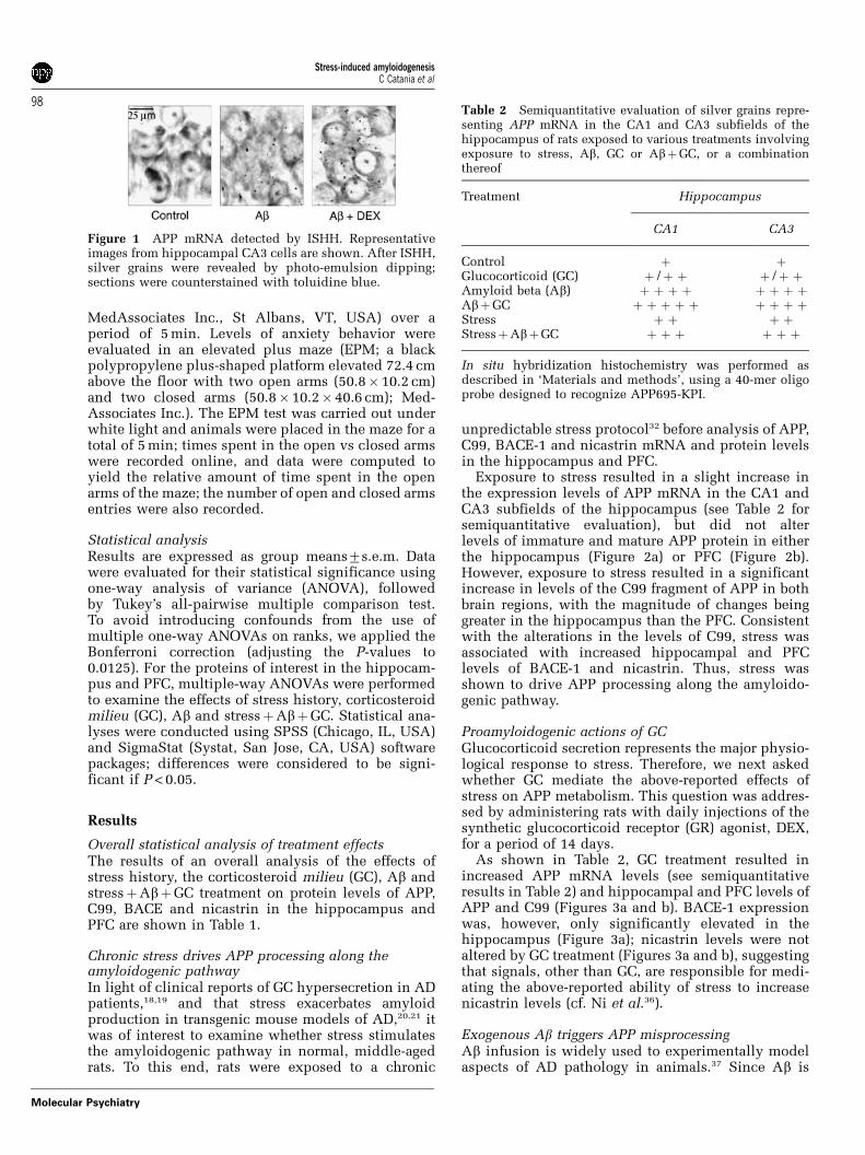
In situ hybridizationParaffin sections (8 mm) were deparaffinized (seeabove), and fixed in 4% p-formaldehyde in phos-phate-buffered saline (PBS) containing 0.1% diethyl-pyrocarbonate (DEPC; Sigma). After washing (PBS–DPEC), sections were acetylated (0.1 M triethanol-amine, 0.25% acetic anhydride in DPEC), delipidatedin chloroform (5 min), dehydrated through a gradedseries of ethanols and air dried. A 40-mer oligo-nucleotide probe (GCTGGCTGCCGTCGTGGGAACTC
GGACTACCTCCTCCACA) was used to detect APP695-KPI.34,35 Slides were hybridized with an antisense orsense (control) 35S-dATP-labeled oligoprobe (106 c.p.m.per slide; overnight at 42 1C), after which they werestringently washed (1� saline sodium citrate (SSC)for 15 min at 55 1C). After sequential immersion 1�SSC, H2O, 65% ethanol and 95% ethanol, slides wereair dried and dipped in photographic emulsion (1:1Kodak NTB2 in distilled water) and exposed for 1month. Sections were then developed, counterstained(toluidine blue) and viewed under bright field orpolarized light to view cells and silver grains,respectively. Hybridization signal on captured images(all slides) was scored by two independent observers(unaware of the treatment details) on a scale of 0–5; theraters followed common scoring criteria and the resultsshown in Table 1 are the means of the scores obtainedby the individual raters. Representative in situ hybridi-zation (ISH) images are shown in Figure 1.
Assays of cognitive performance and emotionalityHippocampus-dependent spatial reference memorywas assessed using the Morris water maze over 4 days(four trials per day), as previously described.25 Datawere recorded using a video-tracking system (View-point, Champagne au Mont d’Or, France).
Emotional state was evaluated by monitoringlocomotion, exploratory behavior and anxiety. Loco-motor and exploratory behavior were assessed (totaldistances traveled and number and duration ofrearings) online in an open-field arena (43.2�43.2 cm; transparent acrylic walls and white floor;
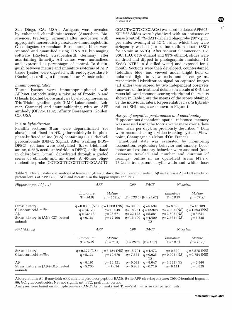
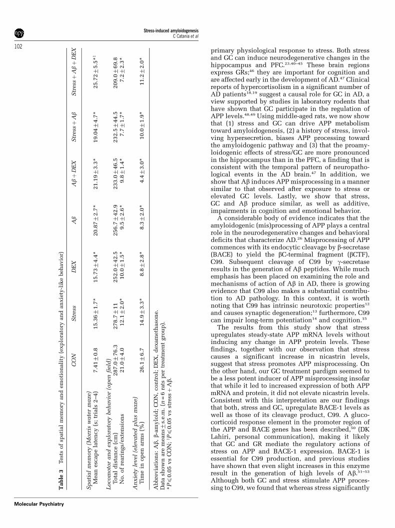
Table 1 Overall statistical analysis of treatment (stress history, the corticosteroid milieu, Ab and stressþAbþGC) effects onprotein levels of APP, C99, BACE and nicastrin in the hippocampus and PFC
Hippocampus (d.f.6, 36) APP C99 BACE Nicastrin
Immature(F = 54.9)
Mature(F = 132.2) (F = 130.3) (F = 25.07)
Immature(F = 19.9)
Mature(F = 37.2)
Stress history q = 0.0336 (NS) q = 1.608 (NS) q = 30.03 q = 5.592 q = 8.829 q = 16.189Glucocorticoid milieu q = 13.178 q = 10.649 q = 18.231 q = 12.928 q = 2.903 (NS) q = 1.293 (NS)Ab q = 13.416 q = 26.671 q = 32.175 q = 5.884 q = 3.598 (NS) q = 8.651Stress history in (AbþGC)-treatedanimals
q = 9.161 q = 12.466 q = 15.490 q = 4.409(NS)
q = 2.563 (NS) q = 5.835
PFC (d.f.6, 36) APP C99 BACE Nicastrin
Immature(F = 15.2)
Mature(F = 35.4) (F = 26.3) (F = 17.7)
Immature(F = 18.5)
Mature(F = 15.8)
Stress history q = 0.377 (NS) q = 3.424 (NS) q = 15.791 q = 4.472 q = 9.629 q = 3.575 (NS)Glucocorticoid milieu q = 5.131 q = 10.676 q = 7.865 q = 0.925
(NS)q = 0.998 (NS) q = 0.754 (NS)
Ab q = 8.195 q = 10.521 q = 6.042 q = 8.047 q = 1.333 (NS) q = 6.948Stress history in (AbþGC)-treatedanimals
q = 5.706 q = 7.654 q = 6.933 q = 6.719 q = 9.111 q = 8.829
Abbreviations: Ab, b-amyloid; APP, amyloid precursor peptide; BACE, b-site APP cleaving enzyme; C99, C-terminal fragment99; GC, glucocorticoids; NS, not significant; PFC, prefrontal cortex.Analyses were based on multiple one-way ANOVAs on ranks and Tukey’s all pairwise comparison tests.
Stress-induced amyloidogenesisC Catania et al
97
Molecular Psychiatry
MedAssociates Inc., St Albans, VT, USA) over aperiod of 5 min. Levels of anxiety behavior wereevaluated in an elevated plus maze (EPM; a blackpolypropylene plus-shaped platform elevated 72.4 cmabove the floor with two open arms (50.8�10.2 cm)and two closed arms (50.8� 10.2� 40.6 cm); Med-Associates Inc.). The EPM test was carried out underwhite light and animals were placed in the maze for atotal of 5 min; times spent in the open vs closed armswere recorded online, and data were computed toyield the relative amount of time spent in the openarms of the maze; the number of open and closed armsentries were also recorded.
Statistical analysisResults are expressed as group means7s.e.m. Datawere evaluated for their statistical significance usingone-way analysis of variance (ANOVA), followedby Tukey’s all-pairwise multiple comparison test.To avoid introducing confounds from the use ofmultiple one-way ANOVAs on ranks, we applied theBonferroni correction (adjusting the P-values to0.0125). For the proteins of interest in the hippocam-pus and PFC, multiple-way ANOVAs were performedto examine the effects of stress history, corticosteroidmilieu (GC), Ab and stressþAbþGC. Statistical ana-lyses were conducted using SPSS (Chicago, IL, USA)and SigmaStat (Systat, San Jose, CA, USA) softwarepackages; differences were considered to be signi-ficant if P < 0.05.
Results
Overall statistical analysis of treatment effectsThe results of an overall analysis of the effects ofstress history, the corticosteroid milieu (GC), Ab andstressþAbþGC treatment on protein levels of APP,C99, BACE and nicastrin in the hippocampus andPFC are shown in Table 1.
Chronic stress drives APP processing along theamyloidogenic pathwayIn light of clinical reports of GC hypersecretion in ADpatients,18,19 and that stress exacerbates amyloidproduction in transgenic mouse models of AD,20,21 itwas of interest to examine whether stress stimulatesthe amyloidogenic pathway in normal, middle-agedrats. To this end, rats were exposed to a chronic
unpredictable stress protocol32 before analysis of APP,C99, BACE-1 and nicastrin mRNA and protein levelsin the hippocampus and PFC.
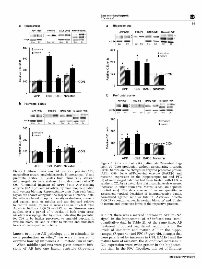
Exposure to stress resulted in a slight increase inthe expression levels of APP mRNA in the CA1 andCA3 subfields of the hippocampus (see Table 2 forsemiquantitative evaluation), but did not alterlevels of immature and mature APP protein in eitherthe hippocampus (Figure 2a) or PFC (Figure 2b).However, exposure to stress resulted in a significantincrease in levels of the C99 fragment of APP in bothbrain regions, with the magnitude of changes beinggreater in the hippocampus than the PFC. Consistentwith the alterations in the levels of C99, stress wasassociated with increased hippocampal and PFClevels of BACE-1 and nicastrin. Thus, stress wasshown to drive APP processing along the amyloido-genic pathway.
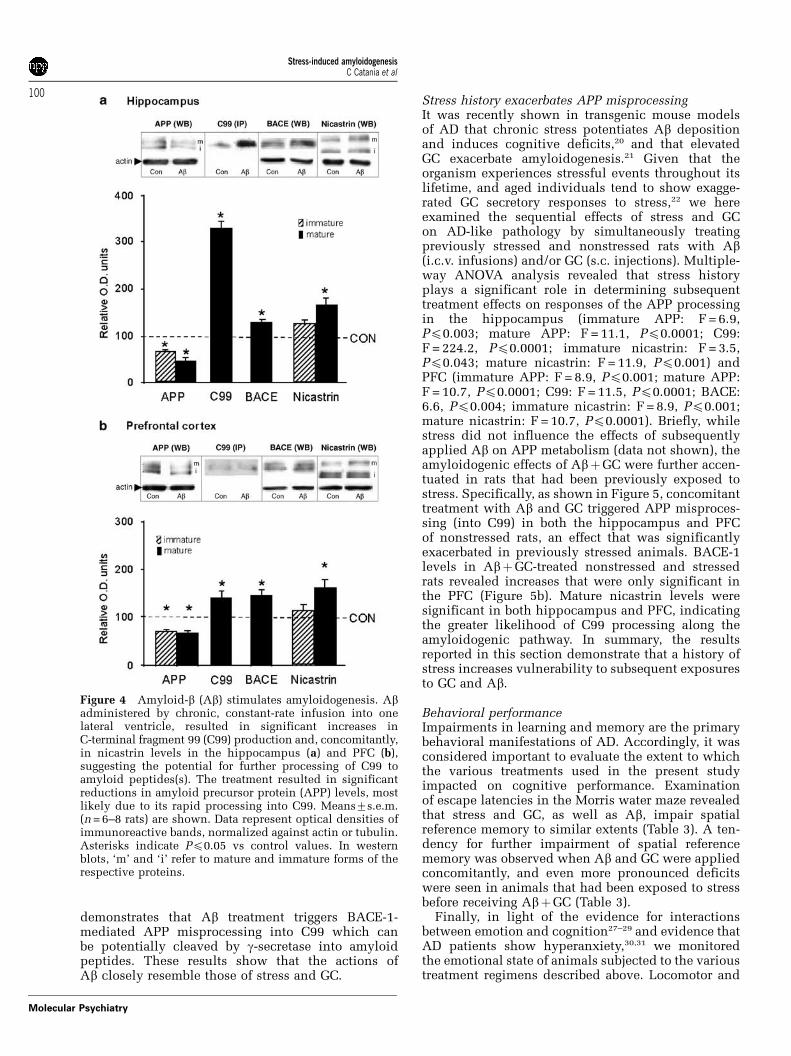
Proamyloidogenic actions of GCGlucocorticoid secretion represents the major physio-logical response to stress. Therefore, we next askedwhether GC mediate the above-reported effects ofstress on APP metabolism. This question was addres-sed by administering rats with daily injections of thesynthetic glucocorticoid receptor (GR) agonist, DEX,for a period of 14 days.
As shown in Table 2, GC treatment resulted inincreased APP mRNA levels (see semiquantitativeresults in Table 2) and hippocampal and PFC levels ofAPP and C99 (Figures 3a and b). BACE-1 expressionwas, however, only significantly elevated in thehippocampus (Figure 3a); nicastrin levels were notaltered by GC treatment (Figures 3a and b), suggestingthat signals, other than GC, are responsible for medi-ating the above-reported ability of stress to increasenicastrin levels (cf. Ni et al.36).
Exogenous Ab triggers APP misprocessingAb infusion is widely used to experimentally modelaspects of AD pathology in animals.37 Since Ab is
Figure 1 APP mRNA detected by ISHH. Representativeimages from hippocampal CA3 cells are shown. After ISHH,silver grains were revealed by photo-emulsion dipping;sections were counterstained with toluidine blue.
Table 2 Semiquantitative evaluation of silver grains repre-senting APP mRNA in the CA1 and CA3 subfields of thehippocampus of rats exposed to various treatments involvingexposure to stress, Ab, GC or AbþGC, or a combinationthereof
Treatment Hippocampus
CA1 CA3
Control þ þGlucocorticoid (GC) þ /þ þ þ /þ þAmyloid beta (Ab) þ þ þ þ þ þ þ þAbþGC þ þ þ þ þ þ þ þ þStress þ þ þ þStressþAbþGC þ þ þ þ þ þ
In situ hybridization histochemistry was performed asdescribed in ‘Materials and methods’, using a 40-mer oligoprobe designed to recognize APP695-KPI.
Stress-induced amyloidogenesisC Catania et al
98
Molecular Psychiatry
known to induce AD pathology and to stimulate itsown production in vitro,38 we were interested toexamine how Ab influences APP metabolism in vivo.
When middle-aged rats were given constant infu-sions of Ab into one lateral ventricle (Frautschy
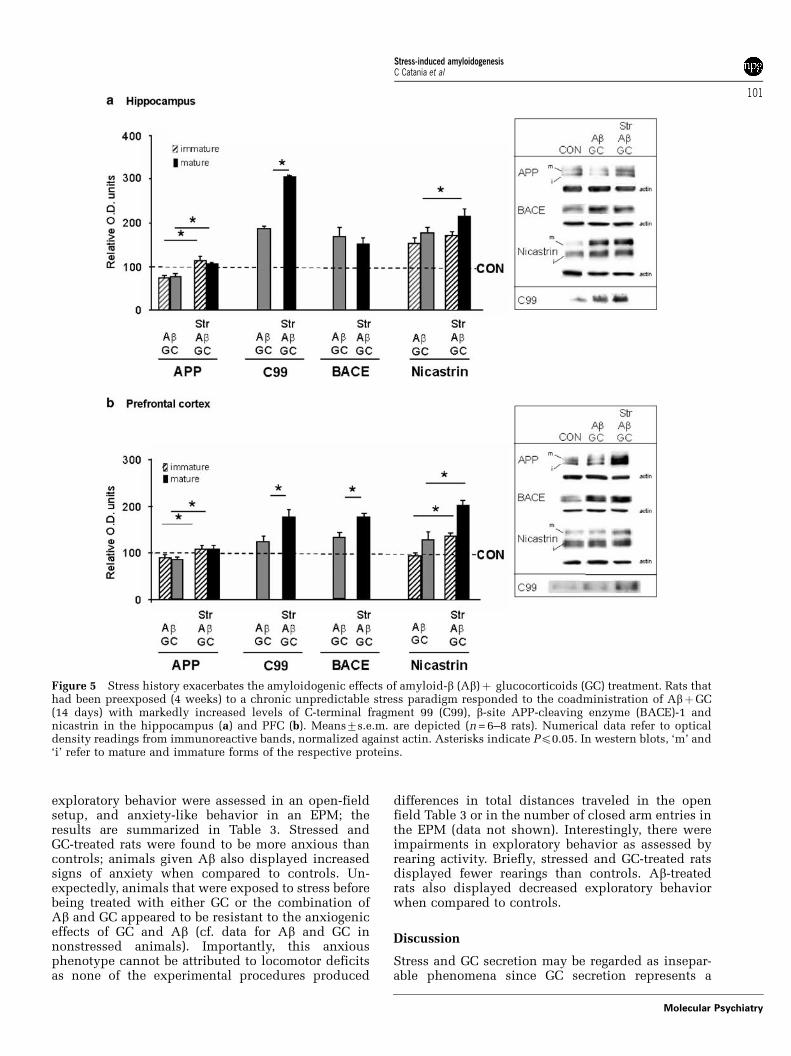
et al.39), there was a marked increase in APP mRNAsignal in the hippocampi of Ab-infused rats (semi-quantitative data in Table 2). At the same time, Abtreatment produced significant reductions in thelevels of immature and mature APP in the hippo-campus (Figure 4a) and PFC (Figure 4b), changes thatwere paralleled by increases in C99, BACE-1 and themature form of nicastrin; the Ab-induced increases inC99 expression were twice greater in the hippocam-pus than in the PFC. Together, this set of findings
Figure 2 Stress drives amyloid precursor protein (APP)metabolism toward amyloidogenesis. Hippocampal (a) andprefrontal cortex (b) lysates from chronically stressedmiddle-aged rats were analyzed for their contents of APP,C99 (C-terminal fragment of APP), b-site APP-cleavingenzyme (BACE)-1 and nicastrin, by immunoprecipitationand western blotting. Representative blots from each brainregion are shown alongside the respective numerical data;the latter are based on optical density evaluations, normali-zed against actin or tubulin and are depicted relativeto control (CON) values as means7s.e.m. (n = 6–8 rats).Asterisks indicate Pp0.05 vs CON values. Stressors wereapplied over a period of 4 weeks. In both brain areas,nicastrin was upregulated by stress, indicating the potentialfor C99 to be further processed to amyloid peptide. Inwestern blots, ‘m’ and ‘i’ refer to mature and immatureforms of the respective proteins.
Figure 3 Glucocorticoids (GC) stimulate C-terminal frag-ment 99 (C99) production without upregulating nicastrinlevels. Shown are the changes in amyloid precursor protein(APP), C99, b-site APP-cleaving enzyme (BACE)-1 andnicastrin expression in the hippocampus (a) and PFC(b) of middle-aged rats that had been treated with DEX, asynthetic GC, for 14 days. Note that nicastrin levels were notincreased in either brain area. Means7s.e.m. are depicted(n = 6–8 rats). The data emerged from semiquantitativeassessment (optical densities) of immunoreactive bands,normalized against actin or tubulin. Asterisks indicatePp0.05 vs control values. In western blots, ‘m’ and ‘i’ referto mature and immature forms of the respective proteins.
Stress-induced amyloidogenesisC Catania et al
99
Molecular Psychiatry
demonstrates that Ab treatment triggers BACE-1-mediated APP misprocessing into C99 which canbe potentially cleaved by g-secretase into amyloidpeptides. These results show that the actions ofAb closely resemble those of stress and GC.
Stress history exacerbates APP misprocessingIt was recently shown in transgenic mouse modelsof AD that chronic stress potentiates Ab depositionand induces cognitive deficits,20 and that elevatedGC exacerbate amyloidogenesis.21 Given that theorganism experiences stressful events throughout itslifetime, and aged individuals tend to show exagge-rated GC secretory responses to stress,22 we hereexamined the sequential effects of stress and GCon AD-like pathology by simultaneously treatingpreviously stressed and nonstressed rats with Ab(i.c.v. infusions) and/or GC (s.c. injections). Multiple-way ANOVA analysis revealed that stress historyplays a significant role in determining subsequenttreatment effects on responses of the APP processingin the hippocampus (immature APP: F = 6.9,Pp0.003; mature APP: F = 11.1, Pp0.0001; C99:F = 224.2, Pp0.0001; immature nicastrin: F = 3.5,Pp0.043; mature nicastrin: F = 11.9, Pp0.001) andPFC (immature APP: F = 8.9, Pp0.001; mature APP:F = 10.7, Pp0.0001; C99: F = 11.5, Pp0.0001; BACE:6.6, Pp0.004; immature nicastrin: F = 8.9, Pp0.001;mature nicastrin: F = 10.7, Pp0.0001). Briefly, whilestress did not influence the effects of subsequentlyapplied Ab on APP metabolism (data not shown), theamyloidogenic effects of AbþGC were further accen-tuated in rats that had been previously exposed tostress. Specifically, as shown in Figure 5, concomitanttreatment with Ab and GC triggered APP misproces-sing (into C99) in both the hippocampus and PFCof nonstressed rats, an effect that was significantlyexacerbated in previously stressed animals. BACE-1levels in AbþGC-treated nonstressed and stressedrats revealed increases that were only significant inthe PFC (Figure 5b). Mature nicastrin levels weresignificant in both hippocampus and PFC, indicatingthe greater likelihood of C99 processing along theamyloidogenic pathway. In summary, the resultsreported in this section demonstrate that a history ofstress increases vulnerability to subsequent exposuresto GC and Ab.
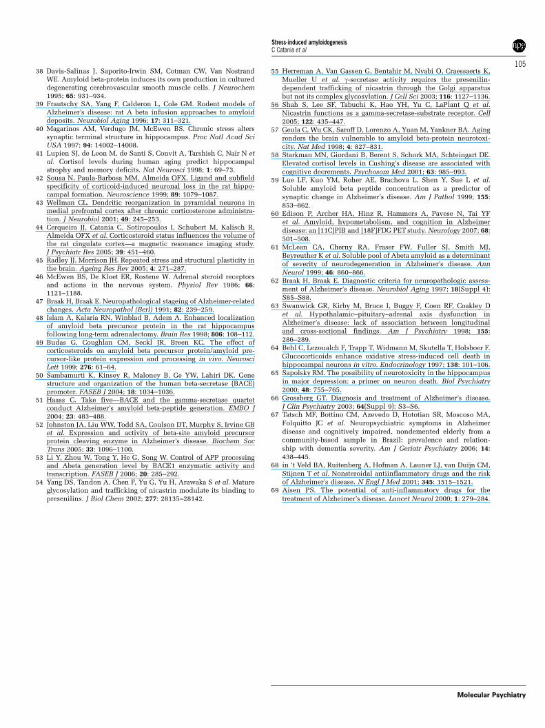
Behavioral performanceImpairments in learning and memory are the primarybehavioral manifestations of AD. Accordingly, it wasconsidered important to evaluate the extent to whichthe various treatments used in the present studyimpacted on cognitive performance. Examinationof escape latencies in the Morris water maze revealedthat stress and GC, as well as Ab, impair spatialreference memory to similar extents (Table 3). A ten-dency for further impairment of spatial referencememory was observed when Ab and GC were appliedconcomitantly, and even more pronounced deficitswere seen in animals that had been exposed to stressbefore receiving AbþGC (Table 3).
Finally, in light of the evidence for interactionsbetween emotion and cognition27–29 and evidence thatAD patients show hyperanxiety,30,31 we monitoredthe emotional state of animals subjected to the varioustreatment regimens described above. Locomotor and
Figure 4 Amyloid-b (Ab) stimulates amyloidogenesis. Abadministered by chronic, constant-rate infusion into onelateral ventricle, resulted in significant increases inC-terminal fragment 99 (C99) production and, concomitantly,in nicastrin levels in the hippocampus (a) and PFC (b),suggesting the potential for further processing of C99 toamyloid peptides(s). The treatment resulted in significantreductions in amyloid precursor protein (APP) levels, mostlikely due to its rapid processing into C99. Means7s.e.m.(n = 6–8 rats) are shown. Data represent optical densities ofimmunoreactive bands, normalized against actin or tubulin.Asterisks indicate Pp0.05 vs control values. In westernblots, ‘m’ and ‘i’ refer to mature and immature forms of therespective proteins.
Stress-induced amyloidogenesisC Catania et al
100
Molecular Psychiatry
exploratory behavior were assessed in an open-fieldsetup, and anxiety-like behavior in an EPM; theresults are summarized in Table 3. Stressed andGC-treated rats were found to be more anxious thancontrols; animals given Ab also displayed increasedsigns of anxiety when compared to controls. Un-expectedly, animals that were exposed to stress beforebeing treated with either GC or the combination ofAb and GC appeared to be resistant to the anxiogeniceffects of GC and Ab (cf. data for Ab and GC innonstressed animals). Importantly, this anxiousphenotype cannot be attributed to locomotor deficitsas none of the experimental procedures produced
differences in total distances traveled in the openfield Table 3 or in the number of closed arm entries inthe EPM (data not shown). Interestingly, there wereimpairments in exploratory behavior as assessed byrearing activity. Briefly, stressed and GC-treated ratsdisplayed fewer rearings than controls. Ab-treatedrats also displayed decreased exploratory behaviorwhen compared to controls.
Discussion
Stress and GC secretion may be regarded as insepar-able phenomena since GC secretion represents a
Figure 5 Stress history exacerbates the amyloidogenic effects of amyloid-b (Ab)þ glucocorticoids (GC) treatment. Rats thathad been preexposed (4 weeks) to a chronic unpredictable stress paradigm responded to the coadministration of AbþGC(14 days) with markedly increased levels of C-terminal fragment 99 (C99), b-site APP-cleaving enzyme (BACE)-1 andnicastrin in the hippocampus (a) and PFC (b). Means7s.e.m. are depicted (n = 6–8 rats). Numerical data refer to opticaldensity readings from immunoreactive bands, normalized against actin. Asterisks indicate Pp0.05. In western blots, ‘m’ and‘i’ refer to mature and immature forms of the respective proteins.
Stress-induced amyloidogenesisC Catania et al
101
Molecular Psychiatry
primary physiological response to stress. Both stressand GC can induce neurodegenerative changes in thehippocampus and PFC.23,40–45 These brain regionsexpress GRs;46 they are important for cognition andare affected early in the development of AD.47 Clinicalreports of hypercortisolism in a significant number ofAD patients18,19 suggest a causal role for GC in AD, aview supported by studies in laboratory rodents thathave shown that GC participate in the regulation ofAPP levels.48,49 Using middle-aged rats, we now showthat (1) stress and GC can drive APP metabolismtoward amyloidogenesis, (2) a history of stress, invol-ving hypersecretion, biases APP processing towardthe amyloidogenic pathway and (3) that the proamy-loidogenic effects of stress/GC are more pronouncedin the hippocampus than in the PFC, a finding that isconsistent with the temporal pattern of neuropatho-logical events in the AD brain.47 In addition, weshow that Ab induces APP misprocessing in a mannersimilar to that observed after exposure to stress orelevated GC levels. Lastly, we show that stress,GC and Ab produce similar, as well as additive,impairments in cognition and emotional behavior.
A considerable body of evidence indicates that theamyloidogenic (mis)processing of APP plays a centralrole in the neurodegenerative changes and behavioraldeficits that characterize AD.26 Misprocessing of APPcommences with its endocytic cleavage by b-secretase(BACE) to yield the bC-terminal fragment (bCTF),C99. Subsequent cleavage of C99 by g-secretaseresults in the generation of Ab peptides. While muchemphasis has been placed on examining the role andmechanisms of action of Ab in AD, there is growingevidence that C99 also makes a substantial contribu-tion to AD pathology. In this context, it is worthnoting that C99 has intrinsic neurotoxic properties12
and causes synaptic degeneration;13 furthermore, C99can impair long-term potentiation14 and cognition.15
The results from this study show that stressupregulates steady-state APP mRNA levels withoutinducing any change in APP protein levels. Thesefindings, together with our observation that stresscauses a significant increase in nicastrin levels,suggest that stress promotes APP misprocessing. Onthe other hand, our GC treatment pardigm seemed tobe a less potent inducer of APP misprocessing insofarthat while it led to increased expression of both APPmRNA and protein, it did not elevate nicastrin levels.Consistent with this interpretation are our findingsthat both, stress and GC, upregulate BACE-1 levels aswell as those of its cleavage product, C99. A gluco-corticoid response element in the promoter region ofthe APP and BACE genes has been described,50 (DKLahiri, personal communication), making it likelythat GC and GR mediate the regulatory actions ofstress on APP and BACE-1 expression. BACE-1 isessential for C99 production, and previous studieshave shown that even slight increases in this enzymeresult in the generation of high levels of Ab.51–53
Although both GC and stress stimulate APP proces-sing to C99, we found that whereas stress significantlyT
able
3T
est
sof
spati
al
mem
ory
an
dem
oti
on
ali
ty(e
xp
lora
tory
an
dan
xie
ty-l
ike
beh
avio
r)
CO
NS
tress
DE
XAb
Abþ
DE
XS
tress
þAb
Str
ess
þAbþ
DE
X
Sp
ati
al
mem
ory
(Morr
isw
ate
rm
aze)
Mean
esc
ap
ela
ten
cy
(s;
tria
ls2–4)
7.4
17
0.8
15.3
67
1.7
*15.7
37
4.4
*20.8
77
2.7
*21.1
97
3.3
*19.0
47
4.7
*25.7
27
5.5
*z
Locom
oto
ran
dexp
lora
tory
beh
avio
r(o
pen
field
)T
ota
ld
ista
nce
(cm
)287.07
76.3
278.77
11
252.07
42.5
256.77
42.9
233.07
46.5
232.57
44.5
209.07
69.8
No.
of
reari
ngs/
exte
nsi
on
s21.07
4.0
12.17
2.0
*10.07
1.5
*9.57
2.6
*9.87
1.4
*7.77
1.7
*7.27
2.3
*
An
xie
tyle
vel
(ele
vate
dp
lus
maze)
Tim
ein
op
en
arm
s(%
)26.17
6.7
14.97
3.3
*8.87
2.8
*8.37
2.0
*4.47
3.0
*10.07
1.9
*11.27
2.0
*
Abbre
via
tion
s:Ab,
b-am
ylo
id;
CO
N,
con
trol;
DE
X,
dexam
eth
aso
ne.
Data
show
nare
mean
s7s.
e.m
.(n
=6
rats
per
treatm
en
tgro
up
).*Pp
0.0
5vs
CO
N;z Pp
0.0
5vs
stre
ssþ
Ab.
Stress-induced amyloidogenesisC Catania et al
102
Molecular Psychiatry
increases nicastrin levels, GC actions are limited tothe point of C99 production. Given the adverseactions of C99 on neuronal function and survival, aswell as behavior,12–15 the potential importance ofGC-induced increases in C99 production for AD-likepathology is nevertheless significant.
Nicastrin, and in particular its glycosylated matureform, is an essential component of the g-secretasecomplex;54,55 because of its role in the recognition andpresentation of substrate (C99) to presenilin-1 and -2,nicastrin is crucial for the generation of Ab.7,56 Ourobservations that stress (but not GC) increasesnicastrin levels, especially those of the mature form,suggest that stress can recruit additional mechanismsthat allow it to carry C99 metabolism through toAb production (cf. Ni et al.36) On the other hand,the possibility that GC can eventually generate Ab(for example, by acting on other members of theg-secretase complex) cannot be excluded; technicallimitations of existing assays unfortunately precludeddirect measurements of rat Ab in the present study.
Since increased Ab production is causally relatedto AD pathology, exogenous administration of Abpeptides into the brain has been extensively used innormal rodents and primates to reproduce the neuro-patholgical and behavioral features of early stageAD.37,39,57 Using the Ab i.c.v. infusion paradigm inmiddle-aged rats, we here observed that Ab upregu-lates APP mRNA levels, but reduces APP proteinlevels; at the same time, Ab treatment results inincreased tissue levels of C99 and nicastrin. Thus,Ab appears to accelerate APP metabolism, increasingthe potential for amyloid peptide generation. Theresemblance of the Ab-induced changes to thosefound after exposure to stress and GC supports theview that stress and GC may contribute to theetiopathogenesis of AD. Moreover, since Ab, stressand GC all have negative effects on cognition,21,22,58 itwould appear that Ab contributes to the molecularsignaling machinery that mediates the behavioralactions of stress and GC. Lastly, it should be notedthat the Ab infusion protocol used here appropriatelymodels the preclinical stages of AD insofar that thepathobiochemical changes observed here occurred ina gradiential manner, being stronger in the hippo-campus than in the PFC (cf. Braak and Braak47). Whileour soluble Ab infusion paradigm did not result indetectable amyloid depositions, it should be notedthat the role of Ab plaques in the pathogenesis ofAD is contentious.59,60 Recent work indicates a strongcorrelation between soluble Ab levels and the severityof memory deficits in AD patients.61 Importantly,soluble Ab is known to trigger synapse loss,4 an earlyevent in the pathogenesis of AD.
Interactions between endogenous and exogenousfactors (for example, age, mutations and Ab produc-tion on the one hand, and stress on the other) areimportant determinants of the onset and progress ofAD. The organism experiences intermittent stressorsthroughout life, and its endogenous production ofAb increases with age.62 The results of recent studies
in transgenic animal models showed that chronicstress potentiates Ab deposition and induces cogni-tive deficits21 and that amyloid production is exacer-bated when GC levels are elevated.20 In the presentwork, we attempted to mimic typical lifetime eventsby exposing rats to a chronic unpredictable stressparadigm before subsequent treatment with GCand/or central infusions of Ab. The results clearlydemonstrate that stress history exacerbates the APPmisprocessing effects of Ab and GC, and, strikingly,also activated the amyloidogenic pathway in the PFC,an area that had otherwise proven to be relativelyresilient to the individual stimuli. Importantly, thecombinatorial treatment resulted in a deteriorationof spatial reference memory that was greater thanthat induced by the individual treatments. Thus,stress history/previous exposure to high GC levels canmarkedly worsen the effects of subsequent exposuresto stress/GC and Ab on AD-like biochemical andbehavioral pathology; accordingly, stress may be animportant precipitating and exacerbating factor inAD. The present findings provide experimentalsupport for the implication of stress as a contributoryfactor in early AD disease.63 Based on this andprevious studies,64,65 we suggest that stress (and GC)make neurons more vulnerable to the pathologicalactions of Ab, including Ab-induced stimulation ofAPP misprocessing.
To sum up, we have demonstrated that (1) stress/GCcan contribute to AD pathology by driving APP meta-bolism toward the production of C99 and, eventually,of Ab, and (2) that stress history ‘primes’ the brain togenerate greater amounts of amyloidogenic peptide(s)upon subsequent exposures to GC and Ab. Thesefindings prompt the hypothesis that the effects ofstress/GC on neuronal atrophy may be mediated byC99 and/or Ab, and that they may ultimately becausally related to disruptions of behavior that aretypical of AD. The observation that Ab, like stress andGC, can increase emotionality is interesting in view ofthe increasing recognition of reciprocal regulatoryrelationships between cognition and emotion,27–29
reports that many AD patients are hyperanxious,66
and the implication of stress and GC in disorders ofanxiety67 and cognition.22,59 Importantly, togetherwith previously established links between depressionand the increased risk for developing AD,31 thepresent findings suggest that Ab may be a commondenominator underlying these various stress-relateddisorders, and indicate the potential importanceof including histories of stress and GC therapy inanamnesic data. Lastly, our results reiterate theneed for judicial use of GC therapy in older subjects,especially in light of the poor efficacy of GC to slowthe progression of AD.68,69
Acknowledgments
We thank Dieter Fischer and Rainer Stoffel forexcellent technical assistance, Carola Hetzel foradministrative help, and Drs Keiro Shirotani and
Stress-induced amyloidogenesisC Catania et al
103
Molecular Psychiatry
Ayako Yamamoto for helpful advice. Isabel Matos andLucilia Pinto are thanked for help with histologicalpreparation and scoring, Dr Sam Gandy for providingthe 369 antibody and Dr Alexandre Patchev for helpwith image preparation. CC and IS were supported bystipends from the Max Planck Society and EU MarieCurie Training Fellowships (at University CollegeLondon, UK). The collaboration between the Germanand Portuguese laboratories was supported throughthe German–Portuguese Luso-Alemas Program(DAAD/GRICES). This study was conducted withinthe framework of the EU-supported integrated project‘CRESCENDO’ (Contract FP6-018652).
References
1 Guillozet AL, Weintraub S, Mash DC, Mesulam MM. Neurofibril-lary tangles, amyloid, and memory in aging and mild cognitiveimpairment. Arch Neurol 2003; 60: 729–736.
2 Cleary JP, Walsh DM, Hofmeister JJ, Shankar GM, Kuskowski MA,Selkoe DJ et al. Natural oligomers of the amyloid-beta proteinspecifically disrupt cognitive function. Nat Neurosc 2005; 8:79–84.
3 Almeida CG, Tampellini D, Takahashi RH, Greengard P, Lin MT,Snyder EM et al. Beta-amyloid accumulation in APP mutantneurons reduces PSD-95 and GluR1 in synapses. Neurobiol Dis2005; 20: 187–198.
4 Roselli F, Tirard M, Lu J, Hutzler P, Lamberti P, Livrea P et al.Soluble beta-amyloid1-40 induces NMDA-dependent degradationof postsynaptic density-95 at glutamatergic synapses. J Neurosci2005; 25: 11061–11070.
5 Terry RD, Masliah E, Salmon DP, Butters N, DeTeresa R, Hill Ret al. Physical basis of cognitive alterations in Alzheimer’s disease:synapse loss is the major correlate of cognitive impairment. AnnNeurol 1991; 30: 572–580.
6 Olariu A, Tran MH, Yamada K, Mizuno M, Hefco V, Nabeshima T.Memory deficits and increased emotionality induced by b-amyloid(25–35) are correlated with the reduced acetylcholine release andaltered phorbol dibutyrate binding in the hippocampus. J NeuralTransm 2001; 108: 1065–1079.
7 De Strooper B. Nicastrin: gatekeeper of the gamma-secretasecomplex. Cell 2005; 122: 318–320.
8 Bayer TA, Wirths O, Majtenyi K, Hartmann T, Multhaup G,Beyreuther K et al. Key factors in Alzheimer’s disease: beta-amyloid precursor protein processing, metabolism and intra-neuronal transport. Brain Pathol 2001; 11: 1–11.
9 Lorenzo A, Yankner BA. Beta-amyloid neurotoxicity requires fibrilformation and is inhibited by congo red. Proc Natl Acad Sci USA1994; 91: 12243–12247.
10 Weldon DT, Rogers SD, Ghilardi JR, Finke MP, Cleary JP, O’Hare Eet al. Fibrillar beta-amyloid induces microglial phagocytosis,expression of inducible nitric oxide synthase, and loss of a selectpopulation of neurons in the rat CNS in vivo. J Neurosci 1998; 18:2161–2173.
11 Mattson MP. Pathways towards and away from Alzheimer’sdisease. Nature 2004; 430: 631–639.
12 Yankner BA, Dawes LR, Fisher S, Villa-Komaroff L, Oster-GraniteML, Neve RL. Neurotoxicity of a fragment of the amyloid precursorassociated with Alzheimer’s disease. Science 1989; 245: 417–420.
13 Oster-Granite ML, McPhie DL, Greenan J, Neve RL. Age-dependentneuronal and synaptic degeneration in mice transgenic for the Cterminus of the amyloid precursor protein. J Neurosci 1996; 16:6732–6741.
14 Nalbantoglu J, Tirado-Santiago G, Lahsaini A, Poirier J, GoncalvesO, Verge G et al. Impaired learning and LTP in mice expressing thecarboxy terminus of the Alzheimer amyloid precursor protein.Nature 1997; 387: 500–505.
15 Berger-Sweeney J, McPhie DL, Arters JA, Greenan J, Oster-GraniteML, Neve RL. Impairments in learning and memory accompanied
by neurodegeneration in mice transgenic for the carboxyl-terminusof the amyloid precursor protein. Mol Brain Res 1999; 66: 150–162.
16 Lorenzo A, Yuan M, Zhang Z, Paganetti PA, Sturchler-Pierrat C,Staufenbiel M et al. Amyloid beta interacts with the amyloidprecursor protein: a potential toxic mechanism in Alzheimer’sdisease. Nat Neurosci 2000; 3: 460–464.
17 Heredia L, Lin R, Vigo FS, Kedikian G, Busciglio J, Lorenzo A.Deposition of amyloid fibrils promotes cell-surface accumulationof amyloid beta precursor protein. Neurobiol Dis 2004; 16:617–629.
18 Hartmann A, Veldhuis JD, Deuschle M, Standhardt H, Heuser I.Twenty-four hour cortisol release profiles in patients withAlzheimer’s and Parkinson’s disease compared to normal controls:ultradian secretory pulsatility and diurnal variation. NeurobiolAging 1997; 18: 285–289.
19 Elgh E, Lindqvist Astot A, Fagerlund M, Eriksson S, Olsson T,Nasman B. Cognitive dysfunction, hippocampal atrophy andglucocorticoid feedback in Alzheimer’s disease. Biol Psychiatry2006; 59: 155–161.
20 Green KN, Billings LM, Roozendaal B, McGaugh JL, LaFerla FM.Glucocorticoids increase amyloid-beta and tau pathology ina mouse model of Alzheimer’s disease. J Neurosci 2006; 26:9047–9056.
21 Jeong YH, Park CH, Yoo J, Shin KY, Ahn SM, Kim HS et al. Chronicstress accelerates learning and memory impairments and increasesamyloid deposition in APPV717I-CT100 transgenic mice, anAlzheimer’s disease model. FASEB J 2006; 20: 729–731.
22 Lupien SJ, Fiocco A, Wan N, Maheu F, Lord C, Schramek T et al.Stress hormones and human memory function across the lifespan.Psychoneuroendocrinology 2005; 30: 225–242.
23 Cerqueira JJ, Pego JM, Taipa R, Bessa JM, Almeida OF, Sousa N.Morphological correlates of corticosteroid-induced changesin prefrontal cortex-dependent behaviors. J Neurosci 2005; 25:7792–7800.
24 Sousa N, Almeida OFX. Corticosteroids: sculptors of the hippo-campal formation. Rev Neurosci 2002; 13: 59–84.
25 Cerqueira JJ, Taipa R, Almeida OFX, Sousa N. Specific configura-tion of dendritic degeneration in pyramidal neurons of the medialprefrontal cortex induced by differing corticosteroid regimen.Cereb Cortex 2006; 17: 1998–2006.
26 Selkoe DJ. Alzheimer’s disease: genotypes, phenotypes, andtreatments. Science 1997; 275: 630–631.
27 Dolan RJ. Emotion, cognition, and behavior. Science 2002; 298:1191–1194.
28 Ochsner KN, Gross JJ. The cognitive control of emotion. TrendsCogn Sci 2005; 9: 242–249.
29 Phelps EA. Emotion and cognition: insights from studies of thehuman amygdala. Annu Rev Psychol 2006; 57: 27–53.
30 Ownby RL, Harwood DG, Barker WW, Duara R. Predictors ofanxiety in patients with Alzheimer’s disease. Depress Anxiety2000; 11: 38–42.
31 Ownby RL, Crocco E, Acevedo A, John V, Loewenstein D.Depression and risk for Alzheimer disease: systematic review,meta-analysis, and metaregression analysis. Arch Gen Psychiatry2006; 63: 530–538.
32 Sousa N, Almeida OFX, Holsboer F, Paula-Barbosa MM, MadeiraMD. Maintenance of hippocampal cell numbers in young and agedrats submitted to chronic unpredictable stress. Comparison withthe effects of corticosterone treatment. Stress 1998; 2: 237–249.
33 Paxinos G, Watson C. The Rat Brain in Stereotaxic Coordinates,4thedn. Academic Press: San Diego, 1998.
34 Sola C, Mengod G, Probst A, Palacios KM. Differential regional andcellular distribution of a-amyloid precursor RNAs containingand lacking the Kunitz protease inhibitor domain in the brain ofhuman, rat and mouse. Neuroscience 1993; 53: 267–295.
35 Panegyres PK. The effects of excitotoxicity on the expression of theamyloid precursor protein gene in the brain and its modulation byneuroprotective agents. J Neural Transm 1998; 105: 463–478.
36 Ni Y, Zhao X, Bao G, Zou L, Teng L, Wang Z et al. Activation ofbeta(2)-adrenergic receptor stimulates gamma-secretase activityand accelerates amyloid plaque formation. Nat Med 2007; 12:1390–1396.
37 Stephan A, Phillips AG. A case for a non-transgenic animal modelof Alzheimer’s disease. Genes Brain Behav 2005; 4: 157–172.
Stress-induced amyloidogenesisC Catania et al
104
Molecular Psychiatry
38 Davis-Salinas J, Saporito-Irwin SM, Cotman CW, Van NostrandWE. Amyloid beta-protein induces its own production in cultureddegenerating cerebrovascular smooth muscle cells. J Neurochem1995; 65: 931–934.
39 Frautschy SA, Yang F, Calderon L, Cole GM. Rodent models ofAlzheimer’s disease: rat A beta infusion approaches to amyloiddeposits. Neurobiol Aging 1996; 17: 311–321.
40 Magarinos AM, Verdugo JM, McEwen BS. Chronic stress alterssynaptic terminal structure in hippocampus. Proc Natl Acad SciUSA 1997; 94: 14002–14008.
41 Lupien SJ, de Leon M, de Santi S, Convit A, Tarshish C, Nair N etal. Cortisol levels during human aging predict hippocampalatrophy and memory deficits. Nat Neurosci 1998; 1: 69–73.
42 Sousa N, Paula-Barbosa MM, Almeida OFX. Ligand and subfieldspecificity of corticoid-induced neuronal loss in the rat hippo-campal formation. Neuroscience 1999; 89: 1079–1087.
43 Wellman CL. Dendritic reorganization in pyramidal neurons inmedial prefrontal cortex after chronic corticosterone administra-tion. J Neurobiol 2001; 49: 245–253.
44 Cerqueira JJ, Catania C, Sotiropoulos I, Schubert M, Kalisch R,Almeida OFX et al. Corticosteroid status influences the volume ofthe rat cingulate cortex—a magnetic resonance imaging study.J Psychiatr Res 2005; 39: 451–460.
45 Radley JJ, Morrison JH. Repeated stress and structural plasticity inthe brain. Ageing Res Rev 2005; 4: 271–287.
46 McEwen BS, De Kloet ER, Rostene W. Adrenal steroid receptorsand actions in the nervous system. Physiol Rev 1986; 66:1121–1188.
47 Braak H, Braak E. Neuropathological stageing of Alzheimer-relatedchanges. Acta Neuropathol (Berl) 1991; 82: 239–259.
48 Islam A, Kalaria RN, Winblad B, Adem A. Enhanced localizationof amyloid beta precursor protein in the rat hippocampusfollowing long-term adrenalectomy. Brain Res 1998; 806: 108–112.
49 Budas G, Coughlan CM, Seckl JR, Breen KC. The effect ofcorticosteroids on amyloid beta precursor protein/amyloid pre-cursor-like protein expression and processing in vivo. NeurosciLett 1999; 276: 61–64.
50 Sambamurti K, Kinsey R, Maloney B, Ge YW, Lahiri DK. Genestructure and organization of the human beta-secretase (BACE)promoter. FASEB J 2004; 18: 1034–1036.
51 Haass C. Take five—BACE and the gamma-secretase quartetconduct Alzheimer’s amyloid beta-peptide generation. EMBO J2004; 23: 483–488.
52 Johnston JA, Liu WW, Todd SA, Coulson DT, Murphy S, Irvine GBet al. Expression and activity of beta-site amyloid precursorprotein cleaving enzyme in Alzheimer’s disease. Biochem SocTrans 2005; 33: 1096–1100.
53 Li Y, Zhou W, Tong Y, He G, Song W. Control of APP processingand Abeta generation level by BACE1 enzymatic activity andtranscription. FASEB J 2006; 20: 285–292.
54 Yang DS, Tandon A, Chen F, Yu G, Yu H, Arawaka S et al. Matureglycosylation and trafficking of nicastrin modulate its binding topresenilins. J Biol Chem 2002; 277: 28135–28142.
55 Herreman A, Van Gassen G, Bentahir M, Nyabi O, Craessaerts K,Mueller U et al. g-secretase activity requires the presenilin-
dependent trafficking of nicastrin through the Golgi apparatusbut not its complex glycosylation. J Cell Sci 2003; 116: 1127–1136.
56 Shah S, Lee SF, Tabuchi K, Hao YH, Yu C, LaPlant Q et al.Nicastrin functions as a gamma-secretase-substrate receptor. Cell2005; 122: 435–447.
57 Geula C, Wu CK, Saroff D, Lorenzo A, Yuan M, Yankner BA. Aging
renders the brain vulnerable to amyloid beta-protein neurotoxi-city. Nat Med 1998; 4: 827–831.
58 Starkman MN, Giordani B, Berent S, Schork MA, Schteingart DE.
Elevated cortisol levels in Cushing’s disease are associated withcognitive decrements. Psychosom Med 2001; 63: 985–993.
59 Lue LF, Kuo YM, Roher AE, Brachova L, Shen Y, Sue L et al.Soluble amyloid beta peptide concentration as a predictor ofsynaptic change in Alzheimer’s disease. Am J Pathol 1999; 155:
853–862.60 Edison P, Archer HA, Hinz R, Hammers A, Pavese N, Tai YF
et al. Amyloid, hypometabolism, and cognition in Alzheimer
disease: an [11C]PIB and [18F]FDG PET study. Neurology 2007; 68:501–508.
61 McLean CA, Cherny RA, Fraser FW, Fuller SJ, Smith MJ,
Beyreuther K et al. Soluble pool of Abeta amyloid as a determinantof severity of neurodegeneration in Alzheimer’s disease. AnnNeurol 1999; 46: 860–866.
62 Braak H, Braak E. Diagnostic criteria for neuropathologic assess-ment of Alzheimer’s disease. Neurobiol Aging 1997; 18(Suppl 4):S85–S88.
63 Swanwick GR, Kirby M, Bruce I, Buggy F, Coen RF, Coakley Det al. Hypothalamic–pituitary–adrenal axis dysfunction inAlzheimer’s disease: lack of association between longitudinal
and cross-sectional findings. Am J Psychiatry 1998; 155:286–289.
64 Behl C, Lezoualch F, Trapp T, Widmann M, Skutella T, Holsboer F.
Glucocorticoids enhance oxidative stress-induced cell death inhippocampal neurons in vitro. Endocrinology 1997; 138: 101–106.
65 Sapolsky RM. The possibility of neurotoxicity in the hippocampus
in major depression: a primer on neuron death. Biol Psychiatry2000; 48: 755–765.
66 Grossberg GT. Diagnosis and treatment of Alzheimer’s disease.
J Clin Psychiatry 2003; 64(Suppl 9): S3–S6.67 Tatsch MF, Bottino CM, Azevedo D, Hototian SR, Moscoso MA,
Folquitto JC et al. Neuropsychiatric symptoms in Alzheimer
disease and cognitively impaired, nondemented elderly from acommunity-based sample in Brazil: prevalence and relation-
ship with dementia severity. Am J Geriatr Psychiatry 2006; 14:438–445.
68 in ‘t Veld BA, Ruitenberg A, Hofman A, Launer LJ, van Duijn CM,
Stijnen T et al. Nonsteroidal antiinflammatory drugs and the riskof Alzheimer’s disease. N Engl J Med 2001; 345: 1515–1521.
69 Aisen PS. The potential of anti-inflammatory drugs for the
treatment of Alzheimer’s disease. Lancet Neurol 2000; 1: 279–284.
Stress-induced amyloidogenesisC Catania et al
105
Molecular Psychiatry
Copyright © 2022 FDOKUMEN