The Neurobiological Bases for Development of Pharmacological Treatments of Aggressive Disorders
Upload
khangminh22Category
view
0download
0
1
BEHAVIOURAL AND NEUROBIOLOGICAL CORRELATES OF MATERNAL
SENSITIVITY IN HEALTHY NEW MOTHERS
A thesis submitted to The University of Manchester for the degree
of Doctor of Philosophy (PhD) in the Faculty of Medical and Human Sciences
2013
ALYA MOHAMED AHMED ELMADIH
SCHOOL OF MEDICINE

2
Table of Contents
LIST OF TABLES ..................................................................................................................... 7
LIST OF FIGURES ................................................................................................................... 9
LIST OF DEFINITIONS ......................................................................................................... 10
LIST OF ABBREVIATIONS .................................................................................................. 11
ABSTRACT ............................................................................................................................. 12
DECLARATION ..................................................................................................................... 13
COPYRIGHT STATEMENT .................................................................................................. 13
DEDICATION ......................................................................................................................... 15
ACKNOWLEDGEMENT ....................................................................................................... 16
THE AUTHOR ........................................................................................................................ 17
Chapter 1: General introduction .......................................................................................... 18
1.1 Background .................................................................................................................... 18
1.1.1. Study I (Chapter 2-5 & Publication 1) .................................................................... 19
1.1.2. Study II (Chapter 6-10 & Publication 2 & 3) ......................................................... 23
1.2. Rationale for Submitting the Thesis in an Alternative Format ..................................... 28
1.3. Publications ................................................................................................................... 30
Study I: Natural variation in maternal sensitivity: What are the possible prenatal and
postnatal predictors in healthy early postpartum mothers?
Chapter 2: Study I Literature Review ................................................................................. 34
2.1. The Prenatal Development of Maternal Sensitivity ...................................................... 34
2.2. What Is Maternal Sensitivity? ....................................................................................... 35
2.3. Why Is Maternal Sensitivity Important? ....................................................................... 37
2.3.1. Child outcomes ....................................................................................................... 37
2.3.2. Parenting quality ..................................................................................................... 41
2.4. How Is Maternal Sensitivity Measured? ....................................................................... 42
2.4.1. Validity of the short single interaction observation ................................................ 42
2.5. Is Maternal Sensitivity a Stable Construct?................................................................... 46
2.6. Factors That Influence Maternal Sensitivity ................................................................. 47
2.6.1. Mother prenatal psychological characteristics ................................................... 51
2.6.2. Socio-demographic and support factors ............................................................. 55

3
2.6.3. Early experiences in family-of-origin ................................................................. 58
2.6.4. Obstetric characteristics ..................................................................................... 61
2.6.5. Infant temperamental behaviours....................................................................... 63
2.7. Summary ....................................................................................................................... 64
2.8. Study I Objectives ......................................................................................................... 66
Chapter 3: Study 1 Methodology .......................................................................................... 67
3.1. Sample ........................................................................................................................... 67
3.2. Measures ........................................................................................................................ 70
3.2.1. The Edinburgh Postnatal Depression Scale (EPDS) (Cox et al., 1987) ................. 70
3.2.2. The Hospital Anxiety and Depression (HADS) rating scale (Zigmond &
Snaith, 1983) ..................................................................................................................... 70
3.2.3. Maternal-Fetal Attachment Scale (MFAS) (Cranley, 1981) .................................. 70
3.2.4. Parental Bonding Instrument (PBI) (Parker et al., 1979) ....................................... 71
3.2.5. Childhood Trauma Questionnaire (CTQ) (Bernstein et al., 1997) ......................... 71
3.2.6. The Oslo 3-items social support scale (Dalgard, 1996) .......................................... 72
3.2.7. The Manchester Assessment of Caregiver-Infant Interaction (MACI) (Wan et
al., 2012, 2013 online) ...................................................................................................... 72
3.2.8. Infant Behaviour Questionnaire Revised Very Short Form (IBQ-R-v. short)
(Gartstein & Rothbart, 2003) ............................................................................................ 75
3.3. Procedure ....................................................................................................................... 75
3.3.1. Time 1 (Pregnancy) ................................................................................................ 75
3.3.2. Time 2 (4-6 months postpartum) ............................................................................ 76
3.4. Statistical Analyses ........................................................................................................ 78
3.4.1. Sample size ............................................................................................................. 78
3.4.2. Data analyses .......................................................................................................... 78
3.5. Ethical Considerations ................................................................................................... 79
3.5.1. Ethics ...................................................................................................................... 79
3.5.2. Benefits to research participants ............................................................................. 79
3.5.3. Safety considerations .............................................................................................. 80
Chapter 4: Study I Results .................................................................................................... 81
4.1. Sample Characteristics .................................................................................................. 81
4.2. Characteristics of Mother-Infant Interaction (Manchester Assessment of
Caregiver-Infant Interaction; MACI rating) ......................................................................... 83
4.3. Preliminary Analyses (association of prenatal/postnatal factors with maternal
sensitivity) ............................................................................................................................ 83

4
4.3.1. Prenatal variables .................................................................................................... 83
4.3.2. Postnatal variables .................................................................................................. 87
4.4. Main analyses: Predictors of maternal sensitivity (Stepwise regression) ..................... 87
Chapter 5: Study I Discussion ............................................................................................... 90
5.1. Overview of the Findings .............................................................................................. 90
5.2. Mother-Infant Interaction .............................................................................................. 91
5.3. Predictors of Maternal Sensitivity ................................................................................. 91
5.4. Associations between Maternal Sensitivity and Prenatal/Postnatal Variables .............. 94
5.4.1. Prenatal variables .................................................................................................... 94
5.4.2. Postnatal variables .................................................................................................. 97
Publication 1 ........................................................................................................................... 99
Study II: Neurobiological mechanisms underlying maternal behaviour in humans: Do
the brain and endocrine responses to infant stimuli in less sensitive mothers differ from
those in sensitive mothers?
Chapter 6: Study II Literature Review (A) ....................................................................... 100
6.1. Oxytocin (Background) ............................................................................................... 100
6.1.1. Effects of oxytocin ................................................................................................ 100
6.2. Animal Studies ............................................................................................................ 102
6.2.1. Oxytocin and maternal behaviour in animal ......................................................... 102
6.2.2. Cross-generational transmission of maternal behaviour in animals ..................... 104
6.3. Human Studies ............................................................................................................ 105
6.3.1. Oxytocin and parental behaviour in human .......................................................... 105
6.3.2. The cross-generational transmission of OT in humans ........................................ 108
6.3.3. Oxytocin and own perceived parenting experience .............................................. 109
6.3.4. Maternal behaviour and breastfeeding as a proxy measure for OT levels........... 110
6.3.5. Oxytocin and social relationships’ stress in mothers and women ........................ 111
6.4. Validity of Plasma OT in Reflecting True Levels of OT in Humans .......................... 117
6.5. Summary ..................................................................................................................... 118
Chapter 7: Study II Literature Review (B)...................................................................... 120
7.1. The Brain Basis of Maternal Sensitivity ..................................................................... 120
7.2. Animal Studies ............................................................................................................ 120
7.2.1. The dopaminergic reward system in forebrain and midbrain ............................... 121
7.2.2. The emotional regulation pathways in amygdala and septal regions .................. 121
5
7.2.3. The sensation-driven thalamocingulate region ..................................................... 122
7.3. Human Studies ............................................................................................................ 122
7.3.1. Parental brain responses to auditory stimuli ......................................................... 123
7.3.2. Parental responses to visual stimuli ...................................................................... 126
7.3.3. Maternal brain responses in relation to maternal behaviour ................................. 131
7.4. Reasons for Inconsistency in Findings between Imaging Studies .............................. 144
7.5. Summary ..................................................................................................................... 146
7.6. Objectives of Study II (oxytocin & fMRI scanning) ................................................... 147
7.7. Hypotheses of Study II ................................................................................................ 149
Chapter 8: Study II Methodology (Oxytocin and fMRI Scanning) ............................... 151
8.1. Sample ......................................................................................................................... 151
8.2. Procedure ..................................................................................................................... 155
8.2.1. Oxytocin................................................................................................................ 155
8.2.2. fMRI scanning ..................................................................................................... 156
8.3. Statistical Analyses ...................................................................................................... 158
8.3.1. Sample size ........................................................................................................... 158
8.3.2. Data analysis ......................................................................................................... 159
8.4. Ethical Consideration .................................................................................................. 160
8.4.1. Ethics .................................................................................................................... 160
8.4.2. Safety considerations ............................................................................................ 161
Chapter 9: Study II Results (Oxytocin & fMRI scanning) .............................................. 162
9.1. Sample ......................................................................................................................... 162
9.2. Plasma Oxytocin Results ............................................................................................. 164
9.2.1. Preliminary analyses ............................................................................................. 164
9.2.2. Main analysis ........................................................................................................ 167
9.2.3. Relationship between plasma OT and own perceived parenting experience ....... 169
9.3. fMRI Results ............................................................................................................... 171
9.3.1. Preliminary analysis .............................................................................................. 171
9.3.2. Whole brain analysis of maternal brain responses................................................ 171
9.3.3. Comparisons between high and low sensitivity mothers ..................................... 173
9.3.4. Correlations of plasma oxytocin with ROI ........................................................... 177
Chapter 10: Study II Discussion (Oxytocin & fMRI scanning) ...................................... 179
10.1. Plasma Oxytocin Discussion ..................................................................................... 179

6
10.1.1. Overview of the findings .................................................................................... 179
10.1.2. Why baseline OT was high in LSMs? ................................................................ 180
10.1.3. Why does OT drop in HSMs?............................................................................. 183
10.1.4. Does oxytocin has a dual action? ........................................................................ 185
10.2. fMRI Scanning Discussion ........................................................................................ 187
10.2.1. Overview of the findings .................................................................................... 187
10.2.2. Main effect .......................................................................................................... 188
10.2.3. Comparison of brain activation between HSMs and LSMs ............................... 188
10.2.4. Correlation between plasma oxytocin and brain activation ................................ 190
Publication 2 ......................................................................................................................... 193
Publication 3 ......................................................................................................................... 194
Chapter 11: Summary and conclusion ............................................................................... 195
11.1. The Aims of The Thesis ............................................................................................ 195
11.2. Summary of the Findings .......................................................................................... 196
11.3. Clinical Implications of the Findings ........................................................................ 200
11.4. Limitation of the Thesis ............................................................................................ 201
11.5. Directions for Future Research .................................................................................. 203
11.6. Conclusions ............................................................................................................... 206
REFERENCES ....................................................................................................................... 207
Appendix A ............................................................................................................................ 226
Appendix B ............................................................................................................................ 228
Total word count is 76,040

7
LIST OF TABLES
Table 2.1: Examples of commonly used measures for assessing maternal sensitivity
through short observation ..................................................................................................... 45
Table 3.1: Sample characteristics of women followed up postnatally (N = 80) and the
drop-out (N = 25) ................................................................................................................. 69
Table 3.2: A brief description of rating definitions for maternal sensitivity on MACI
.............................................................................................................................................. 74
Table 3.3: Summary of the measures used in Study I ......................................................... 77
Table 4.1: Demographic characteristics of the sample (N = 80) ......................................... 82
Table 4.2: Correlations of prenatal variables with maternal sensitivity .............................. 86
Table 4.3: Stepwise regression examining prenatal predictors of maternal sensitivity....... 89
Table 6.1: Studies demonstrating the influence of oxytocin in parent infant bond ........... 114
Table 7.1a: Maternal brain responses to own infant using auditory stimuli ..................... 134
Table 7.1b: Maternal brain responses to own infant using auditory stimuli ..................... 136
Table 7.2a: Maternal brain responses to own infant using still pictures ........................... 138
Table 7.2b: Maternal brain responses to own infant using still pictures ........................... 140
Table 7.3: Maternal brain responses to own infant using video clips ............................... 142
Table 8.1: The demographic and obstetric characteristics of the sample grouped by
level of maternal sensitivity ............................................................................................... 154
Table 9.1: Comparinson for the demographic and obstetric characteristics of mothers
grouped by level of maternal sensitivity ............................................................................ 163
Table 9.2: Mean oxytocin levels (pg/ml) measured before and after play-interction
among mothers grouped by level of maternal sensitivity ................................................... 164

8
Table 9.3: Comparing demographic and obstetric characteristics of mothers grouped
by level of maternal sensitivity excluding the outlier ........................................................ 166
Table 9.4: . Correlations between plasma oxytocin and self reported own parenting
experience in mothers grouped by level of maternal sensitivity ........................................ 169
Table 9.5: Contrasts testing BOLD signals and ‘main effect’ in response to own and
unknown infant stimuli among the whole sample. ............................................................. 172
Table 9.6: Areas of significant BOLD activation within ROI, when comparing high
sensitivity with low sensitivity mothers ............................................................................. 175

9
LIST OF FIGURES
Figure 1.1: Time chart for data collection times ................................................................. 29
Figure 2.1: A theoratical model for factors that influence maternal sensitivity .................. 50
Figure 8.1: Distributions of high sensitivity and low sensitivity mothers in relation to
maternal sensitivity distribution of the whole sample ........................................................ 153
Figure 8.2: Model representing the order of video clips as viewed by mothers while
in the scanner ...................................................................................................................... 157
Figure 9.1: Box plots and means of plasma oxytocin measured before and after
mother-infant interaction among the high sensitivity and low sensitivity mothers............ 168
Figure 9.2: The relationship between baseline plasma oxytocin and own maternal
overprotection among low sensitivity and high sensitivity mothers .................................. 170
Figure 9.3: Maternal brain activation in response to infant stimuli .................................. 176
Figure 9.4: Correlation between BOLD activation in the right superior temporal
gyrus (STG) and post interaction plasma oxytocin among high sensitivity mothers (N
= 15) ................................................................................................................................... 178
Figure 10.1: A model representing the possible role of oxytocin in stress regulation
among high sensitivity and low sensitivity mothers .......................................................... 185

10
LIST OF DEFINITIONS
Antagonist: A chemical that acts by reducing the physiological activity of
another chemical substance
Antenatal: The period of gestation (refers to the infant)
Antepartum: The period of time before birth (refers to the mother)
Multigravida: A pregnant woman who had a previous pregnancy
Multiparous: A woman who has given birth more than once
Nulliparous: Has not given birth previously
Parity: The number of times that a woman has given birth to a fetus
Parturition: The process of giving birth
Perinatal: The period of time around an infant’s birth
Postnatal: The period immediately after birth (refers to the infant)
Postpartum: The period of time following birth (refers to the mother)
Primigravida: A woman during her first pregnancy
Primiparous: A woman who has given birth once

11
LIST OF ABBREVIATIONS
AAI: Adult Attachment Interview
ANOVA: Analysis Of Variance
AVP: Vasopressin
BA: Brodmann’s Area
BNST: Bed Nucleus of Stria Terminalis
BOLD: Blood Oxygenation Level
Dependence
CSD: Caesarean Section Delivery
CTQ: Child Trauma Questionnaire
DNA: Deoxyribonucleic acid
EPDS: Edinburgh Postnatal Depression
Scale
FDR: False Discovery Rate
fMRI: functional Magnetic Resonance
Imaging
FWE: Family Wise Error
HADS: Hospital Anxiety and Depression
Scale
HSMs: High Sensitivity Mothers
IBQ-R-v. short: Infant Behaviour
Questionnaire Revised very short form
LG-ABN: Licking and Grooming –Arched
Back Nursing (i.e. maternal care in animal)
LSMs: Low Sensitivity Mothers
MAAS: Maternal Antenatal Attachment
Scale
MACI: Manchester Assessment of
Caregiver-Infant Interaction
MBQS: Maternal Behaviour Q-sort
MFA: Maternal Fetal Attachment
MFAS: Maternal Fetal Attachment
Scale
MFR: Maternal Fetal Relationship
MII: Mother-infant interaction
MPOA: Medial preoptic area
NAcc: Nucleus Accumbens
NICHD: National Institute of Child
Health and Human Development
OT: Oxytocin
OTKO: Oxytocin knockout
OTR: Oxytocin Receptor
PAI: Prenatal Attachment Inventory
PBI: Parental Bonding Instrument
PVN: Paraventricular Nucleus
ROI: Region of Interest Analysis
SD: Standard Deviation
SES: Socioeconomic status
SON: Supraoptic nucleus
STAI-T: State Trait Anxiety
Inventory
VD: Vaginal Delivery
VTA: Ventral Tegmental Area
WTCRF: The Wellcome Trust
Clinical Research Facility

12
Behavioural and Neurobiological Correlates of Maternal Sensitivity in Healthy New
Mothers
Alya Mohamed Ahmed Elmadih
Doctor of Philosophy (PhD)
The University of Manchester
June 2013
ABSTRACT
Background: In spite of the importance of maternal sensitivity as a construct that
fosters secure attachment and promotes a child’s social and cognitive development, no
routine clinical screening currently identifies mothers at risk of poor maternal
sensitivity. This is partly because researchers have not identified all the factors that
influence maternal sensitivity. As a result, parenting interventions to promote maternal
sensitivity and optimise child outcomes tend to focus on clinical groups. Thus, more
attention is needed to identify possible determinant factors.
The neurobiological mechanisms underlying natural variation in maternal sensitivity
(i.e. sensitive and less sensitive mothers) are poorly understood, especially the putative
role of the hormone Oxytocin (OT). Literature has suggested that this variation in
maternal sensitivity is an outcome of interaction between maternal OT, as well as social
factors (e.g. perceived parenting) and this interaction charts the discrete profile of the
maternal brain that is mediated by stress- and reward-related neural systems. To date no
study examined for the neurobiological correlates of maternal sensitivity in a distinct
group of mothers representing natural variations in maternal sensitivity. Methods: Out
of 105 women recruited from community antenatal clinics during their pregnancy, to
complete a set of self-reported questionnaires assessing their psychosocial
characteristics, a total of 80 new (i.e. early postpartum) mothers and their infants were
followed up and underwent evaluation of maternal sensitivity at 4-6 months postpartum.
Using a stepwise regression, we examined for predictors of maternal sensitivity among
the sample (Study I). Later, at 7-9 months postpartum, 30 mothers, representing
extremes in maternal sensitivity, were selected from this sample of 80: 15 mothers with
higher scores (high sensitivity mothers - HSMs), and 15 with lower scores for maternal
sensitivity (low sensitivity mothers - LSMs), underwent functional Magnetic Resonance
Imaging (fMRI) to examine their brain responses when viewing videos of their own and
an unknown infant. Maternal plasma OT levels were also measured before and
following an interactive play with their infant (Study II). Results: Mothers’ self-
reported experience of own parental care, and household income, independently
predicted maternal sensitivity, accounting for 17% of the variance. Comparing mothers
grouped by maternal sensitivity level, HSMs showed a drop in their plasma OT levels
following the interaction with their infant. HSMs also showed significant brain
activation in the right superior temporal gyrus in response to own infant (compared to
unknown infant) when compared to LSMs. By contrast LSMs did not show any change
in their plasma OT levels following interaction with their infant, and their brain
responses to own infant did not show any significant brain activation when compared to
HSMs. Conclusions: The findings may have implications for future novel approaches
for early assessment of mothers at risk of low maternal sensitivity so they could be
targeted by specialised assessments and consequently interventions to improve their
parenting (Study I). Maternal sensitivity is accompanied by neural correlates that could
act as a biomarker for future intervention studies that target vulnerable mothers (Study
II).

13
DECLARATION
The author declares that no portion of the work referred to in the thesis has been
submitted in support of an application for another degree or qualification of this or any
other university or other institute of learning
COPYRIGHT STATEMENT
i. The author of this thesis (including any appendices and/or schedules to this
thesis) owns certain copyright or related rights in it (the “Copyright”) and s/he
has given The University of Manchester certain rights to use such Copyright,
including for administrative purposes.
ii. Copies of this thesis, either in full or in extracts and whether in hard or
electronic copy, may be made only in accordance with the Copyright, Designs
and Patents Act 1988 (as amended) and regulations issued under it or, where
appropriate, in accordance with licensing agreements which the University has
from time to time. This page must form part of any such copies made.
iii. The ownership of certain Copyright, patents, designs, trade marks and other
intellectual property (the “Intellectual Property”) and any reproductions of
copyright works in the thesis, for example graphs and tables (“Reproductions”),
which may be described in this thesis, may not be owned by the author and may
be owned by third parties. Such Intellectual Property and Reproductions cannot
and must not be made available for use without the prior written permission of
the owner(s) of the relevant Intellectual Property and/or Reproductions.
iv. Further information on the conditions under which disclosure, publication and
commercialisation of this thesis, the Copyright and any Intellectual Property

14
and/or Reproductions described in it may take place is available in the
University IP Policy (see
http://www.campus.manchester.ac.uk/medialibrary/policies/intellectual-
property.pdf), in any relevant Thesis restriction declarations deposited in the
University Library, The University Library’s regulations (see
http://www.manchester.ac.uk/library/aboutus/regulations) and in The
University’s policy on presentation of Theses.

15
DEDICATION
To the precious memory of my loving parents, whom I miss beyond words and whose
words of encouragement ring in my ears.
To the memory of my beloved sister, the great mother, and the great teacher, Amal
Elmadih, who taught me the value of knowledge. You left a void that could never be
replaced. Words are not enough to say thank you for everything you have done for me.
To my dear husband Adel who has been a great source of motivation and inspiration
throughout this PhD, and to my two sons Kareem and Ahmed. Thank you for being very
considering and supportive, especially at times when I was not there in the way which I
would have liked to be.
To my wonderful brothers Hatim and Elsidig, my lovely sisters Sidega, Samia, Adella,
Wisal, Enaam, Sara, and their little families.

16
ACKNOWLEDGEMENT
I wish to thank the committee members who were more than generous in donating their
expertise and precious time to appraise this work. I am greatly indebted to a number of
people who helped me tremendously through my work on this project. I would like to
express my sincere gratitude to my supervisor, Professor Kathryn Abel, for her
continuous support, motivation and belief in my capacity. Kathryn, what I have learned
while working with you will be an everlasting experience to support me in my career
and during all times. I could not have found a better supervision and support. I was
equally fortunate to have Dr Ming Wai Wan as a co supervisor. Ming, I learnt a lot
about research from you, thank you very much for all your advice and guidance. I am
also thankful to my advisor, Professor Jonathan Hill, and his feedback which
significantly improved the quality of this work. I am also indebted to Dr Rebecca Elliott
and Dr Darragh Downey for their great supervision on the imaging part of this thesis.
Darragh, thank you for taking the time to teach and train me about imaging! You were
always there to support, so thank you very much. I am also very grateful to Dr Richard
Drake for his continuous support and to Professor Steve Williams for his contribution to
the planning of the imaging part of this study. Not to forget thanking Professor Chris
Roberts for his assistance in calculating the sample size for this project. Obviously, this
study would have not been possible without the contribution of the lovely mothers and
their infants, thank you for donating your time and effort to lead this study.
This study was supported by the Wellcome Trust Clinical Research Facility (WTCRF)
(Manchester), the Magnetic Resonance Imaging Facility (MRIF), and the Centre for
Women’s Mental Health, University of Manchester, UK. We are very grateful to the
radiographers at the WTCRF and the University of Manchester Laboratory for their
assistance in this study.

17
THE AUTHOR
Alya Elmadih graduated in 2002 from the Faculty of Medicine, University of
Khartoum, Sudan. Following her graduation, she worked in a range of medical
specialities before she moved to the UK in 2006, where she passed The Professional and
Linguistic Assessments Board (PLAB) exam and obtained full registration with the
General Medical Council. Through her university years and early career years, Alya
found a special interest in academic work and decided to complete a PhD to combine
her academic interest with her career. Given her passion about psychiatry, Alya has
been secured a placement in the psychiatry speciality training programme and she is due
to start her training in August 2013, hoping to pursue a career in child and adolescent
psychiatry.

18
Chapter 1: General introduction
1.1. Background
Maternal sensitivity is generally considered within developmental psychology as a
mother’s “ability for perception, accurate interpretation of baby’s signals, and
appropriate responsiveness” (Ainsworth et al., 1978). The degree of appropriateness and
responsiveness in fine-grained maternal behaviours during interaction with their infants,
in quality as well as quantity, is distinct from parenting style or maternal caregiving,
though the two are usually thought to be related (Mercer & Ferketich, 1995; Meins,
1997; Demers et al., 2010). A sensitive mother responds properly to her infant’s
displayed emotions by affirming positive emotions and reassuring in the context of
negative ones (Sroufe, 2000). She also knows whether her behaviour is or is not
adequate for her infant’s needs (Kivijarvi et al., 2001) and modifies her responses
accordingly in keeping with current circumstances and the child's developmental level
(Pianta et al., 1989).
Such vigilance by the mother allows her to titrate responses appropriately, which
gradually facilitates the child’s exploratory interface with the novel world and allows
the child time to adjust to and explore new situations (if appropriate) (Lohaus et al.,
2004). Such flexibility of maternal sensitivity around the infant’s needs does not affect
the high face validity and moderate to high stability of the maternal sensitivity
construct; indeed, these aspects allow it to be measured longitudinally and across
different situations (De Wolff & Ijzendoorn, 1997; Lindhiem et al., 2011).
Evidence consistently supports the role of maternal sensitivity in fostering secure
attachment (Ainsworth et al., 1978; Pederson et al., 1998) and in promoting the child’s
social, emotional and cognitive development (Kemppinen et al., 2006; Landry et al.,

19
2001; Lohaus et al., 2001; Mills-Koonce et al., 2007; Warren & Simmens, 2005). Low
maternal sensitivity, however, may be associated with poorer infant outcomes (Alink et
al., 2008; Downer & Pianta, 2006; Kochanska & Kim, 2012). Such outcomes might
include behavioural problems (Alink et al., 2008; Kochanska & Kim, 2012) and poor
school performance (Downer & Pianta, 2006). Low sensitivity may be associated with
‘harsh’ or even abusive parenting (Joosen et al., 2012; Lindhiem et al., 2011), which in
its turn results in a range of mental disorders when children reach adulthood (see review
by Norman et al., 2012).
This thesis aimed to provide better understanding for the natural variation in maternal
sensitivity by examining healthy new (i.e. early postpartum) mothers. The thesis
comprises two studies, Study I and Study II. The time frame for the data collection is
presented in Figure 1.1.
1.1.1. Study I (Chapter 2-5 & Publication 1)
Natural variation in maternal sensitivity: What are the possible prenatal and postnatal
predictors in healthy new (early postpartum) mothers?
Much research suggests that parenting interventions can be effective at improving
maternal sensitivity (e.g. Bakermans-Kranenburg et al., 2003); however, their
translation into service delivery has been very limited. This is in part due to two key
reasons: First, no routine clinical screening currently identifies mothers at risk of poor
maternal sensitivity. As a result, parenting interventions to promote maternal sensitivity
and optimise child outcomes tend to focus on clinical groups (e.g. mothers with mental
illness) (Murray et al., 2003; Forman et al., 2007; Barlow et al., 2008); or be promoted
as a general population parenting measure such as the recent ‘CAN parent scheme’
(Parent scheme from Government, 2012). Second, to date, researchers have not
identified all the factors that influence maternal sensitivity and study findings have been

20
somewhat inconsistent (Drake et al., 2007). This is possibly because of inconsistent
measures used to examine maternal sensitivity that range from self-reporting (Shin et
al., 2006; Drake et al., 2007) to observational measures (Downer & Pianta, 2006; Moore
et al., 2009; Strathearn et al., 2012). In addition, some factors were examined more than
others (Evans, 2008).
Previous studies suggested the following broad factors were related to maternal
sensitivity: (1) social context (such as socioeconomic status (SES) and social support);
(2) maternal prenatal mental state (such as anxiety, depression, and attachment to the
fetus); (3) early care experiences of the mother in her family of origin; and (4) obstetric
characteristics and infant temperament.
Firstly, evidence to date suggests that the most robust finding is a positive association
between maternal sensitivity and SES (Evans, 2008), including maternal education
(Pederson et al., 1990; Sacker et al., 2002) – with higher SES or education conferring a
tendency to higher maternal sensitivity. Socioeconomic and educational variables may
confer a range of advantages such that lower SES exposes the mother to a range of
environmental risk factors and deprivations that challenge their sensitivity and
emotional capacity (McAdoo, 2002; Evans, 2008). Social support, including partner
support, may have a ‘buffering effect’ which prevents or alleviates the perception of
parenting as a stressor (Andresen & Telleen, 1992). Social support has been positively
associated with maternal sensitivity in studies of postnatal mothers (e.g. Pauli-Pott et
al., 2003) and in a prenatal study which used self-rated measures of maternal sensitivity
(Shin et al., 2006).
Secondly, a depressive mood and anxiety, even at non-clinical levels, impairs the
communication of emotions between the mother and her infant (Blumberg, 1980, see
Tronick & Reck, 2009). Studies have found a significant correlation between higher

21
postnatal maternal depression scores and lower maternal sensitivity in a community
sample (Campbell et al. 2007; Mills-Koonce et al., 2008). Although a large body of
research suggests that depression and anxiety impede maternal sensitivity (e.g.
Campbell et al., 2007), the focus of much maternal depression research has been
concerned with its impact on infant health and development without measuring maternal
sensitivity (e.g. Kaplan et al., 2008). In addition, only a few focus on examining the
impact of anxiety on maternal sensitivity (Nicol-Harper et al., 2007). Feelings of
maternal ‘attachment’ towards the unborn child (Muller, 1996; Mercer & Ferketich,
1990) have also been associated with the quality of maternal interaction in community
samples (Bloom, 1995; Siddiqui & Hagglof, 2000).
Thirdly, consistent with the ‘internal working model’ of attachment theory (Bowlby,
1969), early positive care experiences provide adults with the emotional and cognitive
resources, and broader social learning, to provide sensitive caregiving themselves
(Lindhiem et al., 2011). By contrast, experience of poor care, neglect and trauma in
childhood confer later difficulties in parenting and maternal sensitivity to their own
infants (Cicchetti et al., 2006). Fourthly, evidence relating obstetric variables (e.g. mode
of delivery, feeding) to maternal sensitivity are inconsistent (Kuzela et al., 1990;
Poindron, 2005) and need further exploration. Similarly, studies examining the role of
difficult infant temperaments in low sensitive mothering have revealed mixed results
(Crockenberg, 1981; Sroufe, 1985; Ghera et al., 2006). Therefore, while some suggest
highly irritable infants make sensitive mothering more challenging (Crockenberg, 1981;
Van den Boom, 1991; Van Ijzendoorn et al., 2007), others suggest the opposite (see
review by Crockenberg, 1986).
Further exploration of the variables that could influence maternal sensitivity is
important. We focused on the prenatal or early postnatal determinants of maternal

22
sensitivity in order to i) allow for early interventions to be targeted at those most in
need; ii) modify maladaptive patterns of interaction before they become ‘ingrained’ and
iii) optimise interaction during critical periods when the infant brain shows relative
plasticity to positive change (see review by Kolb et al., 2011).
Study I of this thesis asks whether reliably and easily identifiable factors available
prenatally or postnatally could predict future maternal sensitivity. The primary aim of
Study I, therefore, was to provide evidence that would feed into future development of
an easy screening of mothers at risk of low maternal sensitivity in clinical settings. This
would enable us to refine assessments of risk for poor maternal sensitivity and facilitate
early identification of at-risk women so they could be considered for a specialised
assessment of maternal behaviour. A secondary aim was to provide detailed information
of maternal psych-socio-demographic characteristics for Study II of this thesis that
would investigate the neurobiological mechanism underlying natural variation of
maternal sensitivity.
Using a sample of Caucasian UK mothers with no history of mental illness, we
examined whether a range of psycho-socio-demographic factors (collected in the third
trimester of pregnancy and at early postnatal period) could predict the level of maternal
sensitivity assessed through play interaction at 4-6 months postpartum. Out of 148
women recruited from community antenatal clinics during their pregnancy, 105 women
met the eligibility criteria and enrolled during their third trimester of pregnancy (mean =
33.90 antepartum weeks; SD = 3.19) ‘Time 1’. During a 35 minute interview, women
completed a set of self-reported, validated questionnaires which assessed their perceived
parenting experience in family of origin, mood (depression, anxiety), social support,
their relationship (attachment) to their fetus, and childhood trauma (collated at a later
stage). Demographic characteristics were also collected. When the infant was 4-6

23
months (mean = 19.36 postpartum weeks; SD = 2.46), ‘Time 2’, 80 mothers were
visited at their homes for one hour to videotape a 6 minute interactive play between
mothers and their infant. The interaction was later rated for maternal sensitivity. Within
the same visit, mothers also reported their mood, the infant’s temperamental behaviour,
mode of delivery and feeding. Later, the predictive value of the assessed variables on
maternal sensitivity was determined through a stepwise regression analysis.
In accordance with the rationale of the study (examining for predictors of maternal
sensitivity in healthy women), we recruited from community antenatal clinics to avoid
the high risk pregnancies associated with hospital antenatal clinics. Women were
recruited prenatally because: First, we wanted to include a measure of maternal fetal
attachment to see if it is related to maternal sensitivity later during postpartum. Second,
response bias would also be minimised when ‘potential participants’ (i.e. women who
fulfil the main inclusion criteria) were referred through their midwives rather than
through advertisements. Third, we wanted to determine the clinical feasibility of
collecting measures prenatally as this represents an ideal time for screening mothers.
Chapter 2 includes the literature review, followed by methodology (Chapter 3), results
(Chapter 4), and discussion (Chapter 5) for this study (i.e. Study I).
1.1.2. Study II (Chapter 6-10 & publication 2 & 3)
Neurobiological mechanisms underlying maternal behaviour in humans: Do the brain
and endocrine responses to infant stimuli in less sensitive mothers differ from those in
sensitive mothers?
Research in rodents and other mammals has highlighted the importance of the hormone
oxytocin (OT) to facilitate the onset and maintenance of maternal behaviour (Insel,
1990; Rosenblatt et al., 1998; Champagne et al., 2001, 2003, 2007; Champagne, 2008).
Greater levels of this hormone have been linked to greater maternal caregiving. For

24
example, among high ‘licking and grooming’ (i.e. high maternal caregiving) female
dams, significantly higher levels of OT receptors were seen in brain regions implicated
in the expression of maternal behaviour across species, during pregnancy, at parturition
and when nursing pups (Champagne et al., 2001).
Recent evidence from human studies also suggests that higher levels of plasma OT are
found in mothers who report higher maternal fetal attachment during pregnancy (Levine
et al., 2007) and who show greater affectionate behaviours (such as gaze, vocalisations
and positive affect) towards infant at postpartum (Feldman et al., 2007, 2010a; Gordon
et al., 2010). Maternal synchrony (i.e. episodes when mother and infant coordinate their
positive social engagement) was also reported to be positively correlated with the
maternal plasma OT level, while maternal intrusiveness (i.e. inappropriate response
from mother) was not (Atzil et al., 2011). OT levels have also been examined in relation
to maternal own attachment experience (Strathearn et al., 2009). Higher levels of
plasma OT were found among mothers who have secure attachment patterns with their
own mothers compared to those with insecure attachment patterns (Strathearn et al.,
2009). Even among non-parents, plasma OT levels have been positively correlated with
self-reported parental care (Gordon et al., 2008; Feldman et al., 2012).
Recent evidence suggests a separate, but related, role for OT in the regulation of stress
responses and this also appears to be related to previous experiences and difficulties in
interpersonal relationships (Tabak et al., 2011). This includes difficulties in
relationships with own mothers (Taylor et al., 2006; Feldman et al., 2011), own infant
(i.e. interactive stress) (Feldman et al., 2011), or romantic partner (Marazziti et al.,
2006; Taylor et al., 2010; Feldman et al., 2011). All these studies have reported higher
levels of peripheral OT (i.e. plasma or urine) in relation to stress in these social

25
relationships. Thus, while OT appears to be an indicator of social affiliation, it might
also be a ‘signal’ for the need to affiliate with others (Taylor et al., 2010).
Evidence linking OT to maternal behaviour in mothers requires further examination in
mothers whose parenting behaviour has been rigorously defined considering the
construct of maternal sensitivity which includes the quality of the interaction
relationship with own infant. Furthermore, the link between OT and maternal behaviour
should be examined in the light of early perceived parenting experience (Bartz et al.,
2011; Strathearn et al., 2012). Feldman et al. (2011) is the only study examining
maternal OT levels in relation to distress and poor mother-infant play relationships.
However, maternal sensitivity was not rigorously defined, and OT elevation was only
evidenced in urine.
Studies using functional magnetic resonance imaging (fMRI) in parenting research
report a complex set of circuitry and neural networks in response to infant stimuli.
While many studies examine maternal brain responses to infant stimuli (Lorberbaum
2002; Bartels & Zeki, 2004; Ranote et al., 2004; Noriuchi et al., 2008; Swain et al.,
2008a, b; Strathearn et al., 2008; 2009), only a few recent studies have examined the
neural correlates of observed maternal behaviour (Atzil et al., 2011; Musser et al.,
2012). Only one study focuses on maternal sensitivity (Kim et al., 2011), yet not in
distinct maternal sensitivity groups. Maternal behaviour is a composite of multiple
behaviours, with discrete maternal brain activation in relation to each aspect of
behaviour (Musser et al., 2012). This presents the possibility of identifying distinct
pathways to sensitive mothering, and of using changes in brain activation in response to
infant stimuli as potential biomarkers for the development and evaluation of new
diagnostic and treatment strategies in at-risk mothers (Swain, 2008b).

26
Over the past decade, a number of clinical studies have demonstrated the positive effect
of intranasal OT (exogenous OT) on emotion recognition (e.g. Guastella et al., 2010)
and affiliative behaviour between individuals (see review by Striepens et al., 2011;
Riem et al., 2011), including fathers (Weisman et al., 2012). These results provide
evidence for possible future OT intervention studies in vulnerable mothers with poor
sensitivity. If fMRI can discriminate different patterns of brain activation between
mothers at opposite ends of a spectrum of high and low maternal sensitivity and if
plasma OT responses to infants correlate with this, it prepares the way for future
efficient hypothesis testing of the effects of novel interventions in small numbers of
normal volunteers. In other words, a distinct neural profile of ‘higher’ sensitivity
mothers means functional imaging can become a ‘biomarker’ for future interventions
among mothers who receive intranasal OT to improve their parenting.
In summary, the literature suggests that natural variation in patterns of maternal
sensitivity (i.e. high and low maternal sensitivity) results from ‘interaction’ between
maternal oxytocin as well as early and current social experiences (Landgraf et al., 1991;
Strathearn et al., 2011). Such interaction then charts discrete profiles of maternal (brain)
responses to infants which is mediated by neural systems regulating both stress and
reward processing (Swain et al., 2007; Atzil et al., 2011).
Study II aimed to shed the light on the interplay between hormonal and neurological
pathways and to extend evidence for the neurobiological basis of human parenting
(Feldman et al., 2007, 2010a, b; Strathearn et al., 2009; Atzil et al., 2011). In order to
achieve this, we specifically examined differences between women representative of a
general community sample, in whom maternal sensitivity has been rigorously
ascertained as lying at opposite ends of the scale. Variation in normal maternal
sensitivity among healthy mothers was documented as well as its relationship with
27
fMRI blood oxygenation level dependence (BOLD) activation to infant cues and plasma
OT responses to infant challenge paradigms.
To our knowledge, this is the first study to examine simultaneously maternal brain
responses and plasma OT responses in two groups of mothers rigorously defined by
maternal sensitivity, while accounting for demographic differences between the groups.
Plasma OT levels were also examined in relation to mothers’ recall of their own
perceived parenting experience.
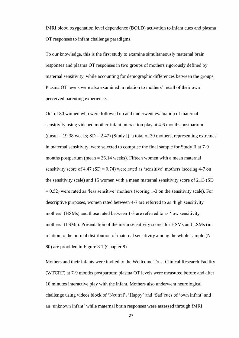
Out of 80 women who were followed up and underwent evaluation of maternal
sensitivity using videoed mother-infant interaction play at 4-6 months postpartum
(mean = 19.38 weeks; SD = 2.47) (Study I), a total of 30 mothers, representing extremes
in maternal sensitivity, were selected to comprise the final sample for Study II at 7-9
months postpartum (mean = 35.14 weeks). Fifteen women with a mean maternal
sensitivity score of 4.47 (SD = 0.74) were rated as ‘sensitive’ mothers (scoring 4-7 on
the sensitivity scale) and 15 women with a mean maternal sensitivity score of 2.13 (SD
= 0.52) were rated as ‘less sensitive’ mothers (scoring 1-3 on the sensitivity scale). For
descriptive purposes, women rated between 4-7 are referred to as ‘high sensitivity
mothers’ (HSMs) and those rated between 1-3 are referred to as ‘low sensitivity
mothers’ (LSMs). Presentation of the mean sensitivity scores for HSMs and LSMs (in
relation to the normal distribution of maternal sensitivity among the whole sample (N =
80) are provided in Figure 8.1 (Chapter 8).
Mothers and their infants were invited to the Wellcome Trust Clinical Research Facility
(WTCRF) at 7-9 months postpartum; plasma OT levels were measured before and after
10 minutes interactive play with the infant. Mothers also underwent neurological
challenge using videos block of ‘Neutral’, ‘Happy’ and ‘Sad’cues of ‘own infant’ and
an ‘unknown infant’ while maternal brain responses were assessed through fMRI

28
scanning. Plasma OT responses and BOLD activation responses were compared
between the two groups (HSMs/LSMs). We also examined for a coordinated association
between maternal BOLD brain activation and the post interaction level of plasma OT
among mothers.
The literature review for Study II is included in Chapters 6 &7, followed by
methodology (Chapter 8), results (Chapter 9), and discussion (Chapter 10). Chapter 11
provides a summary and conclusion of the whole thesis (Study I & Study II).
1.2. Rationale for Submitting the Thesis in an Alternative Format
This thesis is submitted in an alternative (publication) format. The thesis comprises 11
chapters and 3 research manuscripts written up in paper format, all first authored by the
author of this thesis, and pending submission to international, peer-reviewed journals
for publication. Several reasons determined the choice of an alternative format thesis.
First, the study contained novel and important findings, which could have implications
for future approaches to early assessment of mothers at risk of low maternal sensitivity.
The findings might also aid the development of intervention, training and support for
those vulnerable mothers. Second, planning of papers helped build the scope of the
thesis structure at an earlier stage, which helped to provide a more focused and coherent
thesis. Third, this format has provided the author with a great learning experience by
taking her through the discipline of writing research papers; this provides extensive
opportunities for review (including peer reviews when the papers are submitted) and
feedback that strengthens the quality of the work.

29
Figure 1.1. Time chart for data collection times. Note. PBI: Parental Bonding Instrument, MFAS: Maternal Fetal Attachment Scale, Oslo3-
items: social support scale, HADS: Hospital Anxiety and Depression Scale, EPDS: Edinburgh Postnatal Depression Scale, CTQ: Childhood
Trauma Questionnaire, IBQ-R-v.short: Infant Behaviour Questionnaire revised-very short form, fMRI: functional Magnetic Resonance
Imaging, OT: Plasma oxytocin sample. Maternal sensitivity rating was obtained from Time 2 video record of mother-infant interaction.

30
1.3. Publications
The three papers included in this thesis, aimed to achieve a better understanding of the
mechanisms underlying natural variation in maternal sensitivity in healthy mothers. The
author has made a major contribution to the papers including data collection, analysis of
results and writing up. The initial drafts of all the papers included have been written by
the author of this thesis and subsequent editing in response to co-authors has also been
performed by the author. All research materials included in these papers were derived
from the original research undertaken in this thesis.

31
Publication 1: Maternal sensitivity in healthy mothers: Can at-risk maternal sensitivity
be predicted prenatally?
Alya Elmadih, Kathryn M Abel, Rebecca Elliott, and Ming Wai Wan
Abstract
In spite of the large research investment and accumulating evidence that parenting
interventions which optimise infant developmental and mental health can improve
maternal sensitivity, translation of such knowledge into service delivery has been
extremely limited. Interventions are resource-intensive; selecting groups deemed at-risk
(e.g. mothers with mental illness) may not best address the general population’s mental
health. An alternative approach would be to identify those mothers at risk of low
maternal sensitivity in the prenatal period when all early postpartum mothers make
contact with services in order to facilitate delivery of effective intervention early in the
postpartum. Objectives: The primary aim of this study was to identify prenatally
determined psycho-social and demographic factors, which might predict maternal
sensitivity at 6 months postpartum. A secondary aim was to examine whether the
number of psycho-social and demographic factors to which mothers were exposed
predicted lower maternal sensitivity. Design: In the third trimester, 105 healthy,
pregnant women were assessed on simple self-report measures. At 4-6 months
postpartum, 6 minutes of unstructured mother-infant play was videotaped during a
home visit and was blind rated for maternal sensitivity using the Manchester
Assessment of Caregiver-Infant Interaction (MACI). Results: Several prenatally-
measured factors (score of depressive symptoms, experience of own parental care,
parental overprotection, history of trauma, household income, and educational
attainment) were associated with maternal sensitivity at 4-6 months postpartum. Only
two factors (mother’s own reported experience of parental care, and household income)
independently predicted maternal sensitivity, accounting for 17% of the variance. The
number of psychosocial risk factors also predicted lower sensitivity: mothers exposed to
3+ psychosocial risk factors were more likely to show lower sensitivity to their infants.
Conclusion: Relatively simple prenatal ‘screening’ of psycho-social and demographic
risk factors in healthy mothers can identify those who are more likely to be at risk of
low maternal sensitivity. However, asking mothers prenatally about their general social
supports or how well they are bonding with their infants did not predict maternal
sensitivity. Routine assessment of key maternal factors may be a relevant adjunct to
other forms of antenatal health screening.

32
Publication 2: Does oxytocin modulate variation in maternal caregiving in healthy new
mothers?
Alya Elmadih, Ming Wai Wan, Michael Numan, Rebecca Elliott, Darragh Downey, and
Kathryn M Abel
Abstract
Background: The extent to which a mother is sensitive to her infant’s cues and
developmental needs (‘maternal sensitivity’) contributes to the infant’s social and
cognitive development. Animal and recent human studies emphasise a major role for the
neuropeptide Oxytocin (OT) in mediating sensitive caregiving behaviours. To date, no
study has examined OT in relation to extreme variations in human maternal sensitivity.
Methods: : Out of 105 expectant mothers, 80 were followed up and underwent
evaluation for maternal sensitivity at 4-6 months postpartum through 6 minute-free play
interaction with their infants. Of these, 30 enrolled in the current study at 7-9 months
postpartum: 15 ‘sensitive mothers’ (henceforth high sensitivity mothers – HSMs) and
15 ‘less sensitive mothers’ (henceforth low sensitivity mothers – LSMs) underwent
plasma OT measurements before and after 10 minutes of play interactions with their
infants. Results: Consistent with studies of plasma OT and stress in women, but not
with studies of plasma OT and maternal behavior in women, baseline and post-
interaction plasma OT levels were lower amongst HSMs. Only HSMs showed
significant change in plasma OT; with reduction following the play-interaction.
Conclusion: Higher baseline OT levels in healthy LSMs may act as a biomarker for
stress response owing to the demands of caring for an infant or for a gap in own
parenting relationship. OT may be acting to reduce stress and anxiety. By contrast, play
interaction with their infants may be associated with reduced stress (if any) in HSMs, as
suggested by a significant reduction in plasma OT. Plasma OT might represent a useful
biomarker of low maternal sensitivity. Considering mothers in well-defined sensitivity
groups might ‘tap’ on an element of a stress or anxiety coping strategy and might foster
better understanding of parental caregiving behaviour and its potential for modulation
by OT.
33
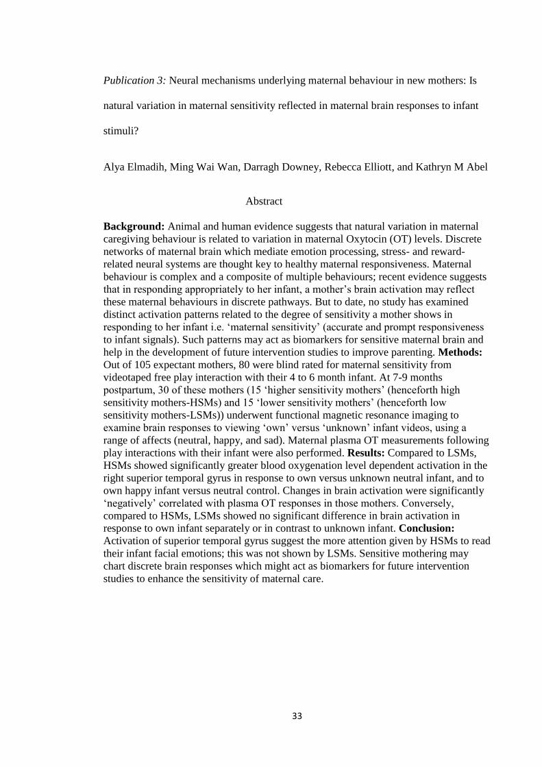
Publication 3: Neural mechanisms underlying maternal behaviour in new mothers: Is
natural variation in maternal sensitivity reflected in maternal brain responses to infant
stimuli?
Alya Elmadih, Ming Wai Wan, Darragh Downey, Rebecca Elliott, and Kathryn M Abel
Abstract
Background: Animal and human evidence suggests that natural variation in maternal
caregiving behaviour is related to variation in maternal Oxytocin (OT) levels. Discrete
networks of maternal brain which mediate emotion processing, stress- and reward-
related neural systems are thought key to healthy maternal responsiveness. Maternal
behaviour is complex and a composite of multiple behaviours; recent evidence suggests
that in responding appropriately to her infant, a mother’s brain activation may reflect
these maternal behaviours in discrete pathways. But to date, no study has examined
distinct activation patterns related to the degree of sensitivity a mother shows in
responding to her infant i.e. ‘maternal sensitivity’ (accurate and prompt responsiveness
to infant signals). Such patterns may act as biomarkers for sensitive maternal brain and
help in the development of future intervention studies to improve parenting. Methods:
Out of 105 expectant mothers, 80 were blind rated for maternal sensitivity from
videotaped free play interaction with their 4 to 6 month infant. At 7-9 months
postpartum, 30 of these mothers (15 ‘higher sensitivity mothers’ (henceforth high
sensitivity mothers-HSMs) and 15 ‘lower sensitivity mothers’ (henceforth low
sensitivity mothers-LSMs)) underwent functional magnetic resonance imaging to
examine brain responses to viewing ‘own’ versus ‘unknown’ infant videos, using a
range of affects (neutral, happy, and sad). Maternal plasma OT measurements following
play interactions with their infant were also performed. Results: Compared to LSMs,
HSMs showed significantly greater blood oxygenation level dependent activation in the
right superior temporal gyrus in response to own versus unknown neutral infant, and to
own happy infant versus neutral control. Changes in brain activation were significantly
‘negatively’ correlated with plasma OT responses in those mothers. Conversely,
compared to HSMs, LSMs showed no significant difference in brain activation in
response to own infant separately or in contrast to unknown infant. Conclusion:
Activation of superior temporal gyrus suggest the more attention given by HSMs to read
their infant facial emotions; this was not shown by LSMs. Sensitive mothering may
chart discrete brain responses which might act as biomarkers for future intervention
studies to enhance the sensitivity of maternal care.

34
Study I: Natural variation in maternal sensitivity: What are the possible prenatal and
postnatal predictors in healthy early postpartum mothers?
Chapter 2: Study I Literature Review
2.1. The Prenatal Development of Maternal Sensitivity
Observations of the intense grief shown by mothers whose infants have died during
birth suggest the existence of a prenatal bond between a mother and her fetus (Kennell
et al., 1970). Pregnancy not only includes the physical development of the fetus, but
also the psychological adjustment of the expectant mother (Rosenblatt, 1998; DiPietro,
2010). This includes the development of a maternal identity, identity of her developing
fetus and her relationship with the fetus (Cranley, 1981; Gloger-Tippelt, 1983). This
combination of anxious preoccupations and pleasurable attachment towards the unborn
infant prepares the mother for ‘motherhood’ (Leckman et al., 1999). This concept can
be referred to as maternal-fetal attachment (MFA), defined as “the extent to which
women engage in behaviours that represent an affiliation and interaction with their
unborn child” (Cranley, 1981) and represents one aspect of the ‘transition to maternity’
that is thought to occur in primiparous women.
Transition to maternity is also evidenced in animals, though with some difference from
humans (Ross et al., 2009). While virgin female rodents are aversive to pups, the
extensive hormonal changes and the physiological events of pregnancy and puerperium
(Moltz et al., 1970; Bridges, 1996) produce an ‘enriched environment’ that encourages
mothers to interact with offspring in order to facilitate the development of maternal
caregiving behaviour (e.g. nest building, pups retrieval) (Fleming et al., 1988). Animal
research has provided increasing insight into the biological mechanisms underlying the

35
transition to maternity, with a special implication for the oestradiol and OT hormones
(Poindron, 2005). Similarly, in humans, the early postpartum constitutes a period of
tremendous hormonal changes that serve adaptive functions in preparation for
caregiving (Workman et al., 2012). The particular implication of OT in the early
development of maternal care behaviour has also been suggested by evidence from
recent human studies, such as Levine et al. (2007) who reported a positive correlation
between maternal-fetal attachment and the steady rise in plasma OT levels during
pregnancy and the first postpartum month.
Following the infant’s birth, the human mother-infant relationship continues to develop,
with this caregiving now expressed as observable behaviours such as touch, ‘motherese’
vocalisation and affection (Feldman et al., 2007). Similarly in animals, pup-licking,
grooming and arched-back nursing (LG-ABN) are seen (Champagne et al., 2001).
However, in both humans and animals broad natural variations in these caregiving
behaviours have been reported, ranging from high to mid to low levels of maternal care
(Feldman et al., 2007; Gordon et al., 2008; Champagne et al., 2001).
2.2. What Is Maternal Sensitivity?
Mary Ainsworth’s definition of maternal sensitivity has long been considered the ‘gold
standard’ definition, that is: “the ability for perception and accurate interpretation of
baby’s signals, and appropriate responsiveness” (Ainsworth et al., 1978). Others have
since focused on particular aspects of this definition, such as the awareness of the
child’s affective state (Crawley & Spiker, 1983) or the behavioural response to infant
cries (Crockenberg, 1981; Egeland & Farber, 1984). The appropriateness of the
mother’s response is also highlighted as an important element of maternal sensitivity by
some (e.g. Crittenden, 1981; Smith & Pederson, 1988), whereas others concentrate on

36
the timing of these maternal responses (e.g. Fish et al., 1991). Chibucos & Kail (1981)
have talked about a physical component of maternal sensitivity in the form of
appropriate handling. Crittenden (1981) and Crnic et al. (1984) believe that allowing
the infant time to respond before further stimulation leads to reciprocal interaction
which, according to Marfo (1992), results in the interaction being mutually rewarding.
A sensitive mother, according to Skinner (1985), shows that her primary concern is for
the child and not herself, in the concept of particular attitude. Nover et al. (1984) see
sensitivity as the infant’s free exploratory play with no interference, which introduces
the idea of sensitivity being non-intrusive (Crawley & Spiker, 1983; Smith & Pederson,
1988). Others consider maternal sensitivity as the emotional availability of the mother
to the child (Kivijarvi et al., 2001). Fonagy et al. (1994) and Meins (1997) raise the
notion of mind-mindedness, which suggests maternal sensitivity to the infant’s mental
state, rather than to her/his physical state.
‘Maternal sensitivity’ is thus a relatively broad concept that includes a variety of
interrelated affective and behavioural caregiving attributes (Thompson, 1997) in
keeping with that defined by Ainsworth and her colleagues (Shin et al., 2008). A
sensitive mother responds properly to her infant’s displayed emotions by affirming the
positive emotions or reassuring about the negative ones (Sroufe, 2000). A sensitive
mother also modifies her responses according to the child's developmental level (Pianta
et al., 1989), thus gradually allowing the child time to cope with and explore new
situations (if appropriate) (Lohaus et al., 2004). In this beautiful ‘reciprocal dance’, a
sensitive mother knows whether her behaviour is or is not adequate for her infant’s
needs (Kivijarvi et al., 2001).

37
2.3. Why Is Maternal Sensitivity Important?
2.3.1. Child outcomes
i. Attachment
Ainsworth et al. (1978) were the first researchers to examine the relationship between
parental behaviours and attachment “the proximity seeking behaviour between the child
and his main caregiver” Bowlby (1969; 1973; 1980) when they observed 26 middle-
class mother-infant dyads from Baltimore. Mothers were visited at their homes for 4
hours every month during the first year of life. During these visits, a variety of maternal
behaviours were assessed, namely sensitivity, acceptance, cooperation and accessibility.
When they assessed attachment at 12 months, they found a strong correlation between
security of attachment and sensitivity (r = 0.78). Accordingly, they suggested
‘sensitivity’ to be the crucial factor that fosters attachment. Nonetheless, this strong
association between sensitivity and attachment has not been left without challenge as
some subsequent studies found only moderate relationship between sensitivity and
attachment security (e.g. Egeland & Farber, 1984; Smith & Pederson, 1988; Teti et al.,
1995; Vondra et al., 1995; Seifer et al., 1996; Beckwith et al., 1999).
The less stronger association between maternal sensitivity and attachment found by
some studies (as compared to Ainsworth’s strong association) may be attributed to: a)
different measures used by studies to assess maternal sensitivity (e.g. Beckwith et al.,
1999); b) different methodologies among different studies (i.e. context, assessment
duration); c) lack of consensus on the conceptualisation of ‘maternal sensitivity’ (Meins
et al., 2001); with some researchers using the term ‘maternal sensitivity’
interchangeably with the terms ‘maternal responsiveness’ (e.g. Blank et al., 1985; De
Wolff & Van Ijzendoorn, 1997) and/or ‘maternal competency’ (e.g. Pianta et al., 1989;
Zahr & Cole, 1991), which can be confusing; d) the crudeness of Ainsworth’s maternal

38
sensitivity scale (De Wolff & Ijzendoorn, 1997) such that some components of the
measure are difficult to define as the child grows, e.g. ‘promptly’ (Lohaus et al., 2004).
This diversity in studies’ approaches is illustrated by the findings of two meta-analysts,
Goldsmith & Alansky, (1987) and De Wolff & Ijzendoorn, (1997), who reported small
to medium size effect sizes for the relationship between sensitivity and attachment (r =
0.10 to 0.30 and r = 0.24, respectively).
A main limitation of Goldsmith & Alansky (1987) is that they derived their effect size
from only 12 studies in which a variety of measures (other than sensitivity) were
examined in relation to attachment. Furthermore, although De Wolff & Ijzendoorn
(1997) only include studies that used the ‘Strange Situation’ (Ainsworth & Bell, 1970),
they were having a problem in dealing with: a) studies that have multiple outcomes
(multiple variables to represent maternal sensitivity), and b) studies with multiple
assessments at different time points. These manoeuvres are likely to have affected the
size effect they reported when assessing the association between maternal sensitivity
and security of attachment.
On the other hand, studies that adhered to Ainsworth’s methodology (i.e. long, frequent
visits) reported a strong positive relationship between maternal sensitivity and security
of attachment (e.g. Isabella, 1993). A bigger relationship between maternal sensitivity
and attachment was also reported by others who assessed sensitivity at a single visit,
such as Pederson et al. (1998) who reported a strong correlation between the two
concepts (r = 0.51). Recent studies, which assess maternal sensitivity longitudinally,
have reported that maternal sensitivity at 4 months is a predictor for attachment security
when the infant is 2 years old (Bigelow et al., 2010). Furthermore, interventions that
were effective in enhancing parental sensitivity were also more effective in enhancing

39
attachment security, which supports the notion of a strong role of sensitivity in shaping
attachment (Bakermans-Kranenburg et al., 2003).
ii. Infant social, emotional and cognitive development
The appropriate responsiveness accounted for by sensitive maternal care “instils an
awareness of the caregiver’s availability and reliability, thus promoting a sense of
security and trust” in the infant towards her/his caregiver (Lohaus et al., 2001). It is this
trust which facilitates the social and emotional development of the infant (e.g. Landry et
al., 2001; Kivijarvi et al., 2001). As part of the National Institute of Child Health and
Human Development (NICHD) study, Belsky & Fearon (2002) report positive
correlations between maternal sensitivity (observed at 24 months) and different
dimensions of child development (i.e. social competence, expressive language,
receptive language and school readiness).
Infant cognitive development which depends on materials and stimulation provided by
the mother inside or outside the home to facilitate infant intellectual learning and
development (Crosnoe et al., 2010) has also been shown to link to maternal sensitivity.
For example, Downer & Pianta (2006) examined the relationship between maternal
sensitivity and cognitive development in 832 infants who were followed from one
month old until school grade 1 (about 4-5 years old). Children’s cognitive function was
assessed both at school entry (54 months) and at the middle of grade 1 using mother’s
and teachers’ reports. Maternal sensitivity was observed at 6, 15, 24, 36 and 54 months.
They reported that children who had experienced more sensitive maternal care scored
higher in maths and phoneme knowledge when assessed at grade 1. These findings
suggested that early maternal sensitivity influences child cognitive development and,
therefore, may also predict later academic performance. Although the study excludes

40
those with unfavourable conditions, e.g. young mothers, mothers who live in dangerous
neighbourhoods, the strength of this study came from the large sample size.
In another large cohort study, Pearson et al. (2011) investigate the relationship between
maternal responsiveness and infant mental development among 732 mother-infant
dyads. Maternal responsiveness was observed at 12 months through book reading task
(using the Thorpe Interaction Measure) and infant mental development assessed at both
18 months (using the Griffiths scales) and at 4 years (using the Wechsler Preschool and
Primary Scale of Intelligence). They reported that maternal responsiveness was
positively associated with infant mental development at 18 months. Although maternal
responsiveness was not related to infant cognitive ability at 4 years, this could be related
to the difference in the measures used at each time point.
iii. Child behaviour
Whereas sensitive maternal behaviours are positively associated with the infant’s
positive mood, as well as social and play behaviour (Kivijarvi et al., 2001; Kemppinen
et al., 2006), a mother’s inability to respond sensitively to her infant’s cues may lead to
negative emotional and behavioural outcomes (Denham & Moser, 1994). Several
studies have suggested that maternal ‘insensitivity’ is a precursor of high levels of child
aggression. For example, in a study investigating the relationship between maternal
sensitivity, discipline (i.e. rules that set limits for the child) and child aggression among
117 mothers and their children, maternal sensitivity and child aggression were both
observed at an average of 26 months (Alink et al., 2008). Their results showed that
negative disciplines are related to child aggression only when mothers are insensitive. In
another study, sensitive mothering during playing games and playful conversation was
predictive of low levels of aggression at school age (Olson et al., 2000). It is possible
that less maternal sensitivity means less support to help the children manage their

41
negative emotions independently, which results in an increase of negative behaviours
(Alink et al., 2008). Interestingly, evidence also suggests that higher levels of maternal
sensitivity in early childhood also contributed to later reductions in child anxiety and
depressive symptoms in temperamentally-vulnerable children (Warren & Simmens,
2005).
2.3.2. Parenting quality
In addition to the influence of early maternal sensitivity on child developmental
outcomes, maternal sensitivity in infancy also influences the quality of parenting in later
years. Older studies have found a link between maternal intrusiveness (i.e. lack of
respect for infant autonomy) –a concept that is linked to insensitive behaviour - and
abusive parenting (e.g. Lyons-Ruth et al., 1987). Recently, Joosen et al. (2012) assessed
maternal sensitivity among 73 mothers and their infants (using Ainsworth’s sensitivity
scale) at 3 and 6 months during bath and play interactions. Harsh parenting was then
assessed through three home visits when the infant was 2 years old. Mothers were
provided with an empty bag and some toys (on the floor) and instructed to ask the infant
to clean the toys off the floor into the bag. Verbal harsh discipline (e.g. impatient,
yelling) or physical (e.g. slapping, grabbing the child) harsh disciplines were then
assessed. Low maternal sensitivity was found to be a predictor of harsh parenting or
‘harsh discipline’ during toddlerhood. The strength of this study is due to that their
observational paradigm included non-distress and distressing interaction (through the
Still Face Paradigm), which provides a comprehensive view of maternal sensitive
responsiveness (Lindhiem et al., 2011).

42
2.4. How Is Maternal Sensitivity Measured?
There is great variation in the methods and procedures used to assess maternal
sensitivity (De Wolf & Van Ijzendoorn, 1997), with rating methods ranging from
maternal interviews to observations. A maternal interview reflects the mother’s
subjective perception of her relationship with her infant. Observational measures (in
which a trained rater evaluates an episode of mother-infant interaction blind to other
family information) are more objective and likely to be a more accurate representation
of the quality of maternal caregiving behaviour. Therefore, observation remains the
most frequently used measure. However, observational behavioural assessments may
also vary in their setting, some taking place at home (e.g. Gordon et al., 2010) and
others in a laboratory (e.g. Atzil et al., 2011). The context of the observed interaction
between mother and infant also varies; while some studies examine the unstructured
interaction between the mother and her infant (e.g. feeding and playing) (Gordon et al.,
2010), others observe a challenging situation (e.g. infant frustration) (Atkinson et al.,
2005). Furthermore, while some studies depend on long visits over a few months (e.g.
Ainsworth et al., 1978; Downer & Pianta, 2006), others are based on a single interactive
episode of a few minutes (e.g. Evans et al., 2007; Moore et al., 2009; Strathearn et al.,
2012). Infant ages at sensitivity assessment time also vary widely (Lohaus et al., 2004),
which might have an implication for the reliability of the assessment method (Lindhiem
et al., 2011).
2.4.1. Validity of the short single interaction observation
The robust reports of detailed and frequent observations of mother-infant interaction do
not necessarily imply limited reliability of short, single, time-point assessments (Mc
Elwain & Booth, 2006). This is supported by the findings of Evans et al. (2007) who
examined the validity of short videotaped, mother-infant laboratory observation against

43
the lengthy home assessment. Evans et al. observed 85 mother-infant dyads at home at
6, 12, and 24 months for 2-3 hours each. Maternal sensitivity was rated using MBQS
(90 items). At 24 months, mother-infant dyads were observed again in the laboratory for
10 minutes and maternal sensitivity was rated using MBQS-Revised (81 items). Their
results showed significant correlation in the maternal sensitivity rating from the short
videotaped laboratory interaction with the lengthy home assessment (r = 0.45; p < 0.01).
These findings support further the use of short observational assessments which are
time efficient, thus more convenient for use by clinicians. It is possible that the lengthy
duration of observation may increase the identification of positive (or negative)
interactions, yet the use of short observation has also been proved to be a reliable
indicator for these interactions. Currently, there is a range of short observational
validated measures used in rating maternal sensitivity with no ‘gold standard’
(Lindhiem et al., 2011). Table 2.1 represents examples of the commonly used measures
in rating maternal sensitivity through short observations.
In the current study, we rated maternal sensitivity through a short observation of the
mother-infant play interaction, using the Manchester Assessment of Caregiver-Infant
Interaction (MACI; Wan et al., 2012, 2013 online). This measure evaluates parent-
infant interaction on seven 1-7 scales, including maternal sensitivity. MACI was
adapted from the existing previously validated measures of mother-infant interaction,
the Global Ratings Scales of Mother-Infant Interaction (Murray et al., 1996) and the
CARE-Index (Crittenden, 1979-2004), both of which are suitable for use from 2 month
infant age or younger, respectively. The MACI was developed in 2008 to meet the need
for an assessment that is: (1) based on a normative developmental model (therefore with
demonstrated validity in healthy samples, despite its use in at-risk infants); (2) relatively
brief, and so suitable for quite large samples, as coding is resource intensive; (3)
suitable for longitudinal measurement at about 6-15 months (in normative samples).

44
The MACI defines caregiver sensitivity as: “Contingent and appropriate behavioural
responses to identified infant behaviours as is required and optimal to meet the infant’s
immediate and developmental needs”. Thus, it is similar to the original definition by
Ainsworth et al. (1978) - the mother’s ability to perceive infant signals and respond
promptly and appropriately - and focuses on quality. This construct of sensitivity is the
most commonly assessed in measures of maternal sensitivity (De Wolff & Van
Ijzendoorn, 1997). The MACI has assessed maternal sensitivity in the home and lab,
with pilot data showing high consistency of maternal sensitivity between contexts (r =
0.68; p = 0.008) (Green et al., 2013).

45
Table 2.1: Examples of commonly used measures for assessing maternal sensitivity through short mother-infant interaction observation
Measure Author Age Setting Situation Length Behaviour
evaluated
Main
advantage
(s)
Main
disadvantage
(s)
Ainsworth’s
Maternal
Sensitivity Scale
Ainsworth
et al.,
1978
Birth-
12
months
Home
Distressed
/
Non-
distressed
5 min Bi-dimensional
scale
Cost-
effective and
time-efficient
Needs long
training; crude
The CARE-Index Crittende,
1979–
2004
Birth-
30
months
Home,
clinic,
etc.
Non-
distressed
3 min 7 scales Applied by
para-
professionals
Needs extensive
training;
expensive
The Global Ratings
Scale
Murray et
al., 1996
2
months-
6
months
Home,
clinic,
etc.
Non-
distressed
5 min 13 mothers’
behaviours, 7
infants’
behaviours and
5 dyadic
behaviours
Quick to rate,
suitable for
clinical and
non-clinical
groups
Small age
ranges
The NICHD
version of the
ORCE
The
NICHD
Study,
1997
6, 15,
24 and
36
months
Home
or child
care
centre
Distressed
/
Non-
distressed
15 min 8 features of
caregiver’s
interaction, and
5 features of
child’s
behaviour
Developed as
part of the
largest,
longest-term
study of child
care
Needs long
training

46
2.5. Is Maternal Sensitivity a Stable Construct?
Despite the importance of maternal sensitivity as a concept, there are some
inconsistencies in the literature related to its stability (i.e. continuity within the same
mother) (Pianta et al., 1989; Lohaus et al., 2004). Many researchers assume the stability
of maternal sensitivity across development (see De Wolff & Ijzendoorn, 1997); some
researchers have tested this stability in longitudinal designs using a variety of
assessment methods. Evidence from these studies showed high (Grossmann et al., 1985;
Joosen et al., 2012), moderate (e.g. Vizziello et al., 2000; Kivijarvi et al., 2001) or even
slight (Beckwith et al., 1999) stability for maternal sensitivity. When Grossmann et al.
(1985) replicated Ainsworth’s et al. (1978) study, they observed 49 mother-infant dyads
of middle class families at 2, 6, 10 months (at home). Maternal sensitivity (assessed by
Ainsworth’s scale) was found to be highly stable across all assessments (r = 0.61, 0.58,
and 0.50; all p < 0.001). In the aforementioned study, Joosen et al. (2012) also assessed
maternal sensitivity, among 73 mothers and their second child both at 3 and 6 months
(during bathing and play interaction for 5 and 15 minutes respectively). They reported a
high stability in maternal sensitivity between the two assessments (r = 0.58; p < 0.01).
In contrast, a few researchers have reported only low stability in maternal sensitivity
(e.g. Beckwith et al., 1999; Lohaus et al., 2004). In the study by Lohaus et al. (2004), 60
primiparous mothers and their infants were observed during free play interactions at
home, both at 3 months and 12 months, for 1 hour at each time. Although, they found
low stability for maternal sensitivity over time (r = 0.19), this could be attributed to their
pure primiparous sample who lack experience with parenting and this could be reflected
in their interaction behaviour. Perhaps the greater confidence and competency which
was recorded by 12 months among these primiparous mothers (as reported by the

47
authors) was also interpreted as greater sensitivity as compared to their rating at 3
months postpartum. Consequently, a solely primiparous sample might affect the
stability of their assessment.
Although Grossmann et al. (1985) examined a wider infant age range than that in
Lohaus’s study and also observed a mixture of situations (e.g. feeding, play, infant
frustration) they found high to moderate stability for maternal sensitivity. This is
possibly because Grossmann et al. provide a more comprehensive assessment for
sensitivity by considering a range of interactions in their assessment, which is a
representation of mother and infant daily routines.
Despite the notion that higher maternal sensitivity ratings are found in relation to infant
distress compared to non-distress (McElwain & Booth-LaForce, 2006), this is actually
reflecting the flexibility of sensitive mothering as a dynamic concept around the infant’s
needs, rather than instability in the construct itself. In other words, infants need change
with the situation (or infant developmental stage). Therefore, it is to be expected that
maternal sensitivity would also change in order to accommodate this in order to allow
for ‘appropriate responsiveness’ (Ainsworth et al., 1978). Yet, this flexibility does not
affect the high face validity and the moderate to high stability of the maternal sensitivity
construct, allowing for it to be measured longitudinally and in different situations
(Lindhiem et al., 2011).
2.6. Factors That Influence Maternal Sensitivity
In spite of the importance of maternal sensitivity, and although much research has been
invested in this area, and has shown that parenting interventions can be effective in
improving maternal sensitivity (e.g. Bakermans-Kranenburg et al., 2003), translation
into service delivery has been very limited. This is due to two reasons. First, no routine

48
clinical screening currently identifies mothers at risk of poor maternal sensitivity. As a
result, parenting interventions to promote maternal sensitivity and optimise child
outcomes tend to focus on the clinical group, i.e. mothers with mental illness such as
postnatal depression (e.g. Murray et al., 2003; Forman et al., 2007; Barlow et al., 2008).
However, it is increasingly clear that far more children are exposed to poor parenting
than such a restricted approach to risk identification is likely to access (Barlow et al.,
2008). An alternative is to offer a more personalised approach focusing most efforts on
those at-risk identifiable in the prenatal or early postnatal period. This is ideally when
all early postpartum mothers make contact with health services, and when mother-infant
dynamics may still be relatively malleable to interventions.
Second, to date researchers have not been able to identify all the factors that influenced
maternal sensitivity and their findings were not always consistent. Much of the variance
in maternal sensitivity remains unexplained (Drake et al., 2007). This is because a range
of maternal behavioural outcomes were investigated, including maternal ‘competency’
and ‘responsiveness’, with no particular focus on maternal sensitivity (Bornstein et al.,
2007) especially in healthy mothers. Two particular studies have identified multiple
postnatal predictors of ‘self-rated’ maternal sensitivity using self-rating ‘mother’s
identity with baby’ as a proxy measure for maternal sensitivity (Shin et al., 2006; Drake
et al., 2007). Mothers’ reported sensitivity toward their 6-week-old infants strongly
predicted by (retrospectively-reported) maternal-fetal attachment in a sample of 196
mothers (Shin et al., 2006). Employment status, social support and infant gestational
age at birth were also significant predictors for maternal sensitivity. Comparatively, in
the other study (N = 177), perceived life satisfaction, self-esteem and number of
children (parity) independently predicted ‘maternal responsiveness’ at 2-4 months
postpartum while maternal education, income and maternal age did not (Drake et al.,
2007). However, in both studies the outcome measured was concerned more with

49
maternal affective experience (self-reported) rather than behavioural sensitivity
observed in interaction.
Another reason for the lack of comprehensive evidence in what might predict maternal
sensitivity is that some researchers focus on the early postpartum period (e.g. Shin et al.
2006), while some confine their study to a special group (adolescents) (e.g. Bloom,
1995). Studies also focus on maternal sensitivity as a predictor for infant behavioural
and developmental outcomes rather than focusing on factors that influence sensitivity
(Susman-Stillman, 1996; Bakermans-Kranenburg & Van Ijzendoorn, 2006).
Nonetheless, and among the limited evidence, the literature has highlighted three broad
prenatal categories that might influence maternal sensitivity: a) mother’s prenatal
psychological characteristics, b) socio-demographic and support factors, c) early
experience in family of origin, and two further categories that might be assessed
postnatally: d) obstetric variables, and e) infant temperamental behaviour. Here, we
review the evidence for these categories being associated with variation in maternal
sensitivity. Figure 2.1 outlines a theoratical model for these categories in relation to
maternal sensitivity.

50
Figure 2.1 A theoratical model of factors that influence maternal sensitivity, based
on current empirical evidence (model used to guide the literature review and data
analyses). Note: MFR: Maternal-Fetal Relationship, SES: Socioeconomic status.

51
2.6.1. Mother prenatal psychological characteristics
i. Maternal mental state (depression and anxiety)
Depressive mood, even at non-clinical level, impairs the communication of emotions
between the mother and her infant (Blumberg, 1980, see Tronick & Reck, 2009) and
subsequently hinders maternal sensitivity (Sidor et al., 2011). This is because
depression tends to make a mother less emotionally available to her infant;
consequently, she may find it difficult to interpret and respond promptly to her infant’s
cues (Kemppinen et al., 2006; Tronick & Reck, 2009). Depressed mother migh also lack
the feeling of reward from social interaction, which makes sensitive responsiveness
more challenging (Kemppinen et al., 2006). Most of the studies which examine the
impact of depression on maternal sensitivity do so among clinically depressed samples
(e.g. Murray et al., 1996) and only a few studies consider community (non-clinical)
samples (e.g. Mills-Koonce et al., 2008; Sidor et al., 2011).
Studies have found significant correlation between postnatal higher maternal depression
scores and lower maternal sensitivity in a community sample (Campbell et al., 2007;
Mills-Koonce et al., 2008). Larger longitudinal studies, like the NICHD study (1999),
were also able to demonstrate the impact of maternal depression on sensitivity. In that
study, 1,215 women and their children were followed from birth to 36 months.
Depression was assessed by self-report questionnaire at 1, 6, 15, and 36 months and
maternal sensitivity was observed through mother-infant play interaction at the same
sessions. Mothers who reported chronic symptoms of depression (through 36 months)
were found to score the least sensitivity rating when compared with those who had not
experienced depression symptoms (NICHD, 1999).
Similar findings were reported by Campbell et al. (2007) who investigated the
trajectories of maternal depressive symptoms among a community sample of mothers

52
and their 11 months to 7 years old children. They found that the rating for maternal
sensitivity was generally high and increased when depressive symptoms were low and
that maternal sensitivity was low or decreased when depressive symptoms were high.
The mechanism underlying poor parenting in non-clinically depressed mothers is not
clear; whether it is purely related to the effect of symptoms or to other unfavourable
factors that co-exist with depressive mood (e.g. poverty, lack of social support and
marital difficulties) remains to be examined (Tronick & Reck, 2009).
Among the few studies that were unable to demonstrate a relationship between
depression and maternal sensitivity, some methodological issues were noticed. For
example, a recent study by Sidor et al. (2011) examined maternal postpartum depressive
symptoms among a non-clinical sample of 106 women (using the Edinburgh Postnatal
Depression Scale-EPDS) while maternal sensitivity was observed (using the CARE-
index). Although they did not find a correlation between maternal sensitivity and mood
symptoms, the fact that their sample was chosen as a socially deprived sample (e.g.
poverty, alcoholism, etc.) might conceal such relationship.
As with depression, only a few studies consider community sample when
examining the impact of maternal anxiety on sensitive responsivness. Among
these, evidence suggests that anxiety decreases the mother’s perceptual abilities
and leads to less engagement when interacting with her infant (Blumberg, 1980),
which might also decrease maternal sensitivity. Nicol-Harper et al. (2007)
investigated the impact of maternal anxiety (using State Trait Anxiety Inventory-
STAI-T) on mother–infant observed interaction in a community sample of 32
mothers with high anxiety (scored 40 or above in STAI-T) and 32 with low anxiety
(scored 34 or less in STAI-T). High trait anxiety mothers showed lower sensitive
responsiveness when compared to low anxiety trait mothers. The strength of this

53
study came from the fact that participants were screened for depression and
excluded from the study if they showed depressive symptoms. This ensured that
any group difference in mother-infant interaction would primarily be due to
anxiety and not due to depression; given the comorbidity between the two (Pollack,
2005). In a study that examined self-regulation in toddlers, women who reported
symptoms of anxiety scored less optimally than healthy controls on maternal
sensitivity (Feldman & Klein, 2003).
ii. Maternal–fetal attachment
Some researchers argue that maternal prenatal attachment is not 'attachment' in the
sense used in attachment theory (Bowlby, 1969), but is a multifaceted construct guided
by the caregiving system, and is reciprocal to the attachment system. Therefore, some
suggest that maternal-fetal attachment (MFA) or maternal fetal ‘relationship’ (MFR) is
more concerned with the need to protect the infant, therefore having more in common
with caregiving (Walsh, 2010). Cranley (1981) suggests that the Maternal Fetal
Attachment Scale (MFAS) measures “the extent to which a woman engages in
behaviours that represent an affiliation and interaction with her unborn child”. Muller
(1996) emphasises that their measure, Prenatal Attachment Inventory (PAI), assesses
the “unique, affectionate relationship that develops between a woman and her fetus”.
Moreover, Condon & Corkindale (1997) suggest that their Maternal Antenatal
Attachment Scale (MAAS) focuses mainly on “thoughts about the fetus”.
Research on the MFR has focused on factors that predict this early relationship (e.g.
mood state, social support, demographic factors) (see Alhusen, 2008; den Bergh &
Simons, 2009) rather than its relationship to postpartum mother-infant interaction or
child outcomes. However, MFR has been positively related to a range of self-reported
outcomes (as proxy for maternal sensitivity) such as self-reported separation anxiety at

54
1-2 months postpartum (Muller, 1996); ‘how a mother feels about her baby’ at 1 week
and 8 months postpartum (Mercer & Ferketich, 1990); and identity with the baby at 1 to
6 weeks postpartum (Shin et al., 2006).
Accordingly, evidence for a relationship between MFR and an observed parent-infant
relationship is very limited (e.g. Siddiqui & Hagglof, 2000; Fuller, 1990; Bloom, 1995).
Bloom (1995), who assessed MFR among 47 pregnant teenage women using MFAS,
found that the total MFAS score in the 3rd trimester (and not 1st or 2
nd) was correlated
with the number of affectionate behaviours in feeding in the first week postpartum (r =
0.34). Specifically, ‘attributing characteristics to the fetus’ subscale in the 3rd
trimester
was correlated with affectionate behaviours (r = 0.32) and caretaking behaviours (r =
0.34). However, these MFAS results have not been replicated in a wider demographic
of mothers. In addition some researchers have raised concerns about the reliability of
MFAS when used as ‘indivisual scales’ as opposing to an ‘overall measure’ (Galbally et
al., 2011). Siddiqui & Hagglof (2000), who assessed prenatal attachment to the fetus
among 100 pregnant women during the last trimester of their pregnancy, found that the
PAI score was positively related to face-to-face interaction at 3 months postpartum,
particularly in maternal stimulation behaviours using proximal behaviours, and
negatively with maternal non-involvement.
In summary, investigating the unique relationship between the expectant mother and her
unborn infant has provided some evidence to suggest that prenatal attachment is
important for the postnatal relationship. However, findings are mixed, with a reliance on
self-reported measures to assess mother-infant postpartum outcomes (e.g. Muller, 1996;
Shin et al., 2006), and a focus on the MFR as an outcome in itself (Alhusen, 2008).

55
2.6.2. Socio-demographic and support factors
i. Socioeconomic characteristics (education/financial status)
Some studies suggest that maternal sensitivity correlates positively with maternal
education, possibly because: a) educated mothers know that their infant uses them as the
main source of information and learning (Walker et al., 1986); b) a better SES, since
education grants mothers a higher job status, which is more likely to allow flexibility
with child care to fit her parenting role (Pederson et al., 1990); or c) a combination of
the two previous factors. However, some studies like the aforementioned study by
Drake et al. (2007) fail to find such an association between education and maternal
responsiveness. Perhaps at early postpartum (2-4 months after delivery) the influence of
education on maternal behaviour may not be as powerful as other factors, such as
hormones, in shaping maternal behaviours (Feldman et al., 2007). Thus, maternal
education may become a more important influence at a later stage postpartum when the
infant becomes more interactive.
One of the most robust findings, with respect to factors associated with maternal
sensitivity, is SES. Low-income is associated with psychological stresses and
diminished social support (Evans, 2008). Among 223 mothers assessed with their 13-
year-old children at home, children in lower income families reported receiving low
maternal responsiveness, and that their mothers were also highly stressed (Evans, 2008).
Although this study depended on self-report measures (completed by children) to assess
maternal responsiveness; their finding suggests that maternal psychological stress may
act as a mediator in the relationship between poverty and maternal responsiveness.
Others also report lower maternal sensitivity associated with lower SES mothers (e.g.
Sacker et al., 2002; Bornstein et al., 2007). It is possible that, being of lower SES

56
exposes mothers to a range of environmental risk factors and deprivations that challenge
their sensitivity to their infant and their emotional capacity (McAdoo, 2002; Evans,
2008). On the other hand, both higher social functioning and better social support exist
among higher SES mothers and might explain better interactions with infants (Eccles &
Harold, 1996). Although a study by Pederson et al. (1998) did not find an association
between maternal sensitivity at 13 months (assessed by MBQS) and maternal income
among the 60 mothers in their study, this is possibly due to their homogenous higher
income sample (mean $45,000/year) compared to the lower income sample in other
studies (e.g. Evans, 2008).
ii. Social support
Social support refers to “the emotional, instrumental or financial aid that’s obtained
from social relationship including partner’s contribution” (Bloom, 1994), has a positive
role in improving parenting and, in particular, maternal sensitivity (Stiles, 2010). It has
been suggested that the positive impact of social support on parenting works through
preventing or alleviating the perception of parenting as a stressful duty (i.e. the
buffering effect) (Andresen & Telleen, 1992). Emotional support also acts in a similar
way by improving maternal self-esteem and raising the mother’s confidence in order to
tackle the parenting role (Andresen & Telleen, 1992). Pauli-Pott and colleagues (2003)
found that marital and emotional support positively correlated with maternal sensitivity.
Similarly, Feldman & Klein (2003) reported a positive correlation between the father’s
involvement at 3 months and the level of maternal sensitivity at 9 months. Even among
mothers with mental illness, successful parenting is also related to stability within the
family and having good financial and social support (Abel et al., 2005).
iii. Demographic factors (maternal age at infant birth/primigravidas)

57
Giardino et al. (2008) examined maternal responsiveness in 56 adolescent mothers (14-
19 years) compared with 49 adult mothers (older than 19 years) at 6 weeks to 6 months
postpartum, using self-report of alertness and sympathy, observation of mother-infant
interaction, and measurement of physiological changes associated with responsiveness
(e.g. maternal heart rate, corticosterone level). Their findings revealed that, while there
was no difference between the two groups of mothers (adolescence/adult) in the self-
reported measures which rated their maternal competency, the maternal sensitivity
rating through observation showed less attentive responses to infants among the
youngest mothers. In addition, the youngest mothers showed lower physiological
responsiveness (i.e. assessed by maternal heart rate/corticosterone level) towards
infants’ cries compared with adult mothers. The authors relate the lower levels of
maternal sensitivity among the younger group of mothers to their lower level of social
support.
However, others like Demers et al. (2010) relate the low sensitivity among adolescent
mothers to their lower levels of mind-mindedness (i.e. the capacity of the mother to
comment appropriately on their infant’s internal states) (Meins, 1997). Demers et al.
(2010) examined mind-mindedness among 29 adult mothers (21 years or above) and 69
adolescent mothers (20 years or below) and maternal sensitivity was observed at 18
months postpartum (using MBQS for sensitivity). Maternal sensitivity, which was
significantly higher among adult mothers compared to the younger mothers, was
positively related to levels of mind-mindedness, which was also higher among this
group compared to the younger group. Although in general, older mothers may be more
likely to be rated as more sensitive than teenage mothers, the relationship between
maternal age and maternal sensitivity is not consistently reported and may not be linear
(Drake et al., 2007).
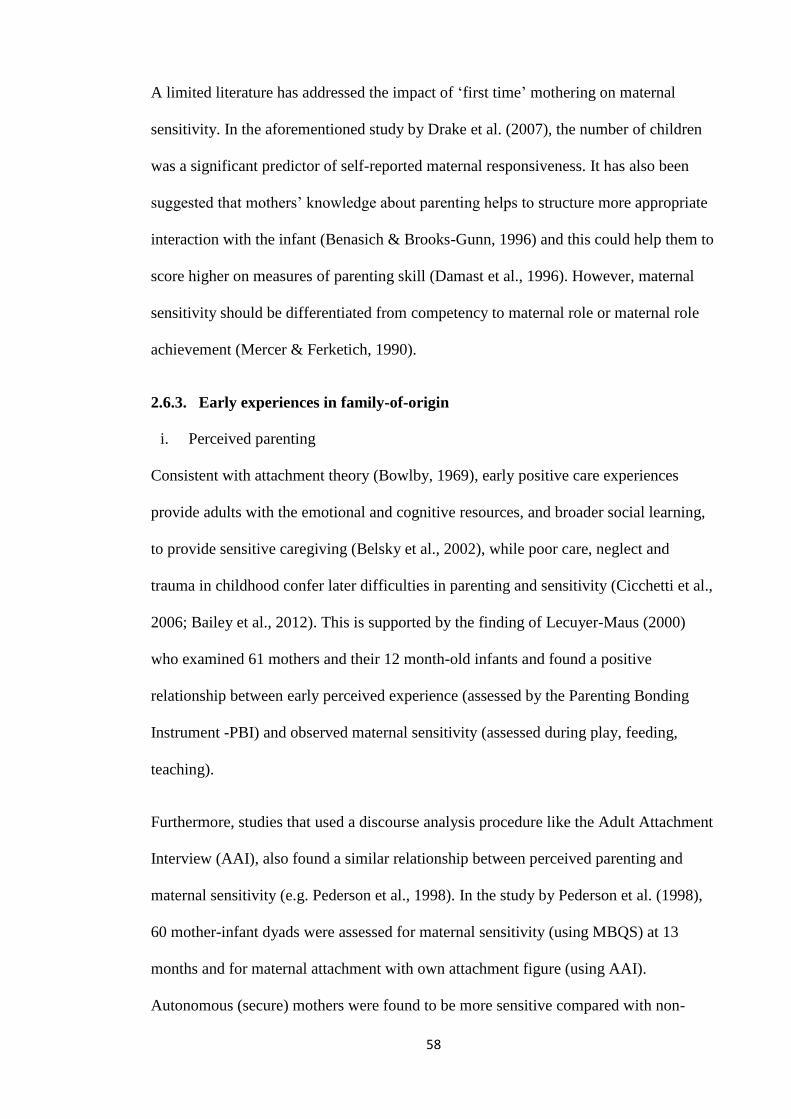
58
A limited literature has addressed the impact of ‘first time’ mothering on maternal
sensitivity. In the aforementioned study by Drake et al. (2007), the number of children
was a significant predictor of self-reported maternal responsiveness. It has also been
suggested that mothers’ knowledge about parenting helps to structure more appropriate
interaction with the infant (Benasich & Brooks-Gunn, 1996) and this could help them to
score higher on measures of parenting skill (Damast et al., 1996). However, maternal
sensitivity should be differentiated from competency to maternal role or maternal role
achievement (Mercer & Ferketich, 1990).
2.6.3. Early experiences in family-of-origin
i. Perceived parenting
Consistent with attachment theory (Bowlby, 1969), early positive care experiences
provide adults with the emotional and cognitive resources, and broader social learning,
to provide sensitive caregiving (Belsky et al., 2002), while poor care, neglect and
trauma in childhood confer later difficulties in parenting and sensitivity (Cicchetti et al.,
2006; Bailey et al., 2012). This is supported by the finding of Lecuyer-Maus (2000)
who examined 61 mothers and their 12 month-old infants and found a positive
relationship between early perceived experience (assessed by the Parenting Bonding
Instrument -PBI) and observed maternal sensitivity (assessed during play, feeding,
teaching).
Furthermore, studies that used a discourse analysis procedure like the Adult Attachment
Interview (AAI), also found a similar relationship between perceived parenting and
maternal sensitivity (e.g. Pederson et al., 1998). In the study by Pederson et al. (1998),
60 mother-infant dyads were assessed for maternal sensitivity (using MBQS) at 13
months and for maternal attachment with own attachment figure (using AAI).
Autonomous (secure) mothers were found to be more sensitive compared with non-

59
autonomous (ambivalence/avoidance) mothers. Even among teenage mothers (N = 74)
higher levels of maternal sensitivity were found among secure mothers compared to
ambivalence/avoidance mothers (Ward & Carlson, 1995). Although such experiences
rely on adult retrospective report, there are no strong reasons to suspect that prenatal
measurement would differ from postnatal or from earlier in adulthood (e.g. Talbot et al.,
2009).
ii. Childhood maltreatment
Old literature suggest that early childhood adversity (maltreatment) has implications for
future parenting (Belsky, 1984), and a range of self-reported maladaptive parenting was
identified in relation to maltreatment. Among the studies that focus on maternal
sensitivity as an outcome, high risk samples were considered (mothers with mental
illness, substance abuse or mothers whose child is removed from the home) (Lindhiem
et al., 2011) as part of interventions to improve their parenting skills (Cicchetti et al.,
2006).
Of the available research that examined low risk samples, three studies rated maternal
sensitivity in the observation setting. The first study (Moehler et al., 2007) assessed
emotional availability (including sensitivity and non-intrusiveness) through 3 minutes of
play interaction among 58 mothers with a history of abuse (scored above threshold in
Child Trauma Questionnaire-CTQ) and 61 control mothers (scored below threshold in
CTQ), both recruited through advertisement at 5 months postpartum. Results showed
that mothers with a history of abuse scored significantly lower in non-intrusiveness (p <
0.02) and scored borderline significantly lower in sensitivity (p < 0.08). However, given
that this is a low risk sample, their results, even at a trend, might still be considered
interesting. The second study (Bailey et al., 2012), adopts a similar methodology to the
first study, among 82 mothers of 4-6 year olds, but in order to increase the variability of

60
the sample participants had to have at least one social risk factor (i.e. young age at birth,
low income, single) to be included in the study. Emotional availability (sensitivity,
intrusiveness, hostility) was observed through play interaction, mother-directed clean up
and shared snacks with the infant. Maternal maltreatment was reported using the
History of Maltreatment and Trauma Form (CTQ was used to validate its use). The
results partially supported the first study: the maternal self-reported history of childhood
physical and emotional maltreatment was associated with hostility towards their own
child, while experience of sexual abuse was associated with maternal self-concern
regarding their parental competence.
Contrary to the previous two studies, the third study (Pereira et al., 2012) examined
maternal sensitivity in the home setting among 291 mothers and their 16 month-old
infants through 2 hours of observation for mother-infant daily routine (using MBQS).
Parenting stress was assessed through self-reporting and through interaction observation
(20 minutes toy deprivation task and 6 minutes when mother completed questionnaire
during infant presence). Lower sensitivity was rated among those who reported
maltreatment and those who experienced current parenting stress. They also reported
that maternal distress was a significant mediator for the relationship between maternal
sensitivity and maltreatment. This mediation through maternal distress is interesting and
might also explain the borderline or the insignificant findings of the other two studies
(Moehler et al., 2007; Bailey et al., 2012) with regard to the link between maltreatment
and maternal sensitivity in particular. The three studies suggest the impact of childhood
maltreatment on mother-child interaction and on maternal sensitivity, which is also
detectable among the community sample (e.g. Pereira et al., 2012).
A maternal history of abuse not only impedes development of healthy maternal
sensitivity but may also act as predictor of risk for child abuse. About 30% of mothers

61
who abuse their children were themselves abused (Knutson, 1995). The mechanisms
underlying this relationship are not clear, but the high stress levels and poor quality of
parenting (Hill et al., 2001; Pereira et al., 2012) associated with maltreatment are likely
to play a role in this.
2.6.4. Obstetric characteristics
i. Mode of delivery
Animal studies have linked vaginal delivery (VD) to better quality maternal caregiving
behaviour when compared with Caesarean section (CSD) (Poindron, 2005). Some have
suggested this may be because of the vagino-cervical stimulation which occurs during
vaginal delivery and which triggers the release of OT. In humans, however, no strong
evidence connects vaginal delivery to better quality of maternal care or to higher level
of OT. For example, most of the recent studies (albeit with rather small and limited
samples) that examine maternal plasma OT levels, at baseline or following interaction
with infant, do not find an association between OT and vaginal delivery (e.g. Feldman
et al., 2007; Atzil et al., 2011).
However, indirect evidence from functional imaging (fMRI) does suggest that, in
response to infants’ cries at 2–4 weeks, mothers who delivered vaginally show more
BOLD responses in brain areas related to maternal emotion, reward (amygdala) and
motivations (cingulate cortex) compared to women who delivered by CSD (Swain et al.,
2008a). It remains unclear whether mode of delivery would still influence maternal
behaviour assessed at later stages during infant development.
ii. Mode of feeding
Breastfeeding represents a close physical contact between mother and infant and has
been suggested to enhance the maternal infant bond (Feldman & Eidelman, 2003).
However, findings are inconsistent in this regard. Previously, some studies were unable

62
to support the relation between maternal sensitivity and breastfeeding. For example,
Pridham et al. (2001) found no relationship between breastfeeding and maternal
sensitivity observed through mother-infant interaction during feeding practice among 99
mothers and their infants at 1, 4, 8 and 12 months. Similarly, Drake et al. (2007)
suggested that breastfeeding was not a significant predictor of maternal responsiveness.
This might be attributed to the sample and measures of these studies, such as including
premature and low birthweight infants in Pridham et al., and the use of self-report
measures to rate sensitivity in the study by Drake et al.
On the other side, some studies links breastfeeding to greater maternal sensitivity. In a
study that examines the relationship between breastfeeding, maternal sensitivity and
attachment, Britton et al. (2006) recruited 152 women during pregnancy. Maternal
sensitivity was observed during feeding (using the Nursing Child Assessment Satellite
Training Feeding Scale) both at 3 and 6 months. They reported that mothers who
breastfed showed greater sensitivity at 3 months compared to bottle feeding mothers
and that the duration of breastfeeding correlated positively with the sensitivity rating.
Similar findings were reported by Tharner et al. (2012) among 675 mothers. In that
study, both mode of feeding and duration of breastfeeding were assessed at 2 to 6
months postpartum, while maternal sensitivity was observed at 14 months during free
interaction for 13 minutes. The duration of breastfeeding was positively related to a
higher maternal sensitivity rating. None of the two studies controlled for demographic
factors related to breastfeeding, such as maternal age, education and SES, the thing
which might have confounded their findings.
Recently, and as part of an fMRI study, Kim et al. (2011) assessed 9 breastfeeding
mothers and 8 formula-feeding mothers for maternal sensitivity (using Coding
Interactive Behaviour), observing this through mother-infant interactive play at home at

63
3–4 months postpartum. In this small sample, breastfeeding mothers showed higher
sensitivity ratings (p = 0.05). However, with the exception of the study by Britton et al.,
none of these studies provides a strong evidence for the implication of breastfeeding or
its duration in sensitive responsiveness, and thus further studies are needed to resolve
inconsistency in this regard.
2.6.5. Infant temperamental behaviours
Researchers have focused on infant temperaments (i.e. individual personality
differences in infants and young children) as a factor that could make sensitive
mothering more challenging (Van Ijzendoorn et al., 2007). Studies examining infant
temperament in relation to maternal behaviour have revealed mixed results. Thus,
highly irritable infants may receive less sensitive care (as a result of their difficult
temperaments) (Crockenberg, 1981; Van den Boom 1991). Van den Boom (1991)
observed maternal sensitivity and infant temperament at monthly intervals over 6
months among 15 irritable and 15 non-irritable infants. Mothers of non-irritable infants
were found to be more sensitive than those of irritable infants. This agrees with Thomas
& Chess (1977), who suggest that infant temperament can make maternal
responsiveness either highly challenging or less challenging. Whereas mothers of
children with an ‘easy’ temperament can be more reassured about the adequacy of their
parenting, those of ‘difficult’ temperament may be more likely to be distressed and
unsure about their maternal care behaviour. The latter effect might hinder maternal self-
efficacy (Bandura, 1977) and consequently maternal sensitivity.
Conversely, some mothers might interpret their infant’s temperament as a request for
more care and accordingly show more responsive maternal care with irritable infants
(see review by Crockenberg, 1986). According to Sroufe (1985), this is related to the

64
fact that parents attempt to be flexible around their infant’s needs and hence give more
attention to those who demand it. Ghera et al. (2006) examined 56 mothers and their 9
month-old infants (the latter were previously rated with negative reactivity through
laboratory observation in another part of the study) for maternal sensitivity. Maternal
sensitivity was observed during a 1 hour session that included feeding, free play,
caregiving (using Ainsworth’s scale). They used the Infant Behaviour questionnaire
(IBQ) as a measure for maternal perception of the infant’s ‘soothability’. Their results
showed that the relationship between infant negative temperament and maternal
sensitivity is positive when the mother’s perceive her infant as ‘soothable’ and negative
when she perceived her/him as unsoothable. Therefore, the mothers of negatively
reactive infants, who perceive their infants as not soothable or perceived herself as‘not
capable’ of soothing her/him, are at risk of providing low sensitivity care.
In contradiction to other studies that used maternal self-report measures (e.g. IBQ) to
assess infant temperament, Ghera et al. used such measure as an index for how the
mother perceived her child as soothable, rather than as an actual assessment for
temperament. This clarifies some ambiguity in the literature in this matter. For instance,
evidence suggests that studies that used observational measures to rate temperament
(e.g. Crockenberg & Acredolo, 1983; Crockenberg, 1981; Van den Boom, 1994), tend
to support a link between high irritability in infants and less sensitive maternal care. By
contrast, only a few of those studies which rely on parent rating of infant temperament
support this link (e.g. Campbell, 1979).
2.7. Summary
Natural variations in maternal sensitivity have been observed (Belsky et al., 2002).
Generally, insensitive mothering has been linked to poor infant emotional, social and

65
cognitive outcomes (Beckwith & Cohen, 1989; Kemppinen et al., 2006; Kivijarvi et al.,
2001). The literature implies many individual factors which negatively influence
maternal sensitivity. Maternal mood (depression/anxiety), even at non-clinical levels,
has been suggested to have a negative impact on maternal sensitivity (Tronick & Reck,
2009). Prenatal maternal attachment to the unborn child appears to be important for the
postnatal relationship, although there is no particular focus on maternal sensitivity
(Alhusen, 2008). Social support (Andresen & Telleen, 1992), maternal education
(Pederson et al., 1990) and financial stability (Evans, 2008) are all positively related to
maternal sensitivity. The mother’s experience in her family of origin has a significant
influence on a mother’s own future sensitivity with her children: better quality parental
care provides a good ‘model’ to follow when caring for own children (Lindhiem et al.,
2011). Conversely, maltreatment experiences (even in low risk samples) negatively
affect a mother’s sensitive responses (Pereira et al., 2012). Primiparity, mode of
delivery and feeding show less robust association with subsequent maternal care
behaviour and may be significantly confounded (e.g. Swain et al., 2008a). Similarly,
mixed results are also seen in relation to difficult temperament of the infant (Ghera et
al., 2006).
Targeted interventions are difficult when many of the variables that influence sensitivity
are still unknown. This warrants further exploration for variables that could influence
maternal sensitivity, especially those which could be determined prenatally so
intervention could start in the early postpartum period before mother-infant patterns of
interaction have become ‘ingrained’ and while the infant brain shows relative plasticity
to positive change. We attempt to evaluate a more comprehensive set of factors thought
to influence maternal sensitivity in order to answer this key question: In healthy
mothers, can we identify prenatally or early postnatally which mothers are at risk of low
maternal sensitivity at 4-6 months postpartum?

66
2.8. Study I Objectives
Using a sample of Caucasian UK mothers with no history of mental illness, the aim of
the current study was to examine whether a range of psycho-social and demographic
factors collated prenatally and postnatally could predict how sensitive mothers were
toward their infants during observed play interaction in their home environment. We
examined whether maternal sensitivity at 4-6 months is predicted by (1) the prenatally
collated information regarding the mother’s current mental state (depression, anxiety,
maternal-fetal attachment), the mother’s early experiences of being parented (care,
overprotection) and maltreatment, socio-demographic characteristics (household
income, educational level, maternal age) and social support; (2) the postnatally collated
information regarding infant temperamental behaviour as perceived by mothers.

67
Chapter 3: Study I Methodology
3.1. Sample
Recruitment took place at 6 community antenatal clinics in Greater Manchester region
in the North West of England between February 2011 and October 2011. For the
purposes of inclusion, women must be Caucasian, of 18 to 40 years old, fluent English
speakers, in the third trimester of pregnancy (28 weeks or more), with no current
psychiatric illness and not on psychotropic medications. Women with any known
abnormality or malformation in the current pregnancy, and who scored above threshold
for depression (see below), were excluded. Although women were also excluded from
follow-up if they scored above threshold for depression or their infants were diagnosed
with medical, sensory or developmental disorders, none fell under either criterion.
Following informed consent (see below), 105 women participated in the antenatal phase
(Time 1) at a mean of 33.9 antepartum weeks (SD = 3.19) and all agreed to be contacted
for follow-up postnatally.
Of 105 women included in Time 1, a total of 80 women and their infants were followed
up at 4-6 months postpartum (mean = 19.36 weeks; SD = 2.46) (Time 2), with 25
women dropping out before Time 2 and therefore excluded from the analyses: 14
mothers were lost to follow-up, 9 discontinued participation and 2 were no longer
eligible (1 infant was not living with the mother, 1 infant death). There was no
difference in demographic characteristics between those who dropped (N = 25) and the
final sample included in the analysis (N = 80) except in number of married women
which was higher in the latter (Table 3.1). Also, no difference between the drop out and
the follow up sample in regard to scores of measures completed at Time 1 (Appendix
A).

68
Several reasons contribute to the selection of inclusion criteria. For example, since
behaviours representing an affiliation with the infant are different across cultures
(Alhusen, 2008), having a multi-cultural sample might demand a bigger sample to
account for variability across cultures, which might not be feasible in the current study.
Therefore, we confined our recruitment to white women. Furthermore, only fluent
English speakers were included due to lack of validated translated versions from the
questionnaires used in the study. We had to exclude women with current psychiatric
illness to avoid being confounded by factors related to their own illnesses rather than to
the actual parenting experience (Murray et al., 1996). Knowing about fetal
malformation or abnormality might affect the mothers’ responses towards the fetus and
later after birth; these infants find difficulties in expressing their emotions in explicit
ways (Van Ijzendoorn et al., 2007; Landry et al., 2001). Maternal sensitivity was
assessed during 4-6 months rather than very early postpartum to ensure that mothers
were more settled in their parenting role. Finally, mother infant play interaction was
assessed at the women’s own homes to maximise the reliability of this assessment by
providing a naturalistic atmosphere during the interaction.

69
Table 3.1. Sample characteristics for women followed up postnatally (N = 80) and
the drop-out (N = 25)
Characteristic Women
followed up
in Time 2
(N = 80)
Drop-out
after Time 1
(N = 25)
t (103)
Chi-
square
test
p-value
Mean [SD]
Maternal age
(years)
29.59 [5.53] 27.96 [6.62] - 1.22 0.22
Gestational age
(weeks)
34.10 [3.25] 33.28 [2.97] - 1.12 0.77
Average maternal
education (years)
14.06 [2.80] 14.15 [2.43] - 1.87 0.43
Average annual
household income
(thousand pounds)
28.13 [4.26] 27.63 [4.64] - 1.76 0.23
Frequency (%)
Married/cohabiting 69 (86.3) 17 (68.0) 4.28 0.04
Primigravidas 41 (51.3) 13 (52.0) 0.01 0.95
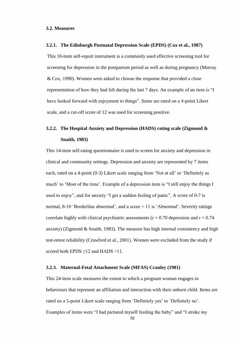
70
3.2. Measures
3.2.1. The Edinburgh Postnatal Depression Scale (EPDS) (Cox et al., 1987)
This 10-item self-report instrument is a commonly used effective screening tool for
screening for depression in the postpartum period as well as during pregnancy (Murray
& Cox, 1990). Women were asked to choose the response that provided a close
representation of how they had felt during the last 7 days. An example of an item is “I
have looked forward with enjoyment to things”. Items are rated on a 4-point Likert
scale, and a cut-off score of 12 was used for screening positive.
3.2.2. The Hospital Anxiety and Depression (HADS) rating scale (Zigmond &
Snaith, 1983)
This 14-item self-rating questionnaire is used to screen for anxiety and depression in
clinical and community settings. Depression and anxiety are represented by 7 items
each, rated on a 4-point (0-3) Likert scale ranging from ‘Not at all’ or ‘Definitely as
much’ to ‘Most of the time’. Example of a depression item is “I still enjoy the things I
used to enjoy”, and for anxiety “I get a sudden feeling of panic”. A score of 0-7 is
normal, 8-10 ‘Borderline abnormal’, and a score > 11 is ‘Abnormal’. Severity ratings
correlate highly with clinical psychiatric assessments (r = 0.70 depression and r = 0.74
anxiety) (Zigmond & Snaith, 1983). The measure has high internal consistency and high
test-retest reliability (Crawford et al., 2001). Women were excluded from the study if
scored both EPDS ≥12 and HADS >11.
3.2.3. Maternal-Fetal Attachment Scale (MFAS) Cranley (1981)
This 24-item scale measures the extent to which a pregnant woman engages in
behaviours that represent an affiliation and interaction with their unborn child. Items are
rated on a 5-point Likert scale ranging from ‘Definitely yes’ to ‘Definitely no’.
Examples of items were “I had pictured myself feeding the baby” and “I stroke my

71
tummy to quiet the baby when there is too much kicking”. In the current study, two
modification were made: as most women would know the fetus’ gender by the time of
recruitment (the third trimester), item 13 (“I have decided on a name for a baby boy”)
was omitted and item 10 (“I have decided on a name for a girl baby”) was re-worded to
include the word ‘boy’. Similar modification was done by Levine et al. (2007) to
accommodate for their third trimester sample. The MFAS coefficient of reliability was
reported as 0.85 (Cranley, 1981).
3.2.4. Parental Bonding Instrument (PBI) Parker et al. (1979)
This self-report measure examines an adult’s retrospective report of own parents’ caring
behaviours (25 items for each parent) during the first 16 years of life, consisting of
‘care’ (12 items) and ‘overprotection’ (13 items) scales. Items are rated on a 4-point
Likert scale ranging from ‘Very like’ to ‘Very unlike’. An example of a care item is
“Spoke to me in a warm and friendly voice” and of overprotection is “Tried to control
everything I did”. Participants were asked to score their parents as they remembered
them in the first 16 years. The PBI possessed good internal consistency (Parker et al.,
1979). Examination of test-retest reliability over 20 years supports the construct and
convergent validity of the measure over an extended period of time which, was found to
be independent of mood effects (Wilhelm et al., 2005).
3.2.5. Childhood Trauma Questionnaire (CTQ) Bernstein et al. (1997)
This 28-item retrospective self-report inventory screens for a history of childhood and
adolescent abuse and neglect involving three subscales assessing abuse (Emotional,
Physical, and Sexual) and two neglect subscales (Emotional and Physical) on a 5-point
Likert scale (5 being the most negative response). The scale also includes three items to
detect underreporting ‘denial/minimisation’ (items 10, 16 and 22). The measure is
interpreted as an overall sum for all the scales. The reliability coefficient for the CTQ

72
scales ranged from satisfactory to excellent, with Test-retest reliability revealing high
stability for the measure ranging between r = 0.79 and 0.86 for all scales (Bernstein et
al., 1994).
3.2.6. The Oslo 3-items social support scale (Dalgard, 1996)
This 3-item scale covers different areas of social support. Participants reported the
number of people close to them, ease of getting help from neighbours, and concern
shown by others. Responses are rated in a 5-point Likert scale for items 1 and 3 and in
4-point Likert scale for item 2. Results are interpreted as a cumulative overall score
which ranges from 3-14. One modification was made to item 1 in the current study:
“How easily can you get help from neighbours if you should need it?” was modified to
include ‘friends’ to emphasise the role of friends in social support as a more up-to-date
indication of social support, particularly in urban areas, as early piloting found that
women understood this mainly to mean immediate neighbourhood support.
3.2.7. The Manchester Assessment of Caregiver-Infant Interaction (MACI), Wan
et al., 2012, 2013 online)
This observational measure of caregiver-infant interaction evaluates global features
regarding the quality of interactions on a 7-point scale in 7 domains, including 2
caregiver scales (sensitive responsiveness and non-directiveness), 3 infant scales
(attentiveness to caregiver, positive affect and liveliness), and 2 dyadic scales (mutuality
and intensity of engagement). The measure is an adaptation of existing global scales of
caregiver-infant interaction (Murray et al., 1996; Blazey et al., 2008) and the CARE-
Index (Crittenden, 1979-2004) and has been validated for use in the 6-15 month-age
range (Wan et al., 2012, 2013 online). In the current study, inter-rater reliability was
tested for 30% of videotaped parent-infant interactions. Using absolute agreement inter-
class correlation coefficients, moderate to high agreement was demonstrated as follows:

73
maternal sensitivity: r = 0.70, p = 0.001; maternal non-directiveness: r = 0.55, p =
0.002; infant attentiveness to parent: r = 0.50, p = 0.004; infant positive affect: r = 0.62,
p = 0.001; infant liveliness: r = 0.47, p = 0.001; mutuality: r = 0.72, p = 0.001; and
intensity of engagement: r = 0.68, p = 0.001. Any disagreements in the complete sample
were resolved by both raters re-reviewing the clips to reach consensus.
The current study focused on one MACI domain: caregiver ‘sensitive responsiveness’,
defined as the caregiver’s “contingent and appropriate behavioural responsiveness to
identified infant behaviours as is required and optimal to meet the infant’s immediate
and developmental needs. In this measure, responsiveness considers quality, and its
broad definition includes an attentive attitude, and appropriate engagement, support and
structuring in response to both infant behaviour and a lack of behaviour.” The rating of
‘sensitive responsiveness’ varies from (1) ‘minimally responsive/sensitive’ to (7) ‘very
responsive/sensitive’ (Table 3.2; see Appendix B for full manual descriptions). This
definition of ‘caregiver sensitive responsiveness’ is similar to the original definition of
Ainsworth et al. (1978) of ‘maternal sensitivity’ (the mother’s ability to perceive infant
signals and respond promptly and appropriately) and is the most commonly assessed
construct of sensitivity in measures of maternal sensitivity (De Wolff & Van
Ijzendoorn, 1997). Thus, to avoid confusion, this MACI domain is henceforth referred
to as ‘maternal sensitivity’. MACI-rated maternal sensitivity has demonstrated
moderately high stability between 7 to 14 months (r = 0.48; Wan et al., 2013 online). It
is also independent of variables we do not expect to be related to interaction in
normative populations, including infant non-verbal developmental level and infant
gender (Wan et al., 2012).

74
Table 3.2. A brief description of rating definitions for maternal sensitivity on the
MACI measure
Rating Defining feature
1. Minimally
responsive/sensitive
Little evidence: Generally does not respond, or
responses are insensitive
2. Slightly
responsive/sensitive
Occasional or very moderate sensitive
responding
3. Fairly
responsive/sensitive
Scattered evidence; ‘fair’ but misses
opportunities, or takes over insensitively
4. Somewhat
responsive/sensitive
A mixed picture: sometimes responsive or
consistently mildly responsive
5. Generally
responsive/sensitive
Clear examples offset by scattered or mildly
insensitivity and/or responding
6. Responsive/sensitive Clearly evident, substantially outweighing
insensitivity/unresponsiveness
7. Very
responsive/sensitive
Consistent pattern of moderate to highly
sensitive responding throughout

75
3.2.8. Infant Behaviour Questionnaire Revised Very Short Form (IBQ-R-v. short)
(Gartstein & Rothbart, 2003)
This revised version of the original Infant Behaviour Questionnaire (Rothbart, 1981)
was developed in 2003 and refined in 2008
(http://www.bowdoin.edu/~sputnam/rothbart-temperament-questionnaires). The
measure assesses the dimensions of temperament in infants of 3 to 12 months, as
reported by parents. The IBQ-R-v. short measure consists of 37 items covering 3 “broad
scales”: ‘surgency/extraversion’ (the degree to which a child is generally happy, active,
seeking stimulation and enjoys vocalising) and ‘negative affect’ (the degree to which a
child is not easily calmed), and ‘effortful control’ (the degree to which a child can focus
attention, is not easily distracted) (Rothbart, 1981). Mothers rated the frequency of
specific temperament-related behaviours observed over the past week on a 7-point
Likert scale ranging from ‘Never’ to ‘Always’ and scored as 1 to 7 with 7 being the
highest frequency for behaviour. An example of a Surgency item is “When tossed
around playfully, how often did the baby laugh?” of a Negative affect item “When tired,
how often did your baby show distress?”, and of Effortful Control “When singing or
talking to your baby, how often did s/he soothe immediately?” Results are interpreted as
an average for each scale separately.
All measures used in Study I are included in Appendix B.
3.3. Procedure
3.3.1. Time 1 (Pregnancy)
After completing the informed consent, women were interviewed in private at the
antenatal clinic for 35 minutes, during which they completed the EPDS, HADS, MFAS,

76
PBI, and the Oslo 3-items social support scale. Detailed demographic information was
also collected.
3.3.2. Time 2 (4-6 months postpartum)
After prior arrangement, eligible and contactable mothers (N = 80) were visited in their
own homes at 4-6 months postpartum (mean = 19.36 weeks; SD = 2.46) at a time when
the infant was healthy and likely to be alert. Mothers were instructed to play with their
infant for 6 minutes on a floor mat as they would normally do, using toys (as supplied)
or not as they wished, ignoring the researcher’s presence. Mothers were asked not to
have visitors or other members of the family present during the interaction recording.
Interactions were stopped and later resumed if the infant got distressed, sick or upon the
mother’s request. Interactions were videotaped and later rated by an independent trained
researcher blind to participant information using the MACI. Mothers were administered
a structured interview which consisted of obtaining socio-demographic, obstetric and
perinatal data, and were administered the EPDS, HADS, IBQ-R-v.short, in addition to
CTQ which was delayed to this visit due to the sensitive nature of its questions.
Informed consent was completed at the beginning of the visit. Table 3.3 summarises the
measures used in Study I (Time 1 and Time 2).
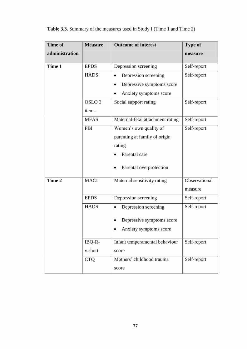
77
Table 3.3. Summary of the measures used in Study I (Time 1 and Time 2)
Time of
administration
Measure Outcome of interest Type of
measure
Time 1 EPDS Depression screening Self-report
HADS Depression screening
Depressive symptoms score
Anxiety symptoms score
Self-report
OSLO 3
items
Social support rating Self-report
MFAS Maternal-fetal attachment rating Self-report
PBI Women’s own quality of
parenting at family of origin
rating
Parental care
Parental overprotection
Self-report
Time 2 MACI Maternal sensitivity rating Observational
measure
EPDS Depression screening Self-report
HADS Depression screening
Depressive symptoms score
Anxiety symptoms score
Self-report
IBQ-R-
v.short
Infant temperamental behaviour
score
Self-report
CTQ Mothers’ childhood trauma
score
Self-report

78
3.4. Statistical Analyses
3.4.1. Sample size
A priori power calculation was performed before recruitment to determine the sample
size. The sample size of 80 is ample enough to detect relationships with up to 10
variables using regression analysis to detect a correlation of 0.21-0.25 (seen in the
literature in the variables of interest, e.g. Tarkka, 2003; Shin et al., 2006) at a power of
0.81, a medium size effect (0.45) and an alpha level of 0.05. Taking into account
dropouts and non responses over the two data collection times, a sample size of 105
women was selected at baseline (Time 1 of the study).
3.4.2. Data analyses
Sample descriptions were presented in frequency and descriptively, as appropriate.
All variables were checked for their distribution, and logarithm or square root
transformations were performed on variables that were skewed (showing less than - 0.8
or more than 0.8 skew). Pearson or Spearman correlations examined associations
between key measure variables and in relation to maternal sensitivity. Assessments for
covariates were performed and partial correlations were considered as appropriate.
Because women were assessed for maternal sensitivity at different postpartum time
(range: 16-26 weeks), infant age (at Time 2) was controlled for in all correlational
analyses that included maternal sensitivity and also when maternal sensitivity was a
dependent variable (regression analyses). Data were analysed using SPSS 19, and the
level of significance for all analyses was set at 0.05.

79
3.5. Ethical Considerations
3.5.1. Ethics
Information about the study - including the aims of the study, what it involved in
relation to participants, the nature of the questionnaires, as well as confidentiality issues
- was explained to women verbally by the researcher and in written format through the
information sheet. Women were given time to think about their participations and to ask
freely should they have any query related to the study. Informed consent was obtained
from each woman before each time they were involved in the study. Women were
aware that their participation was entirely voluntary and they were free to withdraw at
any time without giving a reason.
All personal data were kept anonymous, and all study data (paper, electronic, videotape)
were stored in accordance with the Data Protection regulation at the University of
Manchester, and Data Protection Act (1998). This study was approved by the North
West 8 Research Ethics Committee-Greater Manchester East (Study Ref: 10/H1013/69).
3.5.2. Benefits to research participants
The women in the current study were very keen to participate and they enjoyed
contributing to a study that might help to make a difference for some other mothers.
One woman who sent a thank you letter wrote “...I hope the study helps women who
have not been able to find that magical bond I have with my daughter”. Nevertheless, as
compensation for the time spent by women as a result of their participation, women
were given a value of £3 pounds Johnson’s baby starter pack at Time 1 and a value of
£15 pounds Boots shopping vouchers at Time 2.

80
3.5.3. Safety considerations
Although the Time 2 visit was conducted at the mothers’ home, a padded floor mat was
provided and safety was assessed to ensure it was safe for the infant to play on the floor
(e.g. no electrical sockets near the floor mat). The infant was always accompanied by
the mother at all times. When the mother was busy completing the questionnaires, the
infant had to be fastened in her/his seat to ensure her/his safety.

81
Chapter 4: Study I Results
The results are presented as follows: (1) Descriptive sample characteristics; (2)
Characteristics for the mother-infant interaction; (3) Preliminary analysis of each
measure (prenatal/postnatal) and its association with maternal sensitivity; (4) Main
analyses to test the predictive value of assessed variables on maternal sensitivity (using
stepwise regression).
4.1. Sample Characteristics
Within the final sample that followed up postnatally (N = 80 women) (Table 4.1), 61
(76.3%) had a natural delivery, 41 (51.2%) were primiparous, and 7 (8.8%) were less
than 21 years at time of infant birth. All women had healthy singleton term babies, a
mean birthweight of 3.45 kilograms (SD = 0.45), and 41 (51.3%) were female. More
than half of infants (n = 46; 57.5%) were breastfed (exclusive or combined). At the time
of follow-up (Time 2), the majority of mothers (n = 68, 85.0%) were on maternity leave,
9 (11.3%) were not employed, and 3 (2.9%) were back in work either as part time (n =
2) or full time (n = 1).

82
Table 4.1. Demographic characteristics for the sample (N = 80)
Demographic characteristic Statistics
(N = 80)
Mean [SD]
Maternal age (years) 29.59 [5.53]
Gestational age at Time1 (weeks)
Infant age at Time 2 (weeks)
34.10 [3.25]
19.36 [2.46]
Frequency (%)
Primiparity 41 (51.2)
Married/cohabiting
69 (86.3)
Household income/year:
£15000 or less
£16000 -£25000
£26000 -£35000
£36000-£45000
£45000 and above
£46000 and above
19 (23.8)
12 (15.0)
17 (21.3)
11 (13.8)
21 (26.3)
21 (26.3)
Highest educational level:
Secondary (GCSE)
Post-secondary education (A-
level or equivalent)
University degree and
postgraduate
26 (32.4)
23 (28.8)
31 (38.8)

83
4.2. Characteristics of Mother-Infant Interaction (Manchester Assessment of
Caregiver-Infant Interaction; MACI rating)
All the 7 domains (scales) within the MACI were normally distributed. After controlling
for infant age, maternal sensitive responsiveness (maternal sensitivity) (mean = 3.31;
SD = 1.31), correlated significantly with most other scales characterising mother-infant
interaction (Appendix A). These were maternal non-directiveness (mean = 3.13; SD =
1.30) (r = 0.50; p < 0.01), mutuality (mean = 2.95; SD = 0.97) (r = 0.72; p < 0.01),
intensity of engagement (mean = 4.06; SD = 0.88) (r = 0.28; 0.01), and infant
attentiveness (mean = 3.40; SD = 0.96) (r = 0.23; 0.05), but not with infant positive
affect (4.26; SD = 1.03) (r = 0.07; 0.56) or infant liveliness (mean = 3.18; SD = 0.97) (r
= 0.01; 0.99). On the other hand, the other maternal domain of MACI (maternal non-
directiveness) was only correlated with mutuality (r = 0.38; p < 0.01).
4.3. Preliminary Analyses (association of prenatal/postnatal factors with maternal
sensitivity)
Variables were examined for correlation with maternal sensitivity to determine what
would be examined as predictors in the main analysis.
4.3.1. Prenatal variables
i. Hospital Anxiety and Depression rating Scale (HADS) score
Depression score was based on HADS score and EPDS was only used as a screening
tool. We limited our inclusion to women who scored below threshold in both EPDS and
HADS depression (i.e. below 12 and 11, respectively). Mean EPDS for the included
women was 6.40 (SD = 4.03). The sample produced relatively low anxiety (mean =
5.56; SD = 3.43) and depression scores (mean = 3.39; SD = 2.72). Most anxiety and

84
depression scores (the latter transformed as was positively skewed) were within the
‘normal’ range. However, five women scored in the abnormal range for anxiety (score =
11, 11, 14, 16 and 18). One participant scored in the abnormal range on the depression
scale (score = 12) but had scored below the threshold on the EPDS (score = 11) and
therefore fitted within our inclusion criteria. Although 16 women scored ‘borderline
abnormal’ on anxiety, most of them (n = 12) were within the normal range in both
anxiety and depression scales. Anxiety and depression scores showed a strong
correlation (r = 0.65; p < 0.01). Accordingly their combined score was also examined.
Maternal sensitivity was correlated with depression score (r = - 0.27; p = 0.02) more
significantly than with anxiety score (r = - 0.21; p = 0.07) or the combined score (r = -
0. 24; p = 0.04). Accordingly, depression score was examined as a predictor variable in
the main analysis presented later.
ii. Maternal-Fetal Attachment Scale (MFAS) score
The mean score for MFAS was 94.70 (SD = 10.10). Maternal sensitivity was not
correlated with MFAS total score (r = 0.06; p = 0.60).
iii. Parental Bonding Instrument (PBI) scores
The PBI included ‘care’ scale and ‘overprotection’ scale for each own parent. Six
women reported absence of their own father, and accordingly their PBI paternal reports
were not completed. Maternal care (mean = 29.16; SD = 8.70) and paternal care (mean
= 26.14; SD = 9.76) (both transformed as they were negatively skewed) were
significantly correlated (r = 0.39; p < 0.01). Therefore, mean combined ‘parental care’
score (mean = 27.81; SD = 7.50) was analysed. Similarly, maternal overprotection
(mean = 11.85; SD = 6.26) and paternal overprotection (mean = 11.47; SD = 6.93) were
also highly correlated, and hence a mean combined ‘parental overprotection’ score
(mean = 11.61; SD = 5.64) was analysed. Maternal sensitivity was correlated more

85
strongly with parental care score (r = 0.31; p = 0.01) than with parental overprotection
score (r = - 0.24; p = 0.03). Therefore, parental care was included in the final analysis.
iv. Childhood Trauma Questionnaire score
Considering the sensitive nature of the CTQ questionnaire, it was completed at Time 2.
The overall CTQ score (mean = 32.25; SD = 14.14) was negatively correlated with
maternal sensitivity (r = - 0.29; p = 0.01) (after transformation), and therefore, was
included as a candidate predictor in the main regression analysis. It is worth mentioning
that our results showed 16 women (20.0%) with evidence of denial on the CTQ report
(scored 3 on denial items). However, when we re-ran the analyses excluding those
women, the results did not show significant change and therefore those women were not
excluded from the analyses.
v. Socioeconomic status (SES)
Controlling for maternal age, maternal education and household income were strongly
correlated with each other (Spearman’s r = 0.62; p < 0.01) as well as with maternal
sensitivity (Spearman’s r = 0.23; p 0.04, Spearman’s r = 0.27; p = 0.02, respectively).
Given the strong correlation related to the latter, household income was selected to
represent SES in the final analysis.
vi. The Oslo 3-items social support scale score
We considered the overall sum of the 3 items (mean = 12.09; SD = 1.95). Controlling
for parity, there was no significant correlation between the Oslo score (transformed as it
was negatively skewed) (negative trend) and maternal sensitivity (r = - 0.20; p = 0.07).
vii. Demographic characteristics related to parenting
Women who were less than 21 years, as expected at time of infant birth, (n = 7; 8.8%)
showed a trend for lower maternal sensitivity (mean = 2.43; SD = 3.38) compared with

86
older mothers (mean = 3.38; SD = 1.31); [F (1, 77) = 3.42; p = 0.07]. Single marital
status (n = 11; 13.7%) also showed a trend for lower maternal sensitivity (mean = 2.64;
SD = 1.43) compared with married or cohabiting mothers (mean = 3.41; SD = 1.2); [F
(1, 77) = 3.01; p = 0.09]. However given the small sample size for theses two variables
and the insignificant findings, no further analyses were performed.
A summary of the correlations between prenatal variables and maternal sensitivity is
presented in Table 4.2.
Table 4.2. Correlations of prenatal variables with maternal sensitivity
Variable Correlation with
maternal sensitivity
HADS anxiety - 0.21
HADS depression - 0.26*
Maternal fetal attachment 0.06
Parental care 0.31*
Parental overprotection - 0.24*
Overall CTQ score - 0.29*
Oslo social support score - 0.21
Household income1
Education1
0.27*
0.23*
*p < 0.05, 1: ordinal variable (Spearman’s correlation coefficient).

87
4.3.2. Postnatal variables
i. Hospital Anxiety and Depression rating Scale score
HADS anxiety (mean = 4.91; SD = 2.98) and depression scores (2.91, SD = 2.26)
generally remained low postnatally. Three participants scored above the threshold for
‘abnormal’ anxiety scores (score = 11, 13 and 19). None scored above the threshold on
the depression scores. The majority of women were within the normal range in both
scales (n = 65; 81.3%). Although a proportion of women (n = 10) scored borderline
abnormal on anxiety, most of them (n = 8) were within the normal range for depression
scale. Same as prenatally, postnatal anxiety and depression scores (the latter
transformed as was positively skewed) were significantly correlated (r = 0.50; p < 0.01),
and accordingly their combined score (mean = 0.44; SD = 3.43) was also examined.
However, maternal sensitivity was not correlated with anxiety (r = - 0.08; p = 0.50) or
depression scores (r = - 0.04; p = 0.75) separately or as a combined score (r = 0.07; p =
0.54). It is worth mentioning that prenatal and postnatal HADS depression scores were
strongly correlated with each other (r = 0.32; p = 0.01), and the same for anxiety scores
(r = 0.60; p < 0.01).
ii. Infant Behaviour Questionnaire Revised Very Short Form score
The three scales within IBQ-R-v.short, namely Surgency (mean = 3.80; SD = 0.82),
Negative affect (mean = 3.03; SD = 1.01) and Effortful Control (mean = 5.33; SD =
0.64), were not correlated with maternal sensitivity (r = 0.01 to 0.13; p = 0.25 to 0.93).
4.4. Main Analysis: Predictors of maternal sensitivity (Stepwise regression)
Based on the preliminary analyses, no postnatal factor was significantly associated with
maternal sensitivity; therefore, only prenatal factors were entered into a stepwise
regression. Four prenatal variables which were significantly correlated with maternal
88
sensitivity were included in the regression analyses, namely: depression score, own
parental care, childhood trauma, and household income in addition to infant age to
account for its variability. The results are presented in Table 4.3. In the first model,
infant age was entered, and then variables were added one at a time. In the second
model, depression scores accounted for significant variance of maternal sensitivity but
that was shared by parental care as evidence by cancellation of the depression effect
when parental care was entered (model 3) (parental care correlated negatively with
depression score, r = - 0.24; p = 0.02). When childhood trauma was added in the fourth
model, it cancelled the effect of parental care (parental care correlated negatively with
childhood trauma, r = - 0.76; p < 0.01). However, the childhood trauma score was not
even significant and it did not add any improvement in the overall regression.
Accordingly, childhood trauma was removed from the regression model. When
household income was entered in the fifth model, it proved to be a significant predictor
of maternal sensitivity, along with parental care. Therefore, about 17% of the variance
in maternal sensitivity is predicted from prenatal variables, with parental care received
by the mother and household income accounting for most of the variance.

89
Table 4.3. Stepwise regression examining prenatal predictors of maternal
sensitivity (adjusted R2) = 0.17
Model Variable Beta P-value Cumulative
adjusted R2
1 Infant age - 0.14 0.24 0.01
2 Infant age
Depression
score
- 0.13
- 0.23
0.27
0.05*
0.05
3 Infant age
Depression
score
Parental care
- 0.16
- 0.17
0.29
0.16
0.15
0. 01*
0.12
4 Infant age
Depression
score
Parental care
Childhood
trauma
- 0.16
- 0.16
0.28
- 0.03
0.17
0.17
0.12
0.89
0.10
5 Infant age
Depression
score
Parental care
Household
income
- 0.14
- 0.12
0.25
0.26
0.20
0.30
0.03*
0.03*
0.17
*p < 0.05

90
Chapter 5: Study I Discussion
5.1. Overview of the Findings
The present study (Study I) aimed to examine whether readily identifiable prenatal or
postnatal factors could help determining mothers at risk of low maternal sensitivity at 4-
6 months postpartum, in order to aid parenting interventions in the planning and
targeting of scarce resources to parenting outcomes.
Six prenatal variables were significantly correlated with maternal sensitivity: maternal
depression score, parental care, parental overprotection, household income, maternal
education, childhood trauma score, and two of these were significant predictors for
maternal sensitivity at 4-6 months postpartum (parental care, household income).
Conversely, maternal fetal attachment, maternal social support, young maternal age as
expected at birth (< 21 years), and single marital status were not related to maternal
sensitivity.
Although postnatal variables were measured at the same time as mother-infant
interaction video data was collated (on which maternal sensitivity was evaluated), none
of the postnatal variables were associated with maternal sensitivity, namely maternal
postnatal mood scores (anxiety, depression), and maternal report of infant temperament.
Accordingly, none of them was examined as a predictor. Our findings from the stepwise
regression revealed that own parental care and household income were significant
predictors of maternal sensitivity accounting for 17% of the variance in maternal
sensitivity after controlling for infant age at time of sensitivity assessment.
The discussion includes sub-sections including discussion of the mother-infant
interaction characteristics follow by our main findings from the regression model.

91
Interesting findings from the preliminary analysis, including the relationships between
prenatal and postnatal variables with maternal sensitivity, were also discussed to inspire
future hypotheses.
5.2. Mother-Infant Interaction
Our findings revealed that maternal sensitivity was correlated significantly with most
other scales characterising parent-infant interaction, namely maternal scale (maternal
non-directiveness), dyadic scales (mutuality, intensity of engagement) and an infant
scale (infant attentiveness). Thus, maternal sensitivity ratings in this cohort appear to
largely reflect the quality of overall interaction, and to a lesser extent the quality of
infant behaviour. On the other hand, the other maternal domain (i.e. non-directiveness)
correlated only with mutuality, signifying the unique contribution of maternal
sensitivity in mother-infant interaction quality.
5.3. Predictors of Maternal Sensitivity
The first main finding, the mother’s own reported level of parental care (maternal +
paternal), as measured prenatally using the PBI, was found to be associated with her
behavioural sensitivity to her own child. This is in line with attachment theory (Bowlby,
1969) as well as with other studies like Lecuyer-Maus (2000) and Pederson et al.
(1998). This finding, suggests that a positive care experience serves as an internalised
model for more sensitive ways of interacting with own infant (Ward & Carlson, 1995).
Furthermore, this impact of maternal own parental experience on maternal sensitivity
towards own children is consistent with the animal literature which reports a role for the
early environment in shaping maternal caregiving behaviour towards own offspring
(Francis, 1999, 2000; Champagne et al., 2007; Champagne, 2008). The concordance

92
between our findings and other studies that use a discourse analysis procedure
(Pederson et al., 1998; Lindhiem et al., 2011) also support the reliability of self-report
measures (i.e. PBI) in assessing own early parental care.
Another interesting finding related to parenting experience is also shown from the
regression analysis. Although initial depression scores accounted for significant
variance in maternal sensitivity, significant variance was shared with parental care.
Therefore, the effect of maternal depressed mood prenatally on later sensitivity (if any)
appears to be attributable to negative early care experiences, but not specifically to
trauma. Other studies of healthy populations also showed increase in depression scores
among those who showed low parental care or high parental overprotection (Hill et al.,
2001; Avagianou & Zafiropoulou, 2008). Our results suggest that a mother’s own
experiences of less optimum/negative early care might place her at later risk of both a
more depressed mood and lower behavioural sensitivity. Although one can expect that
PBI is a self-report measure, and therefore the mother’s own negative responses in this
instrument may be the artefact of a depressed mood. The concordance between our
findings and other studies that use a discourse analysis procedure (Pederson et al., 1998;
Lindhiem et al., 2011) support the reliability of self-report measures in assessing
previous parental care. Furthermore, studies like Hill et al. (2001) have assessed
depressive mood through an adapted measure (Schedule for Affective Disorders and
Schizophrenia), and they were also able to support the relationship between parental
care and depression.
The second main finding from the regression analysis, in regard to household income
(represents SES) as a significant predictor of maternal sensitivity, is also consistent with
other studies (e.g. Pederson et al., 1990; Sacker et al., 2002; Evans, 2008). This
relationship between SES and maternal sensitivity may be because higher SES mothers

93
have a more infant-centred approach to parenting than lower SES mothers (Ziv et al.,
2000), and therefore are more capable in seeking resources to help their parenting skills
(Bornstein & Bradley, 2003). The higher social functioning among higher SES mothers
in general might also explain better interactions with infants (Eccles & Harold, 1996).
This parental focus on infant fine-grained behaviours is exactly what is measured by the
MACI (i.e. the sensitivity rating measure used in the current study). On the other hand,
being of lower SES exposes mothers to a range of environmental risk factors and
deprivations that challenge their sensitivity to their infant and their emotional capacity
(McAdoo, 2002).
A previous study has reported SES as a significant predictor for infant responsiveness
(Bornstein et al., 2007). Therefore, one can speculate that high SES not only works
directly to enhance maternal sensitivity, but also through improving the reciprocal
interaction between the mother and her infant through increasing infant responsiveness.
Surprisingly, some researchers like Pederson et al. (1998) and Drake et al. (2007) did
not find this relationship between SES and maternal sensitivity. However, this could be
related to their methodological approaches as mentioned earlier. In comparison, in the
current study women represent a wide range of social classes and maternal sensitivity
was assessed through observation which allowed better chance to uncover this
relationship.
The childhood trauma score was not a significant predictor of maternal sensitivity.
Although previous studies among community samples reported negative associations
between maltreatment and different aspect of maternal behaviours, including non-
intrusiveness (Moehler et al., 2007) and hostility (Bailey et al., 2012), only one study
was able to show maltreatment as a predictor of maternal sensitivity (Pereira et al.,
2012). One might expect this is due to a low level of trauma in the community sample.

94
However, Bailey et al. (2012) increased the variability of their sample by only including
women who have at least one social risk factor (i.e. young age at birth, low income,
single), yet their findings revealed maltreatment to be related only to hostility towards
the child and not to maternal sensitivity. Although Pereira et al. (2012) reported that
maltreatment is a predictor of maternal sensitivity in a low-risk sample, yet they also
reported that this relationship is only significant when mediated by maternal distress
(observed and self-reported). The sample size of Pereira et al. was also much bigger
compared with the current study, or the samples of Moehler et al. or Bailey et al. (N =
291 vs. 80, 58 and 82, respectively).
Although our stepwise regression model explains only 17% of the variance in maternal
sensitivity, yet this is higher than some other studies like Drake et al. (2007) who
examined the predictive capacity of multiple variables and could explain for 15% of
variance in maternal sensitivity.
5.4. Associations between Maternal Sensitivity and Prenatal/Postnatal Variables
5.4.1. Prenatal variables
Aside from household income, our other proxy measure of SES (maternal education)
was also positively associated with maternal sensitivity. This is possibly due to higher
SES provided by education (Pederson et al., 1990), appreciation among educated
mothers of their role in the infant’s learning (Walker et al., 1986), or to more positive
disciplinary techniques adopted by an educated mother and hence enabling more
sensitive care (Augustine et al., 2009).
Unlike older studies such as Fuller (1990) and Siddiqui & Hagglof (2000), we did not
find a relationship between maternal fetal attachment and maternal sensitivity. Several

95
explanations might account for this. First, we assessed maternal sensitivity much later
(4-6 months) than Fuller or Siddiqui & Hagglof (first weeks postpartum). Therefore,
maternal-fetal attachment may no longer be related to maternal interaction style at a
later stage in postpartum and the interaction style by then is more related to other
measures reflecting maternal and infant current characteristics rather that how the
mother felt during pregnancy. Second, although Siddiqui & Hagglof (2000) reported
that PAI score was positively related to face-to-face interaction at 3 months postpartum,
particularly in maternal stimulation behaviours using proximal behaviours, and
negatively with maternal non-involvement, PAI was unrelated to maternal
responsiveness to infant behaviour. Therefore, MFR may be predictive of later maternal
involvement and stimulation (rather than sensitive responsiveness), and thus highlight
mothers at risk of withdrawal and neglect, rather than of low sensitive interaction per se.
Third, different measures have been used to measure MFR, for example while MFAS
emphasises maternal behaviours (Cranley, 1981), the Prenatal Attachment Inventory
(PAI) emphasises affiliation with regards to the prenatal relationship with the fetus
(Muller, 1993). Although MFAS is the most widely used measure for studying the MFR
(Beckwith et al., 1999), the subscales were theoretically rather than empirically derived,
with high internal consistency only for 2 factors, while other measures (e.g. PAI,
Maternal Antenatal Attachment Scale) have too few psychometrics published to date
(Alhusen, 2008). Although there is some evidence to suggest that maternal fetal
attachment might be related to future maternal responsive behaviour, better examination
of this link should be considered (see review by DiPietro, 2010).
Continuity of affect states (depression and anxiety) from the prenatal period to
postpartum period (at non-clinical levels) has been reported previously (Kaplan et al.,
2008). Similarly, in the current study, both prenatal and postnatal HADS depression

96
scores were strongly correlated with each other (as were anxiety scores). However,
depression scores in the prenatal period, but not at postpartum, significantly correlated
with maternal sensitivity (negatively). One explanation is that, we found higher prenatal
depressive symptoms compared to postnatal time (mean depression score 3.39 vs. 2.91
respectively, but statistically not significant). Thus the relationship between depressive
symptoms and maternal sensitivity was only apparent prenatally especially with the
relatively low depression scores among the sample in general.
Neither the prenatal nor the postnatal anxiety score correlated with maternal sensitivity,
inconsistent with other studies (e.g. Nicol-Harper et al., 2007). However, this could be
attributed to the relatively low anxiety scores in our sample. Interestingly, in spite of
limiting our inclusion to mothers who scored below the threshold for depression
screening, we were still able to show a relationship between depression scores and
maternal sensitivity, indicating the high impact of depression (even at a non-clinical
level) on parenting (Tronick & Reck, 2009).
Unlike other older studies (Goldstein et al., 1996; Barclay et al., 1997), we did not find
an association between the level of maternal social support and maternal sensitivity.
However, similar results were found by Han (2002) who did not find a difference in
maternal sensitivity between those who received physical and psychological support
and those who did not. Three reasons might contribute to our findings: First, some argue
that it is the support from the spouse, rather than from other people, which is related to
maternal responsiveness (Levitt et al., 1986; Crockenberg, 1988; Erel & Burman, 1995).
Second, our measure focuses on quantitative nature of social support rather than
qualitativeone with the latter suggested by literature as being more relevant to maternal
responsiveness (Crockenberg, 1988).The first reasoning is supported by our finding of a
trend for lower maternal sensitivity among single mothers compared with married or

97
cohabiting mothers. Third, we assessed social support during pregnancy, yet the
mother’s demand for social support increases during the busy time following the
infant’s arrival (Andresen & Telleen, 1992) and, therefore, the mother’s perception of
her social support might have changed after delivery when maternal sensitivity was
assessed.
5.4.2. Postnatal variables
None of the postnatal variables were significantly related to maternal sensitivity, namely
postnatal mood scores (anxiety, depression) and maternal self-report of infant
temperament. Maternal sensitivity was not related to dimensions of infant temperament
as rated by mothers. However, comparing our results to Ghera et al. (2006) who found a
positive correlation between IBQ score (as a measure of infant as being soothable) and
maternal sensitivity, it is important to note that all the infants included in their study
were previously rated as ‘difficult temperamental’ infants. Given that difficult
temperament was only represented by about 10-15% of infants (Thomas & Chess,
1977), our sample (N = 80) might not be ample enough to show a big number of those
infants.
In addition, some researchers argue that the relationship between maternal caregiving
and infant temperament might depend on the mother’s characteristics and beliefs, as
well as the balance of stress and support in her life, e.g. mood state, social support, SES
(Stifter & Wiggins, 2004). In other words, it is the presence of other favourable (or
unfavourable) factors which lead a mother to perceive her infant’s difficult temperament
as either a request for more attention or as a strain (Sroufe, 1985; Mertesacker et al.,
2004). This explains why many of those who used 'observational' measures to rate
temperament supported the link between high irritability in infants and less sensitive
care (e.g. Crockenberg & Acredolo, 1983; Crockenberg, 1981; Van den Boom, 1994),

98
whereas only a few of those who rely on parent rating of temperament support this link
(e.g. Campbell, 1979). Ghera et al. (2006) also suggested that the relationship between
an infant’s difficult temperament and maternal sensitivity is positive if the mother
perceives her infant as soothable and negative if she perceives her/him as unsoothable.
The strength and limitation of the study (Study I) as well as clinical implication and
future directions are included in the conclusion chapter (Chapter 11).

99

1
Page 1 of 20
Maternal sensitivity in healthy mothers: Can at-risk maternal sensitivity be
predicted prenatally?
Authors
1Alya Elmadih
1 Kathryn M Abel
2 Rebecca Elliott
1 Ming Wai Wan
Affiliations, and author addresses
1 Community Based Medicine, Institute of Brain, Behaviour and Mental Health,
University of Manchester, Manchester, M13 9PL, UK.
2 Neuroscience and Psychiatry Unit, Institute of Brain, Behaviour and Mental Health,
University of Manchester, Manchester, M13 9PL, UK.
Corresponding Author
Ming Wai Wan
4rd
Floor East Jean McFarlane Building
University of Manchester
M13 9PL
UK
+44 1612750731
E-mail address: [email protected]
The total number of words of the manuscript, including entire text from title page to
figure legends: 4237
The number of words of the abstract: 312
The number of figures: 1
The number of tables: 3

2
SYNOPSIS
In spite of the large research investment and accumulating evidence that parenting
interventions which optimise infant developmental and mental health can improve
maternal sensitivity, translation of such knowledge into service delivery has been
extremely limited. Interventions are resource-intensive; selecting groups deemed at-risk
(e.g. mothers with mental illness) may not best address the general population’s mental
health. An alternative approach would be to identify those mothers at risk of low
maternal sensitivity in the prenatal period when all early postpartum mothers make
contact with services in order to facilitate delivery of effective intervention early in the
postpartum. Objectives: The primary aim of this study was to identify prenatally
determined psycho-social and demographic factors, which might predict maternal
sensitivity at 6 months postpartum. A secondary aim was to examine whether the
number of psycho-social and demographic factors to which mothers were exposed
predicted lower maternal sensitivity. Design: In the third trimester, 105 healthy,
pregnant women were assessed on simple self-report measures. At 4-6 months
postpartum, 6 minutes of unstructured mother-infant play was videotaped during a
home visit and was blind rated for maternal sensitivity using the Manchester
Assessment of Caregiver-Infant Interaction (MACI). Results: Several prenatally-
measured factors (score of depessive symptoms, experience of own parental care, own
parental overprotection, history of trauma, household income, and educational
attainment) were associated with maternal sensitivity at 4-6 months postpartum. Only
two factors (mother’s own reported experience of parental care, and household income)
independently predicted maternal sensitivity, accounting for 17% of the variance. The
number of psychosocial risk factors also predicted lower sensitivity: mothers exposed to
3+ psychosocial risk factors were more likely to show lower sensitivity to their infants.
Conclusion: Relatively simple prenatal ‘screening’ of psycho-social and demographic
risk factors in healthy mothers can identify those who are more likely to be at risk of
low maternal sensitivity. However, asking mothers prenatally about their general social
supports or how well they are bonding with their infants did not predict maternal
sensitivity. Routine assessment of key maternal factors may be a relevant adjunct to
other forms of antenatal health screening.
INTRODUCTION
Early ‘preventative’ parenting interventions that take place in infancy aim to promote
positive infant development and mental health by enhancing maternal sensitivity – the
mother’s accurate perception and prompt response to infant signals. The rationale for
targeting maternal sensitivity is that evidence from developmental psychology supports
its role in fostering secure attachment and in child social, emotional, and cognitive
development (e.g. Landry et al., 2001; Lohaus et al., 2001; Warren & Simmens, 2005;
Kemppinen et al., 2006; Mills-Koonce et al., 2008). Low maternal sensitivity is
associated with later harshness or difficulties with parenting (e.g. Lindhiem et al., 2011;
Joosen et al., 2012) and a range of poorer child outcomes, such as behavioural problems

3
(Downer & Pianta, 2006; Alink et al., 2008; Kochanska & Kim, 2012). Although much
research has been invested and has shown that such parenting interventions can be
effective in improving maternal sensitivity (e.g. Bakermans-Kranenberg et al., 2003),
translation into service delivery has been very limited (Barlow et al., 2008) and has
tended to focus on specific at-risk groups (Forman et al., 2007). Effective interventions
are almost certainly too resource-intensive for a universal approach, yet those most in
need are least likely to engage in services. An alternative is to offer a more personalised,
tiered approach that focuses efforts on those identified to be at risk in the prenatal
period when all new mothers make contact with health services and when mother-infant
dynamics may still be relatively malleable. Whether factors identified prenatally could
predict future maternal sensitivity is the focus of the current study.
No studies to our knowledge have examined the predictive value of multiple prenatally-
measured factors on observed maternal sensitivity. Since continuity in maternal
sensitivity is likely to be due largely to continuity of stable social contexts (Van Ryzin
et al., 2011), it is possible that sensitivity can be predicted from associated socio-
contextual factors already present before the infant’s birth, assuming their continued
stability. Two studies have identified multiple postnatal predictors of self-rated
‘maternal sensitivity’. Among the data that could potentially be collated prenatally,
mothers’ reported sensitivity towards their 6-week-old infants strongly predicted
(retrospectively-reported) maternal-fetal attachment in a Korean sample (Shin et al.,
2006). Employment status and social support were also significant predictors. In a US
study, perceived life satisfaction and self-esteem independently predicted ‘maternal
responsiveness’ at 2-4 months postpartum (Drake et al., 2007). However, in both
studies, the outcome measured was concerned more with maternal affective experience
rather than behavioural sensitivity observed in interaction. One study identified prenatal
predictors of (6-month) maternal sensitivity to distress, but the factors do not lend
themselves to application in the health context (Leerkes, 2010).
As well as considering the independent predictive value of single risk factors,
cumulative ‘contextual’ risk models have been widely conceptualised as developmental
models (e.g. Sameroff & Seifer, 1983; Sameroff, 2006) that suggest that no single risk
factor has a long-lasting effect on a child. Instead, it is the aggregate number of risk
factors that alters developmental trajectories. Although cumulative risk models have
predicted poor child and adolescent outcomes (e.g. Pasco Fearon & Belsky, 2011;

4
Watamura et al., 2011), they have rarely examined maternal sensitivity as an outcome.
In a study by Mertesacker et al. (2004) only a combination of risk factors at 4 months,
rather than any single risk factor, predicted 4-8 month maternal sensitivity change, but
the significant factors were either all infant-related (for instance, depression and anxiety
scores with respect to their infants) or low social support was combined with at least
one negative infant-related factor. The implication is that the mother’s thoughts and
feelings concerning her infant significantly contribute to maternal sensitivity, which
cannot be measured until postpartum.
Studies that have focused on single factors that may be measurable prenatally and have
been related to maternal sensitivity can be grouped into the following broad categories:
(1) social context (such as socioeconomic status (SES) and social support); (2) maternal
prenatal mental state (such as anxiety, depression, and attachment to the fetus); (3) early
care experiences of the mother in her family of origin. Firstly, evidence to date suggests
that the most robust finding is a positive association between maternal sensitivity and
SES (Bornstein et al., 2007; Evans, 2008), including maternal education (Sacker et al.,
2002) – with higher SES or education conferring higher sensitivity. Social support,
which has a ‘buffering effect’ on parenting stress, has been positively associated with
maternal sensitivity in postnatal studies (e.g. Pauli-Pott et al., 2003) and in a prenatal
study, which has used self-rated measures of maternal sensitivity (Shin et al., 2006).
Secondly, some evidence suggests the effect of maternal prenatal mental states with
respect to affect states (depression and anxiety) on maternal responsiveness (Grant et
al., 2009) with continuity between the pre- and postnatal mental states reported by some
(Kaplan et al., 2008). Furthermore, maternal-fetal attachment (MFA) towards the
(unborn) child (Muller, 1996) has also been associated with maternal interaction quality
in community samples (Siddiqui & Hagglof, 2000; Campbell et al. 2007; Mills-Koonce
et al., 2008). Third, consistent with attachment theory (Bowlby, 1969), early positive
care experiences provide adults with the emotional and cognitive resources, and broader
social learning, to provide sensitive caregiving (Lindhiem et al., 2011), while poor care,
neglect, and trauma in childhood confer later difficulties in parenting and sensitivity
(Cicchetti et al., 2006; Bailey et al., 2012).
The aim of the current study was to examine, in a community sample, whether a range
of psycho-socio-demographic factors collated prenatally could predict how sensitive
mothers were towards their infants during observed play interaction in their home

5
environment. We examined whether (1) maternal sensitivity is predicted by the
prenatally collated information regarding the mother’s current mental state (depression,
anxiety, maternal-fetal attachment), the mother’s early experiences of being parented
(care, overprotection) and maltreatment, socioeconomic status (household income,
educational level), and social support; (2) low maternal sensitivity would be predicted
by the number of psycho-socio-demographic risk factors present out of the following
eight variables: young maternal age, single marital status, low social support,
unemployment household, no education, low household income, own parental neglect.
METHODS
Participants
Participants were recruited from six community antenatal clinics in the northwest
region of England (Greater Manchester). Of 148 Caucasian pregnant women, 105 met
the eligibility criteria (18-40 years, 28 weeks or more, no current psychiatric illness,
and scored less than the threshold for depression scores) and enrolled in ‘Time1’ (T1)
of the current study.
At 4-6 months postpartum, ‘Time 2’ (T2), 14 women were lost to follow-up, 9
withdrew from the study, and 2 were no longer eligible (1 baby was not living with the
mother and 1 baby death). No significant difference was seen in the demographic
characteristics between the drop out sample (N = 25) and the follow-up sample (N =
80), except for number of married/cohabiting women which was higher in the follow-
up sample. Among the remaining 80 women who were followed up at T2, 61 (76.3%)
had a natural delivery, 41 (51.2%) were primiparous, and 7 (8.8%) were less than 21
years at time of infant birth. All women had healthy singleton term babies, mean
birthweight was 3.45 kilograms (SD = 0.45), and 41 (51.3%) were girls. More than half
of infants (n = 46; 57.5%) were breast fed (exclusive or combined). At T2 the majority
of mothers (n = 68; 85.0%) were home on maternity leave, 9 (11.3%) were not
employed and 3 (2.9%) were back to work. Table 1 shows the characteristics of the
women at T1 and T2. The study was approved by the North West 8 Research Ethics
Committee-Greater Manchester East (Ref: 10/H1013/69) and informed consent was
obtained at each time of the study.

6
Table 1 about here ‘Demographic characteristics for the women enrolled in the study
at T1 (N = 105) and T2 (N = 80)’.
Measures
The Edinburgh Postnatal Depression Scale (EPDS) (Cox et al., 1987). This is a 10-item
self-report instrument used to screen for depression in the postpartum and antenatal
periods. Items are rated on a 4-point Likert scale. A cut-off score of 12 was used for
screening positive.
The Hospital Anxiety and Depression (HADS) rating scale (Zigmond & Snaith, 1983).
This is a 14-item self-rating questionnaire used to screen for anxiety and depression in
normative and clinical samples. Items are rated on a 4-point Likert scale.
The Oslo 3-items social support scale (Dalgard, 1996). This is a 3-item scale covering
different areas of social support (family, friends, and neighbourhood). It provides a
cumulative overall score.
Maternal-Fetal Attachment Scale (MFAS) (Cranley, 1981). This 24-item self-report
measure assesses women’s representation of affiliation towards their unborn child.
Items are rated on a 5-point Likert scale, interpreted as an overall score. In the current
study, item 13 (‘I have decided on a name for a baby boy’) was omitted and item 10 (‘I
have decided on a name for a girl baby’) was re-worded to include the word ‘boy’.
Parental Bonding Instrument (PBI) (Parker et al., 1979). This self-report measure
examines an adult’s retrospective report of her parents’ caring behaviours (25 items for
each parent) during the first 16 years of life, consisting of ‘care’ (12 items) and
‘overprotection’ (13 items) scales. Items are rated on a 4-point Likert scale.
Childhood Trauma Questionnaire (CTQ) (Bernstein et al., 1994). This 28-item
retrospective self-report inventory screens for a history of childhood and adolescent
abuse (emotional, physical, and sexual) and neglect (emotional and physical) on a 5-
point Likert scale.
The Manchester Assessment of Caregiver-Infant Interaction (MACI) (Wan et al., 2012,
2013 online). This observational measure of caregiver-infant interaction evaluates
global features regarding the quality of interactions on a 7-point scale in 7 domains,

7
including 2 caregiver scales (sensitive responsiveness and non-directiveness), 3 infant
scales (attentiveness to caregiver, positive affect, and liveliness), and 2 dyadic scales
(mutuality and intensity of engagement). MACI was adapted by one of the authors
(MWW; Wan et al., 2012) from the validated global scales of caregiver-infant
interaction (Murray et al., 1996; Blazey et al., 2008) and the CARE-Index. MACI-rated
maternal sensitivity has demonstrated high inter-rater reliability at 6-15 months of age (r
= .74; p < .001) (Wan et al., 2013 online).
In the current study, 30% of videotaped parent-infant interactions were rated
independently by a second trained coder blind to all participant information. Using
absolute agreement inter-class correlation coefficients, moderate to high agreement was
demonstrated in all scales (r = .47 to .72; all ps < .004), including maternal sensitivity (r
= .70; p < .001). Any disagreements in the complete sample were resolved by both
raters re-reviewing the clips to reach consensus.
The current study focused on one MACI domain: caregiver sensitive responsiveness
(henceforth ‘maternal sensitivity’), defined as the caregiver’s ‘contingent and
appropriate behavioural responsiveness to identified infant behaviours as is required and
optimal to meet the infant’s immediate and developmental needs’. The rating of
‘sensitive responsiveness’ varies from (1) minimally responsive/sensitive’ to (7) very
responsive/sensitive’ (see Appendix for sensitive responsiveness domain descriptions).
Procedure
In the third trimester of pregnancy (T1), the following measures were completed by
mothers in the antenatal clinic: EPDS, HADS, MFAS, PBI, Oslo 3-item scale, and
questions on demographic and participant characteristics. At 4-6 months postpartum
(T2), a videotaped 6 minute mother-infant interaction episode of unstructured play was
collated during a home visit. Mothers were instructed to play with their infants for 6
minutes on a floor mat as they normally would do, using toys or not, ignoring the
researcher’s presence. Mothers also completed the CTQ (not completed in T1 due to the
sensitive nature of the questions), EPDS, and HADS.
Data analysis
Examination for covariates was performed, covarying for particular characteristics as
necessary, such as infant age. A preliminary analysis to examine correlation

8
relationships with maternal sensitivity was first performed. Next we ran two regression
analyses to answer our questions about which prenatal characteristics could predict
maternal sensitivity. First, a stepwise regression was performed to test for maternal
sensitivity predictors from the variables assessed in the preliminary analyses above.
Second, a binary logistic regression was undertaken to test whether low maternal
sensitivity (MACI rating of 1-3) could be predicted by the number of risk factors
present in mothers (as opposed to the presence of a particular factor).
RESULTS
Preliminary analysis
Mother-infant interaction characteristics. Maternal sensitivity (M = 3.31; SD = 1.31)
correlated significantly with most other scales characterising parent-infant interaction,
namely, maternal nondirectiveness (mean = 3.13; SD = 1.30) (r = .50; p < .01),
mutuality (mean = 2.95; SD = 0.97) (r = .72; p < .01), intensity of engagement (mean =
4.06; SD = 0.88) (r = .28; .01), and infant attentiveness (mean = 3.40; SD = 0.96) (r =
.23; .05).
Correlations of prenatal variables with maternal sensitivity. Variables were examined
for correlation with maternal sensitivity to determine what would be examined as
predictors in the main analysis. All correlations are shown in Table 2.
Maternal mental state. Given the exclusion of high EPDS scorers from the study (mean
EPDS for included women = 6.40; SD = 4.03), HADS anxiety (M = 5.56; SD = 3.43)
and HADS depression scores (M = 3.39; SD = 2.72), were generally low. HADS
depression score (transformed), but not anxiety score, correlated with maternal
sensitivity.
Maternal fetal attachment score. MFAS (M = 94.70; SD = 10.10) was not correlated
with maternal sensitivity.
Maternal own parenting experience. PBI maternal care (M = 29.16; SD = 8.70) and
paternal care (M = 26.14; SD = 9.76) (6 missing due to absence of own father) were
significantly correlated, after transformation (r = .39; p < .01), so combined into a
‘parental care’ score. Similarly, PBI maternal overprotection (M = 11.85; SD = 6.26)
and paternal overprotection (M = 11.47; SD = 6.93) were highly correlated (r = .48; p <

9
.01), so combined into a ‘parental overprotection’ score. Maternal sensitivity was
correlated with both parental care and parental overprotection.
Childhood maltreatment score. The 5 CTQ subscales were highly inter-correlated (r =
.47 to .62; p < .01; all transformed) and their overall score (M = 32.25; SD = 14.14) was
negatively correlated with maternal sensitivity.
Sociodemographic. Household income and maternal education were strongly correlated
with each other (Spearman’s r = .62; p < .01) and with maternal sensitivity. Household
income was chosen as a proxy indicator for socioeconomic status (SES) due to its
stronger correlation.
Social support score (M = 12.09; SD = 1.95) was not correlated with maternal
sensitivity. However, a trend of higher maternal sensitivity was found in
married/cohabiting mothers (M = 3.41; SD = 1.2) compared with single mothers (M =
2.4; SD = 1.43) F (1, 77) = 3.01; p = .09.
Table 2 about here ‘Correlations of prenatal variables with maternal sensitivity’.
Presence of multiple social risk factors. Accumulation of multiple risk factors proposed
by literature to impact upon early parent-child relationships were explored in the current
study. These were: less than 21 years’ maternal age, single marital status,
unemployment household, no educational attainment, poor social support (< 9 Oslo total
score), low household income (≤ £15,000/year), neglectful maternal parenting (PBI
maternal care score ≤ 27 + maternal overprotection score ≤ 27), neglectful paternal
parenting (PBI paternal care score ≤ 24 + paternal overprotection score ≤ 24). Maternal
sensitivity generally decreased with increase in number of social risk factors (Figure 1),
and mothers who reported three or more social risk factors (n = 14; 17.5%) showed
lower maternal sensitivity (mean = 2.36; SD = 1.08) compared with mothers who had
two or fewer factors (n = 66) (mean = 3.50; SD = 1.27); F (1, 77) = 9.15, p < .01.
Figure 1 about here ‘Maternal sensitivity ratings across the sample (n = 80) by the
number of psycho-socio-demographic risk factors present’.
Main analyses: Predictors of maternal sensitivity
Stepwise regression analysis. Four variables were entered, one at a time, into the
regression model, in addition to infant age to adjust for its variability (Table 3).

10
Depression scores accounted for significant variance of maternal sensitivity after
adjusting for infant age (Table 3, model 2), and this was shared by parental care (model
3). Parental care was significantly predictive of low maternal sensitivity, but CTQ score,
added in model 4, cancelled the effect of parental care, possibly owing to the significant
correlation between the two variables (r = -.76; p < .01). However, CTQ was not a
significant predictor of low maternal sensitivity and it did not improve the regression
model, and was therefore removed from the model. Household income (model 5) was a
significant predictor of high maternal sensitivity along with parental care. Overall, 17%
of the variance in maternal sensitivity was predicted from readily collected prenatal
variables, with parental care received by the mother herself and household income
accounting for most of the variance.
Table 3 about here ‘Stepwise regression examining prenatal predictors of maternal
sensitivity among the sample (N = 80) (adjusted R2) = .17’.
Logistic regression analysis. A binary logistic regression was performed to examine if
the number of social deprivation factors significantly predict low maternal sensitivity
(MACI 1-3; n = 40). Significant predictive capacity (Beta = -.43; p = .02; Nagelkerke
R2 = .12; p = .03) was found. When a more stringent definition of low maternal
sensitivity was adopted (MACI rating of 1-2; n = 27) similar results emerged
(Nagelkerke R2 = .13; p = .02).
DISCUSSION
The present study examines whether variables collected prenatally could predict
maternal sensitivity at 4-6 months postpartum. Our findings revealed that women’s own
experiences as recipients of parental care, and household income, were significant
independent predictors of maternal sensitivity, accounting for 17% of the variance, after
controlling for infant age. Presence of multiple risk factors (young maternal age, single
maternal status, poor social support, no educational attainment, low household income,
unemployment household, own neglectful maternal parenting, and neglectful paternal
parenting) were predictive of low maternal sensitivity and in this sample account for
12% of the variance.
The finding of an association between own parenting experience and later maternal
sensitivity accords with attachment theory (Bowlby, 1969) and is consistent with other

11
studies which used recall measures like PBI (e.g. Lecuyer-Maus, 2000) or a discourse
analysis procedure such as the Adult Attachment Interview (e.g. Lindhiem et al., 2011),
and suggests that a positive care experience serves as an internalised model for more
sensitive ways of interacting with own infant (Belsky et al., 2005). In our data, the
association of maternal sensitivity and childhood maltreatment was precluded by a
negative correlation between childhood maltreatment and parental care (the latter was a
predictor in the model) which is in line with other studies (e.g. Hill et al., 2001).
Our finding that socioeconomic status (household income) was a significant predictor of
postnatal maternal sensitivity is consistent with other more recent studies (e.g. Sacker et
al., 2002; Evans, 2008). There are a range of possible social explanations for this. For
example, higher SES mothers may have more support which in turn allows them greater
capacity to provide a more infant-centred approach to parenting than lower SES mothers
(Ziv et al., 2000). In addition, and perhaps as a result, they may be more interested in
seeking materials which facilitate and support their own parenting skills (Eccles &
Harold, 1996; Bornstein & Bradley, 2003). On the other hand, low socioeconomic
status exposes mothers potentially to a range of deprivations that challenge their
capacity to deliver sensitive maternal care (McAdoo, 2002), including their cognitive or
affective deficits which would make mother predisposed to low maternal sensitivity.
Where studies have not found a relationship between SES and maternal sensitivity, this
is likely to reflect methodological issues such as the high income of the sample studied
(e.g. Pederson et al., 1998) or the use of a self-assessment measure for maternal
sensitivity (e.g. Drake et al., 2007).
Unlike Fuller (1990) and Siddiqui & Hagglof (2000), we did not report a relationship
between maternal fetal attachment and maternal sensitivity. Siddiqui & Hagglof (2000)
found that MFA (assessed at last trimester) was related to face-to-face interaction at 3
months postpartum, particularly in maternal stimulation behaviours using proximal
behaviours, and negatively with maternal non-involvement. However, the MFA score
was not unrelated to ‘maternal responsiveness’ – which may be most akin to sensitivity
in their study. Therefore, MFA may be predictive of later maternal involvement and
stimulation (rather than sensitive responsiveness), and thus highlight mothers at risk of
withdrawal and neglect, rather than of low sensitive interaction per se. Other researchers
who support this relationship either confined their mother-infant interaction assessment
to the first few days postpartum (e.g. Fuller, 1990) or focused on a particular group (e.g.

12
adolescent) (Bloom, 1995) with results that have not been replicated in a wider
demographic of mothers.
Unlike other older studies (Barclay et al., 1997; Goldstein et al., 1996) we did not find
an association between level of maternal social support and maternal sensitivity. Some
argue that it is the support from the spouse rather than from other people which is
related to maternal responsiveness (Erel & Burman, 1995). This is further supported by
our finding of lower maternal sensitivity among single mothers (significance at a trend),
compared with married or cohabiting mothers.
Having more than one risk factor at the same time may present an overwhelming
challenge to early postpartum mothers, resulting in a decline in the ability of the mother
to pay attention to their infant and a decline in sensitive maternal behaviour
(Mertesacker et al., 2004).
Strengths and Limitations
This study has some important strengths related to the relatively large, consecutive
population sampling of a community group of expectant mothers. Given the known
variation of maternal care in the population, we were particularly interested to look at
maternal sensitivity predictors that would (1) be suitable to collect in routine antenatal
care, (2) be easily collected by researchers and clinicians, and (3) would reflect a
majority of women attending antenatal clinics rather than a smaller clinical risk sample.
We also confined our recruitment to Caucasian women in order to minimise known
cultural differences between maternal care behaviour in mothers (Alhusen et al., 2008),
especially with the bigger number of variables we are already examining. In so doing,
we acknowledge that this limits the generalisability of our findings to non-Caucasian
mothers. However, our sample still has a mixed sociodemographic characteristic.
Although the sample from which we derived the 80 mothers (in whom maternal
sensitivity was assessed) was relatively large (N = 105), it was still quite small for
uncovering all factors that might influence maternal sensitivity. Furthermore, we
assessed early experiences using self-report measures (i.e. PBI, CTQ) which might be
subjected to underreporting (Ballestrem et al., 2005). Nonetheless, good psychometric
properties and good predictive validity have been reported for both PBI and CTQ
(Wilhelm et al. 2005; Bernstein et al. 1994).
Clinical implication and future research

13
Our findings highlight the potential for early assessment of parental care. Positive
parenting support to improve parenting sensitivity is a limited resource which needs
careful identification of children at greatest risk of maladaptive care if we are to
optimise children’s future outcomes. Information that can readily be collected prenatally
in routine clinical settings may provide both an invaluable and potentially highly cost-
effective way in which to assess mothers most at risk, and thus the potential to intervene
early in the mother-infant relationship. Targeting parenting interventions of mothers at
risk of low maternal sensitivity may aid more appropriate distribution of resources and
better planning of services. Future research needs to replicate these findings in a larger
sample with wider ethnic diversity to assess the utility of these as predictor variables to
other ethnic and minority groups and assess the cost effectiveness of implementing
simple screening questionnaires as part of routine antenatal assessment.
ACKNOWLEDGEMENTS
This study was supported by the Centre for Women’s Mental Health, University of
Manchester, UK. We are very grateful for the enormous contributions of mothers and
infants to this study.
REFERENCES
Alhusen, J. L. (2008). A Literature Update on Maternal-Fetal Attachment. Journal of
Obstetric, Gynecologic, & Neonatal Nursing 37, 315-28.
Alink, L. R. A., Mesman, J., Van Zeijl, J., Stolk, M. N., Juffer, F., Bakermans-
Kranenburg, M. J., Van IJzendoorn, M. H., and Koot, H. M. (2008). Maternal
Sensitivity Moderates the Relation between Negative Discipline and Aggression
in Early Childhood. Parenting and Early Aggression 18, 99-120.
Bailey, H., De Oliveira, C. A.,Wolfe, V. V., Evans, E. M., and Hartwick, C. (2012). The
impact of childhood maltreatment history on parenting: a comparison of
maltreatment types and assessment methods. Child Abuse & Neglect 36, 236-46.
Bakermans-Kranenburg, M. J., Van IJzendoorn, M. H., and Juffer, F. (2003). Less is
more: meta-analyses of sensitivity and attachment interventions in early
childhood. Psychological Bulletin 129, 195-15.
Ballestrem, C., Straus, M., and Kachele, H. (2005). Contribution to the epidemiology of
postpartum depression in Germany - implications for the utilisation treatment.
Archives of Women’s Mental Health 8, 29-35.
Barclay, L., Everitt, L., Rogan, F., Schmied, V., and Wyllie, A. (1997). Becoming a
mother – an analysis of women’s experience of early motherhood. Journal of
Advanced Nursing 25, 719-28.
Barlow, J., Schrader McMillan, A., and Kirkpatrick, S. (2008). Health-led parenting
interventions in pregnancy and early years. Research Report DCSF-RW070.

14
Belsky, J., Jaffee, S. R., Sligo, J., Woodward, L., and Silva, P. A. (2005).
Intergenerational transmission of warm-sensitive stimulating parenting: a
prospective study of mothers and fathers of 3-year-olds. Child Development 76,
384-96.
Bernstein, D., Fink, L., Handelsman, L., Foote, J., Lovejoy, M., Wenzel, K., Sapareto,
and E., Ruggiero, J. (1994). Initial reliability and validity of a new retrospective
measure of child abuse and neglect. The American Journal of Psychiatry 151,
1132-6.
Blazey, L., Leadbitter, K., Holt, C., and Green, J. (2008). Attachment behaviours and
parent-child interaction in pre-school autism. London, UK: International
Meeting for Autism Research. Poster.
Bloom, K. (1995). The development of attachment behaviors in pregnant adolescents.
Nursing Research 44, 284-9.
Bornstein, M. H., and Bradley, R. H. (2003). Socioeconomic status, parenting, and child
development. Mahwah, NJ: Lawrence Erlbaum Associates, Inc.
Bornstein, M. H., Hendricks, C., Haynes, O. M., and Painter, K. M. (2007). Maternal
sensitivity and child responsiveness: associations with social context, maternal
characteristics, and child characteristics in a multivariate analysis. Infancy 12,
189-22.
Bowlby, J. (1969). Attachment and loss: Vol.1 Attachment. New York: Basic Books.
Campbell, S. B., Matestic, P., von Stauffenberg, C., Mohan, R., and Kirchner, T.
(2007). Trajectories of maternal depressive symptoms, maternal sensitivity, and
children’s functioning at school entry. Developmental Psychology 43, 1202-15.
Cicchetti, D., Rogosch, F. A., and Toth, S. L. (2006). Fostering secure attachment in
infants in maltreating families through preventive interventions. Development
and Psychopathology 18, 623-49.
Cox, J. L., Holden, J. M., and Sagovsky, R. (1987). Detection of postnatal depression:
development of the 10-item Edinburgh Postnatal Depression Scale. British
Journal of Psychiatry 150, 782-6.
Cranley, M. (1981). Development of a tool for the measurement of maternal attachment
during pregnancy. Nursing Research 30, 281-4.
Dalgard, O. (1996). Community health profile: a tool for psychiatric prevention. In
Promotion of Mental Health. eds D.R. Trent & C. A. Reed. Aldershot: Avebury
Press. 5.
Downer, J. T., and Pianta, R. C. (2006). Academic and cognitive functioning in first
grade: associations with earlier home and child care predictors and with
concurrent home and classroom experiences. School Psychology Review 35, 11-
30.
Drake, E. E., Humenick, S. S., Amankwaa L., Younger J., and Roux G. (2007).
Predictors of maternal responsiveness. Journal of Nursing Scholarship 39, 119-
25.
Eccles, J., and Harold, A. (1996). Family involvement in children’s and adolescents’
schooling. Family-school links: How do they affect educational outcomes? In A.
Booth & J. Dunn (Eds.), 3-34.
Erel, O., and Burman, B. (1995). Interrelatedness of marital relations and parent-child
relations: a meta-analytic review. Psychological Bulletin 118, 108-32.
Evans, G. W. (2008). Poverty and maternal responsiveness: The role of maternal stress
and social resources. International Journal of Behavioral Development 32, 232-
37.
Forman, D., O’Hara, M. W., Stuart, S., Gorman, L. L., Larsen, K. E., and Coy, K. C.
(2007). Effective treatment for postpartum depression is not sufficient to

15
improve the developing mother-child relationship. Development and
Psychopathology 19, 585-602.
Fuller, J. (1990). Early patterns of maternal attachment. Health Care for Women
International 11, 433-46.
Goldstein, L. H., Diener, M. L., Mangelsdorf, S. C. (1996). Maternal characteristics and
social support across the transition to motherhood: associations with maternal
behavior. Journal of Family Psychology 10, 60-71.
Grant, K., McMahon, C., Austin, M. P., Reilly, N., Leader, L., and Ali, S. (2009).
Maternal prenatal anxiety, postnatal caregiving and infants’ cortisol responses to
the still face procedure. Developmental psychobiology 51, 625-37.
Hill, J., Pickles, A., Burnside, E., Byatt, M., Rollinson, L., Davis, R., and Harvey, K.
(2001). Child sexual abuse, poor parental care and adult depression: evidence for
different mechanisms. British Journal of Psychiatry 179, 104-9.
Joosen, K. J., Mesman, J., Bakermans-Kranenburg, M. J., and Van IJzendoorn, M. H.
(2012). Maternal sensitivity to infants in various settings predicts harsh
discipline in toddlerhood. Attachment & human development 14, 101-17.
Kaplan, L. A., Evans, L., and Monk, C. (2008). Effects of mothers’ prenatal psychiatric
status and postnatal caregiving on infant biobehavioral regulation: can prenatal
programming be modified? Early Human Development 84, 249-56.
Kemppinen, K., Kumpulainen, K., Raita-Hasu, J., and Moilanen, I. (2006). The
continuity of maternal sensitivity from infancy to toddler age. Journal of
Reproductive and infant Psychology, 24, 199-212.
Kochanska, G., and Kim, S. (2012). Difficult temperament moderates links between
maternal responsiveness and children’s compliance and behavior problems in
low-income families. Journal of Child Psychology and Psychiatry 54, 323-32.
Landry, S. H., Smith, K. E., Swank, P. R., Assel, M. A., and Vellet, S. (2001). Does
early responsive parenting have a special importance for children’s development
or is consistency across early childhood necessary? Developmental Psychology
37, 387-403.
Lecuyer-Maus, E. A. (2000). Maternal sensitivity and responsiveness, limit-setting
style, and relationship history in the transition to toddlerhood. Issues in
Comprehensive Pediatric Nursing 23, 117-39.
Leerkes, E. M. (2010). Predictors of maternal sensitivity to infant distress. Parenting:
Science and Practice 10, 219-39.
Lindhiem, O., Bernard, K., and Dozier, M. (2011). Maternal sensitivity: within-person
variability and the utility of multiple assessments. Child Maltreatment 16, 41-50.
Lohaus, A., Keller, H., Ball, J., Elben, C., and Voelker, S. (2001).The concept of
maternal sensitivity: components and relations to warmth and contingency.
Parenting: Science and Practice 1, 267-84.
McAdoo, H. P. (2002). African American parenting. Applied parenting 4, 47-58.
Mertesacker, B., Bade, U., Haverkock, A., and Pauli-Pott, U. (2004). Predicting
maternal reactivity/sensitivity: the role of infant emotionality, maternal
depressiveness/anxiety, and social support. Infant Mental Health Journal 25, 47-
61.
Mills-Koonce, W. R., Gariepy, J. L., Sutton, K., and Cox, M. J. (2008). Changes in
maternal sensitivity across the first three years: are mothers from different
attachment dyads differentially influenced by depressive symptomatology?
Attachment & Human Development 10, 299-317.
Muller, M. E. (1996). Prenatal and postnatal attachment: a modest correlation. Journal
of Obstetric, Gynecologic & Neonatal Nursing 25, 161-6.

16
Murray, L., Fiori-Cowley, A., Hooper, R., and Cooper, P. (1996). The impact of
postnatal depression and associated adversity on early mother-infant interactions
and later infant outcome. Child Development 67, 2512 -26.
Parker, G., Tupling, H., and Brown, L. B. (1979). Parental bonding instrument. British
Journal of Medical Psychology 52, 1-10.
Pasco Fearon, R. M., and Belsky, J. (2011). Infant–mother attachment and the growth of
externalizing problems across the primary-school years. Journal of Child
Psychology and Psychiatry 52, 782-91.
Pauli-Pott, U., Mertesacker, B., Bade, U., Haverkock, A., and Beckman, D. (2003).
Parental perceptions and infant temparament development. Infant Behavior and
Development 26, 27-48.
Pederson, D. R., Gleason, K. E., Moran G., and Bento, S. (1998). Maternal attachment
representations, maternal sensitivity, and the infant-mother attachment
relationship. Developmental Psychology 34, 925-33.
Sacker, A., Schoon, I., and Bartley, M. (2002). Social inequality in educational
achievement and psychosocial adjustment throughout childhood: magnitude and
mechanisms. Social Science & Medicine 55, 863-80.
Sameroff, A. J. (2006). Identifying risk and protective factors for healthy child
development. In A.Clarke-Steward & J.Dunn (Eds.), Families count: Effects on
child and adolescent development New York: Cambridge University Press, 53-
78.
Sameroff, A. J., and Seifer, R. (1983). Familial risk and child competence. Child
Development 54, 1254-68.
Shin, H., Park, Y. J., and Kim, M. J. (2006). Predictors of maternal sensitivity during
the early postpartum period. Journal of Advanced Nursing 55, 425-34.
Siddiqui, A., and Hagglof, B. (2000). Does maternal prenatal attachment predict
postnatal mother-infant interaction? Early Human Development 59, 13-25.
Van Ryzin, M. J., Carlson, E. A., and Sroufe, L. A. (2011). Attachment discontinuity in
a high-risk sample. Attachment & human development 13, 381-401.
Wan, M. W., Green, J., Elsabbagh, M., Johnson, M., Charman, T., and Plummer, F.;
BASIS Team. (2012). Parent-infant interaction in infant siblings at risk of
autism. Research in Developmental Disabilities 33, 924-32.
Wan, M. W., Green, J., Elsabbagh, M., Johnson, M., Charman, T., and Plummer, F.; the
BASIS Team. (2013 online). Quality of interaction between at-risk infants and
caregiver at 12-15months is associated with 3-year autism outcome. Journal of
Child Psychology and Psychiatry In press.
Warren, S. L., and Simmens, S. J. (2005). Predicting toddler anxiety, depressive
symptoms: Effects of caregiver sensitivity on temperamentally vulnerable
children. Infant Mental Health Journal 26, 40-55.
Watamura, S. E., Phillips, D. A., Morrissey, T. W., McCartney, K., and Bub, K. (2011).
Double jeopardy: poorer social-emotional outcomes for children in the NICHD
SECCYD experiencing home and child-care environments that confer risk.
Child Development 82, 48-65.
Wilhelm, K., Niven, H., Parker, G., and Hadzi-Pavlovic, D. (2005). The stability of the
Parental Bonding Instrument over a 20-year period. Psychological Medicine 35,
387-93.
Zigmond, A. S., and Snaith, R. P. (1983). The hospital anxiety and depression scale.
Acta Psychiatrica Scandinavica 67, 361-70.
Ziv, Y., Aviezer, O., Gini, M., Sagi, A., and Koren-Karie, N. (2000). Emotional
availability in the mother-infant dyad as related to the quality of infant-mother
attachment relationship. Attachment & Human Development 2, 149-69.

17
Table 1. Demographic characteristics for the women enrolled in the study at T1 (N =
105) and T2 (N = 80)
Demographic
characteristic
Time 1
(N = 105)*
Time 2
(N = 80)
Mean [SD]
Maternal age (years) 29.20 [5.82] 29.59 [5.53]
Gestational age (weeks) 33.90 [3.19] 34.10 [3.25]
Frequency (%)
Primiparavity/primiparity 54 (51.4) 41 (51.2)
Married/cohabiting 86 (81.9)
69 (86.3)
Household income/year:
£15,000 or less
£16,000–£34,000
£35,000 or more
30 (28.6)
38 (36.2)
37 (35.2)
19 (23.8)
29 (36.2)
32 (40.0)
Highest educational level:
Secondary (GCSE)
Post-secondary education
(A-level or equivalent)
University degree and
postgraduate
38 (36.2)
32 (30.4)
35(33.4)
26 (32.4)
23 (28.8)
31 (38.8)
*Original (initial) sample.

18
Table 2. Correlations of prenatal variables with maternal sensitivity
Variables Correlation with
maternal
sensitivity
HADS anxiety - .21
HADS depression - .26*
Maternal fetal attachment .06
Parental care .31*
Parental overprotection - .24*
Overall CTQ score - .29*
Oslo social support score - .21
Household income1
Education1
.27*
.23*
*p < .05, **p < .01. Note.1: Ordinal variable (Spearman’s)
Figure 1. Maternal sensitivity ratings across the sample (N = 80) by the
number of psycho-socio-demographic risk factors present

19
Table 3. Stepwise regression examining prenatal predictors of maternal
sensitivity among the sample (N = 80) (adjusted R2) = .17
Model Variable Beta P-value Cumulative
adjusted R2
1 Infant age - .14 .24 .01
2 Infant age
Depression score
- .13
- .23
.27
.05*
.05
3 Infant age
Depression score
Parental care
- .16
- .17
.29
.16
.15
. 01*
.12
4 Infant age
depression score
Parental care
CTQ
- .16
- .16
.28
- .03
.17
.17
.12
.89
.10
5 Infant age
Depression score
Parental care
Household
income
-.14
-.12
.25
.26
.20
.30
.03*
.03*
.17
*p < .05

20
APPENDIX
A brief description of rating definitions for caregiver sensitivity on the MACI
Rating Defining feature
1. Minimally
responsive/sensitive
Little evidence: Generally does not
respond, or responses are insensitive.
2. Slightly
responsive/sensitive
Occasional or very moderate sensitive
responding.
3. Fairly
responsive/sensitive
Scattered evidence; ‘fair’ but misses
opportunities, or takes over insensitively
4. Somewhat
responsive/sensitive
A mixed picture: sometimes responsive or
consistently mildly responsive.
5. Generally
responsive/sensitive
Clear examples offset by scattered or
mildly insensitivity and/or responding.
6.
Responsive/sensitive
Clearly evident, substantially outweighing
insensitivity/unresponsiveness.
7. Very
responsive/sensitive
Consistent pattern of moderate to highly
sensitive responding throughout.

100
Study II: The neurobiological mechanisms underlying maternal behaviour in humans:
Do less sensitive mothers’ brain and endocrine responses to infant stimuli differ from
sensitive mothers?
Chapter 6: Study II Literature Review (A)
6.1. Oxytocin (Background)
OT is a nonapeptide hormone synthesised mainly by the magnocellular neurons of the
supraoptic (SON) and paraventricular (PVN) nuclei of the hypothalamus (Lee et al.,
2009) and to a lesser extent from the spinal cord, bed nucleus of stria terminalis (BNST)
(Carter & Murphy, 1989). Local synthesis in some peripheral tissues has also been
reported, including the heart, thymus, gastrointestinal tract and testes (Gimpl &
Fahrenholz, 2001). A range of brain regions receive OT projections, including the
medial preoptic area (MPOA), nucleus accumbens (NAcc), amygdala, hippocampus,
and ventral tegmental area (Ross & Young, 2009). This explains the wide distribution of
this hormone in the body and brain (Feldman et al., 2011). Parturition, distension of the
cervix, suckling, social recognition and pair bond might all act as stimuli for OT release
(Lee et al., 2009). Depending on the stimulus, stored OT is released episodically from
the posterior pituitary into the blood (peripheral effect) and the brain (central effect)
(Gimpl & Fahrenholz, 2001).
6.1.1. Effects of Oxytocin
OT has well known ‘peripheral effects’ in humans and other mammals. These include
its classical action in the mammary tissues to help with milk ejection, through the ‘let-
down’ reflex (Gimpl & Fahrenholz, 2001), as well as on the uterus to facilitate
myometrial contractility and parturition (Fuchs et al., 1995). OT is increasingly known

101
to have ‘central effects’ on behaviours related to social affiliation. In humans, such
actions include enhancing feelings of trust, possibly by minimising amygdala activation
to fear (Baumgartner et al., 2008; Meyer-Lindenberg, 2008). OT also increases social
memory and face recognition (Lee et al., 2009), possibly through increased eye gaze to
faces (Gimpl & Fahrenholz, 2001). OT works in a reciprocal way with the
hypothalamic-pituitary-adrenal axis (HPA) which mediates stress responses
(Dabrowska et al., 2011) and therefore exhibits an anxiolytic effect (anti-stress effect) by
reducing the level of stress hormones in both humans (Legros, 2001) and rats
(Stachowiak et al., 1995). This anti-stress effect also facilitates the initiation of
breastfeeding (Uvnas-Moberg, 1998) and affiliation (social bonding) between
individuals (Grippo et al., 2007).
The connection of OT with the dopaminergic reward system facilitates its role in
romantic pair bonding (Williams et al., 1992) by the initiation of sexual behaviour and
sexual maturity (Kow & Pfaf, 1998). Similarly, this connection with a reward system
also helps the reinforcement of the infant’s value to its mother (Cardinal et al., 2002)
and consequently mother-infant bonding (Galbally et al., 2011). The crucial role that
OT plays in mediating parental behaviour in animals (e.g. Champagne at al., 2001,
2007, 2008) and humans in particular (Feldman et al., 2007; Gordon et al., 2010; Atzil
et al., 2011; Galbally et al., 2011) is central to this thesis and therefore will be the focus
of this chapter.
Because animal studies have directly examined the role of OT in the neurobiology of
maternal bonding (Insel & Young, 2001), an outline of evidence from animal models is
also provided in this chapter.

102
6.2. Animal Studies
OT is implicated in promoting positive maternal behaviour across a variety of animal
species, including the licking and grooming of female rats (Landgraf et al., 1991), and
the olfactory-based recognition of ewes by sheep (Levy et al., 1995; Meddle et al.,
2007). The quality of maternal care that is received by an animal influences the future
quality of care provided to its own offspring (cross-generational transmission of
maternal behaviour) (Francis et al., 1999; Champagne, 2008), partially because animals
adopt the same OT profile as their reared mothers (Champagne, 2008). Thus, the onset
and maintenance of maternal behaviour have a hormonal and environmental basis
(maternal care), in which OT plays a key role (Rosen et al., 2008).
6.2.1. Oxytocin and maternal behaviour in animal
Animal studies have directly examined the role of OT in maternal behaviour. In a
pioneer study, Pedersen & Prange (1979) studied alloparental behaviour (i.e. nurturing
behaviour that develops toward fostered pups) in virgin female rats after they were
injected with OT, Vasopressin (AVP) or saline into the cerebral ventricles. Forty-two
percent of the virgin females injected with OT displayed a full range of maternal
behaviour (grooming, crouching over pups, licking pups, nest building and pup
retrieval) towards foster pups within two hours. Conversely, none of the saline or AVP-
treated virgin rats displayed these behaviours (Pedersen & Prange, 1979). In a follow up
study, the effect of OT on maternal behaviour was reported to be dose-dependent, with
higher doses of OT reported to elicit a greater range of maternal responses (Pedersen et
al., 1982).
More recently, higher levels of OT receptor (OTR) density were found in the NAcc of
virgin female prairie voles which displayed alloparental behaviour compared to those
who ignored or attacked pups (Olazabal & Young, 2006). Within the same study,

103
administration of an OTR antagonist into the NAcc prevented expression of alloparental
behaviours towards pups. In another study, the injection of OTR antagonist into the
right lateral ventricle led to reduced postpartum maternal behaviour in rats (Van
Leengoed et al., 1987). Although the antagonist did not interfere with parturition, a
marked delay was observed in the onset of pup licking and grooming and other maternal
behaviours among antagonist-treated mothers and, after one hour, two out of the six
mothers had not yet picked up a single pup. On the other hand, the saline-injected
controls started gathering the pups immediately following parturition, and showed all
the elements of maternal behaviour within 10 minutes.
During pregnancy and when nursing, changes in the brain’s OT levels and OTR have
been reported to occur in the female rat brain (Lee et al., 2009), particularly in the
ventral septum (Landgraf et al., 1991), SON, PVN and dorsal hippocampus (Landgraf et
al., 1992). Significant increases in OTR expression are also seen during parturition
throughout the brain in rats (Meddle et al., 2007). This increase in OTR expression
facilitates the formation of a bond between mother and offspring (Leng et al., 2008). An
increase in OTR expression is seen in the PVN and SON of postpartum female prairie
voles (Wang et al., 2000), in the PVN, SON, and lateral hypothalamus of postpartum
rabbits (Caba et al., 1996) and in the olfactory bulb of ewes at parturition (Levy et al.,
1995).
Furthermore, high levels of OT immuno-reactive fibres were also found in brain areas
implicated in maternal care in some animals (Rosen et al., 2008). Recent development
of genetic ‘knockout’ experiments using mice with a deletion in either the OT or OTR
gene have supported the previous findings in relation to the role of OT (Lee et al.,
2009). OT knockout (OTKO) and OTR knockout (OTRKO) mice were able to mate and
give birth without incident (Young et al., 1997; Takayangi et al., 2005) but were unable

104
to lactate (Nishimori et al., 1996); as a result, their pups were unable to survive. These
mice were also reported to be more offensively aggressive, showing more infanticidal
(i.e. kills pups) behaviour than controlled mice in the same environment (Ragnauth et
al., 2005). These findings suggest that, at least in rodents, OT is essential for nursing
and caregivng but not for parturition or reproductive behaviour.
6.2.2. Cross-generational transmission of maternal behaviour in animals
The quality of maternal behaviour in the early postpartum exhibits a measurable stable
individual difference (Meaney, 2001; Champagne at al., 2003), which has an important
impact on the physiology and behaviour of the offspring (see Champagne & Meaney,
2007). In a cross-fostering rat study by Francis et al. (1999), the biological female of
‘low’ licking and grooming arched-back nursing (LG-ABN) mothers reared by ‘high’
LG-ABN foster-mothers were reported to show similarly high levels of LG-ABN (seen
by their foster-mothers) when they become adult themselves and handle their own pups.
On the other hand, the biological offspring of high LG-ABN reared by ‘low’ LG-ABN
foster-mothers were reported to show similarly low levels of LG-ABN (seen by their
foster-mothers) when they handled their own pups. More recently, Champagne (2008)
reported that ‘high’ licking and grooming mothers were also seen to show a high
density of brain OTR similar to that of their high licking and grooming foster-mothers
(Champagne, 2008). This suggests that maternal affiliative care induces non-genomic
(epigenetic) changes in OTR expression in brain areas related to maternal motivation
and behaviour such as the medial preoptic area, the lateral septum, and the bed nucleus
of the stria terminalis in high LG-ABN lactating rats (Francis et al., 2000). Such
changes in the offspring’s gene are believed to be through Deoxyribonucleic acid
(DNA)-methylation (Francis et al., 2002).

105
The quality of maternal care also has an epigenetic impact on stress
reactivity/physiology and the behaviour of the offspring (see Champagne & Meaney,
2007). In rodents, for example, the offspring of high LG mothers showed an increase in
the expression of glucocorticoid receptors in the hippocampus (Zhang & Meaney, 2010)
and decreases in hypothalamus-pituitary-adrenal axis response to stress, which
enhanced their learning and memory ability (Liu et al., 1997). This underscores the
combined effect of maternal care and the OT system in shaping offspring reward and
stress pathways (Feldman, 2011). It worth noting that the expression and distribution of
OT in the brain and its link to maternal behaviour show substantial variation between
species (Ross et al., 2009). It is therefore important that the link between OT and social
bonding/maternal caregiving behaviour is studied in humans, albeit considering the
limitations and difficulties associated with such studies in humans.
6.3. Human Studies
6.3.1. Oxytocin and parental behaviour in humans
A number of recent studies have attempted to shed light on the role of OT in human
parenting behaviour. Two studies examined OT in women during pregnancy and the
early postpartum period. In the first study, Feldman et al. (2007) examined plasma OT
levels in 62 women during the first trimester (T1), third trimester (T2) and the first
postpartum month (T3). In this study, maternal behaviours (gaze, vocalisation, touch,
and affect) were observed during unstructured play in the first postpartum month.
Mothers were interviewed to assess levels of attachment representation towards their
infants, preoccupation and infant checking behaviours. Plasma OT levels measured
prenatally (T1) and postnatally (T3) were significantly correlated with maternal
behaviour, attachment representation and infant checking behaviour. Furthermore, high
106
plasma OT at T1 predicted the amount of (postpartum) maternal behaviour, suggesting
that OT plays a role in the quality and quantity of maternal behaviour in humans in the
early postpartum period.
The second study examined the same cohort in a study of the relationship between
maternal plasma OT levels during pregnancy and the mother’s self-reported attachment
to her fetus (using MFAS) in the third trimester (Levine et al., 2007). Interestingly, they
reported that pregnant women showed five distinct patterns of plasma OT change:
levels that decreased, that remained stable, that increased across pregnancy but dropped
at postpartum, that dropped during pregnancy and increased at postpartum, and that
steadily increased across all time points. While OT levels were not found to be
correlated with MFAS score among the whole sample, significantly higher MFAS
scores were found among women who showed a steady increase in plasma OT across
all time points compared to women who showed other profiles. The variability in
pattern of plasma OT in women during pregnancy is consistent with previous studies in
pregnant women (De Geest et al., 1985; Dawood et al., 1978) and highlights the
importance of taking multiple measurements when assessing plasma OT.
Both Feldman et al. (2007) and Levine et al. (2007) found high stability of plasma OT
levels across assessments within each individual woman. This lends support to the
validity of using plasma OT to assess OT in women. Neither study found any
association between OT levels and demographic variables, including mode of feeding.
Yet, they emphasised the association between plasma OT and the evolving mother-
infant bond during pregnancy, at least for some women, as per the findings of Levine et
al. (i.e. those who show a rise in plasma OT). The authors strengthened their design by
keeping potential confounders ‘constant’ (by including only married or cohabiting

107
women). However, a wider approach to sampling women should be considered in future
studies.
In the first study to examine plasma OT levels in both mothers and fathers, Gordon et
al. (2010) studied 80 couples during the second and sixth postpartum months. At each
visit, plasma OT was obtained from both parents and interactions between each of the
parents and their infants were videotaped and coded for parental affectionate behaviours
(i.e. infant-focused speech, vocalisation, and affectionate touch) and stimulatory
behaviours (i.e. tactile stimulation and object presentation). An overall increase in
plasma OT levels was observed throughout the study period, with the plasma OT levels
of mothers and fathers being positively correlated at both assessments. Plasma OT was
specifically positively correlated with the affectionate parenting behaviour among
mothers and to stimulatory parenting behaviour among fathers, suggesting gender
difference in relation to OT’s role in parenting. The strength of this study came from the
larger sample size and the naturalistic home setting in which observations of maternal
behaviour were made. In addition this study was the first to link the mother’s plasma
OT profile to maternal behaviours later in the postpartum (2 and 6 months), and
therefore add usefully and consistently to the findings in early postpartum (Feldman et
al., 2007).
A similar design was undertaken by Feldman et al. (2010a) who, in addition to
measuring plasma OT, also measured salivary OT among 112 mothers and fathers (not
couples) when their infants were 4 to 6 months old. Parent and infant had play
interaction (assessed for affectionate and stimulatory behaviour as earlier), after which
plasma and saliva samples were measured. Plasma and salivary OT were positively
correlated both within each parent, supporting the validity of both measures in assessing
OT in humans (Hoffman et al., 2012). Consistently with Gordon et al. (2010), OT

108
measurements (plasma and salivary) were positively correlated with affectionate
parenting behaviour in women and to stimulatory parenting behaviour in fathers.
In another study which was a part of an fMRI study, Atzil et al. (2011) rated 23 mothers
for their synchronous behaviour “episodes when mother and infant coordinate their
positive social engagement” and intrusive behaviour “inappropriate responses from
mother” with their 4-6 months infants (assessed through play interaction). Plasma OT
levels were positively correlated with maternal-infant synchronous behaviour but not
with intrusive behaviour. The micro-analytical assessments of maternal sensitive
behaviour in this study are less crude compared with earlier studies (Feldman et al.,
2010a, b) and also provide better insights into OT and maternal behaviour.
6.3.2. The cross-generational transmission of OT in humans
In an attempt to examine the epigenetic pattern of maternal behaviour (i.e. the effect of
early caregiving experience) in humans, Feldman et al. (2010b) replicated their previous
study (i.e. Feldman et al., 2010a), but this time with a smaller sample of 55 mothers and
fathers (not couples) and their 4 to 6 month-old infants. Plasma OT was obtained from
parents while salivary OT was taken from both parents and infants before and after the
15 minute-play interaction. Interaction was coded for parent behaviour (gaze, affect,
vocalisation and touch) and infant behaviour (gaze, affect, vocalisation and touch), and
rated as two composites: affect synchrony and infant social engagement. Similar
findings to that of Feldman et al. (2010a), in regards to positive correlation between OT
and parental behaviour, were also reported here. Interestingly, parent and infant OT
levels were correlated positively with each other, providing evidence for the role of OT
in mediating trans-generational behaviour in humans, equivalent to that reported in the
rodent literature (Francis et al., 2000; Champagne et al., 2007).

109
6.3.3. Oxytocin and own perceived parenting experience
Examining the associations between OT and perceived bonding with own parents have
been the focus of two OT studies among adults and one study among children. In the
first study, Strathearn et al. (2009) assess the attachment representations of 61 pregnant
women with their own mothers (using the AAI). Later at 7 months postpartum, plasma
OT was examined before and following mother-infant play (physical and mirror-based)
interaction in 15 women with secure attachment and 15 with insecure-avoidant
attachment with own mother. Although baseline plasma OT did not differ between
secure and insecure mothers, mothers with secure attachment showed higher post-
interaction OT levels than mothers with insecure attachment (Strathearn et al., 2009).
However, this difference disappeared when the interaction was mirror-based instead of
physical, suggesting that the mother’s own attachment experience and OT profile are
apparent with parameters which relate to physical contact (e.g. touch), rather than to
other modes of social interaction. This is consistent with earlier findings that link OT to
maternal affectionate behaviour (rather than stimulatory behaviour) (Feldman et al.,
2010a, b; Gordon et al., 2010). Within the same study, maternal fMRI responses to
infant visual stimuli (pictures) at 11 months was related to increased activation in
mesocorticolimbic rewards regions (areas implicated in OT and dopamine reward
processing) among secure mothers compared to insecure-avoidant mothers (further
details of this brain activation were discussed in Chapter 7). Although the authors
speculate that OT might be a mechanism by which dopamine induces its effect in
emotional reward behaviours, further evidence is needed when OT and brain activation
are measured simultaneously (Galbally et al., 2011).
The second study examined the relationship between plasma OT and perceived own
parenting experience in adults who have no children (non-parent). Gordon et al. (2008)
measured the plasma OT of 45 women and men while their perceived bonding with

110
their own parents were assessed using the PBI. Plasma OT levels were positively
correlated with PBI parental care scores (maternal and paternal care). Early experience
of parenting may not only influence parenting behaviour towards own children
(Strathearn et al., 2009), but also the physiological hormonal profile of individuals,
including non-parents. This is also evidenced by the correlated OT levels between
infants and their parent in the study by Feldman et al. (2010b).
In the third study, Fries et al. (2005) measured urinary OT in children (4.5 years old)
who were raised by their own parents (n = 21) and children who were raised in
orphanages (n = 18; adopted by the time of the study). Children interacted physically
with both their mothers (foster or biological) and also with unfamiliar adults during
video games while in the adult’s lap for 30 minutes. This was performed in two
occasions at 7 day intervals. Basal urinary OT levels were calculated as an average of
the first urine of the day over four days. Although there was no difference in baseline
OT measurement between the two groups (biological/adopted), children raised by their
parents showed borderline significantly higher urinary OT (p = 0.06) following the
interaction compared to those who were adopted. In spite of their statistically
insignificant finding, it is interesting that the increase in children’s urinary OT
following the interaction was not related to whether the interacting adult was the mother
or an unfamiliar adult. The authors did not include a measure to assess relationships
between those children and their mothers in either of the groups.
6.3.4. Maternal behaviour and breastfeeding as a proxy measure for OT levels
In addition to research directly assessing plasma OT levels, two studies examine the
relationship between maternal care and breastfeeding, where breastfeeding is used as a
proxy for elevated plasma OT level. Feldman & Eidelman (2003) observed 86 mothers
of premature infants for interaction prior to their hospital discharge. Mothers were

111
classified according to their milk production into ‘minimal’, ‘medium’, and ‘substantial’
amounts of milk. Infants in the three groups were matched for birthweight, gestational
age, medical risk and family demographics. At 37 weeks, those who expressed
substantial amounts of milk (more than 75% of infant nutrition) (as compared to the
other two groups) exhibited more maternal postpartum affectionate touch. In the other
study, an fMRI study, 9 breastfeeding mothers and 8 formula-feeding mothers were
assessed for maternal sensitivity at 3 to 4 months, observed though interaction during
feeding (Kim et al., 2011). Breastfeeding mothers showed higher sensitivity ratings
compared to formula-feeding mothers. Similarly, their brain responses showed more
activation in areas implicated in maternal behaviour, including striatum, amygdala and
superior frontal gyrus (details of brain activation are discussed in Chapter 7).
Although the two studies provide interesting findings, neither provides direct evidence
for the role of oxytocin in human maternal behaviour. In addition, the first study
(Feldman & Eidelman, 2003) was among premature infants who interact less than their
full term counterparts (Muller-Nix et al., 2004). In addition, mothers perceive premature
infants as fragile, which lead to more overprotection (directiveness) that might
challenge sensitive responsivness (Singer et al., 1999). Many recent studies which
reported differences in plasma OT responses between mothers did not find significant
differences between mothers in relation to mode of feeding, including one study by the
author of the first study (Feldman et al., 2007). This suggests the importance of
including a direct measurement for OT in such studies.
6.3.5. Oxytocin and social relationships’ stress in mothers and women
In the first study to explore the relationship between OT and different human
attachment relationships, Feldman et al. (2011) used self-report measures to assess
attachment with own parents as well as with romantic partner among 71 mothers and 41

112
fathers. Relationship with own infant was also observed through interactive play at 4 to
6 months infant age. Plasma, salivary and urinary OT levels were assessed before and
after 15 minutes play interaction with infants. Parents who were more synchronous with
their infants in terms of affective expression showed higher plasma and salivary OT
levels than ‘low’ synchrony parents, supporting previous findings that link OT to more
positive parenting behaviour (Feldman et al., 2007; Gordon et al., 2010). However,
among mothers, post-interaction urinary OT (which was not correlated with plasma or
salivary OT) was positively correlated with anxiety in romantic attachment (i.e.
relationship with partner), self-reported parenting stress, and interactive stress (i.e.
proportion of time when the infant shows negative reactivity while the mother tries to
re-engage her/him during ‘observed’ interactive play). Urinary OT levels were also
reported to show a negative trend with own parenting care. In addition to the role of OT
to indicate social affiliation, their findings suggest a role for OT in stress regulation
similar to that reported by other studies in women (Turner et al., 2002; Marazziti et al.,
2006; Taylor et al., 2006, 2010; Tabak et al., 2011).
Studies examining stress in women suggest that OT might also be a marker for social
relationship stress, at least among women (Turner et al., 1999; 2002; Marazziti et al.,
2006; Taylor et al., 2006, 2010; Tabak et al., 2011). This positive relationship between
OT and stress may be related to the well known anti-anxiety and anti-stress effect of OT
(Numan & Woodside, 2010). In other words, OT may be released in stressful situations
in order to decrease or moderate stress responsiveness (Marazziti et al., 2006). In a
study assessing stress in partnership among 85 adults in stable relationships (62%
female and 38% male), plasma OT was positively significantly correlated with
relationship distress in women (while plasma vasopressin correlated with relationship
distress in men) (Taylor et al., 2010). In a further study by the same group, among 73
post menopausal women, plasma OT was negatively correlated in relationship to their

113
own mother, partner, and marginally significant in relation to their best friend (Taylor et
al., 2006). Similarly, Tabak et al. (2011) reported positive correlation between plasma
OT levels and post conflict (with partner, a relative or a friend) anxiety and decreased
levels of forgiveness among 35 women.
Elevation of OT in response to stress in women might be confined to relationship
distress and interpersonal difficulties rather than general stress. This is suggested by
unsuccessful efforts to increase OT through laboratory stress induction using ‘Trier
Social Stress Test’ (Ditzen et al., 2007). In addition, with the exception of the ‘anti-
stress’ effect, most behavioural and physiological effects induced by OT can be blocked
by administration of OT antagonist, suggesting a different receptor or mechanism
(Uvnas-Moberg, 1998). Thus, the pathways by which OT mediates effects on stress and
on social affiliation may be different (Taylor et al., 2010).
These findings suggest a dual role for OT in pro-social behaviours, so while OT
increases as a result of affiliative contact it also increases as a ‘signal for demand to
affiliate with others as the relationship is threatened’ (Taylor et al., 2010). It is therefore
important to note that maternal sensitivity is a construct that requires a reciprocal
interactive relationship between a mother and her infant, and similar to other
relationships might also encounter some difficulties.
Table 6.1 outlines studies that have examined the role of OT in human parenting
behaviour.

114
Table 6.1. Studies demonstrating the influence of oxytocin in the parent-infant bond
Study
Sample
Measures
Maternal behaviour
measured/outcome
variables
Time/method
of OT
assessment
Summary of findings
OT among parents
Feldman et al.
(2007)
N = 62
Pregnant healthy
females
YIPTA,
Mother-infant play
interaction
Plasma OT/Affectionate
maternal behaviour
First, third
trimester, and
postpartum
Plasma OT at early
pregnancy and postpartum are
related to: maternal behaviour
(gaze, vocalisation, touch,
and affect)
Levine et al.
(2007)
N = 66
Pregnant healthy
females
MFAS
Plasma OT/MFAS First, third
trimester, and
postpartum
Significant correlation
between OT and MFAS in
women with rise in OT
through pregnancy
Gordon et al.
(2010)
N = 160
couples
Healthy new
mothers and
fathers (couples)
Parent-infant play
interaction
Affectionate/
Stimulatory parenting
behaviour/Plasma OT
At 2 months
and 6 months
Parental OT is related to the
parents’ affectionate
(mothers) or stimulatory
contact (fathers) with infant
Feldman et al.
(2010a)
Healthy mothers
or fathers
BDI, STAI
Interactive play
Affectionate/Stimulatory
parenting behaviour/
At 4-6
months
OT level in plasma and saliva
interrelated OT increased

115
Study
Sample
Measures
Maternal behaviour
measured/outcome
variables
Time/method
of OT
assessment
Summary of findings
N = 112
parents
Plasma & salivary OT
(parents)
baby only in mothers with high
affectionate contact, and
fathers with high stimulatory
contact
Feldman et al.
(2011)
N = 112
(71 mothers)
Healthy mothers
or fathers
Plasma, salivary,
urinary OT (parent),
interaction play.
PSI, YIPTA, PBI,
AAS
Affect synchrony/
Anxiety in romantic
attachment/ Parenting
stress/Interactive stress
4-6 months
Higher plasma and salivary
OT levels in high affect
synchrony parents.
Urinary OT positively
correlated with anxiety in
romantic attachment,
parenting stress, and
interactive stress
Cross-generational transmission of OT and parenting behaviour
Strathearn et
al. (2009)
N = 30
Healthy pregnant
women
Third trimester =
AAI, PDQ, BDI
7 months = BDI,
PNAS + play session
+ plasma OT
11 months = fMRI
14 months = Bayley
Scale
Adult attachment/
Plasma OT/Maternal
brain responses
7 months
Secure mothers have higher
plasma OT and more brain
activity in reward areas in
response to infant faces
compared to insecure mothers

116
Study
Sample
Measures
Maternal behaviour
measured/outcome
variables
Time/method
of OT
assessment
Summary of findings
Feldman et al.
(2010b)
N = 55 parents
Healthy mothers
or fathers
BDI, STAI,
interaction play
session. Plasma OT
(parent), salivary OT
(parent/infants)
Parent behaviour/Infant
behaviour
4-6 months
Parent and infant OT levels
were positively correlated,
and both positively correlated
with greater parent behaviour
and affect synchronicity
Fries et al.
(2005)
N = 18
(fostered)
N = 21
(biological)
Fostered and
biological
children
Urinary OT and
vasopressin
(children)
Children’s urinray OT
assessed following
interaction during
computer game with
parent and other adult
4.5 years Following interaction, OT
was higher in children raised
by biological parents
compared with fostered
children
Gordon et al.
(2008)
N = 45
Healthy males
and females (non-
parents)
PBI, plasma OT,
Salivary cortisol
STAI, BDI, AAS
Recall of perceived
parenting ‘adult
attachment’ /Plasma OT
24 years Early perceived parenting
correlated positively with
salivary OT levels
Note. OT: Oxytocin, PRL: Prolactin. MFAS: Maternal Fetal Attachment Scale AAI: Adult Attachment Interview, PDQ: Personality Disorder
Questionnaire, BDI: Beck Depression Inventory. AAS: Adult Attachment styles. PBI: Parental bonding instrument. PNAS: positive and
negative affect schedule, fMRI: Functional Magnetic resonance imaging STAI: State trait anxiety inventory. YIPTA: Yale inventory of
parental thoughts and actions. CSD: Caesarean section delivery, VD: Vaginal delivery, STAI: State trait anxiety inventory. ELISA: Enzyme
Link Immunosorbent Assay, PSI: Parenting stress index.

117
6.4. Validity of Plasma OT in Reflecting True Levels of OT in Humans
While animal studies have focused on measuring centrally-produced OT, humans has
been reliant on peripheral OT assessments (plasma, saliva, urine) as convenient
indicators for centrally acting OT levels (Fries et al., 2005; Feldman et al., 2010 a, b;
Bick & Dozier, 2010). This is because assessing brain OT, using cerebrospinal fluid
(CSF) measurement, in humans is a technically difficult and stressful procedure and
thus less likely to be feasible in healthy volunteers. In addition, being a stressful
procedure might affect the reliability of the OT measurement. While this might be
considered as a limitation of human studies in general (Modahl et al., 1998), recent
studies provide some evidence that peripheral measures of OT are reliable indicators of
the centrally acting OT, for example:
a. Recent human studies were able to show modulation of peripheral OT in relation to
social affiliation, including parental behaviours (Feldman et al., 2007; Gordon et al.,
2010).
b. fMRI studies show coordination of peripheral OT levels and BOLD activation in
OT-rich brain areas (Strathearn et al., 2009; Atzil et al., 2011).
c. The high individual stability of OT from early pregnancy to the postpartum period
(Dawood et al., 1978; De Geest et al., 1985; Feldman et al., 2007; Levine et al.,
2007) suggests that baseline OT levels are relatively stable, except during
physiological processes such as breastfeeding (Feldman & Eidelman, 2003). Yet,
these are controllable measures in any methodological design.
d. Feldman et al. (2012) examined the link between plasma OT, variation in OTR gene
and variation in CD38 gene (the latter is a multifunctional molecule that is
implicated in axonal release of OT from hypothalamic neurons) in relation to

118
different parenting behaviours among 532 mothers, fathers and non-parents. They
examined the presence of ‘risk alleles’ on the OTR (rs2254298/rs1042778 SNPs)
and the risk alleles for the CD38 (rs3796863 SNP), all of which are known to be
associated with increased risk of developmental disorders and social dysfunction
(e.g. autism) (Lerer et al., 2010). They reported that individuals with higher risk
alleles on OTR or CD38 had lower levels of plasma OT and the effects were similar
across the whole group (mother, father, non-parents), suggesting some validity in
measuring peripheral OT as a reflection of central neuropathology.
e. Abnormalities in the hypothalamic production of OT might be reflected in the
plasma level, as in a study of Modahl et al. (1998) where plasma OT was
significantly lower in autistic children compared to unaffected children and these
levels did not increase with age as they did with other children.
f. Administration of intranasal OT to fathers (N = 35) increased the father’s salivary
OT levels dramatically and induced more social behaviour (touches, engagement)
with own infant during interactive play (Weisman et al., 2012). This supports
further the validity of peripheral OT measurement.
6.5. Summary
Animal studies emphasise the importance of OT in mediating maternal caregiving
behaviours (Pedersen & Prange, 1979; Van Leengoed et al., 1987; Olazabal & Young,
2006). There is growing evidence in humans to suggest that OT plays a key role in
promoting high quality parenting (Feldman et al., 2007, 2010a; Atzil et al., 2011; for a
review, see Galbally et al., 2011). Specifically, rising plasma OT from the first to third
trimester of pregnancy appears to predict maternal bonding to the fetus (Levine et al.,
2007). OT is also related to social behaviour among parents when they interact with
their own infants (Feldman et al., 2010a; Gordon et al., 2010), and among infants when

119
they interact with their parents (Feldman et al., 2010b). OT has been positively
correlated with attachment representation with own parents (Strathearn et al., 2009) and
to memory about own parenting care (Gordon et al., 2008). However, evidence linking
maternal sensitivity to breastfeeding as proxies for OT secretion was inconsistent
(Feldman & Eidelman, 2003; Feldman et al., 2007; Kim et al., 2011) and merits further
assessment, especially at the late stages during postpartum. Recent evidence suggests
that OT might also be a marker for social relationship stress, in women (Turner et al.,
1999; 2002; Marazziti et al., 2006; Taylor et al., 2006, 2010; Tabak et al., 2011)
including mothers (Feldman et al., 2011)
In spite of promising evidence linking OT to maternal behaviour in humans, these data
require further examination in a group of mothers whose maternal behaviour has been
rigorously defined. Only one such study to date has considered grouping mothers
according to their parenting style, i.e. ‘intrusiveness’ or ‘synchronous’ behaviour (Atzil
et al., 2011). However, they did not consider sensitive responsiveness as a
‘comprehensive’ concept based on the reciprocal relationship between mother and
infant, but rather as fine grained behaviours which are part of the sensitivity concept.
This warrants further examination for the role of OT in maternal sensitive caregiving
behaviour.

120
Chapter 7: Study II Literature Review (B)
7.1. The Brain Basis of Maternal Sensitivity
The neurobiology of maternal cargiving behaviour has been investigated widely in
animal (Insel & Young, 2001) and recently in humans using functional magnetic
resonance imaging (fMRI) (Lorberbaum et al., 2002; Bartels & Zeki, 2004; Strathearn
et al., 2008). However, evidence from animals of brain changes related to parenting is
far in advance compared to the human literature (Champagne et al., 2001; Champagne,
2008). Given that aspects of basic maternal behaviour are likely to be shared across
mammalian species (Lee et al., 2009), this chapter opens with an outline for the
evidence from animal literature before reviewing the evidence from human studies.
7.2. Animal Studies
Animal studies have suggested that pregnancy hormones induce changes in female
brains which prepare a new mother’s brain for the transition to maternity. These
changes follow the rise of progesterone and oestrogen during early pregnancy, which
significantly alter the properties of neurons in specific brain regions such as the
somatosensory cortex, the hippocampus, and the amygdala (Rasia-Filho et al., 2004). In
addition, significant changes in the oxytocin system occur throughout pregnancy,
parturition and lactation (Lee et al., 2009). Because animal behaviour in rodents
involves a composite of motor activity such as arched back nursing, licking, grooming
and nest building, different brain regions are activated as a result of this (Gammie,
2005). Three brain regions have been consistently implicated in animal maternal
caregiving behaviour: (a) the dopaminergic reward system in the forebrain and midbrain
which is crucial for motivation to caregiving in new mothers (Numan & Numan, 1997;

121
Strathearn et al., 2011); (b) the emotional regulation pathways in amygdala and septal
regions (Slotnick & Nigrosh, 1975; Numan et al., 2010), and (c) the sensation-driven
thalamocingulate region (Panksepp et al., 1994; Pereira & Morrell, 2011).
7.2.1. The dopaminergic reward system in forebrain and midbrain
The medial preoptic area (MPOA) is believed to have a central role in mammalian
parenting (Numan & Stolzenberg, 2009). Lesions involving MPOA or the nearby
ventral part of the bed nucleus of the stria terminalis (VBNST) cause disruption in
maternal behaviour (Tsuneoka et al., 2010); and oestradiol injection into any of these
areas enhances mammalian maternal behaviour (Numan & Insel, 2003). The neural
projections from these two areas regulate maternal behaviours through interactions with
dopamine containing neurons in the ventral tegmental area (VTA) and the substantia
nigra (Numan & Stolzenberg, 2009). Disruption of this pathway may negatively affect
maternal behaviour (Numan & Numan, 1997; Numan & Insel, 2003).
7.2.2. The emotional regulation pathways in amygdala and septal regions
Limbic circuits involving septal regions and the amygdala have been suggested to play a
role in mammalian parenting. For instance, rodents with septal lesions are more likely to
commit infanticide (Novakova et al., 1993). Such lesions also inhibit nest building, and
pup retrieval (Numan & Insel, 2003). The amygdala appears to be involved both in
facilitating and inhibiting parental behaviour. While lesions of the amygdala reverse the
avoidance behaviour in nulliparous female rats (Numan et al., 1993) similar lesions
have been found to inhibit maternal affiliation in animals (Oxley & Fleming, 2000;
Numan et al., 2010). Recent studies suggested a more complex role for the amygdala in
parenting that involves both detection of threat and processing of reward and biological
valence that are required for social behaviour (Swain et al., 2007; Adolphs et al., 2010).

122
7.2.3. The sensation-driven thalamocingulate region
Anterior cingulate is rich in opiate receptors (Wise & Herkenham, 1982), which have
been implicated in maternal retrieval of young (Panksepp et al., 1994). Research
demonstrates cingulate activity in association with maternal behaviour in rats (Lonstein
et al., 1998; Pereira & Morrell, 2011) and partial disruption of maternal behaviour (e.g.
placing pup outside the nest while continue caregiving) following cingulate lesions in
mice (Slotnick & Nigrosh, 1975) are inconsistent. Swain et al. (2007) argue that the
cingulate might not be essential for parenting, but might be involved in the organisation
of a range of complex behaviours, including parenting.
Compared to other mammals, human parenting is likely to be a more complex
behaviour with many environmental and social influences (Kentner et al., 2010).
Humans do not generally exhibit a transition from avoidance of the new young infants
to a maternal behaviour style, as is observed in rodent species (Novakova et al., 1993).
Therefore, generalisation of results from animal studies to humans requires some
caution (Bick & Dozier, 2010; Kentner et al., 2010).
7.3. Human Studies
The non-invasive and highly sensitive fMRI has added greatly to our knowledge about
parenting, by mapping the changes in the brain’s hemodynamic response (BOLD
response) in relation to neuronal activity (Swain, 2010). Activation of neural cells in
response to a stimulus leads to energy consumption within the brain (Raichle & Mintun,
2006); as a result, local changes in the relative concentrations of oxyhaemoglobin and
deoxyhaemoglobin occur (Ogawa et al., 1993). The differences between oxygenated
and deoxygenated haemoglobin provide a signal that is detected by the scanner. Brain
activity during the scanning represents an integration of activity over blocks of several

123
seconds (Raichle & Mintun, 2006). Studies present varieties of stimuli to participants
(e.g. parents) during these blocks, in order to generate maps related to specific thoughts
or behaviour (e.g. Lorberbaum et al., 1999; Ranote et al., 2004; Strathearn et al., 2008).
fMRI studies of parental responsiveness have employed a variety of infant stimuli with
earlier researchers using infant auditory stimuli (the cry) (Lorberbaum et al., 1999), and
later studies developing visual stimuli including still pictures (Strathearn et al., 2008,
2009), or more ecologically valid infant visual stimuli (video) (Ranote et al., 2004;
Noriuchi et al. 2008; Atzil et al., 2011).
7.3.1. Parental brain responses to auditory stimuli
Own infant cries or an unknown infant cries: The use of infant cries in human studies of
parental brain responses was first used by Lorberbaum et al. (1999). They recruited 7
mothers with a youngest child of less than 3.5 years to have fMRI scanning while they
listened to blocks of 30 seconds of a recording of their own infant’s cries and 30
seconds of control sounds (white noise). In this small preliminary study, Lorberbaum et
al. report increased activity in the anterior cingulate and right medial prefrontal cortex
with the infant cries compared to control sounds. Despite the small sample size that was
considered in their final analyses (N = 4), and the long time since the subjects last gave
birth, their findings were interesting and suggest the involvement of anterior cingulate
in maternal empathy and urge to care in response to own infant cry (Liotti et al., 2000).
In a follow up study, Lorberbaum and colleagues replicated the previous study in 10
breastfeeding, first-time mothers with much younger children (4-8 weeks postpartum).
Women listened to a recording of a standard infant cry (not from their own infants) as
well as to control sounds. Activity in response to infant cries as compared to white noise
was found in areas which are implicated in maternal behaviour in rats, including, the
midbrain, hypothalamus, dorsal and ventral striatum, and the lateral septal region,

124
providing some evidence of consistency with animal findings. In addition Lorberbaum
et al. also reported activation in regions responsible for planning of appropriate
behaviour, including the right orbitofrontal cortex, and limbic areas of the anterior and
posterior cingulate. Their findings implicated the involvement of mesocorticolimbic
reward circuitry in human maternal behaviour; later studies using visual stimuli seemed
to support this (e.g. Strathearn et al., 2009).
As a part of a study that investigated the effect of the parent’s gender on brain
responses, Seifritz et al. (2003) examined 20 mothers up to 3 years postpartum. Mothers
were presented with an unknown child’s laughs or cries. They reported greater
responses to crying, in the amygdala which is associated with processing emotional
salience which can be positive or negative, and accordingly involved in motivation as
well as empathy (Breiter et al., 1996). However, contrary to the findings of Lorberbaum
et al. (1999; 2002), Seifritz et al. reported decreased activity in the anterior cingulate to
the infant stimuli. This inconsistency is not surprising given that the anterior cingulate
has many subdivisions, each involved in a different social and emotional processing
pathway (Bartels & Zeki, 2004). Swain (2008b) argues that this inconsistency might
also be due to the use of an event-related design in the study by Seifritz et al., in which
stimuli were presented for only 6 seconds as compared to 30 seconds blocks of stimulus
presentation by Lorberbaum et al.
Comparing own infant cries and an unknown infant cries: Comparing the effect of own
and unknown infant cries, Swain et al. (2003) conducted a longitudinal study including
scans and interviews in a group of 9 new mothers at 2-4 weeks postpartum. Mothers
listen to 30 seconds of their own infants’ cries as well as to a standard unknown cries
and control sounds. Their findings suggested that some brain regions were more active
in response to own infant’s cries compared to other cries. These include

125
mesocorticolimbic reward regions including the midbrain, basal ganglia, and cingulate
cortex. These activation patterns in response to own infant stimuli may indicate
involvement of a maternal motivational, reward pathway, as well as maternal learning in
response to own infant cries. Swain et al. also reported activation in the amygdala and
the insula, which are associated with emotional processing and empathy (the latter is
also part of the mirroring neurons-see below) reflecting an increase in maternal arousal
in response to own infant crying consistent with previous studies (Lorberbaum et al.,
2002; Seifritz et al., 2003).
Swain et al. repeated the previous study in the same mothers at 3- 4 months postpartum
(Swain et al., 2004a). Interestingly, while activation of mesocorticolimbic reward
pathways (i.e. midbrain and cingulate cortex) was still found in response to own infant
cries as compared to other infant cries, mothers did not show the same activation in the
amygdala and insula. Instead, there was increased activation in areas related to the
cognitive ability to read others’ emotions (i.e. Theory of Mind), such as the medial
prefrontal cortex and to areas involved in neurohormonal regulation, such as the
hypothalamic regions. The changes in maternal brain activation reported by Swain’s
two studies might reflect the development of the parent-infant relationship (Swain,
2008b), which is accompanied by changes in maternal brain responses, parallel to that
seen in animals (Pereira & Morrell, 2011). This might also explain for the different
findings reported in the two studies by Lorberbaum et al. (1999, 2002).
Consistent with emerging data that OT regulates social bonding in parenting, Swain et
al. (2008a) examine the effect of mode of delivery (as a proxy of OT levels) on maternal
brain responses. They undertook fMRI at 2-4 weeks postpartum in 6 mothers who
delivered vaginally (VD) and 6 mothers who had Caesarean deliveries (CSD). In this
small preliminary study VD mothers’ brains showed significantly greater BOLD
126
activation than CSD mothers’ brains to own infant-cry in: limbic regions rich in OT
receptors including the thalamus, and hypothalamus; motivational areas such as the
caudate nucleus (striatum); emotional processing like the insula; areas for empathy like
superior temporal gyrus and areas which involved in all the three function (motivation,
emotion, and empathy) such as the amygdala. They also reported activation in areas for
planning behaviour such as superior frontal gyrus. It is worth mentioning that the insula
and superior temporal gurus are part of ‘mirroring neurons’ (along with inferior frontal
gyrus) (Iacoboni & Dapretto, 2006) and thus they facilitate reading others minds
(Theory of Mind) (Gallagher & Frith, 2003). These findings in VD mothers may follow
the release of oxytocin during parturition and vaginal delivery (Dawood et al., 1978; De
Geest et al., 1985). However, at 2-4 weeks, maternal hormones (including OT) might be
relatively high (see review by Feldman, 2012) and pain and stress in connection to the
surgery (i.e. CSD) might still be evidenced. Further research is needed to examine
whether the mode of delivery predicts differences in maternal brain activation to infant
stimuli at later postpartum stages.
7.3.2. Parental brain responses to visual stimuli
Studies examining parental brain responses to infant visual stimuli compared to non-
human control stimuli have generally, and not unexpectedly, found activation of the
visual processing areas comprising the occipital and temporal cortices (Strathearn et al.,
2008), and the fusiform face area (Sergent et al., 1992). Several studies have compared
maternal responses to own infant versus unknown infants, using infant photographs
(pictures) or infant-moving stimuli (video) with varying emotional valences. For
example, in a small preliminary study, Nitschke et al. (2004) reported that primiparous
mothers (N = 6), 2-4 months postpartum exhibited greater activation in bilateral
orbitofrontal cortex when viewing smiling pictures of their own versus unfamiliar
infants. Orbitofrontal cortex is associated with the perception of positive emotions

127
(Elliott et al., 2003) and also receives dopaminergic projections from the periaqueductal
gray of the midbrain, which is rich in OTR (Lee et al., 2009) and therefore might
represent reward value for mothers to their own infant’s cues (Strathearn et al., 2011).
Nitschke et al. also found that activation of orbitofrontal cortex correlated positively
with pleasant maternal mood ratings, signifying a role for maternal mood in parental
brain responsiveness (Swain et al., 2008b), and also suggesting mothers’ empathy with
their infant’s emotional state.
Using a similar approach, Bartels & Zeki (2004) measured brain activity in 19 healthy
mothers while viewing photographs of their own, familiar, and unknown children
between the ages of 9 months and 3.5 years. In this larger study of a rather wide range
of child ages, these authors reported that, compared to pictures of familiar or unknown
children, pictures of own children were more likely to activate brain areas mediating
the emotionally rewarding aspects of maternal care behaviour such as the anterior
cingulate, the insula, and dopaminergic reward regions including the striatum of the
basal ganglia, thalamus, and the periaqueductal grey of the midbrain. By contrast, they
found deactivation in the prefrontal cortex, the posterior cingulate cortex, and the
amygdala. Bartels & Zeki proposed that their results suggest that human parent-infant
attachment has a “push-pull” mechanism with activation of reward pathways and
deactivation of ‘negative-avoidance circuits’. However, areas which were deactivated in
that study (i.e. prefrontal cortex, the posterior cingulate cortex, and the amygdala) were
not particularly a negative-avoidance circuit and activation of these areas were reported
by others (Seifritz et al., 2003; Ranote et al., 2004; Swain et al., 2008a) in response to
infant stimuli. This represents the inconsistency in the functions ascribed to each brain
region by different studies, which lead to different interpretation, and consequently act
as a limitation of the current literature. However, certain brain regions which have
several roles such as the amygdala; which implicated in emotion regulation salience –

128
which can be either positive (Ranote et al., 2004), or negative as in arousal (Seifritz et
al., 2003, Swain et al., 2003, 2008a), might also contribute to the mixed literature
findings.
Leibenluft et al. (2004) use pictures of much older own and unknown children (i.e. 5-12
years), again in a relatively small sample of 7 mothers being asked to confirm the facial
identity of the child in the picture. Using pictures of own children versus unknown
children, they replicated the findings of Bartels & Zeki, but, unlike the latter, they
reported activation of the amygdala, insula and posterior cingulate, supporting the role
of these areas in emotional salience similar to other studies (e.g. Lorberbaum et al.,
2002; Seifritz et al., 2003, Swain et al., 2003). Activation of amygdala in response to
pictures of own infant versus other infant, was also reported among much younger
children ages 2-4 months (N = 14) by Swain et al. (2004b). This later, and much larger
study of Swain et al., also reported significant brain activation in reward areas such as
the cingulate cortex, basal ganglia, visual processing areas in the occipital cortex, and in
the brainstem; which was broadly consistent with others (Bartels & Zeki, 2004).
Different emotional affects: In an attempt to assess the effect of different infant
emotional valence on maternal brain activation, Strathearn et al. (2008) conducted an
fMRI study in a relatively large sample size (N = 28) of first time mothers at 5-10
months postpartum. Mothers were shown pictures of their own infant and unknown
infants, all of which were displaying neutral, happy, or sad emotional expressions.
Generally, viewing own infant’s face as compared to unknown infants activated
dopamine rich reward pathways reported by previous studies (Lorberbaum et al., 2002;
Bartels & Zeki, 2004). In response to happy (but not sad) faces of own infant, greater
activation was found in reward areas such as the substantia nigra and ventral tegmental
area of the midbrain, motivation areas including the striatum of the basal ganglia,

129
anterior cingulate and areas for emotional processing and understanding others’ emotion
such as the insula (Singer et al., 2009).
In a follow-up study, Strathearn et al. (2009) attempted to link maternal brain responses
to infant stimuli to mothers’ own attachment representation (assessed by the AAI)
among 30 first-time new mothers. In response to smiling cues of their own infant versus
unknown infant, mothers who were rated as ‘securely attached’ (to their own mothers)
(n = 15) were more likely to show activation in the mesocorticolimbic reward brain
regions. These include the medial prefrontal cortex, ventral striatum, and ventral
tegmental area. In addition, they also found activation in the hypothalamus and pituitary
regions which was positively correlated with the rise in plasma OT (following their
interaction with their infants) among those mothers, supporting the notion that
implicates transgenerational transmission of maternal behaviour (see Chapter 6). Yet,
the fact that plasma OT was measured 4 months before the fMRI scanning makes causal
relationships less reliable. By contrast, ‘insecurely attached’ mothers (n = 15) showed
more activation in the nigrostriatal pathways including dorsolateral prefrontal cortex,
dorsal striatum and substantia nigra of the mid brain, which have also been reported by
others in response to infant stimuli (e.g. Bartels & Zeki, 2004). Viewing their own
infants’ sad cues, securely attached mothers continued to show activation in
mesocorticolimbic region while insecurely attached mothers show activation of the
insula.
In spite of the overlap between the areas activated by the two groups, the authors
proposed that activation of insula among insecure mothers might suggest avoidance or
rejection of infant negative infant cues by those mothers. It is important to note that the
insula is also involved in the experience of emotional affiliative responses between
individuals (Leibenluft et al., 2004; Lenzi et al., 2009) including own infant (Strathearn

130
et al., 2008; Swain et al., 2008a). Therefore, its activation in insecure mothers might be
a representation of a different variant of maternal responsiveness behaviour, especially
with maternal interactive behaviour not assessed in that study. This study by Strathearn
et al. provides the first evidence that link maternal brain correlates to plasma OT
measurements in distinct group of mothers based on attachment representation with
own parents.
Infant video stimuli: Still photographs provide reliable brain activations, but arguably, a
more ecologically salient stimulus is the moving images (videos) of infants, which
provide more naturalistic stimuli. For example, Ranote et al. (2004) compared maternal
brain responsiveness to alternating blocks of own infant, unknown infants, and a neutral
stimulus (moving traffic) among 10 healthy mothers at 4-8 months postpartum. In
response to own infant, they found significantly greater activation in emotion
processing areas such as the amygdala and in bilateral visual processing areas in the
temporal pole and occipital cortex. Activation of main visual processing areas in the
occipital cortex and temporal cortex in studies using video stimuli indicate the dynamic
nature of the stimulus. In response to an unknown infant, Ranote et al. reported more
activation in the orbitofrontal cortex and face processing areas indicating a lack of
familiarity with the unknown infant. In spite of the limited sample size, their findings
paved the way for later studies using a similar paradigm.
Using a similar paradigm of infant video stimuli, Noriuchi et al. (2008) extended the
findings of Ranote et al. among 13 mothers who were shown video clips of their own
and unknown infants at 16 months old. Viewing of own infant compared to unknown
infants resulted in greater activation in areas rich in OT receptors such as the
periaqueductal grey of midbrain, the dorsal and ventrolateral putamen, and areas
associated with emotion processing including the anterior insula consistent with

131
previous findings (Lorberbaum et al. 2002; Bartels & Zeki, 2004; Swain et al., 2008a).
They also reported activation of the dopaminergic reward pathways including the
orbitofrontal cortex similar to Nitschke et al. (2004). The broader brain activation in
response to own infant stimulus reported by Noriuchi et al. as compared to Ranote et al.
might be related to the older infant stimuli used by the former, representing the
development of mother infant bond and accordingly more complicated responses
(Swain et al., 2007).
7.3.3. Maternal brain responses in relation to maternal behaviour
Recently some studies have focused on charting maternal brain correlates in relation to
‘observed’ maternal interactive behaviour with own infant. In the first study, Kim et al.
(2011) examine 9 exclusively breastfeeding mothers (as a proxy representing higher OT
secretion) in comparison to 8 exclusively formula-feeding mothers. Maternal BOLD
activation patterns in response to own infant crying compared to an unknown infant’s
cry at 3-4 weeks postpartum was examined. They reported that breastfeeding mothers
showed greater activation in areas rich in OTR including striatum, areas implicated in
emotional processing, motivation and empathy, such as the amygdala and insula, areas
associated with planning of behaviour such as the superior frontal gyrus, and areas
associated with reading others emotions like the superior temporal gyrus. In this small
study, breastfeeding mothers were rated as showing significantly higher maternal
sensitivity (observed at 3-4 months) compared to formula feeding mothers. Activation
of the amygdala and superior frontal gyrus were also positively correlated with maternal
sensitivity ratings among both groups of mothers (breast and formula feeding),
suggesting that this brain pattern might be related to differences in maternal sensitivity
rather than to the mode of feeding. Furthermore, Kim et al. did not measure maternal
plasma oxytocin levels; limiting the inferences that can be made from this study about
the role of maternal oxytocin in brain. However, these findings do complement more

132
recent evidence that variation in maternal behaviour in animals and humans is
associated with variation in brain responses.
The second study examined the neural correlation of maternal behaviour in a distinct
group of mothers (Atzil et al., 2011). In this study, the difference in brain responses
among 23 mothers grouped according to their observed synchronous (N = 13) or
intrusive (N = 10) caregiving behaviour with their 4-6 month-old infants examined by
fMRI. Baseline plasma OT levels were also measured. Among all mothers, areas
activated through the use of video in the case of own infant compared to an unknown
infant were reported by previous studies (e.g. Bartels & Zeki, 2004) including limbic
motivational pathways such as the right amygdala and left nucleus accumbens.
Activation of these areas was positively correlated with ‘proportion’ of synchronous
behaviour in mothers. Using a region of interest analysis they reported significant
activation in the nucleus accumbens, amygdala, as well as areas implicated in reading
others’ mind, such as superior temporal gyrus, inferior frontal gyrus and the insula
(Iacoboni & Dapretto, 2006), among synchronous mothers. As mentioned earlier, these
areas are implicated in Theory of Mind, and their activation suggests that synchronous
mothers have the ability to understand their infants’ cues (Gallagher & Frith, 2003). On
the other hand, intrusive mothers showed only activation in the right amygdala and left
nucleus accumbens, with greater activation confined to the amygdala.
Atzil et al. used a paradigm for own and an unknown infant video, and, in addition,
another video was shown of the mother playing with her own infant. This makes any
comparison between their results with other studies more complicated. In addition, their
grouping of mothers was not a priori but according to the proportion of synchronous
and invasive behaviours showed by mothers in relation to other mothers included in the
study (no distinct cut-off). This could explain for the big overlap in pattern of brain

133
activation reported by this study. This study provides general understanding about
maternal behaviour rather than concentrate on a specific behavioural domain. For
example, synchronous behaviour might overlaps with sensitivity and harmony each of
which, according to Musser et al. (2012), represents distinct mode of behaviour and
consequently activates different brain areas.
In the third study, Musser et al. (2012) examined brain response to own and unknown
infant’s cries among 22 first time mothers who were rated for, ‘sensitivity’, ‘harmony’
and ‘intrusive’ behaviour with their infant, through mother-infant play interaction (at 18
months). More sensitive behaviour was associated with greater activation in areas
implicated in response inhibition at the frontal pole and areas for reading others mind
such as inferior frontal gyrus in response to own infant as compared to unknown infant.
Mothers who showed more harmony activated areas involved in recalling of memories
such as left hippocampal regions. Mothers who showed more intrusive behaviours
activated the temporal pole and areas suggest empathy with loved ones such as the left
insula. Although their findings have previously been implicated in maternal brain
responses, this study suggests the importance of considering discrete maternal
behaviour when examining neural correlates.
Tables 7.1a, b, 7.2a, b, and 7.3 summarise the findings from studies reviewed in this
chapter.

134
Table 7.1a. Maternal brain responses to own infant using auditory infant stimuli
Author (year)
Lorberbaum et al.
(1999)
Lorberbaum et al. (2002) Swain et al. (2003, 2004a)
Number of participants N = 4 mothers N = 10 mothers N = 9 mothers
Age of infants at time of
scan
3 weeks-3.5 years 4-8 weeks 2-4 weeks
3-4 months
Study design 1.5T, 30s blocks, within-
subjects
1.5T, 30s blocks, within-
subjects
3T, 30s blocks, within-subjects
Contrasts and infant
stimuli used
Cry of own infant >
control noise
Cry of unknown infant >
control noise
Cry of own infant >
unknown >
Control noise
Cry of own infant >
control noise
Septal regions (MPOA/
VBNST/caudate head)
ACT ACT
Midbrain (including
periaqueductal grey)
ACT ACT
Hypothalamus ACT ACT
Thalamus ACT
Limbic structures:
Amygdala ACT (cry – rest) ACT
Anterior cingulate ACT ACT ACT ACT
Middle cingulate ACT
Posterior cingulate ACT
Anterior paracingulate ACT
Hippocampus
Basal ganglia: ACT ACT
Striatum/putamen/ ACT ACT

135
Author (year)
Lorberbaum et al.
(1999)
Lorberbaum et al. (2002) Swain et al. (2003, 2004a)
nucleus accumbens
Lentiform nucleus
Globus pallidus
ACT ACT
Insula
Frontal cortex: ACT ACT ACT ACT
Orbitofrontal/Inferior
frontal gyri
ACT ACT DEACT ACT
Medial frontal gyrus ACT ACT
Superior frontal gyrus
Ventral prefrontal cortex
Precentral gyrus
Gyrus rectus
Temporal/parietal
cortex:
ACT ACT
Temporoparietal cortex ACT ACT
Fusiform gyrus ACT ACT
Temporal/auditory cortex ACT
Parahippocampal/limbic
lobe
Not examined Not examined ACT Not examined
ACT = activated, DEACT = deactivated.

136
Table 7.1b. Maternal brain responses to own infant using auditory infant stimuli
Author (year)
Swain et al. (2008a) Seifritz et al. (2003) Kim et al. (2011) Musser et al. (2012)
Number of participants N = 12 mothers N = 20 mothers N = 17 N = 22
Age of infants at time of
scan
2-4weeks < 3 years 2-4 weeks 18 months
Study design 3T, 30s blocks 1.5T, 6s events, within-
subjects
1.5T, 30s blocks, within-
subjects
3T, blocks, within-subjects
Contrasts and infant
stimuli used
VD > CSD Cry and laugh of
unknown infant
Breastfeeding
mothers>Formula feeding
Cry of own infant >
unknown infant
Sen. Har. Int.
Septal regions (MPOA/
VBNST/caudate head)
Midbrain (including
periaqueductal grey)
Hypothalamus ACT ACT
Thalamus ACT ACT
Limbic structures:
Amygdala ACT ACT (cries only) ACT (correlated with
sensitivity)
ACT ACT
(cries
only)
Anterior cingulate DEACT DEACT
Middle cingulate ACT ACT
Posterior cingulate
Anterior paracingulate
Hippocampus
Basal ganglia: ACT ACT ACT
Striatum/putamen/ ACT
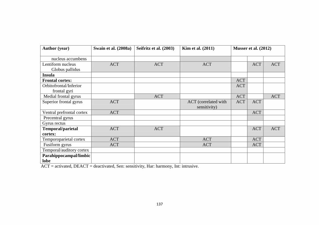
137
Author (year)
Swain et al. (2008a) Seifritz et al. (2003) Kim et al. (2011) Musser et al. (2012)
nucleus accumbens
Lentiform nucleus
Globus pallidus
ACT ACT ACT ACT ACT
Insula
Frontal cortex: ACT
Orbitofrontal/Inferior
frontal gyri
ACT
Medial frontal gyrus ACT ACT ACT
Superior frontal gyrus ACT ACT (correlated with
sensitivity)
ACT ACT
Ventral prefrontal cortex ACT ACT
Precentral gyrus
Gyrus rectus
Temporal/parietal
cortex:
ACT ACT ACT ACT
Temporoparietal cortex ACT ACT ACT
Fusiform gyrus ACT ACT ACT
Temporal/auditory cortex
Parahippocampal/limbic
lobe
ACT = activated, DEACT = deactivated, Sen: sensitivity, Har: harmony, Int: intrusive.

138
Table 7.2a: Maternal brain responses to own infant using still pictures
Author (year) Bartels & Zeki (2004) Leibenluft et al. (2004) Swain et al. (2004b)
Number of participants N = 19 N = 7 N = 9-14
Age of infants at time of
scan
9 months-3.5 years 5-12 years Time 1: 2-4 weeks
Time 2: 3-4 months
Study design 2T, 15s blocks 1.5T, 1.5s events 3T, 30s blocks
Contrasts and infant stimuli
used
Photos of own children >
photos of other known or
unknown children
Photos of own child >
photos of familiar child
Photos of own infant >
control
Photos of unknown
infant > control
Septal regions (MPOA/
VBNST/caudate head)
. ACT
Midbrain (including
periaqueductal grey)
ACT ACT ACT
Hypothalamus ACT
Thalamus ACT ACT ACT ACT
Limbic structures:
Amygdala DEACT ACT ACT
Anterior cingulate ACT ACT ACT ACT
Middle cingulate ACT ACT
Posterior cingulate DEACT ACT
Anterior paracingulate ACT
Hippocampus
Basal ganglia:
Striatum/putamen/
nucleus accumbens
ACT ACT ACT
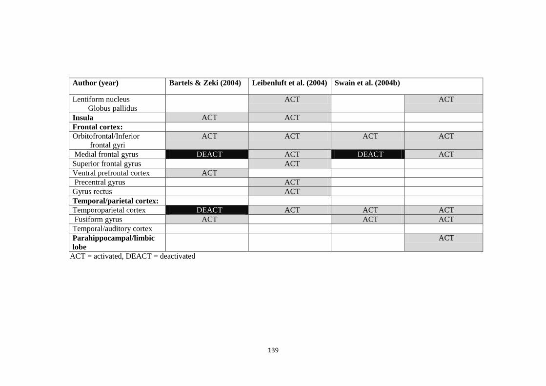
139
Author (year) Bartels & Zeki (2004) Leibenluft et al. (2004) Swain et al. (2004b)
Lentiform nucleus
Globus pallidus
ACT ACT
Insula ACT ACT
Frontal cortex:
Orbitofrontal/Inferior
frontal gyri
ACT ACT ACT ACT
Medial frontal gyrus DEACT ACT DEACT ACT
Superior frontal gyrus ACT
Ventral prefrontal cortex ACT
Precentral gyrus ACT
Gyrus rectus ACT
Temporal/parietal cortex:
Temporoparietal cortex DEACT ACT ACT ACT
Fusiform gyrus ACT ACT ACT
Temporal/auditory cortex
Parahippocampal/limbic
lobe
ACT
ACT = activated, DEACT = deactivated

140
Table 7.2b: Maternal brain responses to own infant using still pictures
Author (year) Nitschke et al. (2004) Strathearn et al. (2008) Strathearn et al. (2009)
Number of participants N = 6 N = 28 N = 30
Age of infants at time of
scan
2-4 months 5-10 months 7 months
Study design 1.5T, 30s blocks 3T, 2s events 3T, 2s events
Contrasts and infant
stimuli used
Photos of own children >
photos of unknown
children
Photos of own > unknown infant
in neutral, happy, and sad
Mothers with secure
pictures of own vs.
unknown child in neutral,
happy, and sad
Photos of own
children > photos of
unknown children
Septal regions (MPOA/
VBNST/caudate head)
Midbrain (including
periaqueductal grey)
ACT (more to happy) ACT (periaqueductal)
Hypothalamus ACT ACT
Thalamus ACT
Limbic structures:
Amygdala
Anterior cingulate ACT (more to happy)
Middle cingulate ACT
Posterior cingulate
Anterior paracingulate
Hippocampus
Basal ganglia:
Striatum/putamen/ ACT (more to happy) ACT (more to sad video)

141
Author (year) Nitschke et al. (2004) Strathearn et al. (2008) Strathearn et al. (2009)
nucleus accumbens
Lentiform nucleus
Globus pallidus
Insula ACT (more to happy) ACT
Frontal cortex:
Orbitofrontal/Inferior
frontal gyri
ACT ACT ACT
Medial frontal gyrus ACT
Superior frontal gyrus ACT (more to happy) ACT
Ventral prefrontal cortex
Precentral gyrus ACT
Gyrus rectus
Temporal/parietal
cortex:
Temporoparietal cortex ACT
Fusiform gyrus ACT
Temporal/auditory cortex ACT ACT
Parahippocampal/limbic
lobe
ACT
ACT = activated, DEACT = deactivated

142
Table 7.3: Maternal brain responses to own infant using video clips
Author (year)
Ranote et al. (2004) Noriuchi et al. (2008) Atzil et al. (2011)
Number of participants N = 10 N = 13 N = 23
Age of infants at time of
scan
4-8 months 16 months 4-6months
Study design 1.5T, 20-40s blocks 1.5T, 32s blocks
Contrasts and infant
stimuli used
Silent video clips of own
children > silent video clips
of unknown children
Silent video clips of own
children > silent video clips
of unknown children
Synchronous mothers
video of own vs.
unknown child (N = 13)
Intrusive mothers
video of own vs.
unknown child
(N = 10)
Septal regions (MPOA/
VBNST/caudate head)
Midbrain (including
periaqueductal grey)
ACT
Hypothalamus ACT
Thalamus ACT
Limbic structures:
Amygdala ACT ACT ++ ACT
Anterior cingulate ACT ACT ACT
Middle cingulate
Posterior cingulate ACT
Hippocampus ACT
Basal ganglia:
Striatum/putamen/
nucleus accumbens
ACT ++ ACT ACT

143
Author (year)
Ranote et al. (2004) Noriuchi et al. (2008) Atzil et al. (2011)
Lentiform nucleus
Globus pallidus
Insula ACT ACT
Frontal cortex:
Orbitofrontal/Inferior
frontal gyri
ACT ACT ACT ACT
Medial frontal gyrus ACT
Ventral prefrontal
cortex
ACT
Dorsomedial prefrontal
cortex
ACT
Precentral gyrus ACT ACT ACT
Temporal/parietal
cortex:
Temporoparietal cortex
Fusiform gyrus ACT
Temporal/auditory
cortex
ACT ACT
Parahippocampal/limbic
lobe
Occipital cortex ACT ACT ACT
Cerebellum ACT
ACT = activated, ++ activation is more significant compare to the other group within the study.

144
7.4. Reasons for Inconsistency in Findings between Imaging Studies
Structures and pathways in a wide variety of maternal brain regions are activated by
infant stimuli. Yet, some specific regions are more consistently implicated, than
others. These include:
1) Thalamic and hypothalamic regions.
2) Frontal cortex, including orbitofrontal cortex.
3) Midbrain regions, including periaqueductal grey.
4) Limbic structures, especially cingulate cortex.
5) The basal ganglia, including striatum, putamen, nucleus accumbens.
6) Insular cortex.
7) Temporoparietal cortex, especially fusiform gyrus.
8) Occipital cortex.
9) The cerebellum.
Some of these areas are also implicated in studies of parental caregiving in animal,
namely midbrain/basal forebrain, limbic and thalamocingulate structures, indicating
consistency with the animal literature.
Overall, studies in humans using functional imaging provide some evidence of
consistently activated maternal brain regions in response to infant’s stimuli (Table
7.1a, b, 7.2 a, b, and 7.3). However, inconsistencies are also notable. For example,
while some studies find amygdala activation (Seifritz et al., 2003; Swain et al.
2003, 2004, 2008a, Ranote et al., 2004; Lenzi, 2009), others report no such
activation (Swain et al., 2004b) or deactivation (Bartels & Zeki, 2004). This lack of
consistency could be explained by several reasons. First, many of the earlier studies
are rather small (e.g. Lorberbaum et al., 1999; Nitschke et al., 2004). Second, age of
infants varies considerably between studies (e.g. Lorberbaum et al., 2002;
145
Leibenluft et al., 2004) and brain activation may change as the mother and infant
develop a relationship and as the infant gets older and more interactive with the
mother. This may result in less activation in areas that support basic, reflexive
caring in younger infants, such as nursing (mesolimbic) and areas that serve
motivation and response to threat (mesocortical dopamine connections) in early
postpartum (see review of Swain, 2010). This notion is supported by the extended
findings of Lorberbaum et al. (2002); when they repeated their earlier study
(Lorberbaum et al., 1999) in mothers at earlier postpartum time (4-8 weeks) and
found more mesolimbic and mesocortical dopamine pathways activation in
response to infant cries.
Third, maternal caregiving is a composite of multiple behaviours, each of which
may have discrete neural correlates (Musser et al., 2012). Therefore, it is important
to know about mothers’ quality of ‘mothering’ (e.g. maternal sensitivity) in order to
interpret maternal brain responses (Barrett & Fleming, 2011). Fourth, individual
maternal experience with their own parents may influence the way women process
their infant cues (Strathearn et al., 2009), and therefore must be considered when
interpreting brain responses. The third and fourth points reflect the variability and
complexity of human maternal behaviour and the capacity to parent (i.e. variation
in maternal caregiving) (Swain et al., 2007).
Fifth, some brain regions subserve a range of different functions, e.g. the amygdala
and insula, which could lead to mixed findings in different studies (Strathearn et al.,
2009; Kim et al., 2011). Sixth, discrepant findings among studies may also be
related to the use of different experimental and analytic paradigms (Barrett &
Fleming, 2011); some studies used fixed effects analyses which do not consider
inter-subject variability and therefore limit inference to the group studied.

146
Conversely, other studies use random effects analyses that account for inter-subject
variability and permit generalisation of findings (Swain et al., 2007). Furthermore,
some studies used block designs (Lorberbaum et al., 2002), while others used event
related designs with relatively shorter stimuli (Seifritz et al., 2003). Finally,
variation in the modality of the stimulus may affect the consistency of findings.
While many studies used own infant stimuli, the use of control stimuli varied
widely (e.g. unknown infants, familiar infant, or neutral scene). Moreover, some
included different emotional valences (Strathearn et al., 2008, 2009) while others
included more cognitively demanding tasks (Leibenluft et al., 2004).
7.5. Summary
While many studies examined maternal brain responses to infant stimuli, far fewer
have examined the neural correlates in relation to maternal behaviour (Atzil et al.,
2011; Musser et al., 2012), and only one study has attempted to focus on maternal
sensitivity (Kim et al., 2011), yet due to design issues, firm conclusions from this
study are limited. Maternal caregiving is a composite of multiple behaviours, and
recent evidence suggests discrete maternal brain activation in relation to each of
these maternal behaviours (Musser et al., 2012).
The aim of the present study was to examine maternal brain responses to own and
unknown infant stimuli using videos in blocks of neutral, happy, and sad cues,
comparing mothers from a community UK population. Mothers were chosen to
represent healthy women in a population and therefore were recruited with no
history of mental illness and to represent the natural variation in maternal
sensitivity (i.e. high sensitivity and low sensitivity mothers). Also in these mothers,
we examined the relationship between maternal BOLD brain activation to infant

147
stimuli, including OT rich brain areas, and levels of plasma OT following mother-
infant interaction. This study was designed to address limitations from previous
studies by including a larger sample (N = 30) of new mothers examined at a limited
postpartum period (7-9 months) while measuring plasma OT and fMRI within the
same session. Furthermore, mothers were assessed for obstetric and demographic
influences which might represent potential confounders of any association and
which might be relevant in mediating some of the differences we may find between
the high and low sensitivity mothers.
To our knowledge, this study is the first to examine both brain and plasma OT
responses to infant stimuli in healthy mothers rigorously defined by their maternal
sensitivity rating. If BOLD activation in fMRI can discriminate between high and
low maternal sensitivity mothers, it would give credence to the notion that
functional imaging is a robust biomarker of ‘maternal sensitivity’. If we can show a
significant relationship between plasma OT responses to infant challenge between
high sensitivity mothers (HSMs) and low sensitivity mothers (LSMs), and, further
make links to imaging patterns, it would imply that modulation of maternal
behaviour by novel interventions could be studied in small numbers of normal
volunteers.
7.6. Objectives of Study II (oxytocin & fMRI scanning)
In this study, we aimed to examine differences in plasma OT and brain correlates
between women representing natural variation in human maternal sensitivity. We
aimed to:

148
1. Examine whether plasma OT is distinct between HSMs and LSMs, both at
baseline and following interaction challenge with their own infant, accounting
for demographic differences between the two groups.
2. Extend previous finding by examining maternal plasma OT in relation to own
perceived parenting experience (Gordon et al., 2008; Strathearn et al., 2009).
3. To confirm and extend previous findings in relation to activation of specific
maternal motivational reward pathways in response to own infant compared to
an unknown infant among all mothers.
4. To examine whether fMRI can discriminate between high and low maternal
sensitivity and whether its modulation by OT can also be detected, more
specifically:
i. To examine differences in maternal BOLD activation between
HSMs and LSMs in response to own infant versus an unknown
infant, regardless of the emotion displayed, as well as when emotion
considered (i.e. neutral, happy and sad).
ii. To examine differences in maternal BOLD activation between
HSMs and LSMs in response to own infant separately (without
comparing it to unknown infant) for each emotional affect.
iii. To examine correlations between maternal BOLD activation in
response to own infant and plasma OT responses following
interaction with own infant.

149
7.7. Hypotheses of Study II
In regards to maternal plasma OT, we hypothesised that:
1. HSMs would show significantly higher plasma OT levels both at baseline
and following infant play-interaction than LSMs.
2. Plasma OT levels in each group of mothers (HSMs/LSMs) will be
positively correlated with positive rating of own parenting experience.
In regards to maternal brain responses, we hypothesised that:
i. We would predict that our main effect comparing own infant with an
unknown infant (among the whole sample) would show greater BOLD
activation in areas previously reported by others (e.g. Ranote et al., 2004;
Bartels & Zeki, 2004), specifically: reward areas in the prefrontal cortex,
limbic areas at the anterior cingulate cortex and the amygdala.
ii. Based on previous evidence that examined maternal neural correlations in
relation to ‘maternal affiliative behaviour’ (Atzil et al., 2011; Musser et al.,
2012), or to ‘proxies of maternal oxytocin responses’ (Swain et al., 2008a;
Kim et al., 2011), we hypothesised that: maternal brain responses to the
contrast of own infant versus an unknown infant video (all affects
combined) would be significantly greater in the HSMs compared to LSMs,
specifically in: areas implicated in OT secretion at the hypothalamus, areas
associated with mirroring the emotions of others at the superior temporal
gyrus, areas for emotional processing at the posterior cingulate gyrus, and
areas for encoding emotional memories at the hippocampal formation.
iii. Greater BOLD activation (in the same areas stated in (ii) will be found in
HSMs compared to LSMs in response to own infant facial affect (neutral,
happy, and sad); when compared to an unknown infant of a similar affect

150
(or when compared to neutral control). However, given this is the first study
to investigate differences in infant emotional affects between two distinct
groups of mothers, sorting of areas in relation to each individual affect were
left as exploratory.
iv. BOLD activation in HSMs (in regions stated earlier in (ii) would correlate
positively with their plasma OT levels following the interactive play with
their infant.

151
Chapter 8: Study II Methodology
(Oxytocin & fMRI Scanning)
8.1. Sample
Study II was initiated at 7-9 months postpartum, with the cohort followed through
from Study I. Exclusion criteria for this study were: (a) any contraindication to
magnetic resonance imaging (MRI) (see MRI declaration form-Appendix B), (b)
pregnancy, (c) left-handed mother, (d) a screen positive for depression (EPDS ≥ 12
and HADS > 11), and (e) mother not living with her infant. Out of 80 mothers who
were followed up and who underwent evaluation of maternal sensitivity at 4-6
months postpartum (Study I-Time 2), 13 were not eligible for MRI (11 left-handed,
2 with contraindication to MRI), and 10 declined or were lost to follow up.
Selection of the final sample was performed among the remaining 57 eligable
mothers.
The final sample consisted of 30 eligible mothers representing opposite ends of
maternal sensitivity distribution at a mean of 35.14 weeks postpartum (SD = 3.26):
15 classified as ‘sensitive’ mothers (blind rated 4-7 on the MACI sensitivity scale;
mean = 4.47; SD = 0.74) and 15 women as ‘less sensitive’ mothers (blind rated 1-3
on the MACI sensitivity scale; mean = 2.13 (SD = 0.52). For description purposes,
the two sub-groups respectively are referred to here as ‘high sensitivity mothers’
(HSMs) and ‘low sensitivity mothers’ (LSMs) (Figure 8.1). The selection of the
HSMs and LSMs were initially determined by taking ratings of 1+ above the SD
(ratings 5-7), and 1+ below the SD (ratings 1-2), respectively, from published

152
MACI data on a healthy sample of 47 mothers at 7 months postpartum (Wan et al.,
2012). However, as this did not provide, in the current sample, the required number
of N = 15 in each group (based on power calculation for the scanning study), the
thresholds were increased, but with no overlap, such that HSM’s included those
rated 4-7 (equating to: general to high sensitive responsiveness) and LSM’s
included those rated 1-3 (equating to: minimal to moderate sensitive responsiveness
(Figure 8.1).
Table 8.1 represents the demographic and obstetric characteristics for the high and
low sensitivity mothers, and statistics comparing demographics between the two
groups are presented with the results (Chapter 9).

153
Figure 8.1. Distributions of high sensitivity mothers (HSMs, N = 15) and low sensitivity mothers (LSMs, N= 15) in relation to maternal sensitivity
distribution for the whole sample (N = 80). Key: Means for high sensitivity mothers (vertical solid line) and low sensitivity mothers (vertical dashed
line) in relation to the larger sample are also presented.

154
Table 8.1. The demographic and obstetric characteristics of the sample
grouped by level of maternal sensitivity
Characteristic High Sensitivity
Mothers (N = 15)
Low Sensitivity
Mothers (N = 15)
Mean [SD]
Maternal age (years) 30.40 [5.37] 27.65 [4.76]
Postpartum stage
(weeks)
35.93 [2.81] 34.29 [0.69]
Infant birthweight
(kilograms)
3.44 [0.44] 3.23 [0.59]
Frequency (%)
Married/cohabiting 13 (86.7) 11 (78.6)
Primiparity 6 (40.0) 9 (64.3)
Highest maternal
education:
Secondary (GCSE)
Post-secondary
education (A-level or
equivalent)
University degree and
postgraduate
3 (20.0)
4 (26.7)
8 (53.3)
6 (40.0)
6 (40.0)
3 (20.0)
Household income/year:
£15000 or less
£16000-£34000
£35000 or more
1 (6.7)
5 (33.3)
9 (60.0)
5 (33.3)
6 (40.0)
4 (26.7)
Infant gender (female) 10 (66.7) 7 (50.0)
Mode of delivery
(vaginal)
10 (66.7) 11 (78.6)
Mode of feeding (breast) 11 (73.3) 7 (50.0)

155
8.2. Procedure
Potentially eligible mothers were contacted and if they showed interest in participation
they were visited at their home to explain further about the study and to confirm their
eligibility after mothers rated their mood using HADS and EPDS. Within this visit,
following informed consent, a video of the infant’s face was obtained to use as a
stimulus during the fMRI. After prior arrangement mothers and infants attended the
WTCRF in Manchester, for a two hour session when three samples of blood were
collected and brain responses to infant stimuli were assessed though 35 minute-fMRI.
8.2.1. Oxytocin
i. Plasma Oxytocin samples
Mothers attended the session at the WTCRF either during menstruation or on their
contraceptive pill free days (early follicular phase). Measurement of plasma OT was
taken before and after mother-infant interactive play with their infants (instructions as
per the earlier play interaction). Blood samples were taken from mothers at the same
time in the early afternoon 1200 -1400 hours, and one hour after the last nursing feed (to
control for the influence of diurnal and physiological changes, respectively). Mothers
were asked to refrain from caffeine/smoking for at least two hours before the sampling.
Three samples of blood (5 ml each) were taken from antecubital veins through an
intravenous cannula. The first sample (OT1) was taken 10 minutes after the mothers
were separated from their infants. Then the mother rejoined their infant and participated
in interactive play for 10 minutes. Immediately after the interaction, the second sample
was taken (OT2) followed by the third sample (OT3) 5 minutes after that. After each
sampling, the cannula was flushed with 2 ml normal saline to maintain patency of the
vein, and 3 ml of blood were discarded before each sample to ensure no dilution
because of the saline. Refreshments were served after the blood sampling and before

156
scanning to overcome the influence of any possible anxiety provoked by the blood draw
on the mother’s brain.
ii. Sample processing & Assays
Each of the three samples (OT1, OT2 & OT3) was drawn into chilled vacutainer tubes
containing lithium heparin injected with 200 ml of Trasylol (aprotinin) 500,000 KIU/ml
blood. OT samples were kept ice-chilled and processed within 10 minutes. Samples
were then centrifuged at 4o C at 3500rpm for 15 minutes, and 500ul supernatants were
transferred to 2 microtubes (aliquot 1 & 2) and stored at - 80o C until transferred on dry
ice, to the University of Manchester Laboratory for analysis. Optimisation to test the
extraction method was performed through pilot samples before the actual analysis.
Determination of the OT was performed using the Max Binding Determination
Competitive Assay protocol on Gen 5 software using a Biotek Plate reader. Samples
were diluted 1 in 4 for assay and then results multiplied by 4 to compensate for dilution.
Oxytocin analyses were performed by a laboratory scientist who was blind to the study
setting and information.
8.2.2. fMRI scanning
i. fMRI paradigms
A series of alternating pre-recorded 20-second video clips was prepared consisting of 3
affect conditions (Neutral, Happy, and Sad) of each of two infants (the participant’s
‘own’ infant, and an ‘unknown’ infant) for use in the fMRI scan. The unknown infant
was matched on age, gender and ethnicity to the mother’s own infant. To provide a
measurement of BOLD signal change in active conditions compared to control
condition, mothers were also presented with neutral control stimuli (i.e. video of slow
moving traffic), which was a standard across subjects and used in a previous research
study (Ranote et al., 2004). Active video blocks were interspersed with resting periods

157
(blank screen) for comparison with active (video) blocks BOLD responses. Videos
lasting 16 minutes were presented in the following order: (Own neutral infant-neutral
control-unknown neutral infant) x 4, REST, (Own happy infant-neutral control-
unknown happy infant) x 4, REST, (Own sad infant- neutral control- unknown sad
infant) x 4, REST (Figure 8.2). All stimuli were displayed from a computer controlled
projector presented on a display screen and relayed to the participant via a mirror placed
above the head while in the MRI scanner.
Figure 8.2. Model representing the order of video clips as viewed by mothers while in
the scanner. Note: 20 sec ‘Neutral control’ was added to the first three blocks of each
emotion.
For the video task we used the following contrasts to test our hypotheses (see Chapter
7):
a. To examine the main effect, among all mothers: all infant videos minus the neutral
control.

158
b. Comparing high and low sensitivity groups: 1) HSMs vs. LSMs (own infant minus
unknown infant) (all affects combined), 2) HSMs vs. LSMs (own neutral minus
unknown neutral infant, own happy minus unknown happy infant, and own sad
minus unknown sad infant), 3) HSMs vs. LSMs (own happy, own neutral, and
own sad) subtracting neutral control.
c. The equivalent of (b) for LSMs vs. HSMs.
ii. fMRI Acquisition
Imaging was performed using a 1.5 Tesla Philips Intera MRI scanner running Explorer
gradients (software version 11.1.4.4). High-resolution T1-weighted structural images
were acquired to exclude any structural abnormality (none were found) and for
coregistration with functional data. The structural scan using SENSE employed a 3D
Contrast Turbo Field Echo Sequence with a temporal resolution (TR) of 9ms and an
echo time (TE) of 4ms with an 8° flip angle producing 140 slices with a voxel size of
0.8 x 0.8 x 1.0 mm. Functional images were acquired using a multi-slice, single shot
echo-planar imaging sequence, generating 29 ascending axial slices (TR = 2.5s, TE =
40ms, 4mm thickness with 0.5mm slice gap, in-plane resolution of 3.4 x 3.4mm). fMRI
data were exported from the scanner as proprietary Philips PAR/REC files. These files
were converted to IMG and HDR files using MRIcro software (Rorden & Brett, 2000).
8.3. Statistical Analyses
8.3.1. Sample size
The sample size was recalculated for Study II to avoid overestimation or
underestimation of the sample (Mumford & Nichols, 2008). The sample size of 30
mothers (15 in each group) is ample enough to produce a power of approximately 90%
(Desmond & Glover, 2002).

159
8.3.2. Data analysis
Demographic and obstetric differences between high sensitivity and low sensitivity
groups of mothers were assessed through independent sample t-test for interval
variables and through Chi-square test for categorical variables.
i. Oxytocin analysis
Pearson correlations were used to explore correlations between plasma OT levels and
other variables and stability of OT across the 3 assessments as well as correlations
between OT and other behavioural measures. A repeated-measures analysis of variance
(ANOVA) was employed to test for a significant change in plasma OT levels over time
between groups (HSMs and LSMs). Analyses were performed using SPSS (version 19)
and p < 0.05 (two-tailed) was considered statistically significant.
ii. fMRI analysis
Imaging data for each participant were pre-processed to reduce artefacts related to
signal components, corrected for motion, and the individual data were spatially
normalised for group analyses using Statistical Parametric Mapping (SPM8;
http://www.fil.ion.ucl.ac.uk) and implemented in MATLAB (Math works Inc.,
Sherborn, MA, USA). All functional images were realigned using a least squares
approach and a 6 parameter (rigid body) spatial translation (Friston et al., 1996). This
was undertaken because in the scanner; participants, while in the head coil, may still
undergo slight head movement. The first image was used as a reference scan and the
following images were translated onto this, correcting for pitch, roll and yaw throughout
the task. Translation and rotation corrections did not exceed 3.0 mm and 2.5° for any
participant.

160
The realigned images were co-registered with the corresponding structural (T1
Weighted) scan and segmented into different tissue classes by matching grey matter to a
grey matter reference template. Tissue classification registers the images with tissue
probability maps (grey matter, white matter and cerebrospinal fluid) adapted from the
International Consortium for Brain Mapping (ICBM) 452 T1-weighted scans.
Following this, the scans were spatially (stereotactically) normalised onto standardised
brain slices. This involves the warp of the images onto a standardised template. The
standardised template conforms to the ICBM, NIH P-20 project and approximates the
stereotactic atlas of Talairach & Tournoux (1988) (Ashburner & Friston, 1997; 1999).
The standardized slices then underwent smoothing using an isotropic Gaussian kernel
filter (10 mm full width half maximum [FWHM] to increase the signal-to-noise ratio by
use of the matched filter theorem (Friston et al., 2000). A parametric model was
employed that enabled the modelling of linear hemodynamic responses. First level
analyses were performed on each subject’s data to generate a single mean image
corresponding to canonical hemodynamic responses convolved with the block design
compared to resting conditions. Second level group analyses were performed using a
factorial ANOVA to contrast the conditions in each task.
8.4. Ethical Consideration
8.4.1. Ethics
Ethical regulations for Study I were also applied in Study II as appropriate; including
detailed information about the study aims, what it involved in relation to participants
(mothers), and ability of mothers to withdraw from the study at any time without giving
any reasons. This was explained to mothers verbally and in written format through the
information sheet. Eligibility for the fMRI was confirmed by completing the MRI

161
declaration form (ISBE; ethics Ref No. 02043, see Appendix B). Informed consents
were obtained from each participant before their participation. Mothers also gave their
permission so their GP would be informed if any abnormality was found in their
structural brain image, and for their infant’s videos to be shown to other participants in
the study. Mothers were given a value of £30 pounds Boots shopping vouchers and a
‘Thank You’ card.
8.4.2. Safety considerations
Mothers’ cannulation and blood withdrawal followed the Standard Operational
Procedure (SOP) at the WTCRF, which is in accordance with the protocol followed in
the Central Manchester University Hospitals, NHS Foundation Trust. Infants’ safety
measures while playing with mother was followed as in Study I (e.g. padded mat, no
electrical sockets etc.). Mothers were given the option to visit the ‘mock’ MRI scanner
at the University of Manchester prior to their actual scanning session to minimise
possible discomfort or anxiety related to scanning.

162
Chapter 9: Study II Results
(Oxytocin & fMRI scanning)
9.1. Sample
Participants for this study (Study II) are 15 HSMs and 15 LSMs who completed Study I
of this project at 4-6 months postpartum (see Chapter 8-Methods). Although household
income and maternal education were both borderline higher among HSMs, the results
were insignificant and therefore they were not controlled for to avoid over adjustment.
In addition, the two groups of mothers did not differ significantly in: maternal age,
marital status, parity (multiparous/primiparous), postpartum stage, infant birthweight,
infant gender, mode of delivery (CSD/ VD), or mode of feeding (bottle/breast) (both at
the 4-6 month visit or the current visit) (Table 9.1).

163
Table 9.1. Comparison of the demographic and obstetric characteristics of mothers
grouped by level of maternal sensitivity (high sensitivity mothers, HSMs and low
sensitivity mothers, LSMs)
Characteristic HSMs
(N = 15)
LSMs
(N = 15)
t (28)
Chi-
square
test
p-
value
Mean [SD]
Maternal age (years) 30.40 [5.37] 27.65 [4.76] - 1.38 0.18
Average maternal
education (years)
15.06 [2.82] 12.80 [2.73] - 1.87 0.07
Average annual household
income (thousand pounds)
33.00 [4.61] 26.33 [4.24] - 2.76 0.09
Infant birthweight
(kilograms)
3.44 [0.44] 3.23 [0.59] - 0.80 0.43
Postpartum stage (weeks) 35.93 [2.81] 34.29 [3.69] - 1.37 0.18
Frequency (%)
Married/cohabiting 13 (86.7) 11 (78.6) 0.56 0.65
Primiparous 6 (40.0) 9 (64.3) 1.71 0.19
Infant gender (female) 10 (66.7) 7 (50.0) 0.83 0.36
Mode of delivery
(vaginal)
10 (66.7) 11 (78.6) 0.51 0.47
Mode of feeding (breast)
At 4-6 month visit
At 7-9 month visit
11 (73.3)
3 (20.0)
7 (50.0)
3 (20.0)
1.68
0.55*
0.19
0.64
*Fisher exact test

164
9.2. Plasma Oxytocin Results
9.2.1. Preliminary analyses
Three samples of plasma oxytocin were collected from each woman before (OT1) and
after (OT2 & OT3) interaction with their infant (see Chapter 8). Two participants did
not have their OT3 assessed (due to technical difficulties) and hence their OT3 was
assigned the same value as OT2. One outlier was excluded from this analysis (scored >
3 SD in all three assessments). The three plasma OT assessments (Table 9.2) were
highly correlated with each other, both among HSMs (r = 0.79-0.96; p < 0.01) and
among LSMs (r = 0.92-0.99; p < 0.01), indicating high levels of OT individual stability.
Table 9.2. Mean oxytocin levels (pg/ml) measured before and
after play-interaction among mothers grouped by level of
maternal sensitivity
Oxytocin
sample
High Sensitivity
Mothers (N = 15)
mean [SD]
Low Sensitivity
Mothers (N = 14)
mean [SD]
OT1 235.09 [83.51] 301.87 [39.15]
OT2 223.67 [83.43] 303.27 [34.67]
OT3 210.01 [81.58] 301.56 [38.12]
Note: OT1: Oxytocin measured before mother-infant
interaction, OT2 & OT3: Oxytocin measured after mother
infant interaction.

165
After excluding the outlier, the two groups were re-examined for possible confounders.
Household income was significantly higher in HSMs compared to LSMs and
accordingly was controlled for in the subsequent analyses. The two groups remained
matched and not significantly different with respect to other demographics including
maternal age, marital status, maternal education, parity, infant birthweight, infant
gender, mode of delivery, mode of feeding, and postpartum stage (Table 9.3).

166
Table 9.3. Comparing demographic and obstetric characteristics of mothers grouped by
level of maternal sensitivity excluding the outlier
Characteristic
HSMs
(N = 15)
LSMs
(N = 14)
t (27)
Chi-
square
test
p-value
Mean [SD]
Maternal age (years) 30.40 [5.37] 27.64 [4.77] - 1.46 0.16
Average maternal
education (years)
15.06 [2.82] 12.77 [2.76] - 1.94 0.06
Average annual
household income
(thousand pounds)
33.00 [4.61] 25.12 [4.13] - 2.11 0.04
Infant birthweight
(kilograms)
3.44 [0.44] 3.20 [0.53] - 1.10 0.28
Postpartum stage
(weeks)
35.93 [2.81] 34.64 [3.22] 1.32 0.20
Frequency (%)
Married/cohabiting 13 (86.7) 11 (78.6) 0.56 0.65
Primiparous 6 (40.0) 8 (57.1) 1.65 0.21
Infant gender
(female)
10 (66.7) 7 (50.0) 0.83 0.36
Mode of delivery
(vaginal)
10 (66.7) 11 (78.6) 0.51 0.47
Mode of feeding
(breast)
3 (20.0) 3 (20.0) 0.55* 0.64
*Fisher exact test

167
9.2.2. Main analysis
Controlling for household income, a repeated measure ANOVA showed no overall
change in the mean level of OT from baseline to post interaction (within subject effect)
Greenhouse-Geisser [F (1.37, 35.51) = 1.54; p = 0.23]. Next, we examine between
subject effect [F (1, 26) = 8.42; p = 0.01] and found significant difference in OT through
the three assessment points when the two groups of sensitivity was compared with this
change confined to HSMs (Figure 9.1). High variance on OT3 measurement among the
HSMs is also evidenced from the graph (Figure 9.1).

168
Figure 9.1. Box plots and means of plasma oxytocin measured before (OT1) and after mother-infant interaction (OT2 and OT3)
among the high sensitivity mothers (HSMs, N = 15) and low sensitivity mothers (LSMs, N = 14), controlling for household income.
Key: The horizontal dashed line represents the means of the three OT assessments among LSMs, and the horizontal solid line
represents means of the three assessments among the HSMs.

169
9.2.3. Relationship between plasma OT and own perceived parenting experience
Controlling for household income, the two groups of mothers did not differ significantly
in their prenatal self-reporting of own: maternal care (transformed) [F (1, 26) = 0.01; p =
0.93], paternal care (transformed) [F (1, 26) = 0.41; p = 0.53], maternal overprotection, [F
(1, 26) = 2.41; p = 0.13], or paternal overprotection [F (1, 26) = 0.52; p = 0.48]. Next, we
examined whether plasma OT was related to these self-reports of own parenting
experience. To simplify interpretation, OT1 (OT pre) and the mean of OT2 and OT3 (OT
post) were considered to test the correlations with perceived parenting experience.
Maternal overprotection was positively correlated with both OT pre and OT post among
LSMs but not among HSMs (Table 9.4; Figure 9.2). Maternal care, paternal
overprotection and paternal care were not correlated with plasma OT (OT pre or OT post) in
any of the groups. Furthermore, OT at all times was not correlated with mode of delivery
or feeding.
Table 9.4. Correlations between plasma OT and self reported own
parenting experience in mothers grouped by level of maternal
sensitivity
HSMs (N = 15) LSMs (N = 14)
OT pre OT post OT pre OT post
Maternal
care1
- 0.38 - 0.21 - 0.21 - 0.07
Maternal
overprotection
0.28 0.07 0.62* 0.63*
Paternal care1 - 0.34 - 0.34 - 0.09 0.18
Paternal
overprotection
- 0.12 - 0.06 0.16 0.03
*p < 0.05. 1: Transformed variable

170
Figure 9.2. The relationship between baseline plasma oxytocin (OT pre) and own
maternal overprotection among: (a) low sensitivity (N = 14), and (b) high sensitivity
mothers (N = 15) (similar relationships also shown with OT post).

171
9.3. fMRI Results
9.3.1. Preliminary analysis
No participants were excluded from this analysis, and therefore the two groups
represent the same characteristics presented at the beginning of this chapter (Table 9.1).
Because one outlier was excluded from OT analyses (> 3SD), and therefore was also
excluded when correlations between BOLD brain activation and plasma OT were
examined.
9.3.2. Whole brain analyses of maternal brain responses
In order not to limit the extent of this exploratory analysis, we started by performing
‘whole brain analyses’ with a significant threshold specified as p ≤ 0.05, Family Wise
Error (FWE) corrected. Prior to testing the specific hypotheses, maternal responses to
all infant stimuli (i.e. own + unknown) were compared with responses to neutral control
stimuli (i.e. video of slow moving traffic). Thus, for analysis purposes, the first contrast
analysed was ‘All infants minus neutral control’. This provided a measurement of
BOLD signal change in active conditions compared to control conditions. Although no
activation reached the threshold level, but subthreshold regions are shown in Table 9.5
for interest.
Next, the main effect was examined after combining all facial affects, to compare
videos of own infant versus videos of an unknown infant ‘own infant minus unknown
infant’. In response to own infant (compared to an unknown infant cues) enhanced
BOLD activation was observed in the right inferior frontal gyrus (BA 47 & 9), and in a
range of subcortical regions, including, left parahippocampal gyrus (BA 34), bilateral
uncus (BA 28), and right anterior cingulate gyrus (BA, 24) (Table 9.5).

172
Table 9.5. Contrasts testing BOLD signals and ‘main effect’ in response to own and an unknown infant stimuli (combined all affects)
among the whole sample (N = 30)
Contrast Area Sub-areas BA R/L Talairach
coordinates
x y z
Z score FWE FDR KE
All infants minus
neutral control
Parietal lobe Postcentral gyrus 1 R
54 - 16 50
4.11
0.08
0.16
991
Temporal
lobe
Middle temporal
gyrus
R 40 - 61 6 4.19 0.08 0.26 991
Own infant minus
unknown infant
Frontal lobe Inferior frontal
gyrus
47
9
R
R
43 26 2
47 11 27
4.50
4.08
0.02
0.01
0.02
0.04
591
591
Limbic lobe
Parahippocampal
gyrus
34 L - 23 2 - 19 4.49 0.02 0.02 156
Uncus 28
28
28
L
R
R
- 29 5 - 23
33 2 - 23
30 5 - 19
4.45
4.55
3.85
0.01 0.01 591
Anterior cingulate
gyrus
24 0 31 23 4.21 0.06 0.03 571
Note. All significant whole brain (Family Wise Error (FWE) ≤ 0.05) corrected for multiple comparison. BA: Brodmann’s area; L: Left, R:
Right; FDR: False Discovery Rate, KE: Cluster size (provided for interest only and not as a significant reference).

173
9.3.3. Comparisons between high and low sensitivity mothers
Comparison of BOLD activation between HSMs and LSMs using whole brain analyses
did not reveal significant results. Therefore, a ‘Region of Interest (ROI)’ with random
effects analysis was set to test our main hypotheses regarding comparisons between the
two sensitivity groups.
Region of Interest Analyses (ROI): Based on our hypothesis a single ROI composite was
hypothesised to include: the right superior temporal gyrus, right posterior cingulate
gyrus, left subthalamic nucleus and left hippocampal formation. We compared brain
activation patterns between sensitivity groups using a two-factor random effects
ANOVA model where ROI differences in BOLD activation were assessed using a
threshold of (FWE) ≤ 0.05, corrected for multiple comparisons. Activation was
calculated between the two groups of mothers, HSMs (N = 15) and LSMs (N = 15) in a
priori ROI. Activation between the two groups was compared using the t-test.
In post hoc analyses, we compared BOLD activation to own versus unknown infant
video between the two sensitivity groups regardless of facial affect [i.e. HSMs versus
LSMs (own infant minus unknown infant); similarly, we compared LSMs versus HSMs.
No significant effects were found with these contrasts in any of the ROIs.
We also compared responses to own versus unknown infant video between the two
sensitivity groups for each facial affect separately [i.e. HSMs versus LSMs (own neutral
infant minus unknown neutral infant), HSMs versus LSMs (own happy infant minus
unknown happy infant)], HSMs versus LSMs (own sad infant minus unknown sad
infant)], and similarly for LSMs versus HSMs. Compared to LSMs, HSMs showed
greater BOLD activation in the right superior temporal gyrus (BA 41) in response to
own neutral infant as compared to unknown neutral infant (Table 9.6, Figure 9.3);
174
whereas, compared with HSMs, LSMs did not show significant BOLD activation in any
of the ROIs. Own infant happy or sad versus unknown happy or sad videos did not
reveal significant differences in BOLD activation when the two groups were compared.
Finally, we compared BOLD responses to different facial affects between HSMs and
LSMs using only own infant stimuli, [i.e. HSMs versus LSMs (own neutral infant minus
neutral control) HSMs versus LSMs (own happy infant minus neutral control), HSMs
versus LSMs (own sad infant minus neutral control)], and similarly for LSMs versus
HSMs. Compared to LSMs, HSMs showed greater BOLD activation in the right
superior temporal gyrus which was extending to the right insula (BA 13) in response to
own happy infant (Table 9.6). By contrast, compared to HSMs, LSMs did not show
significant BOLD activation in response to own happy infant video in any ROIs.
Viewing own neutral or sad infant videos did not reveal significant differences in
BOLD activation in the ROIs when the two groups were compared.

175
Table 9.6. Areas of significant BOLD activation within ROI, when comparing high sensitivity (N = 15) with low sensitivity (N =
15) mothers
Note. All significant ROI (Family Wise Error (FWE) ≤ 0.05) corrected for multiple comparison. BA: Brodmann’s area; L: Left, R:
Right; FDR: False Discovery Rate; KE: Cluster size (provided for interest only and not as a significant reference).
Groups
compared
Contrast ROI BA Talairach
coordinates
x y z
Z score FWE FDR KE
High
sensitivity
mothers
vs.
Low
sensitivity
mothers
mothers
Own neutral infant
minus unknown
neutral infant
Superior temporal
gyrus
41 57 -25 13 3.39 0.05 0.22 25
Own happy infant
minus neutral
control
Superior temporal
gyrus
41
13
43 -32 5
57 -32 18
3.92
3.38
0.01
0.05
0.15
0.16
118
118

176
Figure 9.3. Maternal brain activation in response to infant stimuli: Compared with LSMs, HSMs show greater activation of the right
superior temporal gyrus in response to: (1) own neutral infant versus unknown neutral infant video, and (2) own happy infant versus neutral
control, at ROI-threshold of FWE ≤ 0.05. Structural brain image created from average of all subjects.

177
9.3.4. Correlations of plasma oxytocin with ROI
Repeated measure ANOVA found a between-group correlation between BOLD
activation of ROIs and plasma OT. The activation of the right superior temporal gyrus
among HSMs (compared to LSMs) in response to own happy infant minus neutral
control was negatively correlated with post-interaction plasma OT levels among those
mothers (OT post) (r = - 0.81, p < 0.01). This implies that the greater the BOLD
activation in the right superior temporal gyrus in HSMs when they viewed their infant
happy cues (compared to neutral control), the lower their plasma OT levels following
play interaction with their infant (Figure 9.4). No other significant correlations were
found in other ROIs including, the left subthalamic nucleus.
On the other hand, in LSMs there was no significant correlation between the plasma OT
post and any of the a priori ROIs. Furthermore, no significant correlations were found
between the a priori ROI activations and plasma OT levels when the entire sample was
considered, and significant correlations between BOLD activation and OT post emerged
only when the HSMs group was examined separately, indicating that the origin of the
correlations lies in the HSMs group.

178
Figure 9.4. Correlation between BOLD activation in the right superior temporal gyrus
(STG) and post interaction plasma oxytocin among high sensitivity mothers (N = 15)
R² = 0.66 -0.3
-0.2
-0.1
0
0.1
0.2
0.3
0.4
0.5
0 100 200 300 400
No
rma
ils
ed
% B
OL
D S
ign
al c
ha
ng
e
Mean Plasma Oxytocin Concentration pg/ml
Oxytocin concentration
Linear (Oxytocin concentration)

179
Chapter 10: Study II Discussion
(Oxytocin & fMRI Scanning)
10.1. Plasma Oxytocin Discussion
10.1.1. Overview of the findings
We sought to examine further how natural differences in maternal sensitivity to infant
cues are associated with maternal OT in a community sample of healthy mothers.
Contradictory to our main hypotheses, we report that: (1) HSMs have lower baseline
plasma OT levels compared to LSMs, and (2) following a play-interaction with their
infants, plasma OT levels significantly decrease in HSMs whereas no significant change
was seen among LSMs. In accordance with our secondary hypothesis we report that, at
least in LSMs, baseline and post interaction plasma OT levels were significantly
correlated with own parenting experience. However, contradictory to our hypothesis (1)
this correlation of OT, among LSMs, were positively correlated with unfavourable (not
with favourable) own parenting experience, in particular; own maternal overprotection,
whereas (2) both baseline and post interaction plasma OT levels among HSMs were not
correlated with own parenting experience.
Recent parenting studies that examined correlation of OT to quality of affectionate
behaviour among parents found a positive correlation between plasma OT levels and
maternal affectionate (or paternal stimulatory) behaviour towards the infant (Feldman et
al., 2007, 2010a; Gordon et al., 2010). However, a recent parenting study by the same
group of authors (Feldman et al., 2011) reported higher urinary OT levels among
mothers who experienced gaps in their positive social relationships, including their
relationship with their own mother, romantic partner or own infant (Feldman et al.,

180
2011). This suggests an anti-stress role for OT similar to that reported by other studies
among women (i.e. who were not necessarily mothers) (Taylor et al., 2010), which will
be discussed further in this chapter.
In contrast to other parenting studies which examine correlation of OT with maternal or
paternal behaviour, in the current sample, OT was assessed in two groups of women at
opposite ends of the distributions of maternal sensitivity, selected from a larger
community sample of 80 early postpartum mothers. This megrouping implies women
with distinct sets of affective and behavioural caregiving attributes (Thompson, 1997),
and accordingly might ‘tap’ into an element of stress or anxiety coping strategy, which
could be part of the sensitive responsiveness concept.
Similar to the aforementioned studies on parenting (Feldman et al., 2007; Gordon et al.,
2010, Atzil et al., 2011) we did not find a relationship between baseline plasma OT
levels and mode of delivery or feeding. This suggests no difference in OT between
breastfeeding and formula feeding mothers when plasma OT was not sampled during
feeding (Van der Post et al., 1997). It also suggested that when OT is measured later in
postpartum (7-9 months), it is no longer necessarily reflecting birth history (e.g.
VD/CSD) (Swain et al., 2008a), but rather is more likely to reflect parenting style
(Feldman et al., 2010a). We infer that baseline plasma OT was not different between
primiparous and multiparous women for similar reasons.
10.1.2. Why baseline OT was high in LSMs?
Elevated plasma OT levels have been reported in relation to gaps in positive social
relationships. In a study assessing stress in partnership among 85 adults in stable
relationships (62% women and 38% men), baseline plasma OT was positively
correlated with ratings of distress in relationships in women (while plasma vasopressin
correlated with relationship distress in men) (Taylor et al., 2010). In a further study,
181
among 73 post menopausal women, baseline plasma OT was negatively correlated with
relationships with a woman’s own mother, partner, and marginally significant (also
negatively) with relationship with best friend (Taylor et al., 2006). Similarly, in a study
that examined the relationship between romantic attachment and unstimulated plasma
OT, 45 subjects (33 women and 12 men-not romantically attached to each other)
reported their experience in romantic close relationships as well as their anxiety in these
relationships (Marazziti et al., 2006). In that study, a significant positive correlation was
found between plasma OT levels and anxiety in romantic relationship. The authors
strengthened their findings by accounting for the pulsatile secretion of OT by
considering the mean of three samples collected within one hour. However, their sample
included individuals with and without a current romantic relationship; but they reported
no difference in plasma OT levels with respect to this.
The implication of a role for OT in social relationships’ development and difficulties
was also reported among parenting studies. Although in the study by Feldman et al.
(2011) plasma OT findings were inconsistent with our findings in that parents with high
affective synchrony showed higher plasma OT, their urinary OT findings were
consistent with our results. Urinary OT in that study was significantly and positively
correlated with interactive stress, parenting stress and anxious romantic attachments. A
correlation between plasma OT and urinary OT has been reported in other recent studies
in humans (Hoffman et al., 2012) and animals (Polito et al., 2006). In addition, an
increase in urinary OT was also reported following intravenous injection of exogenous
OT, providing support for the reliability of using urinary OT to reflect systematic
changes in plasma OT (Mitsui et al., 2011). Therefore, the absence of the correlation
between plasma and urinary OT in the study by Feldman et al., is interesting and might
suggest simultaneous multiple roles for the OT; in both reflecting and facilitating
through an anxiolytic pathway, affiliative relationships.

182
In the current study, plasma OT levels in LSMs showed a positive correlation with own
maternal overprotection-which was borderline higher in this group-which might explain
the higher OT levels (at all three assessment times) among LSMs. Although in the
current study there were no differences in marital status between HSMs and LSMs, we
did not assess the quality of the ‘relationship with partner’ and so we cannot exclude
distressed pair bond relationships as an explanation for the elevated plasma OT among
the LSMs in our study.
Higher plasma OT was also reported in relation to more depressive symptoms. Taylor et
al. (2010) found a positive correlation between plasma OT and depression scores in
women who were not depressed. Similarly, Parker et al. (2010) measured OT hourly
over a period of 16 hours among clinically depressed individuals and their control
group, and found higher levels of OT among depressed individuals compared to the
control group. Exploring a similar explanation among our sample (although not
hypothesised) did not reveal differences in depression scores between the two groups of
mothers. However, significantly higher mean depression scores were found among
LSMs when sensitivity cut-off scores were applied to the larger sample (N = 80), t (78)
= 2.50; p = 0.01. Had that difference been represented in our smaller sample (N =15 in
each group), it might have been reflected in an association with plasma OT as reported
in previous studies in women (Taylor et al., 2010; Parker et al., 2010).
In summary, in women plasma OT might act as an anxiolytic or anti-stress hormone
(Neumann et al., 2000, Numan & Woodside, 2010). Elevation among LSMs in the
current study could be a reflection of their higher perceived maternal overprotection (as
a representation for a difficult relationship with own mother), difficulties in pair
bonding relationships or higher depressive symptoms, all of which are associated with

183
isolation or poor social networking (Grippo et al., 2007; Parker et al., 2010). Yet, only
the first explanation was evidenced in the current study.
10.1.3. Why does OT drop in HSMs?
The significant reduction in plasma OT levels among HSMs following interaction with
their infants is consistent with some previous reports e.g. Turner et al. (2002). These
authors examine changes in plasma OT in response to laboratory-induced positive or
negative emotions among 32 nulliparous women. Significant drop in plasma OT was
reported after women viewed positive emotion (i.e. a comedy movie), whereas no
change was found after women viewed negative emotions (sad movie). In another
study, Bick & Dozier (2010) measured urinary OT levels in 26 healthy mothers
following 25 minutes of physical interactions while playing computer games with their
child aged 2.5-4.5 year old and again with an unfamiliar child of a similar age. Urinary
OT was significantly higher following interaction with the unfamiliar child as compared
with own child. They concluded that interaction with an unfamiliar child might
constitute a (socially) stressful affiliative situation. Accordingly, they suggested that OT
increased in order to modulate this stress and in order to ensure mothers exhibit pro-
social behaviour with the unfamiliar child which would then compensate for the absence
of a ‘natural’, biological bond. The authors did not measure pre-interaction urinary OT,
and it is therefore possible that the lower OT levels (following interaction with own
infant), represents a ‘drop’ in OT rather that an increase following interaction with
unfamiliar child, which would be similar to our findings.
In a study that investigated the relationship between adult temperament (using the Adult
Temperament Questionnaire) and plasma OT responses among mothers, Strathearn et
al. (2012) examined plasma OT before and after mother infant interaction among 55
first time mothers and their 6-7 month-old infants. Effortful control (i.e. propensity to

184
focus on executing plans, performing tasks and maintaining focus and attention) was
negatively correlated with the OT response (difference between baseline and post
interaction levels). In other words, the more concentration and attention shown by the
mother during the interaction with her infant, the less likely she experienced an increase
in plasma OT following the interaction. Characteristics related to effortful control are
actually important for sensitive mothering as mothers need focus and attention in order
to respond ‘promptly’ and ‘appropriately’ to infant signals (Ainsworth et al., 1978).
We would therefore expect that the drop in plasma OT among HSMs following their
interaction with their infant is part of the mechanism underlying their sensitive
responsiveness. Thus, HSMs perceive their infant signals as a positive (affiliative) event
and they give appropriate attention and focus to these signals; accordingly their plasma
OT levels drop as a signal of the easy, focused, positive interaction with own infant. By
contrast, LSMs (in our study) may not perceive interaction with their infant as a positive
(affiliative) event and hence restrain/resist the alleviation effect of the socially affiliative
interaction as compared to HSMs. They are also unable to give proper attention and
focus to infant signals, possibly because they do not recognise such signals or attach
appropriate emotional valence to them, and accordingly their plasma OT levels remain
elevated as both a signal of a stressful, unfocused, negative (affiliative) interaction with
own infant (Figure 10.1) and perhaps to enhance (affiliative) behaviours. This is
consistent with Feldman et al. (2010a) who reported no difference in plasma OT levels
among mothers rated as low in affectionate contact following interaction with their
infants. Thus, by classifying mothers according to sensitivity behaviours, we may have
‘tapped’ into stress or anxiety coping, as well as pro-affiliative strategies, at least in part
modulated by OT.

185
Figure 10.1. A model representing the possible role of oxytocin as an anxiolytic (anti-
stress) (pro-affiliative) hormone in HSMs and LSMs: high levels of own maternal
overprotection, and possibly high depression score or distressed pair-bonding have a
stress-inducing effect in LSMs resulting in elevation of plasma OT levels. LSMs
maintain the same stress level after interaction with their infant. HSMs experienced less
stress in life and accordingly less plasma oxytocin levels with marked reduction in their
OT levels following interact with their infants suggesting induction of positive emotions
as a result of interaction. Key: Solid Rectangle: Possible factors with evidence from the
current study, Dashed Rectangle: Proposed factors without evidence from the current
study.
10.1.4. Does oxytocin has a dual action?
The animal literature (Numan & Woodside, 2010) makes it clear that OT serves a dual
role in maternal behavior: It increases maternal motivation and it also decreases stress
and anxiety (it has anxiolytic effects). The latter effect may aid the mother in coping
with difficult circumstances related to infant care. With respect to the human literature,
different methods, which include postpartum stage of the mother-infant dyad and the

186
ways in which mothers are classified, may differentially tap into one or the other of
these two aspects of OT involvement in mother-infant interactions.
Accordingly, some recent studies suggest that, OT increases following affiliative
interaction with own infant (Feldman et al., 2007; Gordon et al., 2010). Others studies,
including the current study, suggest that at least in women, OT is triggered/released as a
signal when there is a need to enhance or promote affiliation with others or when the
social relationship is ‘threatened’ (including pair bonding or parental bonding) (Taylor
et al., 2006, 2010; Bick & Dozier, 2010; Taylor et al., 2010; Feldman et al., 2011;
Tabak et al., 2011). This putative role of OT as a biomarker for a distressed social
relationship may occur primarily in women (Turner et al., 1999; Feldman et al., 2011).
Women may use their close relationships with others in a different way to men: women
may regulate their perception of and responses to stress through the development of
close affiliative bonds (Marazziti et al., 2006). In this way, plasma OT may be secreted
in some women particularly under what they perceive to be stressful situations in order
speciffically to prompt a desire for affiliation (Taylor et al., 2006). Interestingly, this
dual action for OT, as a pro-social, pro-affiliative as well as an anxiolytic or anti-stress
hormone might be represented simultaneously within the same individual, as suggested
by Feldman et al. (2011).
Our findings support a role for OT in affiliative bond formation and in stress regulation.
The pathways by which OT might mediate effects on stress and on social affiliation
may be different. Taylor et al. (2010) suggest that, when OT increases as a result of
social affiliation, it does not involve pathways implicated in anti-stress (e.g. dopamine
pathways) (Neumann et al., 2000). Indeed, with the exception of the anti-stress
(anxiolytic) effect, most behavioural and physiological effects induced by OT can be
blocked by administration of OTR antagonist, suggests a different mechanism (Uvnas-
187
Moberg, 1998) when OT works as anxiolytic, yet further research into the role of OT in
human social relationship is needed.
10.2. fMRI Scanning Discussion
10.2.1. Overview of the findings
Using whole brain analysis to examine brain activation to infant video stimuli among
healthy early postpartum mothers at 7-9 months postpartum, we report the following
main findings which were in accordance with our hypotheses: overall, when mothers
viewed videos of their own infants compared to unknown infants (main effect),
significant brain activation was elicited predominantly in 3 areas: i) the anterior
cingulate gyrus, ii) the right inferior frontal gyrus, and iii) the left parahippocampal
gyrus and bilateral uncus.
Using ROI analysis to compare brain responses between high sensitivity and low
sensitivity group of mothers we report the following main findings: in accordance with
our hypothesis, when compared to LSMs, HSMs showed greater BOLD activation in
the right superior temporal gyrus in response to own neutral infant compared to an
unknown infant video or in response to own happy infant compared to a neutral control
stimulus. However, inconsistent with our hypotheses, this BOLD activation among
HSMs was negatively (not positively) correlated with post interaction plasma OT
concentration in those mothers. Also inconsistent with our hypotheses, HSMs did not
show significant activation in other ROI, specifically the hypothalamus, posterior
cingulate gyrus, or the hippocampal formation in response to own infant compared to an
unknown infant video. Compared to HSMs, LSMs did not show significant activation in
any ROI in response to their own infant, separately or when compared to an unknown
infant.

188
10.2.2. Main effect
Previous studies have suggested a role for the anterior cingulate gyrus in emotional
processing (see review by Barrett & Fleming, 2011). The inferior frontal gyrus is
thought to have an important role in the decoding of facial expressions of emotions
(Adolphs, 2002). Lesions in this region lead to an impaired ability to identify facial
emotions in adults (Hornak et al., 1996). The inferior frontal gyrus has also been
identified as one of the areas responsible of mirroring others’ emotions (Theory of
Mind) (Iacoboni & Dapretto, 2006). This suggests that a key to healthy maternal
responsiveness is the capacity of mothers to recognise and respond to her infant’s
emotions; and in young, preverbal infants, this is perhaps especially through mirroring
of emotional expressions and facial cues. Viewing of own infant also activated the
parahippocampal gyrus and uncus; these regions are thought to encode emotional
memories (Phelps et al., 2004; Swain et al., 2008b). Our findings in regards to all
mothers, confirm the findings of previous studies (e.g. Ranote et al., 2004; Swain et al.,
2008a).
10.2.3. Comparison of brain activation between HSMs and LSMs
Compared to LSMs, HSMs showed greater activation of the right superior temporal
gyrus in response to own neutral infant compared to unknown neutral infant stimuli.
This activation continued and extends towards the insula while HSMs viewed their own
happy infant compared to neutral control video.
The superior temporal gyrus contains ‘mirror neurons’ (Iacoboni & Dapretto, 2006)
which are activated in response to observing someone else experiencing an emotion;
accordingly, the ‘observer’ mimics the emotion or action observed (Iacoboni et al.,
1999; Iacboni & Dapretto, 2006). This occurs in response to understanding and
empathising with others ‘Theory of Mind’ (Blakemore & Decety, 2001; Gallagher &

189
Frith, 2003; Rizzolatti & Fabbri-Destro, 2008). However, one can argue that Theory of
Mind suggests reading of thought, which is not necessarily accompanied by empathy
with emotion. Yet, for early postpartum mothers to respond sensitively to their infant’s
needs, understanding infant emotion is a key aspect of sensitive mothering. Therefore
we would anticipate that activation of an area rich in mirror neurons (the superior
temporal gyrus) in HSMs might suggest that HSMs give more attention to read their
infant facial emotion in order to react to this as a part of an internal maternal reflective
function (Brunet et al., 2000; Lenzi et al., 2009), compared to LSMs. This notion is
directly consistent with what attachment theorists have suggested about maternal
sensitivity. Many considered maternal sensitivity as the response to the infant’s mental
state, rather than to her/his physical state (Fonagy et al., 1994; Meins, 1997), and thus a
sensitive mother would expect to respond properly to her infant’s displayed emotions by
affirming the positive emotions or reassuring about the negative ones (Sroufe, 2000).
As mentioned in the literature review earlier (Chapter 7), a few studies considered
examining maternal brain responses in relation to maternal interactive behaviour (Atzil
et al., 2011; Kim et al., 2011; Musser et al., 2012). Yet, among these studies, only Kim
et al. (2011) focus on maternal sensitivity, and they also reported activation of superior
temporal gyrus in response to own infant’s crying, among 9 exclusively breastfeeding
mothers, at 3-4 weeks postpartum which was also positively correlated with maternal
sensitivity ratings in those mothers at 3-4 months postpartum. Activation of the superior
temporal gyrus was also reported in response to own infant crying at 2-4 weeks
postpartum among 6 mothers who delivered vaginally as compared to 6 mothers who
delivered by Caesarean section (Swain et al., 2008a). In another study, at a later
postpartum time point (11 months), mothers who reported having had a secure
attachment (N = 15 ) with their own parents also showed activation of the superior

190
temporal gyrus when viewing own versus an unknown infant, compared to those
reporting an insecure attachment (Strathearn et al., 2009).
In contrast to our expectations, HSMs (as compared to LSMs) did not show activation
in regions involved in hormonal regulation or in motivation such as the thalamus and
the posterior cingulate gyrus respectively. Previous findings are equivocal, with some
studies reported activation of the thalamus in response to infant’s cry (Lorberbaum et
al., 2002; Swain et al., 2008a), while others did not find this activation (Seifritz et al.,
2003). Similarly in regards to the posterior cingulate cortex, while some studies
reported activation of this region in response to infant’s cry (Swain et al., 2003; 2004a),
others did not report such activation in response to infant pictures (Bartels & Zeki et al.,
2004). Yet in the current study lack of thalamic activation among HSMs, was in line
with low plasma OT levels among those mothers (see OT results).
Interestingly the significant difference between HSMs and LSMs in response to infant
stimuli was only in response to neutral emotion and not to happy or sad emotion. This is
possibly because interpreting neutral cues demands more attention from the moher to
decide on the nature of the expression, while happy and sad are ready to get interpreted.
10.2.4. Correlation between plasma oxytocin and brain activation
Previous studies suggest that OT has a dual role, both as ‘affiliative’ hormone and as
‘anti-stress’ hormone (Turner et al., 1999). Among LSMs its relative elevation may be
indicative that they experience high stress levels overall (possibly associated with a lack
of positive social relationships, including a poor relationship with own mother)
(Marazziti et al., 2006; Feldman et al., 2011) (see OT discussion). On the other hand, it
could be that HSMs experience relatively lower stress in their life (with associated
reduced OT levels) and that they also experience significant reduction in plasma OT
following interaction with their own children as a response to the positive reward and

191
calming effect infant interaction evokes for them. Our data support the notion that
HSMs perceived their infant interaction as a more positive event than LSMs (Bick&
Dozier, 2010), as evidenced by the drop in post interaction plasma OT levels among
HSMs. A similar positive reaction towards their own infant is also consistent with their
brain responses showing higher BOLD activation in the superior temporal gyrus which
correlated with lower maternal plasma OT following interaction with their infant. This
negative correlation between maternal brain responses and plasma OT levels provides
further support for our explanation of why we found the lower plasma OT levels among
our higher sensitivity mothers.
Even though, in our study, HSMs showed lower plasma OT levels at all time points
compared to LSMs, their brain responses to own infant were similar to patterns of
activation reported by other imaging studies using proxy measures of high OT levels i.e.
mothers who delivered vaginally (Swain et al., 2008a) or who breastfeed (Kim et al.,
2011). And also not far from brain activation reported after OT administration (e.g.
Riem et al., 2011). This suggests that OT may act through different pathways in the
mediation of affiliative responses compared to its role in modulating stress axis
responsivity (Taylor et al., 2010) and also when administered (exogenous OT) and
when naturally secreted (endogenous OT).
This is the first study to chart the difference in plasma OT response as well as the
neurological response among a group of mothers representing distinct group of natural
variation in maternal sensitivity. Our findings extend previous findings indicating a
central role for OT in maternal caregiving behaviour and an ability of mothers’ brain to
chart their caregiving quality. Our findings suggest that mothers at the higher end of
maternal sensitivity distribution activates brain regions that indicates their ability to
understand their infant emotion and that their plasma OT show a remark change as a

192
result of their positive perception for these signals. Sensitive mothering requires
recognising infant cues/signals, then paying attention and, responding to them
appropriately and contingently (Ainsworth et al., 1978). This complex behaviour
demands a mother to be flexible and able to switch attention appropriately when the
situation requires. She must also be able to retain information about the infant and to
manipulate the environment to make it suitable for the infant. In order to do this,
mothers need a range of affective and cognitive capacities including the ability to pay
attention selectively to some stimuli and ignore less salient stimuli; the ability to read an
infant’s emotions and intact working memory.
The study strength, limitation, clinical implication and directions for future research are
included in the conclusion chapter (Chapter 11).
193

194

1
Page 1 of 19
Does oxytocin modulate variation in maternal caregiving in healthy new mothers?
Authors
1Alya Elmadih
1 Ming Wai Wan
2 Michael Numan
3 Rebecca Elliott
3 Darragh Downey
1 Kathryn M Abel
Affiliations, and author addresses
1 Community Based Medicine, Institute of Brain, Behaviour and Mental Health,
University of Manchester, Manchester, M13 9PL, UK.
2 Michael Numan, Rio Rancho, NM 87144, E-mail address: [email protected].
3 Neuroscience and Psychiatry Unit, Institute of Brain, Behaviour and Mental Health,
University of Manchester, Manchester, M13 9PL, UK.
Corresponding Author
Kathryn M Abel
3rd
Floor East Jean McFarlane Building
University of Manchester
M13 9PL
UK
+44 161 275 0714/32
E-mail address: kathryn.m.abel @ manchester.ac.uk
The total number of words of the manuscript, including entire text from title page to
figure legends: 4183
The number of words of the abstract: 294
The number of figures: 4
The number of tables: 2

2
Abstract
Background: The extent to which a mother is sensitive to her infant’s cues and
developmental needs (‘maternal sensitivity’) contributes to the infant’s social and
cognitive development. Animal and recent human studies emphasise a major role for the
neuropeptide Oxytocin (OT) in mediating sensitive caregiving behaviours. To date, no
study has examined OT in relation to extreme variations in human maternal sensitivity.
Methods: : Out of 105 expectant mothers, 80 were followed up and underwent
evaluation for maternal sensitivity at 4-6 months postpartum through 6 minute-free play
interaction with their infants. Of these, 30 enrolled in the current study at 7-9 months
postpartum: 15 ‘sensitive mothers’ (henceforth high sensitivity mothers – HSMs) and
15 ‘less sensitive mothers’ (henceforth low sensitivity mothers – LSMs) underwent
plasma OT measurements before and after 10 minutes of play interactions with their
infants. Results: Consistent with studies of plasma OT and stress in women, but not
with studies of plasma OT and maternal behavior in women, baseline and post-
interaction plasma OT levels were lower amongst HSMs. Only HSMs showed
significant change in plasma OT; with reduction following the play-interaction.
Conclusion: Higher baseline OT levels in healthy LSMs may act as a biomarker for
stress response owing to the demands of caring for an infant or for a gap in own
parenting relationship. OT may be acting to reduce stress and anxiety. By contrast, play
interaction with their infants may be associated with reduced stress (if any) in HSMs, as
suggested by a significant reduction in plasma OT. Plasma OT might represent a useful
biomarker of low maternal sensitivity. Considering mothers in well-defined sensitivity
groups might ‘tap’ on an element of a stress or anxiety coping strategy and might foster
better understanding of parental caregiving behaviour and its potential for modulation
by OT.
Keywords: Oxytocin, maternal sensitivity, stress regulation, mother-infant interaction.
Introduction
The infants of humans are one of the most immature creatures that depend on its
caregiver for survival and soothing during stressful times (Feldman et al., 2007). The
nonapeptide oxytocin (OT) has been implicated by animal (Francis et al., 2000;
Champagne et al., 2001; Numan & Stolzenberg, 2009; Ross & Young, 2009) and
recently by human studies as an important factor in enhancing social competence and
initiating and promoting maternal caregiving behaviour (Lee et al., 2009; Gordon et al.,
2010; Feldman et al. 2010a, b; Numan & Woodside, 2010). Among humans, mothers
who showed a rising pattern of plasma OT reported higher maternal fetal attachment
(Levine et al., 2007). Similarly, higher plasma OT levels during pregnancy and at the
first postpartum month were correlated with higher levels of maternal postpartum
behaviours, such as gaze, vocalisations, and positive affect in new mothers (Feldman et
al., 2007). Maternal synchrony, which was defined as ‘episodes when mother and infant
coordinate their positive social engagement’ (Atzil et al., 2011) was also found to be

3
correlated with maternal plasma OT levels while maternal intrusiveness ‘inappropriate
response from mother’ was not.
OT was also examined in relation to maternal own attachment experience and higher
levels of plasma OT were reported following mother-infant physical interaction among
mothers with secured attachment (with own parents) compared with those with insecure
attachment (Strathearn et al. 2009). Even among non-parents, plasma OT levels were
positively correlated with self-reported recall of parental care (maternal and paternal
care) (Gordon et al., 2008; Feldman et al., 2012). Similarly among children, urinary OT
in children (4.5 years old) who were raised by their own parents showed a trend for
higher levels compared with children who were raised in orphanages (Fries et al., 2005).
In contrast to the role of OT in affiliative behaviour, recent evidence from women
studies suggests a role for OT in regulation of stress that is related to interpersonal
relationship difficulties (Tabak et al., 2011). These studies reported higher levels of OT
in relation to stress in social relationships, including relationships with romantic
partners (Marazziti et al., 2006; Taylor et al., 2010; Feldman et al., 2011), own mother
(Taylor et al., 2006), or best friend (Taylor et al., 2006). This positive relationship
between OT and stress may be related to the well known anti-anxiety and anti-stress
effect of OT (Numan & Woodside, 2010). In other words, OT may be released in
stressful situations in order to decrease or moderate stress responsiveness (Marazziti et
al. (2006).
In the first study to explore the relationship between OT and different human
attachment relationships, Feldman et al. (2011) recruited 112 mothers and fathers (71
were mothers) and their 4-6-month-old infants. Plasma, salivary, and urinary OT levels
of parents were assessed before and after play interaction with infants. Higher plasma
and salivary OT levels were found amongst parents who showed ‘high’ affect
synchrony towards infants as compared with ‘low’ synchrony parents, supporting
previous findings that link OT to more affectionate parenting behaviour (Feldman et al.,
2007; Gordon et al., 2010). However, among mothers only, post interaction urinary OT
(which was not correlated with plasma or salivary OT) was positively correlated with
anxiety in romantic attachments (i.e. relationships with a partner), with self-reported
parenting stress, and with interactive stress (i.e. proportion of time when infant shows
negative reactivity whilst mother tries to re-engage her/him during interactive play).
Urinary OT levels were also reported to show a negative trend with own parenting care.

4
Their findings suggest a role for OT in stress regulation in mothers similar to that
reported by other studies in women (Turner et al., 2002; Taylor et al., 2006, 2010). OT
appears to be an indicator of social affiliation (Feldman et al., 2007, 2010a, b), but it
might also be a ‘signal’ for the need to affiliate with others (Taylor et al., 2006, 2010;
Tabak et al., 2011). Animal literature suggest that OT has an ‘openness’ to early social
experience, with higher OT receptors density found in relation to enriched early
perceived parenting environment (Champagne, 2008).
To date, parenting studies that focus on OT in relation to maternal affiliative behaviour
have not included a rigorous examination of maternal sensitivity. Yet, maternal
sensitivity is a concept that involves an interactive relationship with own infant
(Ainsworth et al., 1978), as thus might encounter difficulties. In addition, only one
study considered examining mothers’ social relationships, including relationships with
their own parents while also examining their affiliative bond with own infants (Feldman
et al., 2011). In light of the above, the present study examined OT in relation to natural
variations in maternal sensitivity in healthy early postpartum mothers, specifically
examining differences between women representing opposite ends of rating in maternal
sensitivity. Our study also explored the relationship between maternal OT and own
parenting experience. Based on the studies which emphasize a positive relationship
between OT and parenting, we hypothesised that: 1) HSMs will show significantly
higher plasma OT levels at baseline and following interaction with their infants than
LSMs; 2) post-interaction plasma OT in each group of mothers (HSMs/LSMs) will be
positively related to favourable rating of own parenting quality.
Materials and methods
Participants
Women were recruited from community antenatal clinics across the northwest region of
England (Greater Manchester), as part of a larger longitudinal study examining natural
variation in maternal sensitivity. Initially, 105 women who were ethnically white British
with no psychiatric illness, and scored below the threshold on depression screening (see
below), were recruited during their last trimester of pregnancy (mean = 33.90 weeks;
SD = 3.19). Following child birth, 80 women were then followed up and underwent
evaluation of maternal sensitivity using videoed mother-infant interaction play at 4-6
months postpartum (mean = 19.38 weeks; SD = 2.47). Thirty mothers, representing
extremes in maternal sensitivity, were selected from this sample of 80 and at a mean of

5
35.14 weeks postpartum (SD = 3.26) their OT levels were measured before and after a
mother-infant interaction. Figure 1 outlines study recruitment stages. The study protocol
was approved by the North West Research Ethics Committee: Ref: 10/H1013/69 and
written consent was obtained from all women.
Figure 1 about here ‘Chart for data collection times’.
Measures
Manchester Assessment of Caregiver-Infant Interaction (MACI) (Wan et al., 2012, 2013
online)
This observational measure of caregiver-infant interaction evaluates global features of
interaction from 6-mins unstructured play in 7 (2 caregiver, 3 infant, 2 dyadic) scales.
The MACI was developed for research purposes to provide relatively brief rating scales
suitable for a wide age range in infancy, and which would provide variance in the
normal population and be sensitive in at-risk samples. Its scales were modified and
refined from existing validated global scales of caregiver-infant interaction (Murray et
al., 1996; Blazey et al., 2008). The MACI has demonstrated reliability, moderate 6-
month stability and other psychometrics, and its scales are independent of
socioeconomic status, infant gender and maternal age (Wan et al., 2012, 2013 online).
The current study focused on the ‘caregiver sensitive responsiveness’ scale (henceforth
‘maternal sensitivity’), defined as the “the extent to which the infant’s moment-to-
moment behaviour and developmental needs are responded to and supported by the
caregiver, appropriately and contingently”. Sensitivity is rated on a scale from minimal
(1) to high (7) after carefully reviewing the clip several times. In the current study,
acceptable inter-rater agreement was demonstrated on maternal sensitivity (interclass
correlation: r = 0.70; p < 0.001, absolute agreement) based on independent blind ratings
of 30% of interaction clips in the complete sample. Disagreements were resolved by
both raters re-reviewing the clips to reach consensus.
The Edinburgh Postnatal Depression Scale (EPDS) (Cox et al., 1987)
This 10-item self-report instrument is widely used to screen for depression in the
postpartum and antenatal periods. Items are rated on a 4-point Likert-type scale, and a
cut-off score of 12 was used for screening positive.
The Hospital Anxiety and Depression (HADS) rating scale (Zigmond & Snaith, 1983)

6
This 14-item questionnaire is a self-rating instrument to screen for anxiety and
depression (7 items each). Items are rated on a 4-point Likert scale. A score of 11 was
used for screening positive. Women were excluded if they scored both: EPDS ≥ 12 and
HADS-depression > 11.
Parental Bonding Instrument (PBI) Parker et al. (1979)
This self-report measure examines an adult’s retrospective report of parents’ caring
behaviours (25 items for each parent) during the first 16 years of life, consisting of
‘care’ (12 items) and ‘overprotection’ (13 items). Items are rated on a 4-point Likert
scale.
Procedure
Time 1 & 2: Pregnancy & Mother-Infant Interaction
At the third trimester of their pregnancy (Time 1), women completed PBI, EPDS, and
HADS. Mothers were excluded if they scored both EPDS ≥ 12 and HADS-depression >
11 at any time of the study. At 4-6 month postpartum (Time 2), mothers were visited at
home and asked to play with their infant on a floor mat as they normally do, with or
without toys (as supplied), as they wished. The interactions were videotaped for 6
minutes and later rated, using the MACI caregiver sensitivity scale, by a trained
researcher who was blind to study information.
Time 3: Oxytocin measurement
Thirty mothers (15 HSMs and 15 LSMs) and their infants were invited to a clinical
research facility for plasma OT measurement, which was conducted either during
mothers’ menstruation or on contraceptive pill free days. Blood samples were taken at
the same time of day at 12:00–14:00 hours, an hour after the last nursing feed. Mothers
were asked to refrain from caffeine and smoking for at least two hours beforehand.
Three 5 ml samples of blood were taken from antecubital veins through an intravenous
cannula. The first sample (OT1) was taken 10 minutes after mother-infant separation,
followed by reunion and a 10-minute mother-infant play interaction (as described
earlier). The second and third samples were taken immediately post-interaction (OT2)
and 5 minutes later (OT3).
Oxytocin processing & assays
All samples were processed as follows: samples were drawn into chilled vacutainer
tubes containing lithium heparin injected with 200 ml of Trasylol (aprotinin) 500,000

7
KIU/ml blood. OT samples were kept ice-chilled until processed within 10 minutes.
Samples were then centrifuged at 4oC at 3500 rpm for 15 minutes. 500ul supernatants
were transferred to 2 micro tubes (aliquot 1 & 2) and stored at -80o C until transferred
on dry ice to the University lab for analysis. Determination of OT was performed using
the Max Binding Determination Competitive Assay protocol on Gen 5 software using
Biotek Plate reader. Oxytocin analyses were performed by a laboratory scientist who
was blind to all study information.
Statistical analyses
Demographic differences between the two groups of mothers were assessed through
independent sample t test or Chi square test. Pearson correlations were used to examine
correlations between plasma OT levels at the three assessment points as well as
correlations with own parental experience. A repeated-measures analysis of variance
(ANOVA) was employed to test for a significant change in plasma OT levels over time
between groups (HSMs and LSMs). Analyses were performed using SPSS (version 19).
Results
Among the 30 mothers included in the current study: 15 were classified as ‘sensitive’
mothers (blind rated 4-7 on the MACI sensitivity scale; mean = 4.47; SD = 0.74) and 15
mothers classified as ‘less sensitive’ mothers (blind rated 1-3 on the MACI sensitivity
scale; mean = 2.13; SD = 0.52) (Figure 2). For description purposes, the two sub-groups
respectively are referred to here as ‘high sensitivity mothers’ (HSMs) and ‘low
sensitivity mothers’ (LSMs), respectively.
Figure 2 about here ‘Distributions of high sensitivity mothers (HSMs, N = 15) and
low sensitivity mothers (LSMs, N = 15) in relation to maternal sensitivity
distribution of the whole sample (N = 80)’.
Assessing for confounders
One outlier was excluded from analyses (scored > 3 SD in all OT assessments).
Household income was higher in HSMs compared with LSMs and accordingly was
controlled for in the subsequent analysis. The two groups did not differ in maternal age,
marital status, maternal education (in years), parity, infant birthweight, infant gender,
mode of delivery, mode of feeding, or postpartum stage (Table 1).

8
Table 1 about here ‘Comparing demographic and obstetric characteristics of
mothers grouped by level of maternal sensitivity’.
Plasma oxytocin
Three samples were collected from each participant (Table 2). A high level of OT
individual stability was found in HSMs (r = 0.79–0.96; p < 0.01) and LSMs (r = 0.92–
0.99; p < 0.01).
Table 2 about here ‘Mean plasma OT levels (pg/ml) among the high (N = 15)
and low (N = 14) sensitivity group of mothers’
Repeated measure ANOVA showed no overall change in the mean level of OT from
baseline to post interaction (within subject effect) Greenhouse-Geisser [F (1.37, 35.51)
= 1.54; p = 0.23]. Next, we examined between subject effect [F (1, 26) = 8.42; p =
0.01], and found significant difference in OT through the three assessment points when
the two groups of sensitivity were compared (between subject effects) with this change
confined to HSMs (Figure 3). By looking at the graph, we can also see high variance on
OT3 measurement among HSMs.
Figure 3 about here ‘Means and box plots for plasma oxytocin measured before
(OT1) and after mother-infant interaction (OT2 and OT3) among the high
sensitivity mothers (HSMs, N = 15) and low sensitivity mothers (LSMs, N =
14)’.
Relationship between plasma OT and mothers’ own parenting experience
Controlling for household income the two groups did not differ in: own maternal care
(transformed) [F (1, 26) = 0.01; p = 0.93], own paternal care (transformed) [F (1, 26) =
0.41; p = 0.53], own maternal overprotection, [F (1, 26) = 2.41; p = 0.13], or own
paternal overprotection [F (1, 26) = 0.52; p = 0.48].
OT1 (OT pre) and the mean of OT2 and OT3 (OT post) were considered to test the
relation with own parenting experience. Own maternal overprotection was positively
correlated with both OT pre and OT post among LSMs (r = 0.62; p = 0 .02; and r = 0.63; p
0 .02, respectively) but not among HSMs (r = 0.28; p = 0.31; and r = 0.07; p = 0.79,
respectively) (Figure 4). Own maternal care, own paternal care, or own paternal

9
overprotection were not correlated with plasma OT levels in any of the groups (all ps >
0.22).
Figure 4 about here ‘Relationship between baseline plasma oxytocin (OT pre)
and own maternal overprotection among: (a) low sensitivity (N= 14), and (b)
high sensitivity mothers (N = 15).
Discussion
We sought to examine further how natural variation in maternal sensitivity to infant
cues is associated with maternal plasma OT in a community sample of healthy mothers.
Contrary to our main hypotheses, we report that: 1) HSMs have lower baseline plasma
OT levels compared with LSMs, and 2) following a play-interaction with their infants,
plasma OT levels significantly decreased in HSMs whereas no significant change was
seen among LSMs. We also found that greater levels of baseline and post interaction
plasma OT in LSMs were associated with unfavourable reports of their own experience
of being parented (i.e. higher own maternal overprotection).
While these findings were contrary to most of the recent parenting studies which found
positive correlations between plasma OT levels and maternal (or paternal) behaviour
(Feldman et al., 2007; 2010a, b; Gordon et al., 2010; Atzil et al., 2011), it is in line with
other studies that implicate OT in regulation of interpersonal stress in women (Taylor et
al., 2006, 2010) including mothers (Feldman et al., 2011).
In this sample, OT was assessed in women at opposite ends of the distributions of
maternal sensitivity which results in distinct affective and behavioural caregiving
attributes (Thompson, 1997). By classifying mothers according to sensitivity
behaviours, we have ‘tapped’ into stress or anxiety coping strategies, at least in part
modulated by OT (see Numan & Woodside, 2010). As far as we are aware, this is the
first study to find these results in healthy mothers who have been carefully selected for
high and low maternal sensitivity from within the normal distribution.
The animal literature (Numan & Woodside, 2010) makes it clear that OT serves a dual
role in maternal behavior: It increases maternal motivation and it also decreases stress
and anxiety (it has anxiolytic-effects). The latter effect may aid the mother in coping
with difficult circumstances related to infant care. With respect to the human literature,
different methods, which include postpartum stage of the mother-infant dyad and the

10
ways in which mothers are classified, may differentially tap into one or the other of
these two aspects of OT involvement in mother-infant interactions.
Explanations for elevated plasma OT in LSMs
Elevated plasma OT levels were reported in relation to social relationships’ difficulties.
In a study assessing stress in partnership among 85 adults in stable relationships (62%
female and 38% male), plasma OT was significantly correlated with relationship
distress in women (while plasma vasopressin correlated with relationship distress in
men) (Taylor et al., 2010). In a further study by the same group, among 73 post
menopausal women, plasma OT was negatively correlated with relationship with their
own mother, or partner, and also marginally significant with relationship to best friend
Taylor et al. (2006). Similarly, Marazziti et al. (2006) reported positive correlation
between plasma OT levels and romantic relationship anxiety among 45 young subjects
(12 men and 33 women). This suggests plasma OT increases as a ‘signal to affiliate
with others as the pair-bond relationship is threatened’ and therefore OT might act as a
biomarker for a distressed pair-bond relationship (Taylor et al. 2010).
The implication of OT in social relationships’ difficulties was also reported among
parenting studies. Although in the study by Feldman et al. (2011) plasma OT findings
were inconsistent with our findings in that parents with high affective synchrony
showed higher plasma OT, their urinary OT findings were consistent with our results;
that is, urinary OT was positively correlated with interactive stress, parenting stress, and
anxious romantic attachments. Previously, literature has support a significant correlation
between plasma and urinary OT measurements in humans (Hoffman et al., 2012). Thus
the contradictory findings reported by Feldman et al. are a representation of the dual
role that OT plays in affiliation and regulation of stress. In the current study plasma OT
among LSMs was positively correlated with negative recall of own maternal parenting
experience, in particular higher maternal overprotection, which might suggest
difficulties in relationships with their own mothers. We did not assess the quality of the
‘relationship with partner’ and so we cannot exclude distressed pair-bond relationships
as another explanation for the elevated plasma OT among the LSMs in our study.
Explanations for plasma OT drops in HSMs
Our findings suggest that plasma OT levels are reduced in mothers after playing with
their infants, but this is specific to mothers who display high sensitivity towards their
infants. On the other hand, mothers who had previously shown low sensitivity towards

11
their infants during play showed no change in plasma OT level, which tended therefore
to remain high. A previous study has also demonstrated a reduction in plasma OT in
women (n = 32) after a laboratory-induced positive experience (viewing a comedy
film), whereas no change was found after women viewed negative emotions (viewing a
sad film) (Turner et al., 2002). Recently, a drop in maternal plasma OT was reported
when mothers showed higher levels of ‘effortful control’ during interaction with own
infants, which is a prerequisite of sensitive mothering (Strathearn et al., 2012).
Moreover, urinary OT in mothers (n = 26) was significantly higher following interaction
with the unfamiliar child (2.5–4.5years) as compared with own child (Bick & Dozier,
2010). The authors concluded that interaction with an unfamiliar child might constitute
a stressful situation that results in an OT increase in order to modulate this stress.
It is possible that OT in women, as in the current study, acts as an anti-stress modality
against stressors related to own parenting experience, which explains for its elevation in
LSMs. We would also expect that the drop in plasma OT among HSMs following their
interaction with their infants is part of the mechanism underlying their sensitive
responsiveness. HSMs perceive their infant signals as a positive event (Turner et al.,
2002), give appropriate attention and focus to these signals (Strathearn et al., 2012), and
their plasma OT levels accordingly drops. On the other hand, LSMs in our study may
not perceive their interaction with their infant as a positive event, they do not give
proper attention and focus to infant signals, and accordingly their OT levels remain
elevated. This is consistent with Feldman et al. (2010a) who reported no difference in
plasma OT levels among mothers rated as low in affectionate contact following
interaction with their infants.
Although several studies implicate elevated OT levels in relational distress and poorer
intimate relationships in women (Taylor et al., 2006, 2010; Marazziti et al., 2006; Tabak
et al., 2011) this is the first study to suggest this in the context of mother-infant play.
This is suggestive of a role for endogenous oxytocin as a biomarker for mothers at risk
of low maternal sensitivity.
Strength and Limitations
As far as we are aware, this is the first study to chart the differences in plasma OT
responses, between mothers selected to represent higher maternal sensitivity and lower
maternal sensitivity. In spite of its strengths, this study has some important limitations.
First, while the sample from which we derived the high and low sensitivity groups was

12
large enough to show a normal distribution of sensitivity and variability among HSMs
and LSMs (N = 80), it was still relatively small to represent the diverse, natural
variation in maternal sensitivity. Yet both the final (N = 30) and the original sample (N
= 80) represent a set of well-matched demographics.
Second, although we classified mothers into two groups according to distinctly different
ratings along a scale (LSMs = minimal to scattered sensitivity; HSMs = mixed to high
sensitivity), we were unable to sample the extremes in the complete sample due to the
overlap between phases of study and the interval needed to blind rate interactions.
Third, inferences about centrally functioning OT from plasma measurement must
remain limited (Modahl et al., 1998) even if many previous studies do show modulation
of peripheral plasma OT in relation to social affiliation (e.g. Gordon et al., 2008) or
parenting brain responses (Strathearn et al., 2009). Fourth, the association we found
between plasma OT and own maternal overprotection among HSMs does not imply a
causal relationship, and there may be other, unidentified factors that could have
accounted for this relationship. Finally, own parenting experience was assessed by a
self-report measure, yet PBI has shown good psychometric properties and convergent
validity over 20 years (Wilhelm et al., 2005).
These preliminary findings require replication in a larger population with additional
dynamic measures of the relationship between OT and the stress response (Neumann et
al., 2000). Plasma OT might usefully be employed in future studies exploring
mechanisms of low sensitivity parenting and the effectiveness of interventions designed
to improve parenting quality.
Acknowledgements
This study was supported by the Wellcome Trust Clinical Research Facility
(Manchester), and the Centre for Women’s Mental Health (University of Manchester,
UK). We are very grateful to all the mothers and infants who have contributed to this
study.
References
Ainsworth, M. D. S., Blehar, M. C., Waters, E., & Wall, S. (1978). Patterns of
attachment: A psychological study of the strange situation. Hillsdale, NJ:
Erlbaum.
Atzil, S., Hendler, T., & Feldman, R. (2011). Specifying the neurobiological basis of
human attachment: brain, hormones, and behavior in synchronous and intrusive
mothers. Neuropsychopharmacology 36, 1-13.

13
Bick, J., & Dozier, M. (2010). Mothers’ and children’s concentrations of oxytocin
following close, physical interactions with biological and non-biological
children. Developmental Psychobiology 52, 100-7.
Blazey, L., Leadbitter, K., Holt, C., & Green, J. (2008). Attachment behaviours and
parent-child interaction in pre-school autism. London, UK: International
Meeting for Autism Research. Poster.
Champagne, F., Diorio, J., Sharma, S., & Meaney, M. J. (2001). Naturally occurring
variations in maternal behavior in the rat are associated with differences in
estrogen-inducible central oxytocin receptors. Proceedings of the National
Academy of Sciences of the United States of America 98, 12736-41.
Champagne, F. A. (2008). Epigenetic mechanisms and the transgenerational effects of
maternal care. Frontiers in Neuroendocrinology 29, 386-97.
Cox, J. L., Holden, J. M., & Sagovsky, R. (1987). Detection of postnatal depression:
development of the 10-item Edinburgh Postnatal Depression Scale. British
Journal of Psychiatry 150, 782-86.
Feldman, R., Weller, A., Zagoory-Sharon, O., & Levine, A. (2007). Evidence for a
neuroendocrinological foundation of human affiliation: plasma oxytocin levels
across pregnancy and the postpartum period predict mother-infant bonding.
Psychological Science 18, 965-70.
Feldman, R., Gordon, I., Schneiderman, I., Weisman, O., & Zagoory-Sharon, O.
(2010a). Natural variations in maternal and paternal care are associated with
systematic changes in oxytocin following parent-infant contact.
Psychoneuroendocrinology. 35, 1133-41.
Feldman, R., Gordon, I., & Zagoory-Sharon, O. (2011). Maternal and paternal plasma,
salivary, and urinary oxytocin and parent-infant synchrony: considering stress
and affiliation components of human bonding. Developmental Science 14, 752-
61.
Feldman, R., Zagoory-Sharon, O., Weisman, O., Schneiderman, I., Gordon, I., Maoz,
R., Shalev, I., & Ebstein, R. P. (2012). Sensitive parenting is associated with
plasma oxytocin and polymorphisms in the OXTR and CD38 genes. Biological
Psychiatry 72, 175-81.
Feldman, R., Gordon, I., & Zagoory-Sharon, O. (2010b). The cross-generation
transmission of oxytocin in humans. Hormones and Behavior 58, 669-76.
Francis, D. D., Champagne, F. C., & Meaney, M. J. (2000). Variations in maternal
behaviour are associated with differences in oxytocin receptor levels in the rat.
Journal of Neuroendocrinology 12, 1145-8.
Fries, A. B. W., Ziegler, T. E., Kurian, J. R., Jacoris, S., & Pollak, S. D. (2005). Early
experience in humans is associated with changes in neuropeptides critical for
regulating social behavior. Proceedings of the National Academy of Sciences of
the United States of America 102, 17237-40.
Gordon, I., Zagoory-sharon, O., Schneiderman, I., Leckman, J.F., Weller, A., &
Feldman, R. (2008). Oxytocin and cortisol in romantically unattached young
adults: associations with bonding and psychological distress. Psychophysiology
45, 349-52.
Gordon, I., Zagoory-Sharon, O., Leckman, J. F., & Feldman, R. (2010). Oxytocin and
the development of parenting in humans. Bilological Psychiatry 68, 377-82.
Hoffman, E. R., Brownley, K. A., Hamer, R. M., & Bulik, C. M. (2012). Plasma,
salivary, and urinary oxytocin in anorexia nervosa: a pilot study. Eating
Behaviors 13, 256-9.
Lee, H. J., Macbeth, A. H., Pagani, J. H., & Young, W. S. (2009). Oxytocin: the great
facilitator of life. Progress in Neurobiology. 88, 127-51.
14
Levine, A., Zagoory-Sharon O., Feldman R., & Weller, A. (2007). Oxytocin during
pregnancy and early postpartum: individual patterns and maternal-fetal
attachment. Peptides 28, 1162-69.
Marazziti, D., Dell'Osso, B., Baroni, S., Mungai, F., Catena, M., Rucci, P., Albanese, F.,
Giannaccini, G., Betti, L., Fabbrini, L., Italiani, P., Del Debbio, P., Lucacchini,
A., & Dell'Osso, L. (2006). A relationship between oxytocin and anxiety of
romantic attachment. Clinical Practice and Epidemiology in Mental Health 28
(no page numbers).
Modahl, C., Green, L., Fein, D., Morris, M., Waterhouse, L., Feinstein, C., & Levin, H.
(1998). Plasma oxytocin levels in autistic children. Biological Psychiatry 43,
270-77.
Murray, L., Fiori-Cowley, A., Hooper, R., & Cooper, P. (1996). The impact of postnatal
depression and associated adversity on early mother-infant interactions and later
infant outcome. Child Development 67, 2512-26.
Neumann, I., Torner, L., & Wigger, A. (2000). Brain oxytocin: differential inhibition of
neuroendocrine stress responses and anxiety-related behaviour in virgin,
pregnant and lactating rats. Neuroscience. 95, 567-75.
Numan, M. & Stolzenberg, D. S. (2009). Medial preoptic area interactions with
dopamine neural systems in the control of the onset and maintenance of
maternal behavior in rats. Frontiers in Neuroendocrinology 30, 46-64.
Numan, M. & Woodside, B. (2010). Maternity: neural mechanisms, motivational
processes, and physiological adaptations. Behavioral Neuroscience 124, 715-41.
Parker, G., Tupling, H., & Brown, L. B. (1979). Parental bonding instrument. British
Journal of Medical Psychology 52, 1-10.
Ross, H. E., & Young, L. J. (2009). Oxytocin and the neural mechanisms regulating
social cognition and affiliative behavior. Frontiers in Neuroendocrinology 30,
534-47.
Strathearn, L., Fonagy, P., Amico, J., & Montague, P. R. (2009). Adult attachment
predicts maternal brain and oxytocin response to infant cues.
Neuropsychopharmacology 34, 2655-66.
Strathearn, L., Iyengar, U., Fonagy, P., & Kim, S. (2012). Maternal oxytocin response
during mother-infant interaction: associations with adult temperament.
Hormones and Behavior 61, 429-35.
Tabak, B. A., McCullough, M. E., Szeto, A., Mendez, A. J., & McCabe, P. M. (2011).
Oxytocin indexes relational distress following interpersonal harms in women.
Psychoneuroendocrinology 36, 115-22.
Taylor, S. E., Gonzaga, G. C., Klein, L.C., Hu, P., Greendale, G.A., & Seeman S.E.
(2006). Relation of oxytocin to psychological stress responses and
hypothalamic-pituitary-adrenocortical axis activity in older women.
Psychosomatic Medicine 68, 238-45.
Taylor, S. E., Saphire-Bernstein, S., & Seeman, T. E. (2010). Are plasma oxytocin in
women and plasma vasopressin in men biomarkers of distressed pair bond
relationships? Psychological Science 21, 3-7.
Thompson, R. A. (1997). Sensitivity and security: new questions to ponder. Child
Development 68, 595-97.
Turner, R. A., Altemus, M., Yip, D. N., Kupferman, E., Fletcher, D., Bostrom, A.,
Lyons, D. M., & Amico, J. A. (2002). Effects of emotion on oxytocin, prolactin,
and ACTH in women. Stress 5, 269-76.
Wan, M. W., Green, J., Elsabbagh, M., Johnson, M., Charman, T., & Plummer, F.;
BASIS Team. (2012). Parent-infant interaction in infant siblings at risk of
autism. Research in Developmental Disabilities 33, 924-32.

15
Wan, M. W., Green, J., Elsabbagh, M., Johnson, M., Charman, T., & Plummer, F.; the
BASIS Team. (2013 online). Quality of interaction between at-risk infants and
caregiver at 12-15months is associated with 3-year autism outcome. Journal of
Child Psychology and Psychiatry In press.
Wilhelm, K., Niven, H., Parker, G., & Hadzi-Pavlovic, D. (2005). The stability of the
parental bonding instrument over a 20-year period. Psychological Medicine 35,
387-93.
Zigmond, A. S., & Snaith, R. P. (1983). The hospital anxiety and depression scale. Acta
Psychiatrica Scandinavica 67, 361-70.
Figure 1. Chart for data collection times. Note: PBI: Parental Bonding Instrument,
HADS: Hospital Anxiety and Depression Scale, EPDS: Edinburgh Postnatal
Depression Scale, HSMs: high sensitivity mothers, LSMs: low sensitivity mothers,
OT: plasma oxytocin sample. Maternal sensitivity (MACI score) was obtained from
Time 2 video record of mother-infant interaction.

16
Figure 2. Distributions of high sensitivity mothers (HSMs, N = 15) and low
sensitivity mothers (LSMs, N = 15) in relation to sensitivity distribution for the
whole sample (N = 80). Key: Means for high sensitivity mothers (vertical solid line)
and low sensitivity mothers (vertical dashed line) in relation to the larger sample are
also shown.

17
Table 1. Comparing demographic and obstetric characteristics of mothers grouped
by level of maternal sensitivity, excluding one outlier (high sensitivity mothers
HSMs, N = 15 and low sensitivity mothers LSMs, N = 14)
Characteristic
HSMs
(N = 15)
LSMs
(N = 14)
t (27)
Chi-
square
test
p-value
Mean [SD]
Maternal age
(years)
30.40 [5.37] 27.64 [4.77] - 1.46 0.16
Average maternal
education (years)
15.06 [2.82] 12.77 [2.76] - 1.94 0.06
Average annual
household income
(thousand pounds)
33.00 [4.61]
26.12 [4.13]
- 2.11
0.04
Infant birthweight
(kilograms)
3.44 [0.44] 3.20 [0.53] - 1.10 0.28
Postpartum stage
(weeks)
35.93 [2.81] 34.64 [3.22] 1.32 0.20
Frequency (%)
Married/cohabiting 13 (86.7) 11 (78.6) 0.56 0.65
Primiparous 6 (40.0) 8 (57.1) 1.65 0.21
Infant gender
(female)
10 (66.7) 7 (50) 0.83 0.36
Mode of delivery
(vaginal)
10 (66.7) 11 (78.6) 0.51 0.47
Mode of feeding
(breast)
3 (20.0) 3 (20.0) 0.55* 0.64
*Fisher exact test
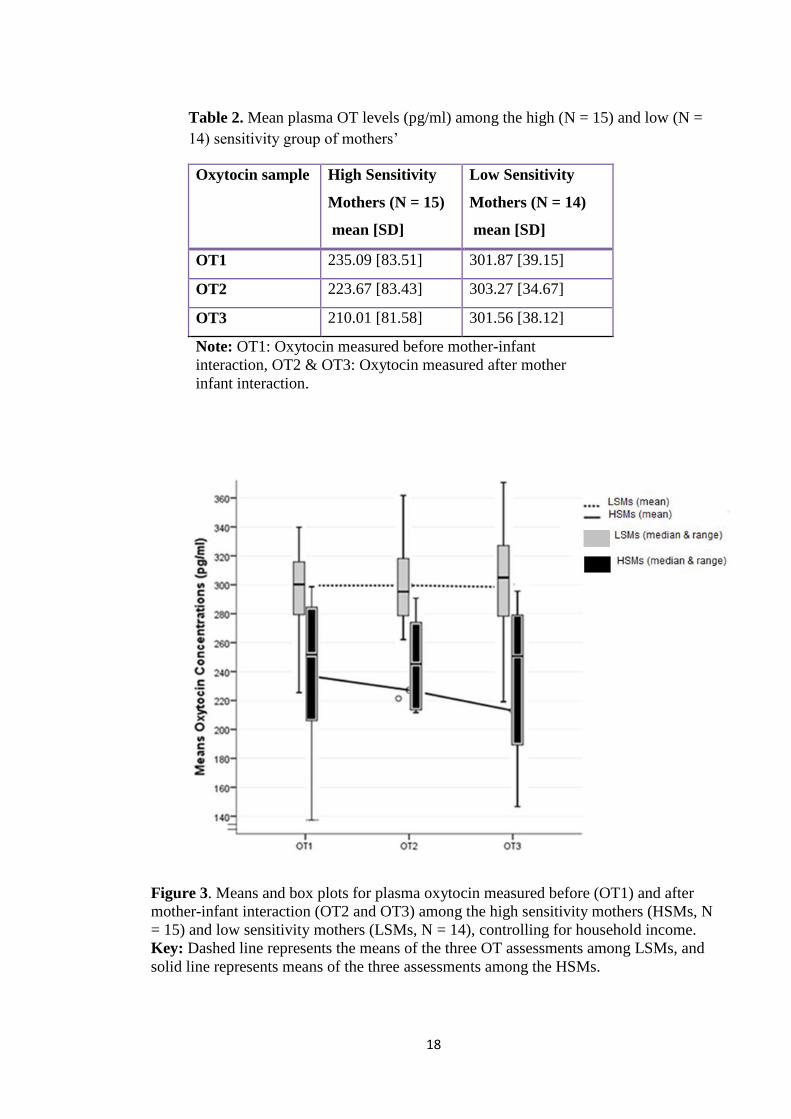
18
Table 2. Mean plasma OT levels (pg/ml) among the high (N = 15) and low (N =
14) sensitivity group of mothers’
Oxytocin sample High Sensitivity
Mothers (N = 15)
mean [SD]
Low Sensitivity
Mothers (N = 14)
mean [SD]
OT1 235.09 [83.51] 301.87 [39.15]
OT2 223.67 [83.43] 303.27 [34.67]
OT3 210.01 [81.58] 301.56 [38.12]
Note: OT1: Oxytocin measured before mother-infant
interaction, OT2 & OT3: Oxytocin measured after mother
infant interaction.
Figure 3. Means and box plots for plasma oxytocin measured before (OT1) and after
mother-infant interaction (OT2 and OT3) among the high sensitivity mothers (HSMs, N
= 15) and low sensitivity mothers (LSMs, N = 14), controlling for household income.
Key: Dashed line represents the means of the three OT assessments among LSMs, and
solid line represents means of the three assessments among the HSMs.

19
Figure 4. The relationship between baseline plasma oxytocin (OT pre) and own maternal
overprotection among: (a) low sensitivity (N = 14), and (b) high sensitivity mothers (N
= 15).

194

1
Page 1 of 21
Neural mechanisms underlying maternal behaviour in new mothers: Is natural
variation in maternal sensitivity reflected in maternal brain responses to infant
stimuli?
Authors
1 Alya Elmadih
1 Ming Wai Wan
2 Darragh Downey
2 Rebecca Elliott
1 Kathryn M Abel
Affiliations, and author addresses
1 Community Based Medicine, Institute of Brain, Behaviour and Mental Health,
University of Manchester, Manchester, M13 9PL, UK.
2 Neuroscience and Psychiatry Unit, Institute of Brain, Behaviour and Mental Health,
University of Manchester, Manchester, M13 9PL, UK.
Corresponding Author
Kathryn M Abel
3rd
Floor East Jean McFarlane Building
University of Manchester
M13 9PL
UK
+44 161 275 0714/32
E-mail address: kathryn.m.abel @ manchester.ac.uk
The total number of words of the manuscript, including entire text from title page to
figure legends: 4799
The number of words of the abstract: 337
The number of figures: 3
The number of tables: 3

2
ABSTRACT
Background: Animal and human evidence suggests that natural variation in maternal
caregiving behaviour is related to variation in maternal Oxytocin (OT) levels. Discrete
networks of maternal brain which mediate emotion processing, stress- and reward-
related neural systems are thought key to healthy maternal responsiveness. Maternal
behaviour is complex and a composite of multiple behaviours; recent evidence suggests
that in responding appropriately to her infant, a mother’s brain activation may reflect
these maternal behaviours in discrete pathways. But to date, no study has examined
distinct activation patterns related to the degree of sensitivity a mother shows in
responding to her infant i.e. ‘maternal sensitivity’ (accurate and prompt responsiveness
to infant signals). Such patterns may act as biomarkers for sensitive maternal brain and
help in the development of future intervention studies to improve parenting. Methods:
Out of 105 expectant mothers, 80 were blind rated for maternal sensitivity from 6
minute videotaped free play interaction with their 4 to 6 month infant. At 7-9 months
postpartum, 30 of these mothers (15 ‘higher sensitivity mothers’ (henceforth high
sensitivity mothers-HSMs) and 15 ‘lower sensitivity mothers’ (henceforth low
sensitivity mothers-LSMs)) underwent functional magnetic resonance imaging to
examine brain responses to viewing ‘own’ versus ‘unknown’ infant videos, using a
range of affects (neutral, happy, and sad). Maternal plasma OT measurements following
10 minute play interactions with their infant were also performed. Results: Compared
to LSMs, HSMs showed significantly greater blood oxygenation level dependent
activation in the right superior temporal gyrus in response to own versus unknown
neutral infant, and to own happy infant versus neutral control. Changes in brain
activation were significantly ‘negatively’ correlated with plasma OT responses in those
mothers. Conversely, compared to HSMs, LSMs showed no significant difference in
brain activation in response to own infant separately or in contrast to unknown infant.
Conclusion: Activation of superior temporal gyrus suggest the more attention given by
HSMs to read their infant facial emotions; this was not shown by LSMs. Sensitive
mothering may chart discrete brain responses which might act as biomarkers for future
intervention studies to enhance the sensitivity of maternal care.
INTRODUCTION
Natural variation in maternal caregiving behaviour, ranging from neglect to optimum
caregiving has been described in both animals (Champagne et al., 2001) and humans

3
(Feldman et al., 2012; Gordon et al., 2010). Low levels of maternal sensitivity (i.e. the
accurate interpretation of infant’s signals and appropriate responsiveness) (Ainsworth et
al., 1978) in particular have been linked to poor infant social, emotional and cognitive
outcomes (NICHD, 1999; Warren & Simmens, 2005). Understanding the neurobiology
of maternal sensitivity is potentially of great importance in providing evidence of
change following interventions that are designed to enhance parenting (e.g. Weisman et
al., 2012). Recent use of functional imaging (fMRI) (Raichle & Mintun, 2006) has
added greatly to our knowledge about brain mechanisms associated with healthy
parenting by mapping blood oxygenation level dependent (BOLD) brain responses to
infant versus non-infant stimuli in vivo. The reported range of BOLD activation patterns
in response to infant stimuli suggests that a complex network is involved in shaping
maternal behaviour (Musser et al., 2012). Such networks include motivational and
reward pathways in the cingulate cortex and prefrontal cortex, areas for emotion
processing such as the amygdala, areas for decoding emotional memories such as the
hippocampus, and areas implicated in OT secretion such as the hypothalamus (Atzil et
al., 2011; Lenzi et al., 2009; Lorberbaum et al., 2002; Ranote et al., 2004; Strathearn et
al., 2009; Swain et al., 2008).
While many studies examined maternal brain responses to infant stimuli (Bartels &
Zeki, 2004; Lenzi et al., 2009; Lorberbaum et al., 2002; Noriuchi et al., 2008; Ranote et
al., 2004; Strathearn et al., 2009; Swain et al., 2008), far fewer have examined the
neural correlates in relation to observed maternal behaviour (Atzil et al., 2011; Musser
et al., 2012); and only one study has attempted to focus on maternal sensitivity (Kim et
al., 2011). Kim et al. (2011) compared the neural correlates of maternal sensitive
responding between nine breastfeeding mothers and eight formula-feeding mothers
following exposure, in an fMRI paradigm, to own and unknown infant cries at 3-4
weeks postpartum. They reported that breastfeeding mothers showed greater activation
in areas involved in emotion processing and empathy, such as the amygdala and insula
(the latter is also part of the mirroring neurons-see below); and in areas associated with
planning behaviour, such as the superior frontal gyrus. Activation of the amygdala and
superior frontal gyrus were positively correlated with observed maternal sensitivity
ratings at 3-4 months. This is interesting especially with the amygdala has been
previously implicated in multiple functions, all of which facilitate maternal behaviour,
such as processing of emotional salience (positive or negative), motivation as well as
empathy (Breiter et al., 1996; Ranote et al., 2004; Swain et al., 2008). However, the

4
findings of Kim et al. have not been replicated in groups rigorously defined to represent
variations in maternal sensitivity.
As far as we are aware, only one study has examined the neural correlates of maternal
behaviour in mothers distinguished specifically on a measure of maternal sensitivity.
Atzil et al. (2011) examined the difference in brain responses among 23 mothers
grouped according to their observed ‘synchronous’ or ‘intrusive’ behaviour with their 4-
6-month infants. In that study, both groups activated limbic motivational pathways,
such as the right amygdala and the left nucleus accumbens. In addition, synchronous
mothers showed significant BOLD activation in areas rich in ‘mirroring neurons’
(Iacoboni, 2009; Iacoboni & Dapretto, 2006), i.e. superior temporal gyrus, inferior
frontal gyrus and the insula, suggesting the ability of synchronous mothers to read and
understand their infant signals (Gallagher & Frith, 2003; Rizzolatti & Fabbri-Destro,
2008). Although synchrony constitutes part of ‘sensitive responsiveness’, it is important
to note that maternal synchrony focuses on more fine-grained behavioural and affect
attunement with the infant, which may be more closely related with brain processes. On
the other hand, maternal sensitivity is probably more linked with many areas of
development. Yet, sensitivity and synchrony are likely to be highly correlated. Mothers
who respond promptly and appropriately to their infant’s behaviours (maternal
sensitivity) are likely to show more coordination in affect, gaze, body movement etc.
(synchrony).
Recently, Musser et al. (2012) examined brain responses to own and unknown infant
cries among 22 first-time mothers who were rated for ‘sensitivity’, ‘harmony’ and
‘intrusive’ behaviour with their 18-month-old infant, through mother-infant play
interaction. They reported that more sensitive behaviour was significantly associated
with greater activation in areas implicated in reward and planning of behaviour in the
frontal cortex, and areas for reading others’ minds, such as the inferior frontal gyrus
(Adolphs, 2002). Mothers who showed more harmony also showed significantly greater
activation in areas involved in recalling memories, such as the left hippocampal regions
(Phelps et al., 2004; Swain et al., 2008); whereas, mothers who showed more intrusive
behaviours activated the temporal pole and areas associated with empathy for loved
ones, such as left insula (Atzil et al., 2011). Although Musser et al. did not compare
BOLD activation between distinct groups of sensitive and less sensitive mothers, and
although brain activation was only examined in relation to proportions of maternal
5
behaviours, their findings support the importance of considering discrete maternal
behaviour when examining neural correlates.
Maternal brain activation has also been linked with maternal oxytocin (OT) levels on
the basis that the animal literature finds a good case for a central role of OT in
mediating maternal care behaviour (Champagne, 2008). Mothers who delivered
vaginally (Swain et al., 2008) or who breastfeed their infants (Kim et al., 2011) have
both been used as a proxy measure for high maternal OT levels. Strathearn et al. (2009)
reported that baseline maternal plasma OT levels were positively correlated with
maternal BOLD activation in thalamocingulate regions (in response to own infant
pictures) among mothers rated as securely attached to their own mothers, and as
compared to mothers rated as insecurely attached to their own mothers. Similarly, Atzil
et al. (2011) reported baseline plasma OT levels were positively correlated with BOLD
activation in motivational brain areas such as the nucleus accumbens in ‘synchronous’
mothers and not in ‘intrusive’ mothers. To date, no study has examined the relationship
between dynamic plasma OT responses to infant challenge and directly observed
behavioural measures of maternal sensitivity.
In the current study, we attempt to extend previous findings by examining maternal
BOLD activation (using fMRI) to own and unknown infant stimuli in a group of well-
characterised mothers from a community UK sample. We compare responses and
relationships between two groups of mothers representing natural variations of maternal
sensitivity (sensitive and less sensitive mothers). We aimed to examine whether
sensitive maternal responsiveness could chart discrete patterns of maternal brain
activation, and whether such activation is correlated with dynamic responses in maternal
plasma OT to infant challenge. We hypothesised that in response to own versus
unknown infant video, mothers rated as demonstrating higher sensitivity would show
significantly greater BOLD activation compared to less sensitive mothers, and this
would be seen specifically in brain regions related to maternal behaviours, including:
hypothalamus, cingulate cortex, hippocampal formation and superior temporal gyrus.
We also hypothesised that maternal brain activation in response to own infant stimuli
among mothers rated as higher sensitivity would be positively correlated with plasma
OT responses following a play interaction with their infants.
MATERIALS AND METHODS
Participants
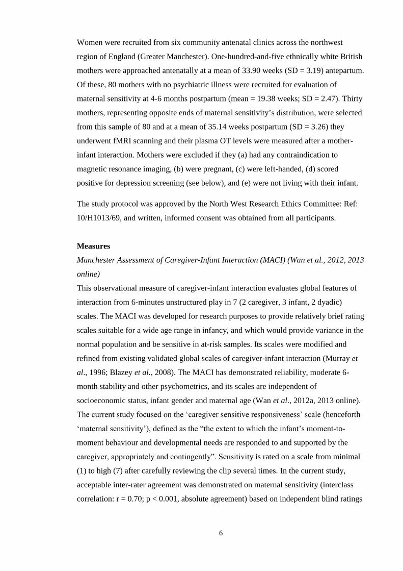
6
Women were recruited from six community antenatal clinics across the northwest
region of England (Greater Manchester). One-hundred-and-five ethnically white British
mothers were approached antenatally at a mean of 33.90 weeks (SD = 3.19) antepartum.
Of these, 80 mothers with no psychiatric illness were recruited for evaluation of
maternal sensitivity at 4-6 months postpartum (mean = 19.38 weeks; SD = 2.47). Thirty
mothers, representing opposite ends of maternal sensitivity’s distribution, were selected
from this sample of 80 and at a mean of 35.14 weeks postpartum (SD = 3.26) they
underwent fMRI scanning and their plasma OT levels were measured after a mother-
infant interaction. Mothers were excluded if they (a) had any contraindication to
magnetic resonance imaging, (b) were pregnant, (c) were left-handed, (d) scored
positive for depression screening (see below), and (e) were not living with their infant.
The study protocol was approved by the North West Research Ethics Committee: Ref:
10/H1013/69, and written, informed consent was obtained from all participants.
Measures
Manchester Assessment of Caregiver-Infant Interaction (MACI) (Wan et al., 2012, 2013
online)
This observational measure of caregiver-infant interaction evaluates global features of
interaction from 6-minutes unstructured play in 7 (2 caregiver, 3 infant, 2 dyadic)
scales. The MACI was developed for research purposes to provide relatively brief rating
scales suitable for a wide age range in infancy, and which would provide variance in the
normal population and be sensitive in at-risk samples. Its scales were modified and
refined from existing validated global scales of caregiver-infant interaction (Murray et
al., 1996; Blazey et al., 2008). The MACI has demonstrated reliability, moderate 6-
month stability and other psychometrics, and its scales are independent of
socioeconomic status, infant gender and maternal age (Wan et al., 2012a, 2013 online).
The current study focused on the ‘caregiver sensitive responsiveness’ scale (henceforth
‘maternal sensitivity’), defined as the “the extent to which the infant’s moment-to-
moment behaviour and developmental needs are responded to and supported by the
caregiver, appropriately and contingently”. Sensitivity is rated on a scale from minimal
(1) to high (7) after carefully reviewing the clip several times. In the current study,
acceptable inter-rater agreement was demonstrated on maternal sensitivity (interclass
correlation: r = 0.70; p < 0.001, absolute agreement) based on independent blind ratings

7
of 30% of interaction clips in the complete sample. Disagreements were resolved by
both raters re-reviewing the clips to reach consensus.
The Edinburgh Postnatal Depression Scale (EPDS) (Cox et al., 1987)
This 10-item self-report instrument is widely used to screen for depression in the
postpartum and antenatal periods. Items are rated on a 4-point Likert-type scale, and a
cut-off score of 12 was used for screening positive.
The Hospital Anxiety and Depression (HADS) rating scale (Zigmond & Snaith, 1983)
This 14-item questionnaire is a self-rating instrument to screen for anxiety and
depression (seven items each). Items are rated on a 4-point Likert scale. Mothers were
excluded if they scored: EPDS ≥ 12 and HADS-depression > 11.
Procedure
Time 1: Mother-Infant Interaction
At 4-6 months postpartum, mothers were visited at home and asked-after a period of
familiarisation -to play with their infant on a floor mat as they normally do, with or
without toys, as they wished. The interactions were videotaped for six minutes and later
rated, using the MACI caregiver sensitivity scale, by a trained researcher blind to study
information. Mothers also completed EPDS, HADS, and obstetric information sheet.
Mothers’ demographic characterestics were collected previously in anther part of the
sudy.
Time 2: fMRI Scanning & Oxytocin Sampling
Video of the infant’s face to be edited for use as fMRI stimuli were obtained during a
preparatory home visit. Approximately two weeks later mothers and infants attended the
Wellcome Trust Clinical Research Facility (WTCRF) where mothers were scanned
using fMRI and their plasma OT samples were collected. During the 35 minute
scanning session, mothers were instructed to view the video of their infant and an
unknown infant as though they were watching TV. For plasma OT measurements, blood
samples were taken from mothers at the same time in the early afternoon, 1200 -1400
hours, and one hour after the last nursing feed. Mothers were asked to refrain from
caffeine and smoking for at least two hours before the sampling.
i. fMRI Paradigms
A series of alternating pre-recorded 20-second video clips consisting of three affect
conditions (neutral, happy, and sad) of participant’s ‘own’ infant and an ‘unknown’

8
infant (matched on age and gender) in addition to a neutral control stimulus (moving
traffic) was prepared for use in the fMRI scan. Videos lasting 16 minutes were
presented in the following order: (Own neutral infant - neutral control - unknown
neutral infant) x 4, REST (i.e. blank screen), (Own happy infant - neutral control -
unknown happy infant) x 4, REST, (Own sad infant - neutral control - unknown sad
infant) x 4, REST.
ii. fMRI Acquisition
Imaging was performed using a 1.5 Tesla Philips Intera MRI scanner running Explorer
gradients (software version 11.1.4.4). High-resolution T1-weighted structural images
were acquired to exclude any structural abnormality (none were found) and for co-
registration with functional data. The structural scan using SENSE employed a 3D
Contrast Turbo Field Echo Sequence with a temporal resolution (TR) of 9ms and an
echo time (TE) of 4ms with an 8° flip angle producing 140 slices with a voxel size of
0.8 x 0.8 x 1.0 mm. Functional images were acquired using a multi-slice, single-shot
echo-planar imaging sequence, generating 29 ascending axial slices (TR = 2.5s, TE =
40ms, 4mm thickness with 0.5mm slice gap, in-plane resolution of 3.4x3.4mm).
iii. fMRI Analysis
Imaging data were preprocessed, individual data corrected for motion, and the
individual data were spatially normalised for group analyses using Statistical Parametric
Mapping (SPM8) and implemented in MATLAB (Mathworks Inc, Sherborn, MA,
USA). All functional images were realigned using a least squares approach and a six-
parameter (rigid body) spatial translation (Friston et al., 1996). The first image was used
as a reference scan and the following images were translated onto this, correcting for
pitch, roll and yaw throughout the task. Translation and rotation corrections did not
exceed 3.0 mm and 2.5° for any participant.
The scans were spatially (stereotactically) normalised onto standardised brain slices.
This involves the warp of the images onto a standardised template. The standardised
template conforms to the ICBM, NIH P-20 project and approximates the stereotactic
atlas of Talairach and Tournoux (1988). The standardised slices then underwent
smoothing using an isotropic Gaussian kernel filter (10mm full width half maximum
[FWHM]) to increase the signal-to-noise ratio by use of the matched filter theorem
(Friston et al., 2000). A parametric model was employed that enabled the modelling of
linear hemodynamic responses. We compared brain activation patterns between our two
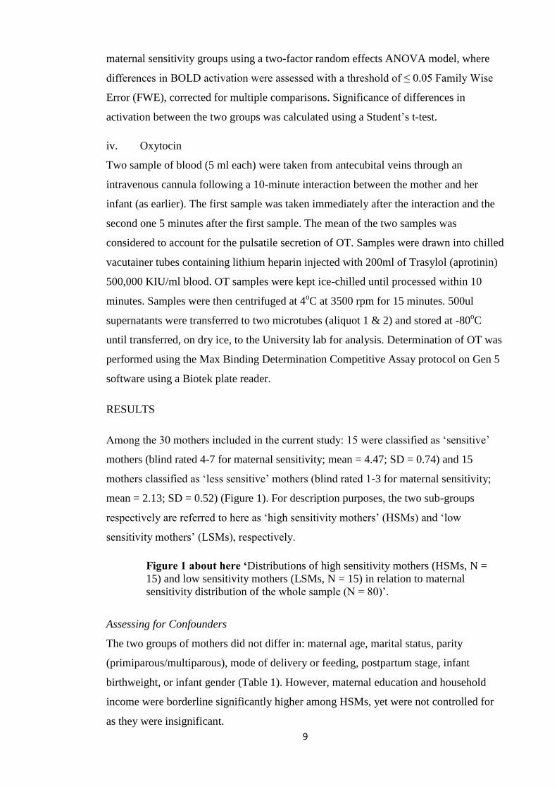
9
maternal sensitivity groups using a two-factor random effects ANOVA model, where
differences in BOLD activation were assessed with a threshold of ≤ 0.05 Family Wise
Error (FWE), corrected for multiple comparisons. Significance of differences in
activation between the two groups was calculated using a Student’s t-test.
iv. Oxytocin
Two sample of blood (5 ml each) were taken from antecubital veins through an
intravenous cannula following a 10-minute interaction between the mother and her
infant (as earlier). The first sample was taken immediately after the interaction and the
second one 5 minutes after the first sample. The mean of the two samples was
considered to account for the pulsatile secretion of OT. Samples were drawn into chilled
vacutainer tubes containing lithium heparin injected with 200ml of Trasylol (aprotinin)
500,000 KIU/ml blood. OT samples were kept ice-chilled until processed within 10
minutes. Samples were then centrifuged at 4oC at 3500 rpm for 15 minutes. 500ul
supernatants were transferred to two microtubes (aliquot 1 & 2) and stored at -80oC
until transferred, on dry ice, to the University lab for analysis. Determination of OT was
performed using the Max Binding Determination Competitive Assay protocol on Gen 5
software using a Biotek plate reader.
RESULTS
Among the 30 mothers included in the current study: 15 were classified as ‘sensitive’
mothers (blind rated 4-7 for maternal sensitivity; mean = 4.47; SD = 0.74) and 15
mothers classified as ‘less sensitive’ mothers (blind rated 1-3 for maternal sensitivity;
mean = 2.13; SD = 0.52) (Figure 1). For description purposes, the two sub-groups
respectively are referred to here as ‘high sensitivity mothers’ (HSMs) and ‘low
sensitivity mothers’ (LSMs), respectively.
Figure 1 about here ‘Distributions of high sensitivity mothers (HSMs, N =
15) and low sensitivity mothers (LSMs, N = 15) in relation to maternal
sensitivity distribution of the whole sample (N = 80)’.
Assessing for Confounders
The two groups of mothers did not differ in: maternal age, marital status, parity
(primiparous/multiparous), mode of delivery or feeding, postpartum stage, infant
birthweight, or infant gender (Table 1). However, maternal education and household
income were borderline significantly higher among HSMs, yet were not controlled for
as they were insignificant.
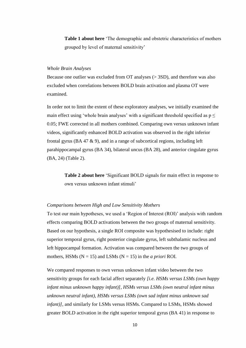
10
Table 1 about here ‘The demographic and obstetric characteristics of mothers
grouped by level of maternal sensitivity’
Whole Brain Analyses
Because one outlier was excluded from OT analyses (> 3SD), and therefore was also
excluded when correlations between BOLD brain activation and plasma OT were
examined.
In order not to limit the extent of these exploratory analyses, we initially examined the
main effect using ‘whole brain analyses’ with a significant threshold specified as p ≤
0.05; FWE corrected in all mothers combined. Comparing own versus unknown infant
videos, significantly enhanced BOLD activation was observed in the right inferior
frontal gyrus (BA 47 & 9), and in a range of subcortical regions, including left
parahippocampal gyrus (BA 34), bilateral uncus (BA 28), and anterior cingulate gyrus
(BA, 24) (Table 2).
Table 2 about here ‘Significant BOLD signals for main effect in response to
own versus unknown infant stimuli’
Comparisons between High and Low Sensitivity Mothers
To test our main hypotheses, we used a ‘Region of Interest (ROI)’ analysis with random
effects comparing BOLD activations between the two groups of maternal sensitivity.
Based on our hypothesis, a single ROI composite was hypothesised to include: right
superior temporal gyrus, right posterior cingulate gyrus, left subthalamic nucleus and
left hippocampal formation. Activation was compared between the two groups of
mothers, HSMs (N = 15) and LSMs (N = 15) in the a priori ROI.
We compared responses to own versus unknown infant video between the two
sensitivity groups for each facial affect separately [i.e. HSMs versus LSMs (own happy
infant minus unknown happy infant)], HSMs versus LSMs (own neutral infant minus
unknown neutral infant), HSMs versus LSMs (own sad infant minus unknown sad
infant)], and similarly for LSMs versus HSMs. Compared to LSMs, HSMs showed
greater BOLD activation in the right superior temporal gyrus (BA 41) in response to

11
own neutral infant as compared to unknown neutral infant (Table 3, Figure 2); whereas,
compared with HSMs, LSMs did not show significant activation in response to own
versus unknown infant contrasts in any of the ROIs. Comparing activation between
HSMs and LSMs in own infant happy or sad versus unknown happy or sad videos did
not reveal significant differences between the two groups.
Finally, we compared BOLD activation to different facial affects between HSMs and
LSMs using only own infant stimuli versus neutral control, [i.e. HSMs versus LSMs
(own neutral infant minus neutral control), HSMs versus LSMs (own happy infant minus
neutral control), HSMs versus LSMs (own sad infant minus neutral control)], and
similarly for LSMs versus HSMs. Compared to LSMs, HSMs showed greater BOLD
activation in the right superior temporal gyrus extending to the right insula (BA 13) in
response to own happy infant (Table 3, Figure 2). By contrast, compared to HSMs,
LSMs did not show significant BOLD activation in response to own happy infant video
in any ROIs. Viewing own neutral or sad infant videos did not reveal significant
differences in BOLD activation in the ROIs when the two groups were compared.
Table 3 about here ‘Areas of significant BOLD activation within ROI in
response to infant stimuli, when comparing high sensitivity (N = 15) and low
sensitivity (N = 15) mothers’
Figure 2 about here ‘Maternal brain activation in response to infant stimuli:
Compared with LSMs, HSMs show greater activation of the right superior
temporal gyrus in response to: (a) own neutral infant versus unknown neutral
infant videos, and (b) own happy infant versus neutral control, at ROI-
threshold of FWE ≤ 0.05. Structural brain image created from average of all
subjects.
Correlations of Plasma Oxytocin with ROI Activation
Post-interaction plasma OT was significantly lower among HSMs (mean = 210.01; SD
= 81.58) compared to LSMs (mean = 301.56; SD = 38.12), F (1, 26) = 9.7; p < 0.01)
(after controlling for household income which was lower in LSMs and after excluding
the outlier). When considering the correlation between ROI activation and plasma OT, a
between-group correlation was found. Activation of right superior temporal gyrus (in

12
response to own happy infant) was negatively correlated with plasma OT levels among
higher sensitivity mothers (r = - 0.81; p < 0.01) (Figure 3).
Figure 3 about here ‘Correlation between BOLD activation in the right superior
temporal gyrus (STG) and post interaction plasma oxytocin among high
sensitivity mothers (n = 15)’.
DISCUSSION
Overall, when early postpartum mothers viewed videos of their own infants compared
to unknown infants, significant brain activation was elicited predominantly in three
areas: i) the anterior cingulate gyrus, ii) the right inferior frontal gyrus, and iii) the left
parahippocampal gyrus and bilateral uncus. This is consistent with previous findings
(e.g. Kim et al., 2011; Swain et al., 2008). In accordance with our hypothesis, when
compared to LSMs, HSMs showed greater BOLD activation in right superior temporal
gyrus in response to own neutral infant compared to an unknown infant video or in
response to own happy infant compared to neutral control stimulus. Inconsistent with
our hypothesis, however: the BOLD activation among HSMs (in response to own happy
infant) was negatively and not positively correlated with post-interaction plasma OT
concentration in those mothers. Furthermore, and inconsistent with our hypothesis,
HSMs did not show significantly greater BOLD activation in any other ROIs,
specifically hypothalamus, posterior cingulate gyrus, or the hippocampal formation, in
response to own versus unknown infant stimuli. When compared to HSMs, LSMs
showed no significantly greater activation in any ROI in response to their own infants.
The superior temporal gyrus has been implicated in the perception of emotions in facial
stimuli and were also understood to contains ‘mirror neurons’ (Iacoboni & Dapretto,
2006), and is thus implicated in understanding and empathising with others’ ‘Theory of
Mind’ (Blakemore & Decety, 2001; Gallagher & Frith, 2003; Rizzolatti & Fabbri-
Destro, 2008). For a postpartum mother to respond sensitively to her infant’s needs,
recognising and understanding another’s emotions (i.e. her infant’s emotion) is a key
aspect of sensitive mothering (Strathearn et al., 2012). Therefore, our findings that
HSMs show significantly greater activation in superior temporal gyrus suggests this is
related to their behaviour. In other words, the findings suggest that HSMs give more

13
attention to read their infant facial emotions, whereas LSMs do not, possibly because of
the superior maternal reflective function among the former (Brunet et al., 2000; Lenzi et
al., 2009). Thus, a sensitive mother must have the capacity to recognise her infant’s
emotional and other cues/signals, pay attention to them and then respond to them by
appropriately mirroring and affirming positive emotions or recognising and reassuring
in relation to the negative ones (Ainsworth et al., 1978; Sroufe, 2000).
Activation of superior temporal gyrus has also been reported in response to infant cry at
2-4 weeks postpartum among exclusively breastfeeding mothers (n = 9) when compared
to formula-feeding mothers (Kim et al., 2011), and in response to own infant cry at 2-4
weeks postpartum among mothers who delivered vaginally (n = 6) as compared to those
who delivered by Caesarean section (n = 6) (Swain et al., 2008). Both these groups
were taken to represent more highly sensitive mothers with higher OT levels.
Contrary to our expectations, HSMs (as compared to LSMs) did not show significantly
greater BOLD activation in regions involved in hormonal regulation, motivation, or
encoding emotional memories such as thalamus, posterior cingulate gyrus, and
hippocampal formation respectively. Previous findings are equivocal, with some studies
reporting greater activation of these areas in response to infant cry (Kim et al., 2011;
Lorberbaum et al., 2002; Swain et al., 2004; Swain et al., 2008), while others did not
(Bartels & Zeki et al., 2004; Seifritz et al., 2003).
Our data support a role for OT in reducing or modifying social stress (Taylor et al.,
2010). Previously, Bick and Dozier (2010) measured urinary OT levels in 26 healthy
mothers following 25 minutes of physical interactions while playing computer games
with their own 2.5- 4.5-year-old children, and again with an unfamiliar child of a similar
age. Urinary OT was significantly higher following interaction with the unfamiliar child
as compared with own child. They concluded that interaction with an unfamiliar child
might constitute a stressful situation and, accordingly, that OT increased in order to
modulate this stress. Similarly, among 32 nulliparous women, a significant drop in
plasma OT was reported after women viewed positively valenced stimuli (i.e. a comedy
movie), whereas no change was found after women viewed negatively valenced stimuli
(a sad movie) (Turner et al., 2002).
Our findings suggest that, compared to LSMs, mothers at the higher end of the maternal
sensitivity distribution (i.e. HSMs) perceived their infants and infant interaction as a
less stressful, more positive event, as evidenced by their lower plasma OT levels

14
following interaction with their infant. They also activated brain regions that indicate a
greater ability to recognise and understand their infant’s emotional cues.
Strength and Limitations
As far as we are aware, this is the first study to chart the differences in neural responses,
as well as plasma OT responses, in mothers who are highly sensitive in contrast to
mothers with low maternal sensitivity. However, the study has some limitations. First,
while the sample from which we derived the high and low sensitivity groups was large
enough to show a normal distribution of sensitivity and variability among HSMs and
LSMs (N = 80), it was still relatively small. Yet both the final (N = 30) and the original
samples represent a set of well-matched demographics. Second, although we classified
mothers into two groups according to distinctly different ratings along a scale (LSMs =
minimal to scattered sensitivity; HSMs = mixed to high sensitivity), we were unable to
sample the extremes in the complete sample due to the overlap between phases of study
and the interval needed to blind rate interactions. Third, although a moderate to high
degree of stability has been reported in maternal sensitivity (e.g. Joosen et al., 2012), we
rated maternal sensitivity at a mean of 11 weeks before the scanning and OT challenge
visit. Previous reports do, however, suggest that MACI-rated maternal sensitivity has
moderately high stability between seven and 14 months (r = 0.48) (Wan et al., 2013
online). Finally, we confined our recruitment to white women (as a requirement for
other parts of the study) which may limit the generalisability of our findings to early
postpartum mothers of non-white-British ethnic origin.
CONCLUSION
Our findings extend previous reports and indicate an ability to chart the quality of
maternal caregiving behaviour through BOLD activation responses to infant stimuli in
vivo. Our findings also suggest that OT may particularly act to facilitate caregiving
behaviour in lower sensitivity mothers. This prepares the way for using changes in brain
activation in response to infant stimuli as potential biomarkers for development and
evaluation of new diagnostic and treatment strategies in at-risk mothers (e.g. Riem et
al., 2011).
ACKNOWLEDGEMENTS
This study was supported by the Magnetic Resonance Imaging facility, the WTCRF
(Manchester), and the Centre for Women’s Mental Health (University of Manchester,

15
UK). We are very grateful to all the mothers and infants who have contributed to this
study.
REFERENCES
Adolphs, R. (2002). Neural systems for recognizing emotion. Current Opinion in
Neurobiology 12, 169-77.
Ainsworth, M. D. S., Blehar, M. C., Waters, E. & Wall, S. (1978). Patterns of
attachment: A psychological study of the strange situation. Hillsdale, NJ:
Erlbaum.
Atzil, S., Hendler, T. & Feldman, R. (2011). Specifying the Neurobiological Basis of
Human Attachment: Brain, Hormones, and Behavior in Synchronous and
Intrusive Mothers. Neuropsychopharmacology 36, 1-13.
Bartels, A. & Zeki, S. (2004). The Neural correlation of maternal and romantic love.
Neuroimage 21, 1155-66.
Bick, J. & Dozier, M. (2010). Mothers’ and Children’s Concentrations of Oxytocin
Following Close, Physical Interactions with Biological and Non-biological
Children. Developmental Psychobiology 52, 100-7.
Blakemore, S. J. & Decety, J. (2001). From the perception of action to the
understanding of intention. Nature Reviews Neuroscience 2, 561-7.
Blazey, L., Leadbitter, K., Holt, C. & Green, J. (2008). Attachment behaviours and
parent–child interaction in pre-school autism. London, UK: International
Meeting for Autism Research. Poster.
Breiter, H. C., Etcoff, N. L., Whalen, P. J., Kennedy, W. A., Rauch, S. L., Buckner, R.
L., Strauss, M. M., Hyman, S.E. & Rosen, B. R. (1996). Response and
habituation of the human amygdala during visual processing of facial
expression. Neuron 17, 875-87.
Brunet, E., Sarfati, Y., Hardy-Bayle, M. C. & Decety, J. (2000). A PET Investigation of
the Attribution of Intentions with a Nonverbal Task. Neuroimage 11, 157-166.
Champagne, F., Diorio, J., Sharma, S. & Meaney, M. J. (2001). Naturally occurring
variations in maternal behavior in the rat are associated with differences in
estrogen-inducible central oxytocin receptors. Proceedings of the National
Academy of Sciences of the United States of America 98, 12736-41.
Champagne, F. A. (2008). Epigenetic mechanisms and the trans-generational effects of
maternal care. Frontiers in Neuroendocrinology 29, 386-97.
Cox, J. L., Holden, J. M. & Sagovsky, R. (1987). Detection of postnatal depression:
Development of the 10-item Edinburgh Postnatal Depression Scale. British
Journal of Psychiatry 150, 782-786.
Feldman, R., Zagoory-Sharon, O., Weisman, O., Schneiderman, I., Gordon, I., Maoz,
R., Shalev, I. & Ebstein, R. P. (2012). Sensitive parenting is associated with
plasma oxytocin and polymorphisms in the OXTR and CD38 genes. Biological
Psychiatry 72, 175-81.
Friston, K. J., Holmes, A., Poline, J. B., Price, C. J. & Frith, C. D. (1996). Detecting
activations in PET and fMRI: levels of inference and power. Neuroimage 4, 223-
35.
Friston, K. J. (2000). The labile brain I. Neuronal transients and nonlinear coupling.
Philosophical Transactions of the Royal Society B: Biological Sciences 355,
215-36.
Gallagher, H. L. & Frith, C. D. (2003). Functional imaging of "theory of mind". Trends
in Cognitive Sciences - Cell 7, 77-83.

16
Gordon, I., Zagoory-Sharon, O., Leckman, J. F. & Feldman, R. (2010). Oxytocin and
the Development of Parenting in Humans. Biological Psychiatry 68, 377-82.
Iacoboni, M. & Dapretto, M. (2006). The mirror neuron system and the consequences of
its dysfunction. Nature Reviews Neuroscience 7, 942-51.
Iacoboni, M. (2009). Neurobiology of imitation. Current Opinion in Neurobiology 19,
661-5.
Joosen, K. J., Mesman, J., Bakermans-Kranenburg, M. J. & Van IJzendoorn, M. H.
(2012). Maternal sensitivity to infants in various settings predicts harsh
discipline in toddlerhood. Attachment & Human Development 14, 101-17.
Kim, P., Feldman, R., Mayes, L. C., Eicher, V., Thompson, N., Leckman, J. F. &
Swain, J. E. (2011). Breastfeeding, brain activation to own infant cry, and
maternal sensitivity. Journal of Child Psychology and Psychiatry 52, 907-15.
Lenzi, D., Trentini, C., Pantano, P., Macaluso, E., Iacoboni, M., Lenzi, G. L. &
Ammaniti, M. (2009). Neural basis of maternal communication and emotional
expression processing during infant preverbal stage. Cerebral Cortex 19, 1124-
33.
Lorberbaum, J. P., Newman, J. D., Horwitz, A. R., Dubno, J. R., Lydiard, R. B.,
Hamner, M. B., Bohning, D. E. & George, M. S. (2002). A potential role for
thalamocingulate circuitry in human maternal behavior. Biological Psychiatry
51, 431-45.
Murray, L., Fiori-Cowley, A., Hooper, R. & Cooper, P. (1996). The impact of postnatal
depression and associated adversity on early mother-infant interactions and later
infant outcome. Child Development 67, 2512-26.
Musser, E. D., Kaiser-Laurent, H. & Ablow, J. C. (2012). The neural correlates of
maternal sensitivity: an fMRI study. Developmental Cognitive Neuroscience 2,
428-36.
NICHD, (1999). Chronicity of maternal depressive symptoms, maternal sensitivity, and
child functioning at 36 months. Developmental Psychology 35, 1297–310.
Noriuchi, M., Kikuchi, Y. & Senoo, A. (2008). The functional neuroanatomy of
maternal love: mother's response to infant's attachment behaviors. Biological
Psychiatry 63, 415-23.
Phelps, E. A. (2004). Human emotion and memory: interactions of the amygdala and
hippocampal complex. Current Opinion in Neurobiology 14, 198-202.
Raichle, M. E. & Mintun, M. A. (2006). Brain Work and Brain Imaging Annual Review
of Neuroscience 29, 449-76.
Ranote, S., Elliott, R., Abel, K. M., Mitchell, R., Deakin, J. F. & Appleby, L. (2004).
The neural basis of maternal responsiveness to infants: an fMRI study.
Neuroreport 15, 1825-29.
Riem, M. M., Bakermans-Kranenburg, M. J., Pieper, S., Tops, M., Boksem, M. A.,
Vermeiren, R. R., Van Ijzendoorn, M. H. & Rombouts, S. A. (2011). Oxytocin
modulates amygdala, insula, and inferior frontal gyrus responses to infant
crying: a randomized controlled trial. Biological Psychiatry 70, 291-7.
Rizzolatti, G. & Fabbri-Destro, M. (2008). The mirror system and its role in social
cognition. Current Opinion in Neurobiology 18, 179-84.
Seifritz, E., Esposito, F., Neuhoff, J. G., Luthi, A., Mustovic, H., Dammann, G., von
Bardeleben, U., Radue, E. W., Cirillo, S., Tedeschi, G. & Di Salle, F. (2003).
Differential sex-independent amygdala response to infant crying and laughing in
parents versus nonparents. Biological Psychiatry 54, 1367-75.
Sroufe, L. (2000). Early relationships and the development of children. Infant Mental
Health Journal 21, 67-74.

17
Strathearn, L., Fonagy, P., Amico, J. & Montague, P. R. (2009). Adult attachment
predicts maternal brain and oxytocin response to infant cues.
Neuropsychopharmacology 34, 2655-66.
Strathearn, L., Iyengar, U., Fonagy, P. & Kim, S. (2012). Maternal oxytocin response
during mother–infant interaction: Associations with adult temperament.
Hormones and Behavior 61, 429-35.
Swain, J. E., Leckman, J. F., Mayes, L. C., Feldman, R., Constable, R. T. & Schultz, R.
T. (2004). Neural substrates and psychology of human parent-infant attachment
in the postpartum. Biological Psychiatry 55.
Swain, J. E., Tasgin, E., Mayes, L. C., Feldman, R., Constable, R. T. & Leckman, J. F.
(2008). Maternal brain response to own baby-cry is affected by Cesarean section
delivery. Journal of Child Psychology and Psychiatry 49, 1042-52.
Talairach, J. & Tournoux, P. (1988). Co-Planar Stereotactic Atlas of the Human Brain.
Thieme, Stuttgart/New York.
Taylor, S. E., Saphire-Bernstein, S. & Seeman, T. E. (2010). Are plasma oxytocin in
women and plasma vasopressin in men biomarkers of distressed pair bond
relationships? Psychological Science 21, 3-7.
Turner, R. A., Altemus, M., Yip, D. N., Kupferman, E., Fletcher, D., Bostrom, A.,
Lyons, D. M. & Amico, J. A. (2002). Effects of emotion on oxytocin, prolactin,
and ACTH in women. Stress 5, 269-76.
Wan, M. W., Green, J., Elsabbagh, M., Johnson, M., Charman, T. & Plummer, F.;
BASIS Team. (2012). Parent-infant interaction in infant siblings at risk of
autism. Research in Developmental Disabilities 33, 924-32.
Wan, M. W., Green, J., Elsabbagh, M., Johnson, M., Charman, T. & Plummer, F.; the
BASIS Team. (2013 online). Quality of interaction between at-risk infants and
caregiver at 12-15months is associated with 3-year autism outcome. Journal of
Child Psychology and Psychiatry In press.
Warren, S. L. & Simmens, S. J. (2005). Predicting toddler anxiety, depressive
symptoms: Effects of caregiver sensitivity on temperamentally vulnerable
children. Infant Mental Health Journal 26, 40-55.
Weisman, O., Zagoory-Sharon, O. & Feldman, R. (2012). Oxytocin administration to
parent enhances infant physiological and behavioral readiness for social
engagement. Biological Psychiatry 72, 982-89.
Zigmond, A. S. & Snaith, R. P. (1983). The hospital anxiety and depression scale. Acta
Psychiatrica Scandinavica 67, 361-70.

18
Figure 1. Distributions of high sensitivity mothers (HSMs, N = 15) and low sensitivity
mothers (LSMs, N = 15) in relation to sensitivity distribution for the whole sample (N =
80). Key: Means for high sensitivity mothers (vertical solid line) and low sensitivity
mothers (vertical dashed line) in relation to the larger sample are also presented.

19
Table 1. The demographic and obstetric characteristics of mothers grouped by level
of maternal sensitivity (high sensitivity mothers - HSMs, and low sensitivity mothers
- LSMs)
Characteristic HSMs
(N = 15)
LSMs
(N = 15)
Statistics
t (28)
Chi-
square
test
p-
value
Mean [SD]
Maternal age
(years)
30.40 [5.37] 27.65 [4.76] - 1.38 0.18
Average maternal
education (years)
15.06 [2.82] 12.80 [2.73] - 1.87 0.07
Average annual
household income
(thousand pounds)
33.00 [4.61]
25.33 [4.24]
- 2.76
0.09
Infant birthweight
(kilograms)
3.44 [0.44] 3.23 [0.59] - 0.80 0.43
Postpartum stage
(weeks)
35.93 [2.81] 34.29 [3.69] - 1.37 0.18
Frequency (%)
Married/cohabiting 13 (86.7) 11 (78.6) 0.56 0.65
Primiparous 6 (40.0) 9 (64.3) 1.71 0.19
Infant gender
(female)
10 (66.7) 7 (50.0) 0.83 0.36
Mode of delivery
(vaginal)
10 (66.7) 11 (78.6) 0.51 0.47
Mode of feeding
(breast)
3 (20.0)
3 (20.0)
0.55*
0.64
*Fisher exact test

20
Table 2. Significant BOLD signals for main affect in response to own versus unknown
infant stimuli (combined all affects) among the whole sample (N = 30)
Contrast Area Sub-areas BA R/
L
Talairach
coordinates
x y z
Z
score
FWE
Own
infant
minus
unknown
infant
Frontal
lobe
Inferior frontal
gyrus
47
9
R
R
43 26 2
47 11 27
4.50
4.08
0.02
0.01
Limbic
lobe
Parahippocampal
gyrus
34 L -23 2 -19 4.49 0.02
Uncus 28
28
28
L
R
R
-29 5 -23
33 2 -23
30 5 -19
4.45
4.55
3.85
0.01
Anterior
cingulate gyrus
24 0 31 23 4.21 0.06
Note. All significant whole brain (Family Wise Error (FWE) ≤ 0.05) corrected for
multiple comparison. BA: Brodmann’s area; L: Left, R: Right.
Table 3. Areas of significant BOLD activation within ROI in response to infant stimuli,
when comparing high sensitivity (N = 15) and low sensitivity (N = 15) mothers.
Note. All significant ROI (Family Wise Error (FWE) ≤ 0.05) corrected for multiple
comparison. BA: Brodmann’s area; L: Left, R: Right.
Groups
compared
Contrast ROI BA Talairach
coordinates
x y z
Z
score
FWE
High
sensitivity
mothers vs.
Low
sensitivity
mothers
Own neutral
infant minus
unknown neutral
infant
Superior
temporal
gyrus
41 57 -25 13 3.39 0.05
Own happy
infant minus
neutral control
Superior
temporal
gyrus
41
13
43 -32 5
57 -32 18
3.92
3.38
0.01
0.05

21
Figure 2. Maternal brain activation in response to infant stimuli: Compared with
LSMs, HSMs show greater activation of the right superior temporal gyrus in
response to: (a) own neutral infant versus unknown neutral infant videos, and (b)
own happy infant versus neutral control, at ROI-threshold of FWE ≤ 0.05.
Structural brain image created from average of all subjects.
Figure 3. Correlation between BOLD activation in the right superior temporal
gyrus (STG) and post-interaction plasma oxytocin among high sensitivity
mothers (N = 15).
R² = 0.66 -0.3
-0.2
-0.1
0
0.1
0.2
0.3
0.4
0.5
0 100 200 300 400 No
rma
ilse
d %
BO
LD
Sig
na
l ch
an
ge
Mean Plasma Oxytocin Concentration pg/ml
Oxytocin
concentration
Linear (Oxytocin
concentration)

195
Chapter 11: Summary and Conclusion
1. The aims of the thesis
2. Summary of the findings
3. Clinical implications of the findings
4. Limitation of the thesis
5. Directions for future research
6. Conclusions
11.1. The Aims of the Thesis
The overall aim of the present thesis was to provide greater insight into the mechanisms
underlying natural variation in maternal sensitivity in healthy mothers. Maternal
sensitivity is a key predictor of child attachment and seems central to healthy child
development. In order to identify mothers at risk of low maternal sensitivity, Study I of
this thesis (Chapters 2, 3, 4 & 5) asked whether factors which could be reliably and
easily identified and which were likely to be readily available pre-or postnatally could
predict future maternal sensitivity. Such knowledge about predictors might help future
‘screening’ of mothers at risk of low maternal sensitivity so they could be targeted for
more detailed postnatal assessment and consequently receive interventions aimed at
improving their parenting capacities.
To achieve this, we approached 105 healthy women antenatally at around 30 weeks of
gestation in community antenatal clinics across the North West of England. Using
validated questionnaires, 80 pregnant women were assessed for possible predictors of

196
maternal sensitivity; maternal sensitivity was subsequently rated at 4-6 months at
postpartum through using video observation of mother-infant play-interaction.
Study II (Chapters 6, 7, 8, 9 & 10) aimed to shed light on the interplay between
hormonal and brain pathways underlying natural variation in maternal sensitivity
(Feldman et al., 2007, 2010a, 2010b; Strathearn et al.; 2009; Atzil et al., 2011). Such
knowledge about the distinct neurobiological profile of ‘higher’ sensitivity mothers
might help prepare the way for future efficient hypothesis testing of the effects of novel
interventions including the use of intranasal OT to improve parenting. Thus, Study II
sought to answer these questions:
1. Can baseline and dynamic measures of plasma OT differentiate between
sensitive and less sensitive mothers?
2. Does natural variation in maternal sensitivity result in discrete brain responses
in response to own infant stimuli? Does this pattern of brain activation
correlates with measures of plasma OT?
To achieve this, we examined differences between healthy (non-depressed) women who
were ascertained from a representative general community sample (N = 105), but whose
maternal sensitivity has been rigorously ascertained (N = 80). We compared women
from this group whose sensitivity lay at opposite extremes of the sensitivity scale (N =
30; i.e. 15 in each extreme group).
11.2. Summary of the Findings
Around a fifth of the variance in postnatal maternal sensitivity can be predicted from
readily collected variables available prenatally. Parental care received by the mother
herself and a measure of family SES via household income accounted for most of

197
the variance. This supports the idea that sensitive maternal care can be predicted, at
least to some degree, and hence early intervention could be considered for those at
risk (Chapter 4).
Another 12% of the variance in maternal sensitivity was predicted by the presence
of more than one social deprivation factor (i.e. young maternal age at birth, no
education, single marital status, low income, unemployment household income, and
early neglected parental care) and sensitivity decreased significantly as the number
of these factors increased.
Other prenatal measures, such as availability of social support, maternal attachment
with the unborn infant, early experience of maltreatment, postnatal factors such as
infant temperament, and postnatal mood scores did not predict postnatal maternal
sensitivity. These results suggest the limited capability of these measures in
predicting how sensitive a mother will be with her infant in a well population
(Chapter 4).
Contrary to our main hypothesis, we reported that: LSMs have higher baseline
plasma OT levels compared to HSMs. We would suggest the high plasma OT levels
in LSMs reflecting possible social stressors related to their relations with others,
including own mothers. These findings support the role of OT as a biomarker for a
distressed social relationship similar to what has previously been reported in studies
in women (Marazziti et al., 2006; Taylor et al., 2006, 2010; Tabak et al., 2011;
Feldman et al. 2011) (Chapter 9 & Chapter 10).
The findings also suggest that OT does not have a unioversal pattern in relation to
affiliative behaviour as it might decrease (not increase) in response to affiliative
social interaction. This warns against using it as a universal biomarker to signal
affiliative behaviour.

198
Also contrary to our main hypothesis: following a play-interaction with their infants,
plasma OT levels significantly decreased in HSMs, whereas no significant change
was seen among LSMs. These results suggest that HSMs perceived their infant’s
signals as a positive event and their plasma OT levels drop accordingly in response
to an easy, focused, positive interaction with their own infant (Taylor et al., 2002;
Strathearn et al., 2012). LSMs may not perceive the interaction with their own infant
as a positive event and hence restrain/resist the alleviating effect of the socially
affiliative interaction as compared to HSMs; accordingly their plasma OT remains
elevated (Chapter 9 & Chapter 10).
In accordance with our secondary hypothesis, we found that plasma OT levels, at
least among LSMs, were significantly correlated with mothers’ own perceived
parenting, supporting the role of early environment in shaping maternal hormonal
profile (Champagne et al., 2007; Champagne, 2008; Strathearn et al., 2009).
However, contrary to our hypothesis, the direction of this correlation was positive
with self-reported unfavourable (not with favourable) own parenting experience, in
particular, self-reported own maternal overprotection, as evidenced among LSMs.
This finding supports a role for OT in regulating stress responsivity that is related to
social relationships, including relationships with own parents (Marazziti et al., 2006;
Taylor et al., 2010; Feldman et al., 2011) (Chapter 9 & 10).
Overall, and in accordance with our hypotheses, when mothers viewed videos of
their own infants compared to unknown infants, significantly greater brain
activation was elicited predominantly in emotion processing areas (the right anterior
cingulate gyrus), areas associated with encoding of facial expressions of emotions
(right inferior frontal gyrus) and areas decoding emotional memories (the left
parahippocampal gyrus and bilateral uncus). This provides support for previous
findings and suggests that a key to healthy maternal parenting is the capacity of

199
mothers to recognise and respond to her infant’s emotions; and in young, preverbal
infants, this is perhaps especially through mirroring of emotional expressions and
facial cues (Chapter 9).
Also in accordance with our main hypothesis, we found that, compared to LSMs,
HSMs showed significantly greater activation in brain areas associated with
mirroring the emotions of others (the right superior temporal gyrus) in response to
own infant compared to an unknown infant or compared to a neutral control. By
contrast, when compared to HSMs, LSMs did not show any brain activation in
response to their own infant stimuli. These results suggest that HSMs might give
more attention to read their infant facial stimuli, compared to LSMs who do not give
enhanced attention and focus to infant signals, possibly because the latter do not
recognise these signals (as evidenced by the absence of brain activation) or do not
attribute added salience to infant signals (Chapter 9).
Contrary to hypothesis, HSMs did not show significantly greater activation in areas
implicated in OT secretion, such as the hypothalamus, or in emotion processing
areas, such as the posterior cingulate gyrus, or the hippocampal formation, in
response to own infant compared to an unknown infant video. However, this may be
consistent with our findings that suggest plasma OT’s anxiolytic/anti-stress role
(Numan & Woodside, 2010) rather than its socially affiliative role in HSMs who
may not require OT to act in this way (Chapter 9).
Inconsistence with our hypotheses the BOLD activation in the right superior
temporal gyrus among HSMs was negatively (not positively) correlated with post
interaction plasma OT concentration in those mothers. Again this is consistent with
the plasma OT findings of those mothers and it supports the anti-stress role that OT
might play in those mothers (Chapter 9, 10).

200
We infer that differences between our present findings and those of other parenting
studies which have assessed plasma OT in relation to maternal behaviour are
because, in the present study, we assessed OT in two groups of women ascertained
specifically to occupy opposite ends of the distributions of maternal sensitivity in
the general population. The other studies, however, did not directly compare groups
of mothers carefully asecrtained for sensitivity, and in many cases used indirect
proxies of sensitivity or OT, such as breastfeeding or mode of delivery (e.g. Kim et
al., 2011). We think our findings could be ‘tapping’ into elements of stress or
anxiety coping, which we suggest are an integral aspect of the concept of sensitive
maternal responsiveness (Chapter 9, 10).
11.3. Clinical Implications of the Findings
The current findings add significantly to current understanding of what underpins
the natural variation in maternal sensitivity in a population of healthy women.
It suggests this variation is related to a combination of behavioural, social and
neurobiological differences.
The results of these studies highlight the possibility of identifying predictors of
lower sensitivity mothering.
Evidence from the present studies and similar studies should facilitate future
development of an ‘affordable’, ‘accessible’, and ‘easy’ tool, for health providers to
screen women antenatally for at risk of low maternal sensitivity.
Were such a tool readily available antenatally and acceptable to women, it would
facilitate targetting of less widely available early interventions to improve their
parenting.
The empirical findings in these studies provide the basis for a new understanding of
the multiple roles of OT in the modulation of maternal caregiving. They provide

201
further support for the notion that OT acts differentially depending on the ability of
an individual to regulate emotion and cope with potentially stressful situations
(Tabak et al., 2011). In the context of maternity, our results may imply that if the
overwhelming experience of infant cues is stressful to a mother, OT may play a
more prominent anxiolytic role than when a mother derives pleasure and reward
from her infant’s cues, or develops a ‘positive emotion’ as a result of interaction
with her infant (Feldman et al., 2011).
Thus, baseline and dynamic plasma OT responses may be a potentially useful
biomarker of maternal sensitivity.
The present studies provide further support for previous findings that maternal brain
activation occurs in response to own infant stimuli in areas associated with emotion
recognition and processing. It contributes additional evidence that suggests sensitive
mothers have a greater capacity to recognise and understand their own infant
emotions compared to less sensitive mothers.
Sensitive maternal responsiveness appears to be accompanied by a discrete neural
correlate. This provides a basis for neural activation patterns acting as a biomarker
for monitoring future interventions that aim to improve parenting behaviour (e.g.
Riem et al., 2011).
The present findings also provide preliminary evidence for possible modulation of
maternal brain responses by OT (i.e. as evidenced by the significant correlation
between plasma OT and BOLD activation), although further studies are needed.
11.4. Limitations of the Thesis
The studies encountered a number of limitations which need to be considered:
The original sample from which we derived the 80 mothers in whom maternal
sensitivity was assessed was relatively large (N = 105). However, a larger sample is

202
more likely to uncover all the most important factors that influence maternal
sensitivity in a population.
Mothers’ own parenting experience and early experience of maltreatment were
assessed through self-report measures (PBI & CTQ). Both measures have shown
good psychometric properties (Bernstein et al. 1994, Wilhelm et al., 2005) and the
convergent validity for PBI was confirmed over 20 years (Wilhelm et al., 2005) but,
given that this appears to be particularly relevant as a source of risk for lower
maternal sensitivity, future research is needed to identify objective easy of
measuring this variable.
Although we classified mothers into two groups (Study II) according to distinctly
different ratings along a scale for maternal sensitivity (LSMs = minimal to scattered
sensitivity; HSMs = mixed to high sensitivity), we were unable to sample the
extremes in the complete sample due to the overlap between phases of study and the
interval needed to blind rate interactions.
Inferences about centrally functioning OT from plasma measurement, although
highly convenient, must remain limited (Modahl et al., 1998). However, many
previous studies do show modulation of peripheral plasma OT in relation to social
affiliation (e.g. Strathearn et al., 2012) and findings from fMRI studies also show
coordinated peripheral OT levels and BOLD activation in OT rich brain areas
(Strathearn et al., 2009; Atzil et al., 2011). Similarly, administration of intranasal
OT has been shown both to increase peripheral OT (saliva) and to increase
affiliative behaviours e.g. affectionate paternal behaviour (Weisman et al., 2012)
(Chapter 6).
We did not include a thorough assessment of mothers’ social relationships or
difficulties, including ‘relationship with partner’. Therefore, we cannot exclude the

203
possibility that the presence of other distressed relationships is associated with the
elevated plasma OT among the LSMs in our studies.
The significant correlation we found between plasma OT and BOLD brain
activation among HSMs does not imply a causal relationship. There might be other
unidentified factors to account for this association.
Because most of our measures are designed to measure behaviours representing
bonding/affiliation (e.g. MFAS, MII), it was essential to consider different
representation of these behaviours across cultures (Alhusen, 2008). Therefore, we
confined our recruitment to white British women, in order to minimise the cultural
differences between mothers. While this is may be seen as a strength for the study
design, it also limits the representativeness of the findings by not reflecting the
ethnic diversity of the community in the sample.
Maternal sensitivity was rated at a mean of 11 weeks prior to the OT measurements
and the scanning assessment visit. We do not think this is likely to be of concern
because several studies have reported moderate to high degree of stability in
maternal sensitivity over this relatively short time period (e.g. Joosen et al., 2012).
Moreover, previously the MACI-rated maternal sensitivity has also demonstrated
moderately high stability between 7 to 14 months (r = 0.48; Wan et al., 2013
online).
11.5. Directions for Future Research
Given the importance of identifying mothers most at risk of low maternal
sensitivity, research must continue investigating key factors which indicate the
future quality of maternal sensitivity, including maternal and infant characteristics
(Chapter 2).

204
Such complex interplay between maternal and child social, environmental, and
psychological factors must be considered by future studies. How they might
modulate the impact of risk of poor maternal sensitivity on the mother-infant
relationship is a key question to be addressed (Mertesacker et al., 2004).
Replication of these findings in a larger sample including wider ethnic and social
diversity may find that the factors identified have a greater predictive value,
especially if the current, less diverse sample may not have been sufficiently varied
to capture such associations.
Research in this area might also benefit from longitudinal designs that examine the
continuity of these influences on the mother and child relationship at later stages of
infant development (e.g. Beckwith et al., 1999).
A great strength of the UK NHS system is that it provides many chances when
broad representations of women are in regular contact with health services. This
provides opportunities for the implementation of routine participation and the
consideration of population-based screening. These and future studies should
consider how best to exploit these opportunities. For example, health visitors have
contact with all mothers following infant birth, and are uniquely placed for early
detection of problems affecting the mother-infant relationship (Appleton et al.,
2013).
Expansion of the work of these studies might include enrolment of teenage mothers,
to evaluate the effect of this young maternal age and its attendant difficulties and
risks on maternal sensitivity. Particular attention should also be given to assess
partner support in more detail and how it might affect maternal sensitivity, or create
resilience or greater risk in the mother-infant relationship, or alter the risk set of the
parenting environment in other ways.

205
Although mother-infant interaction in the play context is a widely used and well-
validated method with which to assess maternal sensitivity, it potentially lacks
information about the mother’s responsiveness to her infant in stressful situations
(McElwain & Booth-LaForce, 2006). Therefore, more comprehensive assessment of
variation in sensitivity (including under more stressful situations) should be
considered by future research.
Further studies replicating our design in a larger population, might be able to
examine whether plasma oxytocin levels mediate the relationship between maternal
overprotection and maternal sensitivity.
Replication of Study II should consider the simultaneous assessment of maternal
urinary or salivary OT along with plasma OT measures. This is especially pertinent
given recent evidence from a parenting study which reported an elevation in urinary
OT in mothers experiencing relationships difficulties (Feldman et al., 2011).
Previous evidence suggests that elevation of plasma OT in relation to stress is only
in the context of stimuli which are social stressors i.e. social relationship difficulties.
However, future studies replicating the present design might usefully combine
measurements of plasma OT with additional measures to evaluate the HPA or stress
axis (e.g. cortisol) for more thorough evaluation of the OT role in stress regulation
(Quirin et al., 2011).
The present study found significantly greater activation in brain areas related to
mothers’ ability to read an infant’s emotion (right superior temporal gyrus) in
HSMs. Future studies are needed to examine further activation patterns using more
detailed stimuli which probe specific aspects of affiliative and cognitive functions in
relation to the complex demands of sensitive mothering.

206
11.6. Conclusions
This set of studies is the first to chart the differences in both plasma OT responses as
well as brain responses among mothers representing distinct ends of the spectrum of
natural variation in maternal sensitivity. In addtion, these are the first studies
systematically to explore maternal characteristics that might contribute to this
variation. Charting the profiles of maternal brain activation associated with sensitive
parenting may be seen as a method of representing how a mother coordinates
affective, social-behavioural and neural systems. A deeper understanding of such
pathways is likely to prove useful in future studies of healthy and high risk mothers.
Overall the findings answered our research questions as follows:
1. Two readily identifiable factors available prenatally could facilitate prediction
of future maternal sensitivity: parental care received by the mother and family
SES (via household income).
2. Plasma OT measures could differentiate between sensitive and less sensitive
mothers.
3. Discrete patterns of brain response to own infant stimuli could be evidenced
by fMRI in sensitive early-postpartum mothers; and this pattern correlates
with plasma OT responses of those mothers.

207
REFERENCES
Abel, K. M., Webb, R. T., Salmon, M. P., Wan, M. W. & Appleby, L. (2005).
Prevalence and predictors of parenting outcomes in a cohort of mothers with
schizophrenia admitted for joint mother and baby psychiatric care in England.
Journal of clinical psychiatry 66, 808-9.
Adolphs, R. (2002). Neural systems for recognizing emotion. Current Opinion in
Neurobiology 12, 169-77.
Adolphs, R. (2010). What does the amygdala contribute to social cognition? Annals of
the New York Academy of Sciences 1191, 42-61.
Ainsworth, M. D. S. & Bell, S. M. (1970). Attachment, exploration, and separation:
Illustrated by the behavior of one-year-olds in a strange situation. Child
Development 41, 49-67.
Ainsworth, M. D. S., Blehar, M. C., Waters, E. & Wall, S. (1978). Patterns of
attachment: A psychological study of the strange situation. Hillsdale, NJ:
Erlbaum.
Alhusen, J. L. (2008). A Literature Update on Maternal-Fetal Attachment. Journal of
Obstetric, Gynecologic & Neonatal Nursing 37, 315-28.
Alink, L. R. A., Mesman, J., Van Zeijl, J., Stolk, M. N., Juffer, F., Bakermans-
Kranenburg, M. J., Van IJzendoorn, M. H. & Koot, H. M. (2008). Maternal
Sensitivity Moderates the Relation between Negative Discipline and Aggression
in Early Childhood. Parenting and Early Aggression 18, 99-120.
Andresen, P. & Telleen, S. L. (1992). The relationship between social support and
maternal behaviors and attitudes: a meta-analytic review. American Journal of
Community Psychology 20, 753-74.
Appleton, J. V., Harris, M., Oates, J. & Kelly, C. (2013). Evaluating health visitor
assessments of mother-infant interactions: a mixed methods study. International
Journal of Nursing Studies 50, 5-15.
Ashburner, J. & Friston, K. (1997). Multimodal image coregistration and partitioning--a
unified framework. Neuroimage 6, 209-17.
Ashburner, J., Friston, K. (1999). Nonlinear spatial normalization using basis functions.
Human Brain Mapping 7, 254-66.
Atkinson, L., Goldberg, S., Raval, V., Pederson, D., Benoit, D., Moran, G., Poulton, L.,
Myhal, N., Zwiers, M., Gleason, K. & Leung, E. (2005). On the Relation
Between Maternal State of Mind and Sensitivity in the Prediction of Infant
Attachment Security. Developmental Psychology 41, 42-53.
Atzil, S., Hendler, T. & Feldman, R. (2011). Specifying the Neurobiological Basis of
Human Attachment: Brain, Hormones, and Behavior in Synchronous and
Intrusive Mothers. Neuropsychopharmacology 36, 1–13.
Augustine, J. M., Cavanagh, S. E. & Crosnoe, R. (2009). Maternal Education, Early
Child Care and the Reproduction of Advantage. Social Forces 88, 1-29.
Avagianou, P. A. & Zafiropoulou, M. (2008). Parental bonding and depression:
personality as a mediating factor. International Journal of Adolescent Medicine
and Health 20, 261-9.
Bailey, H., De Oliveira, C. A., Wolfe, V. V., Evans, E. M. & Hartwick, C. (2012). The
impact of childhood maltreatment history on parenting: a comparison of
maltreatment types and assessment methods. Child Abuse & Neglect 36, 236-46.

208
Bakermans-Kranenburg, M. J., Van IJzendoorn, M. H. & Juffer, F. (2003). Less is
more: meta-analyses of sensitivity and attachment interventions in early
childhood. Psychological Bulletin 129, 195-15.
Bakermans-Kranenburg, M. J. & Van IJzendoorn, M. H. (2006). Gene-environment
interaction of the dopamine D4 receptor (DRD4) and observed maternal
insensitivity predicting externalizing behavior in preschoolers. Developmental
Psychobiology 48, 406–9.
Bandura, A. (1977). Self-efficacy: Toward a unifying theory of behavioral change.
Psychological Review 84, 191-15.
Barclay, L., Everitt, L., Rogan, F., Schmied, V. & Wyllie, A. (1997). Becoming a
mother--an analysis of women's experience of early motherhood. Journal of
Advanced Nursing 25, 719-28.
Barlow, J., Schrader McMillan, A. & Kirkpatrick, S. (2008). Health -led parenting
interventions in pregnancy and early years. Research Report DCSF-RW070.
Barrett, J. & Fleming, A. S. (2011). Annual Research Review: All mothers are not
created equal: Neural and psychobiological perspectives on mothering and the
importance of individual differences. Journal of Child Psychology and
Psychiatry 52, 368–97.
Bartels, A. & Zeki, S. (2004). The Neural correlation of maternal and romantic love.
Neuroimage 21, 1155-66.
Bartz, J. A., Zaki, J., Bolger, N. & Ochsner, K. N. (2011). Social effects of oxytocin in
humans: context and person matter. Trends in Cognitive Sciences 15, 301–9.
Baumgartner, T., Heinrichs, M., Vonlanthen, A., Fischbacher, U. & Fehr, E. (2008).
Oxytocin shapes the neural circuitry of trust and trust adaptation in humans.
Neuron 58, 639–50.
Beckwith, L. & Cohen, S. E. (1989). Maternal responsiveness with preterm infants and
later competency. New Directions for Child and Adolescent Development 43,
75-87.
Beckwith, L., Cohen, S. E. & Hamilton, C. E. (1999). Maternal sensitivity during
infancy and subsequent life events relate to attachment representation at early
adulthood. Developmental Psychology 35, 693-700.
Belsky, J. (1984). The Determinants of Parenting: A Process Model. Child Development
55, 83-96.
Belsky, J. & Fearon, R. M. (2002). Early attachment security, subsequent maternal
sensitivity, and later child development: does continuity in development depend
upon continuity of caregiving? Attachment and Human Development 4, 361-87.
Benasich, A. A. & Brooks-Gunn, J. (1996). Maternal attitudes and knowledge of child-
rearing: Associations with family and child outcomes. Child Development 67,
1186–205.
Bernstein, B., D. P., Ahluvalia, T., Pogge, D. & Handelsman, L. (1997). Validity of the
Childhood Trauma Questionnaire in an adolescent psychiatric population.
Journal of the American Academy of Child & Adolescent Psychiatry 36, 340-
348.
Bernstein, D., Fink, L., Handelsman, L., Foote, J., Lovejoy, M., Wenzel, K., Sapareto,
E. & Ruggiero, J. (1994). Initial reliability and validity of a new retrospective
measure of child abuse and neglect. The American Journal of Psychiatry 151,
1132-6.
Bick, J. & Dozier, M. (2010). Mothers’ and Children’s Concentrations of Oxytocin
Following Close, Physical Interactions with Biological and Non-biological
Children. Developmental Psychobiology 52, 100–7.
209
Bigelow, A. E., MacLean, K., Proctor, J., Myatt, T., Gillis, R. & Power, M. (2010).
Maternal sensitivity throughout infancy: continuity and relation to attachment
security. Infant Behavior and Development 33, 50-60.
Blakemore, S. J. & Decety, J. (2001). From the perception of action to the
understanding of intention. Nature Reviews Neuroscience 2, 561-7.
Blank, D. M., Schroeder, M. A. & Flynn, J. (1985). Major influences on maternal
responsiveness to infants. Applied Nursing Research 8, 34-38.
Blazey, L., Leadbitter, K., Holt, C. & Green, J. (2008). Attachment behaviours and
parent–child interaction in pre-school autism. London, UK: International
Meeting for Autism Research. Poster.
Bloom, J. R. & Kessler, L. (1994). Emotional support following cancer: a test of the
stigma and social activity hypotheses. Journal of Health and Social Behavior 35,
118-33.
Bloom, K. (1995). The development of attachment behaviors in pregnant adolescents.
Nursing Research 44, 284-9.
Blumberg, N. L. (1980). Effects of neonatal risk, maternal attitude, and cognitive style
on early postpartum adjustment Journal of Abnormal Psychology 89, 139-50.
Bornstein, M. H. & Bradley, R. H. (2003). Socioeconomic status, parenting, and child
development. Mahwah, NJ: Lawrence Erlbaum Associates, Inc.
Bornstein, M. H., Hendricks, C., Haynes, O. M. & Painter, K. M. (2007). Maternal
Sensitivity and Child Responsiveness: Associations with Social Context,
Maternal Characteristics, and Child Characteristics in a Multivariate Analysis.
Infancy 12, 189-22.
Bowlby, J. (1969). Attachment and loss: Vol.1 Attachment. New York: Basic Books.
Bowlby, J. (1973). Attachment and loss: Vol. 2. Separation: Anxiety and anger. New
York: Basic Books.
Bowlby, J. (1980). Attachment and loss: Vol. 3. Loss: Sadness and depression. New
York: Basic Books.
Breiter, H. C., Etcoff, N. L., Whalen, P. J., Kennedy, W. A., Rauch, S. L., Buckner, R.
L., Strauss, M. M., Hyman, S.E. & Rosen, B. R. (1996). Response and
habituation of the human amygdala during visual processing of facial
expression. Neuron 17, 875-87.
Bridges, R. S. (1996). Biochemical basis of parental behavior in the rat. In J. S.
Rosenblatt, and C. T. Advances in the Study of Behavior 25, 215-42.
Britton, J. R., Britton, H. L. & Gronwaldt, V. (2006). Breastfeeding, sensitivity, and
attachment. Pediatrics. 118, 1436-43.
Brunet, E., Sarfati, Y., Hardy-Bayle, M. C. & Decety, J. (2000). A PET Investigation of
the Attribution of Intentions with a Nonverbal Task. Neuroimage 11, 157-166.
Caba, M., Silver, R., Gonzalez-Mariscal, G., Jimenez, A. & Beyer, C. (1996). Oxytocin
and vasopressin immunoreactivity in rabbit hypothalamus during estrus, late
pregnancy, and postpartum. Brain Research 720, 7–16.
Campbell, S. B. (1979). Mother-infant interaction as a function of maternal ratings of
temperament. Child Psychiatry and Human Development 10, 67-76.
Campbell, S. B., Matestic, P., von Stauffenberg, C., Mohan, R. & Kirchner, T. (2007).
Trajectories of maternal depressive symptoms, maternal sensitivity, and
children’s functioning at school entry. Developmental Psychology 43, 1202–15.
CAN parent, (2012). Parent scheme from Government. http://www.canparent.org.uk/.
Cardinal, R. N., Parkinson, J.A., Hall, J. & Everitt, B.J. (2002). Emotion and
motivation: The role of the amygdala, ventral striatum, and prefrontal cortex.
Neuroscience & Biobehavioral Reviews 26, 321–52.
Carter, D. A. & Murphy, D. (1989). Independent regulation of neuropeptide mRNA
level and poly (A) tail length. Journal of Biological Chemistry 264, 6601–3.

210
Champagne, F., Diorio, J., Sharma, S. & Meaney, M. J. (2001). Naturally occurring
variations in maternal behavior in the rat are associated with differences in
estrogen-inducible central oxytocin receptors Proceedings of the National
Academy of Sciences of the United States of America 98, 12736-41.
Champagne, F. A., Francis, D. D., Mar, A. & Meaney, M. J. (2003). Variations in
maternal care in the rat as a mediating influence for the effects of environment
on development. Physiology & Behaviour 79, 359-71.
Champagne, F. A. & Meaney, M. J. (2007). Transgenerational effects of social
environment on variations in maternal care and behavioral response to novelty.
Behavioral Neuroscience 121, 1353-63.
Champagne, F. A. (2008). Epigenetic mechanisms and the transgenerational effects of
maternal care Frontiers in Neuroendocrinology 29, 386-97.
Chibucos, T. R. & Kail, P. R. (1981). Longitudinal Examination of Father-Infant
Interaction and Infant-Father Attachment. Merrill-Palmer Quarterly 27, 81-96.
Cicchetti, D., Rogosch, F. A. & Toth, S. L. (2006). Fostering secure attachment in
infants in maltreating families through preventive interventions. Development
and Psychopathology 18, 623-49.
Condon, J. T. & Corkindale, C. (1997). The correlates of antenatal attachment in
pregnant women. British Journal of Medical Psychology 70, 359–72.
Cox, J. L., Holden, J. M. & Sagovsky, R. (1987). Detection of postnatal depression:
Development of the 10-item Edinburgh Postnatal Depression Scale. British
Journal of Psychiatry 150, 782-786.
Cranley, M. (1981). Development of a tool for the measurement of maternal attachment
during pregnancy. Nursing Research 30, 281-4.
Crawford, J. R., Henry, J. D., Crombie, C. & Taylor, E. P. (2001). Normative data for
the HADS from a large non-clinical sample. British Journal of Clinical
Psychology 40, 429–34.
Crawley, S. B. & Spiker, D. (1983). Mother-child interactions involving two year olds
with Down's syndrome: A look at individual differences. Child Development 54,
1312-23.
Crittenden, P. M. (1979-2004). Care Index: Coding Manual. Unpublished manuscript,
Miami, Florida. available from the author. CARE-Index: Coding Manual.
Crittenden, P. M. (1981). Abusing, neglecting, problematic, and adequate dyads:
Differentiating by patterns of interaction. Quarterly Merrill-Palmer 27, 201-18.
Crnic, K. A., Greeenberg, M. T., Robinson, N. M. & Ragozin, A. S. (1984). Maternal
stress and social support: effects on the mother-infant relationship from birth to
eighteen months. American Journal of Orthopsychiatry 54, 224-35.
Crockenberg, S., Fitzgerald & Hiram, E. (1988). Social support and parenting. Theory
and research in behavioral pediatrics. In H. E. Fitzgerald, B. M. Lester & M. W.
Yogman (Eds.) 4, 141–74.
Crockenberg, S. B. (1981). Infant irritability, mother responsiveness, and social support
influences on the security of infant-mother attachment. Child Development 52,
857-65.
Crockenberg, S. B. & Acredolo, C. (1983). Infant temperament ratings: A function of
infants, or mothers, or both? Infant Behavior and Development 6, 61-72.
Crockenberg, S. B. (1986). Are temperamental differences in babies associated with
predictable differences in care giving? New Directions for Child and Adolescent
Development 1986, 53-73.
Crosnoe, R., Leventhal, T., Wirth, R. J., Pierce, K. M. & Pianta, R. C.; NICHD Early
Child Care Research Network. (2010). Family socioeconomic status and
consistent environmental stimulation in early childhood. Child Development 81,
972-87.

211
Dabrowska, J., Hazra, R., Ahern, T. H., Dong Guo, J., McDonald, A. J., Mascagni, F.,
Muller, J. F., Young, L. J. & Rainnie, D. G. (2011). Neuroanatomical evidence
for reciprocal regulation of the corticotrophin-releasing factor and oxytocin
systems in the hypothalamus and the bed nucleus of the stria terminalis of the
rat: Implications for balancing stress and affect. Psychoneuroendocrinology 36,
1312–26.
Dalgard, O. (1996). Community health profile: a tool for psychiatric prevention. In
Promotion of Mental Health. eds D. R. Trent & C. A. Reed. Aldershot: Avebury
Press. 5.
Damast, A. M., Tamis-LeMonda, C. S. & Bornstein, M. H. (1996). Mother–child play:
Sequential interactions and the relation between maternal beliefs and behaviors.
Child Development 67, 1752-66.
Dawood, M. Y., Raghavan, K. S., Pociask, C. & Fuchs, F. (1978). Oxytocin in human
pregnancy and parturition. Obstetrics & Gynecology 51.
De Geest, K., Thiery, M., Piron-Possuyt, G. & Vanden Driessche R. (1985). Plasma
oxytocin in human pregnancy and parturition. Journal of Perinatal Medicine 13,
3-13.
De Wolff, M. S. & Van Ijzendoorn, M. H. (1997). Sensitivity and Attachment: A Meta-
Analysis on Parental Antecedents of Infant Attachment. Child Development 68,
571-91.
Demers, I., Bernier, A., Tarabulsy, G. M. & Provost, M. A. (2010). Mind-mindedness in
adult and adolescent mothers: Relations to maternal sensitivity and infant
attachment. International Journal of Behavioral Development 34, 529–537.
Denham, S. A. & Moser, M. H. (1994). Mothers' Attachment to Infants: Relations with
Infant Temperament, Stress, and Responsive Maternal Behavior Early Child
Development and Care, 98, 1-6.
Desmond, J. E. & Glover, G. H. (2002). Estimating sample size in functional MRI
(fMRI) neuroimaging studies: statistical power analyses. Journal of
Neuroscience Methods 118, 115-28.
DiPietro, J. A. (2010). Psychological and Psychophysiological Considerations
Regarding the Maternal–Fetal Relationship. Infant and Child Development 19,
27–38.
Ditzen, B., Neumann, I. D., Bodenmann, G., Von Dawans, B., Turner, R. A., Ehlert, U.
& Heinrichs, M. (2007). Effects of different kinds of couple interaction on
cortisol and heart rate responses to stress in women. Psychoneuroendocrinology
32, 565-74.
Downer, J. T. & Pianta, R. C. (2006). Academic and Cognitive Functioning in First
Grade: Associations with Earlier Home and Child Care Predictors and with
Concurrent Home and Classroom Experiences. School Psychology Review 35,
11–30.
Drake, E. E., Humenick, S. S., Amankwaa L., Younger J. & Roux G. (2007). Predictors
of Maternal Responsiveness. Journal of Nursing Scholarship 39, 119-25.
Eccles, J. & Harold, A. (1996). Family involvement in children’s and adolescents’
schooling. Family-school links: How do they affect educational outcomes? In A.
Booth & J. Dunn (Eds.), 3-34.
Egeland, B. & Farber, EA. (1984). Infant-mother attachment: factors related to its
development and changes over time. Child Development 55, 753-71.
Elliott, R., Newman, J. L., Longe, O. A. & Deakin, J. F. W. (2003). Differential
response patterns in the striatum and orbitofrontal cortex to financial reward in
humans: a parametric functional magnetic resonance imaging study. The Journal
of Neuroscience 23, 303-7.

212
Erel, O. & Burman, B. (1995). Interrelatedness of marital relations and parent–child
relations: A meta-analytic review. Psychological Bulletin 118, 108–132.
Evans, E. M., Moran, G., Bento, S. & Pederson, D. R. (2007). Assessing Maternal
Sensitivity from Videotaped Recordings: Validity and Practical Applications.
Psychology Presentations Paper 13.
Evans, G. W. (2008). Poverty and maternal responsiveness: The role of maternal stress
and social resources. International Journal of Behavioral Development 32, 232-
37
Feldman, R. & Eidelman, A. I. (2003). Direct and indirect effect of breast milk on
neurobehavioural and cognitive development of premature infants.
Developmental psychobiology 43, 109-19.
Feldman, R. & Klein ,P. S. (2003). Toddlers’ selfregulated compliance to mothers,
caregivers, and fathers: Implications for theories of socialization. Developmental
Psychology 39, 680-92.
Feldman, R., Weller, A., Zagoory-Sharon, O. & Levine, A. (2007). Evidence for a
neuroendocrinological foundation of human affiliation: plasma oxytocin levels
across pregnancy and the postpartum period predict mother-infant bonding.
Psychological Science 18, 965-70.
Feldman, R., Gordon, I., Schneiderman, I., Weisman, O. & Zagoory-Sharon, O.
(2010a). Natural variations in maternal and paternal care are associated with
systematic changes in oxytocin following parent-infant contact.
Psychoneuroendocrinology. 35, 1133-41.
Feldman, R., Gordon, I. & Zagoory-Sharon, O. (2011). Maternal and paternal plasma,
salivary, and urinary oxytocin and parent–infant synchrony: considering stress
and affiliation components of human bonding. Developmental Science 14, 752–
61.
Feldman, R. (2012). Oxytocin and social affiliation in humans. Hormones and Behavior
61, 380–391.
Feldman, R., Zagoory-Sharon, O., Weisman, O., Schneiderman, I., Gordon, I., Maoz,
R., Shalev, I. & Ebstein, R. P. (2012). Sensitive parenting is associated with
plasma oxytocin and polymorphisms in the OXTR and CD38 genes. Biological
Psychiatry 72, 175-81.
Feldman, R., Gordon, I. & Zagoory-Sharon, O. (2010b). The cross-generation
transmission of oxytocin in humans Hormones and Behavior 58, 669-76.
Fish, M., Stifter, C. & Belsky, J. (1991). Conditions of Continuity and Discontinuity in
Infant Negative Emotionality: Newborn to Five Months. Child Development 62,
1525-37.
Fleming, A. S. & Corter, C. (1988). Factors influencing maternal responsiveness in
humans: Usefulness of an animal model Psychoneuroendocrinology 13, 189-12.
Fonagy, P., Steele, M., Steele, H., Higgitt, A. & Target, M. (1994). ‘The Emanuel
Miller Memorial Lecture 1992’. The Theory and Practice of Resilience. Journal
of Child Psychology and Psychiatry 35, 231-57.
Forman, D. R., O'Hara, M. W., Stuart S., Gorman, L. L., Larsen K. E. & Coy, K. C.
(2007). Effective treatment for postpartum depression is not sufficient to
improve the developing mother-child relationship. Development and
Psychopathology 19, 585-02.
Francis, D., Diorio, J., Liu, D. & Meaney, M. J. (1999). Nongenomic transmission
across generations of maternal behavior and stress responses in the rat. Science,
1155–58.
Francis, D. D., Champagne, F. C. & Meaney, M. J. (2000). Variations in maternal
behaviour are associated with differences in oxytocin receptor levels in the rat.
Journal of Neuroendocrinology 12, 1145-8.

213
Francis, D. D., Young, L. J., Meaney, M. J. & Insel, T. R. (2002). Naturally occurring
differences in maternal care are associated with the expression of oxytocin and
vasopressin (V1a) receptors: gender differences. Journal of Neuroendocrinology
14, 349–53.
Fries, A. B. W., Ziegler, T. E., Kurian, J. R., Jacoris, S. & Pollak, S. D. (2005). Early
experience in humans is associated with changes in neuropeptides critical for
regulating social behavior. Proceedings of the National Academy of Sciences of
the United States of America 102, 17237–240.
Friston, K. J., Holmes, A., Poline, J. B., Price, C. J. & Frith, C. D. (1996). Detecting
activations in PET and fMRI: levels of inference and power. Neuroimage 4,
223-35.
Friston, K. J. (2000). The labile brain. I. Neuronal transients and nonlinear coupling.
Philosophical Transactions of the Royal Society B: Biological Sciences 355,
215-36.
Fuchs, A. R., Fields, M. J., Freidman, S., Shemesh, M. & Ivell. (1995). Oxytocin and
the timing of parturition. Influence of oxytocin receptor gene expression,
oxytocin secretion, and oxytocin-induced prostaglandin F2a and E2 release.
Advances in Experimental Medicine and Biology 395, 405-420.
Fuller, J. (1990). Early patterns of maternal attachment. Health Care for Women
International 11, 433-46.
Galbally, M., Lewis, A. J., Ijzendoorn, M. V. & Permezel, M. (2011). The role of
oxytocin in mother-infant relations: a systematic review of human studies.
Harvard Review of Psychiatry 19, 1-14.
Gallagher, H. L. & Frith, C. D. (2003). Functional imaging of "theory of mind". Trends
in Cognitive Sciences - Cell 7, 77-83.
Gammie, S. C. (2005). Current models and future directions for understanding the
neural circuitries of maternal behaviors in rodents. Behavioral and Cognitive
Neuroscience Reviews 4, 119-35.
Gartstein, M. A. & Rothbart, M. K. (2003). Studying infant temperament via the revised
infant behavior questionnaire. Infant Behavior & Development 26, 64-86.
Ghera, M. M., Hane, A. A., Malesa, E. E. & Fox, N. A. (2006). The role of infant
soothability in the relation between infant negativity and maternal sensitivity.
Infant Behavior and Development 29, 289-93.
Giardino, J., Gonzalez, A., Steiner, M. & Fleming, A. S. (2008). Effects of motherhood
on physiological and subjective responses to infant cries in teenage mothers: a
comparison with non-mothers and adult mothers. Hormones and Behavior 53,
149-58.
Gimpl, G. & Fahrenholz, F. (2001). The Oxytocin Receptor System: Structure,
Function, and Regulation Physiological Reviews. 81, 629-83.
Gloger-Tippelt, G. (1983). A process model of the pregnancy course. Human
Development 26, 134-48.
Goldsmith, H. & Alansky, J. A. (1987). Maternal and infant temperamental predictors
of attachment: a meta-analytic review. Journal of consulting and clinical
Psychology 55, 805-16.
Goldstein, L. H., Diener, M. L. & Mangelsdorf, S. C. (1996). Maternal characteristics
and social support across the transition to motherhood: Associations with
maternal behavior. Journal of Family Psychology 10, 60-71.
Gordon, I., Zagoory-sharon, O., Schneiderman, I., Leckman, J. F., Weller, A. &
Feldman, R. (2008). Oxytocin and cortisol in romantically unattached young
adults: Associations with bonding and psychological distress. Psychophysiology
45, 349–52.

214
Gordon, I., Zagoory-Sharon, O., Leckman, J. F. & Feldman, R. (2010). Oxytocin and
the Development of Parenting in Humans. Bilological Psychiatry 68, 377-82.
Green, J., Wan, M. W., Guiraud, J., Holsgrove, S., McNally, J., Slonims, V., Elsabbagh,
M., Charman, T., Pickles, A. & Johnson, M.; The BASIS Team. (2013).
Intervention for Infants at Risk of Developing Autism: A Case Series. Journal of
Autism and Developmental Disorders In press.
Grippo, A. J., Gerena, D., Huang, J., Kumar, N., Shah, M., Ughreja, R. & Carter, C. S.
(2007). Social isolation induces behavioral and neuroendocrine disturbances
relevant to depression in female and male prairie voles.
Psychoneuroendocrinology. 32, 966–80.
Grossmann, K., Grossmann, K. E., Spangler, G., Suess, G. & Unzner, L. (1985).
Maternal sensitivity and newborns' orientation responses as related to quality of
attachment in northern Germany. Monogrphs of the Society for Research in
Child Development 50, 233-56.
Guastella, A. J., Einfeld, S. L., Gray, K. M., Rinehart, N. J., Tonge, B. J., Lambert, T. J.
& Hickie, I. B. (2010). Intranasal oxytocin improves emotion recognition for
youth with autism spectrum disorders. Biological Psychiatry. 67, 692-4.
Han, K. E. (2002). The Relationship of Maternal Self-esteem and Maternal Sensitivity
with Mother-Infant Attachment. Seoul: Unpublished Master's Thesis, Hanyang
University.
Hill, J., Pickles, A., Burnside, E., Byatt, M., Rollinson, L., Davis, R. & Harvey, K.
(2001). Child sexual abuse, poor parental care and adult depression: evidence for
different mechanisms. British Journal of Psychiatry 179, 104-9.
Hoffman, E. R., Brownley, K. A., Hamer, R. M. & Bulik, C. M. (2012). Plasma,
salivary, and urinary oxytocin in anorexia nervosa: a pilot study. Eating
Behaviors 13, 256-9.
Hornak, J., Rolls, E. T. & Wade, D. (1996). Face and voice expression identification in
patients with emotional and behavioural changes following ventral frontal lobe
damage. Neuropsychologia 34, 247-61.
Iacoboni, M., Woods, R. P., Brass, M., Bekkering, H., Mazziotta, J. C. & Rizzolatti, G.
(1999). Cortical mechanisms of human imitation. Science 286, 2526-8.
Iacoboni, M. & Dapretto, M. (2006). The mirror neuron system and the consequences of
its dysfunction. Nature Reviews Neuroscience 7, 942–51.
Insel, T. (1990). Oxytocin and maternal behavior, Mammalian parenting: biochemical,
neurobiological and behavioral determinants. New York: Oxford University
Press, 260–80.
Insel, T. R. & Young, L. J. (2001). The neurobiology of attachment. Nature Reviews
Neuroscience 2, 129-36.
Isabella, R. A. (1993). Origins of Attachment: Maternal Interactive Behavior across the
First Year. Child Development 64, 605-21.
Joosen, K. J., Mesman, J., Bakermans-Kranenburg, M. J. & Van IJzendoorn, M. H.
(2012). Maternal sensitivity to infants in various settings predicts harsh
discipline in toddlerhood. Attachment & human development 14, 101-17.
Kaplan, L. A., Evans, L. & Monk, C. (2008). Effects of Mothers’ Prenatal Psychiatric
Status and Postnatal Caregiving on Infant Biobehavioral Regulation: Can
Prenatal Programming be Modified? Early Human Development 84, 249–56.
Kemppinen, K., Kumpulainen, K., Raita-Hasu, J. & Moilanen, I. (2006). The continuity
of maternal sensitivity from infancy to toddler age. Journal of Reproductive and
infant Psychology, 24, 199 – 12.
Kennell, J., Slyter, H. & Klaus, M. (1970). The mourning response of parents to the
death of a newborn infant. The New England Journal of Medicine 283, 344-49.

215
Kentner, A. C., Abizaid, A. & Bielajew, C. (2010). Modeling dad: animal models of
paternal behavior. Neuroscience & Biobehavioral Reviews 34, 438–51.
Kim, P., Feldman, R., Mayes, L. C., Eicher, V., Thompson, N., Leckman, J. F. &
Swain, J. E. (2011). Breastfeeding, brain activation to own infant cry, and
maternal sensitivity. Journal of Child Psychology and Psychiatry 52, 907-15.
Kivijarvi, M., Voeten, M. J. M., Niemela, P., Raiha, H., Lertola, K. & Piha, J. (2001).
Maternal sensitivity behavior and infant behavior in early interaction. Infant
Mental Health Journal 22, 627-40.
Knutson, J. F. (1995). Psychological characteristics of maltreated children: Putative risk
factors and consequences. Annual Review of Psychology 46, 401-31.
Kochanska, G. & Kim, S. (2012). Difficult temperament moderates links between
maternal responsiveness and children's compliance and behavior problems in
low-income families. Journal of Child Psychology and Psychiatry 54, 323–32.
Kolb, B. & Gibb, R. (2011). Brain Plasticity and Behaviour in the Developing Brain.
Journal - Canadian Academy of Child and Adolescent Psychiatry 22, 265–76.
Kow, L. M. & Pfaf, D. W. (1998). Mapping of neural and signal transduction pathways
for lordosis in the search for estrogen actions on the central nervous system.
Behavioural Brain Research 92, 169–80.
Kuzela, A. L., Cynthia, A. S. & Worobey, J. (1990). Breastfeeding and mother-infant
interactions. Journal of Reproductive and Infant Psychology 8.
Landgraf, R., Neumann, I. & Pittman, Q. J. (1991). Septal and hippocampal release of
vasopressin and oxytocin during late pregnancy and parturition in the rat.
Neuroendocrinology 54, 378–83.
Landgraf, R., Neumann, I., Russell, J. A. & Pittman, Q. J. (1992). Push-pull perfusion
and microdialysis studies of central oxytocin and vasopressin release in freely
moving rats during pregnancy, parturition, and lactation. Annals of the New York
Academy of Sciences, 326–39.
Landry, S. H., Smith, K. E., Swank, P. R., Assel, M. A. & Vellet, S. (2001). Does Early
Responsive Parenting Have a Special Importance for Children's Development or
Is Consistency across Early Childhood Necessary? Developmental Psychology
37, 387-403.
Leckman, J. F., Mayes, L. C., Feldman, R., Evans, D. W., King, R. A. & Cohen, D. J.
(1999). Early parental preoccupations and behaviors and their possible
relationship to the symptoms of obsessive-compulsive disorder. Acta
Psychiatrica Scandinavica 396, 1–26.
Lecuyer-Maus, E. A. (2000). Maternal sensitivity and responsiveness, limit-setting
style, and relationship history in the transition to toddlerhood. Issues in
Comprehensive Pediatric Nursing 23, 117-39
Lee, H. J., Macbeth, A. H., Pagani, J. H. & Young, W. S. 3rd. (2009). Oxytocin: the
Great Facilitator of Life. Progress in Neurobiology. 88, 127-51.
Legros, J. J. (2001). Inhibitory effect of oxytocin on corticotrope function in humans:
are vasopressin and oxytocin ying-yang neurohormones?
Psychoneuroendocrinology. 26, 649–55.
Leibenluft, E., Gobbini, M. I., Harrison, T. & Haxby, J. V. (2004). Mothers' neural
activation in response to pictures of their children and other children. Biological
Psychiatry 56, 225-32.
Leng, G., Meddle, S. L. & Douglas, A. J. (2008 ). Oxytocin and the maternal brain.
Current openion in Pharmacology 8, 731-4.
Lenzi, D., Trentini, C., Pantano, P., Macaluso, E., Iacoboni, M., Lenzi, G. L. &
Ammaniti, M. (2009). Neural basis of maternal communication and emotional
expression processing during infant preverbal stage. Cerebral Cortex 19, 1124-
33.

216
Lerer, E., Levi, S., Israel, S., Yaari, M., Nemanov, L., Mankuta, D., Nurit, Y. &
Ebstein, R. P. (2010). Low CD38 expression in lymphoblastoid cells and
haplotypes are both associated with autism in a family-based study. Autism
Research 3, 293-302.
Levine, A., Zagoory-Sharon O., Feldman R. & Weller, A. (2007). Oxytocin during
pregnancy and early postpartum: Individual patterns and maternal–fetal
attachment Peptides 28, 1162-69.
Levitt, M. J., Weber, R. A. & Clark, M. C. (1986). Social network relationships as
sources of maternal support and well-being. Developmental Psychology 22, 310-
316.
Levy, F., Kendrick, K. M., Goode, J. A., Guevara-Guzman, R. & Keverne, E. B. (1995).
Oxytocin and vasopressin release in the olfactory bulb of parturient ewes:
changes with maternal experience and effects on acetylcholine, gamma-
aminobutyric acid, glutamate and noradrenaline release. Brain Research 669,
197–206.
Lindhiem, O., Bernard, K. & Dozier, M. (2011). Maternal Sensitivity: Within-
PersonVariability and the Utility of Multiple Assessments. Child Maltreatment
16, 41-50.
Liotti, G. (2000). Disorganized attachment, models of borderline states, and
evolutionary psychotherapy. Genes on the couch: Essays in evolutionary
psychotherapy: Psychology Press, 232-56.
Liu, D., Diorio, J., Tannenbaum, B., Caldji, C., Francis, D. & Freedman, A. (1997).
Maternal care, hippocampal glucocorticoid receptors, and hypothalamic-
pituitary-adrenal responses to stress. Science 277, 1659–662.
Lohaus, A., Keller, H., Ball, J., Elben, C. & Voelker, S. (2001). The concept of maternal
sensitivity: components and relations to warmth and contingency. Parenting:
Science and Practice 1, 267-84.
Lohaus, L. K., H., Ball, J., Voelker, S. & Elben, C. (2004). Maternal sensitivity in
interactions with three- and 12-month-old infants: Stability, structural
composition, and developmental consequences. Infant and Child Development
13, 235–52.
Lonstein, J. S., Simmons, D. A., Swann, J. M. & Stern, J. M. (1998). Forebrain
expression of c-fos due to active maternal behaviour in lactating rats.
Neuroscience 82, 267-81.
Lorberbaum, J. P., Newman, J. D., Dubno, J. R., Horwitz, A. R., Nahas, Z., Teneback,
C. C., Bloomer, C. W., Bohning, D. E., Vincent, D., Johnson, M. R., Emmanuel,
N., Brawman-Mintzer, O., Book , S. W., Lydiard, R. B., Ballenger, J. C. &
George, M. S. (1999). Feasibility of using fMRI to study mothers responding to
infant cries. Depression and Anxiety 10, 99-104.
Lorberbaum, J. P., Newman, J. D., Horwitz, A. R., Dubno, J. R., Lydiard, R. B.,
Hamner, M. B., Bohning, D. E. & George, M. S. (2002). A potential role for
thalamocingulate circuitry in human maternal behavior. Biologoical Psychiatry
51, 431-45.
Lyons-Ruth, K., Connell, D. B., Zoll, D. & Stahl, J. (1987). Infants at social risk:
Relations among infant maltreatment, maternal behavior, and infant attachment
behavior. Developmental Psychology 23, 223-32.
Maclean, P. C., Erickson, S. J. & Lowe, J. R. (2009). Comparing emotional reactivity
and regulation in infants born ELGA and VLGA. Infant Behavior and
Development 32, 336-39.
Marazziti, D., Dell'Osso, B., Baroni, S., Mungai, F., Catena, M., Rucci, P., Albanese, F.,
Giannaccini, G., Betti, L., Fabbrini, L., Italiani, P., Del Debbio, P., Lucacchini,

217
A. & Dell'Osso, L. (2006). A relationship between oxytocin and anxiety of
romantic attachment. Clinical Practice and Epidemiology in Mental Health 28.
Marfo, K. (1992). Correlates of maternal directiveness with children who are
develpmentally delayed American Journal of Orthopsychiatry 62, 219-33.
McAdoo, H. P. (2002). African American parenting. Applied parenting 4, 47–58.
McElwain, N. L. & Booth-Laforce, C. (2006). Maternal sensitivity to infant distress and
nondistress as predictors of infant-mother attachment security. Journal of
Family Psychology 20, 247-55.
Meaney, M. J. (2001). Maternal care, gene expression, and the transmission of
individual diffrences in stress reactivity across generations. Annual Review of
Neuroscience 24, 1161-92.
Meddle, S. L., Bishop, V. R., Gkoumassi, E., Van Leeuwen, F. W. & Douglas, A. J.
(2007). Dynamic changes in oxytocin receptor expression and activation at
parturition in the rat brain. Endocrinology 148, 5095–104.
Meins, E. (1997). Security of attachment and maternal tutoring strategies: Interaction
within the zone of proximal development. British Journal of Developmental
Psychology 15, 129-144.
Meins, E. A. F., C., Fradley, E. & Tuckey, M. (2001). Rethinking maternal sensitivity:
mothers' comments on infants' mental processes predict security of attachment at
12 months. Journal of child psychology and psychiatry 42, 637-648.
Mercer, R. T. & Ferketich, S. L. (1990). Predictors of parental attachment during early
parenthood. Journal of Advanced Nursing 15, 268-80.
Mercer, R. T. & Ferketich, S. L. (1995). Experienced and inexperienced mothers'
maternal competence during infancy. Research in Nursing & Health 18, 333-43.
Mertesacker, B., Bade, U., Haverkock, A. & Pauli-Pott, U. (2004). Predicting maternal
reactivity/sensitivity: The role of infant emotionality, maternal
epressiveness/anxiety, and social support. Infant Mental Health Journal 25, 47–
61.
Meyer-Lindenberg, A. (2008). Impact of prosocial neuropeptides on human brain
function. Progress in Brain Research 170, 463–70.
Mills-Koonce, W. R., Gariepy, J. L., Propper, C., Sutton, K., Calkins, S., Moore, G. &
Cox, M. (2007). Infant and parent factors associated with early maternal
sensitivity: A caregiver-attachment systems approach. Infant Behavior &
Development 30, 114-26.
Mills-Koonce, W. R., Gariepy, J. L., Sutton, K. & Cox, M. J. (2008). Changes in
maternal sensitivity across the first three years: are mothers from different
attachment dyads differentially influenced by depressive symptomatology?
Attachment & Human Development 10, 299-317.
Mitsui, S., Yamamoto, M., Nagasawa, M., Kazutaka, M., Kikusui, T., Ohtani, N. &
Ohta, M. (2011). Urinary oxytocin as a noninvasive biomarker of positive
emotion in dogs. Hormones and Behavior 60, 239–43.
Modahl, C., Green, L., Fein, D., Morris, M., Waterhouse, L., Feinstein, C. & Levin, H.
(1998). Plasma oxytocin levels in autistic children. Biological Psychiatry 43,
270-77.
Moehler, E., Biringen, Z. & Poustka, L. (2007). Emotional availability in a sample of
mothers with a history of abuse. American Journal of Orthopsychiatry 77, 624-
28.
Moltz, H. M., Lubin, M., Leon, M. & Numan, M. (1970). Hormonal induction of
maternal behavior in the ovariectomized nulliparous rat. Physiology & Behavior
5, 1373–377.
Moore, G. A., Hill-Soderlund, A. L., Propper, C. B., Calkins, S. D., Mills-Koonce, W.
R. & Cox, M. J. (2009). Mother–infant vagal regulation in the face-to-face still-

218
face paradigm is moderated by maternal sensitivity. Child Development 80,
209–223.
Muller, M. E. (1993). Development of the Prenatal Attachment Inventory. Western
Journal of Nursing Research 15, 199-211.
Muller, M. E. (1996). Prenatal and postnatal attachment: a modest correlation. Journal
of Obstetric, Gynecologic & Neonatal Nursing 25, 161-6.
Muller-Nix, C., Forcada-Guex, M., Pierrehumbert, B., Jaunin, L., Borghini, A. &
Ansermet, F. (2004). Prematurity, maternal stress and mother-child interactions.
Early Human Development and Psychopathology 79, 145-158.
Mumford, J. A. & Nichols, T. (2008). Power calculation for group fMRI studies
accounting for arbitrary design and temporal autocorrelation. Neuroimage 39,
261-68.
Murray, D. & Cox, J. L. (1990). Screening for depression during pregnancy with the
Edinburgh Depression Scale (EPDS). Journal of Reproductive and Infant
Psychology 8, 99-107.
Murray, L., Fiori-Cowley, A., Hooper, R. & Cooper, P. (1996). The impact of postnatal
depression and associated adversity on early mother-infant interactions and later
infant outcome. Child Development 67, 2512 -26.
Murray, L., Cooper, P. J., Wilson, A. & Romaniuk, H. (2003). Controlled trial of the
short- and long-term effect of psychological treatment of post-partum
depression, Impact on the mother-child relationship and child outcome. The
British Journal of Psychiatry 182, 420-27.
Musser, E. D., Kaiser-Laurent, H. & Ablow, J. C. (2012). The neural correlates of
maternal sensitivity: an fMRI study. Developmental Cognitive Neuroscience 2,
428-36.
Neumann, I. D., Wigger, A., Torner, L., Holsboer, F. & Landgraf, R. (2000). Brain
oxytocin inhibits basal and stressinduced activity of the hypothalamo-pituitary-
adrenal axis in male and female rats: partial action within the paraventricular
nucleus. J Neuroendocrinol 12, 235–243.
NICHD, (1997). The effects of infant child care on infant-mother attachment security:
Results of the NICHD study of early child care. Child Development 68, 860-79.
NICHD, (1999). Chronicity of maternal depressive symptoms, maternal sensitivity, and
child functioning at 36 months. Developmental Psychology 35, 1297–310.
Nicol-Harper, R., Harvey, A. G. & Stein A. (2007). Interactions between mothers and
infants: impact of maternal anxiety. Infant Behavior and Development 30, 161-
67.
Nishimori, K., Young, L. J., Guo, Q., Wang, Z., Insel, T. R. & Matzuk, M. M. (1996).
Oxytocin is required for nursing but is not essential for parturition or
reproductive behavior. Proceedings of the National Academy of Sciences of the
United States of America 93, 11699–704.
Nitschke, J. B., Nelson, E. E., Rusch, B. D., Fox, A. S., Oakes, T. R. & Davidson, R. J.
(2004). Orbitofrontal cortex tracks positive mood in mothers viewing pictures of
their newborn infants. Neuroimaging 21, 583-92.
Noriuchi, M., Kikuchi, Y. & Senoo, A. (2008). The functional neuroanatomy of
maternal love: mother's response to infant's attachment behaviors. Biological
Psychiatry 63, 415-23.
Norman, R. E., Byambaa, M., Rumna De, Butchart, A., Scott, J. & Vos, T. (2012). The
Long-Term Health Consequences of Child Physical Abuse, Emotional Abuse,
and Neglect: A Systematic Review and Meta-Analysis. PLOS Medicine 9,
e1001349.
Novakova, V., Sterc, J., Kuchar, S. & Mozes, S. (1993). Maternal-Behavior In Septal
Rat Females. Physiological Research 42, 351-60.

219
Nover, A., Shore, M. F., Timberlake, E. M. & Greenspan, S. I. (1984). The relationship
of maternal perception and maternal behavior: a study of normal mothers and
their infants. American Journal of Orthopsychiatry 54, 210-23.
Numan, M., Numan, M. J. & English, J. B. (1993). Excitotoxic amino acid injections
into the medial amygdala facilitate maternal behavior in virgin female rats.
Hormones and Behavior 27, 56-81.
Numan, M. & Numan, M. J. (1997). Projection sites of medial preoptic area and ventral
bed nucleus of the stria terminalis neurons that express Fos during maternal
behavior in female rats. Journal of Neuroendocrinology 9, 369-84.
Numan, M. & Insel, T. R. (2003). The neurobiology of parental behavior. New York:
Springer-Verlag.
Numan, M. & Stolzenberg, D. S. (2009). Medial preoptic area interactions with
dopamine neural systems in the control of the onset and maintenance of
maternal behavior in rats. Frontiers in Neuroendocrinology 30, 46-64.
Numan, M., Bress, J. A., Ranker, L. R., Gary, A. J., Denicola, A. L., Bettis, J. K.&
Knapp, S. E. (2010). The importance of the basolateral/basomedial amygdala for
goal-directed maternal responses in postpartum rats. Behavioural Brain
Research 214, 368-76.
Numan, M. & Woodside, B. (2010). Maternity: neural mechanisms, motivational
processes, and physiological adaptations. Behavioral Neuroscience 124, 715-41.
Ogawa, S., Menon, R. S., Tank, D. W., Kim, S. G., Merkle, H., Ellermann, J. M. &
Ugurbil, K. (1993). Functional brain mapping by blood oxygenation level-
dependent contrast magnetic resonance imaging. A comparison of signal
characteristics with a biophysical model. Biophysical Journal 64, 803-12.
Olazabal, D. E. & Young, L. J. (2006). Oxytocin receptors in the nucleus accumbens
facilitate "spontaneous" maternal behavior in adult female prairie voles.
Neuroscience, 559-68.
Olson, S. L., Bates, J. E., Sandy, J. M. & Lanthier, R. (2000). Early developmental
precursors of externalizing behavior in middle childhood and adolescence.
Journal of Abnormal Child Psychology 28, 119–33.
Oxley, G. & Fleming, A. S. (2000). The effects of medial preoptic area and amygdala
lesions on maternal behavior in the juvenile rat. Developmental Psychobiology
37, 253-65.
Panksepp, J., Nelson, E. & Siviy, S. (1994). Brain opioids and mother-infant social
motivation. Acta Paediatrica 397, 40-46.
Parker, G., Tupling, H. & Brown, L. B. (1979). Parental bonding instrument. British
Journal of Medical Psychology 52, 1-10.
Parker, K. J., Kenna, H. A., Zeitzer, J. M., Keller, J., Blasey, C. M., Amico, J. A. &
Schatzberga, A. F. (2010). Preliminary evidence that plasma oxytocin levels are
elevated in major depression. Psychiatry Research 178, 359–62.
Pauli-Pott, U., Mertesacker, B., Bade, U., Haverkock, A. & Beckman, D. (2003).
Parental perceptions and infant temparament development. Infant Behavior and
Development 26, 27-48.
Pearson, R. M., Heron, J., Melotti, R., Joinson, C., Stein, A., Ramchandani, P. G. &
Evans, J. (2011). The association between observed non-verbal maternal
responses at 12 months and later infant development at 18 months and IQ at 4
years: a longitudinal study. Infant Behavior and Development 34, 525-33.
Pedersen, C. A. & Prange, A. J. Jr. (1979). Induction of maternal behavior in virgin rats
after intracerebroventricular administration of oxytocin. Proceedings of the
National Academy of Sciences of the United States of America 76, 6661–65.
Pedersen, C. A., Ascher, J. A., Monroe, Y. L. & Prange, A. J. Jr. (1982). Oxytocin
induces maternal behavior in virgin female rats. Science 216, 648–50.

220
Pederson, D. R., Moran, G., Sitko, C., Campbell, K., Ghesquire, K. & Acton, H. (1990).
Maternal sensitivity and the security of infant-mother attachment: a Q-sort
study. Child Development 61, 1974-83.
Pederson, D. R., Gleason, K. E., Moran G. & Bento, S. (1998). Maternal attachment
representations, maternal sensitivity, and the infant-mother attachment
relationship. Developmental Psychology 34, 925-33.
Pereira, J., Vickers, K., Atkinson, L., Gonzalez, A., Wekerle, C. & Levitan, R. (2012).
Parenting stress mediates between maternal maltreatment history and maternal
sensitivity in a community sample. Child Abuse & Neglect 36, 433-7.
Pereira, M. & Morrell, J. I. (2011). Functional mapping of the neural circuitry of rat
maternal motivation: effects of site-specific transient neural inactivation.
Journal of Neuroendocrinology 23, 1020-35.
Phelps, E. A. (2004). Human emotion and memory: interactions of the amygdala and
hippocampal complex. Current Opinion in Neurobiology 14, 198-202.
Pianta, R. C., Sroufe, L. A. & Egeland, B. (1989). Continuity and discontinuity in
maternal sensitivity at 6, 24, and 42 months in a high-risk sample. Child
Development 60, 481-87.
Poindron, P. (2005). Mechanisms of activation of maternal behaviour in mammals.
Reproduction Nutrition Development 45, 341-51.
Polito, A. B., Goldstein, D. L., Sanchez, L., Cool, D. R. & Morris, M. (2006). Urinary
oxytocin as a non-invasive biomarker for neurohypophyseal hormone secretion.
Peptides 27, 2877-84.
Pollack, M. H. (2005). Comorbid anxiety and depression. Journal of Clinical Psychiatry
66, 22-9.
Pridham, K. F., Schroeder, M., Brown, R. & Clark, R. (2001). The relationship of a
mother's working model of feeding to her feeding behaviour. Journal of
Advanced Nursing 35, 741-50.
Quirin, M., Kuhl, J. & Dusing, R. (2011). Oxytocin buffers cortisol responses to stress
in individuals with impaired emotion regulation abilities.
Psychoneuroendocrinology 36, 898-904.
Ragnauth, A. K., Devidze, N., Moy, V., Finley, K., Goodwillie, A., Kow, L. M.,
Muglia, L. J. & Pfaff, D. W. (2005). Female oxytocin gene-knockout mice, in a
semi-natural environment, display exaggerated aggressive behavior. Genes,
Brain, and Behaviour 4, 229-39.
Raichle, M. E. & Mintun, M. A. (2006). Brain Work and Brain Imaging Annual Review
of Neuroscience 29, 449-76.
Ranote, S., Elliott, R., Abel, K. M., Mitchell, R., Deakin, J. F. & Appleby, L. (2004).
The neural basis of maternal responsiveness to infants: an fMRI study.
Neuroreport 15, 1825-29.
Rasia-Filho, A. A., Fabian, C., Rigoti, K. & Achaval, M. (2004). Influence of sex,
estrous cycle and motherhood in dendritic spine density in the rat medial
amygdala revealed by the Golgi method. Neuroscience 126, 839–47.
Riem, M. M., Bakermans-Kranenburg, M. J., Pieper, S., Tops, M., Boksem, M. A.,
Vermeiren, R. R., Van Ijzendoorn, M. H. & Rombouts, S. A. (2011). Oxytocin
modulates amygdala, insula, and inferior frontal gyrus responses to infant
crying: a randomized controlled trial. Biological Psychiatry 70, 291-7.
Rizzolatti, G. & Fabbri-Destro, M. (2008). The mirror system and its role in social
cognition. Current Opinion in Neurobiology 18, 179-84.
Rorden, C. & Brett, M. (2000). Stereotaxic display of brain lesions. Behavioral
Neurology 12, 191–200.

221
Rosen, G. J., De Vries, G. J, Goldman, S. L., Goldman, B. D. & Forger, N. G. (2008).
Distribution of oxytocin in the brain of a eusocial rodent. Neuroscience 155,
809-17.
Rosenblatt, J. S., Olufowobi, A. & Siegel, H. I. (1998). Effects of pregnancy hormones
on maternal responsiveness, responsiveness to estrogen stimulation of maternal
behavior, and the lordosis response to estrogen stimulation. Hormones and
Behavior 33, 104–14.
Ross, H. E. & Young, L. J. (2009). Oxytocin and the neural mechanisms regulating
social cognition and affiliative behavior. Frontiers in Neuroendocrinology 30,
534-47.
Ross, H. E., Freeman, S. M., Spiegel, L. L., Ren, X., Terwilliger, E. F. & Young, L. J.
(2009). Variation in oxytocin receptor density in the nucleus accumbens has
differential effects on affiliative behaviors in monogamous and polygamous
voles. The Journal of Neuroscience 29, 1312–318.
Rothbart, M. K. (1981). Measurement of temperament in infancy. Child Development
52, 569–78.
Sacker, A., Schoon, I. & Bartley, M. (2002). Social inequality in educational
achievement and psychosocial adjustment throughout childhood: Magnitude and
mechanisms. Social Science & Medicine 55, 863–80.
Seifer, R., Schiller, M, Sameroff, A. J., Resnick. S. & Riordan, K. (1996). Attachment,
maternal sensitivity, and infant temperament during the first year of life.
Developmental Psychology 32, 12-25.
Seifritz, E., Esposito, F., Neuhoff, J. G., Luthi, A., Mustovic, H., Dammann, G., Von
Bardeleben, U., Radue, E. W., Cirillo, S., Tedeschi, G. & Di Salle, F. (2003).
Differential sex-independent amygdala response to infant crying and laughing in
parents versus nonparents. Bilological Psychiatry 54, 1367-75.
Sergent, J., Ohta, S. & MacDonald, B. (1992). Functional neuroanatomy of face and
object processing. A positron emission tomography study. Brain 115, 15-36.
Shin, H., Park, Y. J. & Kim, M. J. (2006). Predictors of maternal sensitivity during the
early postpartum period. Journal of Advanced Nursing 55, 425–34.
Shin, H., Park, Y. J., Ryu, H. &Seomun, G., A. (2008). Maternal Sensitivity : aconcept
analysis. Journal of Advanced Nursing 64, 304-14.
Siddiqui, A. & Hagglof, B. (2000). Does maternal prenatal attachment predict postnatal
mother–infant interaction? Early Human Development 59, 13-25.
Sidor, A., Kunz, E., Schweyer, D., Eickhorst, A. & Cierpka, M. (2011). Links between
maternal postpartum depressive symptoms, maternal distress, infant gender and
sensitivity in a high-risk population. Child and Adolescent Psychiatry and
Mental Health 5.
Singer, L. T., Salvator, A., Guo, S., Collin, M., Lilien, L. & Baley, J. (1999). Maternal
psychological distress and parenting stress after the birth of a very low-birth-
weight infant. Journal of the American Medical Association 281, 799-805.
Singer, T., Critchley, H. D. & Preuschoff, K. (2009). A common role of insula in
feelings, empathy and uncertainty. Trends in Cognitive Sciences 13, 334-40.
Skinner, E. (1985). Determinants of mother sensitive and contingent-responsive
behavior: The role of child rearing beliefs and socioeconomic status. Parental
belief system: The psychological consequences for children, 51-82.
Slotnick, B. M. & Nigrosh, B. J. (1975). Maternal behavior of mice with cingulate
cortical, amygdala, or septal lesions. Journal of Comparative Physiology 88,
118–127.
Smith, P. B. & Pederson, D. R. (1988). Maternal sensitivity and patterns of infant-
mother attachment. Child Development 59, 1097-101.

222
Sroufe, L. (2000). Early relationships and the development of children. Infant Mental
Health Journal 21, 67-74.
Sroufe, L. A. (1985). Attachment Classification from the Perspective of Infant-
Caregiver Relationships and Infant Temperament. Child Development 56, 1-14.
Stachowiak, A., Macchi, C., Nussdorfer, G. G. & Malendowicz, L. K. (1995). Effects of
oxytocin on the function and morphology of the rat adrenal cortex: in vitro and
in vivo investigations. Research in Experimental Medicine 195, 265–74.
Stifter, C. & Wiggins, C. (2004). Assessment of disturbances in emotion regulation and
temperament. In R. Del Carmen-Wiggins & A. Carter (Eds.). Handbook of
infant and toddler mental health assessment. New York: Oxford University
Press.
Stiles, A. S. (2010). Case study of an intervention to enhance maternal sensitivity in
adolescent mothers. Journal of Obstetric, Gynecologic & Neonatal Nursing 39,
723-33.
Strathearn, L., Fonagy, P. & Montague, P. R. (2008). What's in a smile? Maternal brain
responses to infant facial cues. Pediatrics 122, 40-51.
Strathearn, L., Fonagy, P., Amico, J. & Montague, P. R. (2009). Adult attachment
predicts maternal brain and oxytocin response to infant cues.
Neuropsychopharmacology 34, 2655-66.
Strathearn, L. (2011). Maternal neglect: oxytocin, dopamine and the neurobiology of
attachment. Journal of Neuroendocrinology 23, 1054-65.
Strathearn, L., Iyengar, U., Fonagy, P. & Kim, S. (2012). Maternal oxytocin response
during mother–infant interaction: Associations with adult temperament.
Hormones and Behavior 61, 429–35.
Striepens, N., Kendrick, K. M., Maier, W. & Hurlemann, R. (2011). Prosocial effects of
oxytocin and clinical evidence for its therapeutic potential. Frontiers in
Neuroendocrinology 32, 426–50.
Susman-Stillman, A., Kalkoske, M., Egeland, B. & Waldman, I. (1996). Infant
temperament and maternal sensitivity as predictors of attachment security Infant
Behavior & Development 19, 33-47.
Swain, J., Tasgin, E., Mayes, L. C., Feldman, R., Constable, R. T. & Leckman, J. F.
(2008b). Baby stimuli and the parent brain: functional neuroimaging of the
neural substrates of parent-infant attachment. Psychiatry 5, 28-36.
Swain, J. E., Leckman, J. F. Mayes, L. C. , Feldman, R., Constable, R. T. & Schultz, R.
T. (2003). The neural circuitry of parent-infant attachment in the early
postpartum. American College of Neuropsychopharmacology.
Swain, J. E., Leckman, J. F., Mayes, L. C., Feldman, R., Constable, R. T. & Schultz, R.
T. (2004a). Neural substrates and psychology of human parent-infant attachment
in the postpartum. Biological Psychiatry 55.
Swain, J. E., Mayes, L. C. & Leckman, J. F. (2004b). The development of parent-infant
attachment through dynamic and interactive signalling loops of care and cry.
Behavioral and brain sciences 27, 472-73.
Swain, J. E., Lorberbaum, J. P., Kose, S. & Strathearn, L. (2007). Brain basis of early
parent–infant interactions: psychology, physiology, and in vivo functional
neuroimaging studies. Journal of Child Psychology and Psychiatry 48, 262-87.
Swain, J. E., Tasgin, E., Mayes, L. C., Feldman, R., Constable, R. T. & Leckman, J. F.
(2008a). Maternal brain response to own baby-cry is affected by cesarean
section delivery. Journal of Child Psychology and Psychiatry 49, 1042-52.
Swain, J. E. (2010). The human parental brain: In vivo neuroimaging. Progress in
Neuro-Psychopharmacolical and Biological Psychiatry 35, 1242–254.

223
Tabak, B. A., McCullough, M. E., Szeto, A., Mendez, A. J. & McCabe, P. M. (2011).
Oxytocin indexes relational distress following interpersonal harms in women.
Psychoneuroendocrinology 36, 115-22.
Takayanagi, Y., Yoshida, M., Bielsky, I. F., Ross, H. E., Kawamata, M., Onaka, T.,
Yanagisawa, T., Kimura, T., Matzuk, M. M., Young, L. J. & Nishimori, K.
(2005). Pervasive social deficits, but normal parturition, in oxytocin receptor-
deficient mice. Proceedings of the National Academy of Sciences of the United
States of America 102, 16096–101.
Talairach, J. & Tournoux, P. (1988). Co-Planar Stereotactic Atlas of the Human Brain.
Thieme, Stuttgart/New York.
Talbot, J. A., Baker, J. K. & McHale, J. P. (2009). Sharing the love: Prebirth adult
attachment status and coparenting adjustment during early infancy. Parenting:
Science and Practice 9, 56-77.
Tarkka, M. T. (2003). Predictors of maternal competence by first-time mothers when
the child is 8 months old. Journal of Advanced Nursing 233-40.
Taylor, S. E., Gonzaga, G. C., Klein, L. C., Hu, P., Greendale, G. A. & Seeman S. E.
(2006). Relation of oxytocin to psychological stress responses and
hypothalamic-pituitary-adrenocortical axis activity in older women.
Psychosomatic Medicine 68, 238-45.
Taylor, S. E., Saphire-Bernstein, S., and Seeman, T. E. (2010). Are plasma oxytocin in
women and plasma vasopressin in men biomarkers of distressed pair bond
relationships? Psychological Science 21, 3-7.
Teti, D. M., Gelfand, D. M., Messinger, D. S. & Isabella, R. (1995). Maternal
Depression and the Quality of Early Attachment: An Examination of Infants,
Preschoolers, and Their Mothers. Developmental Psychology 31, 364-76.
Tharner, A., Luijk, M. P., Raat, H., Ijzendoorn, M. H., Bakermans-Kranenburg, M. J.,
Moll, H. A., Jaddoe, V. W., Hofman, A., Verhulst, F. C. &Tiemeier, H. (2012).
Breastfeeding and its relation to maternal sensitivity and infant attachment.
Journal of Developmental & Behavioral Pediatrics 33, 396-404.
Thomas, A. & Chess, S. (1977). Temperament and development. New York:
Brunner/Mazel.
Thompson, R. A. (1997). Sensitivity and Security: New Questions to Ponder. Child
Development 68, 595-97.
Tronick, E. & Reck, C. (2009). Infants of depressed mothers. Harvard Review of
Psychiatry 17, 147-56.
Tsuneoka, T., Yoshida, S., Tachikawa, K., Kato, T., Numan., M. & Kuroda, K. O.
(2010). Role of anterior commissural nucleus and adjacent areas in regulationg
mouse maternal behaviour. Parental Brain, Neurobiology, Behaviour and the
next generation, Edinburgh.
Turner, R. A., Altemus, M., Enos, T., Cooper, B. & McGuinness, T. (1999).
Preliminary research on plasma oxytocin in normal cycling women:
investigating emotion and interpersonal distress. Psychiatry 62, 97—113.
Turner, R. A., Altemus, M., Yip, D. N., Kupferman, E., Fletcher, D., Bostrom, A.,
Lyons, D. M. & Amico, J. A. (2002). Effects of emotion on oxytocin, prolactin,
and ACTH in women. Stress 5, 269-76.
Uvnas-Moberg, K. (1998). Oxytocin may mediate the benefits of positive social
interaction and emotions. Psychoneuroendocrinology 23, 819–35.
Van den Bergh, B. R. H. & Simons, A. (2009). A review of scales to measure the
mother–fetus relationship. Journal of Reproductive and Infant Psychology 27,
114–26.
224
Van den Boom, D. C. (1991). The influence of infant irritability on the development of
the mother-infant relationship in the first six months of life. The cultural context
of infancy 2, 63-89.
Van den Boom, D. C. (1994). The Influence of Temperament and Mothering on
Attachment and Exploration: An Experimental Manipulation of Sensitive
Responsiveness among Lower-Class Mothers with Irritable Infants Child
Development 65, 1457-77.
Van der Post, J. A., Van Buul, B. J., Hart, A. A., Van Heerikhuize, J. J., Pesman, G.,
Legros, J. J., Steegers, E. A., Swaab, D. F. & Boer, K. (1997). Vasopressin and
oxytocin levels during normal pregnancy: effects of chronic dietary sodium
restriction. Journal of Endocrinology 152, 345—54.
Van IJZendoorn, M. H., Rutgers, A. H., Bakermans-Kranenburg, M. J., Swinkels, H.
N., Van Daalen, E., Dietz, C., Naber, F. B. A., Buitelaar, J. K. & Van Engeland,
H. (2007). Prenatal sensitivity and attachment in children with autism spectrum
disorder: Comparison with children with mental retardation, with language
delays, and with typical development. Child Development 78, 597-608.
Van Leengoed, E., Kerker, E. & Swanson, H. H. (1987). Inhibition of postpartum
maternal behaviour in the rat by injecting an oxytocin antagonist into the
cerebral ventricles. Journal of Endocrinology 112, 275-82.
Vizziello, G. M. F., Ferrero, C. & Musicco, M. (2000). Parent child synchrony of
interaction. In Crittenden, P. M. and Claussen, A.H. (Eds) The organization of
attachment relationships. Maturation, Culture, and Context. Cambridge:
Cambridge University Press 2000, 38-60.
Vondra, J. I., Shaw, D. S. & Kevenides, M. (1995). Predicting infant attachment
classification from multiple, contemporaneous measures of maternal care. Infant
Behavior & Development 18, 415-25
Walker, L. O., Crain, H. & Thompson, E. (1986). Mothering behavior and maternal role
attainment during the postpartum period. The journal of nursing research 35,
352-5.
Walsh, J. (2010). Definitions matter: if maternal-fetal relationships are not attachment,
what are they? Archives of Women's Mental Health 13, 449-51.
Wan, M. W., Green, J., Elsabbagh, M., Johnson, M., Charman, T. & Plummer, F.;
BASIS Team. (2012). Parent-infant interaction in infant siblings at risk of
autism. Research in Developmental Disabilities 33, 924-32.
Wan, M. W., Green, J., Elsabbagh, M., Johnson, M., Charman, T. & Plummer, F.; the
BASIS Team. (2013 online). Quality of interaction between at-risk infants and
caregiver at 12-15months is associated with 3-year autism outcome. Journal of
Child Psychology and Psychiatry In press.
Wang, Z. X., Liu, Y., Young, L. J. & Insel, T. R. (2000). Hypothalamic vasopressin
gene expression increases in both males and females postpartum in a biparental
rodent. Journal of Neuroendocrinology 12, 111–20.
Ward, M. J. & Carlson, E. A. (1995). Associations among Adult Attachment
Representations, Maternal Sensitivity, and Infant-Mother Attachment in a
Sample of Adolescent Mothers. Child Development 66, 69 -79.
Warren, S. L. & Simmens, S. J. (2005). Predicting toddler anxiety, depressive
symptoms: Effects of caregiver sensitivity on temperamentally vulnerable
children. Infant Mental Health Journal 26, 40–55.
Weisman, O., Zagoory-Sharon, O. & Feldman, R. (2012). Oxytocin Administration to
Parent Enhances Infant Physiological and Behavioral Readiness for Social
Engagement. Biological Psychiatry 72, 982-89.

225
Wilhelm, K., Niven, H., Parker, G. & Hadzi-Pavlovic, D. (2005). The stability of the
Parental Bonding Instrument over a 20-year period. Psychological Medicine 35,
387-93.
Williams, J. R., Catania, K. C. & Carter, C. S. (1992). Development of partner
preferences in female prairie voles (Microtus ochrogaster): the role of social and
sexual experience. Hormones and Behaviors 26, 339–49.
Wise, S. P. & Herkenham, M. (1982). Opiate Receptor Distribution In The Cerebral-
Cortex Of The Rhesus-Monkey. Science 218, 387-89.
Workman, J. L., Barha, C. K. & Galea, L. A. (2012). Endocrine substrates of cognitive
and affective changes during pregnancy and postpartum. Behavioral
Neuroscience 126, 54-72.
Young, L. J., Winslow, J. T., Wang, Z., Gingrich, B., Guo, Q., Matzuk, M. M. & Insel,
T. R. (1997). Gene targeting approaches to neuroendocrinology: oxytocin,
maternal behavior, and affiliation. Hormones and Behaviors 31, 221-31.
Zahr, L. & Cole, J. (1991). Assessing Maternal Competence and Sensitivity to
Premature Infants' Cues. Issues in Comprehensive Pediatric Nursing 14, 231-40.
Zhang, T. Y. & Meaney, M. J. (2010). Epigenetics and the environmental regulation of
the genome and its function. Annual Review of Psychology 61, 439–66.
Zigmond, A. S. & Snaith, R. P. (1983). The hospital anxiety and depression scale. Acta
Psychiatrica Scandinavica 67, 361-70.
Ziv, Y., Aviezer, O., Gini, M., Sagi, A. & Koren-Karie, N. (2000). Emotional
availability in the mother-infant dyad as related to the quality of infant-mother
attachment relationship. Attachment & Human Development 2, 149-69.

226
Appendix A: Maternal sensitivity & prenatal variables
Figure 1. Distributions of the sensitive responsiveness (maternal sensitivity) domain of
the MACI domains in the current sample (N = 80)
a
n
d
t
h
e
d
r
o
p
-
Table 1. Inter-correlation of the global aspect of MACI scales in the
sample, controlling for infant age (N = 80)
1 2 3 4 5 6
1. Maternal
sensitivity
-
2. Maternal non
directivness
0.48** -
3. Infant
attentiveness
0.21* - 0.03 -
4. Infant positive
affect
0 .08 - 0.08 0.15 -
5. Infant
liveliness
- 0.01 0.10 0.21 - 0.01 -
6. Mutuality 0.72** 0.38** 0.59** 0.27* 0.17 -
7. Intensity of
engagement
0.28* 0.02 0.45** 0.47** - 0.01 0.47**
*p < 0.05, **p < 0.01.

227
Table 2. Prenatal measures scores comparison between the followed-up
sample (N = 80) and the drop-out (N = 25)
Characteristic Women
followed up
postnatally
(N= 80)
Drop-out
after
prenatal
phase
(N = 25)
Statistics
t (103)
p-value
Mean [SD]
EPDS score 6.40 (4.03) 7.00 (3.80) 0.66 0.51
HADS 1-anxiety
5.56 (3.43) 4.72 (2.67) -1.13 0.26
HADS 1-
depression*
3.39 (2.72) 3.04 (2.86) -0.55 0.58
OSLO score* 12.14 (2.05) 12.04 (1.67) -.22 0.83
MFAS score 94.70
(10.10)
91.56 (9.13) -1.39 0.17
PBI maternal care* 29.16 (8.70) 29.16 (8.51) -.01 1.00
PBI maternal
overprotection
11.85 (6.26) 10.68 (6.60) -0.81 0.42
PBI paternal care* 26.14; 6
missing
(9.75)
23.80 (7.63) -1.09 0.28
PBI paternal
overprotection
11.47; 6
missing
(6.93)
11.40 (6.33) -.05 0.96
*t-test calculation performed for the transformed values (not shown).
Table 3. Comparing own perceived parenting between HSMs and LSMs,
controlling for household income
Measure HSMs (N = 15)
Mean [SD]
LSMs (N = 14)
Mean [SD]
F (1, 26) p-value
Maternal care 1.43 [0.21]* 1.41 [0.19]* 0.01 0.93
Maternal
overprotection
9.80 [5.51] 13.21 [4.62] 2.41 0.13
Paternal care 1.41 [0.16]* 1.27 [0.41]* 0.41 0.53
Paternal
overprotection
10.40 [6.63] 13.93 [6.70] 0.52 0.48
*Value after transformation.

228
Appendix B: Measures used in the study
Hospital Anxiety and Depression Scale (HADS)
Patients are asked to choose one response from the four given for each interview. They should give an immediate response and be dissuaded
from thinking too long about their answers. Instruct the patient to answer how it currently describes their feelings.
I feel tense or 'wound up':
Most of the time
A lot of the time
From time to time, occasionally
Not at all
I still enjoy the things I used to enjoy:
Definitely as much
Not quite so much
Only a little
Hardly at all
I can laugh and see the funny side of
things:
As much as I always could
Not quite so much now
Definitely not so much now
Not at all
I get a sort of frightened feeling as if
something awful is about to happen:
Very definitely and quite badly
Yes, but not too badly
A little, but it doesn't worry me
Not at all
Worrying thoughts go through my mind:
A great deal of the time
A lot of the time
From time to time, but not too often
Only occasionally
I feel cheerful:
Not at all
Not often
Sometimes
Most of the time

229
I get a sort of frightened feeling like
'butterflies' in the stomach:
Not at all
Occasionally
Quite Often
Very Often
I can sit at ease and feel relaxed:
Definitely
Usually
Not Often
Not at all
I have lost interest in my appearance:
Definitely
I don't take as much care as I should
I may not take quite as much care
I take just as much care as ever
I look forward with enjoyment to
things:
As much as I ever did
Rather less than I used to
Definitely less than I used to
Hardly at all
I feel restless as I have to be on the
move:
Very much indeed
Quite a lot
Not very much
Not at all
I feel as if I am slowed down:
Nearly all the time
Very often
Sometimes
Not at all
I get sudden feelings of panic:
Very often indeed
Quite often
Not very often
Not at all
I can enjoy a good book or radio or TV
program:
Often
Sometimes
Not often
Very seldom

230
Edinburgh Postnatal Depression
Scale (EPDS)
In the past 7 days:
1. I have been able to laugh and see the
funny side
of things
❑ As much as I always could
❑ Not quite so much now
❑ Definitely not so much now
❑ Not at all
2. I have looked forward with
enjoyment to things
❑ As much as I ever did
❑ Rather less than I used to
❑ Definitely less than I used to
❑ Hardly at all
*3. I have blamed myself unnecessarily
when things went wrong
❑ Yes, most of the time
❑ Yes, some of the time
❑ Not very often
❑ No, never
4. I have been anxious or worried for
no good reason
❑ No, not at all
❑ Hardly ever
❑ Yes, sometimes
❑ Yes, very often
*5. I have felt scared or panicky for no
very good reason
❑ Yes, quite a lot
❑ Yes, sometimes
❑ No, not much
❑ No, not at all
*6.Things have been getting on top of
me:
❑ Yes, most of the time I haven’t
been able to cope at all
❑ Yes, sometimes I haven’t been
coping as well
as usual
❑ No, most of the time I have
coped quite well
❑ No, I have been coping as well as
ever
*7. I have been so unhappy that I have
had difficulty sleeping
❑ Yes, most of the time
❑ Yes, sometimes
❑ Not very often
❑ No, not at all
*8. I have felt sad or miserable
❑ Yes, most of the time
❑ Yes, quite often
❑ Not very often
❑ No, not at all
*9. I have been so unhappy that I have
been crying
❑ Yes, most of the time
❑ Yes, quite often
❑ Only occasionally
❑ No, never
*10. The thought of harming myself has
occurred to me
❑ Yes, quite often
❑ Sometimes
❑ Hardly ever

231
Infant Behaviour Questionnaire – Revised
Very Short Form
1 2 3 4 5 6 7 NA
Never Very
Rarely
Less
Than
Half the
Time
About
Half the
Time
More
Than
Half the
Time
Almost
Always
Always Does Not
Apply
When being dressed or undressed during the last week, how often did the baby
squirm and/or try to roll away?
1 2 3 4 5 6 7 NA
2. When tossed around playfully how often did the baby laugh?
1 2 3 4 5 6 7 NA
3. When tired, how often did your baby show distress?
1 2 3 4 5 6 7 NA
4. When introduced to an unfamiliar adult, how often did the baby cling to a
parent?
1 2 3 4 5 6 7 NA
5. How often during the last week did the baby enjoy being read to?
1 2 3 4 5 6 7 NA
6. How often during the last week did the baby play with one toy or object for 5-10
minutes?
1 2 3 4 5 6 7 NA
7. How often during the week did your baby move quickly toward new objects?
1 2 3 4 5 6 7 NA
8. When put into the bath water, how often did the baby laugh?
1 2 3 4 5 6 7 NA
9. When it was time for bed or a nap and your baby did not want to go, how often
did s/he whimper or sob?
1 2 3 4 5 6 7 NA
10. After sleeping, how often did the baby cry if someone doesn’t come within a
few minutes?
1 2 3 4 5 6 7 NA

232
11. In the last week, while being fed in your lap, how often did the baby seem
eager to get away as soon as the feeding was over?
1 2 3 4 5 6 7 NA
12. When singing or talking to your baby, how often did s/he soothe immediately?
1 2 3 4 5 6 7 NA
13. When placed on his/her back, how often did the baby squirm and/or turn body?
1 2 3 4 5 6 7 NA
14. During a peekaboo game, how often did the baby laugh?
1 2 3 4 5 6 7 NA
15. How often does the infant look up from playing when the telephone rings?
1 2 3 4 5 6 7 NA
16. How often did the baby seem angry (crying and fussing) when you left her/him
in the crib?
1 2 3 4 5 6 7 NA
17. How often during the last week did the baby startle at a sudden change in body
position (e.g., when moved suddenly)?
1 2 3 4 5 6 7 NA
18. How often during the last week did the baby enjoy hearing the sound of words,
as in nursery rhymes?
1 2 3 4 5 6 7 NA
19. How often during the last week did the baby look at pictures in books and/or
magazines for 5 minutes or longer at a time?
1 2 3 4 5 6 7 NA
20. When visiting a new place, how often did your baby get excited about
exploring new surroundings?
1 2 3 4 5 6 7 NA
21. How often during the last week did the baby smile or laugh when given a toy?
1 2 3 4 5 6 7 NA
22. At the end of an exciting day, how often did your baby become tearful?
1 2 3 4 5 6 7 NA
23. How often during the last week did the baby protest being placed in a confining
place (infant seat, play pen, car seat, etc.)?

233
1 2 3 4 5 6 7 NA
24. When being held, in the last week, did your baby seem to enjoy him/herself?
1 2 3 4 5 6 7 NA
25. When showing the baby something to look at, how often did s/he soothe
immediately?
1 2 3 4 5 6 7 NA
26. When hair was washed, how often did the baby vocalize?
1 2 3 4 5 6 7 NA
27. How often did your baby notice the sound of an airplane passing overhead?
1 2 3 4 5 6 7 NA
28. When introduced to an unfamiliar adult, how often did the baby refuse to go to
the unfamiliar person?
1 2 3 4 5 6 7 NA
29. When you were busy with another activity, and your baby was not able to get
your attention, how often did s/he cry?
1 2 3 4 5 6 7 NA
30. How often during the last week did the baby enjoy gentle rhythmic activities,
such as rocking or swaying?
1 2 3 4 5 6 7 NA
31. How often during the last week did the baby stare at a mobile, crib bumper or
picture for 5 minutes or longer?
1 2 3 4 5 6 7 NA
32. When the baby wanted something, how often did s/he become upset when s/he
could not get what s/he wanted?
1 2 3 4 5 6 7 NA
33. When in the presence of several unfamiliar adults, how often did the baby cling
to a parent?
1 2 3 4 5 6 7 NA
34. When rocked or hugged, in the last week, did your baby seem to enjoy
him/herself?
1 2 3 4 5 6 7 NA
35. When patting or gently rubbing some part of the baby’s body, how often did
s/he soothe immediately?

234
1 2 3 4 5 6 7 NA
36. How often did your baby make talking sounds when riding in a car?
1 2 3 4 5 6 7 NA
37. When placed in an infant seat or car seat, how often did the baby squirm and
turn body?
1 2 3 4 5 6 7 NA
Oslo 3-items social support scale
1. How easy can you get help from friends/neighbors if you should need
it?
1. Very easy 2.Easy 3.Possible 4.Difficult 5.Very difficult
2. How many people are so close to you that you can count on them if
you have serious problems?
0. None 1.1-2 2.3-5 3. 5+
3. How much concern do people show in what you are doing?
1. A lot 2.Some 3.Uncertain 4.Little 5.No concern.

235
Parent Bonding Instrument (PBI): MOTHER FORM * As you remember your MOTHER in your first 16 years would you place a tick in the
most appropriate box next to each question
Likely Moderately
likely
Moderately
unlikely
Very
unlikely
Spoke to me in a warm and
friendly voice
Did not help me as much as I
needed
Let me do those things I liked
doing
Seemed emotionally cold to me
Appeared to understand my
problems and worries
Was affectionate to me
Liked me to make my own
decisions
Did not want me to grow up
Tried to control everything I did
Invaded my privacy
Enjoyed talking things over with
me
Frequently smiled at me
Tended to baby me
Did not seem to understand what I
needed or wanted
Let me decide things for myself
Made me feel I wasn’t wanted
Could make me feel better when I
was upset
Did not talk with me very much
Tried to make me feel dependent
on her/him
Felt I could not look after myself
unless she/he was around
Gave me as much freedom as I
wanted
Let me go out as often as I wanted
Was overprotective of me
Did not praise me
Let me dress in any way I pleased
*Father form is typical

236
Childhood Trauma Questionnaire
When I was growing up.. Never true
Rarely true
Sometimes true
Often true
Very often true
1.I didn’t have enough to eat
2. I knew that there was someone to take care of me and protect me
3.People in my family called me things like stupid, lazy, or ugly
4.My parents were too drunk or high to take care of the family
5.There was someone in my family who helped me feel that I was important or special
6.I had to wear dirty clothes
7.I felt loved
8.I thought that my parents wished I had never been born
9.I got hit so hard by someone in my family that I had to see a doctor
10.There was nothing I wanted to change about my family
11.People in my family hit me so hard that it left me with bruises
12.I was punished with a belt, a board or some other hard object
13.People in my family looked out for each other
14.People in my family said hurtful or insulting things to me
15.I believe that I was physically abused
16.I had a perfect childhood
17.I got hit or beaten so badly that it was noticed by someone like, teacher or doctor
18.I felt that someone in my family hated me
19.People in my family felt close to each other
20.Someone tried to touch me in a sexual way, or tried to make me touch them
21.Someone threatened to hurt me or tell lies about me unless I did something sexual with them
22.I had the best family in the world
23.Someone tried to make me do sexual things or watch sexual things
24.Someone molested me
25.Ibelieve that I was emotionally abused
26.There was someone to take me to doctor if I needed it
27.I believe that I was sexually abused
28.My family was source of strength and support

237
Maternal –Fetal Attachment Scale
I think or do the following Definitely yes
Yes Uncertain No
Definitely no
1.I talk to my unborn baby
2.I feel all the trouble of being pregnant is worth it
3.I enjoy watching my tummy jiggle as the baby kicks inside
4.I pictures myself feeding the baby
5.I’m really looking forward to seeing what the baby looks like
6.I wonder if the baby feels cramped in there
7.I refer to my baby by a nickname
8.I imagine myself taking care of the baby
9.I can almost guess what my baby’s personality will be from the way s/he move around
10.I have decided on a name for a girl baby
11.I do things to try to stay healthy that I would not do if I were not pregnant
12.I wonder if my baby can hear inside of me
13.I have decided on a name for a boy baby
14.I wonder if the baby thinks and feels things inside of me
15.I eat meat and vegetables to be sure my baby gets a good diet
16.It seems my baby kicks and move to tell me its eating time
17.I poke my baby to get him/her to poke back
18.I can hardly wait to hold the baby
19.I try to picture what the baby will look like
20.I stroke my tummy to quiet the baby when there is too much kicking
21.I can tell that the baby has hiccups
22.I feel my body is ugly
23.I give up doing certain things because I want to help my baby
24.I grasp my baby’s foot through my tummy to move it around

238
Brief description of the Manchester Assessment of Caregiver-Infant Interaction
(MACI)
Domain Description Scale extremes
Caregiver
Sensitive responsive-ness
The identification of, and behavioural response to, infant behaviour and signals that are contingent and appropriate to meet the infant’s immediate and developmental needs. An attentive attitude, appropriate engagement, support and structuring in response to infant behaviour (and lack of behaviour).
1=Minimally sensitively responsive
7=Very sensitively responsive
Non-directiveness
A focus on the infant’s experience and agenda as opposed to a caregiver-directed focus. High ‘non-directiveness’ includes accepting and encouraging non-intrusive behaviour, and positive comments reflecting the infant’s experience. Low ‘non-directiveness’ includes demanding, intrusive, and negative behaviours and comments directed at the infant not at the service of promoting infant-initiated behaviour.
1=Highly directive
7=Highly non-directive
Infant
Attentiveness to parent
The amount of visual contact with and amount and quality of interest in the parent directly (particularly in younger infants) and/or through mutual focus in a joint activity (particularly in older infants) as opposed to focus on other environmental stimuli or self-absorption. Considerations include infant body/face orientation toward the caregiver and interest in and acceptance of objects demonstrated by the parent, imitation and social referencing.
1=Inattentive 7=Very highly
attentive
Positive affect
The amount and extent of positive mood, which includes positive expression and vocalisation, and enthausiasm, weighed against negative affect and behaviour, including negative expression, vocalisation and bodily gestures.
1=Highly negative affect
7=Highly positive affect
Liveliness The level of physical activity, independent of the nature of the activity, weighting particularly behaviour initiated by the infant spontaneously over that which is in response to the mother’s actions. Reflex movements and those controlled by the parent (e.g. by manipulating limbs) are not included.
1=Unlively 7=Extremely
lively
Dyadic
Mutuality
The degree of dyadic togetherness, ‘tunefulness’, and sharedness of the play experience, including shared attention, infant acceptance of maternal involvement, playing together, interactive flow, and shared body orientation.
1=Very low mutuality
7=Very high mutuality
Intensity of engagement
The intensity (not quantity) of mutual engagement at its most optimal point, either directly or through mutual focus on a third object. Intensity rates higher with level of interest and positivity, and includes smiles, vocalisations, deepening of interest, and peaks of infant excitement, with laughter or mirroring.
1=Almost no engagement
7=Very intense engagement

239
Copyright © 2022 FDOKUMEN