Identification of a Penta-and Hexapeptide of Islet Amyloid Polypeptide (IAPP) With Amyloidogenic and...
17
Identification of a Penta- and Hexapeptide of Islet Amyloid Polypeptide (IAPP) with Amyloidogenic and Cytotoxic Properties Konstantinos Tenidis 1 , Michaela Waldner 1 , Ju ¨ rgen Bernhagen 2 Wolfgang Fischle 3 , Michael Bergmann 1 , Marco Weber 1,2 Marie-Luise Merkle 2 , Wolfgang Voelter 1 , Herwig Brunner 2 and Aphrodite Kapurniotu 1 * 1 Physiological-chemical Institute, University of Tu ¨ bingen, D-72076 Tu ¨ bingen Germany 2 Laboratory of Biochemistry Chair for Interfacial Engineering, University of Stuttgart, Fraunhofer Institute FhIGB, D-70569 Stuttgart Germany 3 Gladstone Institute of Virology and Immunology, University of California, San Francisco, CA 94141-9100, USA Pancreatic amyloid is found in more than 95 % of type II diabetes patients. Pancreatic amyloid is formed by the aggregation of islet amy- loid polypeptide (hIAPP or amylin), which is a 37-residue peptide. Because pancreatic amyloid is cytotoxic, it is believed that its formation is directly associated with the development of the disease. We recently showed that hIAPP amyloid formation follows the nucleation-dependent polymerization mechanism and proceeds via a conformational transition of soluble hIAPP into aggregated b-sheets. Here, we report that the penta- and hexapeptide sequences, hIAPP(23-27) (FGAIL) and hIAPP(22- 27) (NFGAIL) of hIAPP are sufficient for the formation of b-sheet-con- taining amyloid fibrils. Although these two peptides differ by only one amino acid residue, they aggregate into completely different fibrillar assemblies. hIAPP(23-27) (FGAIL) fibrils self-assemble laterally into unu- sually broad ribbons, whereas hIAPP(22-27) (NFGAIL) fibrils coil around each other in a typical amyloid fibril morphology. hIAPP(20-27) (SNNFGAIL) also aggregates into b-sheet-containing fibrils, whereas no amyloidogenicity is found for hIAPP(24-27) (GAIL), indicating that hIAPP(23-27) (FGAIL) is the shortest fibrillogenic sequence of hIAPP. Insoluble amyloid formation by the partial hIAPP sequences followed kinetics that were consistent with a nucleation-dependent polymerization mechanism. hIAPP(22-27) (NFGAIL), hIAPP(20-27) (SNNFGAIL), and also the known fibrillogenic sequence, hIAPP(20-29) (SNNFGAILSS) exhibited significantly lower kinetic and thermodynamic solubilities than the pentapeptide hIAPP(23-27) (FGAIL). Fibrils formed by all short peptide sequences and also by hIAPP(20-29) were cytotoxic towards the pancreatic cell line RIN5fm, whereas no cytotoxicity was observed for the soluble form of the peptides, a notion that is consistent with hIAPP cytotoxicity. Our results suggest that a penta- and hexapeptide sequence of an appropriate amino acid composition can be sufficient for b-sheet and amyloid fibril formation and cytotoxicity and may assist in the rational design of inhibitors of pancreatic amyloid formation or other amyloidosis-related diseases. # 2000 Academic Press Keywords: islet amyloid polypeptide; amyloid fibrils; nucleation; b-sheet; cytotoxicity *Corresponding author E-mail address of the corresponding author: [email protected] Abbreviations used: Ab, b-amyloid peptide; AD, Alzheimer’s disease; ACN, acetonitrile; AFM, atomic force microscopy; CR, congo red; DMSO, dimethyl sulfoxide; EM, electron microscopy; FAB-MS, fast-atom bombardment mass spectroscopy; Fmoc, 9-fluorenylmethoxycarbonyl; FT-IR, Fourier transform infrared spectroscopy; hIAPP, human islet amyloid polypeptide; IAPP, islet amyloid polypeptide; LD-MS, laser desorption mass spectroscopy; MTT, 3-[4,5-dimethylthiazol-2-yl]-2,5-diphenyltetrazolium bromide; rlAPP, rat islet amyloid polypeptide; RP-HPLC, reverse- phase high-pressure liquid chromatography; R 2 -SSNMR, rotational resonance solid-state NMR; TBTU, 2-(1H- benzotriazole-1-yl)-1,1,3,3-tetramethyluronium tetrafluoroborate; tBu, tert-butyl; TFA, trifluoracetic acid; Trt, trityl. Article No. jmbi.1999.3422 available online at http://www.idealibrary.com on J. Mol. Biol. (2000) 295, 1055–1071 0022-2836/00/041055–17 $35.00/0 # 2000 Academic Press
-
Upload
independent -
Category
Documents
-
view
1 -
download
0
Transcript of Identification of a Penta-and Hexapeptide of Islet Amyloid Polypeptide (IAPP) With Amyloidogenic and...

Article No. jmbi.1999.3422 available online at http://www.idealibrary.com on J. Mol. Biol. (2000) 295, 1055±1071
Identification of a Penta- and Hexapeptide of IsletAmyloid Polypeptide (IAPP) with Amyloidogenic andCytotoxic Properties
Konstantinos Tenidis1, Michaela Waldner1, JuÈ rgen Bernhagen2
Wolfgang Fischle3, Michael Bergmann1, Marco Weber1,2
Marie-Luise Merkle2, Wolfgang Voelter1, Herwig Brunner2
and Aphrodite Kapurniotu1*
1Physiological-chemicalInstitute, University ofTuÈ bingen, D-72076 TuÈ bingenGermany2Laboratory of BiochemistryChair for InterfacialEngineering, University ofStuttgart, Fraunhofer InstituteFhIGB, D-70569 StuttgartGermany3Gladstone Institute of Virologyand Immunology, University ofCalifornia, San Francisco, CA94141-9100, USA
E-mail address of the correspon
Abbreviations used: Ab, b-amylmicroscopy; CR, congo red; DMSOmass spectroscopy; Fmoc, 9-¯uorehuman islet amyloid polypeptide;3-[4,5-dimethylthiazol-2-yl]-2,5-dipphase high-pressure liquid chrombenzotriazole-1-yl)-1,1,3,3-tetrame
0022-2836/00/041055±17 $35.00/0
Pancreatic amyloid is found in more than 95 % of type II diabetespatients. Pancreatic amyloid is formed by the aggregation of islet amy-loid polypeptide (hIAPP or amylin), which is a 37-residue peptide.Because pancreatic amyloid is cytotoxic, it is believed that its formationis directly associated with the development of the disease. We recentlyshowed that hIAPP amyloid formation follows the nucleation-dependentpolymerization mechanism and proceeds via a conformational transitionof soluble hIAPP into aggregated b-sheets. Here, we report that thepenta- and hexapeptide sequences, hIAPP(23-27) (FGAIL) and hIAPP(22-27) (NFGAIL) of hIAPP are suf®cient for the formation of b-sheet-con-taining amyloid ®brils. Although these two peptides differ by only oneamino acid residue, they aggregate into completely different ®brillarassemblies. hIAPP(23-27) (FGAIL) ®brils self-assemble laterally into unu-sually broad ribbons, whereas hIAPP(22-27) (NFGAIL) ®brils coil aroundeach other in a typical amyloid ®bril morphology. hIAPP(20-27)(SNNFGAIL) also aggregates into b-sheet-containing ®brils, whereas noamyloidogenicity is found for hIAPP(24-27) (GAIL), indicating thathIAPP(23-27) (FGAIL) is the shortest ®brillogenic sequence of hIAPP.Insoluble amyloid formation by the partial hIAPP sequences followedkinetics that were consistent with a nucleation-dependent polymerizationmechanism. hIAPP(22-27) (NFGAIL), hIAPP(20-27) (SNNFGAIL), andalso the known ®brillogenic sequence, hIAPP(20-29) (SNNFGAILSS)exhibited signi®cantly lower kinetic and thermodynamic solubilities thanthe pentapeptide hIAPP(23-27) (FGAIL). Fibrils formed by all shortpeptide sequences and also by hIAPP(20-29) were cytotoxic towards thepancreatic cell line RIN5fm, whereas no cytotoxicity was observed for thesoluble form of the peptides, a notion that is consistent with hIAPPcytotoxicity. Our results suggest that a penta- and hexapeptide sequenceof an appropriate amino acid composition can be suf®cient for b-sheetand amyloid ®bril formation and cytotoxicity and may assist in therational design of inhibitors of pancreatic amyloid formation or otheramyloidosis-related diseases.
# 2000 Academic Press
Keywords: islet amyloid polypeptide; amyloid ®brils; nucleation; b-sheet;cytotoxicity
*Corresponding authording author: [email protected]
oid peptide; AD, Alzheimer's disease; ACN, acetonitrile; AFM, atomic force, dimethyl sulfoxide; EM, electron microscopy; FAB-MS, fast-atom bombardment
nylmethoxycarbonyl; FT-IR, Fourier transform infrared spectroscopy; hIAPP,IAPP, islet amyloid polypeptide; LD-MS, laser desorption mass spectroscopy; MTT,henyltetrazolium bromide; rlAPP, rat islet amyloid polypeptide; RP-HPLC, reverse-
atography; R2-SSNMR, rotational resonance solid-state NMR; TBTU, 2-(1H-thyluronium tetra¯uoroborate; tBu, tert-butyl; TFA, tri¯uoracetic acid; Trt, trityl.
# 2000 Academic Press
1056 Amyloidogenic Sequences of IAPP
Introduction
Amyloid formation is the pathological hallmarkof a number of diseases including Alzheimer's dis-ease (AD), the prion protein-related encephalopa-thies and type II diabetes (Cooper et al., 1987;Lansbury, 1996; Sipe, 1994). Although amyloid®brils originate from different proteins, they sharesimilar physicochemical properties including mor-phology and insolubility (Lansbury, 1992; Sipe,1994). At the level of secondary structure, amyloidfrom different polypeptides consists of peptidechains in a predominant b-sheet conformation(Ashburn et al., 1992; Ashburn & Lansbury, 1993;Gasset et al., 1992; Glenner et al., 1988; Hilbich et al.,1991; Inouye et al., 1993; Jarrett et al., 1993;Lansbury, 1992).
Pancreatic amyloid in humans forms throughthe aggregation of islet amyloid polypeptide(hIAPP), which is a 37-amino acid residue poly-peptide (Scheme 1) (Clark et al., 1987; Cooper et al.,1987; Rink et al., 1993; Westermark et al., 1987).hIAPP-derived amyloid is found in more than 95 %of type II diabetes patients and is believed to bedirectly related to b-cell dysfunction and the patho-genesis of the disease (Clark et al., 1987; de Koninget al., 1993; Luskey, 1992). hIAPP in its solubleform is thought to play a role in glucose homeo-stasis, possibly as an insulin counter-regulatoryhormone (Edwards & Morley, 1992; Rink et al.,1993). However, ®brillar aggregates of hIAPP have
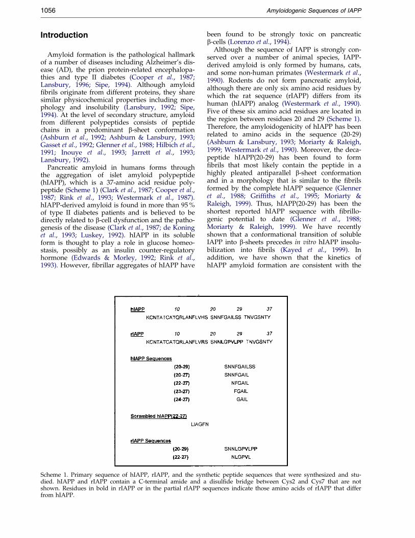
Scheme 1. Primary sequence of hIAPP, rIAPP, and the syndied. hIAPP and rIAPP contain a C-terminal amide and ashown. Residues in bold in rIAPP or in the partial rIAPP sefrom hIAPP.
been found to be strongly toxic on pancreaticb-cells (Lorenzo et al., 1994).
Although the sequence of IAPP is strongly con-served over a number of animal species, IAPP-derived amyloid is only formed by humans, cats,and some non-human primates (Westermark et al.,1990). Rodents do not form pancreatic amyloid,although there are only six amino acid residues bywhich the rat sequence (rIAPP) differs from itshuman (hIAPP) analog (Westermark et al., 1990).Five of these six amino acid residues are located inthe region between residues 20 and 29 (Scheme 1).Therefore, the amyloidogenicity of hIAPP has beenrelated to amino acids in the sequence (20-29)(Ashburn & Lansbury, 1993; Moriarty & Raleigh,1999; Westermark et al., 1990). Moreover, the deca-peptide hIAPP(20-29) has been found to form®brils that most likely contain the peptide in ahighly pleated antiparallel b-sheet conformationand in a morphology that is similar to the ®brilsformed by the complete hIAPP sequence (Glenneret al., 1988; Grif®ths et al., 1995; Moriarty &Raleigh, 1999). Thus, hIAPP(20-29) has been theshortest reported hIAPP sequence with ®brillo-genic potential to date (Glenner et al., 1988;Moriarty & Raleigh, 1999). We have recentlyshown that a conformational transition of solubleIAPP into b-sheets precedes in vitro hIAPP insolu-bilization into ®brils (Kayed et al., 1999). Inaddition, we have shown that the kinetics ofhIAPP amyloid formation are consistent with the
thetic peptide sequences that were synthesized and stu-disul®de bridge between Cys2 and Cys7 that are not
quences indicate those amino acids of rIAPP that differ

Amyloidogenic Sequences of IAPP 1057
nucleation-dependent polymerization mechanism(Jarrett & Lansbury, 1993; Kayed et al., 1999).
Here, we focus on the identi®cation of the short-est sequence of hIAPP which is able to self-assem-ble into b-sheets and amyloid. Secondary structureprediction analysis of the (20-29) region of hIAPPwas performed ®rst and the sequences that werepredicted to form b-sheet structures and appropri-ate control-sequences were synthesized. Kinetics ofinsoluble ®bril formation were studied by turbiditymeasurements at 400 nm and their ®brillogenicpotentials evaluated by electron microscopy (EM),atomic force microscopy (AFM), and the Congored (CR) staining method. Conformations ofinsoluble peptide aggregates were studied byFourier transform infrared (FT-IR) spectroscopy.Finally, the effect of the short hIAPP sequenceson cell viability of a pancreatic cell line was exam-ined.
Results
Identification of minimum length IAPPsequences with fibrillogenic potential
We ®rst performed secondary structure predic-tion analysis of hIAPP(20-29) (SNNFGAILSS),which was established to represent an amyloido-genic sequence of IAPP (Ashburn et al., 1992;Ashburn & Lansbury, 1993; Moriarty & Raleigh,1999; Westermark et al., 1990), by the methoddescribed by Chou & Fasman (1978), and foundthat hIAPP(23-27) (FGAIL) could potentially be theshortest sequence with b-sheet-forming potential.By contrast, no b-sheet-forming potential was pre-dicted for the corresponding rat sequence,rIAPP(23-27) (LGPVL). Since the amyloidogenicityof a peptide sequence has been described to beoften closely related to its b-sheet-forming propen-sity (Ashburn et al., 1992; Ashburn & Lansbury,1993; Jarrett et al., 1993; Lansbury, 1992; Moriarty& Raleigh, 1999), we hypothesized that thesequence hIAPP(23-27) (FGAIL) could be ®brillo-genic. Such an assumption was supported byrotational resonance solid-state NMR (R2-SSNMR)studies that have suggested that the basic unit ofthe hIAPP(20-29) (SNNFGAILSS) ®bril may con-tain the FGAIL sequence in a self-assembled, inter-molecular b-sheet con®guration (Grif®ths et al.,1995).
We synthesized hIAPP(23-27) (FGAIL) as well asthe longer and shorter analogs hIAPP(22-27)(NFGAIL) and hIAPP(24-27) (GAIL), respectively(Scheme 1). Sequences hIAPP(20-29) (SNNFGAILSS), a scrambled version of hIAPP(22-27)(LIAGFN), rIAPP(20-29) (SNNLGPVLPP), andrIAPP(22-27) (NLGPVL) were also synthesizedand used as controls for b-sheet-forming and amy-loidogenic or non-amyloidogenic sequences,respectively (Westermark et al., 1990).
We then tested the ®brillogenic potential of thesepeptides by EM and AFM. Following aging of ahighly concentrated aqueous solution (3.7-9.3 mg/
ml, four to six days incubation time in phosphatebuffer (pH 7.4) or in water), the tetrapeptidehIAPP(24-27) (GAIL) remained soluble. AFMexamination con®rmed that no ®brils were presentin the hIAPP(24-27) (GAIL) solutions. Instead,round and punctuate aggregates formed when thesolution was allowed to dry (Figure 1(a)). By con-trast, supersaturated and aged aqueous suspen-sions of hIAPP(23-27) (FGAIL) and hIAPP(22-27)(NFGAIL) (tested apparent peptide concentrationswere 2.6-5.2 mg/ml for FGAIL and 3.2-6.4 mg/mlfor NFGAIL, in phosphate buffer (pH 7.4), orwater, four to six days incubation time) werefound to consist exclusively of amyloid-like ®brilsand ®bril bundles (Figures 1(b) and (c), and 2(a)and (b)). Although these two peptide sequencesdiffer by only one residue, their ®brillar assemblieswere completely different. hIAPP(23-27) (FGAIL)®brils generally aligned laterally with each other toyield very long (>3 mm) and broad (> 300 nm) rib-bon-like ®bril bundles (Figures 1(b) and 2(a) and(b)). By contrast, hIAPP(22-27) (NFGAIL) ®lamentscoiled along each other and resulted in ®brils withproperties that have been well described for amy-loids, i.e. an axial helical periodicity (about200 nm) (Goldsbury et al., 1997, 1999; Harper et al.,1997a,b; Stine et al., 1996). Of note, no propensitytowards a parallel alignment, as seen with thehIAPP(23-27) (FGAIL) ®laments, was observed forhIAPP(22-27) (NFGAIL) (Figure 1(c)). Judging fromthe EM measurements, hIAPP(22-27) (NFGAIL)®brils had diameters of about 15 to 20 nm. How-ever, it should be noted that the observed branch-ing and coiling features of the hIAPP(22-27)(NFGAIL) ®brils (Figure 1(c)) suggested that thediameters measured represented an assembly of atleast two ®laments (Harper et al., 1997a,b). Amajority of isolated hIAPP(23-27) (FGAIL) ®brils,which may also represent an assembly of at leasttwo ®laments, had diameters of about 17 nmaccording to AFM.
Aged and saturated solutions of hIAPP(20-27)(SNNFGAIL) and hIAPP(20-29) (SNNFGAILSS)also formed ®brils and ®bril bundles (Figure 1(d)and (e)). Fibrils of hIAPP(20-27) (SNNFGAIL)showed a similar helical coiling around each otheras seen for the hIAPP(20-27) (NFGAIL) ®brils andhad diameters of about 6 to 10 nm, as estimated byEM. By contrast, ®laments of hIAPP(20-29)(SNNFGAILSS) had a strong propensity to laterallyself-assemble into a net of broad ribbons. No ®brilformation was observed in aged incubations ofhighly concentrated solutions (6.4 mg/ml or10 mM) of the scrambled hIAPP(22-27) controlpeptide LIAGFN. These solutions, when dried,exclusively consisted of amorphous aggregates(Figure 1(f)). Similarly, highly concentrated sol-utions of the soluble rat sequences rIAPP(22-27)(NLGPVL) (6.1 mg/ml or 10 mM) (Figure 1(g))and rIAPP(20-29) (SNNLGPVLPP) (10 mg/ml or10 mM) (data not shown) consisted of amorphousaggregates only.

Figure 1 (legend shown on p. 1057)
1058 Amyloidogenic Sequences of IAPP
We then applied the CR staining method com-bined with polarization microscopy to test foramyloidogenicity of the IAPP sequences. Amyloid®brils in general, and ®brillar hIAPP in particular,bind CR and exhibit a gold/green birefringenceunder polarized light (Cooper, 1974; Lansbury,1992). Aged hIAPP(23-27) (FGAIL) and hIAPP(22-27) (NFGAIL) suspensions (2.6-5.2 mg/ml forFGAIL and 3.2-6.4 mg/ml for NFGAIL, in phos-phate buffer (pH 7.4), aged for four days) boundCR and exhibited the characteristic green/goldbirefringence (Figure 3(a) for normal ®eld and (b)for polarized light microscopy of hIAPP(23-27)(FGAIL), and Figure 3(c) for normal ®eld and
(d) for polarized microscopy of hIAPP(22-27)(NFGAIL)). hIAPP(20-27) (SNNFGAIL) alsostained with CR and showed strong birefringence(not shown). As a control, aged, supersaturatedsolutions of hIAPP(20-29) (SNNFGAILSS) werealso examined and were found to exhibit stronggreen/gold birefringence, whereas no CR bindingwas found for aged solutions of rIAPP(22-27)(NLGPVL) and rIAPP(20-29) (SNNLGPVLPP) (notshown). Interestingly, aged and highly concen-trated solutions of hIAPP(24-27) (GAIL) (25 mM or9.3 mg/ml), when allowed to dry, yielded longneedles that were visible by light microscopy.These needles appeared birefringent with and
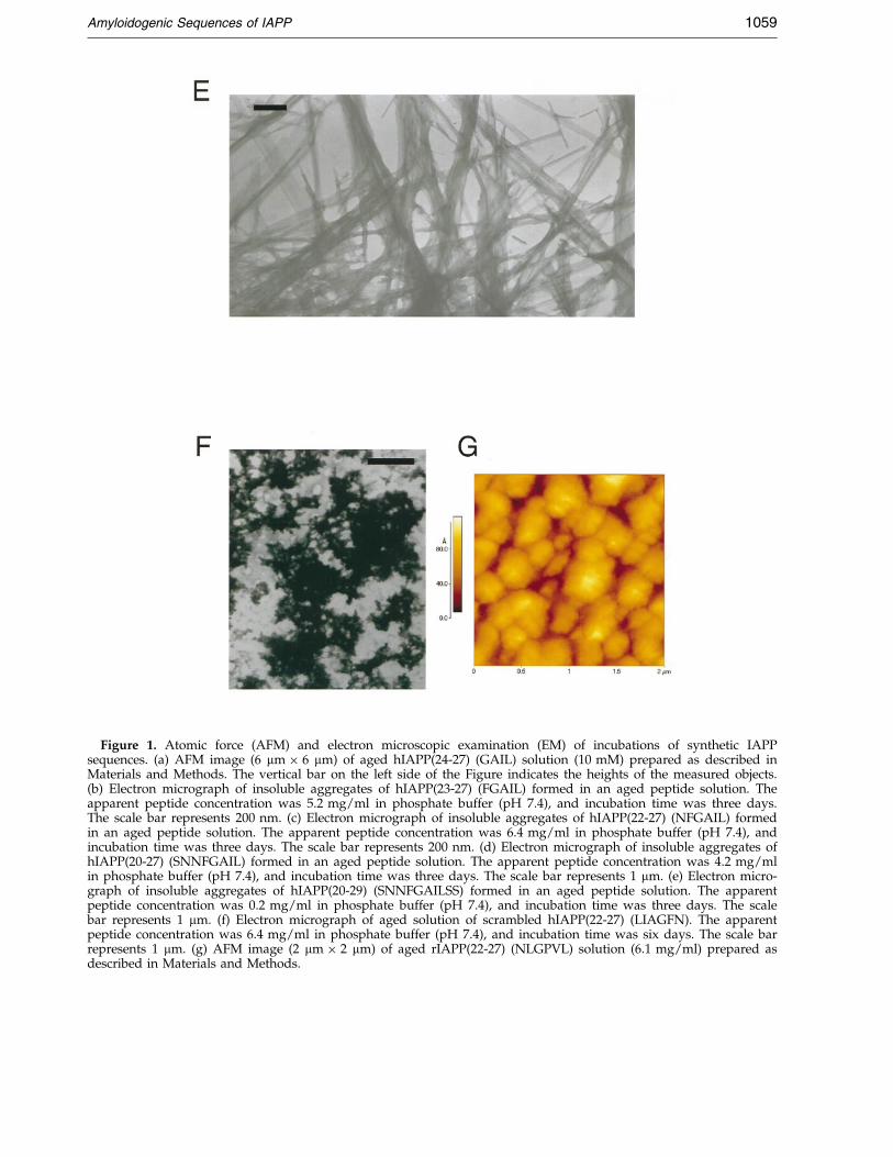
Figure 1. Atomic force (AFM) and electron microscopic examination (EM) of incubations of synthetic IAPPsequences. (a) AFM image (6 mm � 6 mm) of aged hIAPP(24-27) (GAIL) solution (10 mM) prepared as described inMaterials and Methods. The vertical bar on the left side of the Figure indicates the heights of the measured objects.(b) Electron micrograph of insoluble aggregates of hIAPP(23-27) (FGAIL) formed in an aged peptide solution. Theapparent peptide concentration was 5.2 mg/ml in phosphate buffer (pH 7.4), and incubation time was three days.The scale bar represents 200 nm. (c) Electron micrograph of insoluble aggregates of hIAPP(22-27) (NFGAIL) formedin an aged peptide solution. The apparent peptide concentration was 6.4 mg/ml in phosphate buffer (pH 7.4), andincubation time was three days. The scale bar represents 200 nm. (d) Electron micrograph of insoluble aggregates ofhIAPP(20-27) (SNNFGAIL) formed in an aged peptide solution. The apparent peptide concentration was 4.2 mg/mlin phosphate buffer (pH 7.4), and incubation time was three days. The scale bar represents 1 mm. (e) Electron micro-graph of insoluble aggregates of hIAPP(20-29) (SNNFGAILSS) formed in an aged peptide solution. The apparentpeptide concentration was 0.2 mg/ml in phosphate buffer (pH 7.4), and incubation time was three days. The scalebar represents 1 mm. (f) Electron micrograph of aged solution of scrambled hIAPP(22-27) (LIAGFN). The apparentpeptide concentration was 6.4 mg/ml in phosphate buffer (pH 7.4), and incubation time was six days. The scale barrepresents 1 mm. (g) AFM image (2 mm � 2 mm) of aged rIAPP(22-27) (NLGPVL) solution (6.1 mg/ml) prepared asdescribed in Materials and Methods.
Amyloidogenic Sequences of IAPP 1059

Figure 2. Atomic force microscopic examination(AFM) of ®brillar aggregates of hIAPP(23-27) (FGAIL).(a) AFM image (10 mm � 10 mm) of single ®brils of anaged hIAPP(23-27) (FGAIL) solution (apparent peptideconcentration 2.6 mg/ml) prepared as described inMaterials and Methods. (b) AFM image (3.1 mm � 3.1mm) of ribbon-like ®bril bundles of the aged hIAPP(23-27) (FGAIL) solution (see (a)). The vertical bar on theleft side of the Figure indicates the heights of themeasured objects.
1060 Amyloidogenic Sequences of IAPP
without CR staining under polarized light(Figure 3(e) for normal ®eld and (f) for polarizedmicroscopy).
Taken together, the results of the EM, AFM, andCR analyses strongly suggested that hIAPP(23-27)(FGAIL) is the shortest hIAPP sequence necessaryfor amyloid formation.
Aggregation kinetics of the hIAPP sequences
Aggregation of several amyloid-forming poly-peptides in vitro has been suggested to proceed by
nucleation-dependent polymerization (Ashburn &Lansbury, 1993; Harper & Lansbury, 1997; Jarrettet al., 1993; Jarrett & Lansbury, 1993). The existenceof a lag-time prior to protein aggregation, which ispartially or completely eliminated by the additionof preformed aggregates or seeds, is a characteristicfeature of this mechanism. To study self-assemblyof our amyloidogenic sequences, we followed theiraggregation and insolubilization kinetics by turbid-ity measurements at 400 nm (Figure 4) (Ashburn &Lansbury, 1993; Harper & Lansbury, 1997; Jarrettet al., 1993; Jarrett & Lansbury, 1993). In anotherseries of experiments, the results of the turbidityassays were independently con®rmed by the CRbinding assay (data not shown) (Klunk et al.,1989a,b).
For the kinetic aggregation assays, several sol-vents for preparation of stock solutions and var-ious peptide concentrations were tested to obtainreproducible aggregation kinetics. This was necess-ary due to the various experimental factors thatmay strongly affect aggregation kinetics of super-saturated solutions, which are inherently meta-stable (Harper & Lansbury, 1997; Jarrett &Lansbury, 1993; Kayed et al., 1999). For the prep-aration of the peptide stocks, acetonitrile (ACN)and 1,1,1,3,3,3-hexa¯uoro-2-propanol were amongthose examined. As hIAPP(20-27) (SNNFGAIL)and hIAPP(20-29) (SNNFGAILSS) were insolublein these solvents at the required concentrations,stock solutions of all peptide sequences were even-tually prepared in dimethyl sulfoxide (DMSO) andused immediately to avoid formation of pre-aggre-gated peptide seeds (Jarrett & Lansbury, 1993).DMSO is broadly applied as a disaggregatingsolvent, especially for the preparation of stocksolutions of amyloidogenic peptides includingsequences of hIAPP (Ashburn & Lansbury, 1993;Harper et al., 1997b; Jarrett et al., 1993). However,the possibility that stock solutions prepared inDMSO may also contain minute amounts of seedscannot be completely excluded. Filtering of thesolutions was not possible due to strong non-speci®c binding of the peptides to various ®ltermembranes. Yet, the observed concentrationdependence of the lag-time (data not shown) andthe reproducibility of our results indicated that ourfresh stock solutions most likely did not containaggregation nuclei.
The kinetic pro®les of insoluble amyloid for-mation observed for all short sequences werestrongly indicative of a nucleation-dependentpolymerization mechanism (Jarrett & Lansbury,1993). After a lag-time of 30 hours, hIAPP(23-27)(FGAIL) (2.6 mg/ml in phosphate buffer and4 % DMSO, pH 7.4) rapidly self-assembled intoinsoluble amyloid ®brils (Figure 4). Aggregation ofhIAPP(22-27) (NFGAIL) at an identical apparentmolar concentration (3.2 mg/ml) exhibited a sig-ni®cantly shorter lag-time of only 42 minutes(Figure 4), indicating a lower kinetic solubility forthis sequence. hIAPP(20-27) (SNNFGAIL) and thestrongly amyloidogenic sequence hIAPP(20-29)

Figure 3. Examination of the amyloidogenic nature of partial hIAPP fragments by the Congo red (CR) bindingassay. Normal ®eld microscopic examination of aged solutions of (a) hIAPP(23-27) (FGAIL), (c) hIAPP(22-27)(NFGAIL), and (e) hIAPP(24-27) (GAIL) following staining with CR is shown. Microscopic examination under polar-ized light of (b) hIAPP(23-27) (FGAIL), (d) hIAPP(22-27) (NFGAIL), and (f) hIAPP(24-27) (GAIL) is also shown.Supersaturated solutions were prepared and aged for four days as described in Materials and Methods.
Amyloidogenic Sequences of IAPP 1061
(SNNFGAILSS) immediately aggregated under theabove conditions (Figure 4) (Ashburn & Lansbury,1993). By contrast, hIAPP(24-27) (GAIL) was highlysoluble in aqueous buffer and did not aggregateunder these conditions neither at fourfold highermolar concentrations than those applied for theother sequences (Figure 4). Of note, because themolecular mass of the various self-associated pep-tide species is unknown, the concentrations of theamyloidogenic sequences are generally presentedas mass per volume measures. To compare the bio-physical properties (®bril- and b-sheet-formingpotentials, solubilities and cytotoxicities) of thedifferent peptide sequences, solutions or suspen-sions of identical ``apparent'' molar concentrationswere applied, indicated by the use of molarconcentration units. Molarities were calculatedbased on the molecular mass of the monomers,assuming that the peptides were monomeric andcompletely soluble.
Kinetic aggregation studies of the amyloidogenicfragments at various peptide concentrations indi-
cated that the length of the lag-time was inverselyrelated to the apparent peptide concentration (datanot shown), which is another characteristic featureof nucleation-dependent polymerization (Jarrett &Lansbury, 1993). Of importance, the observed lag-times of aggregation for hIAPP(23-27) (FGAIL) and(22-27) (NFGAIL) were immediately eliminated byseeding, further adding to the notion that amyloidformation by the short hIAPP sequences wasnucleation-dependent (Figure 5(a) and (b)).
In agreement with previous reports (Jarrett &Lansbury, 1993), the thermodynamic solubilities ofthe hIAPP sequences were found to dependstrongly on the speci®c peptide sequence. Thermo-dynamic solubilities were determined by quanti®-cation of the amount of soluble peptide at thepoint of thermodynamic equilibrium which wasusually after four days following the beginning ofthe aggregation experiment (Ashburn & Lansbury,1993; Jarrett et al., 1993; Jarrett & Lansbury, 1993).In the post-nucleation equilibrium, hIAPP(23-27)(FGAIL) exhibited the highest thermodynamic

Figure 4. Kinetics of insoluble amyloid formation of partial hIAPP sequences as followed by turbidity at 400 nm.Turbidity of aqueous solutions of hIAPP(24-27) (GAIL), (23-27) (FGAIL), and (22-27) (NFGAIL) in 10 mM phosphatebuffer (pH 7.4), and 4 % DMSO (apparent peptide concentrations were 5 mM) prepared as described in Materials andMethods was followed for several hours. Inset: Kinetics of aggregation of hIAPP sequences (20-27) (SNNFGAIL) and(20-29) (SNNFGAILSS) under the same experimental conditions as for the shorter sequences are shown (time-scale:minutes). Turbidity of aqueous solutions (apparent peptide concentrations were 5 mM) was measured and solutionsprepared as described in Materials and Methods. Data are from one representative experiment of at least three per-formed.
1062 Amyloidogenic Sequences of IAPP
solubility (1.4(�0.2) mg/ml) and hIAPP(20-29)(SNNFGAILSS) the lowest one (0.3(�0.1) mg/ml),while hIAPP(20-27) (SNNFGAIL) exhibited a ther-modynamic solubility that was similar tohIAPP(20-29) (0.5(�0.1) mg/ml) (mean(�SEM) of
Figure 5. Nucleation of amyloid formation by hIAPP sequof traces of preformed ®brils. Kinetics of amyloid formationseeded and non-seeded (a) hIAPP(23-27) (FGAIL) (2.6 mg/mcated. Seeding was performed with 10 % preformed (a) hIAP®brils as described in Materials and Methods. Data are froformed.
at least three independent aggregation assays).Interestingly, a 40 % higher insolubility on a molarbasis was found for hIAPP(22-27) (NFGAIL)(thermodynamic solubility: 1.2(�0.2) mg/ml), ascompared to hIAPP(23-27) (FGAIL).
ences (23-27) (FGAIL) and (22-27) (NFGAIL) via additionare followed by turbidity measurement at 400 nm for
l) and (b) hIAPP(22-27) (NFGAIL) (2.4 mg/ml) as indi-P(23-27) (FGAIL) ®brils and (b) hIAPP(22-27) (NFGAIL)m one representative experiment of at least three per-

Figure 6. Secondary structure of ®brillar aggregates of partial hIAPP sequences, aged hIAPP(24-27) (GAIL) andrIAPP(22-27) (NLGPVL) as assessed by Fourier transform infrared spectroscopy (FT-IR). (a) Spectrum of an agedhIAPP(24-27) (GAIL) solution prepared and measured as described in Materials and Methods. (b) Insoluble ®brils ofhIAPP(23-27) (FGAIL), (c) hIAPP(22-27) (NFGAIL), (d) hIAPP(20-27) (SNNFGAIL), and (e) hIAPP(20-29)(SNNFGAILSS) were collected from kinetic aggregation experiments and FT-IR spectra were measured as describedin Materials and Methods. (f) Spectrum of an aged rIAPP(22-27) (NLGPVL) solution prepared and measured asdescribed in Materials and Methods.
Amyloidogenic Sequences of IAPP 1063
Conformation of the hIAPP sequences in theirfibrillar state as determined by FT-IR
FT-IR spectroscopic studies of the aggregates ofthe amyloidogenic partial sequences and also ofhighly concentrated and aged (3.7 mg/mlor 10 mM, incubation time of four days) solutionsof hIAPP(24-27) (GAIL) were then performed. The®brillar aggregates were obtained from the kineticaggregation assays and were collected after
four days of aggregation. FT-IR spectra typical fora random coil structure mixed with various differ-ent conformations were recorded for agedhIAPP(24-27) (GAIL) solutions (Figure 6(a)). Bycontrast, a sharp absorbance maximum at1633 cmÿ1 was found in the spectrum of hIAPP(23-27) (FGAIL) ®brils. Such an absorbance is typicallyassigned to b-sheet structures (Krimm & Bandekar,1986; Lansbury, 1992) (Figure 6(b)). hIAPP(22-27)

Figure 7. (a) Cytoxicity of ®brillar aggregates of partial sequences of hIAPP as indicated. Various concentrations of®brillar aggregates of hIAPP(23-27) (FGAIL), hIAPP(22-27) (NFGAIL), hIAPP(20-27) (SNNFGAIL), and hIAPP(20-29)(SNNFGAILSS) were added to RIN5fm cells that were plated at a density of 5x10⁵ cells/ml. Following a 20 hoursincubation, cell viability was assessed by measuring the cellular reduction of MTT. (b) Comparison of the cytotoxici-ties of soluble (fresh dissolved) versus the ®brillar forms of the hIAPP fragments as indicated. Lyophilized peptidescorresponding to sequences hIAPP(23-27) (FGAIL), hIAPP(22-27) (NFGAIL), hIAPP(20-27) ((SNNFGAIL), andhIAPP(20-29) (SNNFGAILSS) were dissolved in cell culture medium at an apparent concentration of 500 mM andthen immediately added to the cells at an apparent ®nal concentration of 50 mM. Fibrillar aggregates were alsoapplied onto cells at apparent concentrations of 50 mM. Following incubation, cytotoxicities were assessed asdescribed in (a). Data are percentages of control values (vehicle alone) and are the mean (�SEM) of three indepen-dent experiments with each experiment performed in multiple replicates (n � 6) except for the data of hIAPP(20-27)which are derived from one representative experiment performed in n � 12 replicates.
1064 Amyloidogenic Sequences of IAPP
(NFGAIL) ®brils produced a spectrum with amarkedly broader absorbance maximum at1633 cmÿ1 and a strong shoulder at 1666 cmÿ1,indicating the presence of b-structures mixed withsigni®cant amounts of non-b structures, i.e. ran-dom coil and turns (Figure 6(c)). The spectra of the®brils of hIAPP(20-27) (SNNFGAIL) and hIAPP(20-29) (SNNFGAILSS) had pronounced maxima at1633 cmÿ1 and 1625 cmÿ1, respectively, suggestingsigni®cant b-sheet contents (Figure 6(d) and (e))(Ashburn et al., 1992; Ashburn & Lansbury, 1993;Glenner et al., 1988; Lansbury, 1992; Moriarty &Raleigh, 1999). By contrast, aged and highly con-centrated solutions of the reported non-amyloido-genic rIAPP(20-29) (SNNLGPVLPP) (10 mM or10.1 mg/ml in 2H2O) (not shown) and rIAPP(22-27) (NLGPVL) (10 mM or 6.1 mg/ml in 2H2O),(Figure 6(f)), gave FT-IR spectra that indicated thatnon-b conformations were predominant.
Effect of the hIAPP fragments on the viabilityof a pancreatic bbb-cell line
There are several lines of evidence suggesting anassociation between the pathology of the variousamyloidoses and the cytotoxicity of the ®brillarforms of the respective amyloid-forming peptides,including b-amyloid peptide (Ab), prion protein,and hIAPP (Forloni et al., 1994; Iversen et al., 1995;Lorenzo et al., 1994; Lorenzo & Yankner, 1994; Pike
et al., 1991, 1995). We used the insulin-producingpancreatic cell line RIN5mf (Nishi et al., 1989) totest the cytotoxic effect of fragments hIAPP(23-27)(FGAIL), (22-27) (NFGAIL), (20-27) (SNNFGAIL),and (20-29) (SNNFGAILSS). In preliminary studies,a strong cytotoxic effect of the complete hIAPPsequence on this cell line was found (data notshown); its effective apparent concentration thatcaused 50 % cell damage (EC50) following a 20hours incubation was 1.2 nM. For the assessmentof peptide toxicity, an assay based on the cellularreduction of the dye 3-[4,5-dimethylthiazol-2-yl]-2,5-diphenyltetrazolium bromide (MTT) was used(Kapurniotu et al., 1998; Schubert et al., 1995;Shearman et al., 1995, 1994).
We ®rst tested the effect of preformed ®brillaraggregates of hIAPP(23-27) (FGAIL), (22-27)(NFGAIL), (20-27) (SNNFGAIL), and (20-29)(SNNFGAILSS) obtained from the kinetic aggrega-tion experiments (see above). Fibrillar aggregateswere collected after aggregations had reached athermodynamic equilibrium (four days), quanti-®ed, and added to the cells. We found signi®cantcytotoxicities for the preformed ®brillar aggregatesof the known amyloidogenic sequence hIAPP(20-29) (SNNFGAILSS) as well as for the aggregates ofhIAPP(23-27) (FGAIL) and (22-27) (NFGAIL) thatwere similarily cytotoxic to hIAPP(20-29)(Figure 7(a)). Of note, cytotoxicities of these pep-tides were about 1000-fold lower on a molar basis

Amyloidogenic Sequences of IAPP 1065
than that of the complete hIAPP sequence. Theaggregates of hIAPP (20-27) (SNNFGAIL) wereslightly less toxic than the aggregates of the threeother peptide fragments (Figure 7(a)). As a nega-tive control, sequence LIAGFN that was previouslyfound to be non-amyloidogenic was tested follow-ing aging and no cytotoxicity was found. To con-trol for possible reported effects of DMSO oncytotoxicity (Mattson et al., 1992), peptide ®brilswere also prepared by dissolving or suspendingthe peptides in H2O followed by aging for fourdays prior to application onto the cells. Underthese conditions, the same cytotoxicities wereobserved as for the peptide stocks prepared inDMSO.
We then tested the cytotoxic effect of the freshlydissolved peptides. Peptide stock solutions of thepartial sequences were prepared by directly dissol-ving the lyophilized peptides in cell medium andat an apparent concentration of 500 mM. While thesolutions of peptides (24-27) (GAIL), (23-27)(FGAIL), (22-27) (NFGAIL), and (20-27)(SNNFGAIL) did not contain ®brils at the timepoint of their preparation, the stock solutions ofhIAPP and (20-29) (SNNFGAILSS) already con-tained some ®brillar aggregates due to their lowkinetic solubilities. The absence or presence of®brils at the time of preparation of the stock sol-utions was veri®ed by EM (not shown). Incubationof the cells with freshly dissolved hIAPP(24-27)(GAIL), (23-27) (FGAIL), (22-27) (NFGAIL), and(20-27) (SNNFGAIL) for about 20 hours did notaffect cell viability, whereas non-aged but ®bril-containing hIAPP(20-29) (SNNFGAILSS) was ascytotoxic as its ®brillar form (Figure 7(b)). No inso-luble peptide aggregates on or between the cellscould be detected by light microscopic examinationof the incubations with hIAPP(23-27) (FGAIL), (22-27) (NFGAIL), and (20-27) (SNNFGAIL), whereassigni®cant amounts of such aggregates wereobserved in the hIAPP(20-29) (SNNFGAILSS) orhIAPP incubations. Thus, it is likely that peptideshIAPP(23-27) (FGAIL), (22-27) (NFGAIL), and(20-27) (SNNFGAIL), due to their high degree ofsolubility and their long aggregational lag-times,remained in their soluble forms during the 20 hourincubation period, suggesting that the solubleforms are likely non-toxic.
Discussion
There is increasing evidence suggesting a directassociation of aggregation and ®bril formation byamyloid-forming polypeptides with the pathologicsequelae of amyloidosis-related diseases, includingprion-protein related encephalopathies, AD, andtype II diabetes (Clark et al., 1987; Forloni et al.,1994; Iversen et al., 1995; Lorenzo et al., 1994;Lorenzo & Yankner, 1994; Pike et al., 1991, 1995;Schubert et al., 1995; Thomas et al., 1995). Amyloidconsists of polypeptide chains that are predomi-nantly in a b-sheet conformation (Ashburn et al.,
1992; Ashburn & Lansbury, 1993; Gasset et al.,1992; Hilbich et al., 1991; Inouye et al., 1993; Jarrettet al., 1993; Lansbury, 1992; Zhang & Rich, 1997).The b-sheet-forming propensity of a peptide chainhas been shown to be closely related to its abilityto aggregate into amyloid ®brils (Ashburn et al.,1992; Ashburn & Lansbury, 1993; Gasset et al.,1992; Glenner et al., 1988; Grif®ths et al., 1995;Hilbich et al., 1991; Jarrett et al., 1993; Kapurniotuet al., 1998; Kayed et al., 1999; Kominos, 1995;Lansbury, 1996; Moriarty & Raleigh, 1999; Pikeet al., 1995). Aggregation and amyloid formationby Ab and hIAPP have been suggested to proceedvia a common molecular mechanism that includesa conformational transition from soluble, non-toxicpeptide conformers into b-sheet-containing amy-loid precursors (Kayed et al., 1999; Pike et al., 1995;Shen & Murphy, 1995; Soto & Frangione, 1995).
Our studies aimed to identify the shortestsequence of hIAPP capable of forming b-sheets andamyloid. Secondary structure predictions indicatedthat the sequence hIAPP(23-27) (FGAIL) could bethe shortest hIAPP sequence with a b-sheet-form-ing potential. In fact, aged suspensions ofsequences (23-27) (FGAIL), (22-27) (NFGAIL), (20-27) (SNNFGAIL), and (20-29) (SNNFGAILSS) werefound to exclusively consist of long ®brils and®bril bundles. While the amyloidogenicity ofsequence (20-29) was already known (Ashburn &Lansbury, 1993; Moriarty & Raleigh, 1999;Westermark et al., 1990; Glenner et al., 1988), therehad been no reports with regard to ®bril formationby sequences of amyloidogenic polypeptides asshort as the penta- or hexapeptides investigatedhere. To our knowledge, the shortest sequencesthat have been reported to be able to form amyloid®brils consist of at least eight amino acid residues.These are the octapeptide AGAAAAGA of theprion protein and a ten residue peptide of Ab(Lundberg et al., 1997; Tjernberg et al., 1999).
The ®laments of hIAPP sequence (23-27)(FGAIL) showed a strong propensity to laterallyself-associate into very broad ribbons, whereas the®laments formed by sequences (22-27) (NFGAIL)and (20-27) (SNNFGAIL) were long and thin andcoiled laterally around each other to yield ®brilbundles of pronounced axial helical twisting. A lat-eral helical coiling of ®laments around each otherhas often been described in amyloid ®brils fromseveral different polypeptides, suggesting that thispacking might represent a common ®lament self-assembly mechanism (Goldsbury et al., 1997, 1999;Harper et al., 1997a,b; Stine et al., 1996). On theother hand, studies on Ab have previously relatedthe formation of ribbon-like arrays to the decreasedability of the ®laments to pack laterally in the typi-cal coiled ®ber morphology (Fraser et al., 1994).hIAPP has been reported to form ®brils that arecharacterized by a structural polymorphism, andtwo mechanisms of hIAPP ®brillar self-assemblyhave been proposed. These include the lateralassembly into ribbon- or sheet-like arrays and ®la-ment coiling into coiled ®brils (Goldsbury et al.,
1066 Amyloidogenic Sequences of IAPP
1997, 1999). Thus, the ®brillar assemblies formedby hIAPP(23-27) (FGAIL) and hIAPP(22-27)(NFGAIL) seem to have formed by both self-associ-ation mechanisms in a sequence-dependentmanner. The striking differences between the mor-phologies of the ®laments of the two sequencesthat differ by only one amino acid residue (Asn22)strongly suggest that this residue plays animportant role in directing self-assembly andlateral packing of ®laments. Such a role could bethat its side-chain could be involved in intermole-cular side-chain interactions that lead to a ®lament-stabilizing self-association as previously proposedfor proto®lament assembly of Ab (Fraser et al.,1994). The importance of residue Asn22 for ®brilformation was also con®rmed by our kinetic aggre-gation data and has been also pointed out in avery recent study by Moriarty & Raleigh (1999).
FT-IR spectroscopy of the ®brils of the partialhIAPP sequences suggested that they all containsigni®cant amounts of b-sheet structure (Ashburnet al., 1992; Ashburn & Lansbury, 1993; Grif®thset al., 1995). According to the R2-SSNMR model ofpancreatic amyloid ®brils, hIAPP(23-27) (FGAIL) isstrongly constrained in an intermolecular antipar-allel b-sheet, and a number of the sheet-stabilizingintermolecular interactions in hIAPP(20-29)(SNNFGAILSS) ®brils were found to occurbetween residues in this peptide region (Grif®thset al., 1995). These results are in good agreementwith the high b-sheet and amyloidogenic propen-sity of hIAPP(23-27) (FGAIL) as found in ourwork, and suggest that FGAIL could be the core ofthe hIAPP ®bril.
The sequence motif GAIL is common in the twoamyloidogenic IAPP sequences from cat and man,and has previously been suggested to be crucial fortheir amyloidogenicity (Ashburn et al., 1992;Ashburn & Lansbury, 1993; Grif®ths et al., 1995;Westermark et al., 1990). We found, however, thatthis tetrapeptide alone is not able to aggregate intob-sheets and amyloid-like ®brils in aqueous sol-utions, whereas its N-terminal extension by onlyone residue results in the strongly amyloidogenichIAPP(23-27) (FGAIL). Nevertheless, (24-27)(GAIL) was also found to have a strong propensityto form ordered structures.
Measurement of the kinetics of amyloid for-mation is a complicated process due to the largenumber of different self-associated species and thevariability of their physical properties (Harper &Lansbury, 1997). Therefore, several methods areused, often complementary to each other, to obtainindependent information about the kinetics ofamyloid formation. We used turbidity measure-ments at 400 nm to follow the kinetics of insoluble®bril formation (Ashburn & Lansbury, 1993;Harper & Lansbury, 1997; Jarrett et al., 1993; Jarrett& Lansbury, 1993). It should be noted that turbid-ity measurements at 400 nm only allow for thedetection of aggregates with diameters larger thanthe wavelength of the incident light beam. Thus,formation of small size and possibly visibly soluble
aggregates may be not detectable, and it has beenreported that there are amyloid ®brils that do notexhibit turbidity at 400 nm (Harper & Lansbury,1997; Wood et al., 1996). However, the turbidity-based assays are able to detect formation of insolu-ble amyloid and, therefore, the results of our tur-bidity assays refer to kinetics of insoluble amyloidformation (Ashburn & Lansbury, 1993; Harper &Lansbury, 1997; Jarrett et al., 1993; Jarrett &Lansbury, 1993). Furthermore, the aggregationassays presented here were performed under stir-ring conditions, which has been suggested to poss-ibly generate small ®brils that exhibit turbidity,contrary to ®brils formed in non-stirred solutions(Wood et al., 1996).
Thio¯avine T and CR-amyloid binding-basedassays are used for the kinetic follow up of aggre-gation and amyloid formation (Klunk et al.,1989a,b; LeVine, 1993b; Harper & Lansbury, 1997;Wood et al., 1996, 1995). The exact structural andconformational features of amyloid-forming pep-tides responsible for binding to both compoundsand the binding-induced spectral shifts are not yetunderstood, and among other factors depend on®bril morphology (Klunk et al., 1989a,b; Harper &Lansbury, 1997; Wood et al., 1996). According toone report, hIAPP amyloid fails to induce the typi-cal change in the ¯uorescence spectrum of thio¯a-vin T, thus precluding the use of this dye to followthe kinetics of hIAPP amyloid formation (LeVine,1993a). By contrast, hIAPP amyloid readily bindsto CR (Kayed et al., 1999; Lorenzo & Yankner,1994). Ab ®brils that were visibly soluble and didnot exhibit turbidity at 405 nm were shown tobind to CR (Wood et al., 1996). To exclude thepossibility that amyloid ®brils that were not detect-able by the turbidity assay were already presentduring the lag-time of our aggregational times, wemeasured the aggregation kinetics of of hIAPP(23-27) and hIAPP(22-27) by both turbidity and CR-binding. Identical aggregation kinetics pro®leswere obtained for the same aggregation solutionby both methods, which con®rmed the validity ofthe results of the turbidity assay.
Amyloid formation processes of all amyloido-genic sequences of hIAPP were found to followkinetics that were consistent with the nucleation-dependent polymerization (Jarrett & Lansbury,1993). Kinetic solubilities were in the orderhIAPP(23-27) (FGAIL) < hIAPP(22-27) (NFGAIL)5hIAPP(20-27) (SNNFGAIL) < hIAPP(20-29) (SNNFGAILSS). The thermodynamic solubilities were inthe same order. Thus, the differences in the appar-ent (kinetic) solubilities between these sequencesare due to both kinetic and thermodynamic effects.The large kinetic and thermodynamic solubilitiesof hIAPP(23-27) (FGAIL) suggested that the highb-sheet contents of its ®brillar form did not contrib-ute signi®cantly to ®bril formation rates and stab-ility, factors that may be closely related tomonomer association and the ®bril dissociationequilibrium constants. Thus, the b-sheet content of®brils of a particular sequence may not necessarily

Amyloidogenic Sequences of IAPP 1067
re¯ect its kinetic and thermodynamic solubilities.The observed low kinetic and thermodynamicsolubilities of the longer partial hIAPP sequencesmay be due to additional ®bril-stabilizing inter-actions, i.e. hydrophobic interactions that couldplay a role in a longer sequence, whereas the highentropic cost of self-association would inhibit suchan effect in the short sequences (Jarrett &Lansbury, 1993; Weinreb et al., 1994).
All ®brillar aggregates were cytotoxic to a pan-creatic b-cell line, whereas the soluble forms of thepeptide fragments showed no toxicity. These ®nd-ings were consistent with the previously describedstrong toxicity of hIAPP ®brils and the lack of tox-icity of soluble hIAPP, and show for the ®rst timethat ®brils of hIAPP(20-29) are also cytotoxic(Lorenzo et al., 1994; Lorenzo & Yankner, 1994;May et al., 1993). Interestingly, the fragments thatwere studied were about 1000-fold less toxic than®brillar hIAPP. A possible reason that may accountfor this ®nding might be their about 100 to 1000-fold higher kinetic and/or thermodynamic solubili-ties as compared to the highly insoluble hIAPP(Kayed et al., 1999). This would affect both theiraggregational lag-time and the stability of their®brils towards dissociation (Jarrett & Lansbury,1993).
Here, we show that penta- and hexapeptidesequences of hIAPP, hIAPP(23-27) (FGAIL) andhIAPP(22-27) (NFGAIL), are suf®cient for b-sheetand cytotoxic amyloid ®bril formation. Our studiesalso suggest that, apart from the b-sheet structureitself, there are several yet unidenti®ed confor-mational features that may be involved in themechanism of peptide self-assembly into ®brils.Our results con®rm the dependence of cytotoxicityof an amyloid-forming peptide on its molecularassembly form (Burdick et al., 1992; Iversen et al.,1995; Lorenzo et al., 1994; Lorenzo & Yankner,1994; Pike et al., 1991, 1995). Short sequences con-taining crucial residues for amyloid formation andtheir analogs have been recently used as b-sheetbreakers and inhibitors of Ab amyloid formation(Soto et al., 1998). Thus, the synthetic peptidesequences presented here or rationally designedanalogs thereof may ®nd therapeutic applications,since they could be used as, or assist in, thedevelopment of inhibitors of pancreatic amyloidformation.
Materials and Methods
Peptide synthesis
Syntheses were performed by solid-phase syntheticprotocols on 4-alkoxybenzyl alcohol resin from Bachem(Heidelberg, Germany) using the 9-¯uorenylmethoxycar-bonyl (Fmoc) protecting group for protection of the Na-amino group, the tert-butyl group (tBu) for protection ofthe side-chain of the Ser residue and the trityl group(Trt) for the side-chain of the Asn residue. Couplingswere performed by 2-(1H-benzotriazole-1-yl)-1,1,3,3-tet-ramethyluronium tetra¯uoroborate (TBTU) and a three-fold excess of protected amino acids (Bachem,
Heidelberg, Germany) and coupling reagent was used.Deprotected peptides were obtained by treatment of thepeptide-resin with 95 % (v/v) tri¯uoracetic acid (TFA) atroom temperature for two hours, followed by TFA evap-oration in vacuum at 30 �C. Crude peptides were eitherdissolved in acetic acid and precipitated by the additionof chilled diethyl ether, the pellets dried in vacuum,redissolved in 10 % (v/v) acetic acid and lyophilized, ordissolved in 10 % acetic acid, washed with diethyl etherand lyophilized. Peptides were puri®ed by reverse-phasehigh-pressure liquid chromatography (RP-HPLC) on aNucleosil 100 C-18 semi-preparative column (Grom,Herrenberg, Germany) (100 AÊ pore size, 7-mm pariclesize, 8 mm � 250 mm). Flow rate was 2 ml/minute andthe linear gradient used was from 10 % to 90 % eluent Bin 30 minutes. Eluents used were: A: 10 % acetonitrile(ACN) in water and 0.058 % tri¯uoracetic acid (TFA);and B: 90 % ACN in water and 0.05 % TFA. Characteriz-ation of the HPLC-puri®ed peptides was performed byfast-atom bombardment mass spectroscopy (FAB-MS) orlaser desorption mass spectroscopy (LD-MS) and puritywas further con®rmed by analytical RP-HPLC.
Kinetic aggregation assays
Stock solutions for the assays with ®nal apparent pep-tide concentrations of 5 mM (1.8-5 mg/ml, dependingon the sequence) were prepared by dissolving lyophi-lized forms of the peptides in DMSO at a concentrationof 125 mM. Following immediate mixing and sonicationfor three minutes, stock solutions were then diluted intothe assay buffer in disposable plastic cuvettes (path-length 1 cm) and at room temperature as follows: 40 mlof the peptide stock solution were added to a mixture of920 ml of aqueous 10 mM sodium phosphate buffer(pH 7.4), and 40 ml of 20 mM sodium phosphate buffer(pH 7.4). Immediately thereafter, the solution was stirredby a magnetic stirring bar (3 mm � 10 mm) and turbid-ity (absorbance at 400 nm) was measured over severaltime points. Aggregation assays were always performedin duplicate in the same experimental set and exper-iments were repeated in three independent experimentalsettings. Buffers were always sterile-®ltered (0.22 mm)prior to the experiment. Thermodynamic solubilities arethe mean (�SEM) of three or four independent aggrega-tion experiments, performed as descibed above and weredetermined four days following the start of the aggrega-tion experiment as follows: peptide suspensions werecentrifuged (12,000 g) for ten minutes, supernatantsrecovered and remaining soluble peptide quanti®ed byRP-HPLC analysis using various concentrations of therespective peptide as calibration standards. Pellets weredried over KOH platlets and suspended in water, 2H2O,or cell culture medium at 2.6-5 mg/ml, depending onthe sequence (apparent concentration of 5 mM) to beused for the AFM, FT-IR, and cytotoxicity studies,respectively.
Seeding experiments were performed parallel withaggregation assays of non-seeded solutions and the samestock solutions were used for both assays to excludepossible variations of the lag-time due to different stocks(Harper & Lansbury, 1997; Kapurniotu et al., 1998;Kayed et al., 1999). Because we wanted to probe thenucleation dependence of aggregation of the hIAPP frag-ments, it was necessary to identify conditions above thesaturation level of the peptide solutions that lead to pep-tide aggregation after a signi®cant lag-time (Harper &Lansbury, 1997). Therefore, hIAPP(22-27) (NFGAIL) wasapplied at 2.4 mg/ml (apparent concentration of

1068 Amyloidogenic Sequences of IAPP
3.75 mM) which resulted in a signi®cant aggregationlag-time (ten hours). Both, the aggregation and seedingexperiments of the 2.4 mg/ml hIAPP(22-27) solutionwere performed in 10 mM sodium phosphate buffer(pH 7.4), containing 2 % DMSO and about 0.24 mg/ml(10 %) preformed hIAPP(22-27) (NFGAIL) ®brils. Theseeding experiment of hIAPP(23-27) (FGAIL) was per-formed at 2.6 mg/ml peptide (apparent concentration of5 mM) with 0.26 mg/ml (10 %) preformed hIAPP(23-27)®brils as described above. At this peptide concentrationinsoluble amyloid formation in an unseeded solution setin after a lag-time of about 32 hours.
The results of several turbidity assays were con®rmedby the CR binding assay (Benditt et al., 1970; Klunk et al.,1989a,b) which was performed as follows: at varioustime points, aliquots (40 ml) of solutions used for turbid-ity measurements were removed and mixed with 40 mlof a 25 mM CR solution in 10 mM potassium phosphatebuffer (pH 7.4), containing 150 mM NaCl, reactions incu-bated for 30 minutes at room temperature and boundCR determined as described (Benditt et al., 1970; Klunket al., 1989a; Wood et al., 1996).
FT-IR spectroscopy
Insoluble aggregates formed after four days in a kin-etic aggregation experiment, were collected, quanti®ed,and dried as described above. They were resuspended in2H2O at an apparent concentration of 5 mM, appliedonto a CaF2 plate and air-dried. Spectra were measuredin a Perkin-Elmer spectrophotometer (Spectrum 1000).Spectra of non-amyloidogenic peptides were measuredafter incubation of 10 mM solutions in 2H2O for fourdays at room temperature. The aged solutions were thenapplied onto a CaF2 plate, air-dried, and measured. Thesame peptide preparations that were used for all otherassays were applied also for the FT-IR studies and noadditional puri®cation steps of the HPLC-puri®ed pep-tides were performed to replace potential TFA counter-ions by cloride. Such TFA-replacing procedures areusually performed to exclude the presence of absorbancebands of TFA in the IR spectrum (Moriarty & Raleigh,1999). However, we reasoned that all biophysical andconformational studies on one sequence should be per-formed with a chemically identical peptide preparationto avoid possible effects of different counterions on theproperties of the peptide. In fact, it has often beendemonstrated that biophysical, conformational, and alsocytotoxic properties of amyloidogenic peptides may varydramatically depending on the solvents or salts used fortheir preparation (Kaneko & Tutumi, 1997; Shen &Murphy, 1995). Since no basic residues are present in thesequences studied here, our FT-IR studies should reason-ably allow for a comparative and qualitative study of thesecondary structure differences between the ®brillarforms of the peptides.
Electron microscopy
Incubations of peptides at 1.8-10 mg/ml (apparentconcentrations of 5 to 10 mM) were performed in 10 mMsodium phosphate buffer (pH 7.4), for six days at roomtemperature. Formation of ®brils was assessed afterthree and six days of incubation as recently described byKayed et al. (1999) using a Zeiss EM 109 electron micro-scope operated at 80 kV.
Atomic force microscopy (AFM)
The insoluble aggregates formed in 50 ml of a kineticaggregation experiment (apparent concentration of5 mM) were collected and resuspended in H2O at a ®nalconcentration of 5 mM. The non-amyloidogenic peptideshIAPP(24-27) (GAIL) and the rIAPP sequences were dis-solved at peptide concentrations of 10 mM in water andaged for four days before AFM examination. Followingdilution down to micromolar concentrations, 5 ml wasplaced on a square cover glass (15 mm � 15 mm) andthen air-dried. Images were obtained with an AUTOP-ROBE CP Microscope (Park Scienti®c Instruments, Sun-nyvale, CA) and the non-contact-mode techniqueessentially as recently described (Kayed et al., 1999). Thescanning parameters applied for the images presented inthe Figures were drive amplitude between 8 and 19 Vand scan rate between 1.2 and 1.5 Hz. Diameters ofthe ®brils and dimensions of the aggregates weredetermined as recently described (Kayed et al., 1999;Harper et al., 1997b; Stine et al., 1996).
Congo red staining
A 10 ml aliquot of an aged supersaturated peptide sus-pension or an aged highly concentrated solution (1.8-10 mg/ml, apparent concentrations of 5-10 mM) in10 mM sodium phosphate buffer (pH 7.4), four daysincubation, or a 10 ml aliquot of a kinetic aggregationassay suspension after peptide insolubilization was com-pleted (four days) was allowed to air-dry on a glassmicroscope slide. Staining was performed by theaddition of a solution of 1 mM CR in 100 mM NaCl and10 mM sodium phosphate buffer (pH 7.4), followed bydrying of the solution and rinsing with double-distilledwater to remove excess CR (Jarrett & Lansbury, 1992) orby the addition of a saturated CR solution in 80 % (v/v)ethanol (McLean & Balasubramaniam, 1992). The formermethod was applied for peptides hIAPP(22-27) and (23-27) and the latter for the staining of the highly water-sol-uble peptide hIAPP(24-27) (done at a peptide concen-tration of 25 mM) and the rest of the peptides.Birefringence was determined with an OLYMPUS BH2light microscope (Olympus, Tokyo, Japan) under polar-ized light.
In vitro cytotoxicity studies
The rat insulinoma cell line RIN5fm was obtained byT. E. Rucinsky from the Washington University TissueCulture Support Center and was cultured in RPMI 1640containing 10 % heat-inactivated fetal bovine serum,2 mM L-glutamine, 0.1 mM non-essential amino acids,1 mg/ml glucose, 1 mM sodium pyruvate, and 0.1 mg/ml penicillin/streptomycin. Cells were plated in 96-wellplates at a density of 5 � 105 cells/ml (100 ml/well). Fol-lowing incubation for 24 hours (37 �C, humidi®ed atmos-phere with 5 % CO2), serial dilutions of the suspensionsof the peptide ®brils isolated and quanti®ed from thekinetic aggregation experiments as described above or ofthe freshly dissolved peptides were made in cell culturemedium and 11 ml added into each well. Incubationswere performed for about 20 hours and cell viability wasthen assessed by measuring the cellular reduction ofMTT (Kapurniotu et al., 1998; Shearman et al., 1994).
Amyloidogenic Sequences of IAPP 1069
Acknowledgments
We thank M. Duszenko and G. Duûling for assistancewith the EM, C. Oehr and T. Mayer for assistance withthe AFM measurements, B. Pfeiffer-Guglielmi and B.Hamprecht for help with and use of cell culture facilitiesand R. Bucala and A. Cerami for helpful discussions.This work was supported by an institutional grant ofThe Fraunhofer Institute for Interfacial and BiologicalEngineering (FhIGB, Stuttgart, Germany).
References
Ashburn, T. T. & Lansbury, P. T., Jr (1993). Interspeciesvariations affect the kinetics and thermodynamicsof amyloid plaque formation: Peptide models ofpancreatic amyloid. J. Am. Chem. Soc. 115, 11012-11013.
Ashburn, T. T., Auger, M. & Lansbury, P. T., Jr (1992).The structural basis of pancreatic amyloid for-mation: Isotope-edited spectroscopy in the solidstate. J. Am. Chem. Soc. 114, 790-791.
Benditt, E. P., Eriksen, N. & Berglund, C. (1970). Congored dichroism with dispersed amyloid ®brils, anextrinsic Cotton effect. Proc. Natl Acad. Sci. USA, 66,1044-1051.
Burdick, D., Soreghan, B., Kwon, M., Kosmoski, J.,Knauer, M., Henschen, A., Yates, J., Cotman, C. &Glabe, C. (1992). Assembly and aggregation proper-ties of synthetic Alzheimer's A4/b-amyloid peptide.J. Biol. Chem. 267, 546-554.
Chou, P. Y. & Fasman, G. D. (1978). Empirical predic-tions of protein conformation. Annu. Rev. Biochem.47, 251-276.
Clark, A., Lewis, C. E., Willis, A. C., Cooper, G. J. S.,Morris, J. F., Reid, K. B. M. & Turner, R. C. (1987).Islet amyloid formed from diabetes-associated pep-tide may be pathogenic in type II diabetes. Lancet,ii, 231-234.
Cooper, G. J. S., Willis, A. C., Clark, A., Turner, R. C.,Sim, R. B. & Reid, K. B. M. (1987). Puri®cation andcharacterization of a peptide from amyloid-richpancreases of type 2 diabetic patients. Proc. NatlAcad. Sci. USA, 84, 8628-8632.
Cooper, J. H. (1974). Selective amyloid staining as afunction of amyloid composition and structure. Lab.Invest. 31, 232-238.
de Koning, E. J. P., Bodkin, N. L., Hansen, B. C. &Clark, A. (1993). Diabetes mellitus in Macaca mulattamonkeys is characterized by islet amyloidosis andreduction in b-cell population. Diabetologia, 36, 378-384.
Edwards, B. J. & Morley, J. E. (1992). Amylin. Life Sci.51, 1899-1912.
Forloni, G., Angeretti, N., Chiesa, R., Monzani, E.,Salmona, M., Bugiani, O. & Tagliavini, F. (1994).Neurotoxicity of a prion protein fragment. Nature,362, 543-546.
Fraser, P. E., McLachlan, D. R., Surewicz, W. K.,Mizzen, C. A., Snow, A. D., Nguyen, J. T. &Kirschner, D. A. (1994). Conformation and ®brillo-genesis of Alzheimer Ab peptides with selectedsubstitution of charged residues. J. Mol. Biol. 244,64-73.
Gasset, M., Baldwin, M. A., Lloyd, D. H., Gabriel, J.-M.,Holtzman, D. M., Cohen, F., Fletterick, R. &Prusiner, S. B. (1992). Predicted a-helical regions ofthe prion protein when synthesized as peptides
form amyloid. Proc. Natl Acad. Sci. USA, 89, 10940-10944.
Glenner, G. G., Eanes, D. & Wiley, C. (1988). Amyloid®brils formed from a segment of the pancreatic isletamyloid protein. Biochem. Biophys. Res. Commun.155, 608-614.
Goldsbury, C. S., Cooper, G. J. S., Goldie, K. N., MuÈ ller,S. A., Saa®, E. L., Gruijters, W. T. M., Misur, M. P.,Engel, A., Aebi, U. & Kistler, J. (1997). Polymorphic®brillar assembly of human amylin. J. Struct. Biol.119, 12-27.
Goldsbury, C., Kistler, J., Aebi, U., Arvinte, T. &Cooper, G. J. S. (1999). Watching amyloid ®brilsgrow by time-lapse atomic force microscopy. J. Mol.Biol. 285, 33-39.
Grif®ths, J. M., Ashburn, T. T., Auger, M., Costa, P.,Grif®n, R. G. & Lansbury, P. T., Jr (1995). Rotationalresonance solid-state NMR elucidates a structuralmodel of pancreatic amyloid. J. Am. Chem. Soc. 117,3539-3546.
Harper, J. D. & Lansbury, P. T., Jr (1997). Models ofamyloid seeding in Alzheimer`s disease and scrapie:Mechanistic truths and physiological consequencesof the time-dependent solubility of amyloid pro-teins. Annu. Rev. Biochem. 66, 385-407.
Harper, J. D., Lieber, C. M. & Lansbury, P. T., Jr (1997a).Atomic force microscopic imaging of seeded ®brilformation and ®bril branching by the Alzheimer`sdisease amyloid-b protein. Chem. Biol. 4, 951-959.
Harper, J. D., Wong, S. S., Lieber, C. M. & Lansbury,P. T., Jr (1997b). Observation of metastable Ab amy-loid proto®brils by atomic force microscopy. Chem.Biol. 4, 119-125.
Hilbich, C., Kisters-Woike, B., Reed, J., Masters, C. L. &Beyreuther, K. (1991). Aggregation and secondarystructure of synthetic amyloid bA4 peptides of Alz-heimer`s disease. J. Mol. Biol. 218, 149-163.
Inouye, H., Fraser, P. E. & Kirschner, D. A. (1993). Struc-ture of b-crystallite assemblies formed by Alzheimerb-amyloid protein analogues: analysis by X-ray dif-fraction. Biophys. J. 64, 502-519.
Iversen, L. L., Mortishire-Smith, J., Pollack, S. J. &Shearman, M. S. (1995). The toxicity in vitro ofb-amyloid protein. Biochem. J. 311, 1-16.
Jarrett, J. T., Berger, E. P. & Lansbury, P. T., Jr (1993).The carboxy terminus of the b amyloid protein iscritical for the seeding of amyloid formation: Impli-cations for the pathogenesis of Alzheimer's disease.Biochemistry, 32, 4693-4697.
Jarrett, J. T. & Lansbury, P. T., Jr (1992). Amyloid ®brilformation requires a chemically discriminatingnucleation event: Studies of an amyloidogenicsequence from the bacterial protein OsmB. Biochem-istry, 31, 12345-12352.
Jarrett, L. L. & Lansbury, P. T., Jr (1993). Seeding one-dimensional crystallization of amyloid: a pathogenicmechanism in Alzheimer's disease and scrapie?Cell, 73, 1055-1058.
Kaneko, I. & Tutumi, S. (1997). Conformations of b-amy-loid in solution. J. Neurochem. 68, 438-439.
Kapurniotu, A., Bernhagen, J., Green®eld, N., Al-Abed,Y., Teichberg, S., Frank, R. W., Voelter, W. &Bucala, R. (1998). Contribution of advanced glyco-sylation to the amyloidogenicity of islet amyloidpolypeptide. Eur. J. Biochem. 251, 208-216.
Kayed, R., Bernhagen, J., Green®eld, N., Sweimeh, K.,Brunner, H., Voelter, W. & Kapurniotu, A. (1999).Conformational transitions of islet amyloid poly-

1070 Amyloidogenic Sequences of IAPP
peptide (IAPP) in amyloid formation in vitro. J. Mol.Biol. 287, 781-796.
Klunk, W. E., Pettegrew, J. W. & Abraham, D. J. (1989a).Quantitative evaluation of congo red binding toamyloid-like proteins with a beta-pleated sheet con-formation. J. Histochem. Cytochem. 37, 1273-1281.
Klunk, W. E., Pettegrew, J. W. & Abraham, D. J.(1989b). Two simple methods for quantifying low-af®nity dye-substrate binding. J. Histochem. Cyto-chem. 37, 1293-1297.
Kominos, D. (1995). Towards understanding amyloidaggregation. Biophys. J. 69, 739-740.
Krimm, S. & Bandekar, J. (1986). Vibrational spec-troscopy and conformation of peptides, polypep-tides and proteins. Advan. Protein Chem. 38, 181-364.
Lansbury, P. T., Jr (1992). In pursuit of the molecularstructure of amyloid plaque: New technology pro-vides unexpected and critical information. Biochem-istry, 31, 6865-6870.
Lansbury, P. T., Jr (1996). A reductionist view of Alzhei-mer`s disease. Acc. Chem. Res. 29, 317-321.
LeVine, H., III. (1993a). Thio¯avine T forms uniquely¯uorescent complexes with amyloid structures ofsynthetic Alzheimer's disease b-amyloid peptidesand insulin in solution. In Amyloid Amyloidosis 1993,Proc. 7th Int. Symp. Amyloidosis (Kisilevski, R., ed.),pp. 383-385, Parthenon Publications, New York.
LeVine, H., III (1993b). Thio¯avine T interaction withsynthetic Alzheimer`s disease b-amyloid peptides:detection of amyloid aggregation in solution. ProteinSci. 2, 404-410.
Lorenzo, A. & Yankner, B. A. (1994). b-Amyloid neuro-toxicity requires ®bril formation and is inhibited bycongo red. Proc. Natl Acad. Sci. USA, 91, 12243-12247.
Lorenzo, A., Razzboni, B., Weir, G. C. & Yankner, B. A.(1994). Pancreatic islet cell toxicity of amylin associ-ated with type-2 diabetes mellitus. Nature, 368, 756-760.
Lundberg, K. M., Stenland, C. J., Cohen, F. E., Prusiner,S. B. & Millhauser, G. L. (1997). Kinetics and mech-anism of amyloid formation by the prion proteinH1 peptide as determined by time-dependent ESR.Chem. Biol. 4, 345-355.
Luskey, K. L. (1992). Possible links between amylin anddiabetes. Diabetes Care, 41, 297-299.
Mattson, M. P., Cheng, B., Davis, D., Bryant, K.,Lieberburg, I. & Rydel, R. E. (1992). b-amyloid pep-tides destabilize calcium homeostasis and renderhuman cortical neurons vulnerable to excitotoxicity.J. Neurosci. 12, 376-389.
May, P. C., Boggs, L. N. & Fuson, K. (1993). Neurotoxi-city of human amylin to rat primary hippocampalcultures: Similarity to Alzheimer`s disease amyloidb-neurotoxicity. J. Neurochem. 61, 2330-2333.
McLean, L. R. & Balasubramaniam, A. (1992). Promotionof b-structure by interaction of diabetes-associatedpolypeptide (amylin) with phosphatidylcholine. Bio-chim. Biophys. Acta, 1122, 317-320.
Moriarty, D. F. & Raleigh, D. P. (1999). Effects ofsequential proline substitutions on amyloid for-mation by human amylin20-29. Biochemistry, 38,1811-1818.
Nishi, M., Chan, S. J., Nagamatsu, S., Bell, G. I. &Steiner, D. F. (1989). Conservation of the sequenceof islet amyloid polypeptide in ®ve mammals isconsistent with its putative role as an islet hormone.Proc. Natl Acad. Sci. USA, 86, 5738-5742.
Pike, C. J., Walencewicz, A. J., Glabe, C. G. & Cotman,C. W. (1991). In vitro aging of b-amyloid proteincauses peptide aggregation and neurotoxicity. BrainRes. 563, 311-314.
Pike, C. J., Walencewicz-Wasserman, A. J., Kosmoski, J.,Cribbs, D. H., Glabe, C. G. & Cotman, C. W. (1995).Structure-activity analyses of b-amyloid peptides:contibutions of the b25-35 region to aggregationand neurotoxicity. J. Neurochem. 64, 253-265.
Rink, T. J., Beaumont, K., Koda, J. & Young, A. (1993).Structure and biology of amylin. Trends Pharm. Sci.14, 113-118.
Schubert, D., Behl, C., Lesley, R., Brack, A., Dargusch,R., Sagara, Y. & Kimura, H. (1995). Amyloid pep-tides are toxic via a common oxidative mechanism.Proc. Natl Acad. Sci. USA, 92, 1989-1993.
Shearman, M. S., Hawtin, S. R. & Tailor, V. J. (1995).The intracellular component of cellular 3-(4,5-dimethythiazol-2-yl)-2,5-diphenyltetrazolium bro-mide (MTT) reduction is speci®cally inhibited byb-amyloid peptides. J. Neurochem. 65, 218-227.
Shearman, M. S., Ragan, C. I. & Iversen, L. L. (1994).Inhibition of PC12 cell redox activity is a speci®c,early indicator of the mechanism of b-amyloid-mediated cell death. Proc. Natl Acad. Sci. USA, 91,1470-1474.
Shen, C.-L. & Murphy, R. M. (1995). Solvent effects onself-assembly of b-amyloid peptide. Biophys. J. 69,640-651.
Sipe, J. D. (1994). Amyloidosis. Crit. Rev. Clin. Lab. Sci.31, 325-354.
Soto, C. & Frangione, B. (1995). Two conformationalstates of amyloid b-peptide: implications for thepathogenesis of Alzheimer`s disease. Neurosci.Letters, 186, 115-118.
Soto, C., Sigurdsson, E. M., Morelli, L., Kumar, R. A.,Castano, E. M. & Frangione, B. (1998). b-sheetbreaker peptides inhibit ®brillogenesis in rat brainmodel of amyloidosis: implications for Alzheimer`stherapy. Nature Med. 4, 822-826.
Stine, W. B., Jr., Snyder, S. W., Ladror, U. S., Wade,W. S., Miller, M. F., Perun, T. J., Holzman, T. F. &Krafft, G. A. (1996). The nanometer-scale structureof amyloid-b visualized by atomic force microscopy.J. Protein Chem. 15, 193-202.
Thomas, P. J., Qu, B.-H. & Pedersen, P. L. (1995). Defec-tive protein folding as a basis of human disease.Trends Biochem. Sci. 20, 456-459.
Tjernberg, L. O., Callaway, D. J. E., Tjernberg, A.,Hahne, S., LilliehoÈoÈk, C., Terenius, L., Thyberg, J. &Nordstedt, C. (1999). A molecular model of Alzhei-mer amyloid b-peptide ®bril formation. J. Biol.Chem. 274, 12619-12625.
Weinreb, P. H., Jarrett, J. T. & Lansbury, P. T., Jr (1994).Peptide models of a hydrophobic cluster at theC-terminus of the b-amyloid protein. J. Am. Chem.Soc. 116, 10835-10836.
Westermark, P., EngstroÈm, U., Johnson, K., Westermark,G. T. & Betsholz, C. (1990). Islet amyloid polypep-tide: pinpointing amino acid residues linked toamyloid ®bril formation. Proc. Natl Acad. Sci. USA,87, 5036-5040.
Westermark, P., Wernstedt, C., Wilander, E., Hayden,D. W., O'Brien, T. D. & Johnson, K. H. (1987). Amy-loid ®brils in human insulinoma and islet of Lan-gerhans of the diabetic cat are derived from aneuropeptide-like protein also present in normalislet cells. Proc. Natl Acad. Sci. USA, 84, 3881-3885.

Amyloidogenic Sequences of IAPP 1071
Wood, S. J., Maleeff, B., Hart, T. & Wetzel, R. (1996).Physical, morphological and functional differencesbetween pH 5.8 and 7.4 aggregates of the Alzhei-mer`s amyloid peptide Ab. J. Mol. Biol. 256, 870-877.
Wood, S. J., Wetzel, R., Martin, J. D. & Hurle, M. R.(1995). Prolines and amyloidogenicity in fragments
of the Alzheimer's peptide b/A4. Biochemistry, 34,724-730.
Zhang, S. & Rich, A. (1997). Direct conversion of an oli-gopeptide from a b-sheet to an a-helix: a model foramyloid formation. Proc. Natl Acad. Sci. USA, 94,23-28.
Edited by R. Huber
(Received 14 September 1999; received in revised form 24 November 1999; accepted 24 November 1999)


![Selective and high affinity labeling of neuronal and recombinant nociceptin receptors with the hexapeptide radioprobe [3H]Ac-RYYRIK-ol](https://static.fdokumen.com/doc/165x107/633741c44554fe9f0c05ba75/selective-and-high-affinity-labeling-of-neuronal-and-recombinant-nociceptin-receptors.jpg)