Application of photochemical cross-linking to the study of oligomerization of amyloidogenic proteins
Selective Interception of Gelsolin Amyloidogenic Stretch Results inConformationally Distinct Aggregates with Reduced ToxicityPrabha Arya,† Ankit Srivastava,‡ Suhas V. Vasaikar,‡ Goutam Mukherjee,§ Prashant Mishra,†
and Bishwajit Kundu*,‡
†Department of Biochemical Engineering and Biotechnology, IIT Delhi, New Delhi 110016, India‡Kusuma School of Biological Sciences, IIT Delhi, New Delhi 110016, India§Supercomputing Facility for Bioinformatics and Computational Biology, IIT Delhi, New Delhi 110016, India
*S Supporting Information
ABSTRACT: The pathogenesis of protein misfolding diseasesis attributed to the cytotoxicity caused by amyloidogenicprefibrillar aggregates, rather than mature fibrils. The presenceof one or more amyloidogenic stretches in different proteinshas been proven critical for initiating fibril formation. In thepresent study, we show that two natural compounds, curcuminand emetine, bind tightly (Kd < 1.6 μM) to the coreamyloidogenic stretch (182−192) of gelsolin (AGel). Bindinghappens in different structural orientations, distinctly modulat-ing the amyloidogenic pathway of AGel. While AGel aloneundergoes sigmoidal transition to thioflavin T (ThT)-responsive fibrillar aggregates with clear lag phase, thepresence of curcumin or emetine abolishes the lag phase and produces starkly different, noncytotoxic end products. Atomicforce microscopy revealed that while curcumin augments fibril formation, emetine arrests it at an intermediate aggregated stagewith no fibrillar morphology. FTIR spectroscopy, dynamic light scattering, and ANS fluorescence experiments also suggest thatthese two species are distinct. Curcumin and emetine also differentially affect the preformed amyloids with the former thickeningthe fibrils and the latter releasing reclusive oligomers. MD simulations further provided mechanistic insights of differentialinteraction by the two compounds modulating amyloid formation. The results were also confirmed on the disease-associatedamyloidogenic fragment of gelsolin ( fAGel). Thus, our findings suggest that targeting amyloidogenic stretches in proteins couldbe useful in designing novel molecules against protein misfolding diseases.
KEYWORDS: Amyloid, gelsolin, emetine, curcumin, oligomer, cytotoxicity
Amyloids or fibrillar protein aggregates are pathologicalhallmarks of several localized and systemic neurodegener-
ative diseases.1,2 They form either by proteolysis followed byaggregation or by ordered assembly of misfolded proteins.3,4
More than 30 proteins have been reported to form amyloidsunder certain physicochemical conditions.5 Gelsolin is well-known actin modulating protein, the amyloidogenic form ofwhich causes Finnish type (FAF) or Danish type (FAD)familial amyloidosis. The actual amyloid deposits in these casesare not formed by the full length gelsolin (82 kDa) protein butrather by peptide fragments of sizes 8 and 5 kDa.6 Theseamyloidogenic fragments are generated by erratic proteolyticcleavage of the full length gelsolin having a point mutation atAsp187 to either asparagine (FAF associated) or tyrosine (FADassociated).7
Although several amyloid intervention strategies have beenreported, the molecular mechanisms involved are not fullyunderstood. The existing strategies include mutationalalterations in proteins, β-sheet breaker peptides, chaperone-mediated intervention, molecules that directly disaggregate
preformed amyloids, and high affinity ligands for hyper-stabilization of amyloidogenic proteins.8−12 In addition, smallmolecule modulators of amyloidogenesis not only havepharmaceutical implications but also improve our under-standing of the amyloid forming process.13A growing body ofexperimental data has reported the presence of amino acidstretches called the “amyloidogenic” or the “toxic” stretcheswithin protein molecules.14,15 These stretches typically getexposed upon unfolding, followed by self-assembly withidentical stretches from other protein molecules.16 For differentproteins, sequence as well as physicochemical conditionsrequired for amyloidogenic conversion of these stretchesvaries.17,18 Regardless of these differences, the mere presenceof such stretches in all proteins is by itself a strangecommonality. Previously, our group successfully proved theexistence of toxic stretches in proteins that confer amyloido-
Received: January 13, 2014Accepted: August 13, 2014Published: August 13, 2014
Research Article
pubs.acs.org/chemneuro
© 2014 American Chemical Society 982 dx.doi.org/10.1021/cn500002v | ACS Chem. Neurosci. 2014, 5, 982−992
genic propensity.19,20 Therefore, small molecules interactingdirectly with these stretches are most likely to influence theamyloidogenic pathway both kinetically and thermodynami-cally.In the present study, we targeted the core toxic stretch of
gelsolin, that is, gelsolin182−192 peptide (AGel). This peptide isderived from the 8 or 5 kDa gelsolin fragments that formamyloid deposits in FAF/FAD patients. Due to its ease ofsynthesis and faster kinetics of amyloid formation underphysiologically relevant conditions, it served as a preferredmodel to study gelsolin amyloidosis, as has been reportedpreviously.21,22 Through computational screening, bioactivecompounds binding to AGel were selected from a library ofsmall molecules. Using molecular docking and MD simulations,we narrowed the group to two high affinity ligands againstAGel, emetine and curcumin. We tested these compounds fortheir effect against amyloidogenic conversion of AGel. We showhere that both the compounds induced structural andmechanistic changes in the amyloidogenic pathway of AGel
and produced noncytotoxic end products. Our results alsosuggested a possible molecular mechanism of interactionbetween AGel and the two compounds responsible for theobserved structural and functional differences in the amyloido-genic pathway. The results were reproducible with the 8 kDadisease-associated fragment of gelsolin ( fAGel), indicating thata smaller core peptide stretch may be used successfully for largescale screening of amyloid modulators.
■ RESULTS AND DISCUSSIONDifferential Binding of Curcumin and Emetine to
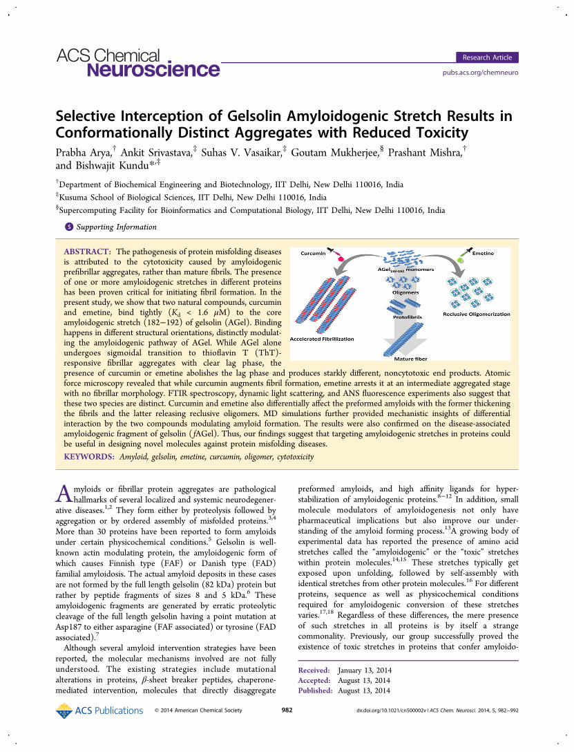
AGel. Amyloidogenic stretches are specific protein sequencesthat act as induction hotspots for intermolecular associations,yielding amyloid fibrils.14 Peptides corresponding to thesestretches have become experimental mimics for studyingamyloidogenesis of their parent proteins. AGel peptide usedin the present study is one such stretch that reportedlycorrelates with gelsolin amyloidogenesis.21,22 Interception ofthis stretch by small molecules is likely to modulate its
Figure 1. Molecular interactions and binding constant determination of AGel with compounds. Structural details of interaction of AGel with (A)curcumin and (B) emetine obtained after docking and confirmation by MD simulations. The blue and red dotted lines represent π−π and H-bonding interactions, respectively. (C) Change in fluorescence spectrum of peptide with increasing curcumin concentrations. A 5 μM peptide wastitrated using continuous injections of curcumin from a 50 μM stock. (D) Change in fluorescence spectrum of emetine with increasing peptideconcentrations. Emetine (5 μM) was titrated using continuous injections of peptide from a 50 μM stock. Panels E and F are corresponding fits forfraction bound versus total ligand concentration to one-site binding model for curcumin and emetine, respectively. All titration were done at 25 °C in0.1 M acetate buffer, pH 5.0.
ACS Chemical Neuroscience Research Article
dx.doi.org/10.1021/cn500002v | ACS Chem. Neurosci. 2014, 5, 982−992983
amyloidogenic pathway. Based on available literature, a libraryof 30 bioactive compounds reported for their potential role inmodulating neuronal diseases (both in vivo and in vitro) wasmade (Supplementary Table 1, Supporting Information). Acomputational screening of this library was done by employingphysicochemical similarity profiles between the compounds andAGel. Following this, molecular docking and moleculardynamics (MD) simulations were carried out for identifyingpotential binders. This resulted in the identification of threehigh affinity ligand-bound complexes, namely, complexes withemetine, capsazepine, and curcumin (Supplementary Table 1,Supporting Information). While curcumin is a known amyloidmodulator and capsazepine a protector of neuronal injury, therole of emetine specifically in amyloid modulation orneuroprotection has never been studied.23,24 The stability ofthese complexes was further ascertained using MD simulations.An all atom RMSD from the initial structures of all threecomplexes was analyzed (Supplementary Figure S1, SupportingInformation). While curcumin and emetine complexes showedstabilization after 5 ns, capsazepine was released from thebound conformation very early (4 ns) and remained in anunbound state, away from the peptide, for the rest of thesimulation time. Thus, only curcumin and emetine were chosenfor further studies. Analysis of interactions in complexes ofcurcumin and emetine with AGel showed significant differ-ences. An aryl ring of curcumin constituted a strong interactionpair by forming a sandwiched π−π interaction with the phenylring of the Phe183 residue of AGel. Additionally, a triplanarassembly of π−π stacked Phe183 and Phe189 with the aryl ringof curcumin stabilized the complex (Figure 1A). In the absenceof structural information about this interaction, a prediction ofaggregation nuclei using WALTZ also showed the involvementof Phe189 (Supplementary Figure S2, Supporting Informa-tion).25 In the case of emetine, the -NH group of itsisoquinoline ring formed a strong H-bond (2.4 Å) with sidechain O atom of the terminal Ser182 of AGel (Figure 1B). Thisapparently blocks further interactions at the N-terminus, whilethe rest of the peptide remains accessible. These bindinginteractions were confirmed by fluorescence spectrometrictitration experiments. This involved titration of the compoundsagainst AGel (or vice versa, see Methods) and monitoring the
change in fluorescence maxima, which were plotted againstincreasing concentrations. Following this, the fraction boundversus total ligand (curcumin/AGel) concentration was bestfitted to a one-site binding model for curcumin (eq 3) and aone-site binding model with Hill slope for emetine (eq 4).Since these experiments were done at ∼20 times lower AGelconcentration than the kinetics experiments, aggregates werenever found. The binding constants (Kd) for ligand tomonomeric AGel were found to be 1.59 ± 0.32 μM forcurcumin and 0.70 ± 0.04 μM for emetine (Figure 1C−F).Therefore, this primary screening created an interestingbackground to explore whether these interactions could affectthe actual amyloidogenic pathway of AGel.
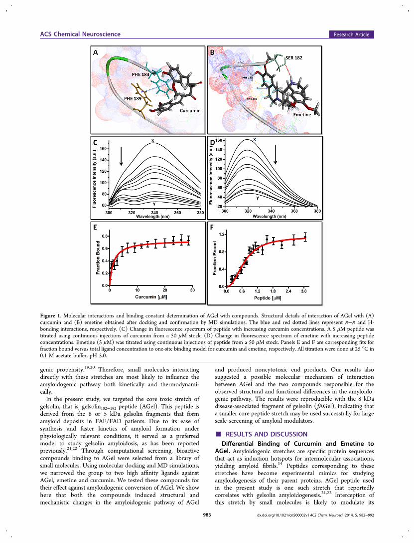
Curcumin and Emetine Distinctly Affect AggregationKinetics and Amyloid Morphology. Having establishedstrong binding, we tested whether these compounds could actas potential modulators against the amyloidogenic conversionof AGel. Hence, amyloid progression of AGel in the presenceor absence of compounds was monitored with time usingthioflavin T (ThT) fluorescence assay and the concomitantmorphological changes were observed using AFM. Aggregationof control AGel progressed with sigmoidal kinetics, showing alag phase of 10 ± 0.3 h, reaching a plateau by 48 h (Figure2A,B). AFM images of the lag, exponential, and stationaryphases, respectively, corresponded to monomeric, oligomericprefibrillar, and fibrillar states with final fiber diameter of 10−12nm (Figure 3, column I). In a way, it resembled the typicalamyloidogenic pathway reported for other proteins.26,27 On theother hand, the presence of curcumin (at all concentrations)resulted in the disappearance of the lag phase and fasterattainment of stationary phase (10−16 h) compared withcontrol (Figure 2A). This observation was supported by AFMimages, where fibrillar aggregates were visible within 12 h ofincubation. Notably, even though the stationary phase isattained in 24 h, there was further increase in the fibrillarcontent up to 48 h, as seen in the AFM images (Figure 3,column II). This suggested that after the saturation phase, thefibers align laterally to produce fully mature fibers withoutsignificant increase in total ThT-binding surfaces. Interestingly,in these samples, typical prefibrillar oligomers were notobserved at any of the experimental time points. The steep
Figure 2. Kinetics of amyloid formation. ThT fluorescence of samples incubated with increasing concentrations of curcumin (A) or emetine (B).The concentrations are mentioned in color codes with lines representing best fit as per given equations. The ThT fluorescence of AGel alone ascontrol is also shown. In each case, aliquots of AGel aggregates, obtained by incubation at 100 μM AGel in 0.1 M acetate buffer, pH 5.0, at 37 °C,were mixed with 10 μM ThT solution in 50 mM sodium phosphate buffer, pH 7.4, and incubated for 10 min before taking fluorescence readings.
ACS Chemical Neuroscience Research Article
dx.doi.org/10.1021/cn500002v | ACS Chem. Neurosci. 2014, 5, 982−992984
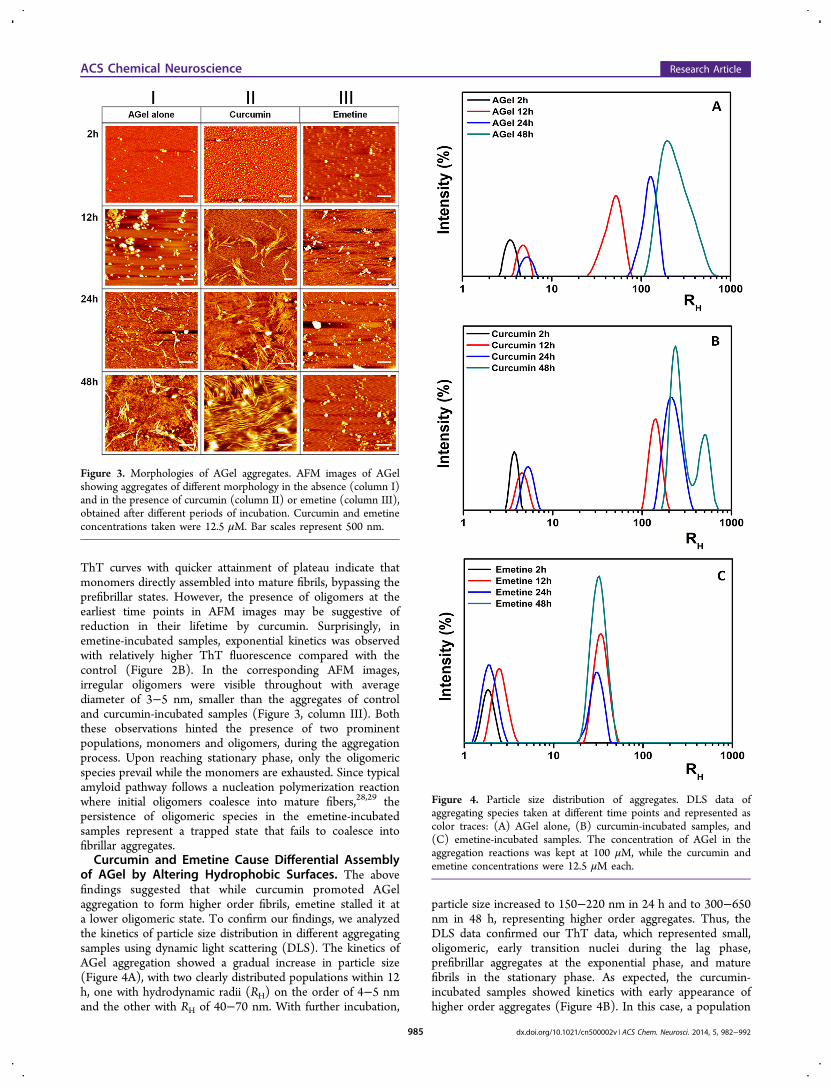
ThT curves with quicker attainment of plateau indicate thatmonomers directly assembled into mature fibrils, bypassing theprefibrillar states. However, the presence of oligomers at theearliest time points in AFM images may be suggestive ofreduction in their lifetime by curcumin. Surprisingly, inemetine-incubated samples, exponential kinetics was observedwith relatively higher ThT fluorescence compared with thecontrol (Figure 2B). In the corresponding AFM images,irregular oligomers were visible throughout with averagediameter of 3−5 nm, smaller than the aggregates of controland curcumin-incubated samples (Figure 3, column III). Boththese observations hinted the presence of two prominentpopulations, monomers and oligomers, during the aggregationprocess. Upon reaching stationary phase, only the oligomericspecies prevail while the monomers are exhausted. Since typicalamyloid pathway follows a nucleation polymerization reactionwhere initial oligomers coalesce into mature fibers,28,29 thepersistence of oligomeric species in the emetine-incubatedsamples represent a trapped state that fails to coalesce intofibrillar aggregates.Curcumin and Emetine Cause Differential Assembly
of AGel by Altering Hydrophobic Surfaces. The abovefindings suggested that while curcumin promoted AGelaggregation to form higher order fibrils, emetine stalled it ata lower oligomeric state. To confirm our findings, we analyzedthe kinetics of particle size distribution in different aggregatingsamples using dynamic light scattering (DLS). The kinetics ofAGel aggregation showed a gradual increase in particle size(Figure 4A), with two clearly distributed populations within 12h, one with hydrodynamic radii (RH) on the order of 4−5 nmand the other with RH of 40−70 nm. With further incubation,
particle size increased to 150−220 nm in 24 h and to 300−650nm in 48 h, representing higher order aggregates. Thus, theDLS data confirmed our ThT data, which represented small,oligomeric, early transition nuclei during the lag phase,prefibrillar aggregates at the exponential phase, and maturefibrils in the stationary phase. As expected, the curcumin-incubated samples showed kinetics with early appearance ofhigher order aggregates (Figure 4B). In this case, a population
Figure 3. Morphologies of AGel aggregates. AFM images of AGelshowing aggregates of different morphology in the absence (column I)and in the presence of curcumin (column II) or emetine (column III),obtained after different periods of incubation. Curcumin and emetineconcentrations taken were 12.5 μM. Bar scales represent 500 nm.
Figure 4. Particle size distribution of aggregates. DLS data ofaggregating species taken at different time points and represented ascolor traces: (A) AGel alone, (B) curcumin-incubated samples, and(C) emetine-incubated samples. The concentration of AGel in theaggregation reactions was kept at 100 μM, while the curcumin andemetine concentrations were 12.5 μM each.
ACS Chemical Neuroscience Research Article
dx.doi.org/10.1021/cn500002v | ACS Chem. Neurosci. 2014, 5, 982−992985
of size ranging between 110 and 190 nm was seen within 12 h,between 150 and 350 nm in 24 h, and between 300 and 700 nmin 48 h. Interestingly, the intermediate species observed incontrol samples (40−70 nm) was altogether absent, confirmingthat curcumin induces the formation of higher order aggregatesbypassing the prefibrillar stages. In contrast, the emetine-incubated samples showed particles with RH on the order of 4−5 nm along with a significant population of 22−50 nm species,consistently present throughout the incubation period (Figure4C). The latter population may actually represent stickyassemblies of nonfibrillar oligomers.Formation of amyloids is believed to proceed with assembly
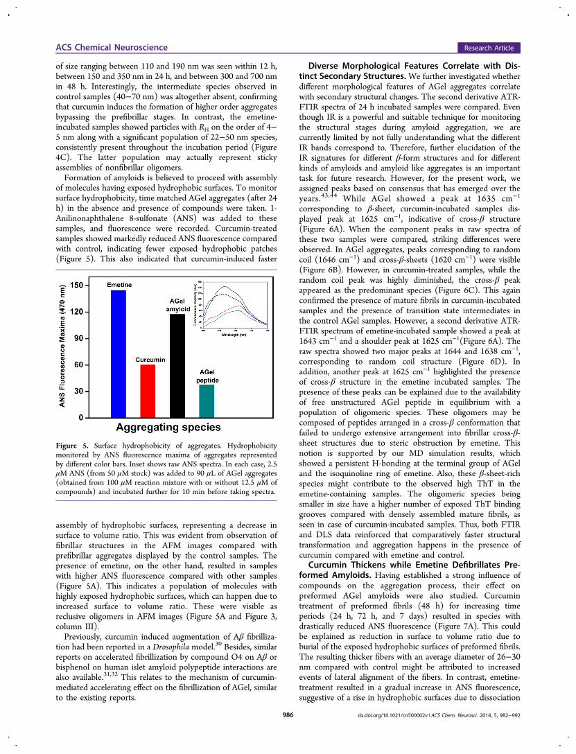
of molecules having exposed hydrophobic surfaces. To monitorsurface hydrophobicity, time matched AGel aggregates (after 24h) in the absence and presence of compounds were taken. 1-Anilinonaphthalene 8-sulfonate (ANS) was added to thesesamples, and fluorescence were recorded. Curcumin-treatedsamples showed markedly reduced ANS fluorescence comparedwith control, indicating fewer exposed hydrophobic patches(Figure 5). This also indicated that curcumin-induced faster
assembly of hydrophobic surfaces, representing a decrease insurface to volume ratio. This was evident from observation offibrillar structures in the AFM images compared withprefibrillar aggregates displayed by the control samples. Thepresence of emetine, on the other hand, resulted in sampleswith higher ANS fluorescence compared with other samples(Figure 5A). This indicates a population of molecules withhighly exposed hydrophobic surfaces, which can happen due toincreased surface to volume ratio. These were visible asreclusive oligomers in AFM images (Figure 5A and Figure 3,column III).Previously, curcumin induced augmentation of Aβ fibrilliza-
tion had been reported in a Drosophila model.30 Besides, similarreports on accelerated fibrillization by compound O4 on Aβ orbisphenol on human islet amyloid polypeptide interactions arealso available.31,32 This relates to the mechanism of curcumin-mediated accelerating effect on the fibrillization of AGel, similarto the existing reports.
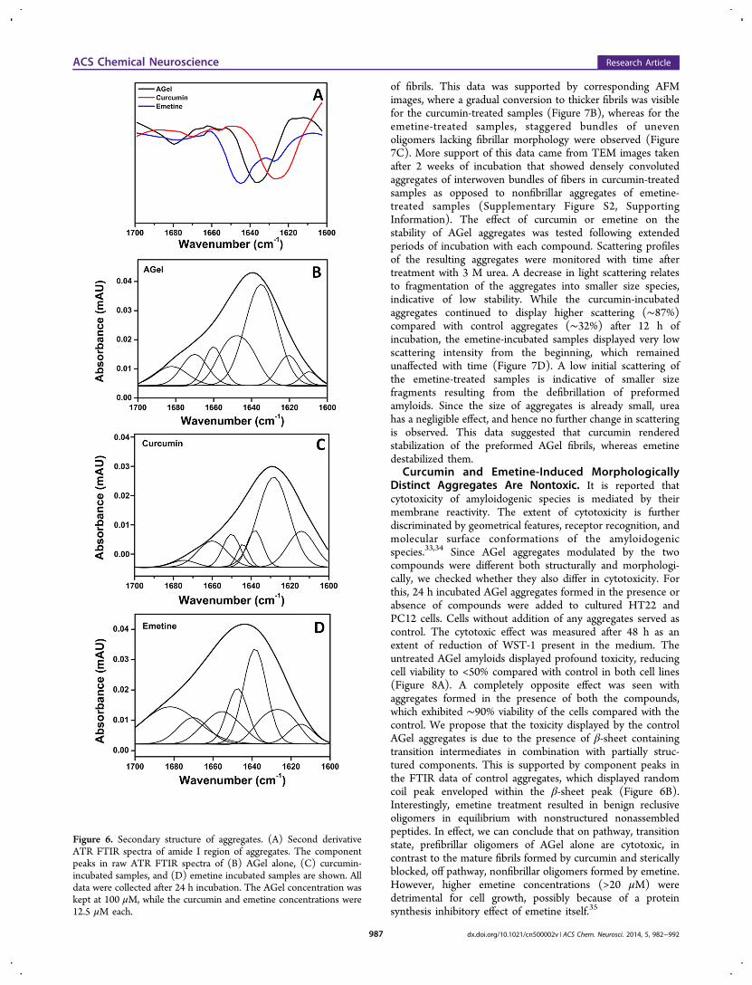
Diverse Morphological Features Correlate with Dis-tinct Secondary Structures. We further investigated whetherdifferent morphological features of AGel aggregates correlatewith secondary structural changes. The second derivative ATR-FTIR spectra of 24 h incubated samples were compared. Eventhough IR is a powerful and suitable technique for monitoringthe structural stages during amyloid aggregation, we arecurrently limited by not fully understanding what the differentIR bands correspond to. Therefore, further elucidation of theIR signatures for different β-form structures and for differentkinds of amyloids and amyloid like aggregates is an importanttask for future research. However, for the present work, weassigned peaks based on consensus that has emerged over theyears.43,44 While AGel showed a peak at 1635 cm−1
corresponding to β-sheet, curcumin-incubated samples dis-played peak at 1625 cm−1, indicative of cross-β structure(Figure 6A). When the component peaks in raw spectra ofthese two samples were compared, striking differences wereobserved. In AGel aggregates, peaks corresponding to randomcoil (1646 cm−1) and cross-β-sheets (1620 cm−1) were visible(Figure 6B). However, in curcumin-treated samples, while therandom coil peak was highly diminished, the cross-β peakappeared as the predominant species (Figure 6C). This againconfirmed the presence of mature fibrils in curcumin-incubatedsamples and the presence of transition state intermediates inthe control AGel samples. However, a second derivative ATR-FTIR spectrum of emetine-incubated sample showed a peak at1643 cm−1 and a shoulder peak at 1625 cm−1(Figure 6A). Theraw spectra showed two major peaks at 1644 and 1638 cm−1,corresponding to random coil structure (Figure 6D). Inaddition, another peak at 1625 cm−1 highlighted the presenceof cross-β structure in the emetine incubated samples. Thepresence of these peaks can be explained due to the availabilityof free unstructured AGel peptide in equilibrium with apopulation of oligomeric species. These oligomers may becomposed of peptides arranged in a cross-β conformation thatfailed to undergo extensive arrangement into fibrillar cross-β-sheet structures due to steric obstruction by emetine. Thisnotion is supported by our MD simulation results, whichshowed a persistent H-bonding at the terminal group of AGeland the isoquinoline ring of emetine. Also, these β-sheet-richspecies might contribute to the observed high ThT in theemetine-containing samples. The oligomeric species beingsmaller in size have a higher number of exposed ThT bindinggrooves compared with densely assembled mature fibrils, asseen in case of curcumin-incubated samples. Thus, both FTIRand DLS data reinforced that comparatively faster structuraltransformation and aggregation happens in the presence ofcurcumin compared with emetine and control.
Curcumin Thickens while Emetine Defibrillates Pre-formed Amyloids. Having established a strong influence ofcompounds on the aggregation process, their effect onpreformed AGel amyloids were also studied. Curcumintreatment of preformed fibrils (48 h) for increasing timeperiods (24 h, 72 h, and 7 days) resulted in species withdrastically reduced ANS fluorescence (Figure 7A). This couldbe explained as reduction in surface to volume ratio due toburial of the exposed hydrophobic surfaces of preformed fibrils.The resulting thicker fibers with an average diameter of 26−30nm compared with control might be attributed to increasedevents of lateral alignment of the fibers. In contrast, emetine-treatment resulted in a gradual increase in ANS fluorescence,suggestive of a rise in hydrophobic surfaces due to dissociation
Figure 5. Surface hydrophobicity of aggregates. Hydrophobicitymonitored by ANS fluorescence maxima of aggregates representedby different color bars. Inset shows raw ANS spectra. In each case, 2.5μM ANS (from 50 μM stock) was added to 90 μL of AGel aggregates(obtained from 100 μM reaction mixture with or without 12.5 μM ofcompounds) and incubated further for 10 min before taking spectra.
ACS Chemical Neuroscience Research Article
dx.doi.org/10.1021/cn500002v | ACS Chem. Neurosci. 2014, 5, 982−992986
of fibrils. This data was supported by corresponding AFMimages, where a gradual conversion to thicker fibrils was visiblefor the curcumin-treated samples (Figure 7B), whereas for theemetine-treated samples, staggered bundles of unevenoligomers lacking fibrillar morphology were observed (Figure7C). More support of this data came from TEM images takenafter 2 weeks of incubation that showed densely convolutedaggregates of interwoven bundles of fibers in curcumin-treatedsamples as opposed to nonfibrillar aggregates of emetine-treated samples (Supplementary Figure S2, SupportingInformation). The effect of curcumin or emetine on thestability of AGel aggregates was tested following extendedperiods of incubation with each compound. Scattering profilesof the resulting aggregates were monitored with time aftertreatment with 3 M urea. A decrease in light scattering relatesto fragmentation of the aggregates into smaller size species,indicative of low stability. While the curcumin-incubatedaggregates continued to display higher scattering (∼87%)compared with control aggregates (∼32%) after 12 h ofincubation, the emetine-incubated samples displayed very lowscattering intensity from the beginning, which remainedunaffected with time (Figure 7D). A low initial scattering ofthe emetine-treated samples is indicative of smaller sizefragments resulting from the defibrillation of preformedamyloids. Since the size of aggregates is already small, ureahas a negligible effect, and hence no further change in scatteringis observed. This data suggested that curcumin renderedstabilization of the preformed AGel fibrils, whereas emetinedestabilized them.
Curcumin and Emetine-Induced MorphologicallyDistinct Aggregates Are Nontoxic. It is reported thatcytotoxicity of amyloidogenic species is mediated by theirmembrane reactivity. The extent of cytotoxicity is furtherdiscriminated by geometrical features, receptor recognition, andmolecular surface conformations of the amyloidogenicspecies.33,34 Since AGel aggregates modulated by the twocompounds were different both structurally and morphologi-cally, we checked whether they also differ in cytotoxicity. Forthis, 24 h incubated AGel aggregates formed in the presence orabsence of compounds were added to cultured HT22 andPC12 cells. Cells without addition of any aggregates served ascontrol. The cytotoxic effect was measured after 48 h as anextent of reduction of WST-1 present in the medium. Theuntreated AGel amyloids displayed profound toxicity, reducingcell viability to <50% compared with control in both cell lines(Figure 8A). A completely opposite effect was seen withaggregates formed in the presence of both the compounds,which exhibited ∼90% viability of the cells compared with thecontrol. We propose that the toxicity displayed by the controlAGel aggregates is due to the presence of β-sheet containingtransition intermediates in combination with partially struc-tured components. This is supported by component peaks inthe FTIR data of control aggregates, which displayed randomcoil peak enveloped within the β-sheet peak (Figure 6B).Interestingly, emetine treatment resulted in benign reclusiveoligomers in equilibrium with nonstructured nonassembledpeptides. In effect, we can conclude that on pathway, transitionstate, prefibrillar oligomers of AGel alone are cytotoxic, incontrast to the mature fibrils formed by curcumin and stericallyblocked, off pathway, nonfibrillar oligomers formed by emetine.However, higher emetine concentrations (>20 μM) weredetrimental for cell growth, possibly because of a proteinsynthesis inhibitory effect of emetine itself.35
Figure 6. Secondary structure of aggregates. (A) Second derivativeATR FTIR spectra of amide I region of aggregates. The componentpeaks in raw ATR FTIR spectra of (B) AGel alone, (C) curcumin-incubated samples, and (D) emetine incubated samples are shown. Alldata were collected after 24 h incubation. The AGel concentration waskept at 100 μM, while the curcumin and emetine concentrations were12.5 μM each.
ACS Chemical Neuroscience Research Article
dx.doi.org/10.1021/cn500002v | ACS Chem. Neurosci. 2014, 5, 982−992987
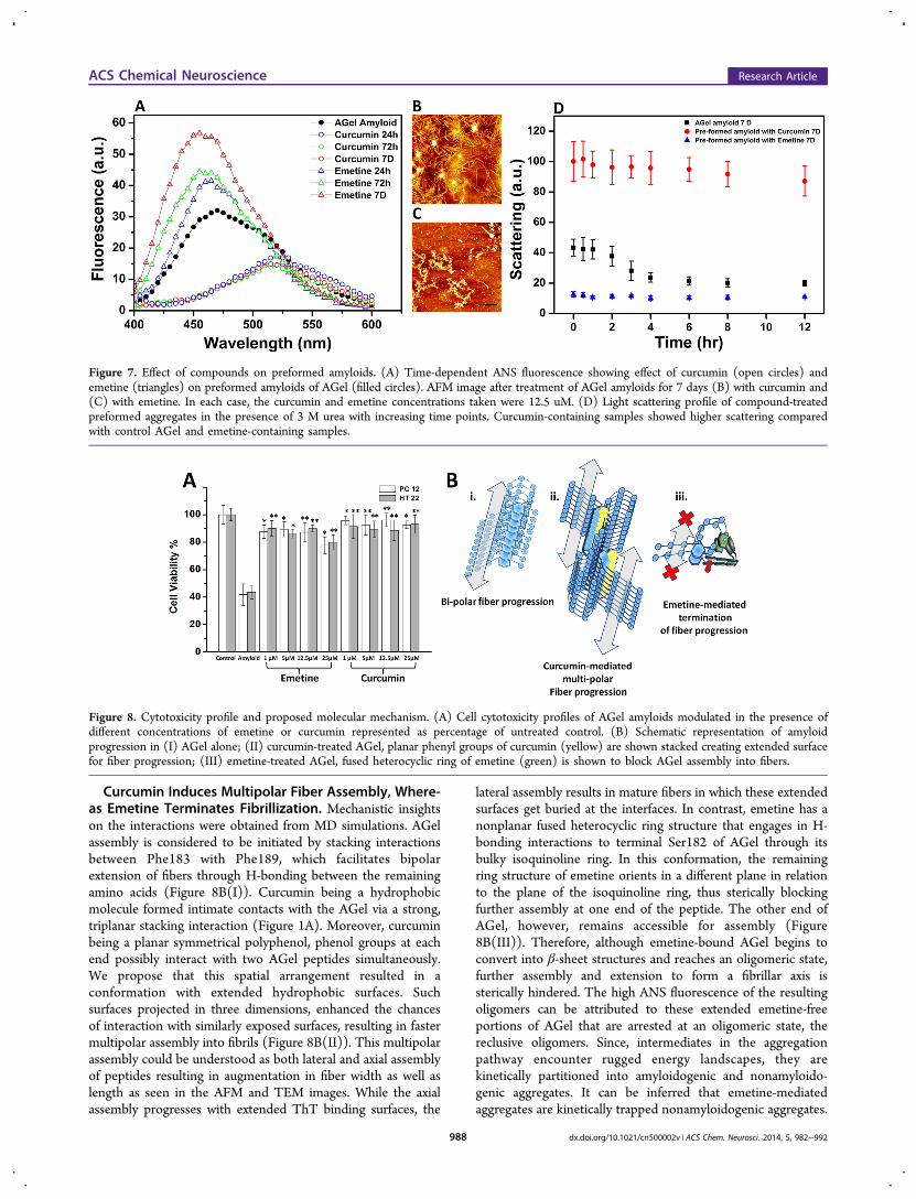
Curcumin Induces Multipolar Fiber Assembly, Where-as Emetine Terminates Fibrillization. Mechanistic insightson the interactions were obtained from MD simulations. AGelassembly is considered to be initiated by stacking interactionsbetween Phe183 with Phe189, which facilitates bipolarextension of fibers through H-bonding between the remainingamino acids (Figure 8B(I)). Curcumin being a hydrophobicmolecule formed intimate contacts with the AGel via a strong,triplanar stacking interaction (Figure 1A). Moreover, curcuminbeing a planar symmetrical polyphenol, phenol groups at eachend possibly interact with two AGel peptides simultaneously.We propose that this spatial arrangement resulted in aconformation with extended hydrophobic surfaces. Suchsurfaces projected in three dimensions, enhanced the chancesof interaction with similarly exposed surfaces, resulting in fastermultipolar assembly into fibrils (Figure 8B(II)). This multipolarassembly could be understood as both lateral and axial assemblyof peptides resulting in augmentation in fiber width as well aslength as seen in the AFM and TEM images. While the axialassembly progresses with extended ThT binding surfaces, the
lateral assembly results in mature fibers in which these extendedsurfaces get buried at the interfaces. In contrast, emetine has anonplanar fused heterocyclic ring structure that engages in H-bonding interactions to terminal Ser182 of AGel through itsbulky isoquinoline ring. In this conformation, the remainingring structure of emetine orients in a different plane in relationto the plane of the isoquinoline ring, thus sterically blockingfurther assembly at one end of the peptide. The other end ofAGel, however, remains accessible for assembly (Figure8B(III)). Therefore, although emetine-bound AGel begins toconvert into β-sheet structures and reaches an oligomeric state,further assembly and extension to form a fibrillar axis issterically hindered. The high ANS fluorescence of the resultingoligomers can be attributed to these extended emetine-freeportions of AGel that are arrested at an oligomeric state, thereclusive oligomers. Since, intermediates in the aggregationpathway encounter rugged energy landscapes, they arekinetically partitioned into amyloidogenic and nonamyloido-genic aggregates. It can be inferred that emetine-mediatedaggregates are kinetically trapped nonamyloidogenic aggregates.
Figure 7. Effect of compounds on preformed amyloids. (A) Time-dependent ANS fluorescence showing effect of curcumin (open circles) andemetine (triangles) on preformed amyloids of AGel (filled circles). AFM image after treatment of AGel amyloids for 7 days (B) with curcumin and(C) with emetine. In each case, the curcumin and emetine concentrations taken were 12.5 uM. (D) Light scattering profile of compound-treatedpreformed aggregates in the presence of 3 M urea with increasing time points. Curcumin-containing samples showed higher scattering comparedwith control AGel and emetine-containing samples.
Figure 8. Cytotoxicity profile and proposed molecular mechanism. (A) Cell cytotoxicity profiles of AGel amyloids modulated in the presence ofdifferent concentrations of emetine or curcumin represented as percentage of untreated control. (B) Schematic representation of amyloidprogression in (I) AGel alone; (II) curcumin-treated AGel, planar phenyl groups of curcumin (yellow) are shown stacked creating extended surfacefor fiber progression; (III) emetine-treated AGel, fused heterocyclic ring of emetine (green) is shown to block AGel assembly into fibers.
ACS Chemical Neuroscience Research Article
dx.doi.org/10.1021/cn500002v | ACS Chem. Neurosci. 2014, 5, 982−992988
Emetine effect thus resembles the effect of epigallocatechingalate (EGCG) on amyloidogenic polypeptides that getconverted to unstructured, off pathway oligomers.36
For ascertaining the significance of our results, we tested theeffect of both the compounds on the disease-associated amyloidfragment of gelsolin ( fAGel). For this, fAGel was expressed as aGST fusion protein from which the peptide was purified afterremoving the tag using precision protease (See SupplementaryMethod and Supplementary Figure S5, Supporting Informa-tion). An early increase in ThT intensity in the case ofcurcumin-incubated samples compared with control fAGelindicated early formation of higher order aggregates. Theemetine-incubated samples also exhibited higher ThT responsecompared with control (Supplementary Figure S6a, SupportingInformation). Thus, the ThT responses were similar to thoseobserved in the case of AGel peptide. The AFM images of 48 hincubated fAGel alone showed fibrillar aggregates, whereas thecurcumin-containing fAGel samples showed dramatic increasein bundled fibrils, possibly by lateral alignment of fibers(Supplementary Figure S6b,c, Supporting Information). Ex-pectedly, the emetine-incubated samples showed large sphericalaggregates, devoid of fibrillar morphology (SupplementaryFigure S6c, Supporting Information). Hence, these resultssubstantiate our findings on modulation of amyloid aggregationusing the core stretch of gelsolin (AGel). These results alsoreassert that the core amyloid stretches are involved in thenucleation of amyloid and can be targeted for controllingamyloid aggregation.
■ CONCLUSION
Small molecule inhibitors of amyloidogenesis have lately beenpromoted as potential therapeutic agents against neuro-degenerative diseases.37,38 We studied the interaction of twoaromatic small molecules, curcumin and emetine, on theamyloidogenic propensity of a gelsolin-derived core amyloido-genic peptide, AGel. From our studies, we conclude thatcurcumin augments aggregation to form a fibrillar state whileemetine stalls the process at an oligomeric state. Since bothstates bypass the intermediate state, they prove to benoncytotoxic in nature. Hence, we conclude that both curcuminand emetine may serve as potential therapeutic agents againstgelsolin amyloidosis. Recent reports suggest that small aromaticmolecules induce dissimilar conformations and toxicities in Aβ.In these cases, the molecular mechanism of interaction is largelyunknown.39,40 Our data, however, clearly demonstrated that thedifferential modulation of AGel aggregates by small moleculesis due to distinct molecular interactions. We conclude thatcurcumin helps lateral arrangement of amyloids by promotingmultipolar assembly, whereas emetine sterically blocks theassembly. We also showed that modulation happens throughdifferential kinetic pathways, resulting in species that aremorphologically and structurally distinct. In our data, thecompounds showed opposite effects on AGel assembly whenused individually. However, it would be interesting to find outtheir combined effect on AGel aggregation. Although, we usedthe core amyloidogenic stretch of gelsolin for quick andefficient identification of small molecule modulators, the datacould be faithfully replicated by the actual disease-associatedamyloid fragment of gelsolin. Thus, we also conclude thatscreening for short stretch aggregation modulators can be apotent way for finding full-length protein aggregationmodulators. These mechanistic insights on small molecule-
based amyloid modulation will pave ways for designing newerstrategies for amyloid intervention.
■ METHODSAggregation Reaction. The synthetic gelsolin D187N peptide
(182-H2NSFNNGNCFILD-CONH2-192) AGel was procured (Gen-Pro, India) with a purity of >95%, confirmed by mass spectrometryand HPLC. Stock solutions of AGel (1 mM) and curcumin (10 mM)were prepared in DMSO (Sigma-Aldrich, USA), whereas emetine (10mM) was made in Milli-Q water, followed by sonication andcentrifugation to remove any residual aggregates and other impurities.The 8 kDa disease associated amyloidogenic fragment ( fAGel) wascloned and expressed as mentioned in the supplementary method.Stock solution of fAGel (500 μM) was made by dissolving lyophilizedpowder in buffer containing 50 mM Tris, pH 8.0. For aggregationreaction, samples containing AGel alone (100 μM), fAGel (25 μM)alone, or fAGel with varying concentrations of compounds (1, 5, 12.5,25, and 50 μM) were made in 0.1 M acetate buffer (pH 5.0)containing 150 mM NaCl and 0.02% NaN3 and incubated at 37 °C,without agitation. These aggregation conditions were optimized afterchecking a series of other buffer and pH conditions. We found thatthese conditions were similar to those reported for aggregation ofdisease-associated gelsolin amyloidogenic fragment.41 For reactionswith preformed amyloids, 12.5 μM of each compound was addedseparately to AGel amyloids obtained after 48 h incubation.
Thioflavin T (ThT) Assay. The kinetics of amyloid formation wasmonitored using the standard thioflavin T (ThT) fluorescence assay.Although aggregation reactions were set up at pH 5.0, for effectiveThT binding, the assay was done at pH 7.4.42 Aliquots (10 μL) weredrawn from each sample and mixed with 10 μM of ThT in 50 mMphosphate buffer, pH 7.4, and incubated for 10 min. Fluorescence wasmeasured in a 1 cm path length cuvette using a LS 55 fluorescencespectrometer (PerkinElmer, MA, USA), keeping the excitation andemission wavelengths at 440 and 485 nm, respectively. The averagedfluorescence data from triplicate reactions for each concentration werefitted using eq 1 (Boltzman-sigmoidal kinetics) or eq 2 (exponentialkinetics) in Origin 8.0:
= − + +−y A A A( /(1 e ))t t k2 1
( )/2
1/2 (1)
where A1 and A2 are the initial and maximum fluorescence,respectively, t1/2 is the time at half-maximum fluorescence, and k isthe rate constant. The lag time was calculated as t1/2 − 2k.
= +y y Aekto (2)
In eq 2, A represents the initial fluorescence, t is time, k is the rateconstant, and yo represents offset.
ANS Fluorescence. ANS from a stock solution of 50 μM (inwater) was added at a final concentration of 2.5 μM to 90 μL aliquotsof AGel aggregates (with or without 12.5 μM compounds) drawn atdifferent time intervals from the aggregation reaction and incubatedfurther for 5 min at 25 °C. Fluorescence scans in the range of 400−600 nm were recorded in a 1 cm path length cuvette with excitationwavelength of 370 nm, keeping the emission and excitation slit at 5 nmeach.
Atomic Force Microscopy. For AFM imaging, aliquots ofsamples were taken, diluted 3-fold in 0.1 M acetate buffer, pH 5.0,and deposited on freshly cleaved mica. Following 2−3 min incubation,they were washed, dried, and imaged in tapping mode using aBioscope Catalyst AFM (Bruker Corporation, Billerica, MA) having aNanoscope V controller. The images were processed using Nanoscopeanalysis, v.1.4.
ATR-FTIR. The attenuated total reflectance (ATR) FTIR spectros-copy was done using a Nicolet 800 FTIR spectrometer (ThermoScientific, USA) with a MCT detector and purged with dry air. The 24h incubated aggregates (with or without 12.5 μM compounds) wereharvested by centrifugation, and the pellet was resuspended in 50 μLof 0.1 M acetate buffer, pH 5.0. A drop of this solution was applied onthe germanium crystal of the horizontal ATR sampling accessory and
ACS Chemical Neuroscience Research Article
dx.doi.org/10.1021/cn500002v | ACS Chem. Neurosci. 2014, 5, 982−992989

scanned. The data was acquired at a resolution of 4 cm−1, and 256scans were averaged per sample. The amide I band in the 1700−1600cm−1 region was analyzed after subtracting the buffer baseline. Thequantification of component peaks in the raw spectra was done usingleast-squares iterative curve fitting to Lorentzian and Gaussian lineshapes. Further, second-derivative analysis for enhancing thecomponent bands in the raw spectra was done using the standardSavitsky−Golay procedure. Assignment of bands was done based onconsensus that has emerged over several years of IR component peakassignment of the amide I region.43,44
Dynamic Light Scattering (DLS). DLS measurements wereperformed at 25 °C using a Malvern Zetasizer Nano ZS (MalvernInstruments, UK) containing a 3 mW helium−neon laser with awavelength of 633 nm and a scattering angle of 173°. Themeasurements were done with correlation times defined on 10 s perrun with 20 runs for each measurement. Aggregating samples in thepresence or absence of compounds (25 μM) at different times pointswere taken for the measurements. The samples incubated in 0.1 Macetate buffer, 150 mM NaCl, pH 5.0, were extracted at different timepoints, centrifuged, and passed through 0.1 μm filters before beingtransferred to the DLS cuvette for measurement. The experimentalresults were analyzed using the built-in Zetasizer software 6.01 usingthe values for dispersant viscosity of 0.89 mPa and refractive index of1.34. The results were plotted as intensity of distribution (%) ofparticles versus hydrodynamic radius (nm).Static Light Scattering. The preformed aggregates of AGel alone,
obtained after 48 h of incubation under aggregating conditions asmentioned above, were harvested by centrifugation at 10000g. Thepellet was resuspended in 200 μL of 0.1 M acetate buffer, pH 5.0,containing 12.5 μM curcumin or emetine and further incubated for 7days. Following this, urea from an 8 M stock solution was added toeach sample to a final concentration of 3 M and incubated forincreasing time points. Rayleigh scattering was monitored at 600 nm,with slit width 10 nm. All measurements were performed in triplicateusing a Cary Eclipse fluorescence spectrophotometer (AgilentTechnologies) at 25 °C in a 10 mm path length quartz cuvette.Cytotoxicity Assay. The HT22 and PC12 cell lines used for this
study were maintained in Dulbecco’s modified Eagle’s medium(DMEM) containing phenol red (Cellclone) supplemented with10% fetal bovine serum (FBS, Gibco, Invitrogen, UK), incubated at 37°C under 5% CO2. About 4 × 10⁴ viable cells were seeded in 96 wellplates with medium replacement after overnight incubation. ControlAGel amyloids or AGel aggregates (25 μL of aggregates resuspendedin PBS) obtained after incubation with various concentrations ofcompounds (1, 5, 12.5, and 25 μM), were added to the cells andfurther incubated for 48 h. Three independent experiments with sixreplicates each were carried out, and the results were averaged. Afterincubation, 10 μL of WST-1 reagent (Biovision, USA) was added toeach well and incubated at 37 °C for 2 h. The absorbance wasmeasured in a microplate reader (Multiskan-GO, Thermo Scientific,USA) at 450 nm, and the percentage cytotoxicity was calculated withrespect to untreated control cells. The microscopic analysis of cellularmorphology was performed using an inverted fluorescence microscope(IX51, Olympus Inc. Japan).Molecular Docking. Based on availability from previously
published literature, 30 bioactive compounds were initially chosenand their structures were retrieved from the PubChem database(Supplementary Table 1, Supporting Information). The D187N FAFmutation was introduced in the AGel stretch of gelsolin (G2 domain,residue 151−258, PDB 3FFN) by choosing the lowest energy rotamerusing the Swiss PDB Viewer.45 Energy minimization of structuralcoordinates of all the ligands and protein molecule (mutant G2domain of gelsolin) was carried out using the AMBER 10 package.46 Apotential ligand binding site near the AGel stretch was identified usingthe AADS active site prediction software.47 Following this, the RASPDprotocol was used to screen the compound library based onphysicochemical properties of the active site residues and the ligandmolecules.48 A second screening was done by docking the top 10RASPD hits at the AGel toxic stretch using ParDOCK, which is allatom energy-based docking software.49 The top three high affinity
ligand-bound complexes were chosen for MD simulations in explicitwater for ascertaining the stability of the complex (SupplementaryTable 1, Supporting Information).
MD Simulations. MD simulations were carried out using theAMBER 10 package on a 320-cores SUN Microsystems cluster. Allcomplexes and the ligand free peptide were initially neutralized byadding an appropriate number of counterions and were solvated inoctahedral boxes containing TIP3P water with 8 Å distance betweenthe peptide and the box boundary. A 1000-step energy minimization ofthe structures using Sander module in the steepest descent was done,followed by a 1500-step minimization in conjugate gradient. Systemswere then heated up to 300 K for 1 ns for equilibrating solutemolecules, followed by production phase MD simulations for 30 ns at300 K. A periodic boundary condition in the NPT ensemble withLangevin temperature coupling and constant pressure of 1 atm withisotropic molecule-based scaling was maintained. During simulations,the Shake algorithm for fixing all covalent bonds containing hydrogenatoms and the particle mesh Ewald (PME) method to treat long-rangeelectrostatic interactions were utilized.50,51 The time step ofsimulations was 2 fs with a cutoff radius of 10 Å for the nonbondedinteractions. The coordinates of the trajectory were saved every 2.5 psof the simulation. The PTRAJ module of AMBER and the VMDsoftware package were utilized to process the coordinate filesgenerated during the MD simulations.
Fluorescence Spectrometric Titrations. Fluorescence spectro-metric titrations were carried out using a longer version of AGel (takenfrom the parent full length gelsolin) containing a tryptophan (AGeltrp,H2N-SWESFNNGNCFILDLG-CONH2) in 0.1 M acetate buffer, pH5.0. The amyloid forming propensities of AGeltrp was ascertained tofollow a similar trend as that of AGel (Supplementary Figure S4,Supporting Information). Peptide solution of 5 μM was titrated usingcontinuous injections from a curcumin stock of 50 μM. However, sincethe emetine excitation wavelength (λ excitation= 284 nm) coincidedwith that of tryptophan, a reverse titration approach was used where 5μM emetine was titrated using continuous injections from a 50 μMAGel stock. With excitation fixed at 280 nm, emission scans wererecorded between 300 and 400 nm, keeping the excitation andemission slits set at 10 nm. Emission maxima (Fmax) at 347 nm (forcurcumin samples) or 318 nm (for emetine samples) were plottedagainst concentration. For all experiments, fluorescence scans wererecorded in triplicate with two accumulations each after a briefincubation of 5 min and averaged. Finally, bound fraction versus totalligand concentration was analyzed using a one-site binding model ofassociation as given below:
+ ⇄P L PL
=K[PL]
[P][L]p
where [P] and [L] represent the peptide and small molecule(curcumin/emetine) concentrations, respectively, and Kp is theassociation constant. The fraction bound versus total ligand(curcumin/AGel) concentration was best fitted to a one-site bindingmodel for curcumin (eq 3) and one-site binding model with Hill slopefor emetine (eq 4).
= +Y B X K X/( )max d (3)
= +Y B X K X/( )maxh
dh h (4)
where X is the concentration of ligand (μM), Y is specific binding, Bmaxis maximum binding, Kd is the dissociation constant, and h is the Hillslope.
■ ASSOCIATED CONTENT*S Supporting InformationSupplementary methods describing purification of disease-associated amyloidogenic fragment of gelsolin ( fAGel), scoresof stability of ligand-bound complexes, RMSD of thecomplexes, Waltz profile of amyloidogenic regions in gelsolin,
ACS Chemical Neuroscience Research Article
dx.doi.org/10.1021/cn500002v | ACS Chem. Neurosci. 2014, 5, 982−992990
morphologies of aggregates visualized by TEM after prolongedincubation, amyloid aggregation of AGeltrp represented by ThTfluorescence and AFM images, and amyloid aggregation offAGel alone and with compounds represented by ThTfluorescence and AFM images. This material is available freeof charge via the Internet at http://pubs.acs.org.
■ AUTHOR INFORMATIONCorresponding Author*Tel: 91-11-26591037. Fax: 91-11 26597530. E-mail: [email protected] ContributionsP.A. and A.S. contributed equally.P.A. and A.S. designed and executed the experiments. while
S.V.V. and G.M. helped in experiments. A.S., P.A., and B.K.wrote the manuscript.FundingP.A. and A.S. acknowledge financial support from CSIR andICMR, Govt of India, respectively.NotesThe authors declare no competing financial interest.
■ ACKNOWLEDGMENTSThe authors thank the Indian Institute of Technology Delhi(IIT Delhi) for infrastructural support and SCFBio, IIT Delhifor computing time. We thank Jasdeep Singh and Fouzia Kalimfor technical help. The authors also thank Prof. J. Gomes forproviding access to his cell culture facility and Dr. Ashish andMr. Sameer Nath for helping in peptide synthesis. We sincerelyacknowledge Dr. A. Patel for providing us purified precisionprotease. We thank Dr. M. Banerjee and Prof. B Jayaram forcritical reading and suggestions.
■ ABBREVIATIONSAGel, gelsolin182−192 D187N mutant peptide; ThT, thioflavinT; ANS, 8-anilino-1-naphthalene sulfonic acid; AFM, atomicforce microscopy; ATR-FTIR, attenuated total reflectance-Fourier transformed infrared spectroscopy; DLS, dynamic lightscattering
■ REFERENCES(1) Eisenberg, D., and Jucker, M. (2012) The amyloid state ofproteins in human diseases. Cell 148, 1188−1203.(2) Chiti, F., and Dobson, C. M. (2006) Protein misfolding,functional amyloid, and human disease. Annu. Rev. Biochem. 75, 333−366.(3) Dobson, C. M. (2003) Protein folding and misfolding. Nature 18,884−890.(4) Merlini, G., and Bellotti, V. (2003) Molecular mechanisms ofamyloidosis. N. Engl. J. Med. 349, 583−596.(5) Buxbaum, J. N., and Linke, R. P. (2012) A molecular history ofthe amyloidoses. J. Mol. Biol. 421, 142−59.(6) Solomon, J. P., Page, L. J., Balch, W. E., and Kelly, J. W. (2012)Gelsolin amyloidosis: Genetics, biochemistry, pathology and possiblestrategies for therapeutic intervention. Crit. Rev. Biochem. Mol. Biol. 47,282−96.(7) De La Chapelle, A., Tolvanen, R., Boysen, G., Santavy, J.,Bleekerwagemakers, L., Maury, C. P. J., and Kere, J. (1992) Gelsolin-derived familial amyloidosis caused by asparagine or tyrosinesubstitution for aspartic acid at residue 187. Nat. Genet. 2, 157−160.(8) Cohen, F. E., and Kelly, J. W. (2003) Therapeutic approaches toprotein-misfolding diseases. Nature 426, 905−909.(9) Soto, C., Sigurdsson, E. M., Morelli, L., Kumar, R. A., Castano, E.M., and Frangione, B. (1998) β-Sheet breaker peptides inhibit
fibrillogenesis in a rat brain model of amyloidosis: implications forAlzheimer’s therapy. Nat. Med. 4, 822−826.(10) Muchowski, P. J., and Wacker, J. L. (2005) Modulation ofneurodegeneration by molecular chaperones. Nat. Rev. Neurosci. 6,11−22.(11) Tomar, R., Garg, D. K., Mishra, R., Thakur, A. K., and Kundu, B.(2013) N-terminal domain of Pyrococcus furiosus l-asparaginasefunctions as a non-specific, stable, molecular chaperone. FEBS J.280, 2688−2699.(12) Peterson, S. A., Klabunde, T., Lashuel, H. A., Purkey, H.,Sacchettini, J. C., and Kelly, J. W. (1998) Inhibiting transthyretinconformational changes that lead to amyloid fibril formation. Proc.Natl. Acad. Sci. U.S.A. 95, 12956−12960.(13) Blazer, L. L., and Neubig, R. R. (2009) Small molecule protein-protein interaction inhibitors as CNS therapeutic agents: currentprogress and future hurdles. Neuropsychopharmacology 34, 126−141.(14) Ventura, S., Zurdo, J., Narayanan, S., Parreno, M., Mangues, R.,Reif, B., Chiti, F., Giannoni, E., Dobson, C. M., Aviles, F. X., andSerrano, L. (2004) Short amino acid stretches can mediate amyloidformation in globular proteins: The Src homology 3 (SH3) case. Proc.Natl. Acad. Sci. U.S.A. 101, 7258−7263.(15) Checler, F., and Vincent, B. (2002) Alzheimer’s and priondiseases: Distinct pathologies, common proteolytic denominators.Trends Neurosci. 25, 616−620.(16) Esteras-Chopo, A., Serrano, L., and Lopez de la Paz, M. (2005)The amyloid stretch hypothesis: Recruiting proteins toward the darkside. Proc. Natl. Acad. Sci. U.S.A. 102, 16672−16677.(17) Tzotzos, S., and Doig, A. J. (2010) Amyloidogenic sequences innative protein structures. Protein Sci. 19, 327−348.(18) Teng, P. K., and Eisenberg, D. (2009) Short protein segmentscan drive a non-fibrillizing protein into the amyloid state. Protein Eng.,Des. Sel. 22, 531−536.(19) Grover, A., Dugar, D., and Kundu, B. (2005) Predictingalternate structure attainment and amyloidogenesis: A nonlinear signalanalysis approach. Biochem. Biophys. Res. Commun. 338, 1410−1416.(20) Rana, A., Gupta, T. P., Bansal, S., and Kundu, B. (2008)Formation of amyloid fibrils by bovine carbonic anhydrase. Biochim.Biophys. Acta 1784, 930−935.(21) Maury, C. P., Liljestrom, M., Boysen, G., Tornroth, T., de laChapelle, A., and Nurmiaho-Lassila, E. L. (2000) Danish type gelsolinrelated amyloidosis: 654G-T mutation is associated with a diseasepathogenetically and clinically similar to that caused by the 654G-Amutation (familial amyloidosis of the Finnish type). J. Clin. Pathol. 53,95−99.(22) Maury, C. P., Nurmiaho-Lassila, E. L., Boysen, G., andLiljestrom, M. (2003) Fibrillogenesis in gelsolin-related familialamyloidosis. Amyloid 1 (Suppl.), 21−25.(23) Yang, F., Lim, G. P., Begum, A. N., Ubeda, O. J., Simmons, M.R., Ambegaokar, S. S., Chen, P. P., Kayed, R., Glabe, C. G., Frautschy,S. A., and Cole, G. M. (2005) Curcumin inhibits formation of amyloidbeta oligomers and fibrils, binds plaques, and reduces amyloid in vivo.J. Biol. Chem. 280, 5892−5901.(24) Ray, A. M., Benham, C. D., Roberts, J. C., Gill, C. H., Lanneau,C., Gitterman, D. P., Harries, M., Davis, J. B., and Davies, C. H. (2003)Capsazepine protects against neuronal injury caused by oxygen glucosedeprivation by inhibiting I(h). J. Neurosci. 23, 10146−10153.(25) Maurer-Stroh, S., Debulpaep, M., Kuemmerer, N., Lopez de laPaz, M., Martins, I. C., Reumers, J., Morris, K. L., Copland, A., Serpell,L., Serrano, L., Schymkowitz, J. W., and Rousseau, F. (2010) Exploringthe sequence determinants of amyloid structure using position-specificscoring matrices. Nat. Methods. 7, 237−242.(26) Wetzel, R. (2006) Kinetics and thermodynamics of amyloidfibril assembly. Acc. Chem. Res. 39, 671−679.(27) Pellarin, R., and Caflisch, A. (2006) Interpreting the aggregationkinetics of amyloid peptides. J. Mol. Biol. 360, 882−892.(28) Lee, J., Culyba, E. K., Powers, E. T., and Kelly, J. W. (2011)Amyloid-β forms fibrils by nucleated conformational conversion ofoligomers. Nat. Chem. Biol. 7, 602−609.
ACS Chemical Neuroscience Research Article
dx.doi.org/10.1021/cn500002v | ACS Chem. Neurosci. 2014, 5, 982−992991
(29) Kundu, B., Maiti, N. R., Jones, E. M., Surewicz, K. A., Vanik, D.L., and Surewicz, W. K. (2003) Nucleation-dependent conformationalconversion of the Y145Stop variant of human prion protein: structuralclues for prion propagation. Proc. Natl. Acad. Sci. U.S.A. 100, 12069−12074.(30) Caesar, I., Jonson, M., Nilsson, K. P., Thor, S., andHammarstrom, P. (2012) Curcumin promotes A-beta fibrillation andreduces neurotoxicity in transgenic Drosophila. PLoS One 7,No. e31424.(31) Bieschke, J., Herbst, M., Wiglenda, T., Friedrich, R. P.,Boeddrich, A., Schiele, F., Kleckers, D., Lopez del Amo, J. M.,Gruning, B. A., Wang, Q., Schmidt, M. R., Lurz, R., Anwyl, R.,Schnoegl, S., Fandrich, M., Frank, R. F., Reif, B., Gunther, S., Walsh, D.M., and Wanker, E. E. (2011) Small-molecule conversion of toxicoligomers to nontoxic β-sheet-rich amyloid fibrils. Nat. Chem. Biol. 8,93−101.(32) Gong, H., Zhang, X., Cheng, B., Sun, Y., Li, C., Li, T., Zheng, L.,and Huang, K. (2013) Bisphenol A accelerates toxic amyloid formationof human islet amyloid polypeptide: A possible link between bisphenolA exposure and type 2 diabetes. PLoS One 8, No. e54198.(33) Fandrich, M. (2012) Oligomeric intermediates in amyloidformation: Structure determination and mechanisms of toxicity. J. Mol.Biol. 421, 427−440.(34) Campioni, S., Mannini, B., Zampagni, M., Pensalfini, A., Parrini,C., Evangelisti, E., Relini, A., Stefani, M., Dobson, C. M., Cecchi, C.,and Chiti, F. (2010) A causative link between the structure of aberrantprotein oligomers and their toxicity. Nat. Chem. Biol. 6, 140−147.(35) Grollman, A. P. (1966) Structural basis for inhibition of proteinsynthesis by emetine and cycloheximide based on an analogy betweenipecac alkaloids and glutarimide antibiotics. Proc. Natl. Acad. Sci. U.S.A.56, 1867−1874.(36) Ehrnhoefer, D. E., Bieschke, J., Boeddrich, A., Herbst, M.,Masino, L., Lurz, R., Engemann, S., Pastore, A., and Wanker, E. E.(2008) EGCG redirects amyloidogenic polypeptides into unstruc-tured, off-pathway oligomers. Nat. Struct Mol. Biol. 15, 558−566.(37) Porat, Y., Abramowitz, A., and Gazit, E. (2006) Inhibition ofamyloid fibril formation by polyphenols: Structural similarity andaromatic interactions as a common inhibition mechanism. Chem. Biol.Drug Des. 67, 27−37.(38) Kroth, H., Ansaloni, A., Varisco, Y., Jan, A., Sreenivasachary, N.,Rezaei-Ghaleh, N., Giriens, V., Lohmann, S., Lopez-Deber, M. P.,Adolfsson, O., Pihlgren, M., Paganetti, P., Froestl, W., Nagel-Steger, L.,Willbold, D., Schrader, T., Zweckstetter, M., Pfeifer, A., Lashuel, H. A.,and Muhs, A. (2012) Discovery and structure activity relationship ofsmall molecule inhibitors of toxic β-amyloid-42 fibril formation. J. Biol.Chem. 287, 34786−34800.(39) Ladiwala, A. R., Dordick, J. S., and Tessier, P. M. (2011)Aromatic small molecules remodel toxic soluble oligomers of amyloidbeta through three independent pathways. J. Biol. Chem. 286, 3209−3218.(40) Ladiwala, A. R., Litt, J., Kane, R. S., Aucoin, D. S., Smith, S. O.,Ranjan, S., Davis, J., Van Nostrand, W. E., and Tessier, P. M. (2012)Conformational differences between two amyloid β oligomers ofsimilar size and dissimilar toxicity. J. Biol. Chem. 287, 24765−24773.(41) Ratnaswamy, G., Koepf, E., Bekele, H., Yin, H., and Kelly, J. W.(1999) The amyloidogenicity of gelsolin is controlled by proteolysisand pH. Chem. Biol. 6, 293−304.(42) Lindgren, M., Sorgjerd, K., and Hammarstrom, P. (2005)Detection and characterization of aggregates, prefibrillar amyloido-genic oligomers, and protofibrils using fluorescence spectroscopy.Biophys. J. 88, 4200−4212.(43) Ridgley, D. M., Ebanks, K. C., and Barone, J. R. (2011) Peptidemixtures can self-assemble into large amyloid fibers of varying size andmorphology. Biomacromolecules 12, 3770−3779.(44) Shivu, B., Seshadri, S., Li, J., Oberg, K. A., Uversky, V. N., andFink, A. L. (2013) Distinct β-sheet structure in protein aggregatesdetermined by ATR-FTIR spectroscopy. Biochemistry 52, 5176−5183.
(45) Guex, N., and Peitsch, M. C. (1997) SWISS-MODEL and theSwiss-PdbViewer: An environment for comparative protein modeling.Electrophoresis 18, 2714−2723.(46) Case, D. A., Darden, T. A., Cheatham,T. E., III, Simmerling, C.L., Wang, J., Duke, R. E., Luo, R., Walker, R. C., Zhang, W., Merz, K.M., Roberts, B., Wang, B., Hayik, S., Roitberg, A., Seabra, G.,Kolossvary, I., Wong, K. F., Paesani, F., Vanicek, J., Liu, J., Wu, X.,Brozell, S. R., Steinbrecher, T., Gohlke, H., Cai, Q., Ye, X., Wang, J.,Hsieh, M.-J., Cui, G., Roe, D. R.,Mathews, D. H., Seetin, M. G., Sagui,C., Babin, V., Luchko, T., Gusarov, S., Kovalenko, A., and Kollman, P.A. (2008) AMBER 10, University of California, San Francisco.(47) Singh, T., Biswas, D., and Jayaram, B. (2011) AADS - Anautomated active site identification, docking and scoring protocol forprotein targets based on physico-chemical descriptors. J. Chem. Inf.Modeling 51, 2515−2527.(48) Mukherjee, G., and Jayaram, B. (2013) A rapid identification ofhit molecules for target proteins via physico-chemical descriptors. Phys.Chem. Chem. Phys. 15, 9107−9116.(49) Gupta, A., Gandhimathi, A., Sharma, P., and Jayaram, B. (2007)ParDOCK: An all atom energy based Monte Carlo docking protocolfor protein-ligand complexes. Protein Pept. Lett. 14, 632−646.(50) Ryckaert, J. P., Ciccotti, G., and Berendsen, H. J. C. (1977)Numerical integration of the cartesian equations of motion of a systemwith constraints: molecular dynamics of n-alkanes. J. Comput. Phys. 23,327−341.(51) Essmann, U., Perera, L., Berkowitz, M. L., Darden, T., Lee, H.,and Pedersen, G. (1995) A smooth particle mesh Ewald method. J.Chem. Phys. 103, 8577−8593.
ACS Chemical Neuroscience Research Article
dx.doi.org/10.1021/cn500002v | ACS Chem. Neurosci. 2014, 5, 982−992992
Copyright © 2022 FDOKUMEN


![Synthesis and evaluation of conformationally restricted N-[2-(3,4-dichlorophenyl)ethyl]-N-methyl-2-(1-pyrrolidinyl)ethylamines at .sigma. receptors. 2. Piperazines, bicyclic amines,](https://static.fdokumen.com/doc/165x107/631289503ed465f0570a465c/synthesis-and-evaluation-of-conformationally-restricted-n-2-34-dichlorophenylethyl-n-methyl-2-1-pyrrolidinylethylamines.jpg)


