The many faces of the helix-turn-helix domain: Transcription regulation and beyond⋆

doi:10.1006/jmbi.2000.3981 available online at http://www.idealibrary.com on J. Mol. Biol. (2000) 301, 553±563
Is Polyproline II Helix the Killer Conformation? ARaman Optical Activity Study of the AmyloidogenicPrefibrillar Intermediate of Human Lysozyme
Ewan W. Blanch1, Ludmilla A. Morozova-Roche2{, Duncan A.E. Cochran3, Andrew J. Doig3, Lutz Hecht1 and Laurence D. Barron1*
1Department of ChemistryUniversity of GlasgowGlasgow G12 8QQ, UK2Oxford Centre for MolecularSciences, New ChemistryLaboratory, University ofOxford, South Parks RoadOxford OX1 3QT, UK3Department of BiomolecularSciences, UMIST, PO Box 88Manchester M60 1QD, UK
{Dr L. A. Morozova-Roche is onfrom the Insitute of Phearetical andBiophysics, Russian Academy of ScPushchino, Moscow Region, Russia
Abbreviations used: ROA, RamanPPII, Poly(L-proline) II; VCD, vibratdichroism.
E-mail address of the [email protected]
0022-2836/00/020553±11 $35.00/0
The amyloidogenic pre®brillar partially denatured intermediate of humanlysozyme, prepared by heating the native protein to 57 �C at pH 2.0, wasstudied using Raman optical activity (ROA). A positive band in the roomtemperature ROA spectrum of the native protein at �1345 cmÿ1, assignedto a hydrated form of a-helix, is not present in that of the pre®brillarintermediate, where a new strong positive band at �1318 cmÿ1 appearsinstead that is assigned to the poly(L-proline) II (PPII)-helical confor-mation. A sharp negative band at �1241 cmÿ1 in the native protein,assigned to b-strand, shows little change in the ROA spectrum of the pre-®brillar intermediate. The disappearance of a positive ROA band at�1551 cmÿ1 assigned to vibrations of tryptophan side-chains indicatesthat major conformational changes have occurred among the ®ve trypto-phan residues present in human lysozyme, four of which are located inthe a-domain. The various ROA data suggest that a substantial loss oftertiary structure has occurred in the pre®brillar intermediate and thatthis is located more in the a-domain than in the b-domain. There is noevidence for any increase in b-structure. The ROA spectrum of hen lyso-zyme, which does not form amyloid ®brils so readily, remains muchmore native-like on heating to 57 �C at pH 2.0. The thermal behaviour ofthe alanine-rich a-helical peptide AK21 in aqueous solution was found tobe similar to that of human lysozyme. Hydrated a-helix therefore appearsto readily undergo a conformational change to PPII structure on heating,which may be a key step in the conversion of a-helix into b-sheet in theformation of amyloid ®brils in human lysozyme. Since it is extended,¯exible, lacks intrachain hydrogen bonds and is fully hydrated in aqu-eous solution, PPII helix has the appropriate characteristics to be impli-cated as a critical conformational element in many conformationaldiseases. Disorder of the PPII type may be a sine qua non for the for-mation of regular ®brils; whereas the more dynamic disorder of the ran-dom coil may lead only to amorphous aggregates.
# 2000 Academic Press
Keywords: human lysozyme; amyloid ®brils; polyproline II helix; Ramanoptical activity; conformational disease
*Corresponding authora leave of absenceExperimental
iences, 142292.
optical activity;ional circular
ing author:
Introduction
Diseases which arise when an underlying pro-tein undergoes a partial unfolding leading to alocal conformational change, with resultant self-association and tissue deposition, have beentermed the conformational diseases by Carrell &Lomas (1997). These diseases are currently the sub-ject of intensive investigations but some detailsremain elusive (Kelly, 1997; Ng & Doig, 1997;
# 2000 Academic Press

554 Polyproline II Helix and Amyloid Formation
Edenhofer et al., 1997; Cohen & Prusiner, 1998;Horiuchi & Caughey, 1999).
A persistent theme is the conversion of a-helix,or some ¯exible loop sequence, into b-sheet. Inmany cases the b-sheet takes the form of amyloid®brils with a ``cross-b`` structure in which thestrands are perpendicular to the long axis of the®bre (Sunde & Blake, 1997). The most celebratedexample occurs in the prion encephalopathieswhere a-helical segments of the normal prion pro-tein are thought to be induced to change to b-sheetby the presence of the scrapie (®brous) form of theprion protein which has less a-helix and more b-sheet than the normal form (Ng & Doig, 1997;Edenhofer et al., 1997; Cohen & Prusiner, 1998;Horiuchi & Caughey, 1999). Other examples arethe Ab peptide, 40 to 42 residues long, which playsa central role in Alzheimer's disease and whichslowly converts from a soluble form to b-aggre-gates with an ordered ®brillar morphology(Baumeister & Eimer, 1998); ®lamentous inclusionsof the brain protein a-synuclein found in Parkin-son's disease, dementia with Lewy bodies andmultiple system atrophy (Serpell et al., 2000); trans-thyretin (Kelly, 1997) and human lysozyme (Boothet al., 1997) mutants which aggregate into amyloidb-®brils thereby causing fatal amyloidoses; expan-sion of glutamine repeats in proteins such as hun-tingtin thereby producing the ®brils associatedwith some neurodegenerative diseases such asHuntington's disease (Perutz, 1999); and diseasesassociated with dysfunctional serpins (Stein &Carrell, 1995), such as emphysema-related liver cir-rhosis from Z-variant a1-antitrypsin in which thereactive centre loop of one molecule inserts intothe b-sheet of another resulting in ®brillar aggrega-tion in the liver (Elliot et al., 1996; Carrell & Lomas,
1997).It is usually assumed that the ®brils and associ-
ated tissue deposits are primarily responsible fordisease. There is, however, another school ofthought suggesting that the structured pre®brillarintermediates are the pathogens (Carrell & Lomas,1997; Lansbury, 1999). For example, a b-sheet-con-taining oligomeric species could bind tightly to anynumber of cellular targets, thereby triggering acytotoxic cascade. According to this view, the®brils and tissue deposits would be an epipheno-menon of the formation of the pathogenic proto®-brils.
Amyloid ®bril formation can also be induced inmany ``normal'' proteins, not usually associatedwith diseases (Lansbury, 1999; Dobson, 1999). Forexample, it has long been known that insulinforms amyloid ®brils if heated above �80 �C atpH 2.0 (Burke & Rougvie, 1972). Other proteinsshown more recently to form ®brils under certainconditions include ®bronectin (Litvinovich et al.,1998), phosphatidylinositol-3-kinase (Guijarro et al.,1998), acylphosphatase (Chiti et al., 1999) andhuman lysozyme (Morozova-Roche et al., 2000).
It has been suggested that most, if not all, pro-teins will form amyloid ®brils under appropriate
conditions, and that the key is the loss of the coop-erative structure of the native state under con-ditions where hydrogen bonds and otherinteractions are still favourable (Chiti et al., 1999).Nonetheless, it is still not clear why some proteinsshow higher tendencies than others to form amy-loid ®brils under given denaturing conditions andwhy non-speci®c aggregates may form rather thanordered ®brils. Having the same fold and functionis not suf®cient: the differences in behaviourappear to originate in differences in the sequences,which impart subtle differences to structural pro-pensities in critical regions. The ¯exible nature ofthese critical regions in native proteins and in par-tially denatured intermediates makes it dif®cult tocharacterise the associated conformations usingconventional solution phase techniques such asNMR and far UV CD. It is usually assumed that b-sheet regions are present in the partially denaturedintermediates, and that these regions are the focusof the irreversible association of protein moleculesto produce the ®brils (Kelly, 1997; Baumeister &Eimer, 1998). Indeed, b-sheet structure has beenobserved in a monomeric fragment of recombinanthuman prion protein under reducing conditions(Jackson et al., 1999). However, another possibilityfor a key structural element that may precede b-sheet formation in these critical regions has nowbeen identi®ed using Raman optical activity(ROA).
ROA measures optical activity associated withvibrational transitions by means of a small differ-ence in the intensity of Raman scattering from chir-al molecules in right- and left-circularly polarisedincident laser light (Na®e, 1997; Barron & Hecht,2000) and provides a new perspective on the sol-ution structure and dynamics of biomolecules(Barron et al., 2000). ROA bears the same relationto conventional Raman spectroscopy as does UVCD to conventional UV absorption spectroscopy.The largest ROA signals are often associated withvibrational coordinates which sample the mostrigid and chiral parts of the structure. In proteinsthese are usually within the peptide backbone andoften give rise to ROA band patterns characteristicof the backbone conformation, unlike the parentRaman spectra in which many bands from theamino acid side-chains often obscure the peptidebackbone bands. As well as bands arising fromsecondary structure (together with a few bandsfrom side-chains), protein ROA spectra also con-tain distinct bands from loops and turns and socan provide information about the tertiary fold ofthe peptide backbone and its changes with pH,
temperature and other variables (Barron et al.,2000).
The extra incisiveness of ROA compared withother spectroscopic techniques has made itespecially useful for probing the conformationalelements present in non-native structures rangingfrom the random coil at one extreme to collapsedpartially structured states such as the molten glo-bule at the other (Barron et al., 2000; Blanch et al.,

Figure 1. MOLSCRIPT diagram of the X-ray crystalstructure of human lysozyme (PDB code 1jsf).
Polyproline II Helix and Amyloid Formation 555
2000; Smyth et al., 2000), as well as conformationalplasticity in native protein states (Blanch et al.,1999). Here, we report ROA results on a partiallydenatured pre®brillar intermediate state of humanlysozyme which appear to reveal the conformationof the critical sequences which may be importantin the formation of amyloid ®brils.
Results and Discussion
ROA measurements on human andhen lysozyme
The structure of human lysozyme, representedas a MOLSCRIPT diagram (Kraulis, 1991) inFigure 1, belongs to the a � b structural class andis very similar to that of other c-type lysozymessuch as that from hen egg white. The structure isdivided into two domains, a and b, by a deep cleftthat contains the active site. There are four a-helices labelled A, B, C and D present in the a-domain which encompasses the N and C-terminalregions of the protein. A short triple-stranded anti-parallel b-sheet and a long loop constitute most ofthe b-domain.
Although having a similar stability to that of henlysozyme with respect to temperature and reducedpH, being greater than that of equine lysozymewhich forms an equilibrium molten globule atpH 2.0 at room temperature (Morozova-Rocheet al., 1997), the thermal denaturation behaviour ofhuman lysozyme is subtly different from that ofhen lysozyme in that, below a pH of �3.0, thermaldenaturation is not a pure two-state process. Com-parison of near and far UV CD of human lysozymeas a function of temperature shows that a partiallyfolded state with at least some characteristics ofthe molten globule exists at elevated temperatures;whereas hen lysozyme shows only a hint of thisbehaviour (Haezebrouck et al., 1995). Furthermore,it has recently been shown that incubation ofhuman lysozyme at �57 �C at pH 2.0, under whichconditions the partially folded state is most highlypopulated, induces the formation of amyloid®brils; but if incubated at 70 �C at pH 2.0, underwhich conditions the fully denatured state is mosthighly populated, only amorphous aggregates areobserved (Morozova-Roche et al., 2000). We havefound that hen lysozyme has a much lower pro-pensity than human lysozyme to form amyloid®brils at �57 �C at pH 2.0.
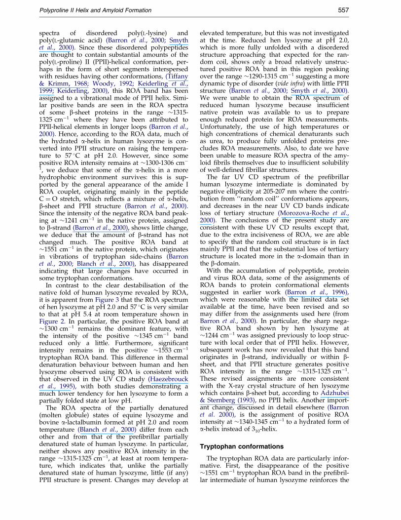
Figure 2 shows the room temperature backscat-tered Raman and ROA spectra of human (top pair)and hen (bottom pair) lysozyme at pH 5.4, underwhich conditions the native folds are preserved.The two ROA spectra are very similar to eachother, demonstrating that the basic structures ofthe two proteins in aqueous solution are similarwith regard to secondary structure, tertiary foldand side-chain conformations. The positive ROAbands at �1300 and 1345 cmÿ1 in the extendedamide III region, where normal vibrational modescontaining largely Ca-H and N-H deformations
and the Ca-N stretch usually contribute, have beenassigned, respectively, to a-helix in a hydrophobicenvironment and to a hydrated form of a-helix inwhich water molecules are hydrogen bonded tothe peptide backbone (Barron et al., 2000; Blanchet al., 2000). Equine lysozyme shows a more intense�1340 cmÿ1 ROA band which has been associatedwith its ability, not shared by the other two lyso-zymes, to bind calcium and also its greater ¯exi-bility as monitored by amide hydrogen-deuteriumexchange protection factors (Blanch et al., 2000). Ahint of a subtle difference in sequences associatedwith hydrated a-helix in human compared withhen and equine lysozyme is the presence of asecond positive peak, at �1333 cmÿ1, on the low-wavenumber side of the positive ROA band at�1345 cmÿ1, that is not observed in the other twolysozymes.
The Raman and ROA spectra of human and henlysozyme at pH 2.0 measured at room temperature(not shown) are very similar to the correspondingpH 5.4 room temperature spectra in Figure 2,demonstrating that under these conditions thestandard native folds are largely preserved.
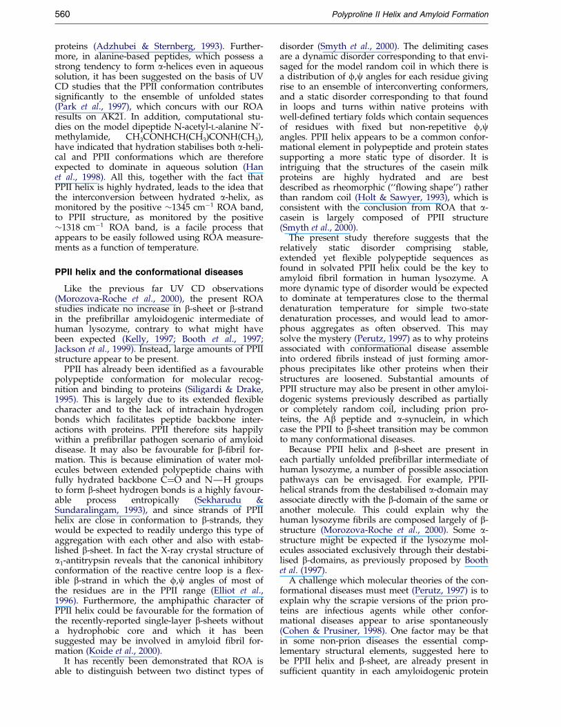
Figure 3 shows the ROA spectra of the two pro-teins at pH 2.0, but measured at 57 �C. That ofhuman lysozyme was measured prior to the onsetof ®bril formation, which was easily detectedthrough large changes in the parent conventionalRaman spectrum (not shown). In particular, new

Figure 2. The backscattered Raman and ROA spectraof native human lysozyme (top pair) and native henlysozyme (bottom pair) in 100 mM acetate buffer(pH 5.4), measured at room temperature (�20 �C).
Figure 3. The backscattered Raman and ROA spectraof human lysozyme (top pair) and hen lysozyme (bot-tom pair) in 200 mM glycine buffer (pH 2.0), measuredat 57 �C.
556 Polyproline II Helix and Amyloid Formation
strong Raman bands appear at �1230 and1671 cmÿ1, which are characteristic for b-sheet(Miura & Thomas, 1995). In contrast, hen lysozymeshowed no sign of ®bril formation over severaldays. As expected (Lansbury, 1999), the rate of®bril formation in human lysozyme was found todepend critically on the protein concentration: forexample, at �100 mg/ml it commences after anhour or two of incubation; at �60 mg/ml it com-mences after �15 hours; and at �20 mg/ml it com-mences after �30 hours. Since there wasinsuf®cient time to collect an ROA spectrum ofacceptable quality of the pre®brillar human lyso-zyme intermediate in a single run at any of theseconcentrations, we summed the ROA data for four
separate measurements made on fresh samplesprior to the onset of ®bril formation: one with aconcentration of �20 mg/ml for �14 hours,another with the same concentration for approxi-mately seven hours, one of �32 mg/ml forapproximately nine hours and one of �64 mg/mlfor approximately seven hours.
Although the pH 2.0 human lysozyme ROAspectrum measured at 57 �C is a little noisier thanthe pH 2.0 and 5.4 spectra measured at room tem-perature, signi®cant changes are readily observed.Most noticeably, the positive �1345 cmÿ1 ROAband has disappeared and a new positive ROAband has appeared at �1318 cmÿ1. A similarpositive band at �1321 cmÿ1 dominates the ROA
Polyproline II Helix and Amyloid Formation 557
spectra of disordered poly(L-lysine) andpoly(L-glutamic acid) (Barron et al., 2000; Smythet al., 2000). Since these disordered polypeptidesare thought to contain substantial amounts of thepoly(L-proline) II (PPII)-helical conformation, per-haps in the form of short segments interspersedwith residues having other conformations, (Tiffany& Krimm, 1968; Woody, 1992; Keiderling et al.,1999; Keiderling, 2000), this ROA band has beenassigned to a vibrational mode of PPII helix. Simi-lar positive bands are seen in the ROA spectraof some b-sheet proteins in the range �1315-1325 cmÿ1 where they have been attributed toPPII-helical elements in longer loops (Barron et al.,2000). Hence, according to the ROA data, much ofthe hydrated a-helix in human lysozyme is con-verted into PPII structure on raising the tempera-ture to 57 �C at pH 2.0. However, since somepositive ROA intensity remains at �1300-1306 cmÿ1, we deduce that some of the a-helix in a morehydrophobic environment survives: this is sup-ported by the general appearance of the amide IROA couplet, originating mainly in the peptideC � O stretch, which re¯ects a mixture of a-helix,b-sheet and PPII structure (Barron et al., 2000).Since the intensity of the negative ROA band peak-ing at �1241 cmÿ1 in the native protein, assignedto b-strand (Barron et al., 2000), shows little change,we deduce that the amount of b-strand has notchanged much. The positive ROA band at�1551 cmÿ1 in the native protein, which originatesin vibrations of tryptophan side-chains (Barronet al., 2000; Blanch et al., 2000), has disappearedindicating that large changes have occurred insome tryptophan conformations.
In contrast to the clear destabilisation of thenative fold of human lysozyme revealed by ROA,it is apparent from Figure 3 that the ROA spectrumof hen lysozyme at pH 2.0 and 57 �C is very similarto that at pH 5.4 at room temperature shown inFigure 2. In particular, the positive ROA band at�1300 cmÿ1 remains the dominant feature, withthe intensity of the positive �1345 cmÿ1 bandreduced only a little. Furthermore, signi®cantintensity remains in the positive �1553 cmÿ1
tryptophan ROA band. This difference in thermaldenaturation behaviour between human and henlysozyme observed using ROA is consistent withthat observed in the UV CD study (Haezebroucket al., 1995), with both studies demonstrating amuch lower tendency for hen lysozyme to form apartially folded state at low pH.
The ROA spectra of the partially denatured(molten globule) states of equine lysozyme andbovine a-lactalbumin formed at pH 2.0 and roomtemperature (Blanch et al., 2000) differ from eachother and from that of the pre®brillar partiallydenatured state of human lysozyme. In particular,neither shows any positive ROA intensity in therange �1315-1325 cmÿ1, at least at room tempera-ture, which indicates that, unlike the partiallydenatured state of human lysozyme, little (if any)PPII structure is present. Changes may develop at
elevated temperature, but this was not investigatedat the time. Reduced hen lysozyme at pH 2.0,which is more fully unfolded with a disorderedstructure approaching that expected for the ran-dom coil, shows only a broad relatively unstruc-tured positive ROA band in this region peakingover the range �1290-1315 cmÿ1 suggesting a moredynamic type of disorder (vide infra) with little PPIIstructure (Barron et al., 2000; Smyth et al., 2000).We were unable to obtain the ROA spectrum ofreduced human lysozyme because insuf®cientnative protein was available to us to prepareenough reduced protein for ROA measurements.Unfortunately, the use of high temperatures orhigh concentrations of chemical denaturants suchas urea, to produce fully unfolded proteins pre-cludes ROA measurements. Also, to date we havebeen unable to measure ROA spectra of the amy-loid ®brils themselves due to insuf®cient solubilityof well-de®ned ®brillar structures.
The far UV CD spectrum of the pre®brillarhuman lysozyme intermediate is dominated bynegative ellipticity at 205-207 nm where the contri-bution from ``random coil'' conformations appears,and decreases in the near UV CD bands indicateloss of tertiary structure (Morozova-Roche et al.,2000). The conclusions of the present study areconsistent with these UV CD results except that,due to the extra incisiveness of ROA, we are ableto specify that the random coil structure is in factmainly PPII and that the substantial loss of tertiarystructure is located more in the a-domain than inthe b-domain.
With the accumulation of polypeptide, proteinand virus ROA data, some of the assignments ofROA bands to protein conformational elementssuggested in earlier work (Barron et al., 1996),which were reasonable with the limited data setavailable at the time, have been revised and somay differ from the assignments used here (fromBarron et al., 2000). In particular, the sharp nega-tive ROA band shown by hen lysozyme at�1244 cmÿ1 was assigned previously to loop struc-ture with local order that of PPII helix. However,subsequent work has now revealed that this bandoriginates in b-strand, individually or within b-sheet, and that PPII structure generates positiveROA intensity in the range �1315-1325 cmÿ1.These revised assignments are more consistentwith the X-ray crystal structure of hen lysozymewhich contains b-sheet but, according to Adzhubei& Sternberg (1993), no PPII helix. Another import-ant change, discussed in detail elsewhere (Barronet al. 2000), is the assignment of positive ROAintensity at �1340-1345 cmÿ1 to a hydrated form ofa-helix instead of 310-helix.
Tryptophan conformations
The tryptophan ROA data are particularly infor-mative. First, the disappearance of the positive�1551 cmÿ1 tryptophan ROA band in the pre®bril-lar intermediate of human lysozyme reinforces the

558 Polyproline II Helix and Amyloid Formation
conclusion above that a signi®cant loss of tertiarystructure has occurred within the a-domain sincethat is where four of the ®ve tryptophan residuesare located. Speci®cally, W28 and W34 are in a-helix B (25-36), W109 is between 310-helix 105-108and a-helix D (110-115), and W112 is in a-helix D(Figure 1). Conformational heterogeneity amongthese tryptophan residues in the pre®brillar inter-mediate could well produce the loss of ROA sig-nals due to cancellation from contributions withopposite signs. In the native state of human lyso-zyme the remaining tryptophan, W64, is located inthe long loop of the b-domain rather than in thesecondary structure. Furthermore, its indole hydro-gen is only weakly protected from hydrogen-deu-terium exchange, unlike those of W109 and W112which show signi®cant protection (Hooke et al.,1994). This suggests that even in the native stateW64 may not be suf®ciently immobilised to makea large contribution to the ROA spectrum and that,in any event, it cannot serve as a marker of the b-sheet structure in the b-domain even though theconformation of the long loop in the b-domainmay be signi®cantly changed in the pre®brillarintermediate.
The rapid folding intermediate of human lyso-zyme has been characterised in detail using hydro-gen exchange pulse labelling techniques andstopped ¯ow CD and ¯uorescence (Hooke et al.,1994). In contrast to hen lysozyme, which ischaracterised by a cooperative folding of the entirea-domain (Radford et al., 1992), the intermediate ofhuman lysozyme has an extended hydrophobiccore based on stabilising interactions of just a-helices A and B and the C-terminal 310-helix, andinvolves W28, W34 and the side-chain of W112. Asimilar hydrophobic core has been detected in thea-domain of both the partially denatured A-stateand the rapid kinetic intermediate of the structu-rally homologous protein equine lysozyme andcharacterised in detail (Morozova-Roche et al.,1997, 1999). It has been suggested that helix C inequine lysozyme is hydrated and is responsible forthe positive �1340 cmÿ1 ROA band shown by thisprotein at neutral and acid pH (Blanch et al., 2000).It is signi®cant that the positive �1551 cmÿ1 ROAband of native equine lysozyme disappears in theA-state, which was attributed to conformationalheterogeneity among W28, W108 and W111 withW108 being the most likely candidate to adopt anon-native conformation (Blanch et al., 2000).
In view of the similarities between the foldingproperties of equine and human lysozyme in con-trast to hen lysozyme, we suggest that helix C inhuman lysozyme is the most likely candidate toundergo the putative transition from a hydrated a-helical conformation to a PPII extended confor-mation in the partially unfolded pre®brillar inter-mediate at pH 2.0 and 57 �C. Helices A, B and Dtogether with the C-terminal 310-helix may remainstable in this state, which would be consistent withthe properties of the rapid folding intermediate.Helix C is located on one side of the cleft separ-
ating the a- and b-domains of human lysozymeand so is sterically the most accessible structurewithin the a-domain to adopt an alternative con-formation and to associate with b-sheet during theformation of ®brils. Since W109 makes contactwith helix C and its indole hydrogen becomes pro-tected from exchange at the same rate as helix Cduring refolding (Hooke et al., 1994; Morozova-Roche et al., 1999), it is a likely candidate to adopta non-native conformation in the partially foldedintermediate of human lysozyme.
ROA measurements on AK21
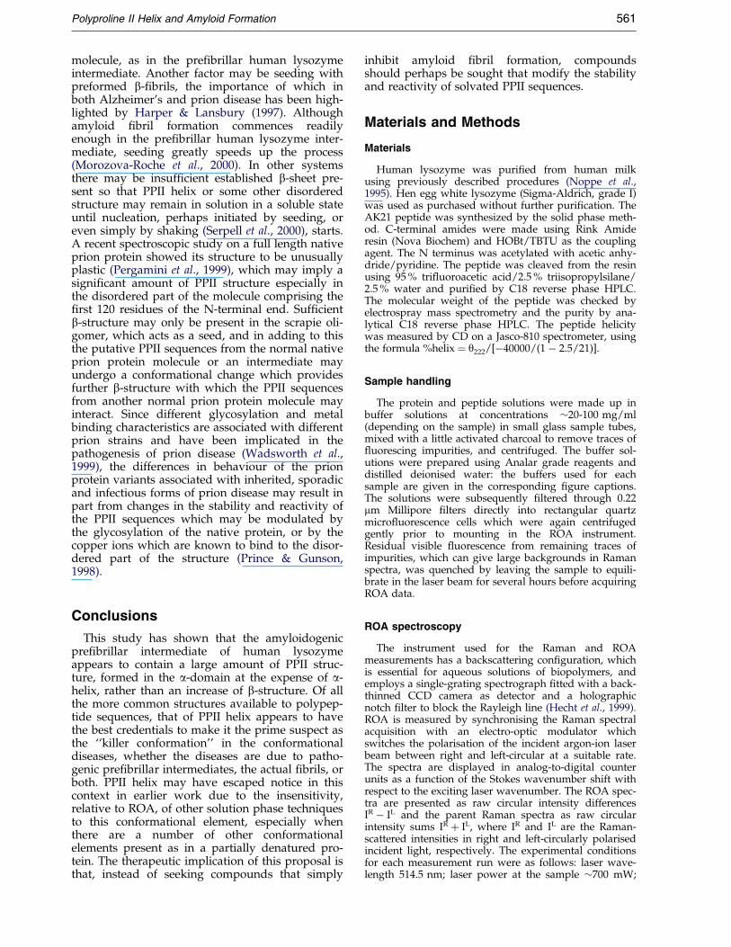
A similar structural change to that detected byROA in human lysozyme at pH 2.0 on warming to57 �C also occurs in a-helical polypeptides. Forexample, as may be seen from Figure 4, on warm-ing the alanine-rich peptide AK21, sequence Ac-AAKAAAAKAAAAKAAAAKAGY-NH2, at pH 7.1from 0 to 30 �C, the positive a-helix ROA band at�1344 cmÿ1 has decayed much more than that at�1308 cmÿ1, and positive ROA intensity has builtup in between. This suggests that, as in humanlysozyme at pH 2.0, the hydrated variant of a-helixconverts into disordered structure, possibly of thePPII type, on warming. Changes in the amide IROA couplet, negative at �1627 cmÿ1 and positiveat �1651 cmÿ1 in the low temperature spectrum,are also consistent with a decrease in a-helix con-tent and an increase in disordered structure sinceat the higher temperature the negative componentis signi®cantly weaker and the positive componentis shifted by �10 cmÿ1 to higher wavenumber(Barron et al., 2000).
UV CD measurements on AK21 indicate �70 %helix at 0 �C and �30 % helix at 30 �C. Althoughthis is compatible with the decrease in helix con-tent observed in the two AK21 ROA spectra, it isnot possible at present to make a direct quantitat-ive comparison, because the two techniques probechiral polypeptide structure in different ways,ROA being more sensitive to short range order.Also UV CD is unable to distinguish the two heli-cal forms manifest in the ROA spectra. Further-more the UV CD measurements were performed atmuch lower concentration than the ROA measure-ments.
Hydrated aaa-helix and PPII helix
Our observations on human lysozyme, AK21and other systems reveal that the variant of a-helixresponsible for the positive �1345 cmÿ1 ROA bandis less stable than that responsible for the positive�1300 cmÿ1 ROA band and has a propensity tochange into PPII structure under destabilising con-ditions. The ®rst variant was suggested to involveextensive hydrogen bonding of water moleculeswith the peptide backbone, while the second wassuggested to be in a more hydrophobic environ-ment (Barron et al., 2000; Blanch et al., 2000). Newinsight into these two a-helix variants may have

Figure 4. The backscattered Raman and ROA spectraof the alanine-rich a-helical peptide AK21 in 10 mMsodium chloride plus 5 mM sodium phosphate buffer(pH 7.5), measured at 0 �C (top pair) and 30 �C (bottompair).
Polyproline II Helix and Amyloid Formation 559
been provided by recent electron spin resonancestudies of double spin-labelled alanine-rich pep-tides which identi®ed a new more open confor-mation of the a-helix (Hanson et al., 1998; Bolin &Millhauser, 1999). Computer modelling suggeststhat the more open geometry leaves the hydrogenbonding network intact but with a changedCÐO � � �N angle which results in a splaying of thebackbone amide carbonyls away from the helixaxis and into solution; consequently, the moreopen structure may be the preferred conformationin aqueous solution since it allows main-chainhydrogen bonding with water molecules (Hansonet al., 1998; Bolin & Millhauser, 1999). If so, anequilibrium would exist between the canonical
form of a-helix, which may be responsible for thepositive �1300 cmÿ1 ROA band, and the moreopen form, which may be responsible for the posi-tive �1345 cmÿ1 ROA band, with the former beingfavoured by a hydrophobic environment and thelatter by an aqueous environment.
PPII helix consists of a left-handed extended heli-cal conformation in which the f and c angles ofthe constituent residues are restricted to values inthe regions ÿ78 � and �146 �, respectively. Thisimparts a perfect threefold rotational symmetry tothe structure (n � ÿ3). The majority of proteinscontain at least one short segment of PPII helixwith a residue composition including not only pro-line, but nearly all other amino acid residues(Adzhubei & Sternberg, 1993; Stapley & Creamer,1999). PPII helix is most often found in longerloops connecting two different or the same types ofsecondary structure. Of particular relevance here isthe fact that the extended nature of PPII helix pre-cludes intrachain hydrogen bonds. However, theorientation and high accessibility of the peptideNH and C�O groups makes them favourable part-ners for hydrogen bonding with side-chains andwith water molecules, both from the proteinhydration shell as well as water molecules incor-porated into the protein structure (Adzhubei &Sternberg, 1993; Stapley & Creamer, 1999). Indeed,main-chain hydrogen bonding with water mol-ecules is the most probable stabilizing factor forPPII-helical segments lying on the protein surface(Adzhubei & Sternberg, 1993). Although in generalapolar amino acid residues are disfavoured in PPIIhelices, nonetheless there does appear to be a sig-ni®cant correlation between hydrophobic pairs ofamino acid residues spaced i, i � 3 so that, due tothe perfect threefold rotational symmetry, PPIIhelices in globular proteins can be considered to beamphipathic (Stapley & Creamer, 1999).
The absence of intrachain hydrogen bonds,together with its inherent ¯exibility, means thatPPII helix is not readily amenable to traditionalmethods of structure determination and this hashindered the recognition of the widespread occur-rence of PPII helices in globular proteins. For freepeptides in solution the PPII conformation is indis-tinguishable from an irregular backbone structureby 1H NMR spectroscopy (Siligardi & Drake,1995). However, it can be recognised for peptidesin solution by means of UV CD (Tiffany & Krimm,1968; Woody, 1992; Makarov, et al., 1993; Siligardi& Drake, 1995) and vibrational circular dichroism(VCD) (Keiderling et al., 1999; Keiderling, 2000). Inglobular proteins the UV CD spectrum of PPIIhelix has been deconvoluted from the UV CD spec-tra of a set of proteins and is very similar to that ofPPII helix in model peptides (Sreerama & Woody,1994).
The special characteristics of PPII helix allow thepolypeptide chain to progress easily from this con-formation to other types, including a-helix and b-sheet, which could explain its prevalence in longerloops connecting secondary structure elements in
560 Polyproline II Helix and Amyloid Formation
proteins (Adzhubei & Sternberg, 1993). Further-more, in alanine-based peptides, which possess astrong tendency to form a-helices even in aqueoussolution, it has been suggested on the basis of UVCD studies that the PPII conformation contributessigni®cantly to the ensemble of unfolded states(Park et al., 1997), which concurs with our ROAresults on AK21. In addition, computational stu-dies on the model dipeptide N-acetyl-L-alanine N0-methylamide, CH3CONHCH(CH3)CONH(CH3),have indicated that hydration stabilises both a-heli-cal and PPII conformations which are thereforeexpected to dominate in aqueous solution (Hanet al., 1998). All this, together with the fact thatPPII helix is highly hydrated, leads to the idea thatthe interconversion between hydrated a-helix, asmonitored by the positive �1345 cmÿ1 ROA band,to PPII structure, as monitored by the positive�1318 cmÿ1 ROA band, is a facile process thatappears to be easily followed using ROA measure-ments as a function of temperature.
PPII helix and the conformational diseases
Like the previous far UV CD observations(Morozova-Roche et al., 2000), the present ROAstudies indicate no increase in b-sheet or b-strandin the pre®brillar amyloidogenic intermediate ofhuman lysozyme, contrary to what might havebeen expected (Kelly, 1997; Booth et al., 1997;Jackson et al., 1999). Instead, large amounts of PPIIstructure appear to be present.
PPII has already been identi®ed as a favourablepolypeptide conformation for molecular recog-nition and binding to proteins (Siligardi & Drake,1995). This is largely due to its extended ¯exiblecharacter and to the lack of intrachain hydrogenbonds which facilitates peptide backbone inter-actions with proteins. PPII therefore sits happilywithin a pre®brillar pathogen scenario of amyloiddisease. It may also be favourable for b-®bril for-mation. This is because elimination of water mol-ecules between extended polypeptide chains withfully hydrated backbone C�O and NÐH groupsto form b-sheet hydrogen bonds is a highly favour-able process entropically (Sekharudu &Sundaralingam, 1993), and since strands of PPIIhelix are close in conformation to b-strands, theywould be expected to readily undergo this type ofaggregation with each other and also with estab-lished b-sheet. In fact the X-ray crystal structure ofa1-antitrypsin reveals that the canonical inhibitoryconformation of the reactive centre loop is a ¯ex-ible b-strand in which the f,c angles of most ofthe residues are in the PPII range (Elliot et al.,1996). Furthermore, the amphipathic character ofPPII helix could be favourable for the formation ofthe recently-reported single-layer b-sheets withouta hydrophobic core and which it has beensuggested may be involved in amyloid ®bril for-mation (Koide et al., 2000).
It has recently been demonstrated that ROA isable to distinguish between two distinct types of
disorder (Smyth et al., 2000). The delimiting casesare a dynamic disorder corresponding to that envi-saged for the model random coil in which there isa distribution of f,c angles for each residue givingrise to an ensemble of interconverting conformers,and a static disorder corresponding to that foundin loops and turns within native proteins withwell-de®ned tertiary folds which contain sequencesof residues with ®xed but non-repetitive f,cangles. PPII helix appears to be a common confor-mational element in polypeptide and protein statessupporting a more static type of disorder. It isintriguing that the structures of the casein milkproteins are highly hydrated and are bestdescribed as rheomorphic (``¯owing shape'') ratherthan random coil (Holt & Sawyer, 1993), which isconsistent with the conclusion from ROA that a-casein is largely composed of PPII structure(Smyth et al., 2000).
The present study therefore suggests that therelatively static disorder comprising stable,extended yet ¯exible polypeptide sequences asfound in solvated PPII helix could be the key toamyloid ®bril formation in human lysozyme. Amore dynamic type of disorder would be expectedto dominate at temperatures close to the thermaldenaturation temperature for simple two-statedenaturation processes, and would lead to amor-phous aggregates as often observed. This maysolve the mystery (Perutz, 1997) as to why proteinsassociated with conformational disease assembleinto ordered ®brils instead of just forming amor-phous precipitates like other proteins when theirstructures are loosened. Substantial amounts ofPPII structure may also be present in other amyloi-dogenic systems previously described as partiallyor completely random coil, including prion pro-teins, the Ab peptide and a-synuclein, in whichcase the PPII to b-sheet transition may be commonto many conformational diseases.
Because PPII helix and b-sheet are present ineach partially unfolded pre®brillar intermediate ofhuman lysozyme, a number of possible associationpathways can be envisaged. For example, PPII-helical strands from the destabilised a-domain mayassociate directly with the b-domain of the same oranother molecule. This could explain why thehuman lysozyme ®brils are composed largely of b-structure (Morozova-Roche et al., 2000). Some a-structure might be expected if the lysozyme mol-ecules associated exclusively through their destabi-lised b-domains, as previously proposed by Boothet al. (1997).
A challenge which molecular theories of the con-formational diseases must meet (Perutz, 1997) is toexplain why the scrapie versions of the prion pro-teins are infectious agents while other confor-mational diseases appear to arise spontaneously(Cohen & Prusiner, 1998). One factor may be thatin some non-prion diseases the essential comp-lementary structural elements, suggested here tobe PPII helix and b-sheet, are already present insuf®cient quantity in each amyloidogenic protein
Polyproline II Helix and Amyloid Formation 561
molecule, as in the pre®brillar human lysozymeintermediate. Another factor may be seeding withpreformed b-®brils, the importance of which inboth Alzheimer's and prion disease has been high-lighted by Harper & Lansbury (1997). Althoughamyloid ®bril formation commences readilyenough in the pre®brillar human lysozyme inter-mediate, seeding greatly speeds up the process(Morozova-Roche et al., 2000). In other systemsthere may be insuf®cient established b-sheet pre-sent so that PPII helix or some other disorderedstructure may remain in solution in a soluble stateuntil nucleation, perhaps initiated by seeding, oreven simply by shaking (Serpell et al., 2000), starts.A recent spectroscopic study on a full length nativeprion protein showed its structure to be unusuallyplastic (Pergamini et al., 1999), which may imply asigni®cant amount of PPII structure especially inthe disordered part of the molecule comprising the®rst 120 residues of the N-terminal end. Suf®cientb-structure may only be present in the scrapie oli-gomer, which acts as a seed, and in adding to thisthe putative PPII sequences from the normal nativeprion protein molecule or an intermediate mayundergo a conformational change which providesfurther b-structure with which the PPII sequencesfrom another normal prion protein molecule mayinteract. Since different glycosylation and metalbinding characteristics are associated with differentprion strains and have been implicated in thepathogenesis of prion disease (Wadsworth et al.,1999), the differences in behaviour of the prionprotein variants associated with inherited, sporadicand infectious forms of prion disease may result inpart from changes in the stability and reactivity ofthe PPII sequences which may be modulated bythe glycosylation of the native protein, or by thecopper ions which are known to bind to the disor-dered part of the structure (Prince & Gunson,1998).
Conclusions
This study has shown that the amyloidogenicpre®brillar intermediate of human lysozymeappears to contain a large amount of PPII struc-ture, formed in the a-domain at the expense of a-helix, rather than an increase of b-structure. Of allthe more common structures available to polypep-tide sequences, that of PPII helix appears to havethe best credentials to make it the prime suspect asthe ``killer conformation'' in the conformationaldiseases, whether the diseases are due to patho-genic pre®brillar intermediates, the actual ®brils, orboth. PPII helix may have escaped notice in thiscontext in earlier work due to the insensitivity,relative to ROA, of other solution phase techniquesto this conformational element, especially whenthere are a number of other conformationalelements present as in a partially denatured pro-tein. The therapeutic implication of this proposal isthat, instead of seeking compounds that simply
inhibit amyloid ®bril formation, compoundsshould perhaps be sought that modify the stabilityand reactivity of solvated PPII sequences.
Materials and Methods
Materials
Human lysozyme was puri®ed from human milkusing previously described procedures (Noppe et al.,1995). Hen egg white lysozyme (Sigma-Aldrich, grade I)was used as purchased without further puri®cation. TheAK21 peptide was synthesized by the solid phase meth-od. C-terminal amides were made using Rink Amideresin (Nova Biochem) and HOBt/TBTU as the couplingagent. The N terminus was acetylated with acetic anhy-dride/pyridine. The peptide was cleaved from the resinusing 95 % tri¯uoroacetic acid/2.5 % triisopropylsilane/2.5 % water and puri®ed by C18 reverse phase HPLC.The molecular weight of the peptide was checked byelectrospray mass spectrometry and the purity by ana-lytical C18 reverse phase HPLC. The peptide helicitywas measured by CD on a Jasco-810 spectrometer, usingthe formula %helix � y222/[ÿ40000/(1 ÿ 2.5/21)].
Sample handling
The protein and peptide solutions were made up inbuffer solutions at concentrations �20-100 mg/ml(depending on the sample) in small glass sample tubes,mixed with a little activated charcoal to remove traces of¯uorescing impurities, and centrifuged. The buffer sol-utions were prepared using Analar grade reagents anddistilled deionised water: the buffers used for eachsample are given in the corresponding ®gure captions.The solutions were subsequently ®ltered through 0.22mm Millipore ®lters directly into rectangular quartzmicro¯uorescence cells which were again centrifugedgently prior to mounting in the ROA instrument.Residual visible ¯uorescence from remaining traces ofimpurities, which can give large backgrounds in Ramanspectra, was quenched by leaving the sample to equili-brate in the laser beam for several hours before acquiringROA data.
ROA spectroscopy
The instrument used for the Raman and ROAmeasurements has a backscattering con®guration, whichis essential for aqueous solutions of biopolymers, andemploys a single-grating spectrograph ®tted with a back-thinned CCD camera as detector and a holographicnotch ®lter to block the Rayleigh line (Hecht et al., 1999).ROA is measured by synchronising the Raman spectralacquisition with an electro-optic modulator whichswitches the polarisation of the incident argon-ion laserbeam between right and left-circular at a suitable rate.The spectra are displayed in analog-to-digital counterunits as a function of the Stokes wavenumber shift withrespect to the exciting laser wavenumber. The ROA spec-tra are presented as raw circular intensity differencesIR ÿ IL and the parent Raman spectra as raw circularintensity sums IR � IL, where IR and IL are the Raman-scattered intensities in right and left-circularly polarisedincident light, respectively. The experimental conditionsfor each measurement run were as follows: laser wave-length 514.5 nm; laser power at the sample �700 mW;
562 Polyproline II Helix and Amyloid Formation
spectral resolution �10 cmÿ1; acquisition time �5-30hours (depending on the sample).
For ROA measurements at other than room tempera-ture (�21 �C), dry temperature-controlled air was blownover the sample from an FTS Systems Model TC-84 Air-jet Crystal Cooler. It was not possible to exceed �60 �Cin these studies due to the possibility of damage to opti-cal elements. The sample temperatures were measuredusing a digital thermometer with a thermocouple probealong the laser beam path through the centre of the celland are accurate to within � � 2 �C.
Acknowledgements
L.D.B. and L.H. thank the BBSRC for a research grantand the EPSRC for a Senior Fellowship for L.D.B. A.J.D.thanks the BBSRC for a research grant and the WellcomeTrust for an equipment grant for the CD spectrometer(reference 057318). L.A.M.-R. is supported by the OxfordCentre for Molecular Sciences. We are grateful to Dr W.Noppe for the generous gift of the wild-type humanlysozyme sample used in this work.
References
Adzhubei, A. A. & Sternberg, M. J. E. (1993). Left-handed polyproline II helices commonly occur inglobular proteins. J. Mol. Biol. 229, 472-493.
Barron, L. D. & Hecht, L. (2000). Vibrational Ramanoptical activity: from fundamentals to biochemicalapplications. In Circular Dichroism: Principles andApplications for Biologists (Berova, N., Nakanishi, K.& Woody, R. W., eds), vol. 667-701, Wiley, NewYork.
Barron, L. D., Hecht, L., Bell, A. F. & Wilson, G. (1996).Recent developments in Raman optical activity ofbiopolymers. Appl. Spectrosc. 50, 619-629.
Barron, L. D., Hecht, L., Blanch, E. W. & Bell, A. F.(2000). Solution structure and dynamics of biomole-cules from Raman optical activity. Prog. Biophys.Molec. Biol. 73, 1-49.
Baumeister, R. & Eimer, S. (1998). Amyloid aggregates,presenilins, and Alzheimer's disease. Ang. Chem.Int. Ed. 37, 2978-2982.
Blanch, E. W., Hecht, L. & Barron, L. D. (1999). Newinsight into the pH-dependent conformationalchanges in bovine b-lactoglobulin from Raman opti-cal activity. Protein Sci. 8, 1362-1367.
Blanch, E. W., Morozova-Roche, L. A., Hecht, L.,Noppe, W. & Barron, L. D. (2000). Raman opticalactivity characterization of native and molten glo-bule states of equine lysozyme: comparison withhen lysozyme and bovine a-lactalbumin. Biopoly-mers: Biospectros. 57, 235-248.
Bolin, K. A. & Millhauser, G. L. (1999). a and 310: thesplit personality of polypeptide helices. Acc. Chem.Res. 32, 1027-1033.
Booth, D. R., Sunde, M., Bellotti, V., Robinson, C. V.,Hutchinson, W. L., Fraser, P. E., Hawkins, P. N.,Dobson, C. M., Radford, S. E., Blake, C. F. & Pepys,M. B. (1997). Instability, unfolding and aggregationof human lysozyme variants underlying amyloid®brillogenesis. Nature, 385, 787-793.
Burke, M. J. & Rougvie, M. A. (1972). Cross-b proteinstructures. I. Insulin ®brils. Biochemistry, 11, 2435-2439.
Carrell, R. W. & Lomas, D. A. (1997). Conformationaldisease. The Lancet, 350, 134-138.
Chiti, F., Webster, P., Taddei, N., Clark, A., Stefani, M.,Ramponi, G. & Dobson, C. M. (1999). Designingconditions for in vitro formation of amyloid proto®-laments and ®brils. Proc. Natl Acad. Sci. USA, 96,3590-3594.
Cohen, F. E. & Prusiner, S. B. (1998). Pathologic confor-mations of prion proteins. Annu. Rev. Biochem. 67,793-819.
Dobson, C. M. (1999). Protein misfolding, evolution anddisease. TIBS, 24, 329-332.
Edenhofer, F., Weiss, S., Winnacker, E. L. & Famulok,M. (1997). Chemistry and molecular biology oftransmissible spongiform encephalopathies. Ang.Chem. Int. Ed. 36, 1674-1694.
Elliot, P. R., Lomas, D. A., Carrell, R. W. & Abrahams,J. P. (1996). Inhibitory conformation of the reactiveloop of a1-antitrypsin. Nature Struct. Biol. 3, 676-681.
Guijarro, J. I., Sunde, M., Jones, J. A., Campbell, I. D. &Dobson, C. M. (1998). Amyloid ®bril formation byan SH3 domain. Proc. Natl Acad. Sci. USA, 95, 4224-4228.
Haezebrouck, P., Joniau, M., van Dael, H., Hooke, S. D.,Woodruff, N. D. & Dobson, C. M. (1995). An equili-brium partially folded state of human lysozyme atlow pH. J. Mol. Biol. 246, 382-387.
Han, W.-G., Jalkanen, K. J., Elstner, M. & Suhai, S.(1998). Theoretical study of aqueous N-acetyl-L-ala-nine N0-methylamide: structures and Raman, VCDand ROA spectra. J. Phys. Chem. B, 102, 2587-2602.
Hanson, P., Anderson, D. J., Martinez, G., Millhauser,G., Formaggio, F., Crisma, M., Toniolo, C. & Vita,C. (1998). Electron spin resonance and structuralanalysis of water soluble, alanine-rich peptidesincorporating TOAC. Mol. Phys. 95, 957-966.
Harper, J. D. & Lansbury, P. T., Jr (1997). Models ofamyloid seeding in Alzheimer's disease and scrapie.Annu. Rev. Biochem. 66, 385-407.
Hecht, L., Barron, L. D., Blanch, E. W., Bell, A. F. &Day, L. A. (1999). Raman optical activity instrumentfor studies of biopolymer structure and function.J. Raman Spectrosc. 30, 815-825.
Holt, C. & Sawyer, L. (1993). Caseins as rheomorphicproteins: interpretation of primary and secondarystructures of the aS1-, b- and k-caseins. J. Chem. Soc.Faraday Trans. 89, 2683-2692.
Hooke, S. D., Radford, S. E. & Dobson, C. M. (1994).The refolding of human lysozyme: a comparisonwith the structurally homologous hen lysozyme.Biochemistry, 33, 5867-5876.
Horiuchi, M. & Caughey, B. (1999). Prion protein inter-conversions and the transmissible spongiform ence-phalopathies. Structure, 7, R231-R240.
Jackson, G. S., Hosszu, L. L. P., Power, A., Hill, A. F.,Kenney, J., Saibil, H., Craven, C. J., Waltho, J. P.,Clarke, A. R. & Collinge, J. (1999). Reversible con-version of monomeric human prion proteinbetween native and ®brilogenic conformations.Science, 283, 1935-1937.
Keiderling, T. A. (2000). Peptide and protein confor-mational studies with vibrational circular dichroismand related spectroscopies. In Circular Dichroism:Principles and Applications for Biologists (Berova, N.,Nakanishi, K. & Woody, R. W., eds), vol. 621-666,Wiley, New York.
Keiderling, T. A., Silva, R. A. G. D., Yoder, G. & Dukor,R. K. (1999). Vibrational circular dichroism spectro-
Polyproline II Helix and Amyloid Formation 563
sopy of selected oligopeptide conformations. Bioorg.Med. Chem. 7, 133-141.
Kelly, J. W. (1997). Amyloid ®bril formation and proteinmisassembly: a structural quest for insights intoamyloid and prion diseases. Structure, 5, 595-600.
Koide, S., Huang, X., Link, K., Koide, A., Bu, Z. &Engelman, D. M. (2000). Design of single-layer b-sheets without a hydrophobic core. Nature, 403,456-460.
Kraulis, P. J. (1991). MOLSCRIPT: A program to pro-duce both detailed and schematic plots of proteinstructures. J. Appl. Crystallog. 24, 946-950.
Lansbury, P. T., Jr (1999). Evolution of amyloid: whatnormal protein folding may tell us about ®brillo-genesis and disease. Proc. Natl Acad. Sci. USA, 96,3342-3344.
Litvinovich, S. V., Brew, S. A., Aota, S., Akiyama, S. K.,Haudenschild, C. & Ingham, K. C. (1998). For-mation of amyloid-like ®brils by self-association ofa partially unfolded ®bronectin type III module.J. Mol. Biol. 280, 245-258.
Makarov, A. A., Adzhubei, I. A., Prostasevich, I. I.,Lobachov, V. M. & Esipova, N. G. (1993). Scanningmicrocalorimetry and circular dichroism study ofmelting of the natural polypeptides in the left-handed helical conformation. J. Protein Chem. 12, 85-91.
Miura, T. & Thomas, G. J., Jr. (1995). Raman spec-troscopy of proteins and their assemblies. In Subcel-lular Biochemistry, Proteins: Structure, Function, andEngineering (Biswas, B. B. & Roy, S., eds), vol. 23,pp. 55-99, Plenum Press, New York.
Morozova-Roche, L. A., Arico-Muendel, C. C., Haynie,D. T., Emelyanenko, V. I., Van Dael, H. & Dobson,C. M. (1997). Structural characterisation and com-parison of the native and A-states of equine lyso-zyme. J. Mol. Biol. 268, 903-921.
Morozova-Roche, L. A., Jones, J. A., Noppe, W. &Dobson, C. M. (1999). Independent nucleation andheterogeneous assembly of structure during foldingof equine lysozyme. J. Mol. Biol. 289, 1055-1073.
Morozova-Roche, L. A., Zurdo, J., Spencer, A., Noppe,W., Receveur, V., Archer, D. B., Joniau, M. &Dobson, C. M. (2000). Amyloid ®bril formation andseeding by wild-type human lysozyme and its dis-ease related mutational variants. J. Struct. Biol. Inthe press.
Na®e, L. A. (1997). Infrared and Raman vibrational opti-cal activity. Annu. Rev. Phys. Chem. 48, 357-386.
Ng, S. B. L. & Doig, A. J. (1997). Molecular and chemicalbasis of prion-related diseases. Chem. Soc. Revs. 26,425-432.
Noppe, W., Hanssens, I. & De Cuyper, M. (1995).Simple two-step procedure for the preparation ofhighly active pure equine milk. J. Chromatog. sect. A,719, 327-331.
Park, S.-H., Shalongo, W. & Stellwagen, E. (1997). Therole of PII conformation in the calculation of pep-
tide fractional helical content. Protein Sci. 6, 1694-1700.
Pergamini, P., Bramanti, E. & Ascoli, G. A. (1999). Struc-tural dependence of the cellular isoform of prionprotein on solvent: spectroscopic characterization ofan intermediate conformation. Bioch. Biophys. Res.Comm. 264, 972-978.
Perutz, M. F. (1997). Mutations make enzyme polymer-ize. Nature, 385, 773-775.
Perutz, M. F. (1999). Glutamine repeats and neurodegen-erative diseases: molecular aspects. TIBS, 24, 58-63.
Prince, R. C. & Gunson, D. E. (1998). Prions are copper-binding proteins. Trends Biochem. Sci. 23, 197-198.
Radford, S. E., Dobson, C. M. & Evans, P. A. (1992). Thefolding of hen lysozyme involves partially struc-tured intermediates and multiple pathways. Nature,358, 302-307.
Sekharudu, Y. C. & Sundaralingam, M. (1993).Hydration of protein secondary structures- the rolein protein folding. In Water and Biological Macromol-ecules (Westhof, E., ed.), vol. 148-162, CRC Press,Boca Raton, FL.
Serpell, L. C., Berriman, J., Jakes, R., Goedert, M. &Crowther, R. A. (2000). Fiber diffraction of synthetica-synuclein ®laments shows amyloid-like cross-bconformation. Proc. Natl Acad. Sci. USA, 97, 4897-4902.
Siligardi, G. & Drake, A. F. (1995). The importance ofextended conformations and, in particular, the PII
conformation for the molecular recognition of pep-tides. Biopolymers, 37, 281-292.
Smyth, E., Syme, C. D., Blanch, E. W., Hecht, L., VasÏaÂk,M. & Barron, L. D. (2000). Solution structure ofnative proteins with irregular folds from Ramanoptical activity. Biopolymers, In the press.
Sreerama, N. & Woody, R. W. (1994). Poly(pro)II helicesin globular proteins: identi®cation and circulardichroic analysis. Biochemistry, 33, 10022-10025.
Stapley, B. J. & Creamer, T. P. (1999). A survey of left-handed polyproline II helices. Protein Sci. 8, 587-595.
Stein, P. E. & Carrell, R. W. (1995). What do dysfunc-tional serpins tell us about molecular mobility anddisease?. Nature Struct. Biol. 2, 96-113.
Sunde, M. & Blake, C. C. F. (1997). The structure ofamyloid ®brils by electron microscopy and X-raydiffraction. Advan. Prot. Chem. 50, 123-159.
Tiffany, M. L. & Krimm, S. (1968). New chain confor-mations of poly(glutamic acid) and polylysine. Bio-polymers, 6, 1379-1382.
Wadsworth, J. D. F., Hill, A. F., Joiner, S., Jackson, G. S.,Clarke, A. R. & Collinge, J. (1999). Strain-speci®cconformation determined by metal ions. Nature CellBiol. 1, 55-59.
Woody, R. W. (1992). Circular dichroism and confor-mations of unordered polypeptides. Advan. Biophys.Chem. 2, 37-79.
Edited by A. R. Fersht
(Received 23 February 2000; received in revised form 21 June 2000; accepted 23 June 2000)
Copyright © 2022 FDOKUMEN